Embed Size (px)
Citation preview
Saad and Kleber G. FranchiniRosa C. Tambascia, Priscila M. Fonseca, Patrícia D. C. Corat, Heitor Moreno, Jr, Mario J. A.
Infused Rats−Expression and Distribution of NOS1 and NOS3 in the Myocardium of Angiotensin II
Print ISSN: 0194-911X. Online ISSN: 1524-4563 Copyright © 2001 American Heart Association, Inc. All rights reserved.
is published by the American Heart Association, 7272 Greenville Avenue, Dallas, TX 75231Hypertension doi: 10.1161/01.HYP.37.6.1423
2001;37:1423-1428Hypertension.
http://hyper.ahajournals.org/content/37/6/1423World Wide Web at:
The online version of this article, along with updated information and services, is located on the
http://hyper.ahajournals.org//subscriptions/
is online at: Hypertension Information about subscribing to Subscriptions:
http://www.lww.com/reprints Information about reprints can be found online at: Reprints:
document. Permissions and Rights Question and Answer this process is available in the
click Request Permissions in the middle column of the Web page under Services. Further information aboutOffice. Once the online version of the published article for which permission is being requested is located,
can be obtained via RightsLink, a service of the Copyright Clearance Center, not the EditorialHypertensionin Requests for permissions to reproduce figures, tables, or portions of articles originally publishedPermissions:
by guest on February 23, 2013http://hyper.ahajournals.org/Downloaded from
Expression and Distribution of NOS1 and NOS3 in theMyocardium of Angiotensin II–Infused Rats
Rosa C. Tambascia, Priscila M. Fonseca, Patrícia D.C. Corat, Heitor Moreno, Jr,Mario J.A. Saad, Kleber G. Franchini
Abstract—Studies have indicated a complex functional interaction between angiotensin (Ang) II and NO in the heart. Thepurpose of the present study was to examine the protein expression and tissue distribution of NO synthases 1 (NOS1)and 3 (NOS3) in the myocardium of rats that underwent continuous infusion of Ang II at 2 different rates (10 and 40ng · kg21 · min21) for 6 days. Mean arterial pressure increased by'15 mm Hg in rats infused with Ang II at 40 ng ·kg21 · min21, but it remained close to the values observed in saline-infused rats ('110 mm Hg) when Ang II was infusedat 10 ng · kg21 · min21. The protein expression of a 160-kDa NOS1 and a 135-kDa NOS3 were found to increase('200%) in the myocardium of rats infused with both subpressor and pressor doses of Ang II. Immunohistochemistrystudies showed that NOS1 and NOS3 are differentially expressed in myocardial cells. NOS1 was detected in cardiacmyocytes and in smooth muscle cells of small and large coronary arteries, whereas NOS3 was detected in theendothelium and in perivascular and interstitial tissues, but NOS3 was not detected in cardiac or smooth muscle cells.Ang II infusion enhanced the tissue immunoreactivity of both isoforms in their specific locations but did not change thedistribution throughout the myocardium. Myocardium staining with anti–angiotensin type 1 (AT1) receptor antibodyindicated that AT1 receptor is expressed in cardiac myocytes, coronary smooth muscle cells, and interstitial andperivascular tissues. Ang II infusion did not change the protein expression and distribution of AT1 receptor in themyocardium. These results indicate that long-term increases in the circulating levels of Ang II modulate the proteinexpression of NOS1 and NOS3 and, consequently, the function of the local myocardial NO system.(Hypertension.2001;37:1423-1428.)
Key Words: angiotensin IIn nitric oxide n nitric oxide synthasen heart n myocardium
Angiotensin (Ang) II modulates cardiac function andcellular growth in response to physiological and patho-
logical processes.1 Many of the short- and long-term effectsof Ang II on cardiac function and structure are due to itsdirect action on cardiac myocytes, vascular smooth musclecells, and cardiac fibroblasts. These effects are mediatedthrough at least 2 different types of receptors, which arebroadly distributed in cardiac cells.2 However, some of thecardiac effects of Ang II occur through the induction andrelease of paracrine/autocrine factors, such as transforminggrowth factor-b1 and endothelin-1.3,4 In this context, in recentyears, experimental evidence has indicated that Ang II andNO influence each other by interacting at various levels ofregulation.5,6 This may have implications not only for thecardiac functions directly influenced by these factors but alsofor the pathogenesis of processes such as myocardial ische-mia and fibrosis.
In general, Ang II and NO exert antagonistic effects incellular function and growth.6,7 The cellular mechanismsresponsible for this antagonism are not clear. In somesystems, this interaction seems to be a simple summation of
the effects of Ang II and NO.7,8 Ang II is able to activate theNO system by inducing the secretion of NO in small and largecoronary arteries. Because NO attenuates the vasoconstrictoreffect of Ang II, this can cause a negative feedback system tolimit the stimulation by Ang II.9 The antagonism of NO onAng II effects is also seen in the growth effect of Ang II oncardiac fibroblasts.10
The mutual regulatory influence of Ang II and NO seemsto extend to gene regulation. Studies performed in angioten-sinogen gene–knockout mice and in rat adrenal medullasuggest that Ang II inhibits the expression of NO synthase(NOS)1.11,12 In rats, however, long-term infusion of highdoses of Ang II increases the expression of NOS1 and NOS3in the renal cortex but reduces NOS1 expression in the renalmedulla.8 The influence of Ang II on the regulation of theconstitutively expressed isoforms of NOS in the myocardiumremains virtually unexplored.
Thus, the present study was designed to examine the effectof long-term increases in circulating levels of Ang II on theexpression, and the cardiac tissue distribution of the consti-tutive isoforms of NOS (ie, NOS1 and NOS3). Experiments
Received September 7, 2000; first decision October 2, 2000; revision accepted December 4, 2000.From the Department of Internal Medicine, School of Medicine, State University of Campinas (UNICAMP), Campinas, SP, Brazil.Correspondence to Kleber G. Franchini, MD, Departamento de Clínica Médica, Faculdade de Ciências Médicas, Universidade Estadual de Campinas,
Cidade Universitária “Zefferino Vaz,” 13081-970 Campinas, SP, Brasil. E-mail [email protected]© 2001 American Heart Association, Inc.
Hypertensionis available at http://www.hypertensionaha.org
1423 by guest on February 23, 2013http://hyper.ahajournals.org/Downloaded from
were also performed to examine the protein expression andthe tissue distribution of angiotensin type 1 (AT1) receptors inthe left ventricle of rats treated or not treated with Ang II.
MethodsThe experiments were performed on male Wistar rats (270 to 300 g)obtained from animal facilities of the State University of Campinas(Campinas, SP, Brazil). All procedures followed the university’sguidelines for the use of animals in experimental studies.
Antibodies and ChemicalsRabbit polyclonal antibodies raised against NOS1, NOS3, and AT1
were purchased from Santa Cruz Biotechnology.125I-labeled proteinA was from Amersham. Ang II was from Calbiochem. All otherreagent grade chemicals were from Sigma.
Rat Instrumentation and ArterialPressure MonitoringAll surgical procedures were performed under aseptic conditions.Rats were anesthetized with a mixture of ketamine (70 mg/kg bodywt IM) and diazepam (6 mg/kg body wt IM) and maintained at 37°C.Tygon-tipped polyvinyl cannulas were placed in the lower abdomi-nal aorta and inferior vena cava throughout the femoral artery andvein, respectively. The cannulas were exteriorized at the back of theneck in a 25-cm length of stainless steel spring (0.5-cm diameter)attached to a swivel (Instech) at the top of an individual cage thatallowed the animal to move freely in its cage while being infused.The animals received single doses of antibiotic (Pentabiótico Veter-inário, 100 mg/kg body wt) and were allowed to recover for 5 daysbefore the study. During this period, 0.9% saline was infusedcontinuously through the venous catheter at a rate of 0.5 mL/h. Afterthis period, saline was substituted for Ang II solutions in 2 differentconcentrations (10 and 40 ng · kg21 · min21) in the experimentalanimals, whereas control animals continued to receive only saline.
Arterial pressure was monitored daily for 6 days for a 1-hourperiod from 3:00 to 4:00PM. The amplified signal was beat-to-beatrecorded and sampled at 100 Hz with WINDAQ-PRO data acquisi-tion software (DATAQ Instruments).
Tissue HomogenizationAt the end of day 6 of Ang II infusion, the animals were anesthetized,hearts were rapidly removed, and the ventricles were mincedcoarsely and homogenized in'10 volumes of solubilization buffer(1% Triton-X 100; 100 mmol/L Tris-HCl (pH 7.4); 100 mmol/Lsodium pyrophosphate; 100 mmol/L sodium fluoride; 10 mmol/LEDTA; 10 mmol/L sodium vanadate; 2 mmol/L PMSF; and 0.1 mgaprotinin/mL) at 4°C with the polytron operated at maximum speedfor 30 seconds. The extracts were centrifuged at 10 000g at 4°C for30 minutes, and the supernatant was used for the assay. Proteinconcentrations were determined with the Bradford dye bindingmethod. The supernatant was treated with Laemmli’s sample buffercontaining 100 mmol/L dithiothreitol and heated in a boiling waterbath for 4 minutes and then subjected to SDS-PAGE (8% bis-acryl-amide) in a Bio-Rad miniature gel apparatus (Mini-Protean, Bio-RadLaboratories). An equal amount of total protein was used for allsamples. Electrotransfer of proteins from the gel to nitrocellulosemembrane was performed for 90 minutes at 120 V (constant).
Protein Analysis by ImmunoblottingThe nitrocellulose membrane was preincubated in blocking buffer(5% nonfat dry milk, 10 mmol/L Tris, 150 mmol/L NaCl, and 0.02%Tween 20) overnight at 4°C. The membrane was then incubated withanti-NOS1, anti-NOS3, or anti-AT1 receptor antibodies diluted in 10mL of blocking buffer (3% BSA instead of nonfat dry milk)overnight at 4°C and washed for 60 minutes in blocking bufferwithout milk or BSA. The blots were subsequently incubated with 2mCi of 125I-labeled protein A (30mCi/mg) in 10 mL of blockingbuffer for 2 hours at room temperature, and then washed again for 30minutes as described above.125I-labeled protein A bound to the
specific antibodies was detected by autoradiography. Band intensi-ties were quantified by optical densitometry of the developedautoradiographs.
Tissue Preparation for ImmunohistochemistryRats were heparinized, deeply anesthetized with pentobarbital so-dium, and euthanized with a lethal dose of lidocaine. The ventricleswere fixed by overnight immersion with 4% paraformaldehyde in 0.1mol/L phosphate buffer, pH 7.4, and processed to inclusion inHistotec (Merck). Sections (5mm) were transferred to poly-L-lysine–coated glass slides. The endogenous peroxidase activity was blockedby treatment with 0.03% H2O2 in 0.1 mol/L PBS at room temperaturefor 30 minutes. The sections were preincubated in blocking buffer(5% nonfat dry milk on 0.1 mol/L PBS) for 45 minutes at 37°C,followed by overnight incubation with the primary antibodies anti-NOS1, anti-NOS3, and anti-AT1 (1:50) at 4°C. The sections wereextensively rinsed in 0.05 mol/L PBS and incubated with peroxidase-conjugated secondary antibodies (1:100) for 2 hours at 25°C. Afterwashing as above, sections were subjected for 5 minutes to freshlyprepared diaminobenzidine that contained H2O2 (0.8%). Secondaryantibody specificity was tested in a series of positive and negativecontrol measurements. In the absence of primary antibodies, appli-cation of secondary antibodies (negative controls) failed to produceany significant staining.
Statistical MethodsData are mean6SEM of absolute (arterial pressure) or percent (blots)values. Differences among mean values were tested with a 2-wayANOVA for repeated measurements. Bonferroni’s multiple-rangetest was used as a post hoc analysis if the probability from the F testwas,0.05.
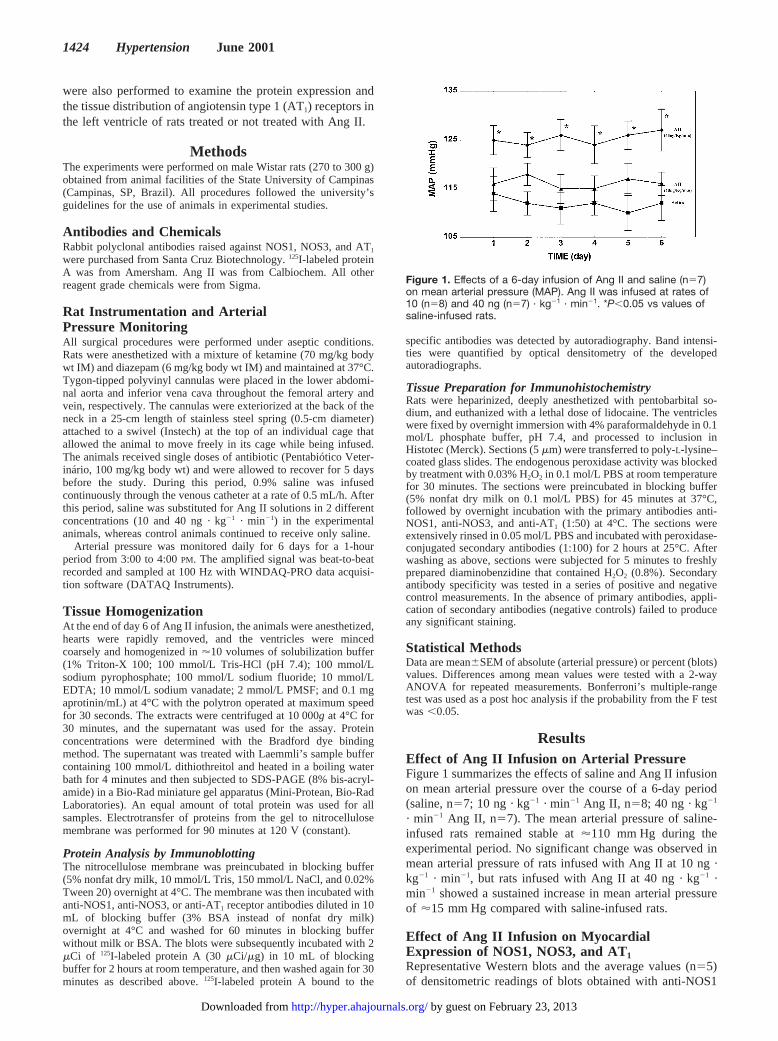
ResultsEffect of Ang II Infusion on Arterial PressureFigure 1 summarizes the effects of saline and Ang II infusionon mean arterial pressure over the course of a 6-day period(saline, n57; 10 ng · kg21 · min21 Ang II, n58; 40 ng · kg21
· min21 Ang II, n57). The mean arterial pressure of saline-infused rats remained stable at'110 mm Hg during theexperimental period. No significant change was observed inmean arterial pressure of rats infused with Ang II at 10 ng ·kg21 · min21, but rats infused with Ang II at 40 ng · kg21 ·min21 showed a sustained increase in mean arterial pressureof '15 mm Hg compared with saline-infused rats.
Effect of Ang II Infusion on MyocardialExpression of NOS1, NOS3, and AT1Representative Western blots and the average values (n55)of densitometric readings of blots obtained with anti-NOS1
Figure 1. Effects of a 6-day infusion of Ang II and saline (n57)on mean arterial pressure (MAP). Ang II was infused at rates of10 (n58) and 40 ng (n57) · kg21 · min21. *P,0.05 vs values ofsaline-infused rats.
1424 Hypertension June 2001
by guest on February 23, 2013http://hyper.ahajournals.org/Downloaded from
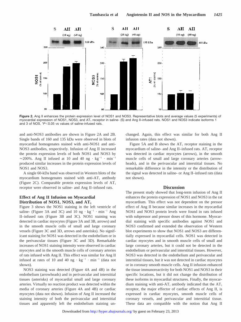
and anti-NOS3 antibodies are shown in Figure 2A and 2B.Single bands of 160 and 135 kDa were observed in blots ofmyocardial homogenates stained with anti-NOS1 and anti-NOS3 antibodies, respectively. Infusion of Ang II increasedthe protein expression levels of both NOS1 and NOS3 by'200%. Ang II infused at 10 and 40 ng · kg21 · min21
produced similar increases in the protein expression levels ofNOS1 and NOS3.
A single 60-kDa band was observed in Western blots of themyocardium homogenates stained with anti-AT1 antibody(Figure 2C). Comparable protein expression levels of AT1
receptor were observed in saline- and Ang II–infused rats.
Effect of Ang II Infusion on MyocardialDistribution of NOS1, NOS3, and AT1
Figure 3 shows the NOS1 staining in the left ventricle ofsaline- (Figure 3A and 3C) and 10 ng · kg21 · min21 AngII–infused rats (Figure 3B and 3C). NOS1 staining wasdetected in cardiac myocytes (Figure 3A and 3B, arrows) andin the smooth muscle cells of small and large coronaryvessels (Figure 3C and 3D, arrows and asterisks). No signif-icant staining for NOS1 was detected in the endothelium or inthe perivascular tissues (Figure 3C and 3D). Remarkableincreases of NOS1 staining intensity were observed in cardiacmyocytes and in the smooth muscle cells of coronary arteriesof rats infused with Ang II. This effect was similar for Ang IIinfused at rates of 10 and 40 ng · kg21 · min21 (data notshown).
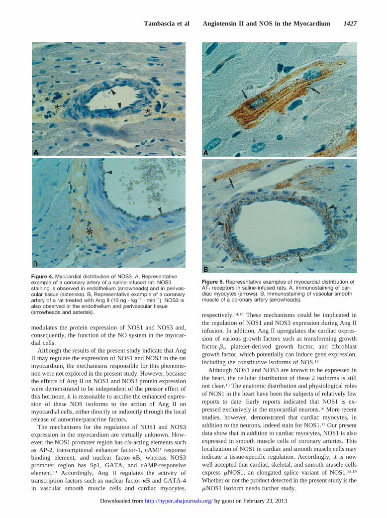
NOS3 staining was detected (Figure 4A and 4B) in theendothelium (arrowheads) and in perivascular and interstitialtissues (asterisks) of myocardial small and large coronaryarteries. Virtually no reaction product was detected within themedia of coronary arteries (Figure 4A and 4B) or cardiacmyocytes (data not shown). Infusion of Ang II increased thestaining intensity of both the perivascular and interstitialtissues and apparently left the endothelium staining un-
changed. Again, this effect was similar for both Ang IIinfusion rates (data not shown).
Figure 5A and B shows the AT1 receptor staining in themyocardium of saline- and Ang II–infused rats. AT1 receptorwas detected in cardiac myocytes (arrows), in the smoothmuscle cells of small and large coronary arteries (arrow-heads), and in the perivascular and interstitial tissues. Noremarkable difference in the intensity or the distribution ofthe signal was detected in saline- or Ang II–infused rats (datanot shown).
DiscussionThe present study showed that long-term infusion of Ang IIenhances the protein expression of NOS1 and NOS3 in the ratmyocardium. This effect was not dependent on the pressoreffect of Ang II because similar increases in the myocardialNOS1 and NOS3 protein levels were found in rats infusedwith subpressor and pressor doses of this hormone. Myocar-dial staining with specific antibodies against NOS1 andNOS3 confirmed and extended the observation of Westernblot experiments to show that NOS1 and NOS3 are differen-tially expressed in myocardial cells. NOS1 was detected incardiac myocytes and in smooth muscle cells of small andlarge coronary arteries, but it could not be detected in theendothelium or perivascular and interstitial tissues. However,NOS3 was detected in the endothelium and perivascular andinterstitial tissues, but it was not detected in cardiac myocytesor in coronary smooth muscle cells. Ang II infusion enhancedthe tissue immunoreactivity for both NOS1 and NOS3 in theirspecific locations, but it did not change the distribution ofthese isoforms in myocardial structures. Finally, the myocar-dium staining with anti-AT1 antibody indicated that the AT1
receptor, the major effector of cardiac effects of Ang II, isexpressed in cardiac myocytes, smooth muscle cells ofcoronary vessels, and perivascular and interstitial tissue.These data are compatible with the notion that Ang II
Figure 2. Ang II enhances the protein expression level of NOS1 and NOS3. Representative blots and average values (5 experiments) ofmyocardial expression of NOS1, NOS3, and AT1 receptor in saline- (S) and Ang II–infused rats. NOS1 and NOS3 indicate isoforms 1and 3 of NOS. *P,0.05 vs values of saline-infused rats.
Tambascia et al Angiotensin II and NOS in the Myocardium 1425
by guest on February 23, 2013http://hyper.ahajournals.org/Downloaded from
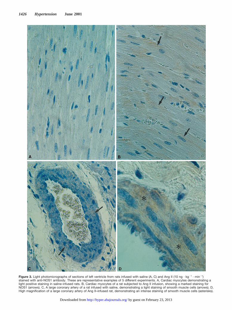
Figure 3. Light photomicrographs of sections of left ventricle from rats infused with saline (A, C) and Ang II (10 ng · kg21 · min21)stained with anti-NOS1 antibody. These are representative examples of 5 different experiments. A, Cardiac myocytes demonstrating alight positive staining in saline-infused rats. B, Cardiac myocytes of a rat subjected to Ang II infusion, showing a marked staining forNOS1 (arrows). C, A large coronary artery of a rat infused with saline, demonstrating a light staining of smooth muscle cells (arrows). D,High magnification of a large coronary artery of Ang II–infused rat, demonstrating an intense staining of smooth muscle cells (asterisks).
1426 Hypertension June 2001
by guest on February 23, 2013http://hyper.ahajournals.org/Downloaded from
modulates the protein expression of NOS1 and NOS3 and,consequently, the function of the NO system in the myocar-dial cells.
Although the results of the present study indicate that AngII may regulate the expression of NOS1 and NOS3 in the ratmyocardium, the mechanisms responsible for this phenome-non were not explored in the present study. However, becausethe effects of Ang II on NOS1 and NOS3 protein expressionwere demonstrated to be independent of the pressor effect ofthis hormone, it is reasonable to ascribe the enhanced expres-sion of these NOS isoforms to the action of Ang II onmyocardial cells, either directly or indirectly through the localrelease of autocrine/paracrine factors.
The mechanisms for the regulation of NOS1 and NOS3expression in the myocardium are virtually unknown. How-ever, the NOS1 promoter region hascis-acting elements suchas AP-2, transcriptional enhancer factor-1, cAMP responsebinding element, and nuclear factor-kB, whereas NOS3promoter region has Sp1, GATA, and cAMP-responsiveelement.13 Accordingly, Ang II regulates the activity oftranscription factors such as nuclear factor-kB and GATA-4in vascular smooth muscle cells and cardiac myocytes,
respectively.14,15 These mechanisms could be implicated inthe regulation of NOS1 and NOS3 expression during Ang IIinfusion. In addition, Ang II upregulates the cardiac expres-sion of various growth factors such as transforming growthfactor-b1, platelet-derived growth factor, and fibroblastgrowth factor, which potentially can induce gene expression,including the constitutive isoforms of NOS.13
Although NOS1 and NOS3 are known to be expressed inthe heart, the cellular distribution of these 2 isoforms is stillnot clear.13 The anatomic distribution and physiological rolesof NOS1 in the heart have been the subjects of relatively fewreports to date. Early reports indicated that NOS1 is ex-pressed exclusively in the myocardial neurons.16 More recentstudies, however, demonstrated that cardiac myocytes, inaddition to the neurons, indeed stain for NOS1.17 Our presentdata show that in addition to cardiac myocytes, NOS1 is alsoexpressed in smooth muscle cells of coronary arteries. Thislocalization of NOS1 in cardiac and smooth muscle cells mayindicate a tissue-specific regulation. Accordingly, it is nowwell accepted that cardiac, skeletal, and smooth muscle cellsexpressmNOS1, an elongated splice variant of NOS1.18,19
Whether or not the product detected in the present study is themNOS1 isoform needs further study.
Figure 4. Myocardial distribution of NOS3. A, Representativeexample of a coronary artery of a saline-infused rat. NOS3staining is observed in endothelium (arrowheads) and in perivas-cular tissue (asterisks). B, Representative example of a coronaryartery of a rat treated with Ang II (10 ng · kg21 · min21). NOS3 isalso observed in the endothelium and perivascular tissue(arrowheads and asterisk).
Figure 5. Representative examples of myocardial distribution ofAT1 receptors in saline-infused rats. A, Immunostaining of car-diac myocytes (arrows). B, Immunostaining of vascular smoothmuscle of a coronary artery (arrowheads).
Tambascia et al Angiotensin II and NOS in the Myocardium 1427
by guest on February 23, 2013http://hyper.ahajournals.org/Downloaded from
The results of the present study show a remarkable differ-ence in the distribution of NOS1 and NOS3 in the myocardialcells. In addition to the expected endothelial location ofNOS3, it was also detected in the interstitial and perivasculartissues. Increases in the staining produced by Ang II infusionwere easily detected in the interstitial and perivascular tis-sues, but no staining could be detected in the endothelium.The reason for the absence of detectable changes in the NOS3staining in the endothelium could be related to the narrowspace occupied by the endothelial cell, which makes conclu-sions difficult in regard to changes in NOS3 expression byimmunohistochemical analysis. The increases of NOS3 pro-tein expression in cardiac interstitial and perivascular tissuesinduced by Ang II could be due to the well-known effect ofangiotensin II on interstitial tissue proliferation and fibrosis.
NOS3 was not detected in significant amounts in cardiacmyocytes. Although this result agrees with some of the earlystudies, it contrasts with more recent studies showing thatcardiac myocytes indeed express NOS3.17 The reason for thisdiscrepancy is not clear but could be related to antibodyspecificity against certain isoforms of NOS3 in cardiacmyocytes.
Finally, we have shown that AT1 receptor is also expressedin cardiac and vascular smooth muscles, as well as ininterstitial and perivascular tissues, the structures in which theincreases in protein expression of NOS1 and NOS3 weredetected. This suggests that Ang II could enhance NOS 1 andNOS3 expression via AT1 receptor. However, the mediationvia the angiotensin type 2 receptors is also possible.
In conclusion, the present study demonstrates that long-term infusion of Ang II is accompanied by an increase in theprotein expression of NOS1 and NOS3 in the cells of ratmyocardium, independent of changes in arterial pressure. Thepresent data also provide evidence that NOS1 and NOS3 aredifferentially distributed in cardiac myocytes, coronary ves-sels, and interstitial tissue and that these NOS isoforms areupregulated by Ang II in these specific locations. The Ang IIupregulation of NOS1 and NOS3 in myocardial cells couldaccount for the impairment of the direct contractile andgrowth effects of this hormone on vascular smooth musclecells, cardiac myocytes, and fibroblasts when plasma or tissuelevels of Ang II are increased. This may favor functions suchas local blood flow, modulation of oxygen consumption, andinhibition of fibroblasts, vascular smooth muscle cells, andcardiac myocyte growth effects of Ang II.
AcknowledgmentsThis study was sponsored by grants from Fundação de Auxílio àPesquisa do Estado de São Paulo (FAPESP) (Proc. 98/11403–7) andConselho Nacional de Desenvolvimento Científico e Tecnológico(CNPq) (Proc. 521098/97-1).
References1. Weber KT, Brilla CG. Pathologic hypertrophy and the cardiac inter-
stitium: fibrosis and the renin-angiotensin-aldosterone system.Circu-lation. 1991;83:1849–1865.
2. Sadoshima J, Izumo S. Molecular characterization of angiotensin II–in-duced hypertrophy of cardiac myocytes and hyperplasia of cardiac fibro-blasts: critical role of the AT1 receptor subtype.Circ Res. 1993;73:413–423.
3. Gray MO, Long CS, Kalinyak JE, Li HT, Karliner JS. Angiotensin IIstimulates cardiac myocyte hypertrophy via paracrine release of TGF-band endothelin from fibroblasts.Cardiovasc Res. 1998;40:352–363.
4. Kupfahl C, Pink D, Friedrich K, Zurbrugg HR, Neuss M, Warnecke C,Fielitz J, Graf K, Fleck E, Zagrosek VR. Angiotensin II directly increasestransforming growth factorb1 and osteopontin and indirectly affectscollagen mRNA expression in the human heart.Cardiovasc Res. 2000;46:463–475.
5. Li D, Tomson K, Yang B, Mehta P, Croker BP, Mehta JL. Modulation ofconstitutive nitric oxide synthase, bcl-2 and Fas expression in culturedhuman coronary endothelial cells exposed to anoxia–reoxygenation andangiotensin II: role of AT1 receptor activation.Cardiovasc Res. 1999;41:109–115.
6. Ritchie RH, Schiebinger RJ, Lapointe MC, Marsh JD. Angiotensin II–induced hypertrophy of adult rat cardiomyocytes is blocked by nitricoxide.Am J Physiol. 1998;275(pt 2):H1370–H1374.
7. Boulanger CM, Heymes C, Benessiano J, Geske RS, Levy BI, VanhouttePM. Neuronal nitric oxide synthase is expressed in rat vascular smoothmuscle cells: activation by angiotensin II in hypertension.Circ Res.1998;83:1271–1278.
8. Chin SY, Pandey KN, Shi S-J, Kobori H, Moreno C, Navar LG. Increasedactivity and expression of Ca21-dependent NOS in renal cortex of AngII-infused hypertensive rats.Am J Physiol. 1999;277(pt):F797–F804.
9. Hennington BS, Zhang H, Miller MT, Granger JP, Reckelhoff JF. An-giotensin II stimulates synthesis of endothelial nitric oxide synthase.Hypertension. 1998;31:283–288.
10. Rizvi MA, Myers PR. Nitric oxide modulates basal and endothelininduced coronary artery vascular smooth muscle cell proliferation andcollagen levels.J Mol Cell Cardiol. 1997;29:1779–1789.
11. Kihara M, Umemura A, Kadota T, Yabana M, Tamura K, Nyuui N,Ogawa N, Murakami K, Fukamizu A, Ishii M. The neuronal isoform ofconstitutive nitric oxide synthase is up-regulated in the macula densa ofangiotensinogen gene-knockout mice.Lab Invest. 1997;76:285–294.
12. Iwai N, Hanai K, Tooyama I, Kitamura Y, Kinoshita M. Regulation ofneuronal nitric oxide synthase in rat adrenal medulla.Hypertension.1995;25:431–436.
13. Papapetropoulos A, Rudic RD, Sessa WC. Molecular control of nitricoxide synthases in the cardiovascular system.Cardiovasc Res. 1999;43:509–520.
14. Ruiz-Ortega M, Lorenzo O, Ruperez M, Konig S, Wittig B, Egido J.Angiotensin II activates nuclear transcription factorkB through AT1 andAT2 in vascular smooth muscle cells: molecular mechanisms.Circ Res.2000;86:1266–1272.
15. Herzig TC, Jobe SM, Aoki H, Molkentin JD, Cowley AW, Izumo S,Markham BE. Angiotensin II type 1a receptor gene expression in theheart: AP-1 and GATA-4 participate in the response to pressure overload.Proc Natl Acad Sci. 1997;94:7543–7548.
16. Shah AM, MacCarthy PA. Paracrine and autocrine effects of nitric oxideon myocardial function.Pharmacol Therap. 2000;86:49–86.
17. Feron O, Bellhassen L, Kobzik L, Smith TW, Kelly RA, Michel T.Endothelial nitric oxide synthase targeting to caveolae: specific inter-actions with caveolin isoforms in cardiac myocytes and endothelial cells.J Biol Chem. 1996;271:22810–22814.
18. Silvagno F, Xia H, Bredt DS. Neuronal nitric oxide synthase m, analternatively spliced isoform expressed in differentiated skeletal muscle.J Biol Chem. 1996;271:11204–11208.
19. Schwarz PM, Kleinert H, Förstermann U. Potential functional signif-icance of brain-type and muscle-type nitric oxide synthase 1 expressed inadventitia and media of rat aorta.Arterioscler Thromb Vasc Biol. 1999;19:2584–2590.
1428 Hypertension June 2001
by guest on February 23, 2013http://hyper.ahajournals.org/Downloaded from


![[Kleber Daum Machado] Teoria Do Eletromagnetismo, (Bookzz.org) (2)](https://img.pdfslide.us/doc/110x75/55cf8aa455034654898ca3cd/kleber-daum-machado-teoria-do-eletromagnetismo-bookzzorg-2.jpg)