Embed Size (px)
Citation preview

1
S4 Biology
MULTICELLULAR ORGANISMS
Unit 2
Part B: I wan’na be like you
NAME:
CLASS:

2
Topic 1: Plant Transport No. Level Rating
R/A/G 16 N5 Plant transport: I can describe the structures of a leaf: upper epidermis, palisade mesophyll,
spongy mesophyll, vein, lower epidermis, guard cells and stomata
17 N5 Plant transport: I have investigated the movement of water and minerals through xylem vessels and
can describe their structure (lignin).
18 N5 Plant transport: I have looked at root hairs under the microscope and can describe the moment of
water through root hair cells and xylem to the leaf (transpiration).
19 N5 Plant transport: I have learned about the process of transpiration and know that it is affected by
wind speed, humidity, temperature and surface area.
20 N5 Plant transport: I have measure the rate of transpiration using a potometer.
21 N5 Plant transport: I have looked at stomata under the microscope and can describe their role in
transpiration
22 N5 Plant transport: I understand that sugar is transported up and down the plant in living phloem tissue 23 N5 I can describe the structure of phloem tissue to include phloem cells with sieve plates and
companion cells
Topic 2: Animal Transport 24 N5 Animal transport and exchange systems: I can state that mammals have blood to transport
nutrients, oxygen and carbon dioxide.
25 N5 Animal transport and exchange systems: I can state that blood contains plasma, red blood cells and
white blood cells
26 N5 Animal transport and exchange systems: I can describe how red blood cells are specialised to
transport oxygen efficiently
27 N5 Animal transport and exchange systems: I understand the role of haemoglobin and oxyhaemoglobin
in the transport of oxygen.
28 N3 KA3 Animal transport and exchange systems: I can describe the body defences against disease and the
role of vaccines
29 N3 KA3 I have learned that the body’s first line of defence is barriers to infection such as skin, tears and
mucus.
30 N3 KA3 I can state how vaccines can provide immunity to disease 31 N5 Animal transport and exchange systems: I can state that white blood cells are part of the immune
system and are involved in destroying pathogens
32 N5 Animal transport and exchange systems: I can name two main types of white blood cells: phagocytes
and lymphocytes
33 N3 KA1
/N5
Animal transport and exchange systems: I have taken part in heart dissections or used models to
examine the structures of the mammalian heart.
34 N5 Animal transport and exchange systems: I can name the chambers of the heart (left atrium, right
atrium, left ventricle, right ventricle) and describe the flow of blood through the heart.
35 N5 Animal transport and exchange systems: I can identify the blood vessels of the human body (aorta,
vena cava, pulmonary arteries, pulmonary veins)
36 N5 Animal transport and exchange systems: I can identify the coronary artery and understand its
importance to healthy functioning of the heart.
37 N5 Animal transport and exchange systems: I know the difference in structure and functions of the
different blood vessels (veins, arteries, capillaries)
Topic 3: Absorption of materials 38 N5 Animal transport and exchange systems: I understand that oxygen and nutrients from food must be
absorbed and delivered to cells for respiration
39 N5 Animal transport and exchange systems: I understand that waste materials such as carbon dioxide
must be removed from cells into the blood stream.
40 N5 Animal transport and exchange systems: I understand that tissues contain capillary networks to
exchange materials at a cellular level

3
41 N5 Animal transport and exchange systems: I can describe the features of the small intestine that
allow food to be absorbed into the bloodstream (large surface area, thin walls, good blood supply,
villi)
42 N5 Animal transport and exchange systems: I can describe the features of the villus and state that
glucose and amino acids move into the bloodstream, fatty acids and glycerol move into the lacteals of
the villi.
43 N3 KA1 Animal transport and exchange systems: I have looked at the structure of the lungs 44 N5 Animal transport and exchange systems: I can describe the features of the alveoli and explain how
oxygen and carbon dioxide are exchanged through the alveolar walls.
Topic 4: Control and communication No. Level Outcome Rating
R/A/G 45 N4 I can discuss why stable conditions are important to the body in order to survive 46 N4 I can name temperature as an external factor 47 N4 I can describe the response of the body to a) increasing and b) decreasing temperature 48 N4 I have researched the causes of diabetes 49 N5 I can state the definitions of “hormone” and the “endocrine system” 50 N5 I understand the terms “target tissues” and “hormone receptor proteins”. 51 N5 I have researched the role of hormones in the body. 52 N4/N5 I have investigated diabetes and its causes including the differences between Type 1 and Type 2
diabetes.
53 N4/N5 I can name blood glucose level as an internal factor and describe how the body responds to both a)
increasing and b) decreasing concentrations
54 N5 I have investigated blood glucose regulation including the role of insulin, glucagon, glycogen,
pancreas and liver.
55 N5 I can state that the Nervous system consists of central nervous system (CNS) and nerves.
56 N4/N5 I have investigated examples of where communication pathways are used, e.g. pain receptors.
57 N5 I can state that the CNS consists of the brain and spinal cord.
58 N4/N5 I have used models to look at the structure and function of brain (N5: including cerebrum,
cerebellum and medulla).
59 N5 I have used diagrams to illustrate the three types or neurons (sensory, inter and motor).
60 N5 I know the key terms receptors, sensory input/stimuli, electrical impulses and describe how
electrical impulses carry messages along neurons.
61 N5 I have used diagrams and animations to investigate synapses and state that a synapse occurs
between neurons. I know that chemicals transfer these messages across synapses.
62 N5 I can describe the structure and function of reflex arcs and their role in protecting the body from
harm. I can state examples of reflex actions and explain their importance (e.g. blinking, iris reflex,
response to pain)

4
For National 5 Biology 2020 Exam……
This is the final topic. Make sure you are revising ALL of the N5 material
from S3 AND S4. Be organised and make sure you have all your notes:

5
Topic 1: Transport
In this topic we will look at how transport is important in living organisms. We
will look at how plants transport food and water to stay alive. In topic 2 we will
look at how animals transport materials around the body.
Plant Transport
Revision
1. What is the process by which plants use light energy to make food?
____________________
2. Write the word equation for this process, labelling raw materials,
products and any other requirements
Photosynthesis and plant structure
Plants need special structures to help them carry out Photosynthesis. The
leaves are the organs of photosynthesis and have special structures and
features to allow them to carry out the process efficiently.
The raw materials for photosynthesis must be delivered to chloroplasts inside
the leaves so that plants can carry out photosynthesis.
For example water and minerals must
be absorbed through special root hair cells
and is carried up to the leaves through special
tubes called _________ _________ .
The food made during photosynthesis is
transported up and down the plant through
special tissue called _____________.
Add labels to the plant to show its organs:
Leaves (Site of photosynthesis)
Roots (Absorb water and minerals)
Stem (Bundles of xylem and phloem)

6
The need for transport
Match the following uses of water to the correct description (revision from Unit 1):
support transport cooling photosynthesis
The diagram below shows the structure of a leaf. Label the structures and
complete the table to show the functions of each.
Use of water Description
Water enters plant cells making them turgid
When water evaporates from the surface of a plant it carries heat away
from the plant
Hydrogen from water combines with carbon dioxide to make glucose
Minerals needed by the plant and sugars produced by photosynthesis must
be moved within the plant
Structure Function
Waxy Cuticle (1, 7)
Upper epidermis (9) & lower epidermis (4)
Palisade mesophyll (2) & spongy mesophyll (8)
Moist air space (3)
Stoma (5)
Guard cells (6)
1 9
8
2
3
7 4
5
6

7
Plants have vascular tissue or “veins”. Vascular tissue is made up of bundles of
xylem and phloem. Your teacher will show you stained xylem vessels (celery) and
pictures of vascular bundles.
Draw a labelled picture of the celery cross section with stained pink bundles of
xylem and the lignified xylem vessels you have seen under the microscope:
Describe the path that water takes as it enters the plant and moves to the
leaves for photosynthesis. You should use your knowledge of osmosis in your
description and the words in the box below (Hint!).
Xylem tissue is ____________ as it is made of xylem cells which have lost
their ends to form a hollow tube. It is also strengthened with ________. These
features allow xylem to transport _________ efficiently.
Phloem tissue is also specialised. Unlike xylem,
phloem is made of _________ cells. It has a
sieve tube to transport food made in
________________ and special companion
cells to control where food is transported to.
Label the diagram to show these features.
Hint:
High water concentration Root hair cells Mesophyll
Low water concentration xylem vessels Moist soil
Celery cross section Xylem Vessels
Phloem Structure
1
3
2

8
Gas exchange at the leaves
Stomata allow gas exchange to take place at the leaves. Your teacher will allow
you to look at stomata under a microscope by doing a leaf peel.
Stomata
Stomata are tiny _________ found on the surface of a
leaf. Plants take in __________ __________ from the air
through these stomata.
Unfortunately water vapour is _______ through the
stomata. To overcome the loss of water the stomata
________ at night when the plant is unable to undergo
photosynthesis.
The ________ cells on either side of the stomata are
responsible for opening and closing the pore.
Transpiration (N5)
_______________ is the name of the process by which water is pulled up the
plant stem against the force of gravity!
Water from the walls of the leaf cells lining the moist air spaces
_____________ and is lost by transpiration via the ____________. In order
to replace these losses, cells draw water from the xylem vessels and set up a
transpiration pull.
The rate of transpiration is affected by a number of factors. We can measure
the rate of transpiration using a potometer. A potometer can be used to
measure the volume of water absorbed by a leafy shoot and we can use this to
work out how quickly transpiration occurs.
The rate of transpiration can vary depending on the environment. Factors which
increase the rate of evaporation of water also increase the rate of
transpiration in a plant. Can you unscramble the letters to show four factors
which increase transpiration rate?
_ _ _ _ / _ _ _ _ _ _ _ _ _ _ _ (PREMATURE HEIGHT)
_ _ _ _ / _ _ _ _ _ (END SWIPED)
_ _ _ _ _ _ _ _ (I HUM TIDY)
_ _ _ _ _ _ _ /_ _ _ _ (AREA CAR FUSE)

9
Transpiration Investigation:
Write a suitable aim for your investigation.
Choose one of the following independent variables (what we are going
to change in our experiment): wind speed, temperature, humidity OR
surface area.
Label the diagram of a simple potometer
Design an experiment to investigate the effect of your chosen variable
on the rate of transpiration in a leafy shoot.
Write a summary of your method.
Write a conclusion for your investigation.
Extra Challenge Questions for the enthusiast……
Using your knowledge of osmosis, predict what would happen to the guard cells
under the following conditions… (Score out the incorrect words in each pair)
Guard
cells
stoma Guard cells will take in/lose water by osmosis as water will enter/leave
the cell from a high water concentration outside to a lower water
concentration inside the guard cells. Cells will become flaccid/turgid and
the stoma pore will open/close.
Guard cells will take in/lose water by osmosis as water will enter/leave
the cell from a high water concentration inside to a lower water
concentration outside the guard cells. Cells will become flaccid/turgid and
the stoma pore will open/close.
Plant Transport:
12 words about plant transport are hidden in
the word search. Can you find them? Use the
hints below…….
1. Gaseous raw material for photosynthesis
2. Cells which form the stomata
3. Tissue that transports food made in photosynthesis
4. Liquid raw material for photosynthesis
5. Cells which control phloem
6. Strengthens xylem vessels
7. Leaf structure that controls gas exchange
8. Waterproof layer that protects leaves
9. Transparent cells present on upper/lower leaf surfaces
10. Cells that contain chlorophyll (palisade/spongy)
11. Force that pulls water from roots to leaves
12. Hollow tubes that transport water
Potometer

10
Topic 2: Animal transport and exchange In the same way that plants need a transport system to move raw materials and
products of photosynthesis, animals need transport systems to ensure raw
materials are delivered to every cell in the body and waste materials are
removed.
Revision:
1. What process inside living cells releases energy from food?
_________________
2. Complete the equation below to show this process:
3. Where does each stage take place in the cell?
Stage 1: ______________ Stage 2: ______________
Enzyme controlled reactions
pyruvate pyruvate
2 ADP + 2 Pi
Stage 1
Stage 2 pyruvate
water
18 ATP
oxygen
Enzyme controlled reactions

11
The raw materials for respiration (_________ and ___________) must be
taken into the body and transported to the cells. Waste materials from cells
must also be transported away. This is the reason that mammals have transport
and exchange systems. Use arrows to match the body systems to the correct
functions:
Digestive system
Respiratory system
Circulatory system
We are now going to look at each transport and exchange system
The Circulatory System
The circulatory system in mammals transports blood containing materials such
as nutrients, oxygen and carbon dioxide.
Blood Composition
Blood contains a watery yellow liquid called __________. Blood gets its red
colour from the red blood cells which contain the red pigment
______________. Haemoglobin carries oxygen.
Red Blood Cell Revision: Can you complete the diagram to show the other
features of red blood cells that allow them to be suited to their function
(remember cells are specialised to help them do a particular job)
Takes in oxygen for respiration and removes carbon dioxide (and water)
Transports food which is broken down to release glucose (for respiration) and allows waste to be removed from the body.
Transports materials such as food, oxygen and waste materials throughout the body.
Contain haemoglobin
to carry oxygen

12
Blood contains approximately 55% plasma, 1% (white blood cells and platelets)
and 44% red blood cells. Use this information to make a graph below.
Blood Composition
We are now going to look at the blood components in more detail.
Haemoglobin is a protein inside the red blood cells. In locations in the body
where there is a high concentration of oxygen, haemoglobin forms
oxyhaemoglobin.
Location: ____________ Colour of blood: ___________________
Blood with a ______ concentration of oxygen is described as oxygenated.
In locations where the oxygen concentration is low, haemoglobin releases oxygen
Location: ____________ Colour of blood: ___________________
The oxygen then diffuses into the cells. Blood with a _____ oxygen
concentration is described as deoxygenated.
Blood components:
Plasma: yellowish liquid containing
water; transports food, etc.
White blood cells: to fight
disease.
Platelets: to clot blood
Red blood cells: to transport
oxygen
Title
X-axis label
Y-axis label
Units?
Plot

13
White blood cells and body defence
White blood cells are part of the immune system and are involved in destroying
pathogens. In S2 you learned about how the body defends itself against
disease.
Revision:
1. What are the three main types of microorganism?
2. What are pathogens?
3. What are the body’s first lines of defence against pathogens?
4. What is the immune system?
5. What are vaccines?
Two main types of white blood cell
In defending the body, there are 2 main types of cells involved:
_____________ which carry out phagocytosis by _________ pathogens
and digesting them (breaking them down).
_____________ which produce antibodies which destroy pathogens.
Antibodies are __________ to particular pathogens.
In the box below, you should draw the two types of white blood cell and
summarise the job that they do to defend the body.
White Blood Cells
Phagocytes Lymphocytes

14
The Circulatory System
The blood allows materials to be transported around the body through the
circulatory system. The circulatory system consists of the __________ ,
__________ __________ and the _________ .
The circulatory system has two circuits allowing blood to flow
through the heart twice. One circuit allows oxygenated blood
to be taken to the cells from the lungs (where _________ is
taken in). The second circuit allows deoxygenated blood to be
taken from the cells to the lungs (where _________
_________ is breathed out).
Label the diagram to show the heart, lungs and tissues of the
body. Your teacher may ask you to shade the oxygenated
blood in RED and the deoxygenated blood in BLUE.
NB: this is a very simplified drawing of the heart and
circulation. On the next page your teacher will give you a more
detailed diagram to complete. It shows the pathway of blood
through the heart, lungs and the main organs of the body.
Blood composition activities:
Make model blood
Body defences poster
Diseases and Vaccines activity (newspaper article / patient leaflet, etc.)
White blood cell types poster
Blood donation activities

15
Pathway of blood through the heart, lungs and body:
The tubes that transport blood are called _____________. In your diagram
above, there are two types of blood vessels named: ___________ and
_______.
In mammals there are three types of blood vessel:
Arteries – which take blood Away from the heart
Veins – which take blood back IN to the heart
Capillaries – which carry blood through tissues and organs.
Later we will look at the arteries, veins and capillaries in more detail.
In order to move blood through the circulatory system a _______ is needed.
The heart is a ____________ pump. When the muscle contracts, it squeezes
blood up and out of the heart.

16
You are now going to look at the structure of the heart (your
teacher may do a dissection of the heart with you!).
The heart has two types of chamber:
Atria – the top chambers of the heart that receive blood.
Ventricles – the bottom chambers of the heart that
squeeze blood upwards and out of the heart.
Label the two atria and the two ventricles. Remember: in
diagrams the parts of the body on the right of the diagram are
always labelled “LEFT” and the parts the left of the diagram are
always labelled “RIGHT”.
You are now ready to look at the structure of the heart and the flow of blood
through it:
__________ side __________ side
17
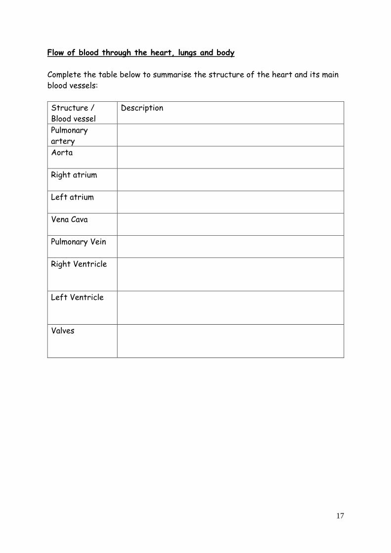
Flow of blood through the heart, lungs and body
Complete the table below to summarise the structure of the heart and its main
blood vessels:
Structure /
Blood vessel
Description
Pulmonary
artery
The pulmonary artery takes deoxygenated blood from the
right ventricle back to the lungs to be oxygenated.
Aorta The aorta takes oxygenated blood from the left ventricle
to the rest of the body.
Right atrium The right atrium contracts and pushes deoxygenated blood
down into the right ventricle.
Left atrium The left atrium contracts and pushes oxygenated blood
down into the left ventricle.
Vena Cava The vena cava takes deoxygenated blood from the rest of
the body into the right atrium.
Pulmonary Vein The pulmonary vein takes oxygenated blood from the lungs
into the left atrium.
Right Ventricle The right ventricle receives blood from the right atrium.
It contracts and pushes deoxygenated blood back towards
the lungs via the pulmonary artery.
Left Ventricle The left ventricle receives blood from the left atrium. It
contracts and pushes oxygenated blood towards the rest
of the body via the aorta.
Valves Valves are found between the atria and ventricles, and
between the ventricles and aorta and pulmonary artery.
They prevent backflow of blood.

18
TASK: Imagine you are a red blood cell. Describe the path you would take as
you move through the body. You should start at the lungs (Hint: use the word
bank below to make sure you have described one full circuit!)
Structure of Arteries, Veins and Capillaries
Arteries take blood ________ from the heart at ______
pressure. Because blood inside the arteries is at high
pressure, the structure of arteries must be able to
withstand the force. They have thick muscular walls and
narrow lumen
Veins take blood ______ ___ the heart. Blood in veins is
under ________ pressure than the blood in the arteries
so they have special ________ which prevent blood from
flowing backwards. They have _________ walls and wide
_______.
Capillaries are the finest vessels of the circulation and
connect arteries to veins. They have ______ walls which
are one cell thick. This allows materials to diffuse quickly
in and out of the blood and body tissues. Capillaries also
form large networks to ensure that all cells receive a blood
supply (capillary networks give a large surface area for the
movement of materials).
left atrium right atrium left ventricle right ventricle
aorta pulmonary vein lungs pulmonary artery
vena cava “drop off oxygen” body tissues “pick up oxygen”
One cell thick
Thick wall,
narrow lumen
Thinner wall,
Wider lumen

19
Capillary network
Why must the blood leaving the body tissues be taken back to the heart?
___________________________________________________________
___________________________________________________________
___________________________________________________________
___________________________________________________________
Use your notes to complete the summary table below. The capillaries have been
completed for you:
Type of blood
vessel
Wall
thickness
Internal
diameter
(lumen)
Valves Blood
pressure Function
Arteries
Capillaries
One cell
thick
Very
narrow
but wide
enough for
blood cells
to squeeze
through
No valves - Capillaries carry blood through
tissues and organs. They allow
materials exchanged between
blood and cells by diffusion.
Veins
Arteries branch forming finer and finer vessels until the capillary network. Here, the
capillary walls are only one cell thick. Oxygen (and nutrients) leave the blood capillary and
diffuse into the body cells from a _____ concentration to a _______ concentration. At
the same time, carbon dioxide (and waste) leave the body cells by diffusion. The capillaries
join together to form wider vessels which carry the blood back to the heart via the veins.
Oxygenated blood blood
Deoxygenated blood
Capillaries

20
Think, Pair, Share:
What are the raw materials needed for respiration?
For each raw material, what organ system allows the material to be absorbed
into the bloodstream?
What features do these organ systems have that allow lots of the material to
be absorbed into the bloodstream?
Consolidate:
Your teacher will help you summarise the class discussion. Write this below:
Coronary Arteries
One of the first arteries to branch off
from the aorta is the _____________
artery. This supplies the heart muscle
itself with blood. Blockage of the coronary
arteries prevents the flow of blood to the
heart muscle. When this happens part of
the heart muscle is deprived of oxygen,
causing the tissue die. This results in a
heart attack. Label the
diagram to show:
aorta, left coronary artery right coronary artery.
Absorption of materials
The raw materials needed for respiration must be absorbed into the
bloodstream so that they can be delivered to the cells.
21
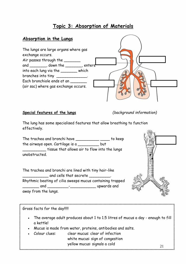
Topic 3: Absorption of Materials
Absorption in the Lungs
The lungs are large organs where gas
exchange occurs.
Air passes through the _______
and _______, down the _______, enters
into each lung via the _______ which
branches into tiny _____________.
Each bronchiole ends at an _______
(air sac) where gas exchange occurs.
Special features of the lungs (background information)
The lung has some specialised features that allow breathing to function
effectively.
The trachea and bronchi have __________ ____ to keep
the airways open. Cartilage is a _________ but
__________ tissue that allows air to flow into the lungs
unobstructed.
The trachea and bronchi are lined with tiny hair-like
___________ and cells that secrete _________.
Rhythmic beating of cilia sweeps mucus containing trapped
_______ and _________-___________ upwards and
away from the lungs.
Gross facts for the day!!!!!
The average adult produces about 1 to 1.5 litres of mucus a day - enough to fill
a kettle!
Mucus is made from water, proteins, antibodies and salts.
Colour clues: clear mucus: clear of infection
white mucus: sign of congestion
yellow mucus: signals a cold

22
Gas Exchange at alveoli
Blood arriving at the lungs is said to be
DEOXYGENATED because it contains a
_____ _______________ of oxygen (high
concentration of carbon dioxide).
Diffusion of oxygen from the alveoli to the red
blood cells occurs (carbon dioxide diffuses from the
plasma into the alveolus). Blood therefore becomes
OXYGENATED as it contains a _______
________________ of oxygen (lower concentration
of carbon dioxide).
The alveoli are well suited to gas exchange because of
their specialised structure. Complete the summary
table below to show these features:
Feature Function
Large surface
area
Allows oxygen to dissolve easily.
Thin lining (only
1 cell thick)
Rapid pick up of oxygen by red blood cells for respiration
rapid removal of Carbon Dioxide.

23
Lung Facts! (TWIG)
Your lungs have about the same volume as a football, and around 300 million
tiny air chambers each on average.
If you laid all of them out on the ground they would cover a badminton court.
Each human lung holds about 3l of air.
A blue whale's lung can hold 2500l of air.
Every day you breathe in and out around 25,000 times.
The average human can hold their breath for up to 60 seconds, but some have
trained themselves to hold their breath for over 20 minutes!
Some divers train themselves to dive for long periods of time
without oxygen
Gas Exchange at alveoli
Oxygen:
At the lungs there is a higher / lower oxygen concentration in the inhaled air
than in the blood entering the alveolus. Oxygen diffuses from the inhaled air
into the blood from a high oxygen concentration to a low concentration. The
oxygen binds to haemoglobin in the red blood cells to form ________________
Carbon dioxide:
At the lungs there is a higher / lower carbon dioxide concentration in the
inhaled air than in the blood entering the alveolus. Carbon dioxide diffuses from
the blood to the air sacs from a high oxygen concentration to a low
concentration.
Project:
Your teacher may give you some information on smoking and the
effects that it can have on the lungs and overall heath. Use your
knowledge of the lung structures to produce a poster or health
information leaflet about smoking and health.

24
Absorption in the Small Intestine
The mammalian digestive system allows large insoluble food to be broken down
into smaller soluble materials. Each of the structure of the digestive system are
well suited to this function.
Enzymes are vital in digestion as they allow large insoluble food to be broken
down into smaller soluble materials….
1. What is the definition of an enzyme?
_______________________________________________________
_______________________________________________________
2. Write down the general equation for an enzyme reaction:
3. All digestive enzymes take big molecules and make them into smaller
molecules. What type of reactions are these? ____________________
The Small Intestine (background information)
This is a long narrow section of the gut which gives a large surface area for the
absorption of food. The function of the small intestine is to __________ the
products of digestion through the wall of the gut and into the
______________.
As partially digested food is moved along the small intestine by peristalsis,
three digestive enzymes from the pancreas continue to breakdown the
remaining _________ ____________ molecules. This prepares the food
molecules for their ______________through the lining of the small intestine.
Revision:
Write the word equation for all enzyme reactions

25
The role of Villi
The internal structure of a villus allows
them to be ideally suited for the
absorption and transport of digested
food.
Villus features:
Thin lining:
_________________________
_________________________
_________________________
Blood capillary:
_________________________
_________________________
_________________________
Central Lacteal:
_________________________
_________________________
_________________________
The small intestine is very efficient at its job because of its ___________ .
The small intestine has a _________ ____________ _________. This is due
to it being very long and also having a ________ inner surface. The inner
surface is also covered in tiny finger-like projections called _______.

26
Absorption of Materials Summary
Now that you have learned the features of the lungs and the digestive system
you should be able to summarise the features that they have in common.
Write notes on the shared features of the lungs and the digestive tract and
describe how these features allow efficient absorption of materials:

27
Topic 4: Control and Communication The maintenance of a constant internal environment by the body is called
_________________. Factors that are kept at a set point include:
• ____________________________
• ____________________________
• ____________________________
Thermoregulation is the regulation of body temperature so that it stays at
a set point of ___°C.
• If the body temperature falls ____ _____ then biological
reactions may happen too _______ for cells to survive.
• If the body temperature rises ____________
then _________ and other cell ___________
may be damaged.
The Brain and the Hypothalamus
•
• The _________________ in the brain
contains the temperature monitoring
centre for the body.
• The hypothalamus receives _______
__________ from structures in the skin
called __________________, which
give information about the surface
temperature of the body. The
hypothalamus also contains its own
thermo-receptors, which are sensitive to
the temperature of the __________.
The hypothalamus responds to the information it gets from thermo-receptors
by sending nerve impulses to __________, such as the skin, to return the body
temperature back to ___________.

28
The response of the body to changes in temperature
Too hot
Too cold
All of the methods to regulate body temperature mentioned so far are
___________________________ - we do not consciously decide to sweat or
to shiver.
In contrast, humans are also able to make___________________________,
for example when we decide to take action to help regulate our body
temperature.
Water balance
For the cells of our body to work properly, it is important that their water
content is maintained at the correct level. This means our body must maintain a
__________ between the water we take in (by drinking and eating) and the
water we lose (through sweating, excretion and breathing out) .
Water balanced is maintained by
the _________. Kidneys control
blood water concentration through
the action of a _________ called ADH.
We are now going to look at some examples
of hormones of the human body.

29
Hormones
Hormones are __________ _____________ that travel around the body in
the ___________.
Hormones are __________ _____________. Hormones are released into the
bloodstream by groups of cells called __________ ________. Hormones are
transported in the ________ __________ to target body tissues where they
bind to cells to produce a response.
Each hormone stimulates a __________ response in either individual cells,
tissues or in other glands. The cell that a hormone acts on is called its
________ _____.
• A specific hormone can only affect cells if the cells have a _________
for it.
• The diagram below shows two cells targeted by two different hormones.
• Hormone one cannot affect the cell on the right because the cell does not
have a receptor for it.
30
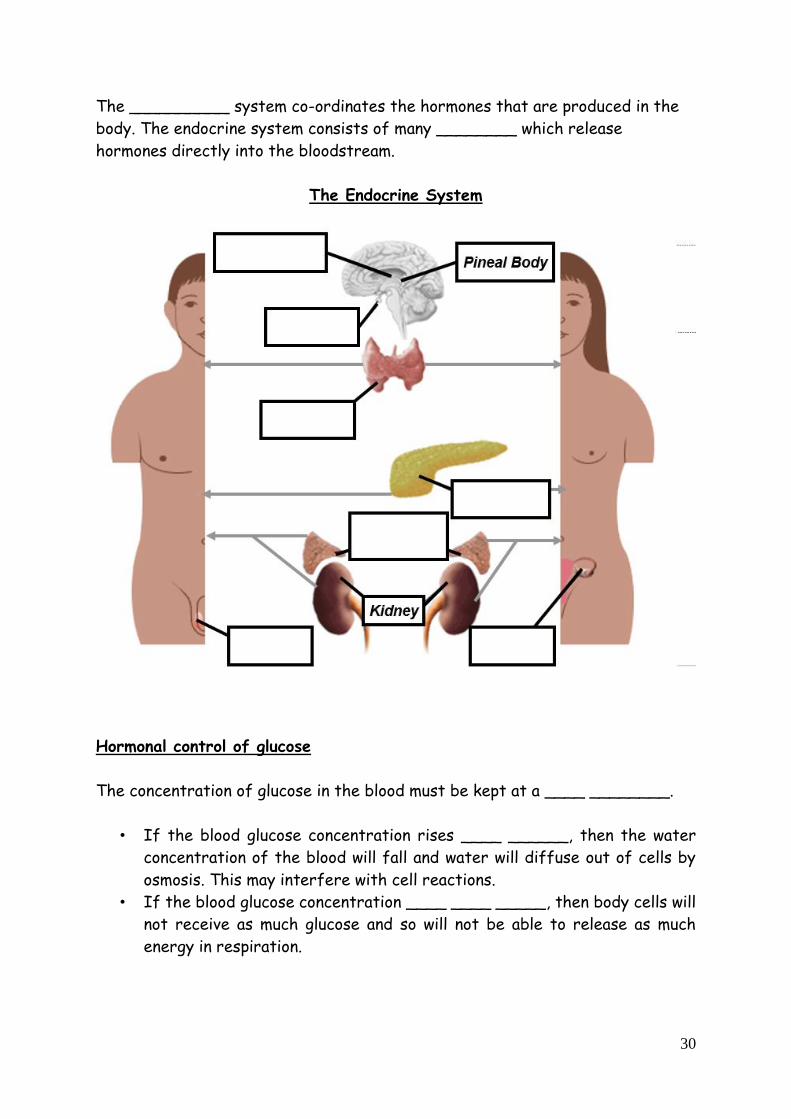
The __________ system co-ordinates the hormones that are produced in the
body. The endocrine system consists of many ________ which release
hormones directly into the bloodstream.
The Endocrine System
Hormonal control of glucose
The concentration of glucose in the blood must be kept at a ____ ________.
• If the blood glucose concentration rises ____ ______, then the water
concentration of the blood will fall and water will diffuse out of cells by
osmosis. This may interfere with cell reactions.
• If the blood glucose concentration ____ ____ _____, then body cells will
not receive as much glucose and so will not be able to release as much
energy in respiration.

31
• The concentration of glucose in the blood is regulated by the action of the
hormones _______ and __________.
• The target cells of these hormones are found in the liver. The liver stores
the body’s glucose in the form of___________ . Glycogen can be broken
down again and the glucose released into the blood when it is needed.
Under what circumstances do you think blood glucose levels will rise?
___________________________________________________________
Under what circumstances do you think blood glucose levels will fall?
___________________________________________________________
Complete the table to show what happens when blood glucose changes from its
set point.
Glucose
level
Hormone
released
Response Effect
Increased
lowers blood
glucose to
normal
Decreased
raises blood
glucose to
normal
32
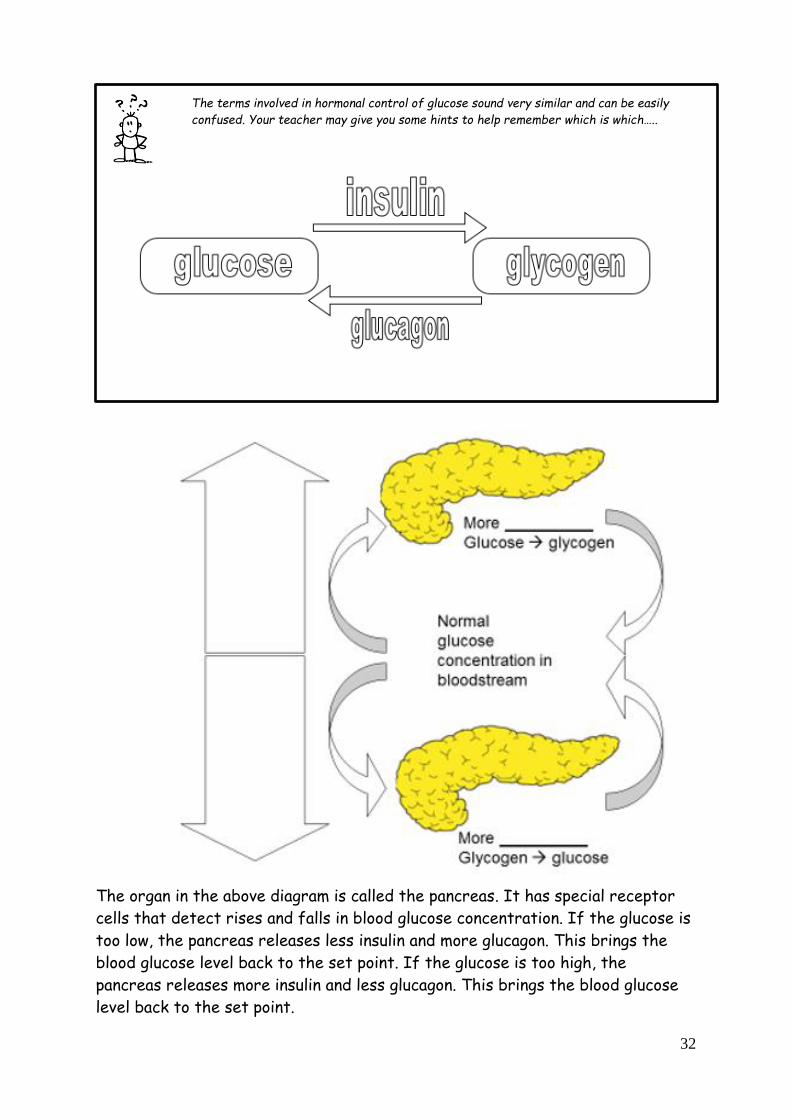
The terms involved in hormonal control of glucose sound very similar and can be easily
confused. Your teacher may give you some hints to help remember which is which…..
The organ in the above diagram is called the pancreas. It has special receptor
cells that detect rises and falls in blood glucose concentration. If the glucose is
too low, the pancreas releases less insulin and more glucagon. This brings the
blood glucose level back to the set point. If the glucose is too high, the
pancreas releases more insulin and less glucagon. This brings the blood glucose
level back to the set point.

33
Additional information
Diabetes
________________ is the body’s _______________ to control its blood
__________________ levels.
After eating, ______________ glucose levels increase much ______________
and take much _______________ to come back normal than someone without
diabetes.
Research investigation – type 1 and type 2 diabetes
Cause
Risk factors
Symptoms
Treatment
34
Control and Communication: The Nervous System
Nervous system
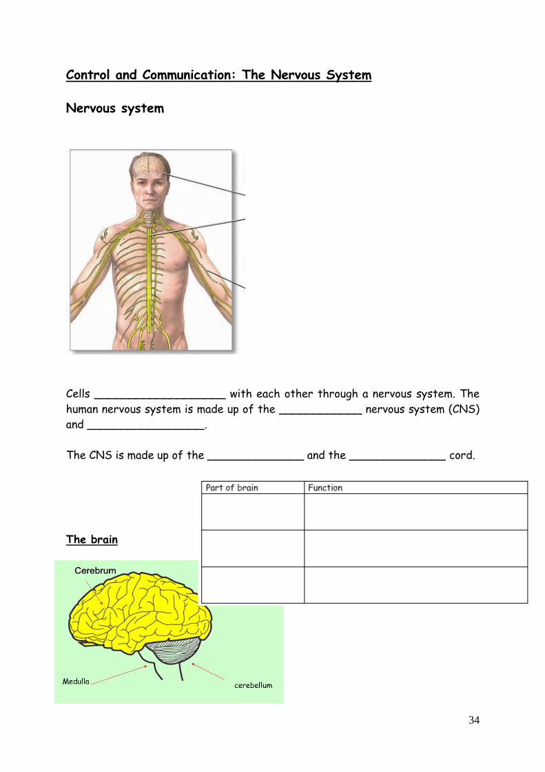
Cells ___________________ with each other through a nervous system. The
human nervous system is made up of the ____________ nervous system (CNS)
and _________________.
The CNS is made up of the ______________ and the ______________ cord.
The brain

35
Neurons
The brain is connected to the rest of the body by nerves, made up of cells called
________________. There are ____ types of neurons.
1. ______________ – carries information from receptors to _________
2. ______________ – carries information from CNS to _____________
3. ______________ – connects sensory and ________________ neurons
Synapses
______________ impulses carry messages along neurons. Neurons have
_____________ between them called ___________________.
__________________ transfer the message across _______________.
This allows signals to be passed along a _____________ of neurons.

36
Summarise the steps involved in synaptic transmission. Use the numbers in the
diagram to help.
Now we are going to look at protective reflexes. Can you think of any
examples of reflex actions that help to protect your body from harm?
1
2
3

37
Experiment
Aim: to see how quickly we can respond to catch a falling ruler.
Results:
Attempt Catch distance (cm)
1
2
3
4
Conclusion:
My eyes contain ______________ which detect the _______________ of
light. They sent impulses to my brain along ______________ neurons. My brain
then sent impulses via relay ______________ to ______________ neurons to
reach _________________ which were my finger __________________,
making me carry out the ______________ of catching the ruler.
Reflex reactions
A ___________ reaction is a rapid _________________ response. It is
quicker as the impulse is only carried to the _____________ cord, not the
____________. It protects the body against _____________, e.g. knee jerk,
burning finger, _______________. The arrangement of the 3 different neurons
is called a _____________ _____.
38
MULTICELLULAR ORGANISMS - I wan’na be like you
WORDBANK
Word Definition Photosynthesis
Upper epidermis
Palisade
Mesophyll
Spongy
Mesophyll
Vein
Lower epidermis
Guard cells
Stomata
Xylem
Lignin
Phloem
Root Hair cells
Potometer
Transpiration
Left atrium
Right atrium
Left ventricle
Right ventricle
Aorta
Vena Cava
Pulmonary artery

39
Pulmonary vein
Coronary Artery
Red blood cells
Haemoglobin
Oxyhaemoglobin
Veins
Arteries
Capillaries
Trachea
Bronchus
Bronchiole
Alveoli
Small intestine
Villus
Lacteal
Glucose
Amino acids
Homeostasis
Endocrine
Gland
Hormone
Target tissue
Receptor protein

40
Diabetes
Pancreas
Insulin
Glucose
Glycogen
Glucagon
Liver
Nervous System
CNS
Cerebrum
Cerebellum
Medulla
Neuron
Sensory Neuron
Inter Neuron
Motor Neuron
Receptor
Stimulus
Response
Effector organ
Electrical
impulse
Synapse
Chemical signal
Reflex arc

41