1
S1064 Evaluation of Epithelial Mesenchymal Transition (EMT) and Cancer Stem Cell (CSC) in Esophageal Adenocarcinoma in Barrett's Esophagus Yutaka Tomizawa, Ganapathy A. Prasad, Louis-Michel Wong Kee Song, Navtej Buttar, Marlys Anderson, Lynn S. Borkenhagen, Kelly T. Dunagan, Lori S. Lutzke, Tsung-Teh Wu, Kenneth K. Wang Background:Epithelial mesenchymal transition(EMT) is a crucial event in the metastatisis of carcinoma. EMT enables epithelial tumors to invade into the mesenchymal submucosa. The key feature of EMT is E-cadherin down-regulation. Snail, Slug and Twist are well recognized transcriptional factors as repressors of E-cadherin expression. Recently, a direct link has been shown between the EMT and cancer stem cell(CSC). CSC has the ability to self-renew and continually sustain tumorigenesis. Cells undergoing an EMT could be the precursors to metastatic carcinoma, perhaps as CSCs. CD133 is recognized marker for gastrointestinal CSCs. Aim:To characterize expressions of Snail, Slug, Twist and CD133 in the metastatic esophageal adenocarcinoma(EAC) in Barrett's esophagus(BE). Method:Formalin-fixed, paraf- fin embedded specimens of surgically treated early EAC were used. All slides were first reviewed by a well experienced GI pathologist, then were immunohistochemically stained for primary antibodies to Snail, Slug, Twist and CD133. We assessed if invading edges of tumor in the submucosa were stained for each antibody, and if there were differences of intensity of staining between in intramucosal cancer cells and in submucosal metastatic cells. The slides were scored by (1) intensity of staining (0=negative, 1=weak, 2=moderate, 3= intense);(2) percentage of epithelial cells staining (0=0-5%, 1=6-25%, 2=26-50%, 3=51- 75%, 4=76-100%);(3) percentage of invading cancer cells staining (same as epithelial cells). Cellular localization (nuclear, cytoplasm, cell surface) and uniformity (focal, general) were also assessed. Result:10 patients were analyzed. All four proteins were expressed in the cancers with uniform staining in both the mucosa and submucosa with Snail being more localized in the nucleus while Slug, Twist and CD133 were exclusively in the cytoplasm. Intensity of staining of metastatic cancer cells in submucosa was similar to those in the mucosa. Semi-quantitative scored analyses of Snail, Slug, Twist and CD133 for overall intensity were 2.5, 2.8, 1.9, and 2.4, respectively. For epithelial tumor cells, the scores were 4.0, 3.8, 3.3, and 3.2, respectively, whereas for invading metastatic cells, 4.0, 3.6, 3.1, and 3.6. Conclusion:This is the first report assessing the expression of known E-cadherin repressors, Snail, Slug, Twist and CSC marker, CD133 in the development of EAC in BE. All invading edges of tumor were found to abundantly express Snail, Slug, Twist and CD133 suggesting that unlike late staged cancers, early staged cancers are predominantly made of cells with metastatic potential which emphasizes the need to completely remove these early cancers. S1065 Feasibility of MicroRNAs as Biomarkers for Barrett's Esophagus Progression: A Pilot Cross-Sectional, Phase 2 Biomarker Study Ajay Bansal, Lane K. Christenson, Xiaoman Hong, Sharad C. Mathur, Amit Rastogi, Tracy Shipe, April D. Higbee, Clark Bloomer, Sachin B. Wani, Neil Gupta, Vikas Singh, Srinivas Gaddam, Prateek Sharma Background: Risk stratification of BE patients, using biomarkers remains an important goal. MicroRNAs have recently been proposed to be novel biomarkers for malignant cancers including colon and breast. Previous studies have evaluated miRNAs in BE in paired (non dysplastic and dysplastic) samples from the same patient but independent patient groups have not been studied. Methods: Paired fresh frozen and H&E specimens (same biopsy sliced into two pieces) from our prospective tissue repository were selected. H&E sections were reviewed by an expert GI pathologist for the percent biopsy area occupied by the lesion of interest—intestinal metaplasia (IM) or high-grade dysplasia (HGD)/esophageal adenocarcinoma (EAC). RNA extraction was performed only from those fresh frozen speci- mens that fulfilled both criteria a) Lesion of interest occupying > 50% biopsy area on paired H&E sections b) RNA Integrity number >6 (Agilent Bioanalyzer 2100). Initially high- throughput miRNA microarrays (LC Sciences, Houston, TX) were performed in 2 patients each with and without HGD/EAC followed by confirmation of results by qRT-PCR (Applied biosystems, Foster City, CA). Using the small nuclear U6 RNA as the normalizer, the ΔΔCT method was used to calculate relative fold change in independent triplicate experiments. Results: 20 BE patients (8 HGD/EAC, 12 IM without dysplasia) were tested. Majority (95%) of samples yielded good RNA amounts (12-25 μg) with all samples with a RIN value > 6 (6.5—8.8). miRNA microarray was followed by qRT-PCR for the following miRNAs- miR- 21, miR-145, miR-let-7-g, miR-191, miR-1826, miR-99b, miR-671-5p, miR-26b, miR-146a, miR-203, miR-16 and miR-574-3p. Final analysis revealed the following miRNAs to be significantly different between patients with and without dysplasia/cancer—miR-21 (1.8 fold), miR-let-7-g (2.0 fold) and miR-1826 (2.5 fold) to be upregulated and miR-145 (1.5 fold), miR-191 (1.7 fold) to be downregulated (all P<0.05). Conclusion: This pilot study showed that select miRNAs are differentially expressed between BE patients with and without HGD/EAC. These results need to be further tested in large prospective trials of BE progression. S1066 Increase Oncogenecity of Bar-T Cells With Continued Exposure to Acid Plus Bile Beyond 56 Weeks Yingxin Kong, Manisha Bajpai, Kiron M. Das Barrett's epithelium (BE) is an acquired metaplastic change at the distal esophagus secondary to chronic GERD. There is a strong relationship between chronic GERD and esophageal adenocarcinoma. Pathogenesis and molecular events involved in this progression is unclear. We earlier reported transformation of a telomerase immortalized benign Barrett's cells (BAR- T) following daily exposure (5 min/day) to acid (pH4) + bile (200μM glycochenodeoxycholic acid for 56 weeks. Molecular changes including COX2, Cdx2, TC22 and p53 were noted as early as 22 weeks, morphological changes at 46 weeks and anchorage independent growth at 56 weeks. Here we report effect of further A+B exposure up to 82 weeks on the oncogenecity of BAR-T cells. Untreated BAR-T cells were grown in parallel as controls. Results: continued S-171 AGA Abstracts exposure to A+B (PH4) showed progressive anchorage independent growth with increase of soft agar colony formation (Table 1). To examine for molecular changes we selected several genes related to intestinal metaplasia, Cdx2 and neoplasia, COX-2, TC22, and p53. TC22 is a novel human propomyosin isoform that is expressed in colon cancer cells but in normal epithelium (Gastroenterology 2002;123:152-62). Molecular changes were measured by RT-PCR at different time of exposure to A+B (Table 2). Progressively increased expression of most of the genes are noted. However, P53 initially increased but at 82 weeks it declined. Apoptosis measured by Annexin v assay was unchanged compared to untreated control up to 48 weeks but decreased by 50% at 82 weeks further supporting increased oncogenecity. Conclusion: Continued A+B exposure to benign BAR-T cells led to neoplastic transformation with increase in oncogenecity beyond 56 weeks. This in-vitro model will help in the understanding of the dynamic molecular mechanism of metaplasiadysplasiacarcinoma in BE. Table 1:A+B Treatment of BAR-T Cells: Colony formation in soft agar* *untreated BAR-T cells did not show any colony formation;a vs c p<0.01, b vs c p<0.01, d or e vs f p<0.001; Table 2: Expression of mRNA (fold change compared with respective untreated control) in BAR-T cells treated with A+B (PH 4) S1067 MicroRNA-143, 145, 192, 194, 215 and Components of the miRNA Processing Apparatus and Gene Silencing Complex are Preferentially Expressed in Barrett's Esophagus Compared to Normal Squamous Epithelium and Esophageal Adenocarcinoma Jantine W. van Baal, Frank P. Vleggaar, Peter D. Siersema BACKGROUND: Barrett's esophagus (BE) is a metaplastic condition of the distal esophagus predisposing to esophageal adenocarcinoma (EAC). MicroRNAs (miRNAs) are single stranded small RNA molecules which regulate gene expression by gene silencing. MiRNAs and mem- bers of the gene silencing complex and miRNA processing machinery are differentially expressed in various gastrointestinal malignancies, however limited data is currently available on their expression in BE and EAC. AIM: To determine the expression of miRNAs and members of the gene silencing and miRNA processing machinery in EAC, BE and SQ. METHODS: RNA and proteins were isolated from biopsies taken during endoscopy from 12 patients with BE, EAC and normal squamous esophagus (SQ). MiRNA expression profiles were analyzed using miRCURY LNA microarrays exhibiting 722 different human miRNAs (Exiqon). Validation was performed using quantitative RT-PCR (Q-RT-PCR) for miRNA- 143, 145, 192, 194, 203, 205, 214 and 215 (U6snRNA was used for normalization). Q- RT-PCR and Western blot were used to determine expression of members of the gene silencing complex (i.e. GW182, Argonaute 1, 2, 3 and 4) and miRNA processing machinery, i.e. Dicer and Drosha (GAPDH was used for normalization of Q-RT-PCR; Actin was used as control for Western blot). RESULTS: Microarray analysis and subsequent validation by Q-RT-PCR showed that miRNA-143, 145, 192, 194 and 215 were significantly upregulated in BE compared to EAC and SQ, miRNA-214 in EAC (3.1 ± 0.6; p<0.05) compared to BE (1.6 ± 0.2) and SQ (0.5 ± 0.1), and miR-203 and 205 in SQ (1.6 ± 0.4; 26.7 ± 7.0, respectively) compared to BE (0.1 ± 0.1, p<0.001; 0.7 ± 0.3, p=0.001, respectively) and EAC (0.1 ± 0.1, p<0.001; 0.4 ± 0.2, p< 0.001, respectively). With regard to the miRNA processing machinery, Drosha mRNA was found to be upregulated in BE (1.2 ± 0.2) compared to SQ (0.5 ± 0.01; p<0.01), whereas Dicer mRNA showed no difference in expression between the various tissues. The components of the gene silencing complex, i.e. Argonaute 1, 2, 3 and 4 mRNA, were found to be upregulated in EAC and BE compared to SQ. Finally, Western blot showed that GW182 and Argonaute 2 protein expression was decreased in SQ. CONCLUSION: We showed that specific miRNAs are preferentially expressed in BE compared to EAC and SQ. In addition, several components of the miRNA processing apparatus and gene silencing complex were also upregulated in BE (Drosha and Argonautes) and downregulated in SQ. As expression was still present in SQ, this may indicate that these components were still active in SQ but at a higher level in BE. The specific miRNAs found in BE may be potentially useful as biomarkers. S1068 Role of Sphingosine 1-Phosphate (S1p) in Human Barrett's Esophagus Carcinogenesis Shijian Chu, Shunlin Ren, Mitchell L. Schubert Rationale. Barrett's esophagus (BE), a metaplastic process in which the normal squamous epithelium is replaced by specialized intestinal epithelium, is a premalignant condition associated with a 125-fold increased risk of esophageal adenocarcinoma. Esophageal aden- ocarcinoma carries a poor prognosis and high mortality rate. The precise pathways responsible for progression of BE to cancer are not known but may involve induction and overexpression AGA Abstracts

S1065 Feasibility of MicroRNAs as Biomarkers for Barrett's Esophagus Progression: A Pilot Cross-Sectional, Phase 2 Biomarker Study

Embed Size (px)

Citation preview

S1064

Evaluation of Epithelial Mesenchymal Transition (EMT) and Cancer Stem Cell(CSC) in Esophageal Adenocarcinoma in Barrett's EsophagusYutaka Tomizawa, Ganapathy A. Prasad, Louis-Michel Wong Kee Song, Navtej Buttar,Marlys Anderson, Lynn S. Borkenhagen, Kelly T. Dunagan, Lori S. Lutzke, Tsung-TehWu, Kenneth K. Wang

Background:Epithelial mesenchymal transition(EMT) is a crucial event in the metastatisis ofcarcinoma. EMT enables epithelial tumors to invade into the mesenchymal submucosa. Thekey feature of EMT is E-cadherin down-regulation. Snail, Slug and Twist are well recognizedtranscriptional factors as repressors of E-cadherin expression. Recently, a direct link hasbeen shown between the EMT and cancer stem cell(CSC). CSC has the ability to self-renewand continually sustain tumorigenesis. Cells undergoing an EMT could be the precursorsto metastatic carcinoma, perhaps as CSCs. CD133 is recognized marker for gastrointestinalCSCs. Aim:To characterize expressions of Snail, Slug, Twist and CD133 in the metastaticesophageal adenocarcinoma(EAC) in Barrett's esophagus(BE). Method:Formalin-fixed, paraf-fin embedded specimens of surgically treated early EAC were used. All slides were firstreviewed by a well experienced GI pathologist, then were immunohistochemically stainedfor primary antibodies to Snail, Slug, Twist and CD133. We assessed if invading edges oftumor in the submucosa were stained for each antibody, and if there were differences ofintensity of staining between in intramucosal cancer cells and in submucosal metastatic cells.The slides were scored by (1) intensity of staining (0=negative, 1=weak, 2=moderate, 3=intense);(2) percentage of epithelial cells staining (0=0-5%, 1=6-25%, 2=26-50%, 3=51-75%, 4=76-100%);(3) percentage of invading cancer cells staining (same as epithelial cells).Cellular localization (nuclear, cytoplasm, cell surface) and uniformity (focal, general) werealso assessed. Result:10 patients were analyzed. All four proteins were expressed in thecancers with uniform staining in both the mucosa and submucosa with Snail being morelocalized in the nucleus while Slug, Twist and CD133 were exclusively in the cytoplasm.Intensity of staining of metastatic cancer cells in submucosa was similar to those in themucosa. Semi-quantitative scored analyses of Snail, Slug, Twist and CD133 for overallintensity were 2.5, 2.8, 1.9, and 2.4, respectively. For epithelial tumor cells, the scores were4.0, 3.8, 3.3, and 3.2, respectively, whereas for invading metastatic cells, 4.0, 3.6, 3.1,and 3.6. Conclusion:This is the first report assessing the expression of known E-cadherinrepressors, Snail, Slug, Twist and CSC marker, CD133 in the development of EAC in BE.All invading edges of tumor were found to abundantly express Snail, Slug, Twist and CD133suggesting that unlike late staged cancers, early staged cancers are predominantly made ofcells with metastatic potential which emphasizes the need to completely remove theseearly cancers.

S1065

Feasibility of MicroRNAs as Biomarkers for Barrett's Esophagus Progression:A Pilot Cross-Sectional, Phase 2 Biomarker StudyAjay Bansal, Lane K. Christenson, Xiaoman Hong, Sharad C. Mathur, Amit Rastogi, TracyShipe, April D. Higbee, Clark Bloomer, Sachin B. Wani, Neil Gupta, Vikas Singh, SrinivasGaddam, Prateek Sharma

Background: Risk stratification of BE patients, using biomarkers remains an important goal.MicroRNAs have recently been proposed to be novel biomarkers for malignant cancersincluding colon and breast. Previous studies have evaluated miRNAs in BE in paired (nondysplastic and dysplastic) samples from the same patient but independent patient groupshave not been studied. Methods: Paired fresh frozen and H&E specimens (same biopsysliced into two pieces) from our prospective tissue repository were selected. H&E sectionswere reviewed by an expert GI pathologist for the percent biopsy area occupied by thelesion of interest—intestinal metaplasia (IM) or high-grade dysplasia (HGD)/esophagealadenocarcinoma (EAC). RNA extraction was performed only from those fresh frozen speci-mens that fulfilled both criteria a) Lesion of interest occupying > 50% biopsy area onpaired H&E sections b) RNA Integrity number >6 (Agilent Bioanalyzer 2100). Initially high-throughput miRNA microarrays (LC Sciences, Houston, TX) were performed in 2 patientseach with and without HGD/EAC followed by confirmation of results by qRT-PCR (Appliedbiosystems, Foster City, CA). Using the small nuclear U6 RNA as the normalizer, the ΔΔCTmethod was used to calculate relative fold change in independent triplicate experiments.Results: 20 BE patients (8 HGD/EAC, 12 IM without dysplasia) were tested. Majority (95%)of samples yielded good RNA amounts (12-25 μg) with all samples with a RIN value > 6(6.5—8.8). miRNA microarray was followed by qRT-PCR for the following miRNAs- miR-21, miR-145, miR-let-7-g, miR-191, miR-1826, miR-99b, miR-671-5p, miR-26b, miR-146a,miR-203, miR-16 and miR-574-3p. Final analysis revealed the following miRNAs to besignificantly different between patients with and without dysplasia/cancer—miR-21 (1.8fold), miR-let-7-g (2.0 fold) and miR-1826 (2.5 fold) to be upregulated and miR-145 (1.5fold), miR-191 (1.7 fold) to be downregulated (all P<0.05). Conclusion: This pilot studyshowed that select miRNAs are differentially expressed between BE patients with and withoutHGD/EAC. These results need to be further tested in large prospective trials of BE progression.

S1066

Increase Oncogenecity of Bar-T Cells With Continued Exposure to Acid PlusBile Beyond 56 WeeksYingxin Kong, Manisha Bajpai, Kiron M. Das

Barrett's epithelium (BE) is an acquired metaplastic change at the distal esophagus secondaryto chronic GERD. There is a strong relationship between chronic GERD and esophagealadenocarcinoma. Pathogenesis and molecular events involved in this progression is unclear.We earlier reported transformation of a telomerase immortalized benign Barrett's cells (BAR-T) following daily exposure (5 min/day) to acid (pH4) + bile (200μM glycochenodeoxycholicacid for 56 weeks. Molecular changes including COX2, Cdx2, TC22 and p53 were notedas early as 22 weeks, morphological changes at 46 weeks and anchorage independent growthat 56weeks. Here we report effect of further A+B exposure up to 82 weeks on the oncogenecityof BAR-T cells. Untreated BAR-T cells were grown in parallel as controls. Results: continued

S-171 AGA Abstracts

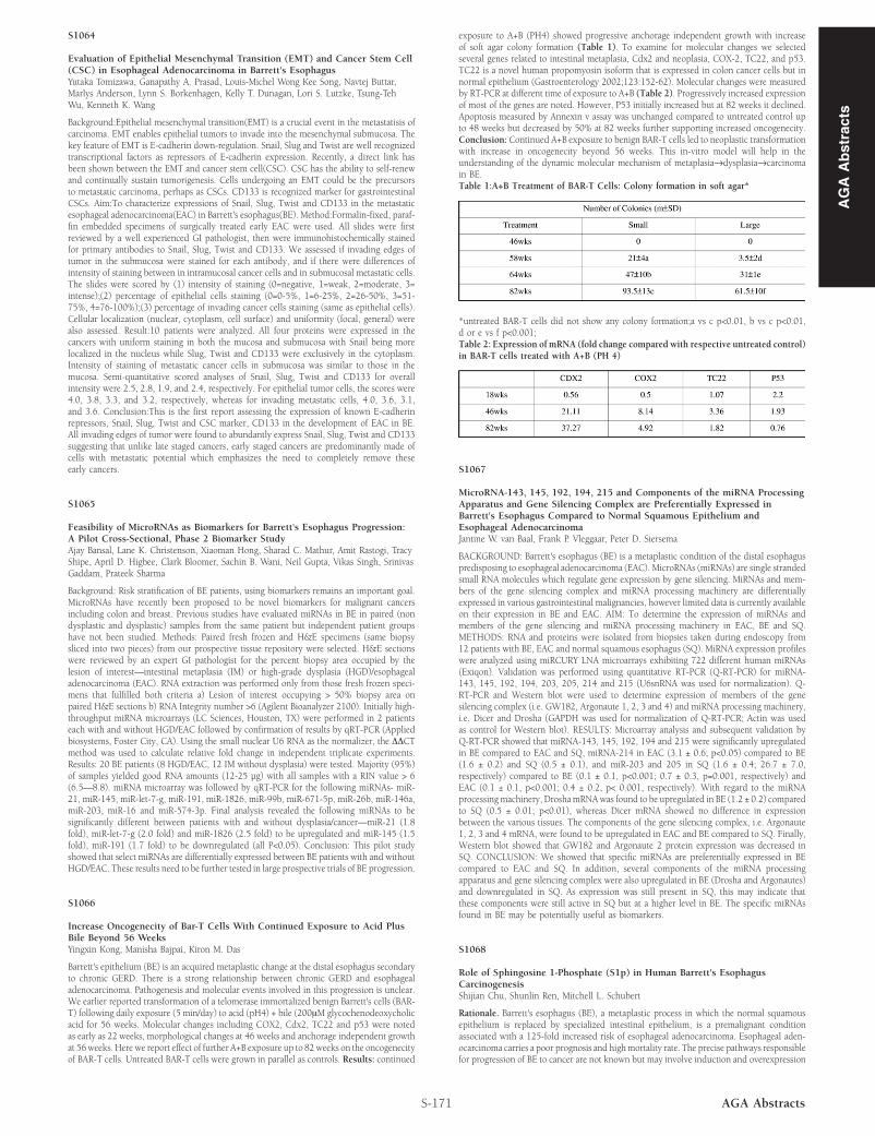
exposure to A+B (PH4) showed progressive anchorage independent growth with increaseof soft agar colony formation (Table 1). To examine for molecular changes we selectedseveral genes related to intestinal metaplasia, Cdx2 and neoplasia, COX-2, TC22, and p53.TC22 is a novel human propomyosin isoform that is expressed in colon cancer cells but innormal epithelium (Gastroenterology 2002;123:152-62). Molecular changes were measuredby RT-PCR at different time of exposure to A+B (Table 2). Progressively increased expressionof most of the genes are noted. However, P53 initially increased but at 82 weeks it declined.Apoptosis measured by Annexin v assay was unchanged compared to untreated control upto 48 weeks but decreased by 50% at 82 weeks further supporting increased oncogenecity.Conclusion: Continued A+B exposure to benign BAR-T cells led to neoplastic transformationwith increase in oncogenecity beyond 56 weeks. This in-vitro model will help in theunderstanding of the dynamic molecular mechanism of metaplasia→dysplasia→carcinomain BE.Table 1:A+B Treatment of BAR-T Cells: Colony formation in soft agar*

*untreated BAR-T cells did not show any colony formation;a vs c p<0.01, b vs c p<0.01,d or e vs f p<0.001;Table 2: Expression of mRNA (fold change compared with respective untreated control)in BAR-T cells treated with A+B (PH 4)

S1067

MicroRNA-143, 145, 192, 194, 215 and Components of the miRNA ProcessingApparatus and Gene Silencing Complex are Preferentially Expressed inBarrett's Esophagus Compared to Normal Squamous Epithelium andEsophageal AdenocarcinomaJantine W. van Baal, Frank P. Vleggaar, Peter D. Siersema

BACKGROUND: Barrett's esophagus (BE) is a metaplastic condition of the distal esophaguspredisposing to esophageal adenocarcinoma (EAC). MicroRNAs (miRNAs) are single strandedsmall RNA molecules which regulate gene expression by gene silencing. MiRNAs and mem-bers of the gene silencing complex and miRNA processing machinery are differentiallyexpressed in various gastrointestinal malignancies, however limited data is currently availableon their expression in BE and EAC. AIM: To determine the expression of miRNAs andmembers of the gene silencing and miRNA processing machinery in EAC, BE and SQ.METHODS: RNA and proteins were isolated from biopsies taken during endoscopy from12 patients with BE, EAC and normal squamous esophagus (SQ). MiRNA expression profileswere analyzed using miRCURY LNA microarrays exhibiting 722 different human miRNAs(Exiqon). Validation was performed using quantitative RT-PCR (Q-RT-PCR) for miRNA-143, 145, 192, 194, 203, 205, 214 and 215 (U6snRNA was used for normalization). Q-RT-PCR and Western blot were used to determine expression of members of the genesilencing complex (i.e. GW182, Argonaute 1, 2, 3 and 4) and miRNA processing machinery,i.e. Dicer and Drosha (GAPDH was used for normalization of Q-RT-PCR; Actin was usedas control for Western blot). RESULTS: Microarray analysis and subsequent validation byQ-RT-PCR showed that miRNA-143, 145, 192, 194 and 215 were significantly upregulatedin BE compared to EAC and SQ, miRNA-214 in EAC (3.1 ± 0.6; p<0.05) compared to BE(1.6 ± 0.2) and SQ (0.5 ± 0.1), and miR-203 and 205 in SQ (1.6 ± 0.4; 26.7 ± 7.0,respectively) compared to BE (0.1 ± 0.1, p<0.001; 0.7 ± 0.3, p=0.001, respectively) andEAC (0.1 ± 0.1, p<0.001; 0.4 ± 0.2, p< 0.001, respectively). With regard to the miRNAprocessingmachinery, DroshamRNAwas found to be upregulated in BE (1.2 ± 0.2) comparedto SQ (0.5 ± 0.01; p<0.01), whereas Dicer mRNA showed no difference in expressionbetween the various tissues. The components of the gene silencing complex, i.e. Argonaute1, 2, 3 and 4 mRNA, were found to be upregulated in EAC and BE compared to SQ. Finally,Western blot showed that GW182 and Argonaute 2 protein expression was decreased inSQ. CONCLUSION: We showed that specific miRNAs are preferentially expressed in BEcompared to EAC and SQ. In addition, several components of the miRNA processingapparatus and gene silencing complex were also upregulated in BE (Drosha and Argonautes)and downregulated in SQ. As expression was still present in SQ, this may indicate thatthese components were still active in SQ but at a higher level in BE. The specific miRNAsfound in BE may be potentially useful as biomarkers.

S1068

Role of Sphingosine 1-Phosphate (S1p) in Human Barrett's EsophagusCarcinogenesisShijian Chu, Shunlin Ren, Mitchell L. Schubert

Rationale. Barrett's esophagus (BE), a metaplastic process in which the normal squamousepithelium is replaced by specialized intestinal epithelium, is a premalignant conditionassociated with a 125-fold increased risk of esophageal adenocarcinoma. Esophageal aden-ocarcinoma carries a poor prognosis and highmortality rate. The precise pathways responsiblefor progression of BE to cancer are not known but may involve induction and overexpression

AG

AA

bst

ract

s