Embed Size (px)
Citation preview
S1
SUPPORTING INFORMATION
Phosphomannose isomerase/GDP-mannosepyrophosphorylase from Pyrococcus furiosus: a thermostablebiocatalyst for the synthesis of guanidinediphosphate-activated and mannose-containing sugar nucleotides
Rahman M. Mizanur and Nicola L. B. Pohl*Department of Chemistry and the Plant Sciences Institute, Gilman Hall, Iowa State University,Ames, Iowa 50011-3111
Table of Contents
Materials S1Bacterial Strains and Growth Conditions S2General Methods S2PCR Amplification, Cloning, Expression and Purification of P. furiosus Enzyme S2Mass Spectrometry S3Enzyme Assays S3Optimal Activity Determination of the Enzyme S4Kinetic Analysis S4Multiple Sequence Alignment S4Enzyme Catalyzed Synthesis of NDP-Sugars and HPLC S4References S5Table S1. Effects of divalent cations on the activity of the P. furiosus enzyme S6Table S2. Substrate specificity of the enzyme S7Figure S1. SDS-PAGE S8Figures S2 and S3. Michaelis-Menten plots S9Figures S4 and S5. Calibration curves for mass spectrometry S11Figures S6 and S7. Sequence alignments S12
Materials. Enzymes and reagents used for the molecular biology procedures, DNA ladders,and deoxynucleotide triphosphates (dNTPs) were purchased from Promega (Madison, WI) orNew England Biolabs (Beverly, MA). Oligonucleotides for DNA amplification weresynthesized by Sigma Genosys (Woodland, TX). Thermostable inorganic pyrophosphatase(IPP) from Thermococcus litoralis (EC 3.6.1.1, M0296S) was purchased from New EnglandBiolabs as a 2000 U/mL 50% glycerol solution in Tris buffer (pH 8.0).Isopropylthiogalactoside (IPTG) was obtained from Labscientific, Livingston, NJ. Proteinmolecular weight standards were obtained from BioRad (Hercules, CA). The QIA Quick gelextraction kit was obtained from Qiagen (Valencia, CA) and the Zero Blunt PCR cloning kit
Supplementary Material (ESI) for Organic & Biomolecular ChemistryThis journal is (c) The Royal Society of Chemistry 2009
S2
was purchased from Invitrogen (Carlsbad, CA). All other chemicals were obtained from SigmaChemical Co. (St. Louis, MO) unless otherwise stated.
Bacterial Strains and Growth Conditions. Genomic DNA from Pyrococcus furiosus (ATCC43587D), obtained from the American Type Culture Collection (Manassas, VA), was used asthe source for the cloning experiments described herein. Oneshot Top10 competent cells(Invitrogen, Carlsbad, CA), Escherichia coli XL-10Blue cells (Stratagene, La Jolla, CA), andPCR-Blunt vectors (Invitrogen, Carlsbad, CA) were used for direct cloning of PCR products.E. coli strain BL21 (DE3) RIPL (Stratagene, La Jolla, CA) was used in combination with theT7 expression system (pET21a vector; Novagen, Madison, WI.) for expression of the sugarnucleotidyltransferase gene. E. coli cells were grown on Luria-Bertani (LB, Sigma, St. Louis,MO) medium at 37 °C on an incubator shaker at 225 rpm. When required, antibiotics wereadded at the following concentrations to make the selective media: carbenicillin 50 µg/mL,kanamycin 50 µg/mL, chloramphenicol 25 µg/mL.
General Methods. Standard procedures to manipulate DNA, including plasmid DNAisolation, restriction enzyme digestion, agarose gel electrophoresis, DNA ligation andtransformation of E. coli, were performed by conventional methods (Sambrook 1989). ThePCR was carried out in an Eppendorf Mastercycler gradient thermocycler (EppendorfScientific Inc. Westbury, NY). Protein was analyzed by sodium dodecyl sulfatepolyacrylamide gel electrophoresis (SDS-PAGE, Tris-HCl 10-20% gradients, Bio-RadLaboratories, Hercules, CA). The gels were stained with Coomassie brilliant blue. Proteinconcentrations were determined with the Bio-Rad protein assay kit according to the method ofBradford (Bradford 1976) using bovine serum albumin as the standard.
PCR Amplification, Cloning, Expression and Purification of P. furiosus Enzyme.Genomic DNA of P. furiosus (ATCC 43587D) was amplified by PCR synthesis using twooligonucleotide primers. The primers were designed in order to construct the G1P-TTe x p r e s s i o n p l a s m i d . T h e f o r w a r d p r i m e r , 5 ’ -AAACCATATGAAGACATTAATTCTTGCTGGAGG -3’, contains an NdeI restriction site(in bold) and the reverse primer, 5’- AAACTCGAGCTAAGCCCTCTGGTAGTCGTCC-3’, contains a XhoI restriction site (in bold) and were synthesized from the putative (Robb2001) mannose-6-phosphate isomerase/mannose-1-phosphate guanylyltransferase (manC)gene of P. furiosus. The amplification reaction mixture contained standard PfuDNApolymerase buffer, 375 µM of dNTPs, 3 ng of each primer, 4 ng of total genomic DNA and2.5 units of Pfu DNA polymerase. The cycling parameters of 94 °C for 2 min 40 sec followedby 30 cycles of 94 °C for 30 s, 56 °C for 45 s and 72 °C for 2 min 15 s, with a final elongationstep of 72 °C for 15 min. The amplified DNA, after agarose gel electrophoresis (1%) waspurified using QIAquick Gel Extraction kit and subcloned into a ZeroBlunt vector usingZeroBlunt PCR cloning kit, and was transformed into Oneshot Top10 and E. coli XL10competent cells to check the correct insert. The resulting construct was then digested withNdeI and XhoI and was ligated to a pET21a vector containing a C-terminal histidine tagsequence (Novagen, Madison, WI) and previously digested with the same restriction enzymes.Aliquots of the ligation mixture were transformed into competent E. coli BL21 (DE3) RIPLcells. Transformants were selected at 37 °C grown on LB medium supplemented withcarbenicillin. The freshly transformed cells containing the desired plasmid were grown in LBuntil the optical density at 600 nm of the cell culture reached 0.5-0.6. Enzyme production wasinitiated by the addition of IPTG (1 mM) and the culture was incubated at 37 °C for the
Supplementary Material (ESI) for Organic & Biomolecular ChemistryThis journal is (c) The Royal Society of Chemistry 2009
S3
additional 3 h. Cells were harvested by centrifugation at 3600 x g for 10 min at 25 °C. Theenzyme was purified essentially at 4 °C unless otherwise stated. The cells were disrupted bysonication (Fisher model 100 Sonic Dismembranator, Fisher Scientific, Pittsburgh, PA), afterwhich unbroken cells and debris were removed by centrifugation (30 min at 10,000 x g). Thesupernatant was then heated at 100 °C for 5 min and the precipitated portion was removed bycentrifugation at 12,000 x g for 20 min. The cleared lysate was then purified by metal chelatechromatography by following the recommended procedures provided by Novagen. Thepurified protein was concentrated and dialyzed into the Tris-HCl buffer (50 mM, pH 7.5) usinga Microcon Centrifugal Filter Device, MWCO 10 kDa (Millipore, Billerica, MA). The proteinwas analyzed by SDS-PAGE analysis. No protein band was detected in SDS-PAGE of an E.coli cell extract harboring a pET21a vector without the manC insert. In order to construct atruncated mutation of the PMI-GMP the C-terminal PMI domain was deleted by PCRamplification of the manC gene using the forward primer described above and a separatelydesigned reverse primer 5’-AAACTCGAG GCG TTA CCA TTC TCG TCC TTT TGA AG-3’with a XhoI restriction site incorporated (underlined). The PCR product was then cloned andexpressed in E. coli BL21 (DE3) RIPL cells following the method described above. Theenzymatic activity, substrate specificity, kinetic properties, and other biochemical functionswere determined following the methods described else where.
Mass Spectrometry. A Shimadzu LCMS 2010 quadrupole mass spectrometer (ShimadzuScientific Instruments, Columbia, MD) equipped with an electrospray ionization (ESI) sourcewas used in negative ion mode. The capillary temperature and the spray voltage were kept at220 °C and 4.5 kV, respectively. The instrument was calibrated by direct infusion ofpolyethylene glycol (PEG) 200, 600, 1000, (1.5 µL/L, 2 µL/L, and 15 µL/L, respectively) andraffinose (50 mg/L) in water/methanol (1:1, v:v) containing ammonium acetate (0.19 mM),0.1% formic acid and 0.1% acetonitrile. For sample analysis the solventacetonitrile/water/triethylamine, (35/65/0.2) was constantly infused into the ion source at 250µL/min by the attached Shimadzu HPLC pump and the samples were injected (30 µL) via theauto sampler adapted to fit two 96-well plates. A preliminary MS chromatogram was obtainedby scanning from 50-700 m/z. To increase the signal to noise ratio, the instrument was set forselected ion monitoring (SIM) mode and all relevant m/z ions were monitored for furtheranalysis of the enzymatic reactions. Inclusion of an Agilent Extend C18 column (2.1 x 50 mm,Agilent, Palo Alto, CA) in the system further increased the signal to noise ratio and decreasedthe appearance of sodium ion adducts by 10-15% without separation of reaction components.Postrun software (LCMS Postrun version 2.02, Shimadzu Scientific Instruments, Columbia,MD) was used to analyze the data from the ESI-MS chromatogram. Peaks were integrated todetermine the relative intensity of each ion species monitored as compared to an internalstandard.
Enzyme Assays. The nucleotidyltransferase activity of the bifunctional P. furiosus enzymewas determined, according to the method described previously (Zea and Pohl 2004), by theformation of GDP-mannose from GTP and of mannose 1-phosphate (Man1P) in the directionof GDP-mannose biosynthesis. The enzymatic reaction was initiated by the addition of Man1P(5 mM) to a reaction mixture of 50 µL containing Na-phosphate buffer (25 mM, pH 7.5),inorganic pyrophosphatase (IPP, 0.2 U), purified enzyme solution (10 µL), MgCl2 (5 mM),and GTP (5 mM). Before adding Man1P the reaction components were incubated at 80 °C for5 min. The reaction was carried out at 80 °C for 5 min and 15 µL of the reaction mixture wasquenched by the addition of 30 µL of 70% methanol/water containing AMP (3 mM) as an
Supplementary Material (ESI) for Organic & Biomolecular ChemistryThis journal is (c) The Royal Society of Chemistry 2009
S4
internal standard. The quenched solutions were centrifuged 10 min at 10,000 x g to precipitatethe protein. Aliquots of the reaction mixtures were diluted with 135 µL of acetonitrile(acetonitrile/water/triethylamine, 35/65/0.2). These samples (5 µL) were subjected to analysisvia ESI-MS to determine the amount of GDP-mannose formed. One unit of enzyme activity isdefined as one micromole of GDP-man was formed per minute. Detection of phosphomannoseisomerase activity of the enzyme was performed at 30°C in 0.5 mL of 25 mM Tris-HCl buffer(pH 7.0) containing 5 mM Co2+, 5 mM mannose-6-phosphate, 5 mM NADP, 10 Uphosphoglucose isomerase and 10 U phosphoglucose dehydrogenase and the appropriateamount of purified manC according to the published method (Wu, 2002). The reduction ofNADP+ was followed by monitoring of UV absorption at 340 nm.
Optimal Activity Determination of the Enzyme. The optimal activity for the P. furiosusenzyme was measured at 80 °C between pH 4.0 to pH 9.6 using 50 mM acetate, phosphate,and Tris-HCl buffer. The optimal temperature was measured at pH 7.5 between 30 °C to 100°C. Relative acceptance of the enzyme to a number of sugar-1-phosphates and NTPs (UTP,dTTP, GTP, ATP and CTP), effects of divalent cations on catalytic conversion and Mg2+ ionconcentrations were determined in Tris buffer (25 mM, pH 7.5) and at 80 °C.
Kinetic Analysis. The values for KM and Vmax were derived from enzymatic reactions run intriplicate and determined from the initial rates of ADP-, CDP-, GDP-, dTDP-, and UDP-mannose or GDP-glucose formation using ESI-MS. The enzymatic reaction was initiated bythe addition of Man1P (25-500 µM) or glucose-1-phosphate to obtain a reaction mixturecontaining Phosphate buffer (25 mM, pH 7.5), inorganic pyrophosphatase (0.2 U), manC (2.5x 10⁻³ U), and NTP (400 µM) with a final volume of 50 µL. Reactions were carried out at 80°C for 5 min and 30 µL of reaction mixture was quenched by addition of 30 µL of 70%methanol/water containing AMP (6 µM) as an internal standard. The quenched solution wascentrifuged and diluted as described above. These samples (30 µL) were subjected to analysisvia ESI-MS to determine the amount of NTP-mannose or GDP-glucose formed. Calibrationcurves for determining the initial activities for nucleotidyltransferase activity of the enzymewere developed following the methods described previously (Zea, 2004). All kinetic data wasfitted with the non-linear regression algorithm in GraphPad Prism version 4 (GraphPadSoftware, San Diego, CA). Error bars on the graphs represent the standard deviation of threeindependent averaged data points.
Multiple Sequence Alignment. A multiple sequence alignment was performed using BLAST(www.ncbi.nih.gov). Motifs were defined as the regions with at least 11 strictly conservedresidues among 30 consecutive positions (Bork, 1996).
manC Catalyzed Synthesis of NDP-Sugars and HPLC. To synthesize GDP-sugars andNDP-mannoses, one equivalent (2 to 3 mg) of sugar-1-phosphate and 1.5 equivalents of NTPsalong with the other reaction components in Na-phosphate buffer (pH 7.5) including inorganicpyrophosphatase (IPP, 2 U) were incubated with purified manC at 80 °C for 1 h. Theformation of product was confirmed by ESI-MS by monitoring of the NDP-sugar. The yielddetermined by ESI-MS was calculated as the difference of the amount of sugar phosphateremaining in the reaction and the initial amount then divided by the initial amount of substrateused in the reaction (Mizanur, 2004). The incubation mixture was loaded on a 5µm SAXphenosphere HPLC column (250 x 10 mm) fitted with a security guard cartridge
Supplementary Material (ESI) for Organic & Biomolecular ChemistryThis journal is (c) The Royal Society of Chemistry 2009
S5
(Phenomenex, CA) after ultrafiltration using a YM-10 membrane (Millipore corporation,Bedford, MA) to remove any protein. The NDP-sugars were eluted with 150 mM ammonium-phosphate buffer (pH 6.0) at ambient temperature and monitored at 254 nm. Fractionscollected were further analyzed by ESI-MS to check the identity of the products, then pooledand lyophilized. Retention times were as follows: GDP-Man (5.4 min), UDP-Man (4.2 min),ADP-Man (5.7 min), CDP-Man (4.6 min), dTDP-Man (4.5 min), GDP-Glc (5.7 min), GDP-Gal (5.4 min), GDP-GlcN (5.7 min), GDP-GlcNAc (5.3 min), GDP-Fuc (6.3 min), whereas allthe nucleotide triphosphates eluted sometime after 7 min. Using similar reaction conditionsthat include inorganic pyrophosphatase, 50 mg scale synthetic reactions were also carried outcontaining one equivalent of sugar-1-phosphate (Man1P, Glc1P, GlcN1P and GlcNAc1P) and1.5 equivalents of GTP with manC.
ReferencesM. M. Bradford, Anal. Biochem. 1976, 72, 248.P. Bork and E. V. Koonin, Curr. Opin. Struct. Biol. 1996, 6, 366.J. Sambrook, E. F. Fritsch and T. Maniatis, Molecular cloning: a laboratory manual, 2nd ed.;
Cold Spring Harbor Laboratory Press: Plainview, NY, 1989.F.T. Robb, D.L. Maeder, J.R. Brown, J. DiRuggiero, M.D. Stump, R.K. Yeh, R.B. Weiss and
D.M. Dunn, Methods Enzymol., 2001, 330, 134-157.B. Y. Wu, R. Zhang, Zheng, C. Guo and P. G. Wang, FEBS Lett. 2002, 519, 87-92.C. J. Zea and N. L. Pohl, Anal. Biochem. 2004, 328, 196.
Supplementary Material (ESI) for Organic & Biomolecular ChemistryThis journal is (c) The Royal Society of Chemistry 2009
S6
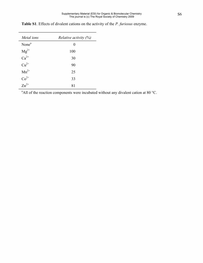
Table S1. Effects of divalent cations on the activity of the P. furiosus enzyme.
Metal ions Relative activity (%)
Nonea 0
Mg2+ 100
Ca2+ 30
Cu2+ 90
Mn2+ 25
Co2+ 33
Zn2+ 81aAll of the reaction components were incubated without any divalent cation at 80 °C.
Supplementary Material (ESI) for Organic & Biomolecular ChemistryThis journal is (c) The Royal Society of Chemistry 2009
S7
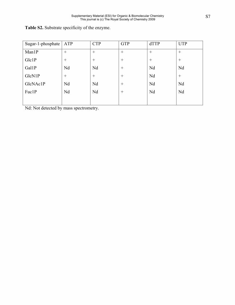
Table S2. Substrate specificity of the enzyme.
Sugar-1-phosphate ATP CTP GTP dTTP UTP
Man1P
Glc1P
Gal1P
GlcN1P
GlcNAc1P
Fuc1P
+
+
Nd
+
Nd
Nd
+
+
Nd
+
Nd
Nd
+
+
+
+
+
+
+
+
Nd
Nd
Nd
Nd
+
+
Nd
+
Nd
Nd
Nd: Not detected by mass spectrometry.
Supplementary Material (ESI) for Organic & Biomolecular ChemistryThis journal is (c) The Royal Society of Chemistry 2009
S8
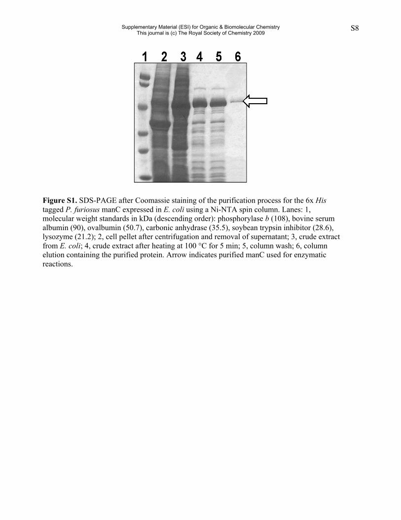
Figure S1. SDS-PAGE after Coomassie staining of the purification process for the 6x Histagged P. furiosus manC expressed in E. coli using a Ni-NTA spin column. Lanes: 1,molecular weight standards in kDa (descending order): phosphorylase b (108), bovine serumalbumin (90), ovalbumin (50.7), carbonic anhydrase (35.5), soybean trypsin inhibitor (28.6),lysozyme (21.2); 2, cell pellet after centrifugation and removal of supernatant; 3, crude extractfrom E. coli; 4, crude extract after heating at 100 °C for 5 min; 5, column wash; 6, columnelution containing the purified protein. Arrow indicates purified manC used for enzymaticreactions.
Supplementary Material (ESI) for Organic & Biomolecular ChemistryThis journal is (c) The Royal Society of Chemistry 2009
S9
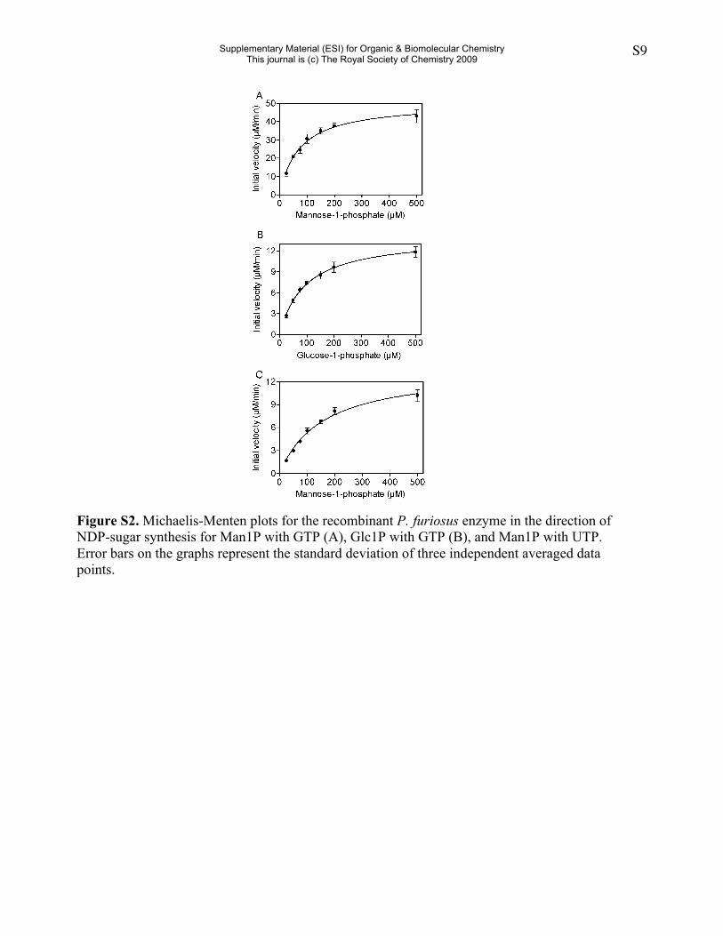
Figure S2. Michaelis-Menten plots for the recombinant P. furiosus enzyme in the direction ofNDP-sugar synthesis for Man1P with GTP (A), Glc1P with GTP (B), and Man1P with UTP.Error bars on the graphs represent the standard deviation of three independent averaged datapoints.
Supplementary Material (ESI) for Organic & Biomolecular ChemistryThis journal is (c) The Royal Society of Chemistry 2009
S10
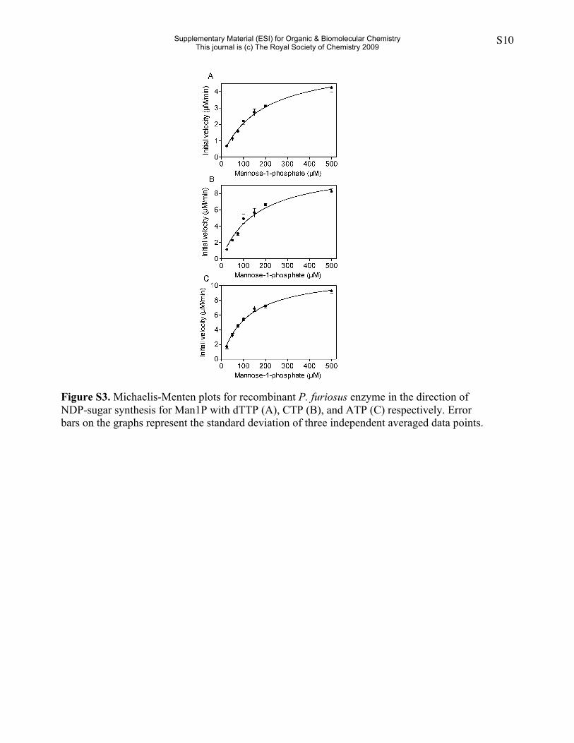
Figure S3. Michaelis-Menten plots for recombinant P. furiosus enzyme in the direction ofNDP-sugar synthesis for Man1P with dTTP (A), CTP (B), and ATP (C) respectively. Errorbars on the graphs represent the standard deviation of three independent averaged data points.
Supplementary Material (ESI) for Organic & Biomolecular ChemistryThis journal is (c) The Royal Society of Chemistry 2009
S11
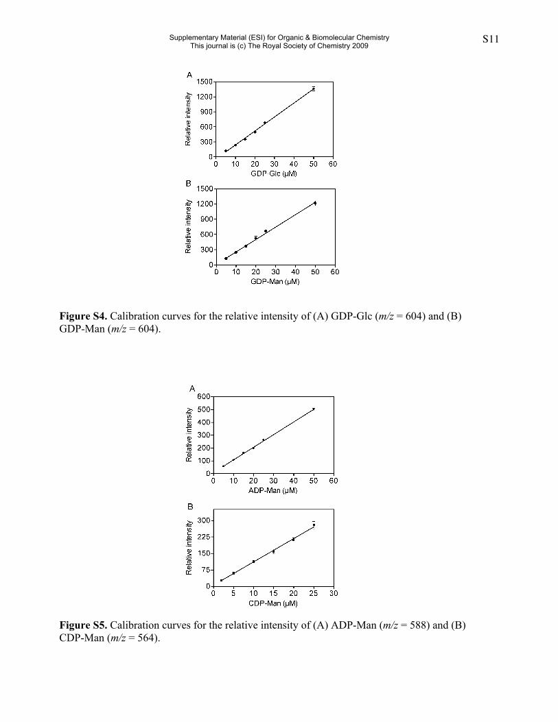
Figure S4. Calibration curves for the relative intensity of (A) GDP-Glc (m/z = 604) and (B)GDP-Man (m/z = 604).
Figure S5. Calibration curves for the relative intensity of (A) ADP-Man (m/z = 588) and (B)CDP-Man (m/z = 564).
Supplementary Material (ESI) for Organic & Biomolecular ChemistryThis journal is (c) The Royal Society of Chemistry 2009
S12
a.1 MKTLILAGGKGTRLWPLSRELMPKQFIKLFS ESLFQKTVKRALY.[1].S.[1].PDEIYVITNKEYRFRVLDDLb.1 MKTLILAGGKGTRLWPLSRELMPKQFIKVFS.[1].KSLFQKTVERALI.[1].S.[1].PKEIFVVTNKEYRFRVLDDLc.6 IKSIILAGGSGTRLWPLSREMYPKQFLKFGD TSLFQETVLRCLE.[1].S.[1].ISEIFVVTNEAQKFFVIGQId.7 LIPCIVSGGSGTRLWPVSRESMPKPFMRLAD.[1].QSLLQKTFLRIAG.[1].P.[1].VARLLTVTNRDLLFRTLDDYe.1 MKTLILAGGKGTRLWPLSREAMPKQFIKVFS.[1].RSLFQKTVERALL.[1].S.[1].PKEIFIVTNKEYKFRVLDDLf.1 MKALILAGGKGTRLWPLSREAMPKQFIKVFS.[1].RSLFQKTVERALI.[1].S.[1].PKEIFVVTNKEYRFRVLDDLg.2 MKALILAGGSGERFWPLSTPETPKQFLKLFG.[1].KSLMRWTFERVLE.[1].M.[1].PKDVIVVTHKDYVERTKKELh.4 LLPVIMAGGAGSRLWPLSRALYPKQFLALTS.[1].LTMLQETLLRLDG L.[1].HLAPLVICNEEHRFIIAEQLi.1 MKILILSGGKGTRLWPLSRENFPKQFIRLFS.[1].HSLFQETVKRAKK.[1].A.[1].DEDIVVITGEKYEWIIRSELj.6 LYPVVMAGGSGSRLWPLSRVLYPKQFLCLKG.[1].LTMLQTTICRLNG V.[1].CESPVVICNEQHRFIVAEQL
a.68 EE.[2].IS.[5].IILEPEAKNTLPAICLGVKAAGE.[2].FAVLPSDHLIKA.[2].EYLNAFRSAEKLSE.[1].YIb.69 NE.[2].VN.[5].ILLEPVGKNTLPAIYWGLKVIDE.[6].VAVLPSDHAIDV.[2].NYIEAFKKAEKLAE.[1].YLc.73 KE.[2].YS.[5].VLIEPEGKNTLPAIFFGMKEIEQ.[6].VGVFSSDHVLDR AAMATIFSAEKLTS.[1].YLd.75 RA.[2].RS.[5].LLLEPVGRNTAPAIAAAALHVQE.[7].LLILPADHLIRD.[2].AFAAAVAEARGLAA.[2].YLe.69 NE.[2].LK.[5].ILLEPVGKNTLPAIYWGLKVIDE.[6].VAVLPSDHAIKV.[2].NYMEAFKKAEKLAE.[1].YLf.69 NE.[2].LK.[5].ILLEPVGKNTLPAIYWGLKVIND.[6].VAVLPSDHAIEV.[2].SYMEAFKKAEKLAE.[1].YLg.70 PE.[2].DE.[1].IIAEPMKKNTAPACFIGTKLADD.[3].VLVLPADHRIPD.[2].KFWKTVKKALDALE.[3].GLh.71 RQ KN.[5].IVLEPVGRNTAPAIALAALRATM.[6].LLVLAADHVIQD.[2].AFIRAVQRAEPLAE.[2].KLi.69 EE.[2].AN.[3].VVVEPEGRNTAPAIALGVKYLIH.[8].VLVLPSDHLIKD.[2].KFVEAVKKGEKYAK.[2].YIj.73 RQ LN.[5].IILEPAGRNTAPAIALAALAAKR.[8].MLVLAADHVIAD.[2].AFRAAVRNAMPYAE.[2].KL
a.135 VTFGITPTRPHTGYGYIKPGK.[ 5].FEVEQFKEKPSRELAEEYVSKG YLWNSGMFVFDSKVFVEELKELAPEb.140 VTFGIKPTKPHTGYGYIKPGE.[ 6].FLVDEFKEKPDLETAKRYVENG YYWNSGMFMFRTTLFMEEARKHAPDc.142 VTFGVVPAFPHTGYGYIKAAE.[ 5].YRVSEFREKPDFETAQKYIEEG CLWNSGMFLFETRLFFEEVKKHAPSd.148 VTFGITPERAETGFGYIEQGA.[ 5].FRVARFVEKPDQATAQSYLDSG.[1].YLWNAGMFCFQAATVLQELERHAPEe.140 VTFGIKPTKPHTGYGYIKPGE.[10].YLVDEFKEKPDLETAKKYVENG YYWNSGMFMFRTSVFMEEARKHAPEf.140 VTFGIKPTKPHTGYGYIKPGE.[10].YLVDEFKEKPDLETARKYVENG YYWNSGMFMFKVSVFMEEARKHSPDg.136 FTFGIVPTRPETGYGYIEIGE.[ 6].HKVAQFREKPDLETAKKFVESG.[1].FLWNSGMFLWKAREFIEEVKVCEPSh.141 VTFGIVPKSPETGYGYIRQGK.[ 6].YQVAAFVEKPDLITAERYLASG.[1].YYWNSGMFVFKASRYLQELDLHRPDi.141 VLFGEKPTYPETGYGYIRLNG.[ 6].YEIEKFEEKPDYEKAKEYVSDG.[1].HFWNCGIFLFTLDRIVKDYTQLMPEj.145 VTFGIVPDLPETGYGYIRRGE.[10].FEVAQFVEKPNLETAQAYVASG.[1].YYWNSGMFLFRAGRYLEELKKYRPD
a.208 .[1].AKVLE.[ 2].EEAYKQIPEASFDYAILEKSGRVAVVPIKTFWSDLGNFDSIYEVMEKDE.[5].KSE.[2].IPb.214 .[2].KAFEE.[ 4].EEAYELAPEISVDYGIMEKTDKAAVVPLNTYWNDLGSFDAVYEALQKDE.[5].EVR.[6].INc.215 .[1].FACFE.[ 5].DEIYACVDNVSIDYGIMEKSDRVAVVKLDQKWSDLGNFAAIYDELEKDS.[5].HEC.[2].MLd.222 .[2].IAARA.[19].AGAFAEAPDISVDYALMERSDKVAVVPCSIGWSDIGSWQALRELSAADE.[5].RGE.[1].VLe.218 .[1].VRAFE.[ 5].EEIYELVPEISVDYGIMEKTDKAAVVPLNTYWNDLGSFDAVYEALDKDE.[5].QVA.[6].INf.218 .[1].VKAFE.[ 5].EEIYELAPEISVDYGIMEKTNKAAVVPLNTYWNDLGSFDAVYEALEKDE.[5].HVT.[6].INg.211 .[2].ENLKD.[ 9].KKAYEKVPSISVDYAVMEKSKKVRVVKADFEWSDVGNWSSVREIEGYTE ESD.[2].ILh.216 .[2].AACKQ.[15].EEAFSSCPDESIDYAVMEKTSDAVVVPLDAQWNDVGCWSALWEINTKDD.[5].RGD.[1].LIi.216 IPFHE.[ 5].IKDFKNFEEISFDYAILERTKKLAVVKMDAGWSDVGSWKAVYDNLPKDE.[5].IGD.[1].KAj.224 .[2].DACEK.[15].EEAFLACPEESVDYAVMERTADAVVVPMDAGWSDVGSWSSLWEISAHTA.[5].HGD.[1].IN
a.277 VDSENNLVITQ.[1].LTALIGLRDLIVIDTDDALLV.[2].b.290 VDSKNNLILTE.[1].LTATVGVEDLIIVDTGDALLV.[2].c.287 LNSDGNLVYSK.[3].IVSLIDIKDMVIVDTSDALLI.[2].d.308 HDVSNCYIDSP.[2].LVGAVGVHDLIIVDTPDALLV.[2].e.294 INSRNNLILTE.[1].LTATVGVEDLIIVDTGDALLV.[2].f.294 VDSRNNLVLTE.[1].LTATVGVEDLVIIDTGDALLV.[2].g.283 VDSDRVFVKTH.[2].PIAVVGLSDVIVIDTPNGILI.[2].h.298 EDTNNSYVYSQ.[2].LIATVGINDLVIVETKDAILV.[2].i.286 MDTECSLLLSQ.[3].LIACIGLEDFVVVGTEDATLI.[2].j.306 HKTENSYVYAE.[2].LVTTVGLKDLVVVQTKDAVLI.[2].
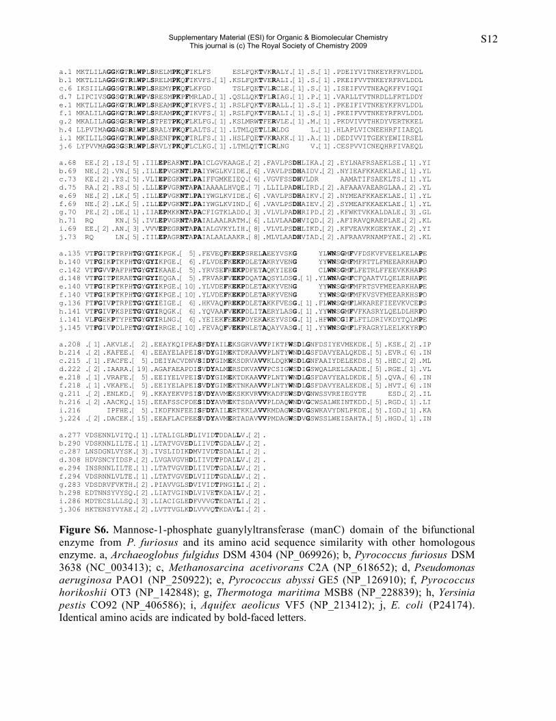
Figure S6. Mannose-1-phosphate guanylyltransferase (manC) domain of the bifunctionalenzyme from P. furiosus and its amino acid sequence similarity with other homologousenzyme. a, Archaeoglobus fulgidus DSM 4304 (NP_069926); b, Pyrococcus furiosus DSM3638 (NC_003413); c, Methanosarcina acetivorans C2A (NP_618652); d, Pseudomonasaeruginosa PAO1 (NP_250922); e, Pyrococcus abyssi GE5 (NP_126910); f, Pyrococcushorikoshii OT3 (NP_142848); g, Thermotoga maritima MSB8 (NP_228839); h, Yersiniapestis CO92 (NP_406586); i, Aquifex aeolicus VF5 (NP_213412); j, E. coli (P24174).Identical amino acids are indicated by bold-faced letters.
Supplementary Material (ESI) for Organic & Biomolecular ChemistryThis journal is (c) The Royal Society of Chemistry 2009
S13
a.278DSENNLVIT.[1].RLTALIGLRDLIVIDTDDALLVARRGEAEKVREVYRLLAEKGDKAVEVHRTAHRPWGSYTVLEENKb.291DSKNNLILT.[1].RLTATVGVEDLIIVDTGDALLVAKKGETQKVKEVYKKLKEENDERAIVHRTAYRPWGSYTVLEEGEc.298 DTEGCFVSS.[2].MVTSLVGVRDLVVVAEKDAILVADRSRCGDVRKMVEILRSKGRPQAEWHASSHRPWGSYRVLEASDd.307 DVEGSYIRS.[2].RLVAVAGLCNVVVVATDDAVLVIDRGKVQDVKQIVERLKKANRDEHALHSTVHRPWGHYRGIDRGEe.298 DSHNCLVHG.[2].KLVSVIGLEDIVVVETKDAMMIAHKDRVQDVKHVVKDLDAQGRSETQNHCEVYRPWGSYDSVDMGGf.297 DCKNTYAYG.[1].RLIAMVGLENVVVVETDDAVLVGHRDRIQEVKEVVSQIKSAGRSEATWHRKVYRPWGAYDSIDMGQg.301 KSQNNYVFS.[2].RLVSLLGVDNLVVIETKDAILVADKSKVQDIKKIVESIKEQGRTEHFCHREVYRPWGKYDSIDHAEh.300 NADNCYLHA.[2].GLVTAVGVKDLIVVQTKDAVLVANTNCVQDVKKIVEKIKLENRHEHITHREVYRPWGKYDSIDFGEi.295 STTNSLVRA.[2].RLVATVGVNNLVVIETADAVLIMDKDQSQDVKKIVSRIKAEGRQEHMHHTTVHRPWGTYQTVDLGDj.297 DTNNSYIYS.[2].RLVATVGINDLIIVETKDALLVANKNKVQSVKEIVGQLKLGSRLEYLQHKEVYRPWGSHDAIAEGV
a.354 SYKIKRITVKPKKRLSLQRHYHRSEHWVVVKGTARIVVDGNEILLRSGESTFVPAGAIHRIENPGKIPLEIIEIQIGEYLb.367 RYKIKRITVLPGKRLSLQLHYHRSEHWVVVRGTAKVRVGDKEFILRPGESTFIPAGVIHRLENPGKVVLEVIETQIGEYLc.375 GFQVKRITVAPGGRLSLQKHRHRAEHWVVVRGCARVTVDDTVADYRESEHIFIPLGAIHRLENPGNDDVELIEVQLGSYLd.384 RFQVKRIVVQPGERLSLQMHHHRAEHWIVVTGTALVTRGAETFLLHENESTYIRAGQTHRLENPGKVPLHLIEVQSGGYLe.375 RFQVKHITVKPGARLSLQMHHHRAEHWIVVSGTAQVTCDDKTFLLTENQSTYIPIASVHRLANPGKIPLEIIEVQSGSYLf.373 RFQVKRITVKPGATLSLQMHHHRAEHWIVVSGTAEVTRGEEVLLLTENQSTYIPLGVTHRLKNPGKLPLELIEVQSGSYLg.378 RYQVKRITVKPGQKLSIQMHHHRSEHWIVVNGTAKIHKGKESFLLTENQSTYIPLGEIHALENPGKVPLELIEVQSGSYLh.377 RYQVKRITVKPGEGISEQQHYHRAEHWIIVAGTAKITIKGEVKILTENESVYIPVGVKHCLENPGKIALELIEVRSGAYLi.372 RHQVKRIMVKPGEKLSVQMHHHRAEHWVVVSGTAKVQNGEREILLTENESTYIPVGVVHALENPGKIPLELIEVQSGSYLj.374 RYHVQHVTIKPGQRTATQIHHHRAEHWVVVSGTARVYRDNESYLVTENESTYIAVGVAHSIENPGKLPLEIIEVRTGSYL
a.434 EEDDIERFEDb.447 GEDDIVRIEDc.455 GEDDIVRLEDd.464 GEDDIVRFEDe.455 GEDDIERLEDf.453 GEDDIVRFEDg.458 GEDDIVRFEDh.457 GEDDIVRFSDi.452 GEDDIVRFSDj.454 EEDDIVRIEH
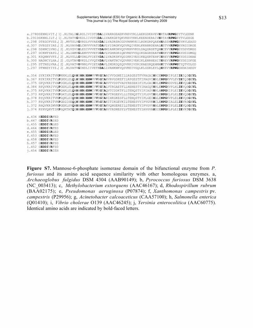
Figure S7. Mannose-6-phosphate isomerase domain of the bifunctional enzyme from P.furiosus and its amino acid sequence similarity with other homologous enzymes. a,Archaeoglobus fulgidus DSM 4304 (AAB90149); b, Pyrococcus furiosus DSM 3638(NC_003413); c, Methylobacterium extorquens (AAC46167); d, Rhodospirillum rubrum(BAA02175); e, Pseudomonas aeruginosa (P07874); f, Xanthomonas campestris pv.campestris (P29956); g, Acinetobacter calcoaceticus (CAA57100); h, Salmonella enterica(Q01410); i, Vibrio cholerae O139 (AAC46245); j, Yersinia enterocolitica (AAC60775).Identical amino acids are indicated by bold-faced letters.
Supplementary Material (ESI) for Organic & Biomolecular ChemistryThis journal is (c) The Royal Society of Chemistry 2009
S14
a
eco 9 VVMAGGTGSRLWPLSRELYPKQFLQLSGDNTLLQTTLLRLSGLSCQKPL-VITNEQHRFV 67pfu 4 LILAGGKGTRLWPLSRELMPKQFIKVFSNKSLFQKTVERALIFSKPKEIFVVTNKEYRFR 63
eco 68 VAEQLREI--NKLNGNIILEPCGRNTAPAIAISAFHALKRNPQE--DPLLLVLAADHVIA 123pfu 64 VLDDLNEIGVNIPEENILLEPVGKNTLPAI----YWGLKVIDESFGDSIVAVLPSDHAID 119
eco 124 KESVFCDAIKNATPIANQGKIVTFGIIPEYAETGYGYIERGELSVPLQGHENTGFYYVNK 183pfu 120 VNENYIEAFKKAEKLA-ENYLVTFGIKPTKPHTGYGYIKPGEKI------SNLGFL-VDE 171 b
eco 184 FVEKPNRETAELYMTSGNHYWNSGIFMFKASVYLEELRKFRPDIYNVCEQVASSSYIDLD 243pfu 172 FKEKPDLETAKRYVENG-YYWNSGMFMFRTTLFMEEARKHAPDVVKAFEEG--------- 221
eco 244 FIRLSKEQFQDCPAESIDFAVMEKTEKCVVCPVDIGWSDVGSWQSLWDISLKSKTGDVC- 302pfu 222 --KTIEEAYELAPEISVDYGIMEKTDKAAVVPLNTYWNDLGSFDAVYEALQKDENGNAVE 279
eco 303 ----KGDILTYDTKNNYIYSESALVAAIGIEDMVIVQTKDAVLVSKKSDVQHVKKIVEML 358pfu 280 VRGFKAKYINVDSKNNLILTER-LTATVGVEDLIIVDTGDALLVAKKGETQKVKEVYKKL 338 c
eco 359 KLQQRTEYISHREVFRPWGKFDSIDQGERYKVKKIIVKPGEGLSLRMHHHRSEHWIVLSG 418pfu 339 KEENDERAIVHRTAYRPWGSYTVLEEGERYKIKRITVLPGKRLSLQLHYHRSEHWVVVRG 398 d
eco 419 TAKVTLGDKTKLVTANESIYIPLGAAYSLENPGIIPLNLIEVSSGDYLGEDDIIRQKERY 478pfu 399 TAKVRVGDKEFILRPGESTFIPAGVIHRLENPGKVVLEVIETQIGEYLGEDDIVRIEDDY 458
eco 479 K 479pfu 459 Q 459
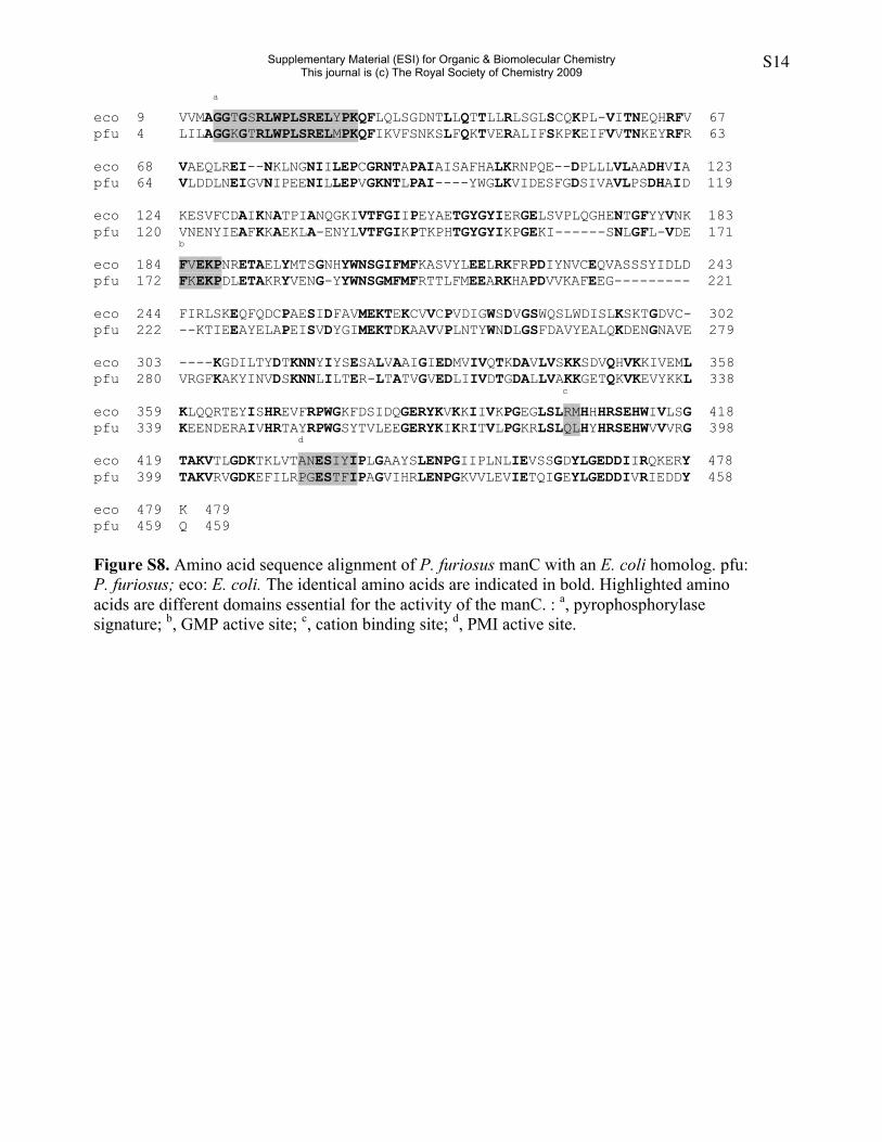
Figure S8. Amino acid sequence alignment of P. furiosus manC with an E. coli homolog. pfu:P. furiosus; eco: E. coli. The identical amino acids are indicated in bold. Highlighted aminoacids are different domains essential for the activity of the manC. : a, pyrophosphorylasesignature; b, GMP active site; c, cation binding site; d, PMI active site.
Supplementary Material (ESI) for Organic & Biomolecular ChemistryThis journal is (c) The Royal Society of Chemistry 2009


![pfc environmental water - SHIMADZU CORPORATIONLiquid Chromatography Mass Spectrometry No.C81 Analysis of PFCs in Environmental Water Using Triple Quadrupole LC/MS/MS [LCMS-8030] Organofluorine](https://img.pdfslide.us/doc/110x75/5f0d2fd27e708231d4391931/pfc-environmental-water-shimadzu-corporation-liquid-chromatography-mass-spectrometry.jpg)