Embed Size (px)
Citation preview
S. Murgo, MD
Chr St-Joseph, MonsErasme Hospital, Brussels
??
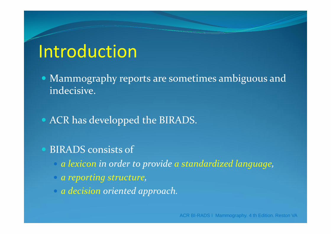
Introduction� Mammography reports are sometimes ambiguous and
indecisive.
� ACR has developped the BIRADS.
� BIRADS consists of � a lexicon in order to provide a standardized language, � a reporting structure,� a decision oriented approach.
ACR BI-RADS – Mammography. 4 th Edition. Reston VA
Introduction� Thanks to the BIRADS
� Reports are more easily understood
� And because the data are acquired in a similiar fashion, they are more easily pooled or compared with others.
Structure of reports� After administrative data, the report should include:� Clinical informations� Number of views by breast� Comparison with previous MG� The type of breast tissues� Significants findings� Correlation ultrasounds� Conclusion with the definition of the next action.
An other form of standardizationAn other form of standardization
Clinical informations
� The indication of examination: screening or clinical anomaly.
� The principal risk factors as a personal or a familial history of BC, a genetic anomaly as BRCA 1 or 2, a previous result of biopsy as an AEH, SHT ...
� The known benign anomalies as a FA or a cyst…
Views by breast� This allows to assess the quality of an exam.
� For example, � A screening mammogram à A single view
mammography overlooks as much as 25% of BC (1)� The evaluation of a new cluster of calcifications àML
projection with magnification.� A new architectural distortion àML projection and/or
focal compression…
(1) Wald NJ. BMJ 1995;311:1189-1193
Previous MG� The comparison with any pertinent previous MG
� This is important in order to � detect a new anomaly for example an asymmetry of
density� to confirm the stability of a breast lesion.
Kopans DB. Breast Imaging. 2d Edition. Lippincott -Raven
Type of breast tissues� In the BIRADS, mammographic breast composition is
described with 4 categories:1. fatty breast (<25% glandular tissue)
2. scattered fibroglandular densities (≥ 25% )
3. heterogeneously dense (≥ 50%)
4. extremely dense (≥ 75%)
ACR BI-RADS – Mammography. 4 th Edition. Reston VA
Type of breast tissues� This classification is subjective and there is discordances
between readers. Nevertheless, It provides to the referring physician an estimate of the sensitivity of the mammography.� ranges from 60% in extremely dense breasts to 90% in fatty breasts (1).� in the categories 3 and 4, it is well demonstrated that ultrasounds
increase the detection rate of BC (↑ 16 %) (2).
� If a implant is present, it should be stated in the report
(1) Kopans DB. Breast Imaging. 2d Edition. Lippincott –Raven(2) Fritz K. Eur Radiol 2010; 20(5): 1085-1092.
Significant Findings� Description of significant findings with the lexicon of
the ACR.� Avoid exotic or histological terminology� Use radiographically appropriate terms � with the exception of some benign pathognomonic
lesions such as intramammary lymph nodes, calcified fibro-adenomas, vascular calcifications and the fat containing lesions.
Kopans DB. Breast Imaging. 2d Edition. Lippincott -Raven
Significant Findings� Calcifications� Mass� Focal asymmetry� Architectural distortion� Associated findings
Kopans DB. Breast Imaging. 2d Edition. Lippincott -Raven

Standardization and quality of communicationStandardization and quality of communication
Calcifications� FOR CALCIFICATIONS, some characteristics should
be described as defined in the lexicon of BIRADS:
� the morphology, � the number, � the distribution, � the size of the cluster, � the associated findings and � the location (using the face of a clock or 4 quadrans).
ACR BI-RADS – Mammography. 4 th Edition. Reston VA
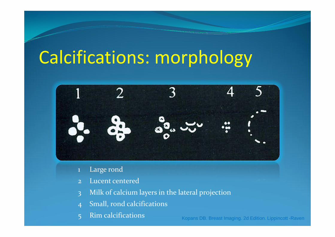
Calcifications: morphology
1 Large rond
2 Lucent centered
3 Milk of calcium layers in the lateral projection
4 Small, rond calcifications
5 Rim calcifications Kopans DB. Breast Imaging. 2d Edition. Lippincott -Raven
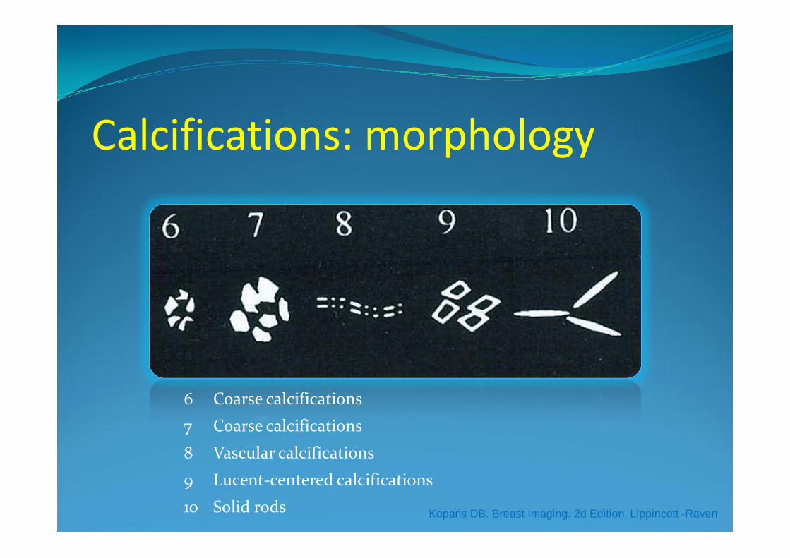
Calcifications: morphology
6 Coarse calcifications
7 Coarse calcifications
8 Vascular calcifications
9 Lucent-centered calcifications
10 Solid rods Kopans DB. Breast Imaging. 2d Edition. Lippincott -Raven
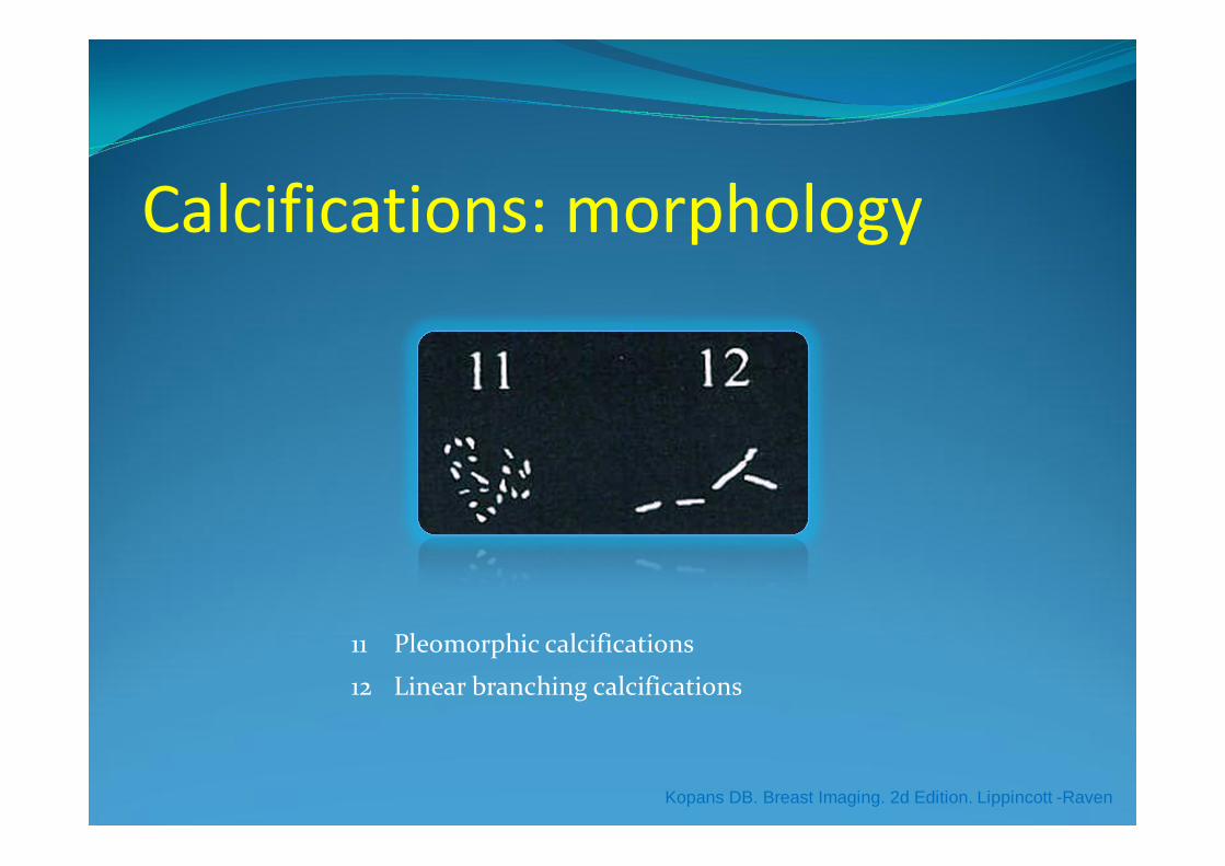
Calcifications: morphology
11 Pleomorphic calcifications
12 Linear branching calcifications
Kopans DB. Breast Imaging. 2d Edition. Lippincott -Raven
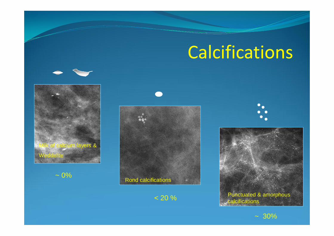
Milk of calcium layers &
Weddelite
Rond calcifications
Punctuated & amorphouscalcifications
Calcifications
~ 0%
< 20 %
~ 30%
Rond calcifications
Calcifications
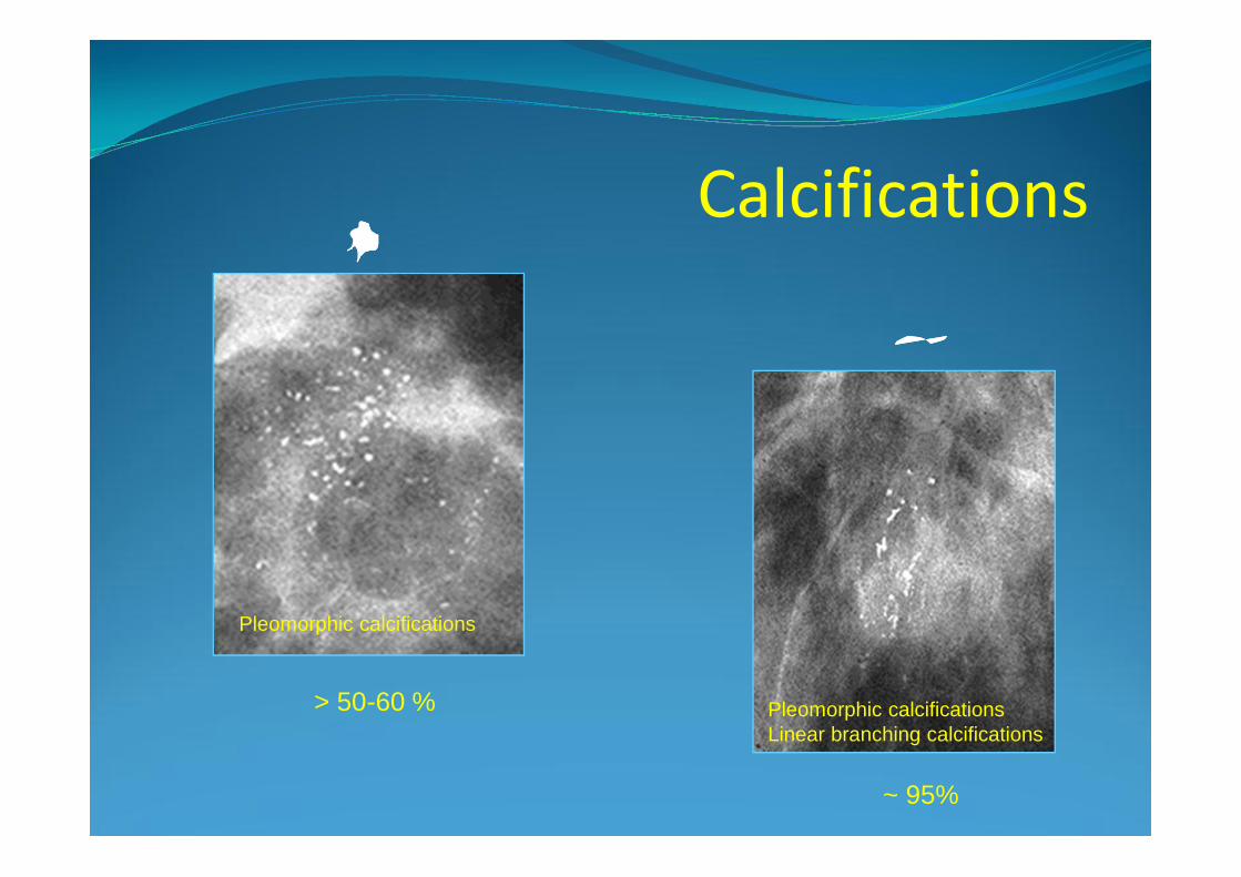
Pleomorphic calcifications
Pleomorphic calcificationsLinear branching calcifications
> 50-60 %
~ 95%
Mass� A Mass is a space occupying lesion seen in two
different projections. If a potential mass is seen in only a single projection, it should be called a density until its three-dimensionality is confirmed.
ACR BI-RADS – Mammography. 4 th Edition. Reston VA
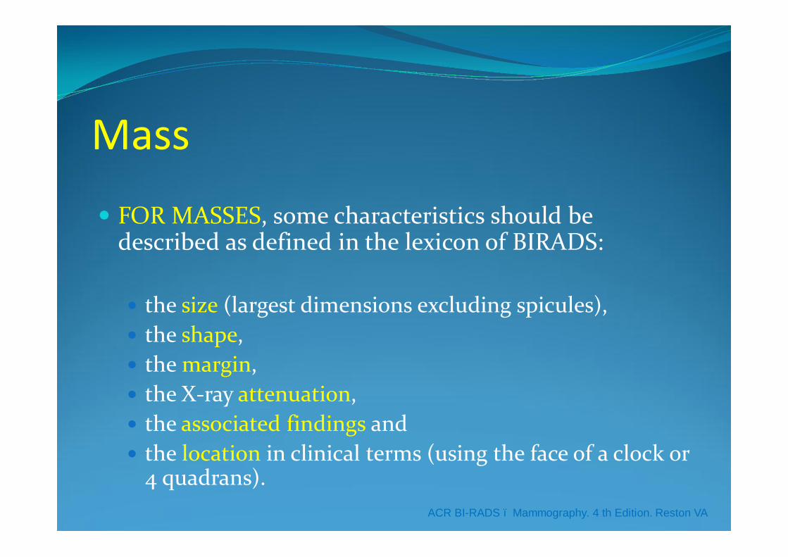
Mass� FOR MASSES, some characteristics should be
described as defined in the lexicon of BIRADS:
� the size (largest dimensions excluding spicules),� the shape, � the margin, � the X-ray attenuation, � the associated findings and� the location in clinical terms (using the face of a clock or
4 quadrans).ACR BI-RADS – Mammography. 4 th Edition. Reston VA
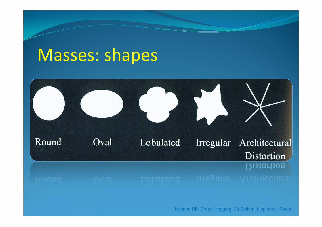
Masses: shapes
Kopans DB. Breast Imaging. 2d Edition. Lippincott -Raven
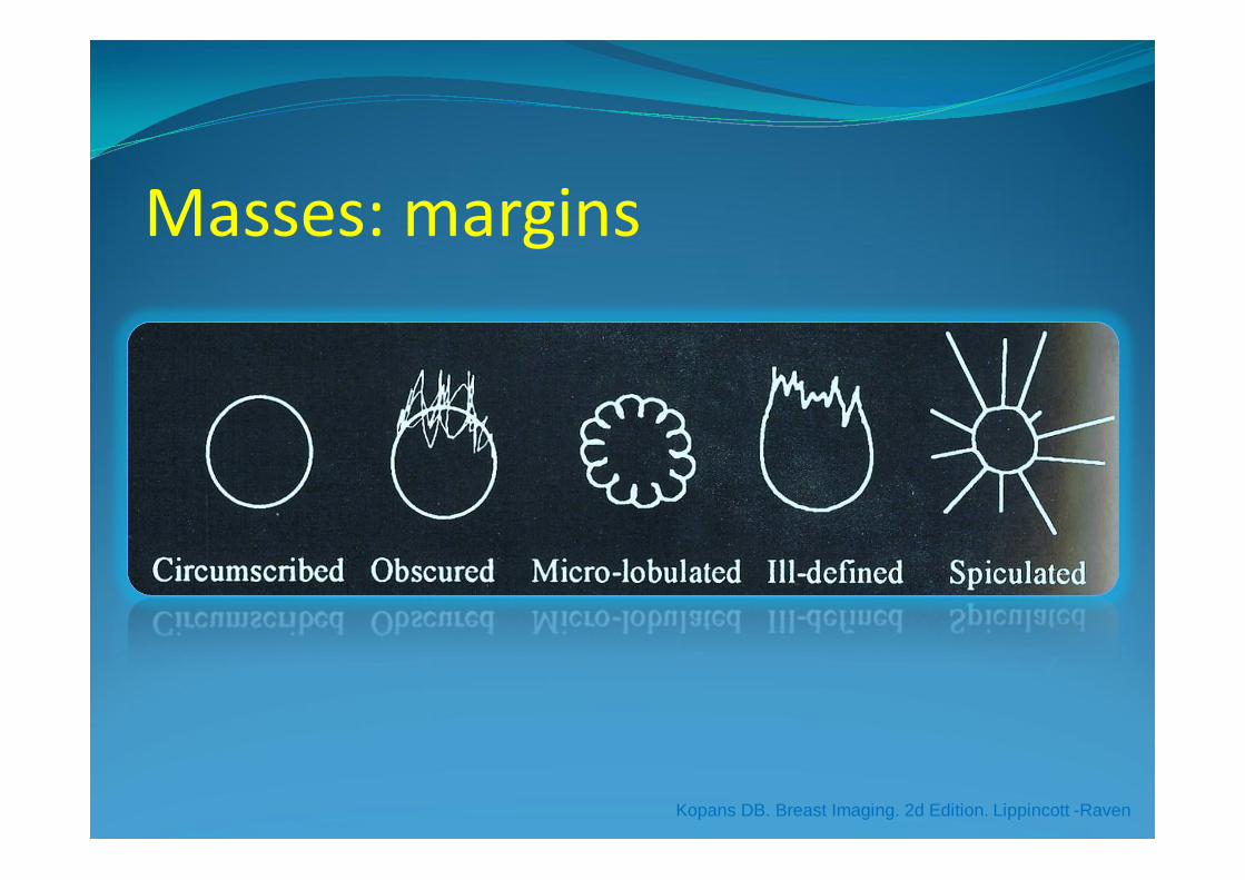
Masses: margins
Kopans DB. Breast Imaging. 2d Edition. Lippincott -Raven
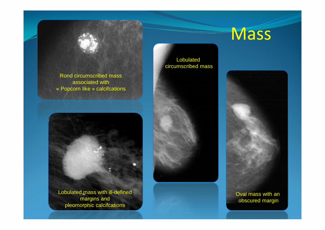
Rond circumscribed mass associated with
« Popcorn like » calcifcations
Lobulated mass with ill-defined margins and
pleomorphic calcifcations
Lobulatedcircumscribed mass
Oval mass with anobscured margin
Mass
A standardizated round mass withobscured marginsA standardizated round mass withobscured margins
Focal asymmetry� It is an asymmetry of tissue density visible on two
views, but it cannot be described using the other shapes.
� It could represent a normal breast, but additional imaging may reveal a true mass or an distortion.
ACR BI-RADS – Mammography. 4 th Edition. Reston VA
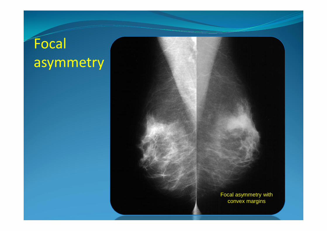
Focal asymmetry with convex margins
Focal asymmetry
An other exampleof asymmetryAn other exampleof asymmetry
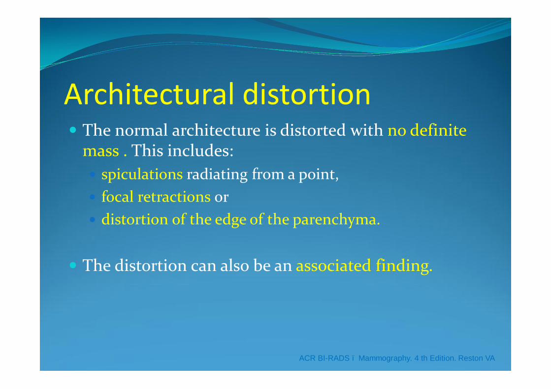
Architectural distortion� The normal architecture is distorted with no definite
mass . This includes:� spiculations radiating from a point, � focal retractions or � distortion of the edge of the parenchyma.
� The distortion can also be an associated finding.
ACR BI-RADS – Mammography. 4 th Edition. Reston VA
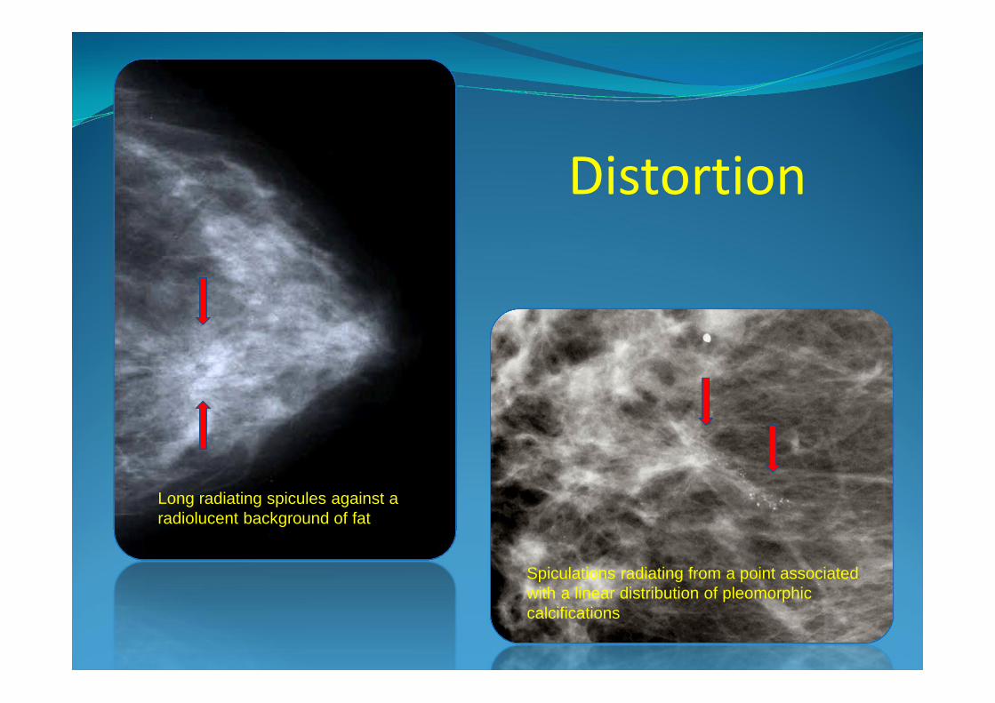
Spiculations radiating from a point associated with a linear distribution of pleomorphic calcifications
Long radiating spicules against a radiolucent background of fat
Distortion
A complete distortion of the presentation
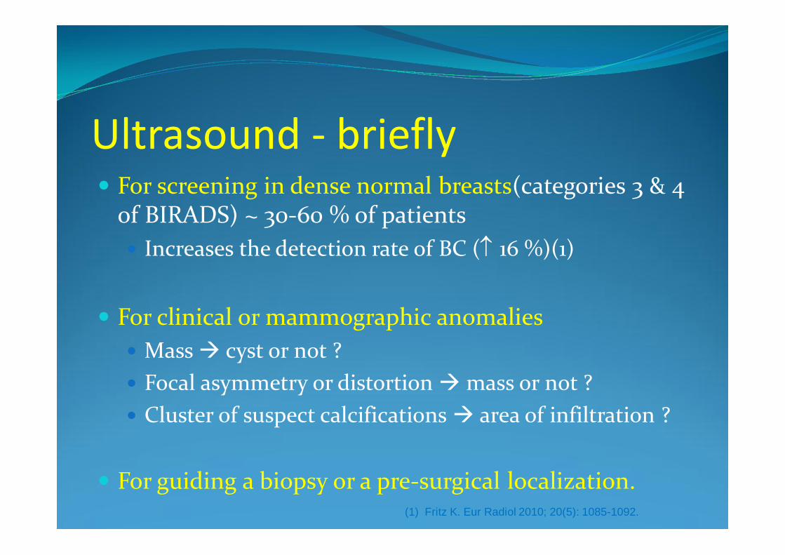
Ultrasound - briefly� For screening in dense normal breasts(categories 3 & 4
of BIRADS) ~ 30-60 % of patients� Increases the detection rate of BC (↑ 16 %)(1)
� For clinical or mammographic anomalies� Mass à cyst or not ?� Focal asymmetry or distortion àmass or not ?� Cluster of suspect calcifications à area of infiltration ?
� For guiding a biopsy or a pre-surgical localization.(1) Fritz K. Eur Radiol 2010; 20(5): 1085-1092.
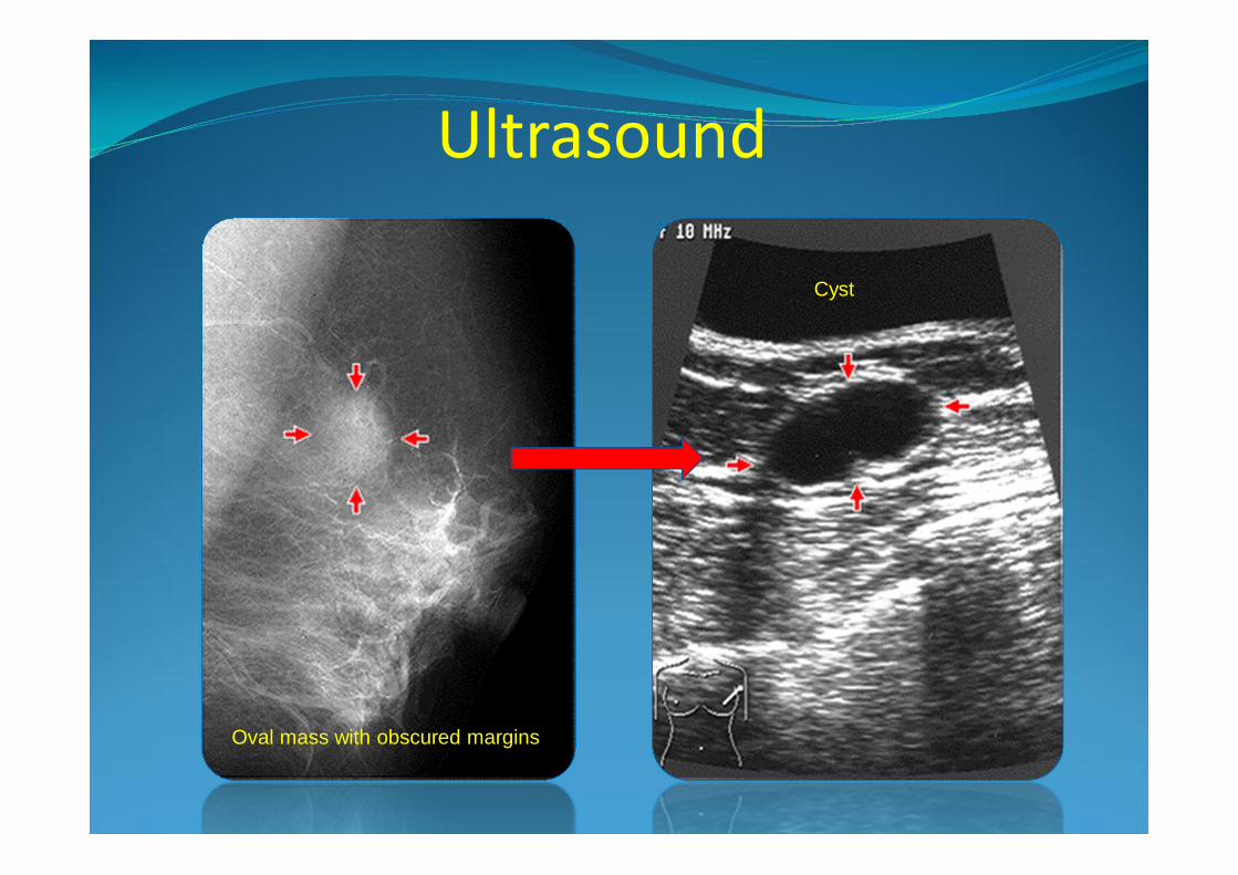
Oval mass with obscured margins
Cyst
Ultrasound
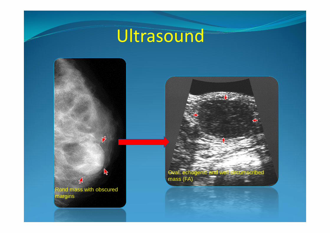
Oval, echogenic and well circumscribed mass (FA)
Rond mass with obscured margins
Ultrasound
� Don’t forget to confirm or not the correlation between clinical or mammographic anomalies and sonographic anomalies.
� If the correlation is not sure, inject a small volume of contrast medium under ultrasound in the surrounding of an anomaly and perform a new mammography (in CC and ML projection) to insure the correlation.
Ultrasound - briefly
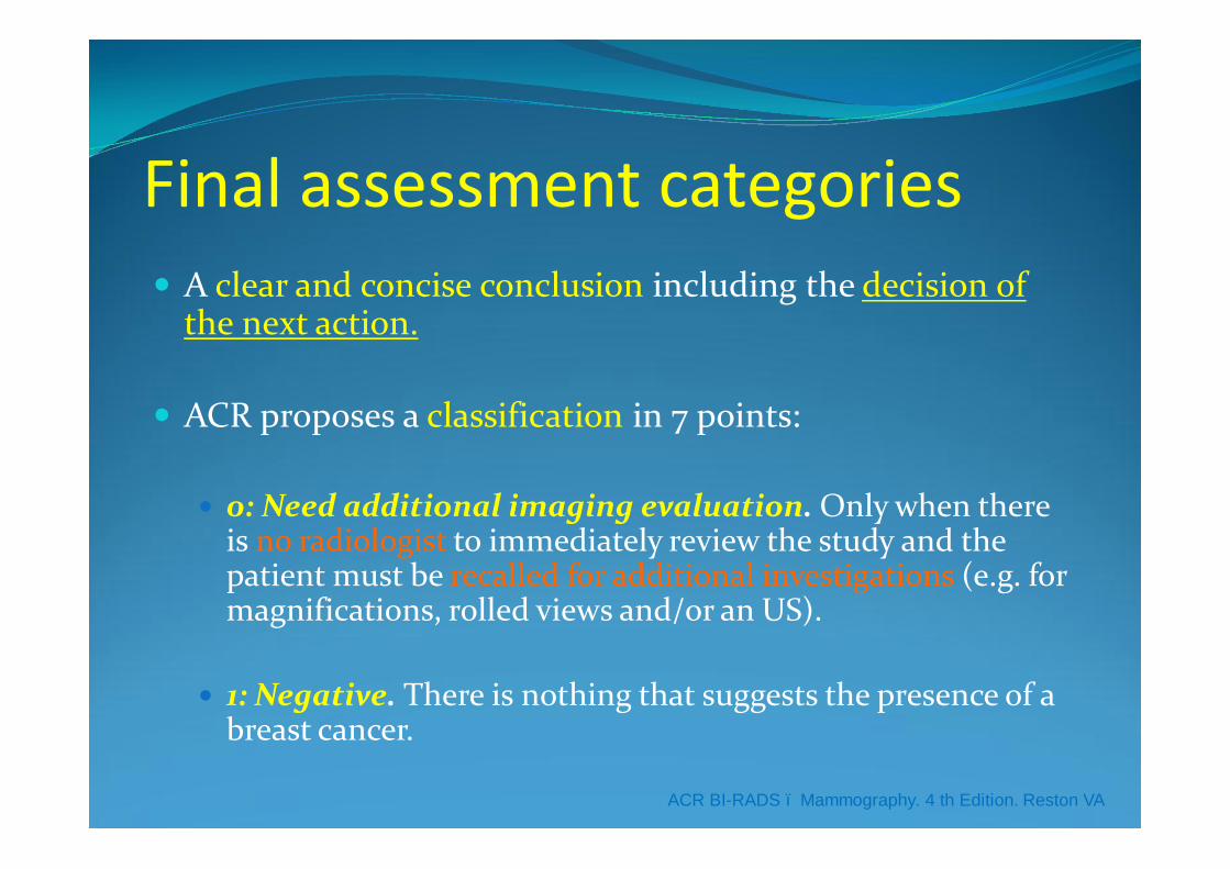
Final assessment categories� A clear and concise conclusion including the decision of
the next action.
� ACR proposes a classification in 7 points:
� 0: Need additional imaging evaluation. Only when there is no radiologist to immediately review the study and the patient must be recalled for additional investigations (e.g. for magnifications, rolled views and/or an US).
� 1: Negative. There is nothing that suggests the presence of a breast cancer.
ACR BI-RADS – Mammography. 4 th Edition. Reston VA
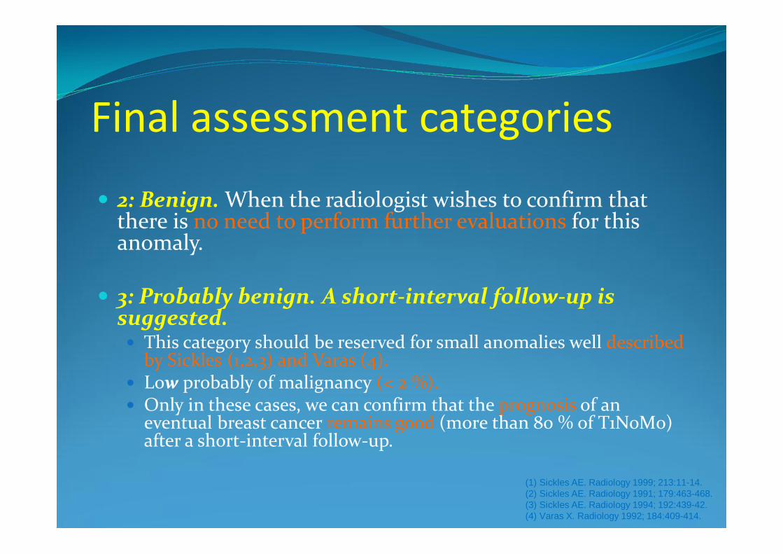
� 2: Benign. When the radiologist wishes to confirm that there is no need to perform further evaluations for this anomaly.
� 3: Probably benign. A short-interval follow-up is suggested. � This category should be reserved for small anomalies well described
by Sickles (1,2,3) and Varas (4). � Low probably of malignancy (< 2 %).� Only in these cases, we can confirm that the prognosis of an
eventual breast cancer remains good (more than 80 % of T1N0M0) after a short-interval follow-up.
Final assessment categories
(1) Sickles AE. Radiology 1999; 213:11-14.(2) Sickles AE. Radiology 1991; 179:463-468.(3) Sickles AE. Radiology 1994; 192:439-42.(4) Varas X. Radiology 1992; 184:409-414.
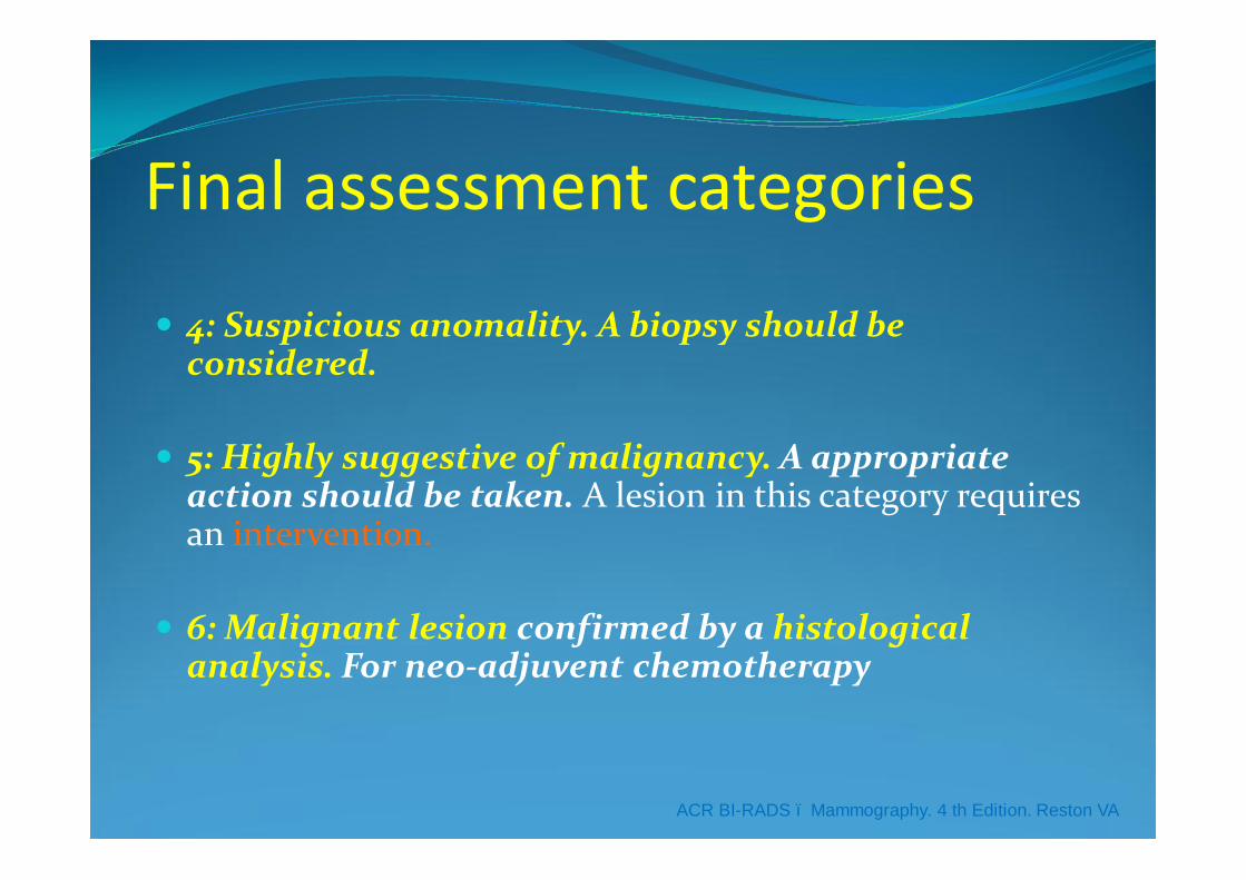
� 4: Suspicious anomality. A biopsy should be considered.
� 5: Highly suggestive of malignancy. A appropriate action should be taken. A lesion in this category requires an intervention.
� 6: Malignant lesion confirmed by a histological analysis. For neo-adjuvent chemotherapy
Final assessment categories
ACR BI-RADS – Mammography. 4 th Edition. Reston VA
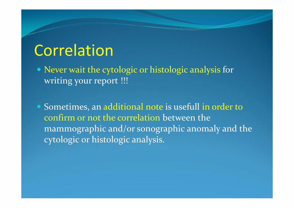
Correlation� Never wait the cytologic or histologic analysis for
writing your report !!!
� Sometimes, an additional note is usefull in order to confirm or not the correlation between the mammographic and/or sonographic anomaly and the cytologic or histologic analysis.
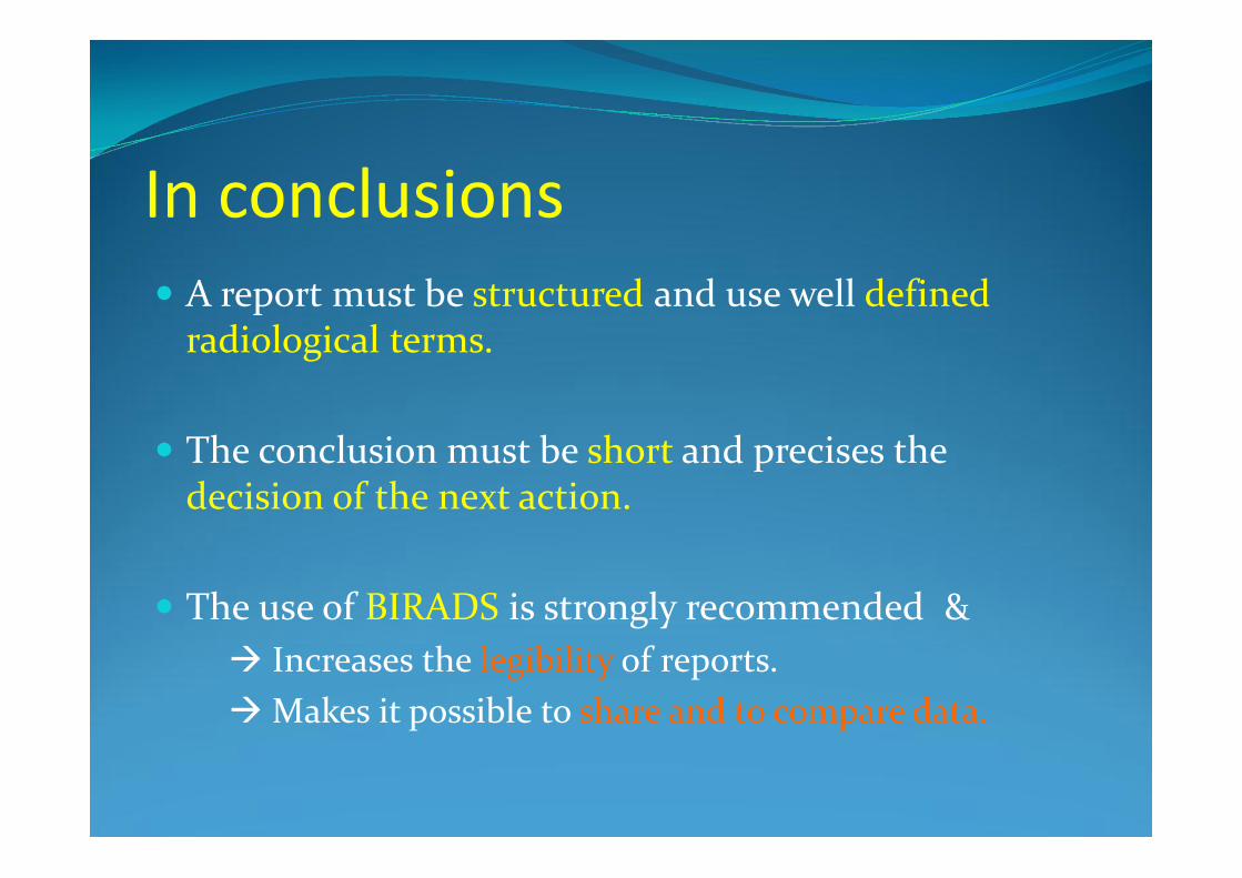
In conclusions� A report must be structured and use well defined
radiological terms.
� The conclusion must be short and precises the decision of the next action.
� The use of BIRADS is strongly recommended …à Increases the legibility of reports.àMakes it possible to share and to compare data.

Now if you becomemore « Standard », I have won my match.
Now if you becomemore « Standard », I have won my match.

Standardization …Standardization …