Embed Size (px)
Citation preview

S-layers in lactobacilli: structural characteristics and putative role in
surface and probiotic properties of whole bacteria
P. Mobili, E. Gerbino, E. E. Tymczyszyn, and A. Gómez-Zavaglia
Centro de Investigación y Desarrollo en Criotecnología de Alimentos (Conicet La Plata, UNLP), 1900 La Plata, Argentina.
The S-layer is a crystalline bidimensional array of proteic or glycoproteic subunits that represents the outermost layer of several species of archea and bacteria. The subunits are linked to each other and to the underlying cell surface (either the peptidoglycan wall or the outer membrane) by non-covalent forces, generally assembled in lattices with oblique, square or hexagonal symmetry.
Several specific functions have been assigned to the S-layer from different microorganisms (protective coats, molecular sieves, molecule and ion traps, cell adhesion and surface recognition, virulence factors). These potential functions have converted the S-layers in attractive targets for biophysical studies and structural research, particularly in regard to their possible nanotechnological applications.
The presence of S-layer proteins has been reported in many species of probiotic lactobacilli. In fact, some probiotic properties of whole bacteria, such as adhesion, aggregation or pathogens inhibition have been related with the occurrence of particular types of S-layers.
Different approaches have been used to study the S-layers in lactobacilli, as well as their role in surface and/or probiotic properties. An indirect approach consists in trying to infer S-layer functions through the comparison of specific bacterial properties (i.e.: aggregation, haemagglutination, adhesion to prokaryotic or eukaryotic cells and to extracellular matrix components), before and after the extraction of the protein [1-8]. A direct approach consists in studying the capacity of isolated S-layer proteins to adhere or associate to other bacteria, eukariotic cells (i.e.: yeasts or enterocytes), extracellular matrix components or inert surfaces as well as to inhibit the growth and/or the activity of pathogens [3-5].
From both approaches, it turns out that S-layer functions in lactobacilli are not only species but also strain specific, and this fact makes difficult the prediction of a given property in S-layer carrying lactobacilli. This correlation can be established if a structural analysis of the S-layers is performed. In this regard, different researchers have studied the nucleotidic sequence of several lactobacilli S-layer protein genes, the primary and secondary structure of proteins (based on predictions from primary structure or from spectroscopic analysis) and posttranslational modifications such as glycosylation. In addition, different structural characteristics of the S-layer proteins have been correlated with the surface and/or probiotic properties of whole microorganisms [8,9].
In this mini-review, an overview of the S-layer proteins from lactobacilli is carried out. The different techniques used for their structural and functional characterization are discussed.
Keywords: probiotic; S-layer; surface properties; aggregation; adherence; structural analysis; vibrational spectroscopy.
1. General overview of S-layer
S-layers are macromolecular paracrystalline arrays of proteins or glycoproteins commonly found in bacteria and archaea. They were described for the first time as "macromolecular monolayers" in the cell wall of Spirillum sp [10]. However, it was only after the seventies when the S-layer attracted the attention of the scientific community. From those years are the descriptions of patterns of bidimensional regular arrays with different symmetries in Lactobacillus fermentum [11], Aquaspirillum sp. [12, 13], Bacillus sphaericus [14], Deinococcus radiodurans [15] and Lactobacillus brevis [16], among others. Finally, it was in 1987, within the scope of the "Second International Workshop on S-
layers”, when researchers agreed in the use of the term "S-layer" to denote these superficial structures [17]. The S-layer has been described for ca. 600 microbial species, belonging to almost all of the taxonomic groups from the domains Archaea and Bacteria, either Gram (-) or Gram (+) [9, 18]. Among Archaea, the S-layer represents the only component outer the plasmatic membrane in most of the cases. Within Gram positive microorganisms, the S-layer is associated to cell wall peptidoglycan, in general through a secondary cell wall polymer (SCWP). On the contrary, among Gram negative bacteria, the S-layer is associated to the lipopolysaccharides of the outer membrane [18, 19] (Figure 1). No matter the nature of the cell component to which the S-layer is attached, this porous structure represents the outermost interaction surface with cell environment. Figure 1 depicts an electronic microscopy image of the supramolecular architecture of the three major classes of prokaryotic cell envelopes containing S-layers in Archaea, Gram negative and Gram positive microorganisms.
_______________________________________________________________________________________

Fig. 1. Electron micrographs of thin sections of: a) an archaeon (Sulfolobus acidocaldarius), b) a Gram-negative bacterium (Aeromonas salmonicida) and c) a Gram-positive bacterium (Bacillus thuringiensis), all of which possess a crystalline cell surface layer (S-layer). CW, Gram-positive cell wall; OM, outer membrane, PG, peptidoglycan layer; PM, plasma membrane; S, S-layer. Scale bar = 50 nm [19]
The topography of S-layers has been extensively investigated by means of electron microscopy, which is considered the most suitable method for identifying the presence of S-layers on intact cells [17]. Indeed, electron microscopy allows the determination of the arrangement of the protein subunits, the analysis of the general geometric pattern, the estimation of the pore size and other topological parameters [12, 13, 20-22]. Different electron microscopy preparation techniques can be applied to obtain images adequately reproducing the native structure of the S-layers, minimizing as much as possible the observation of preparation artifacts. Among them, one can mention negative staining, staining with auro thioglucose for low dose electron microscopy, cryoelectron microscopy, freezedrying or freeze-etching and metal-shadowing, and ultrathin sectioning [23-28]. In particular, freeze-etching preparations have demonstrated to be reliable to provide evidence for the dynamic process of assembly of S-layer proteins on growing and dividing cells [19, 22, 25-27, 29]. Figure 2 depicts images obtained after the preparation of S-layers using two of the mentioned techniques (ultrathin sectioning and negative staining).
Fig. 2. Transmission electron micrographs of (A) thin section of Lactobacillus kefir CIDCA 8321; (B) negative staining of surface protein extracted from L. kefir CIDCA 8321 with guanidine hydrochloride [2].
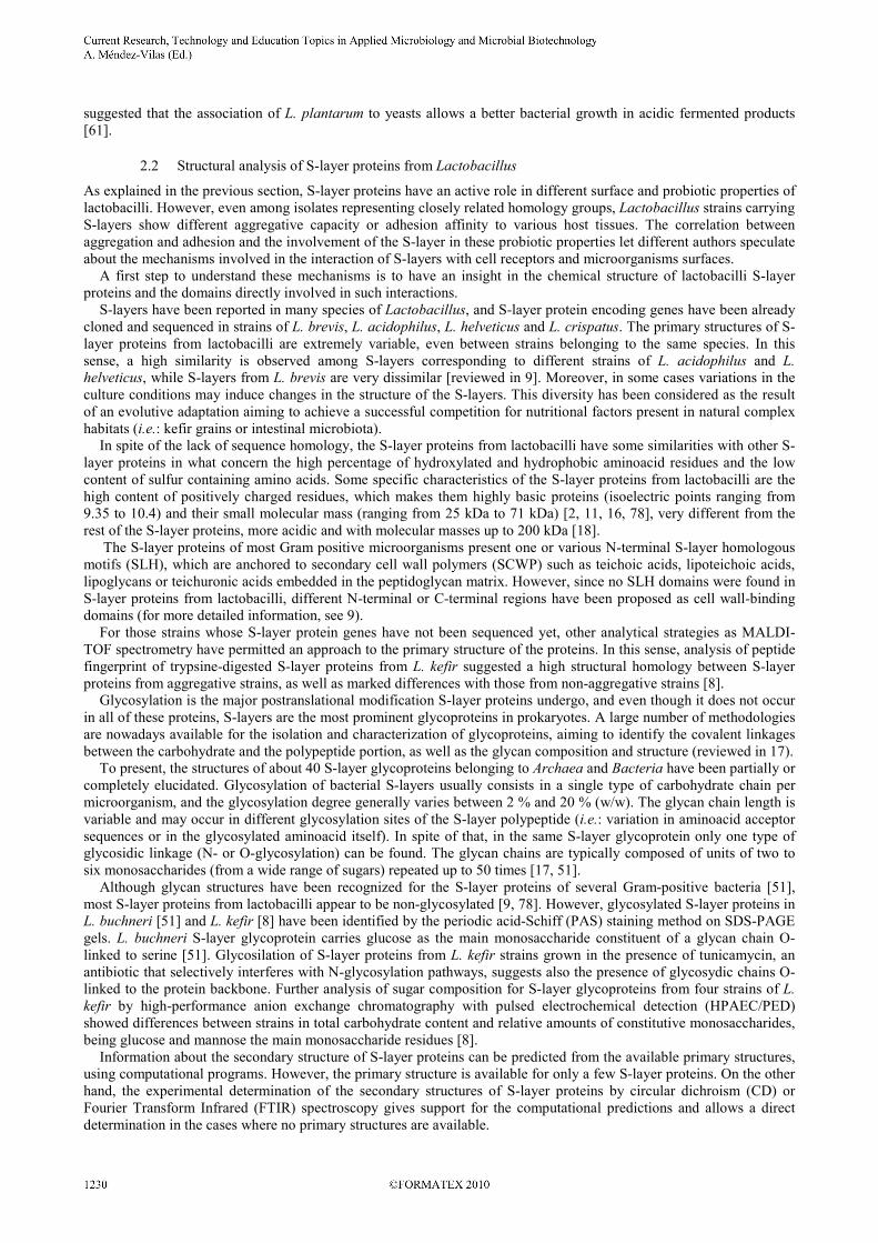
More recently, atomic force microscopy (AFM) has become a powerful tool for the investigation and characterization of S-layers [30-34]. AFM is particularly valuable because the preparation of samples does not require dehydration (as occurs in conventional electron microscopy) and therefore, they can be observed in their native environment, preventing the observation of artifacts resulting from the drying process [30, 32, 34, 35]. In this regard, AFM allows the determination of the shape, thickness, cell dimensions and mechanical properties of S-layers at subnanometer resolution, thus reflecting their molecular architecture [33, 34]. Figure 3 depicts images of S-layers as observed by AFM. This particular topography of S-layers regarding their porosity, regularity, symmetry and self auto-assembly after being removed have converted this surface structure in an attractive target for biophysical and nanotechnological studies as well as for biomedical applications [18, 29, 36-42].
A B
_______________________________________________________________________________________

Fig. 3. (A) High resolution topograph of the inner S-layer surface (outline: hole position with ~90-nm diameter). (B) Fourier-filtered image of A. (C) Average (left) and symmetrized average (right) topographs. (D) High-resolution topograph of the outer S-layer surface (outline: hole position with ~200-nm diameter). (E) Fourier filtered image of D. (F) Average (left) and symmetrized average (right) topographs. Dashed outlines in C and F delineate the structural pore in the center of the flower (f) and triangular (t) shaped surfaces of B15 Å in diameter. Scale bars: 90 nm (A), 20 nm (B), 10 nm (C), 50 nm (D), 20 nm (E) and 10 nm (F). [43]
Based on the information obtained from the above mentioned methodologies, the structure of the S-layer could be schematized as shown in Figure 4. The S-layer crystal lattice may have oblique (p1, p2), tetragonal (p4), or hexagonal (p3, p6) symmetry. Depending on the pattern, a morphological unit of the S-layer consists of one, two, three, four, or six protein subunits. The distance between subunit centers varies from 2.2 to 35 nm, and the S-layer height is 5-25 nm. Pore size varies from 2 to 8 nm, and different pores may occur in one S-layer. It is important to point out that a significant diversity in regard to the lattice symmetry and dimensions can be observed within strains of the same bacterial species. In addition, in some organisms two or even more superimposed S-layer lattices have been identified [22, 27, 41, 42].
Fig. 4. Schematic drawing illustrating the various S-layer lattice types. In the oblique lattice, one morphological unit (red) consists of one (p1) or two (p2) identical subunits. Four subunits constitute one morphological unit in the square (p4) lattice type, whereas the hexagonal lattice type is either composed of three (p3) or six (p6) subunits [44].
Several functions have been attributed to the S-layer. For example, the role of S-layers as protective coats, cell shape determinants, molecular sieves, molecule and ion traps, adhesion sites for exoenzymes as well as structures involved in cell adhesion and surface recognition have been reported [refs see, for example: 18, 19, 45, 46]. Many of these functions are still hypothetical and need further experimental data to be confirmed. The great diversity of the S-layers, even within microorganisms belonging to the same species, may contribute for the difficulty to ascribe a general function for all of them.
2. S-layer in lactic acid bacteria
Lactic acid bacteria are Gram positive microorganisms characterized by the production of lactic acid as a result of sugar fermentation. Within this phylogenetically diverse group, the genus Lactobacillus, with its over hundred species recognized to date, represents the largest one. This genus is also very diverse and includes species commonly found in different natural habitats such as plants, food and also as members of the normal human and animal microbiota of the gastrointestinal and genitourinary tracts [47-49]. Lactobacilli belong to the generally recognized as safe (GRAS) microorganisms by the American Food and Drug Administration (FDA). In addition, several lactobacilli have demonstrated to be health-promoting factors to their host [50] and for this reason, their use as probiotics is widely extended.
_______________________________________________________________________________________

The presence of S-layer has been described in many species of lactobacilli such as Lactobacillus acidophilus, L. helveticus, L. casei, L. brevis, L. buchneri, L. fermentum, L. bulgaricus [41], L. plantarum [51], L. crispatus [52] and more recently in L. kefir and L. parakefir isolated from kefir grains [2]. Different research groups have devoted efforts to understand the role of S-layers from certain strains of lactobacilli, as it is now thought that they play an important role in the maintenance of cellular functions. Considering that purified S-layers are stable toward non-physiological pH, radiation, temperature, some kind of proteolysis, high pressures and detergent treatments [31, 53, 54], a protection role against hostile factors has been proposed for these superficial structures. In this regard, resistance to lysozyme treatment has been related to the presence of an S-layer as the outermost envelope of L. helveticus ATCC 12046 [55]. Moreover, when the S-layer is extracted with chaotropic agents such as 5M lithium chloride, microorganisms become more sensitive toward aggressive environments such as the gastro intestinal juices [56]. In this sense, the S-layers from L. brevis and L. kefir have demonstrated to efficiently coat liposomes and confer them a greater stability when exposed to bile salts, pancreatic extract, pH changes and thermal shocks [57], opening interesting perspectives in the development of vehicles for oral administration of drugs or vaccines, specially taking into account the GRAS status of lactic acid bacteria (reviewed in 9).
2.1 Role of the S-layer in the surface properties of lactobacilli
In the gastro-intestinal tract, the epithelial surface is covered by a layer of mucus that protects epithelial cells from damage and pathogens and also provides a habitat and nutrients for the intestinal microflora [58]. When mucosa is damaged by trauma or microbial infection, bacteria may contact subepithelial tissue structures such as basement membrane and extracellular matrix (ECM) proteins [5, 59]. Different model systems have been developed to select strains that are able to bind to the different above mentioned structures, including cultured intestinal cell lines of human origin such as Caco-2 [59, 60], HT-29 and Intestine 407 cells [6, 61] and purified proteins as mucin [59, 62], ECM proteins such as fibronectin, laminin and types I and IV collagens [6, 7, 63, 64] as well as artificial basement membrane preparations such as Matrigel [5, 52]. As the outermost layer in different species of lactobacilli, the S-layer is in direct contact with bacterial environment and thus may be involved in many of their surface properties. Indeed, different studies found lactobacilli S-layer proteins to mediate bacterial aggregation as well as adhesion to epithelial cells and to intestinal components like mucus or ECM proteins. For this reason, the investigation of surface properties of lactobacilli bearing S-layers and their correlation with specific probiotic properties represent a way to understand both the mechanisms involved in probiotic properties and the functions of the S-layers in lactobacilli [5]. A direct approach to accomplish this objective consists in studying the capacity of isolated S-layer proteins to adhere or associate to other bacteria, eukariotic cells (i.e.: yeasts or enterocytes), ECM components or inert surfaces as well as to inhibit the growth and/or the activity of pathogens [3-5]. An indirect approach consists in trying to infer S-layer functions through the comparison of specific bacterial properties (i.e.: aggregation, haemagglutination, adhesion), before and after the extraction of S-layer proteins [2-9]. In this regard, it must be taken into account that it still remains unclear whether treatments aiming to remove S-layer proteins could at the same time eliminate other surface molecules also important for the adhesion [7, 9].
2.1.1 Aggregation
Aggregation denotes an interaction process among microorganisms that are genetically equal (autoaggregation) or different (coaggregation). Among some probiotic strains, autoaggregation appears to be the first step for adhesion to intestinal epithelial cells and, on the other hand, bacteria with coaggregation abilities may form a barrier preventing colonization by pathogenic microorganisms [65, 66], so the aggregative capacity could also be considered as a potentially probiotic attribute. When aggregation occurs, the formation of bacterial clumps can be observed at the microscope. These clumps tend to precipitate from the microbial suspension. A simple method to quantitatively evaluate aggregation consists in the determination of the optical density decrease of an undisturbed microbial suspension at regular intervals. For coaggregation assays, the absorbance of a mixture composed of the involved microorganisms (ratio: 1:1) is registered. Determination of these properties on bacterial strains before and after the removal of S-layers may confirm the role of such proteins in the interaction [4]. The removal of the S-layer by treatment with 5M LiCl reduces the autoaggregation ability of the strains L. acidophilus M92 [67] and L. crispatus ZJ001 [68], as well as the autoaggregation of L. kefir CIDCA 8321 and its coaggregation with the yeast Saccharomyces lipolytica CIDCA 812 [2, 4], strongly suggesting that these processes are mediated by lactobacillar S-layer proteins (Figure 5). The S-layer from L. kefir CIDCA 8321 seems also to be involved in agglutination of human red blood cells, but not in adhesion to Caco-2 cells [2-4]. This is in contrast with the S-layer of some strains of L. acidophilus that is involved in both autoaggregation and adherence to Caco-2 cells [67, 69]. However, the S-layer is not the only structure involved in aggregation, as demonstrated in the S-layer bearing L. crispatus ZJ001, which exhibits a strong autoaggregating and hemagglutinating phenotype that is not markedly affected by treatment with LiCl [68].
_______________________________________________________________________________________

I
0 2 4 10 20 30
0.4
0.6
0.8
1.0
OD 550 nm
Time (min.)
II
III
IV
A B
I II
III IV
Coaggregation with S. lipolytica
Autoaggregation
Control
L. kefir CIDCA 8321
5M LiCl pretreated
I
0 2 4 10 20 30
0.4
0.6
0.8
1.0
OD 550 nm
Time (min.)
II
III
IV
I
0 2 4 10 20 30
0.4
0.6
0.8
1.0
OD 550 nm
Time (min.)
II
III
IV
A B
I II
III IV
Coaggregation with S. lipolytica
Autoaggregation
I II
III IV
Coaggregation with S. lipolytica
Autoaggregation
Control
L. kefir CIDCA 8321
5M LiCl pretreated
Fig. 5A. Autoaggregation kinetics of L. kefir CIDCA 8321 (○, □) and a mixture of L. kefir CIDCA 8321 with S. lipolytica CIDCA 812 (●,■), obtained before (○,●) or after (□,■) the treatment of lactobacilli with 5M LiCl. B. Optical microscopy (Gram staining, 1000X) of L. kefir CIDCA 8321 (I, II) and a mixture of L. kefir CIDCA 8321 with S. lipolytica CIDCA 812 (III, IV), obtained before (I, III) or after (II, IV) the treatment of lactobacilli with 5M LiCl.
2.1.2 Adhesion to mucus and ECM proteins
The S-layers of certain lactobacilli have demonstrated to act as adhesins, mediating the binding of these bacteria to specific components of the ECM. Direct experimental evidence of this activity has been obtained for the S-layer proteins of L. crispatus JCM 5810 (CbsA), L. brevis ATCC 8287 (SlpA) and L. brevis OLL2772. CbsA is responsible for the adhesion of this strain to solubilized as well as immobilized type I and IV collagens [52, 70]. CbsA heterologously expressed in a non-adhesive L. casei strain turned this strain into adhesive to both immobilized collagen and laminin, as well as to collagen- and laminin-containing regions in chicken colon and ileum [63, 71]. SlpA mediates the binding to immobilized fibronectin [6] and laminin [64], as was demonstrated by expression of SlpA fragments on chimeric flagella and by surface plasmon resonance, respectively. The S-layer protein from L. brevis strain OLL2772 showed a very strong adherence to the human A-antigen expressed in the intestinal mucosa, a molecule also recognized by some viral and bacterial pathogens, so this L. brevis strain could prevent infection by blocking the pathogen receptor. Indirect evidence of adhesive activity was obtained by the use of chemical extraction methods (treatment with 2M guanidine hydrochloride or 5M LiCl) for the removal of S-layer material. This kind of depletion of S-layer proteins from bacterial surface greatly reduced the adhesion of L. brevis ATCC 8287 to immobilized fibronectin and laminin [6, 72], the adhesion of strains of L. plantarum to porcine gastric mucin and ECM proteins [59], the adhesion of L. crispatus to Matrigel [5] and the binding of some strains of L. amylovorus to fibronectin and laminin, whereas increased the adhesiveness to laminin in other L. amylovorus strains [72].
2.1.3 Adhesion to epithelial cells
The adhesion to epithelial cells has also been attributed to the S-layers. In this sense, the expression of the receptor-binding region of the L. brevis S-layer protein in Lactococcus lactis provided nonadhesive lactococci with the ability to adhere to the human intestinal epithelial cell line Intestine 407 [1]. A surface layer protein mutant in L. acidophilus NCFM showed a great reduction in adhesion ability to Caco-2 cells [13]. In addition, a positive relationship has also been suggested between the ability to adhere to chicken epithelia and the presence of an S-layer in an avian isolate of L. acidophilus [69]. However, the molecular mechanisms involved in this binding process have not been explained. Chemical removal of the S-layer proteins significantly reduced the adhesion of L. brevis ATCC 8287 to the human intestinal cell lines Caco-2 and Intestine 407, the endothelial cell line EA-hy926 and the urinary bladder cell line T24 [6], the adhesion of L. acidophilus ATCC 4356 and M92 to avian and porcine intestinal epithelial cells in vitro [67, 70] the adhesion of L. plantarum to Caco-2 cells [59] and the adhesion of L. crispatus ZJ001 to HeLa cells [68]. The functional role of the S-layer proteins in L. crispatus ZJ001 adhesion was also confirmed by an antibody mediated inhibition assay using a polyclonal antibody against the S-layer protein [68].
_______________________________________________________________________________________

On the other hand, the removal of S-layers did not affect the adhesion of L. kefir strains to Caco-2 cells [2, 3]. This behavior suggests that other proteins covalently bound to the bacterial surface or other molecules such as lipoteicoic acids [60] or lectin-like molecules [74] can mediate the adhesion to enterocyte-like cells in vitro.
2.1.4 Activity against pathogens
S-layer proteins with adhesive properties could contribute to lactobacilli probiotic activity by the inhibition of the binding of pathogens to host tissues. This can be achieved through direct competition for attachment sites on human intestinal cells, ECM and mucus proteins, or by the blockage of pathogen surface adhesins. In this regard, it has been reported that the S-layer proteins from L. crispatus JCM 5810 are able to inhibit the adhesion of enterotoxigenic E. coli to laminin and to the basement membrane preparation Matrigel, probably due to the competition for the binding sites in laminin molecules [5]. More recently, it has been demonstrated that the pretreatment of human intestinal epitelium (HEp-2) cells with S-layer extracts from L. helveticus R0052 inhibits the adhesion of E. coli 0157:H7 [75] and that the coincubation of E. coli O157:H7 or Salmonella typhimurium with S-layer proteins from L. crispatus ZJ001 prevents the adhesion of the pathogens to HeLa cells through a competitive exclusion mechanism [68]. Coaggregation with pathogens can explain the mechanisms involved in probiotic properties of some lactobacilli. In this regard, different L. kefir strains carrying S-layer are able to coaggregate with Salmonella enterica serovar Enteritidis. Coincubation of L. kefir CIDCA 8321 (an aggregative strain) with Salmonella clearly protects Caco-2/TC-7 cells against Salmonella invasion. On the contrary, invasion is not prevented when Salmonella is coincubated with non-aggregative L. kefir strains. Preincubation of Salmonella with culture supernatants containing S-layer proteins or with purified S-layer proteins from aggregative L. kefir strains decreases the association of the pathogen to Caco-2/TC-7 cells besides preventing its invasion, greatly reducing the epithelial cell damage caused by the pathogen (Figure 6). Immunoblotting assays performed on these pretreated Salmonella demonstrated that L. kefir S-layer proteins remain associated to Salmonella surface, suggesting a direct interaction among L. kefir S-layer proteins and Salmonella surface adhesins could be involved in this probiotic activity of L. kefir [3].
A B C
Fig. 6. Scanning electron micrographs of: A: Uninfected Caco-2/TC7 cells; B: Caco-2/TC7cells infected during 40 min with Salmonella enteritidis; C: Caco-2/TC7cells infected during 40 min with Salmonella enteritidis pretreated (1 h at 37 °C) with spent culture supernatants of L. kefir CIDCA 8321 containing S-layer proteins [3]
2.1.5 Role of the S-layer in the development of complex microbial communities
There is vast evidence suggesting that aggregation exerts a direct influence on the development of structured microbial communities as biofilms [55]. Kefir grains behave as 3D biofilms in which lactic acid bacteria and yeasts are naturally immobilized in a matrix of protein and polysaccharide [2, 77]. Several L. kefir strains isolated from kefir bear an S-layer, and some of them are able to coaggregate with the kefir yeast Saccharomyces lipolytica CIDCA 812. The treatment of lactobacilli with 5M LiCl completely abolished the coagregation, strongly suggesting this process is mediated by lactobacillar S-layer proteins (Figure 5). The diminishing in coaggregation observed when L. kefir S-layer proteins are added to a mixture of L. kefir CIDCA 8321 and S. lipolytica CIDCA 812 provided additional support for this idea. It was also demonstrated that this interaction is mediated by a lectin-like activity of L. kefir S-layer proteins recognizing sugars on yeast surface [4]. Therefore, it would not be surprising that S-layer proteins with lectin-like activity could have a role in the interaction of lactobacilli with other microorganisms and also with glycoproteins or polysaccharides in kefir grain matrix. The capacity of aggregative L. kefir strains to produce S-layer proteins in milk supplemented with growth factors, sustain this hypothesis [2]. Aggregation properties of kefir strains could also have an important role in the maintenance of the species' balance and also on the number of microorganisms present in the grain along time. In this regard, microorganisms associated to the kefir grains may have some advantages over other microorganisms not taking part in the biofilm. These advantages include the resistance to low pH, low nutrient concentrations and suboptimal temperatures. In this sense, it has been
_______________________________________________________________________________________
suggested that the association of L. plantarum to yeasts allows a better bacterial growth in acidic fermented products [61].
2.2 Structural analysis of S-layer proteins from Lactobacillus
As explained in the previous section, S-layer proteins have an active role in different surface and probiotic properties of lactobacilli. However, even among isolates representing closely related homology groups, Lactobacillus strains carrying S-layers show different aggregative capacity or adhesion affinity to various host tissues. The correlation between aggregation and adhesion and the involvement of the S-layer in these probiotic properties let different authors speculate about the mechanisms involved in the interaction of S-layers with cell receptors and microorganisms surfaces. A first step to understand these mechanisms is to have an insight in the chemical structure of lactobacilli S-layer proteins and the domains directly involved in such interactions. S-layers have been reported in many species of Lactobacillus, and S-layer protein encoding genes have been already cloned and sequenced in strains of L. brevis, L. acidophilus, L. helveticus and L. crispatus. The primary structures of S-layer proteins from lactobacilli are extremely variable, even between strains belonging to the same species. In this sense, a high similarity is observed among S-layers corresponding to different strains of L. acidophilus and L. helveticus, while S-layers from L. brevis are very dissimilar [reviewed in 9]. Moreover, in some cases variations in the culture conditions may induce changes in the structure of the S-layers. This diversity has been considered as the result of an evolutive adaptation aiming to achieve a successful competition for nutritional factors present in natural complex habitats (i.e.: kefir grains or intestinal microbiota). In spite of the lack of sequence homology, the S-layer proteins from lactobacilli have some similarities with other S-layer proteins in what concern the high percentage of hydroxylated and hydrophobic aminoacid residues and the low content of sulfur containing amino acids. Some specific characteristics of the S-layer proteins from lactobacilli are the high content of positively charged residues, which makes them highly basic proteins (isoelectric points ranging from 9.35 to 10.4) and their small molecular mass (ranging from 25 kDa to 71 kDa) [2, 11, 16, 78], very different from the rest of the S-layer proteins, more acidic and with molecular masses up to 200 kDa [18]. The S-layer proteins of most Gram positive microorganisms present one or various N-terminal S-layer homologous motifs (SLH), which are anchored to secondary cell wall polymers (SCWP) such as teichoic acids, lipoteichoic acids, lipoglycans or teichuronic acids embedded in the peptidoglycan matrix. However, since no SLH domains were found in S-layer proteins from lactobacilli, different N-terminal or C-terminal regions have been proposed as cell wall-binding domains (for more detailed information, see 9). For those strains whose S-layer protein genes have not been sequenced yet, other analytical strategies as MALDI-TOF spectrometry have permitted an approach to the primary structure of the proteins. In this sense, analysis of peptide fingerprint of trypsine-digested S-layer proteins from L. kefir suggested a high structural homology between S-layer proteins from aggregative strains, as well as marked differences with those from non-aggregative strains [8]. Glycosylation is the major postranslational modification S-layer proteins undergo, and even though it does not occur in all of these proteins, S-layers are the most prominent glycoproteins in prokaryotes. A large number of methodologies are nowadays available for the isolation and characterization of glycoproteins, aiming to identify the covalent linkages between the carbohydrate and the polypeptide portion, as well as the glycan composition and structure (reviewed in 17). To present, the structures of about 40 S-layer glycoproteins belonging to Archaea and Bacteria have been partially or completely elucidated. Glycosylation of bacterial S-layers usually consists in a single type of carbohydrate chain per microorganism, and the glycosylation degree generally varies between 2 % and 20 % (w/w). The glycan chain length is variable and may occur in different glycosylation sites of the S-layer polypeptide (i.e.: variation in aminoacid acceptor sequences or in the glycosylated aminoacid itself). In spite of that, in the same S-layer glycoprotein only one type of glycosidic linkage (N- or O-glycosylation) can be found. The glycan chains are typically composed of units of two to six monosaccharides (from a wide range of sugars) repeated up to 50 times [17, 51]. Although glycan structures have been recognized for the S-layer proteins of several Gram-positive bacteria [51], most S-layer proteins from lactobacilli appear to be non-glycosylated [9, 78]. However, glycosylated S-layer proteins in L. buchneri [51] and L. kefir [8] have been identified by the periodic acid-Schiff (PAS) staining method on SDS-PAGE gels. L. buchneri S-layer glycoprotein carries glucose as the main monosaccharide constituent of a glycan chain O-linked to serine [51]. Glycosilation of S-layer proteins from L. kefir strains grown in the presence of tunicamycin, an antibiotic that selectively interferes with N-glycosylation pathways, suggests also the presence of glycosydic chains O-linked to the protein backbone. Further analysis of sugar composition for S-layer glycoproteins from four strains of L. kefir by high-performance anion exchange chromatography with pulsed electrochemical detection (HPAEC/PED) showed differences between strains in total carbohydrate content and relative amounts of constitutive monosaccharides, being glucose and mannose the main monosaccharide residues [8]. Information about the secondary structure of S-layer proteins can be predicted from the available primary structures, using computational programs. However, the primary structure is available for only a few S-layer proteins. On the other hand, the experimental determination of the secondary structures of S-layer proteins by circular dichroism (CD) or Fourier Transform Infrared (FTIR) spectroscopy gives support for the computational predictions and allows a direct determination in the cases where no primary structures are available.
_______________________________________________________________________________________

According to CD measurements, approximately 20 % of the amino acids in S-layer proteins are organized as α-helices and about 40 % occur as β-sheets [29, 79]. According to the results of the computed secondary structure predictions, the unprocessed S-layer proteins from L. brevis, L. acidophilus, L. crispatus and L. helveticus seem to contain in average 14 % of α-helices (including the α-helix-rich signal peptide), 39 % of extended strands and 47 % of random coils [9], while those from L. gasseri and L. johnsonii seem to contain 26 to 32 % of β-sheet, alternated with α-helices. Fourier Transform Infrared spectroscopy (FTIR) is a well-established methodology that has been extensively used for the elucidation and analysis of the secondary structure of proteins [80-83]. FTIR Amide I (1720-1600 cm-1) band is mainly generated by C=O stretching vibrations of peptide unions, thus it is strongly influenced by hydrogen bonds giving rise to elements of secondary structure. Each of the protein secondary structural elements like the α-helix, β-sheets and random coil structures, generates a component band that contributes to the Amide I band contour. Empirical association was made between the position of Amide I band and the position and relative areas of component bands after its deconvolution and the type and relative content of secondary structure in the protein [80, 84-89]. FTIR has been used for the characterization of S-layer proteins from a few microorganisms, such as Acetogenium
kivui [90] and Bacillus sphaericus [91]. Among lactobacilli, this spectroscopic methodology has been used for the characterization of S-layer proteins from L. kefir and L. brevis with different surface properties (Figure 7) [92]. According to these authors, the S-layer proteins from different L. kefir strains contain 23 to 42 % of β-sheet and 13 to 21 % of α-helix structure. The differences observed in the aggregative capacities of L. kefir strains can be correlated with the secondary structures of their S-layer proteins, being the β-sheet content higher for non-aggregative strains than for the aggregative ones. These results suggest that the secondary structures of S-layer proteins of analyzed lactobacilli play an important role in determining the surface properties of whole bacteria [92].
c
b
a
Absorbance
1720 1680 1640 1600
Wavenumber (cm-1)Absorbance
ββββ
c
1720 1680 1640 1600
Wavenumber (cm-1)
ββββ
Absorbance
1720 1680 1640 1600
Wavenumber (cm-1)
b
ββββββββ
αααα
Absorbance
1720 1680 1640 1600
Wavenumber (cm-1)
ββββαααα
ββββ
ββββ
a A B
c
b
a
Absorbance
1720 1680 1640 1600
Wavenumber (cm-1)
Absorbance
1720 1680 1640 1600
Wavenumber (cm-1)Absorbance
ββββ
c
1720 1680 1640 1600
Wavenumber (cm-1)
ββββ
Absorbance
ββββ
c
1720 1680 1640 1600
Wavenumber (cm-1)
ββββ
Absorbance
1720 1680 1640 1600
Wavenumber (cm-1)
b
ββββββββ
αααα
Absorbance
1720 1680 1640 1600
Wavenumber (cm-1)
b
ββββββββ
αααα
Absorbance
1720 1680 1640 1600
Wavenumber (cm-1)
ββββαααα
ββββ
ββββ
a
Absorbance
1720 1680 1640 1600
Wavenumber (cm-1)
ββββαααα
ββββ
ββββ
a A B
Fig. 7 A. Unresolved spectra for lyophilized S-layer proteins from L. kefir CIDCA 8344 (spectrum a), CIDCA 8348 (spectrum b) and L. brevis ATCC 8287 (spectrum c) in the Amide I region. B. Unresolved FTIR spectra and curve fitting analysis in the Amide I region for lyophilized S-layer proteins from strains L. kefir CIDCA 8348 (a) and 8344 (b) and L. brevis ATCC 8287 (c). Experimental data (thin solid lines), individual Gaussian components (solid lines) and best fit (dashed lines) are shown [92] In the case of L. kefir strains, the immunochemical reactivity of their S-layer proteins also revealed structural differences among aggregative and non aggregative strains. In this regard, the anti-S-layer monoclonal antibody 5F8 is able to recognize S-layers only from aggregative L. kefir strains. This specificity denotes the existence of at least one epitope that is present only in S-layer proteins from aggregative strains [8].
3. Conclusion
The S-layer has been discovered in the middle seventies. After more than thirty years, investigations dealing with this superficial structure and its forming units are still very active. Different issues contribute to the current state of the art. Among them, one can mention the great diversity, even within S-layers from microorganisms belonging to the same species, which makes difficult a systematic approach aiming to get general conclusions. Besides that, the potentialities of S-layers in nanotechnology, nanoelectronics and biorremediation, open new perspectives in the study of this protein. In this review, we briefly reported the up to date investigations dealing with S-layers. The focus was put on the S-layers from lactobacilli, assessing a correlation between some probiotic properties of whole microorganisms and the structure of their S-layers. In particular, a good correlation exists between the aggregation properties of L. kefir strains and the chemical structure of their S-layer proteins. The investigation of S-layers represents nowadays a multidisciplinary field involving scientists with complementary know-how in disciplines such as Physical-Chemistry, Biophysics, Biochemistry, Molecular Biology, Biotechnology, Microbiology, Immunology and Nanotechnology among many others. An approach based on the complementary backgrounds of these scientists, certainly enriches the perspectives of the different programs carried out. Great advances have been developed in the S-layers' research. However, considering the interdisciplinary of the field, a long way still remains to be undergone.
_______________________________________________________________________________________

Acknowledgements The supports by the Argentinean Agency for Scientific and Technological Promotion (ANPCyT PICT(2006)/00068 and PICT(2008)/0145, by CYTED (Network 108RT0362) are gratefully acknowledged. AGZ and EET are members of the Research Career CONICET.
References
[1] Åvall- Jääskeläinen S, Lindholm A, Palva A. Surface display of the receptor-binding region of the Lactobacillus brevis S-layer protein in Lactococcus lactis provides nonadhesive lactococci with the ability to adhere to intestinal epithelial cells. Applied Environmental Microbiology. 2003; 69: 2230-2236.
[2] Garrote GL, Delfederico L, Bibiloni R, Abraham AG,. Pérez PF, Semorile L, De Antoni GL. Lactobacilli isolated from kefir grains: Evidence of the presence of S-layer proteins. Journal of Dairy Research. 2004; 71: 222-230.
[3] Golowczyc MA, Mobili P, Garrote GL, Abraham AG, De Antoni GL. Protective action of Lactobacillus kefir carrying S-layer protein against Salmonella enterica serovar Enteritidis. International Journal of Food Microbiology. 2007; 118: 264-273.
[4] Golowczyc MA, Mobili P, Garrote GL, Serradell MA, Abraham AG, De Antoni, GL. Interaction between Lactobacillus kefir and Saccharomyces lipolytica isolated from kefir grains: Evidence for lectin-like activity of bacterial surface proteins. Journal of Dairy Research. 2009; 76: 111-116.
[5] Horie M, Ishiyama A, Fujihira-Ueki Y, Sillanpää J, Korhonen TK, Toba T. Identification of Lactobacillus crispatus by polymerase chain reaction targeting S-layer protein gene. Journal of Applied Microbiology. 2002; 92: 396-403.
[6] Hynönen U, Westerlund-Wikström B, Palva A, Korhonen TK. Identification by flagellum display of an epithelial cell- and fibronectin-binding function in the S1pA surface protein of Lactobacillus brevis. Journal of Bacteriology. 2002; 184: 3360-3367.
[7] Jakava-Viljanen M, Palva A. Isolation of surface (S) layer protein carrying Lactobacillus species from porcine intestine and faeces and characterization of their adhesion properties to different host tissues. Veterinary Microbiology. 2007; 124: 264-273.
[8] Mobili P, Serradell M, Trejo S, Avilés Puigvert F, Abraham A, De Antoni G. Heterogeneity of S-layer proteins from from aggregating and non-aggregating Lactobacillus kefir strains Antonie van Leeuwenhoek 2009; 95, 363-372.
[9] Åvall-Jääskeläinen S, Palva A. Lactobacillus surface layers and their applications. FEMS Microbiology Reviews.2005; 29: 511-529.
[10] Engelhardt H. Are S-layers exoskeletons? The basic function of protein surface layers revisited. Journal of Structural Biology. 2007; 160: 115-124.
[11] Kawata T, Masuda K, Yoshino K, Fujimoto M. Regular array in the cell wall of Lactobacillus fermenti as revealed by freeze etching and negative staining. Japanese Journal of Microbiology. 1974; 18: 469-476.
[12] Beveridge TJ, Murray RGE. Superficial macromolecular arrays on the cell wall of Spirillum putridiconchylium. Journal of Bacteriology. 1974; 119: 1019-1038.
[13] Beveridge TJ, Murray RGE. Surface arrays on the cell wall of Spirillum metamorphum. Journal of Bacteriology. 1975; 124: 1529-1544.
[14] Aebi U, Smith PR, Dubochet J. A study of the structure of the T layer of Bacillus brevis. Journal of Supramolecular and
Cellular Biochemistry. 1973; 1: 498-522. [15] Baumeister W, Kuebler O. Topographic study of the cell surface of Micrococcus radiodurans. Proceedings of the National
Academy of Sciences of the United States of America. 1978; 75: 5525-5528. [16] Masuda K, Kawata T. Ultrastructure and partial characterization of a regular array in the cell wall of Lactobacillus brevis.
Microbioly and Immunology. 1979; 23: 941-53. [17] Sleytr UB, Sára M, Pum D, Schuster B, Messner P, Schäffer C. Self-assembly protein systems: Microbial S-layers. In:
Biopolymers. Polyamides and Complex Proteineceous Matrices (A. Steinbüchel, and S.R. Fahnestock, eds.), 2002, vol. 7, pp. 285-338, Wiley-VCH, Weinheim.
[18] Sleytr UB, Sára M, Pum D, Schuster, B. Characterization and use of crystalline bacterial cell surface layers. Progress in Surface Science. 2001; 68: 231-278.
[19] Sleytr UB, Beveridge TJ. Bacterial S-layers: structure, function relationships and their biotechnological applications Trends in Microbiology. 1999; 7: 253-260.
[20] Hovmoller S, Sjogren A, Wang DN. The structure of crystalline bacterial surface layers. Progress in Biophysics and Molecular
Biology. 1988; 51: 131-163. [21] Sara M, Sleytr UB. S-layer proteins. Journal of Bacteriology. 2000; 182: 859-868. [22] Sleytr, UB, Messner, P. Crystalline surface layers in procaryotes. Journal of Bacteriology. 1988; 170: 2891-2897. [23] Kellenberger E and Kistler J. In: Unconventional electron microscopy for molecular structure determination. (W. Hoppe and R.
Mason, eds), 1979 pp. 49-79. F. Vieweg & Sohn, Braunschweig. Germany. [24] Lepault, J, Pitt, T. Projected structure of unstained, frozen-hydrated S-layer of Bacillus brevis. EMBO Journal. 1984; 3: 101-
105. [25] Sleytr UB, Glauert AM. Analysis of regular arrays of subunits on bacterial surfaces: evidence for a dynamic process of
assembly. Journal of Ultrastructure Research. 1975; 50: 103-116. [26] Sleytr, UB. Regular arrays of macromolecules on bacterial cell walls: Structure, chemistry, assembly, and function.
International Review of Cytology. 1978; 53: 1-64. [27] Sleytr UB, Messner P, Pum D. Analysis of crystalline bacterial surface layers by freeze-etching, metal shadowing, negative
staining and ultrathin sectioning methods. Microbiology. 1988; 20: 29-60. [28] Woodcock, CL, Baumeister, W. Different representations of a protein structure obtained with different negative stains.
European Journal of Cell Biology. 1990; 51: 45-52. [29] Sleytr, UB, Huber, C, Ilk, N, Pum, D, Schuster B, Egelseer EM. S-layers as a tool kit for nanobiotechnological applications
FEMS Microbiology Letters. 2007; 267: 131-144.
_______________________________________________________________________________________

[30] Czajkowsky DM, Shao Z. Submolecular resolution of single macromolecules with atomic force microscopy. FEBS Letters. 1998; 430: 51-54.
[31] Engelhardt H, Peters J. Structure research on surface layers: A focus on stability, surface layer homology domains, and surface layer-cell wall interaction. Journal of Structural Biology. 1998; 124: 276-302.
[32] Schabert FA, Henn C, Engel A. Native Escherichia coli OmpF porin surfaces probed by atomic force microscopy. Science. 1995; 268: 92-94.
[33] Scheuring S, Freiss-Husson F, Engel A, Rigaud J.-L., Ranck J-L. High resolution topographs of the Rubrivivax gelatinosus light-harvesting complex 2. EMBO Journal.2001; 20: 3029-3035.
[34] Scheuring S, Stahlberg H, Chami M, Houssin C, Rigaud J-L, Engel A. Charting and unzipping the surface layer of Corynebacterium glutamicum with the atomic force microscope. Molecular Microbiology. 2002; 44: 675-684.
[35] Seelert H, Poetsch A, Dencher NA, Engel A, Stahlberg H, Müller DJ. Proton powered turbine of a plant motor. Nature. 2000; 405: 418-419.
[36] Pum D, Sleytr UB. The application of bacterial Slayers in molecular nanotechnology. Trends Biotechnology. 1999; 17: 8-12. [37] Schuster B, Pum D, Sara M, Sleytr UB. S-layer proteins as key components of a versatile molecular construction kit for
biomedical nanotechnology. Mini Reviews in Medicinal Chemistry 2006; 6: 909-920. [38] Schuster B, Sleytr UB. Composite S-layer lipid structures. Journal of Structural Biology. 2009; 168: 207-216. [39] Sleytr UB Basic and applied S-layer research: an overview. FEMS Microbiological Reviews. 1997; 20: 5-12. [40] Sleytr UB, Bayley H, Sára M, Breitwieser A, Küpcü S, Mader C, Weigert S, Unger FM, Messner P, Jahn-Schmid B, Schuster
B, Pum D, Douglas K, Clark NA, Moore JT, Winningham TA, Levy S, Frithsen I, Pankovc J, Beale P, Gillis HP, Choutov DA, Martin KP. Application of S-layers. FEMS Microbiolical Reviews. 1997; 20: 151-175.
[41] Messner P, Sleytr UB. Crystalline bacterial cell surface layers. Advances in Microbiology and Physiology. 1992; 33: 213-275. [42] Sleytr UB, Messner P, Pum D, Sara M. Crystalline bacterial cell surface proteins. Austin, TX: Academic Press. 1996.
biomedical nanotechnology. Mini Reviews in Medicinal Chemistry. 2006; 6: 909-920. [43] Gonçalves RP, Agnus G, Sens P, Houssin C, Bartenlian B, Scheuring S. Two-chamber AFM: probing membrane proteins
separating two aqueous compartments. Nature Methods. 2006; 3: 1007-1012. [44] Sleytr UB, Sára M. Bacterial and archaeal S-layer proteins: structure-function relationships and their biotechnological
applications. Trends in Biotechnology 1997; 15: 20-26. [45] Debabov VG. Bacterial and Archaeal S-Layers as a subject of nanobiotechnology. Molecular Biology. 2004; 38: 482-493.
Translated from Molekulyarnaya Biologiya. 2004; 38: 578-591. [46] Mertig M, Kirsch R, Pompe W, Engelhardt H. Fabrication of highly oriented nanocluster arrays by biomolecular templating.
European Physical Journal D. 1999; 9: 45-48. [47] Axelsson L. Lactic acid bacteria: classification and physiology. In: Lactic Acid Bacteria: Microbiology and Functional Aspects
(Salminen, S. and von Wright, A., Eds.),1998 pp. 1–72. Marcel Dekker, Inc., New York, USA. [48] Kandler O, Weiss N. Regular, non-sporing Grampositive rods. In: Bergey's Manual of Systematic Bacteriology (Sneath, P.H.A.,
Mair, N.S., Sharpe, M.E. and Holt, J.G., Eds.), 1986 pp. 1208–1234. Williams and Wilking, Baltimore, USA. [49] Stiles ME, Holzapfel WH. Lactic acid bacteria of foods and their current taxonomy. International Journal of Food
Microbiology. 1997; 36: 1-29. [50] Tuohy M, Probert HM, Smejkal CW, Gibson GR. Using probiotics and prebiotics to improve gut health. Drug Discovery
Today. 2003; 8: 692-700. [51] Möschl A, Schäffer C, Sleytr UB, Messner P, Christian R, Schulz G. Characterization of the S-layer glycoproteins of two
lactobacilli. In: Advances in Bacterial Paracrystalline Surface Layers (Beveridge TJ and Koval SF, Eds.), 1993. pp. 281-284. Plenum, New York, USA.
[52] Toba T, Virkola R, Westerlund B, Bjorkman Y, Sillanpaa J, Vartio T, Kalkkinen N, Korhonen TK. A collagen-binding S-layer protein in Lactobacillus crispatus. Applied and Environmental Microbiology. 1995; 61: 2467-2471.
[53] Baumeister W, Barth M, Hegerl R, Guckenberger R, Hahn M, Saxton WO. Three-dimensional structure of the regular surface layer (HPI layer) of Deinococcus radiodurans. Journal of Molecular Biology. 1986; 187: 241-250.
[54] Chami M, Bayan N, Peyret JL, Gulik-Krzywicki T, Leblon G, Shechter E. The S-layer protein of Corynebacterium glutamicum is anchored to the cell wall by its C-terminal hydrophobic domain. Molecular Microbiology. 1997; 23: 483-492.
[55] Lortal S, Van Heijenoort J, Gruber K, Sleytr UB. S-layer of Lactobacillus helveticus ATCC 12046: Isolation, chemical characterization and re-formation after extraction with lithium chloride. Journal of General Microbiology. 1992; 138: 611-618.
[56] Frece J, Kos B, Svetec IK, Zgaga Z, Mrša V, Šušković J. Importance of S-layer proteins in probiotic activity of Lactobacillus acidophilus M92. Journal of Applied Microbiology. 2005; 98: 285-292.
[57] Hollmann A, Delfederico L, Glikmann G, De Antoni GL, Semorile L, Disalvo A Characterization of liposomes coated with S-layer proteins from lactobacilli. Biochimica et Biophysica Acta. 2007; 1768: 393-400.
[58] Tuomola EM, Ouwehand AC, Salminen SJ. Human ileostomy glycoproteins as a model for small intestinal mucus to investigate adhesion of probiotics. Letters in Applied Microbiology. 1999; 28, 159-163.
[59] Tallon R, Arias S, Bressollier P, Urdaci MC. Strain- and matrix-dependent adhesion of Lactobacillus plantarum is mediated by proteinaceous bacterial compounds. Journal of Applied Microbiology. 2007; 102: 442-451.
[60] Granato D, Perotti F, Masserey I, Rouvet M, Golliard M, Servin A, Brassart D. Cell surface-associated lipoteichoic acid acts as an adhesion factor for attachment of Lactobacillus johnsonii La1 to human enterocyte-like Caco-2 cells Applied and Environmental Microbiology. 1999; 65: 1071-1077.
[61] Adlerberth I, Ahrné S, Johansson M-L, Molin G, Hanson LÅ, Wold AE. A mannose-specific adherence mechanism in Lactobacillus plantarum conferring binding to the human colonic cell line HT-29. Applied and Environmental Microbiology. 1996; 62: 2244-2251.
[62] Tuomola EM, Ouwehand AC, Salminen SJ. Chemical, physical and enzymatic pre-treatments of probiotic lactobacilli alter their adhesion to human intestinal mucus glycoproteins. International Journal of Food Microbiology. 2000; 60, 75-81.
_______________________________________________________________________________________

[63] Antikainen J, Anton L, Sillanpää J, Korhonen TK. Domains in the S-layer protein CbsA of Lactobacillus crispatus involved in adherence to collagens, laminin and lipoteichoic acids and in self-assembly. Molecular Microbiology. 2002; 46: 381-394.
[64] De Leeuw E, Li X, Lu W. Binding characteristics of the Lactobacillus brevis ATCC 8287 surface layer to extracellular matrix proteins. FEMS Microbiology Letters. 2006; 260: 210-215.
[65] Boris S, Suárez JE, Barbés C. Characterization of the aggregation promoting factor from Lactobacillus gasseri, a vaginal isolate. Journal of Applied Microbiology. 1997; 83: 413-420.
[66] Reid G, Hawthorn L-A, Mandatori R, Cook RL, Beg HS. Adhesion of lactobacilli to polymer surfaces in vivo and vitro. Microbial Ecology. 1988; 16: 241-251.
[67] Kos B, Šušković J, Vuković S, Šimpraga M, Frece J, Matošić S. Adhesion and aggregation ability of probiotic strain Lactobacillus acidophilus M92. Journal of Applied Microbiology. 2003; 94: 981-987.
[68] Chen X, Xu J , Shuai J, Chen J, Zhang Z, Fang W. The S-layer proteins of Lactobacillus crispatus strain ZJ001 is responsible for competitive exclusion against Escherichia coli O157:H7 and Salmonella typhimurium. International Journal of Food Microbiology. 2007; 115: 307–312.
[69] Schneitz C, Nuotio L, Lounatmaa K. Adhesion of Lactobacillus acidophilus to avian intestinal epithelial cells mediated by the crystalline bacterial cell surface layer (S-layer). Journal of Applied Bacteriology. 1993; 74: 290-294.
[70] Sillanpää J, Martínez B, Antikainen J, Toba T, Kalkkinen N, Tankka S, Lounatmaa K, Keränen J, Höök M, Westerlund-Wikström B, Pouwels PH, Korhonen TK. Characterization of the collagen-binding S-layer protein CbsA of Lactobacillus crispatus. Journal of Bacteriology. 2000; 182: 6440-6450.
[71] Martínez B, Sillanpaä J, Smit E, Korhonen TK, Pouwels PH. Expression of cbsA encoding the collagen-binding S-protein of Lactobacillus crispatus JCM5810 in Lactobacillus casei ATCC 393T. Journal of Bacteriology. 2000; 182: 6857-6861.
[72] Jakava-Viljanen M, Avall-Jääskeläinen S, Messner P, Sleytr UB, Palva A. Isolation of three new surface layer protein genes (slp) from Lactobacillus brevis ATCC 14869 and characterization of the change in their expression under aerated and anaerobic conditions. Journal of Bacteriology. 2002; 184: 6786-6795.
[73] Buck BL, Altermann E, Svingerud T, Klaenhammer TR. Functional analysis of putative adhesion factors in Lactobacillus acidophilus NCFM. Applied and Environmental Microbiology. 2005; 71: 8344-8351.
[74] Mukai T, Arihara K, Itoh H. Lectin-like activity of Lactobacillus acidophilus strain JCM 1026. FEMS Microbiology Letters. 1992; 98: 71-74.
[75] Johnson-Henry KC, Hagen KE, Gordonpour M, Tompkins TA, Sherman PM. Surface-layer protein extracts from Lactobacillus helveticus inhibit enterohaemorrhagic Escherichia coli O157:H7 adhesion to epithelial cells. Cellular Microbiology. 2007; 9: 356-367.
[76] Nikolaev YuA, Plakunov VK. Biofilm-"city of microbes" or an analogue of multicellular organisms? Microbiology. 2007; 76: 149-163.
[77] Angulo L, Lopez E, Lema C. Microflora present in kefir grains of the Galician region (north-west of Spain). Journal of Dairy Research. 1993; 60: 263-267.
[78] Masuda K, Kawata T. Distribution and chemical chracterization of regular arrays in the cell walls of strains of the genus Lactobacillus. FEMS Microbiology Letters. 1983; 20: 145-150.
[79] Sleytr UB, Egelseer EM, Ilk N, Pum, D, Schuster, B. S-Layers as a basic building block in a molecular construction kit. FEBS Journal. 2007; 274: 323-334.
[80] Dong A, Huang P, Caughey WS. Protein secondary structures in water from second-derivative amide I infrared spectra. Biochemistry. 1990; 29: 3303-3308.
[81] Matheus S, Friess W, Mahler HChr. FTIR and DSC as analytical tools for high-concentration protein formulations. Pharmacological Research. 2006; 23: 1350-1363.
[82] Takeda N, Kato M, Taniguchi Y. Pressure- and thermally-induced reversible changes in the secondary structure of ribonuclease A studied by FT-IR spectroscopy. Biochemistry. 1995; 34: 5980-5987.
[83] Vidgren G, Palva I, Pakkanen R, Lounatmaa K, Palva A. S-layer protein gene of Lactobacillus brevis: cloning by polymerase chain reaction and determination of the nucleotide sequence. Journal of Bacteriology. 1992; 174: 7419-7427.
[84] Byler DM, Susi H. Examination of the secondary structure of proteins by deconvolved FTIR spectra. Biopolymers. 1986; 25: 469-487.
[85] Cheng SS, Chittur K, Krishnan Ch, Sukenik LA, Culp K, Lewandowska J. The Conformation of fibronectin on self-assembled monolayers with different surface composition: An FTIR/ATR study. Colloid and Interface Sciences. 1994; 162: 135-143.
[86] Costantino HR, Carrasquillo KG, Cordero RA, Mumenthaler M, Hsu CC, Griebenow K. Effect of excipients on the stability and structure of lyophilized recombinant human growth hormone. Journal of Pharmacological Sciences. 1998; 87: 1412-1420.
[87] Susi H, Byler DM. Fourier Deconvolution of the amide I Raman band of proteins as related to conformation. Applied Spectroscopy. 1988; 42: 819-826.
[88] Torii H, Tasumi M. in: Mantsch H, Chapman D (Eds.), Infrared Spectroscopy of Biomolecules, Wiley-Liss Inc., MA, USA, 1996, pp. 1–18, Chapter 1.
[89] Yang W-J, Griffiths PR, Byler DM, Susi H. Protein conformation by infrared spectroscopy: Resolution enhancement by Fourier self-deconvolution. Applied Spectroscopy. 1985; 39: 282-287.
[90] Lupas A, Engelhardt H, Peters J, Santarius U, Volker S, Baumeister W. Domain structure of the Acetogenium kivui surface layer revealed by electron crystallography and sequence analysis. Journal of Bacteriology. 1994; 176: 1224-1233.
[91] Fahmy K, Merroun M, Pollmann K, Raff J, Savchuk O. Secondary structure and Pd(II) coordination in S-layer proteins from Bacillus sphaericus studied by infrared and X-ray absorption spectroscopy. Biophysical Journal. 2006; 91: 996-1007.
[92] Mobili P, Londero A, Roseiro T, Eusebio E, De Antoni GL, Fausto R, Gómez-Zavaglia A. Characterization of S-layer proteins of Lactobacillus by FTIR spectroscopy and differential scanning calorimetry. Vibrational Spectroscopy. 2009; 50: 68-77.
_______________________________________________________________________________________


![[Skill Build] Slayer](https://img.pdfslide.us/doc/110x75/55cf9d1b550346d033ac4795/skill-build-slayer.jpg)