Embed Size (px)
Citation preview

Presented at the ICLR AI for social good workshop 2019
RXRX1:AN IMAGE SET FOR CELLULAR MORPHOLOGICALVARIATION ACROSS MANY EXPERIMENTAL BATCHES
James Taylor∗, Berton Earnshaw∗, Ben Mabey∗, Mason Victors∗ & Jason Yosinski∗,†∗Recursion Pharmaceuticals, †Uber AI Labs{james.taylor,berton.earnshaw,ben.mabey,mason.victors}@recursionpharma.com, [email protected]
ABSTRACT
High-throughput screening techniques are commonly used in many fields of biol-ogy. However, it is well known that non-biological artifacts arising from variabil-ity in the technical execution of different experimental batches confound high-throughput screens measurements. These batch effects obscure biological con-clusions, and it is therefore necessary to account for them. While a number oftechniques have been proposed, to our knowledge there is not a publicly availablebiological dataset designed specifically for the systematic study of batch effectcorrection. To this end we announce the release of RxRx1, a set of 125,514 high-resolution fluorescence microscopy images of human cells under 1,108 geneticperturbations in 51 experimental batches across four cell types. Visual inspectionof the images by batch makes it clear that the set indeed demonstrates significantbatch effects. In this paper we describe the image set in detail. We also propose aclassification task designed to study batch effect correction on these images, andprovide some baseline results for the task. Our goal in releasing this image set is toencourage researchers across various disciplines to develop effective methods forremoving batch effects that generalize well to unseen experimental batches and toshare these methods with the scientific community.
1 INTRODUCTION
High-throughput screening techniques are in common use in many biological fields, including genet-ics (Echeverri & Perrimon, 2006; Zhou et al., 2014) and drug discovery (Broach et al., 1996; Macar-ron et al., 2011; Swinney & Anthony, 2011; Boutros et al., 2015). Such techniques are capable ofgenerating large amounts of data that, when coupled with modern machine learning methods, couldhelp in answering fundamental questions in biology. These techniques may also help ameliorate theproblem of the exponential rise in the cost of developing an approved drug, which is now estimatedto be well over $2 billion (Scannell et al., 2012; DiMasi et al., 2016). However, creating such largevolumes of biological data necessarily requires the data to be generated in experimental batches, orgroups of experiments executed at similar times under similar conditions. Even when experimentsare carefully designed to control for technical variables such as temperature, humidity, and reagentconcentration, the measurements taken from these screens are confounded by non-biological arti-facts that arise from variability in the technical execution of each batch. These batch effects createfactors of variation within the data that are irrelevant to the biological variables under study, but areunfortunately often correlated with them. It is therefore necessary to correct for batch effects beforedrawing any biological conclusions from measurements taken from high-throughput screens (Leeket al., 2010; Parker & Leek, 2012; Soneson et al., 2014; Nygaard et al., 2016).
Many computational methods have been designed for dealing with batch effects (Leek et al., 2010;Chen et al., 2011; Lazar et al., 2012; Parker & Leek, 2012; Leek et al., 2012; Goh et al., 2017;Shaham et al., 2017), yet to our knowledge there are no publicly-available biological datasets thatwere systematically created to study them. Here we announce the public release of such a dataset,which we call RxRx1. The dataset consists of images of human cells under more than 1,100 differentgenetic perturbations across 51 experimental batches and four cell types. We also propose a machinelearning task that gauges the effectiveness of the batch effect correction method — correctly classify
1

Presented at the ICLR AI for social good workshop 2019
←
Figure 1: 6-channel faux-colored composite image of HUVEC cells (left) and individual channels(rest): nuclei (blue), endoplasmic reticuli (green), actin (red), nucleoli and cytoplasmic RNA (cyan),mitochondria (magenta), and Golgi (yellow). The similarity in content between some channels isdue in part to the spectral overlap between the fluorescent stains used in those channels.
Figure 2: A 384-well plate. Experiments used to generate the images in this dataset were run in thewells of such plates. Photo courtesy of Greiner Bio One International GmbH.
the genetic perturbation present in each image in a held-out set of batches. In order for the classifierto generalize to unseen batches, it must learn to separate biological and technical factors in testimages and make predictions only on the biological factors.
This dataset and task will be of interest to the rapidly growing community of researchers applyingmachine learning methods to complex biological data sets, especially those working with high-content phenotypic screens (Angermueller et al., 2016; Kraus et al., 2016; Caicedo et al., 2017;Kraus et al., 2017; Ando et al., 2017; Chen et al., 2018). The specific task of removing batch effectsis relevant to the broader life sciences community and can provide insights that enable researchersto develop improved methods for working with other biological datasets. In addition, we hope thedataset is of interest to the larger community of machine learning researchers working in computervision, especially those in the areas of domain adaptation, transfer learning, and k-shot learning.
2 DESCRIPTION OF THE DATASET
The image set was produced by Recursion Pharmaceuticals in its automated high-throughput screen-ing laboratory. It is comprised of fluorescence microscopy images of human cells of four differenttypes — HUVEC, RPE, HepG2, and U2OS — which were acquired using a 6-channel variation ofthe Cell Painting imaging protocol (Bray et al., 2016). In Figure 1, we show an example image.
The six channels of an image illuminate the different parts of the cell population in the field ofview: nuclei, endoplasmic reticuli, actin, nucleoli and cytoplasmic RNA, mitochondria, and Golgi.The images themselves are the result of running 51 different instances of the same type of exper-iment. Each experiment instance is comprised of four 384-well plates (see Fig. 2), used to isolatepopulations of cells into wells. The wells are laid out on each plate in a 16×24 grid, but only the
2

Presented at the ICLR AI for social good workshop 2019
Figure 3: Images of four different siRNA phenotypes in HUVEC (same experiment and plate).
Figure 4: Images of the same siRNA in four cell types: HUVEC, RPE, HepG2, U2OS.
wells in the inner 14×22 grid are used since the outer wells are most susceptible to environmentalfactors. Of these 308 usable wells, one remains untreated to provide a negative control. The restof the 307 wells receive exactly one small interfering ribonucleic acid, or siRNA, at a fixed con-centration. Each siRNA is designed to knockdown a single target gene via the RNA interferencepathway, reducing the expression of the gene and its associated protein (Tuschl, 2001). However,siRNAs are known to have significant but consistent off-target effects via the microRNA pathway,creating partial knockdown of many other genes as well. The overall effect of siRNA transfection isto perturb the morphology, count, and distribution of cells in each well, creating a distinct phenotypeassociated with each siRNA. The phenotype is sometimes visually recognizable from the images,but often the specific difference in cell morphology is subtle and hard to detect to the human eye(see Fig. 3).
In each experiment, the same 30 siRNA appear on every plate as a control set for the plate. Thesecontrol siRNA target different genes and produce a variety of phenotypic effects that, taken in com-bination with the single untreated well, provide a set of useful reference wells for each plate. The1,108 remaining wells of each experiment (277 wells×4 plates) receive 1,108 different siRNA.These non-control siRNA target different genes than each other and the genes of the control siRNA.Notice that while the control siRNA appear on each plate, each non-control siRNA appears at mostonce in each experiment. We say at most once because, although rare, it happens that either ansiRNA is not transferred into its well, resulting in an additional untreated well on the plate, or an op-erational error is detected by quality control procedures and renders the well unsuitable for inclusionin the dataset.
When the images were originally acquired from the microscope, they were of spatial resolu-tion 2048×2048, but in order to make the dataset more manageable, they were downsampled to1024×1024 and cropped to the center 512×512 field of view. The image set contains two non-overlapping 512×512 fields of view per well. Therefore, there could be as many as 125,664 images(= 51 experiments×4 plates/experiment×4 wells/plate×2 images/well), but, because of operationalerrors, a number of images were removed, resulting in 125,514 actual images in the dataset.
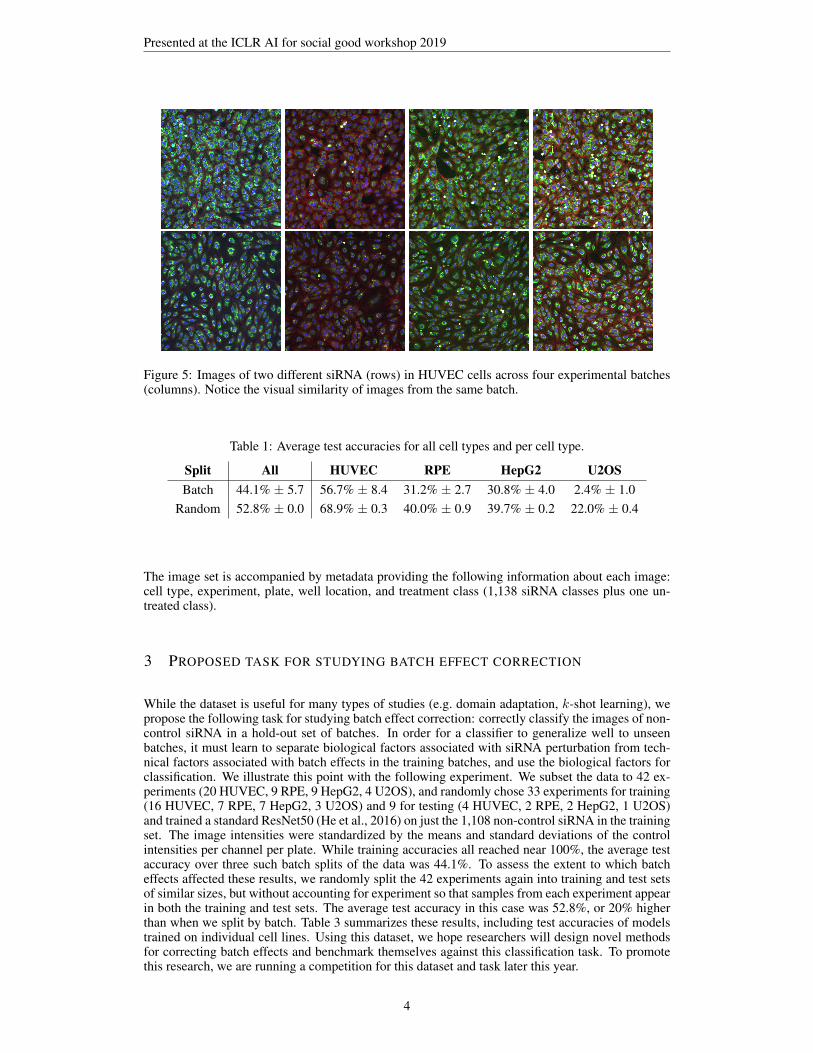
As was mentioned, the entire dataset consists of 51 experiments: 24 in HUVEC, 11 in RPE, 11in HepG2, and 5 in U2OS. Figure 4 shows the phenotype of a single siRNA in the four differentcell types. Each of the 51 experiments was run in a different batch, resulting in images that exhibittechnical effects (e.g. differences in temperature, humidity, siRNA concentration) that are commonto the batch but distinct from other batches (see Fig. 5). It is this feature of the dataset that makes itparticularly suited for studying batch effects and methods for correcting them.
3
Presented at the ICLR AI for social good workshop 2019
Figure 5: Images of two different siRNA (rows) in HUVEC cells across four experimental batches(columns). Notice the visual similarity of images from the same batch.
Table 1: Average test accuracies for all cell types and per cell type.
Split All HUVEC RPE HepG2 U2OSBatch 44.1% ± 5.7 56.7% ± 8.4 31.2% ± 2.7 30.8% ± 4.0 2.4% ± 1.0
Random 52.8% ± 0.0 68.9% ± 0.3 40.0% ± 0.9 39.7% ± 0.2 22.0% ± 0.4
The image set is accompanied by metadata providing the following information about each image:cell type, experiment, plate, well location, and treatment class (1,138 siRNA classes plus one un-treated class).
3 PROPOSED TASK FOR STUDYING BATCH EFFECT CORRECTION
While the dataset is useful for many types of studies (e.g. domain adaptation, k-shot learning), wepropose the following task for studying batch effect correction: correctly classify the images of non-control siRNA in a hold-out set of batches. In order for a classifier to generalize well to unseenbatches, it must learn to separate biological factors associated with siRNA perturbation from tech-nical factors associated with batch effects in the training batches, and use the biological factors forclassification. We illustrate this point with the following experiment. We subset the data to 42 ex-periments (20 HUVEC, 9 RPE, 9 HepG2, 4 U2OS), and randomly chose 33 experiments for training(16 HUVEC, 7 RPE, 7 HepG2, 3 U2OS) and 9 for testing (4 HUVEC, 2 RPE, 2 HepG2, 1 U2OS)and trained a standard ResNet50 (He et al., 2016) on just the 1,108 non-control siRNA in the trainingset. The image intensities were standardized by the means and standard deviations of the controlintensities per channel per plate. While training accuracies all reached near 100%, the average testaccuracy over three such batch splits of the data was 44.1%. To assess the extent to which batcheffects affected these results, we randomly split the 42 experiments again into training and test setsof similar sizes, but without accounting for experiment so that samples from each experiment appearin both the training and test sets. The average test accuracy in this case was 52.8%, or 20% higherthan when we split by batch. Table 3 summarizes these results, including test accuracies of modelstrained on individual cell lines. Using this dataset, we hope researchers will design novel methodsfor correcting batch effects and benchmark themselves against this classification task. To promotethis research, we are running a competition for this dataset and task later this year.
4

Presented at the ICLR AI for social good workshop 2019
REFERENCES
D Michael Ando, Cory McLean, and Marc Berndl. Improving phenotypic measurements in high-content imaging screens. bioRxiv, pp. 161422, 2017.
Christof Angermueller, Tanel Parnamaa, Leopold Parts, and Oliver Stegle. Deep learning for com-putational biology. Molecular systems biology, 12(7):878, 2016.
Michael Boutros, Florian Heigwer, and Christina Laufer. Microscopy-based high-content screening.Cell, 163(6):1314–1325, 2015.
Mark-Anthony Bray, Shantanu Singh, Han Han, Chadwick T Davis, Blake Borgeson, Cathy Hart-land, Maria Kost-Alimova, Sigrun M Gustafsdottir, Christopher C Gibson, and Anne E Carpenter.Cell painting, a high-content image-based assay for morphological profiling using multiplexedfluorescent dyes. Nature protocols, 11(9):1757, 2016.
James R Broach, Jeremy Thorner, et al. High-throughput screening for drug discovery. Nature, 384(6604):14–16, 1996.
Juan C Caicedo, Sam Cooper, Florian Heigwer, Scott Warchal, Peng Qiu, Csaba Molnar, Aliaksei SVasilevich, Joseph D Barry, Harmanjit Singh Bansal, Oren Kraus, et al. Data-analysis strategiesfor image-based cell profiling. Nature methods, 14(9):849, 2017.
Chao Chen, Kay Grennan, Judith Badner, Dandan Zhang, Elliot Gershon, Li Jin, and Chunyu Liu.Removing batch effects in analysis of expression microarray data: an evaluation of six batchadjustment methods. PloS one, 6(2):e17238, 2011.
Hongming Chen, Ola Engkvist, Yinhai Wang, Marcus Olivecrona, and Thomas Blaschke. The riseof deep learning in drug discovery. Drug discovery today, 23(6):1241–1250, 2018.
Joseph A DiMasi, Henry G Grabowski, and Ronald W Hansen. Innovation in the pharmaceuticalindustry: new estimates of r&d costs. Journal of health economics, 47:20–33, 2016.
Christophe J Echeverri and Norbert Perrimon. High-throughput rnai screening in cultured cells: auser’s guide. Nature Reviews Genetics, 7(5):373, 2006.
Wilson Wen Bin Goh, Wei Wang, and Limsoon Wong. Why batch effects matter in omics data, andhow to avoid them. Trends in biotechnology, 35(6):498–507, 2017.
Kaiming He, Xiangyu Zhang, Shaoqing Ren, and Jian Sun. Deep residual learning for image recog-nition. In Proceedings of the IEEE conference on computer vision and pattern recognition, pp.770–778, 2016.
Oren Z Kraus, Jimmy Lei Ba, and Brendan J Frey. Classifying and segmenting microscopy imageswith deep multiple instance learning. Bioinformatics, 32(12):i52–i59, 2016.
Oren Z Kraus, Ben T Grys, Jimmy Ba, Yolanda Chong, Brendan J Frey, Charles Boone, and Brenda JAndrews. Automated analysis of high-content microscopy data with deep learning. Molecularsystems biology, 13(4):924, 2017.
Cosmin Lazar, Stijn Meganck, Jonatan Taminau, David Steenhoff, Alain Coletta, Colin Molter,David Y Weiss-Solıs, Robin Duque, Hugues Bersini, and Ann Nowe. Batch effect removal meth-ods for microarray gene expression data integration: a survey. Briefings in bioinformatics, 14(4):469–490, 2012.
Jeffrey T Leek, Robert B Scharpf, Hector Corrada Bravo, David Simcha, Benjamin Langmead,W Evan Johnson, Donald Geman, Keith Baggerly, and Rafael A Irizarry. Tackling the widespreadand critical impact of batch effects in high-throughput data. Nature Reviews Genetics, 11(10):733,2010.
Jeffrey T Leek, W Evan Johnson, Hilary S Parker, Andrew E Jaffe, and John D Storey. The svapackage for removing batch effects and other unwanted variation in high-throughput experiments.Bioinformatics, 28(6):882–883, 2012.
5
Presented at the ICLR AI for social good workshop 2019
Ricardo Macarron, Martyn N Banks, Dejan Bojanic, David J Burns, Dragan A Cirovic, TinaGaryantes, Darren VS Green, Robert P Hertzberg, William P Janzen, Jeff W Paslay, et al. Impactof high-throughput screening in biomedical research. Nature reviews Drug discovery, 10(3):188,2011.
Vegard Nygaard, Einar Andreas Rødland, and Eivind Hovig. Methods that remove batch effectswhile retaining group differences may lead to exaggerated confidence in downstream analyses.Biostatistics, 17(1):29–39, 2016.
Hilary S Parker and Jeffrey T Leek. The practical effect of batch on genomic prediction. Statisticalapplications in genetics and molecular biology, 11(3), 2012.
Jack W Scannell, Alex Blanckley, Helen Boldon, and Brian Warrington. Diagnosing the decline inpharmaceutical r&d efficiency. Nature reviews Drug discovery, 11(3):191, 2012.
Uri Shaham, Kelly P Stanton, Jun Zhao, Huamin Li, Khadir Raddassi, Ruth Montgomery, and YuvalKluger. Removal of batch effects using distribution-matching residual networks. Bioinformatics,33(16):2539–2546, 2017.
Charlotte Soneson, Sarah Gerster, and Mauro Delorenzi. Batch effect confounding leads to strongbias in performance estimates obtained by cross-validation. PloS one, 9(6):e100335, 2014.
David C Swinney and Jason Anthony. How were new medicines discovered? Nature reviews Drugdiscovery, 10(7):507, 2011.
Thomas Tuschl. Rna interference and small interfering rnas. Chembiochem, 2(4):239–245, 2001.
Yuexin Zhou, Shiyou Zhu, Changzu Cai, Pengfei Yuan, Chunmei Li, Yanyi Huang, and WenshengWei. High-throughput screening of a crispr/cas9 library for functional genomics in human cells.Nature, 509(7501):487, 2014.
6