Embed Size (px)
Citation preview
Rules & regulations and
Requirements
Regarding the attendance in the lectures, any student exceed 15% 0f the total will not allow to set the final exam.
Please the rules not allow any student to sign in the behalf of his colleague, otherwise,……..????!!!
All required instruments and manual textbook by Nairn
Clinic uniform and clean white coat.
Cross-infection control measures (hair, nails….).
Attendance the first 15 minutes.
Our patients????!!!!.
You should sign the procedure you had finish 15 minutes before the end of the session in the logbook from your supervisor, otherwise it will account for, with date. Because this will give time to your colleague to be ready for his/her patient.
Two arches per semester. OR one C0-Cr RPD if possible
All CD steps will be carried out in the hospital lab.
Materials will be given to all from the first week (4-green sticks and 3-compound).
PRINCIPLES OF EXAMAING
AND SELECTING CANDIDATES
FOR
COMPLETE DENTURE
TREATMENT
Dr Salah Al-Omoush * Course Coordinator ( theory).
• Generally speaking, prosthesis may be defined as an appliance which replaces the lost and congenitally missing tissue. While prosthetic in general term is the art and science of designing and fitting an artificial substitute to replace lost or missing tissue. Dental prosthetic “prosthodontics “ fitting of appliances in the oral cavity such as artificial dentures (conventional CD ,immediate CD or RPD , Overdenture retained by natural root or implant , replica CD and fixed partial dentures “bridges”. Extra oral prosthesis such as Obturator, eye and nose.

• By definition: examination and
diagnosis are the process of a
careful investigation of the facts to
determine the nature of the case or
the problem. Therefore, this can be
very helpful to assess the normal
and the abnormal situations.
• This cannot be accomplished in a short time. “spending more time in examining and planning than doing, doing becomes much easier.”
Dr Thomas, 1974
• Therefore, dentists or clinicians must know the anatomy and the physiology of the oral cavity and human body in general. In addition to the psychological make –up of the patient, in order to make a clear and highly successful and predictable treatment. These mainly depend on skilled clinician, skilled dental technician (naturally-born or acquired i.e. effort, practice ,critical evaluation) and the patient’s variables.


The basic requirements for impression making
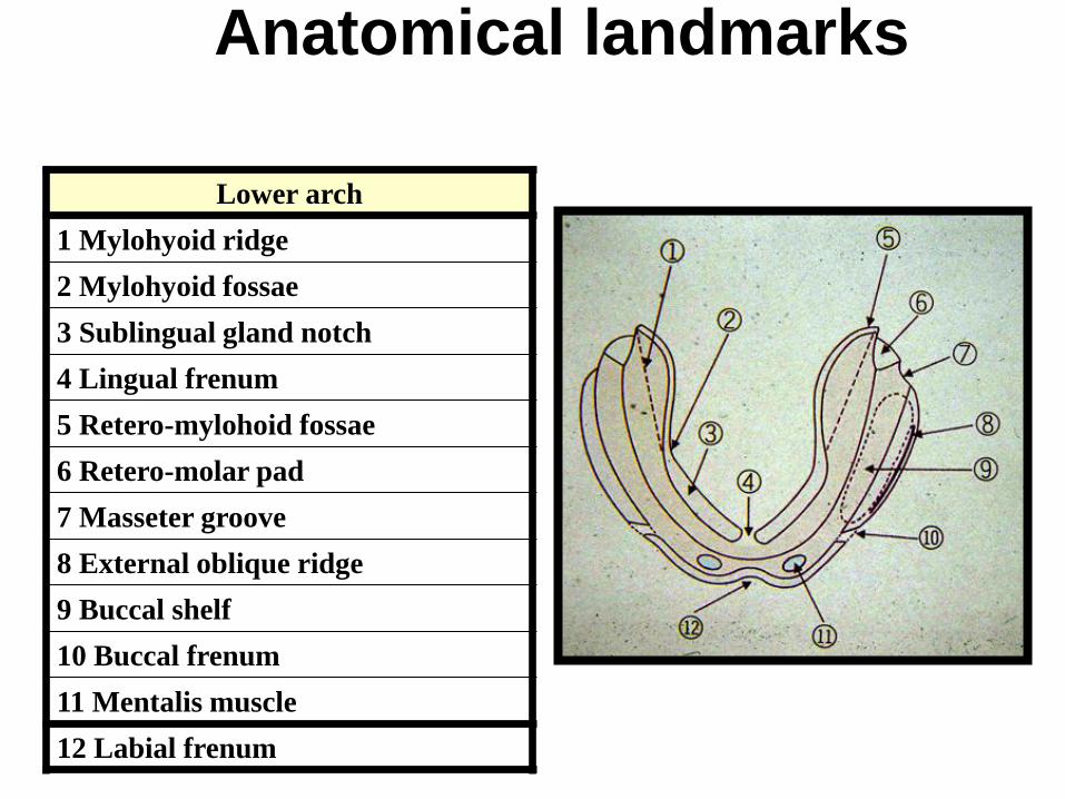
Anatomical landmarks
Lower arch
1 Mylohyoid ridge
2 Mylohyoid fossae
3 Sublingual gland notch
4 Lingual frenum
5 Retero-mylohoid fossae
6 Retero-molar pad
7 Masseter groove
8 External oblique ridge
9 Buccal shelf
10 Buccal frenum
11 Mentalis muscle
12 Labial frenum

Upper arch
1 Incisive papilla
2 Labial frenum
3 Buccal frenum
4 Median raphe
5 Maxillary tuberosity
6 Anterior vibrating line
7 Posterior vibrating line
8 Fovea palatine
9 Hamular notch
10 Pterygo-mandibular raphe

Patient’s variables Patient’s attitude toward complete denture
Patient’s oral condition
Patient’s dentist relationship
Patient’s personally
Patient’s socio –economic status
Patient’s demography factors (Age & sex)
Patient’s previous denture experience

These can be assessed by
1. History taking
2. Examining the Patient’s oral cavity
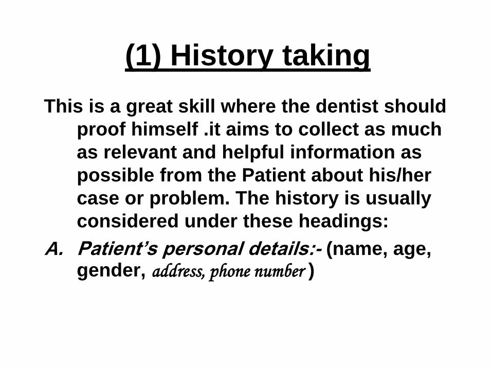
(1) History taking
This is a great skill where the dentist should
proof himself .it aims to collect as much
as relevant and helpful information as
possible from the Patient about his/her
case or problem. The history is usually
considered under these headings:
A. Patient’s personal details:- (name, age, gender, address, phone number )
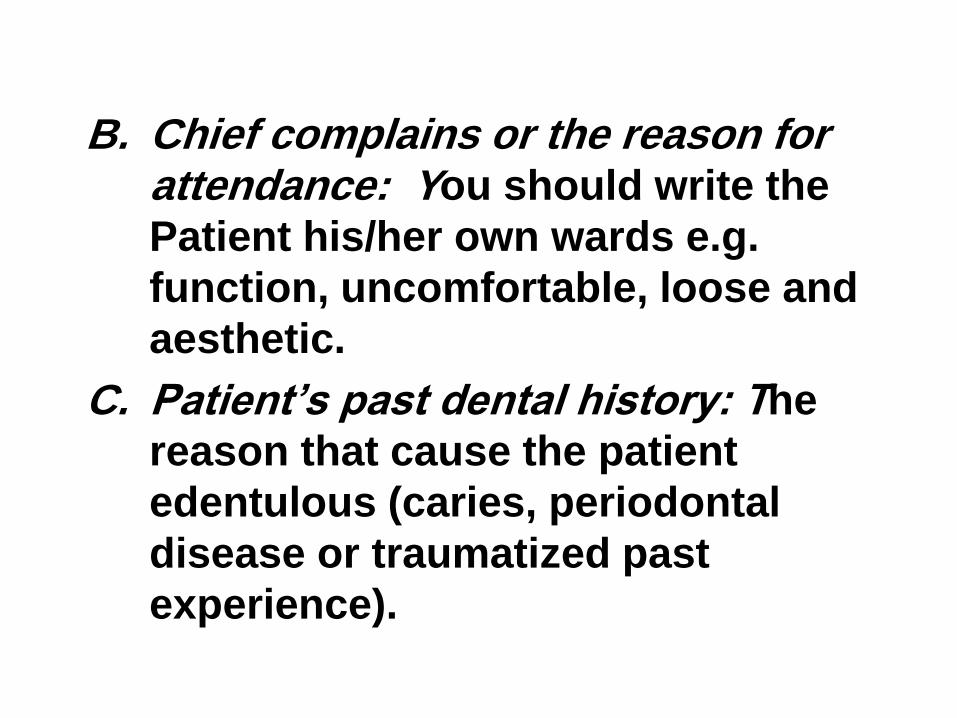
B. Chief complains or the reason for attendance: You should write the
Patient his/her own wards e.g.
function, uncomfortable, loose and
aesthetic.
C. Patient’s past dental history: The
reason that cause the patient
edentulous (caries, periodontal
disease or traumatized past
experience).
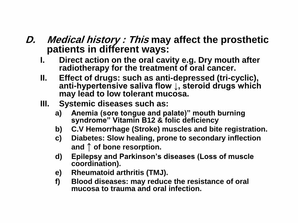
D. Medical history : This may affect the prosthetic patients in different ways:
I. Direct action on the oral cavity e.g. Dry mouth after radiotherapy for the treatment of oral cancer.
II. Effect of drugs: such as anti-depressed (tri-cyclic), anti-hypertensive saliva flow ↓, steroid drugs which may lead to low tolerant mucosa.
III. Systemic diseases such as: a) Anemia (sore tongue and palate)” mouth burning
syndrome” Vitamin B12 & folic deficiency
b) C.V Hemorrhage (Stroke) muscles and bite registration.
c) Diabetes: Slow healing, prone to secondary inflection
and ↑ of bone resorption.
d) Epilepsy and Parkinson’s diseases (Loss of muscle coordination).
e) Rheumatoid arthritis (TMJ).
f) Blood diseases: may reduce the resistance of oral mucosa to trauma and oral infection.
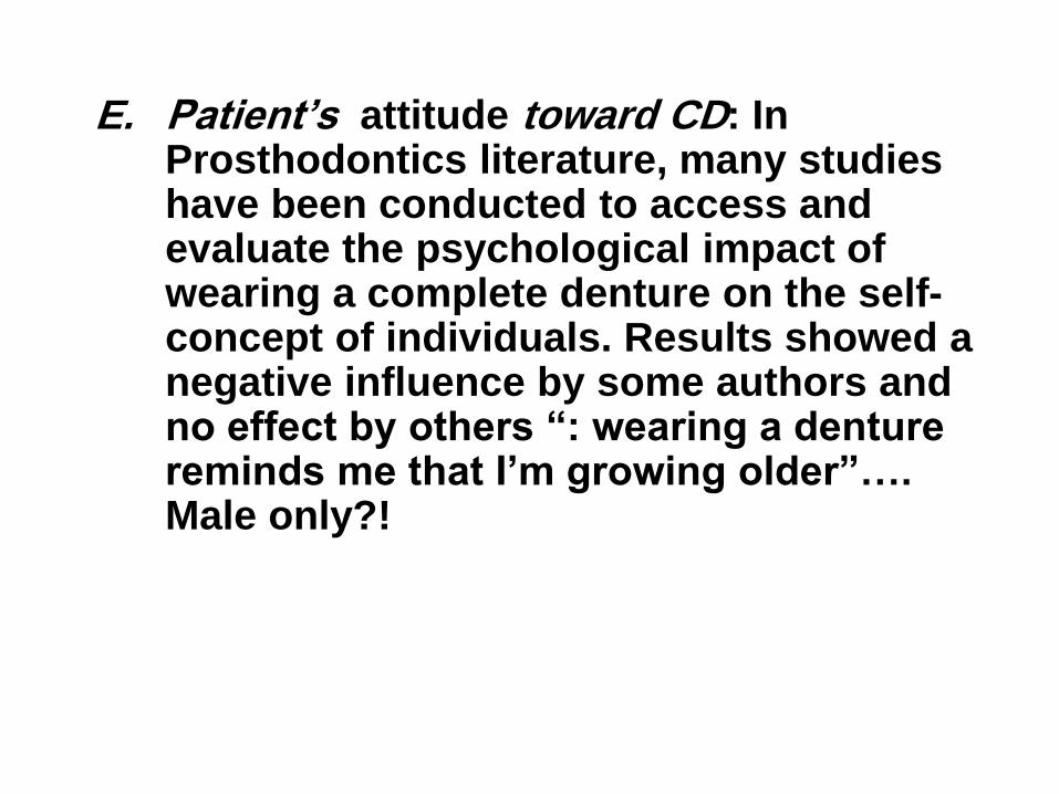
E. Patient’s attitude toward CD: In Prosthodontics literature, many studies have been conducted to access and evaluate the psychological impact of wearing a complete denture on the self-concept of individuals. Results showed a negative influence by some authors and no effect by others “: wearing a denture reminds me that I’m growing older”…. Male only?!

From the clinical point of view, nearly all edentulous patients whom they consult the dental clinic want to have dentures, but not all of them are willing to play a positive part in making such denture satisfactory. Therefore, patient’s attitude towards CDs denture is found in these ways:
I. New denture wearers may have negative (bad) or positive (good) experience gained from parent’s relatives or friends.
II. Patient’s with transitional RPD. If this denture has been worn with comfort, this may make the patient to tolerate and accept the future CD without complaints. Therefore, it should be explained to the patient the different between the two treatment strategies as the later (CD) may need a lot of effort and skills to control them esp. the lower one with respect to retention?!
III. Patients with previous denture experience. It may be very retentive, comfortable for the first 5-year from the time of new denture construction → high expectations? This should be explained to the patient that bone resorption and morphological changes usually increased with time.
F. Past denture experience : (Bad or good)
G. Existence of old dentures: Most CD patients volunteer information about problems with there existence dentures. While others deny. Therefore, questions must be asked to collect as much information as possible regarding: Length of edentulism
Number of dentures they used
The age of existing denture. There fore, a thoroughly clinical examination must be carried out for the retention, stability, fitting and polished surface and the occlusal vertical dimension (OVD). In addition
• Degree of wear “attrition of acrylic teeth “ if Sever OVD remark a new one
• If the patient was comfortable with the old denture from all aspects functionally and aesthetically any change or alteration in the denture design will effect the patient’s “adaptation process” especially for the elderly patients →Copy or Replica dentures
• If the existing dentures were clinically satisfactory, the patient may be Denture collector or
10-15% they never accept dentures due to psychological reasons (Clinical psychiatric).

H. patient’s age and Occupation:- generally speaking the adaptation process of
individual tends to deteriorate with increasing age.
I. patient-dentist relationship and the socio-economic status: -
They should be an empathic interaction between any therapist and patient in all kind of treatment. It is the clinician responsibility to build up such as a positive relationship → Low class people rarely concentrate on aesthetic, while the upper or high class just the opposite “level of education”.
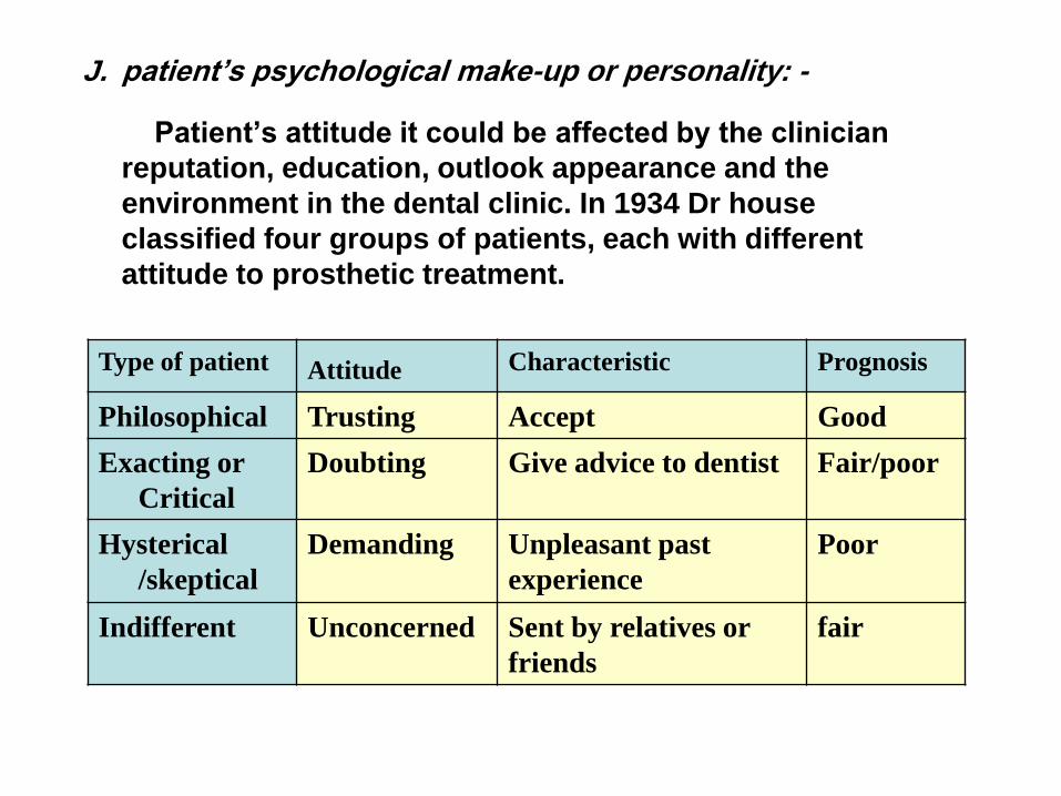
J. patient’s psychological make-up or personality: -
Patient’s attitude it could be affected by the clinician
reputation, education, outlook appearance and the
environment in the dental clinic. In 1934 Dr house
classified four groups of patients, each with different
attitude to prosthetic treatment.
Type of patient Attitude Characteristic Prognosis
Philosophical Trusting Accept Good
Exacting or
Critical
Doubting Give advice to dentist Fair/poor
Hysterical
/skeptical
Demanding Unpleasant past
experience
Poor
Indifferent Unconcerned Sent by relatives or
friends
fair


(2) ORAL EXAMINATION. I. Extra Oral Examination.
II. Intra Oral Examination.

I. Extra Oral Examination. This should be begins as soon as the patient enters
the dental clinic.
The following points should be examined: -
A. Extra Oral Examination of patients:
1. Appearance and patient’s personally i.e. if the
patient well groomed (smart)? Aesthetic? The
psychological traits e.g. introvert extrovert,
aggressive or calm.
2. Clinching or grinding habits.
3. Facial color and any obvious swelling or
asymmetry.
4. Palpation of submandibular and cervical lymph
nodes.
5. TMJ (clicking, pain or crepitus), any deviation
during closure or Limitation on opening.
6. Skeletal base relationship (Class I, II, III)??!

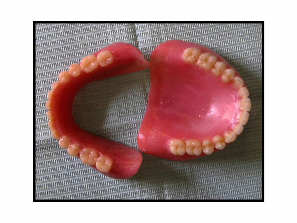
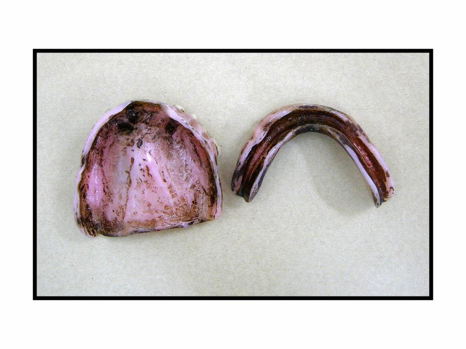
B. Extra Oral Examination of existing Dentures
1. Degree of acrylic teeth wears.
2. Oral hygiene → degree of cleaning of the denture.
3. Type of denture → design and materials used in construction (flangeless denture, clear acrylic, nylon, etc).
4. To select the custom trays, size and color of acrylic teeth….etc.


II. Intra Oral Examination.
This aims to screen and evaluate the
health of the patient’s oral cavity to plan
the construction of dentures on sound
and healthy foundation. This also
subdivided into: A. Visual Examination (Naked eye).
B. Digital Examination.
C. Radiographic Examination.
D. Specific tests.
A. Visual Examination (Naked eye).
1. Color of the oral mucosa: Any variation from normal must be investigated i.e. whitish, reddish patches. It may due to:
Mechanical causes: a. Over-extension of the denture periphery: In new denture
wearer →red line → if continue →ulcer. While in case of old denture wearer due to bone resorption in particular the lower ridge→ overgrowth (denture fissuratum).
b. Ill-fitting dentures: Continuous irritation → food debris → bacterial growth→ calculus deposition→ denture hyperplasia.
c. Continuous wearing of an upper denture: Day and night wearing may cause chronic inflammation called (denture stomatits) in form of granular or papillary hyperplasia
d. Faulty occlusion: Inflammation or ulcer over the crest of the alveolar ridge due to failure in recording the retruded contact Position (RCP). Or on the side of ridge due to cuspal interference.
e. Rubber suction disc
f. Small spicules of the alveolar ridge: Due to traumatic extraction.
g. Miscellaneous: such as systemic diseases which may show an oral manifestation e.g. lichens planus, white lesions and pre-cancerous lesions. Those patients should be referred to oral medicine dept. or their physicians before starting any prosthetic treatment.
2. Size and shape of the alveolar ridge: It should be evaluated carefully if may need surgical intervention or any special technique.
3. Shape of the hard palate: For early prognosis
4. Depth of the sulci: For peripheral seal.
5. Interference factors: Such as the size of the tongue, muscle attachments, freni and the tightness of the lips.
6. Buried roots: usually associated with inflammation and should be confirmed by X-ray.
7. Sinus tract: usually an infected area in bone either from a buried root or sequestra (X-ray).
8. Unilateral swelling: Abnormal swelling is more likely to be pathological. Bilateral bony existosis.
B) Digital examination (Palpation)
Using the index finger pressure over the examined area. If pain felt…. Male always deny?!!
1. Firmness of the ridge: Usually the alveolar ridge is firm and the normal one is composed of bone covered by a thin layer of pink mucosa. In resorbed ridge → think fibrous mucosa→ flabby ridge.
2. Irregularities of the alveolar ridge: Sharpe spikes nodules and knife-edge ridge.
Management:
If painful on pressure →pre-prosthetic surgery after X-ray.
If recent extraction wait for complete bone remolding (at least 3-6 months).
Relief the denture at that area if not severe or contraindicated to surgery.
III. Maxillary tuberositites: Should be confirmed if
it’s hard or soft one.
Severe bony undercut →Surgical removal
Bilateral bony undercut →Remove one side
Fibrous pendulous one →little inter arch space →
Remove surgically.
• (4) Mylohyoid ridge: Relief the denture at that area if surgery is
contraindicated.
Sharpe prominent → Remove surgically
• (5) Tone of facial muscles “Age”.
• (6) Tendency to retch.
• (7) Presence of Tori:
Mandibular Tori: Between 1st and 2nd premolar
Maxillary Tori: Hard palate, single or multi-nodular.
(C) Radiographic Examination
OPG or peri-apical radiographs
Ideally a panoramic radiograph should be taken
routinely for every edentulous patient before
starting any treatment plan to look for any: Buried roots or impacted teeth.
Localized infected area, swelling.
Irregular sharp ridge.
(D) Specific test: Blood test, biopsy, saliva flow ……… etc.