Embed Size (px)
Citation preview
478 Mol. BioSyst., 2013, 9, 478--491 This journal is c The Royal Society of Chemistry 2013
Cite this: Mol. BioSyst.,2013,9, 478
Cloning and sequencing of the kedarcidin biosyntheticgene cluster from Streptoalloteichus sp. ATCC 53650revealing new insights into biosynthesis of theenediyne family of antitumor antibiotics†
Jeremy R. Lohman,a Sheng-Xiong Huang,a Geoffrey P. Horsman,b Paul E. Dilfer,a
Tingting Huang,a Yihua Chen,b Evelyn Wendt-Pienkowskib and Ben Shenz*abcd
Enediyne natural product biosynthesis is characterized by a convergence of multiple pathways,
generating unique peripheral moieties that are appended onto the distinctive enediyne core.
Kedarcidin (KED) possesses two unique peripheral moieties, a (R)-2-aza-3-chloro-b-tyrosine and an iso-
propoxy-bearing 2-naphthonate moiety, as well as two deoxysugars. The appendage pattern of these
peripheral moieties to the enediyne core in KED differs from the other enediynes studied to date with
respect to stereochemical configuration. To investigate the biosynthesis of these moieties and expand
our understanding of enediyne core formation, the biosynthetic gene cluster for KED was cloned from
Streptoalloteichus sp. ATCC 53650 and sequenced. Bioinformatics analysis of the ked cluster revealed
the presence of the conserved genes encoding for enediyne core biosynthesis, type I and type II
polyketide synthase loci likely responsible for 2-aza-L-tyrosine and 3,6,8-trihydroxy-2-naphthonate
formation, and enzymes known for deoxysugar biosynthesis. Genes homologous to those responsible
for the biosynthesis, activation, and coupling of the L-tyrosine-derived moieties from C-1027 and
maduropeptin and of the naphthonate moiety from neocarzinostatin are present in the ked cluster,
supporting 2-aza-L-tyrosine and 3,6,8-trihydroxy-2-naphthoic acid as precursors, respectively, for the
(R)-2-aza-3-chloro-b-tyrosine and the 2-naphthonate moieties in KED biosynthesis.
Introduction
Kedarcidin (KED) was isolated from Streptoalloteichus sp. ATCC53650 (originally strain L585-6) as a chromoprotein antitumorantibiotic in 1992.1–5 The KED apoprotein primary sequenceof 114 amino acids was determined by Edman degradation2
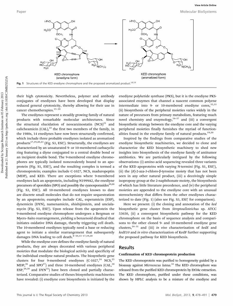
(Fig. S1, ESI†), and the solution structure solved by NMRspectroscopy.3 The structure of the KED chromophore was firstestablished on the basis of an extensive spectroscopic analysisin 1992.4,5 It has since been revised twice according to totalsyntheses6,7 with the final revised structure shown in Fig. 1(also see Fig. S2, ESI†). KED belongs to the enediyne family ofantitumor antibiotics, which are of great interest as potentanticancer agents. They possess a reactive enediyne core that isable to abstract hydrogens from the deoxyribose backbone ofDNA. Molecular oxygen can then react with the newly formedcarbon-centered radicals, leading to site-specific single-stranded or double-stranded breaks, as well as interstrandcrosslinks, and ultimately to cell death.8–14 The potent anti-cancer activity of enediynes is offset in clinical applications by
a Department of Chemistry, The Scripps Research Institute, Jupiter,
Florida 33458, USAb Division of Pharmaceutical Sciences, School of Pharmacy,
University of Wisconsin-Madison, Madison, Wisconsin 53705, USAc Department of Molecular Therapeutics, The Scripps Research Institute, Jupiter,
Florida 33458, USAd Natural Products Library Initiative at The Scripps Research Institute,
The Scripps Research Institute, Jupiter, Florida 33458, USA
† Electronic supplementary information (ESI) available: The amino acid sequenceof KedA in comparison with other known apoproteins (Fig. S1, ESI†), the originaland revised structures of the KED chromophore (Fig. S2, ESI†), enediyne naturalproducts whose structures have been determined (Fig. S3, ESI†), HPLC and MSanalysis of the KED chromophore (Fig. S4, ESI†), SDS-PAGE analysis of thepurified KedF (Fig. S5, ESI†), comparative analysis of the KED, C-1027, andMDP gene cluster supporting the proposed pathway for (R)-2-aza-3-chloro-b-tyrosine in KED biosynthesis (Fig. S6, ESI†), and comparative analysis of theKED, NCS, and MDP gene cluster supporting the proposed pathway for 3-hydroxy-7,8-dimethoxy-6-isopropoxy-2-naphthoic acid in KED biosynthesis. See DOI:10.1039/c3mb25523a‡ The Scripps Research Institute, 130 Scripps Way, #3A1, Jupiter, Florida 33458,USA. E-mail: [email protected]; Fax: +1 561 228-2472; Tel: +1 561 228-2456.
Received 16th November 2012,Accepted 20th January 2013
DOI: 10.1039/c3mb25523a
www.rsc.org/molecularbiosystems
MolecularBioSystems
PAPER
Dow
nloa
ded
by S
crip
ps R
esea
rch
Inst
itute
on
05 F
ebru
ary
2013
Publ
ishe
d on
21
Janu
ary
2013
on
http
://pu
bs.r
sc.o
rg |
doi:1
0.10
39/C
3MB
2552
3A
View Article OnlineView Journal | View Issue
This journal is c The Royal Society of Chemistry 2013 Mol. BioSyst., 2013, 9, 478--491 479
their high cytotoxicity. Nevertheless, polymer and antibodyconjugates of enediynes have been developed that displayreduced general cytotoxicity, thereby allowing for their use incancer chemotherapies.15–20
The enediynes represent a steadily growing family of naturalproducts with remarkable molecular architectures. Sincethe structural elucidation of neocarzinostatin (NCS)21 andcalicheamicin (CAL),22 the first two members of the family, inthe 1980s, 14 enediynes have now been structurally confirmed,which include three probable enediynes isolated as aromatizedproducts17,19,20,23 (Fig. S3, ESI†). Structurally, the enediynes arecharacterized by an unsaturated 9- or 10-membered carbacyclicring featuring a diyne conjugated to a central double bond oran incipient double bond. The 9-membered enediyne chromo-phores are typically isolated noncovalently bound to an apo-protein (Fig. S1, ESI†), and the resulting complex is termed achromoprotein; examples include C-1027, NCS, maduropeptin(MDP), and KED. There are exceptions where 9-memberedenediynes lack an apoprotein, including N1999A2, the enediyneprecursors of sporolides (SPO) and possibly the cyanosporasides19,20
(Fig. S3, ESI†). All 10-membered enediynes known to dateare discrete small molecules that do not require sequestrationby an apoprotein; examples include CAL, esperamicin (ESP),dynemicin (DYN), namenamicin, shishijimicin, and unciala-mycin (Fig. S3, ESI†). Upon release from the apoprotein the9-membered enediyne chromophore undergoes a Bergman orMyers–Saito rearrangement, yielding a benzenoid diradical thatinitiates oxidative DNA damage, thereby triggering cell death.The 10-membered enediynes typically need a base or reducingagent to initiate a similar rearrangement that subsequentlydamages DNA leading to cell death.8–10,15–17,19,20
While the enediyne core defines the enediyne family of naturalproducts, they are always decorated with various peripheralmoieties that modulate the biological activity and specificity ofthe individual enediyne natural products. The biosynthetic geneclusters for four 9-membered enediynes (C-1027,24 NCS,25
MDP,26 and SPO27) and three 10-membered enediynes (CAL,28
ESP,29,30 and DYN31) have been cloned and partially charac-terized. Comparative studies of theses biosynthetic machinerieshave revealed: (i) enediyne core biosynthesis is initiated by the
enediyne polyketide synthase (PKS), but it is the enediyne PKS-associated enzymes that channel a nascent common polyeneintermediate into 9- or 10-membered enediyne cores,32,33
(ii) biosynthesis of the peripheral moieties varies widely in thenature of precursors from primary metabolism, featuring muchnovel chemistry and enzymology,34–57 and (iii) a convergentbiosynthetic strategy between the enediyne core and the varyingperipheral moieties finally furnishes the myriad of function-alities found in the enediyne family of natural products.19,20
Inspired by the findings from comparative studies of theenediyne biosynthetic machineries, we decided to clone andcharacterize the KED biosynthetic machinery to shed newinsights into biosynthesis of the enediyne family of antitumorantibiotics. We are particularly intrigued by the followingobservations: (i) amino acid sequencing revealed three variantsof the KED apoproteins with varying N-termini (Fig. S1, ESI†),(ii) the (R)-2-aza-3-chloro-b-tyrosine moiety that has not beenseen in any other natural product, (iii) a deceivingly simpleisopropoxy group at the 2-naphthonate moiety, the biosynthesisof which has little literature precedence, and (iv) the peripheralmoieties are appended to the enediyne core with an unusualstereochemistry that differs from the other enediynes charac-terized to date (Fig. 1) (also see Fig. S3, ESI† for comparison).
Here we present: (i) the cloning and annotation of the kedbiosynthetic gene cluster from Streptoalloteichus sp. ATCC53650, (ii) a convergent biosynthetic pathway for the KEDchromophore on the basis of sequence analysis and compari-sons to the other cloned 9- and 10-membered enediyne geneclusters,24–31 and (iii) in vivo characterization of kedE andkedE10 and in vitro characterization of KedF further supportingthe proposed pathway for KED biosynthesis.
ResultsConfirmation of KED chromoprotein production
The KED chromoprotein was purified to homogeneity guided by abioassay against Micrococcus luteus.34 The KED chromophore wasreleased from the purified KED chromoprotein by EtOAc extraction.The KED chromophore, purified under these conditions, wasshown by HPLC analysis to be a mixture of the enediyne and
Fig. 1 Structures of the KED enediyne chromophore and the proposed aromatized product.58,59
Paper Molecular BioSystems
Dow
nloa
ded
by S
crip
ps R
esea
rch
Inst
itute
on
05 F
ebru
ary
2013
Publ
ishe
d on
21
Janu
ary
2013
on
http
://pu
bs.r
sc.o
rg |
doi:1
0.10
39/C
3MB
2552
3AView Article Online
480 Mol. BioSyst., 2013, 9, 478--491 This journal is c The Royal Society of Chemistry 2013
aromatized forms, and the enediyne form was completely convertedinto the aromatized form at room temperature overnight; theidentities of both enediyne and aromatized forms of the KEDchromophore were confirmed by high resolution mass spectro-metry (Fig. S4, ESI†). For the enediyne form of the KED chromo-phore, high resolution electrospray ionization mass spectrometry(HRESIMS) yielded an [M + H]+ ion at m/z 1030.37338 for theenediyne form of the KED chromophore, consistent with its pre-dicted molecular formula of C53H60N3O16Cl (calculated [M + H]+ ionat m/z 1030.37349).2,5 For the aromatized form of the KED chromo-phore, HRESIMS revealed an [M + H]+ ion at m/z 1032.39109,consistent with the molecular formula of C53H62N3O16Cl (calculated[M + H]+ ion at m/z 1032.38914), differing from the enediyne formby the presence of two additional protons as would be predicted forthe aromatized KED chromophore (Fig. 1 and Fig. S4, ESI†).58,59
These results re-confirm that Streptoalloteichus sp. ATCC 53650 inour possession harbors functional KED biosynthetic machinery.1–5
Under the conditions described, the isolated yield of the KEDchromoprotein complex is estimated to be 50 mg L�1.2,5
Cloning, sequencing, and annotation of the ked gene cluster
The enediyne PKS gene is the hallmark of enediyne biosyn-thetic clusters,32,33 and as such, degenerate primers previously
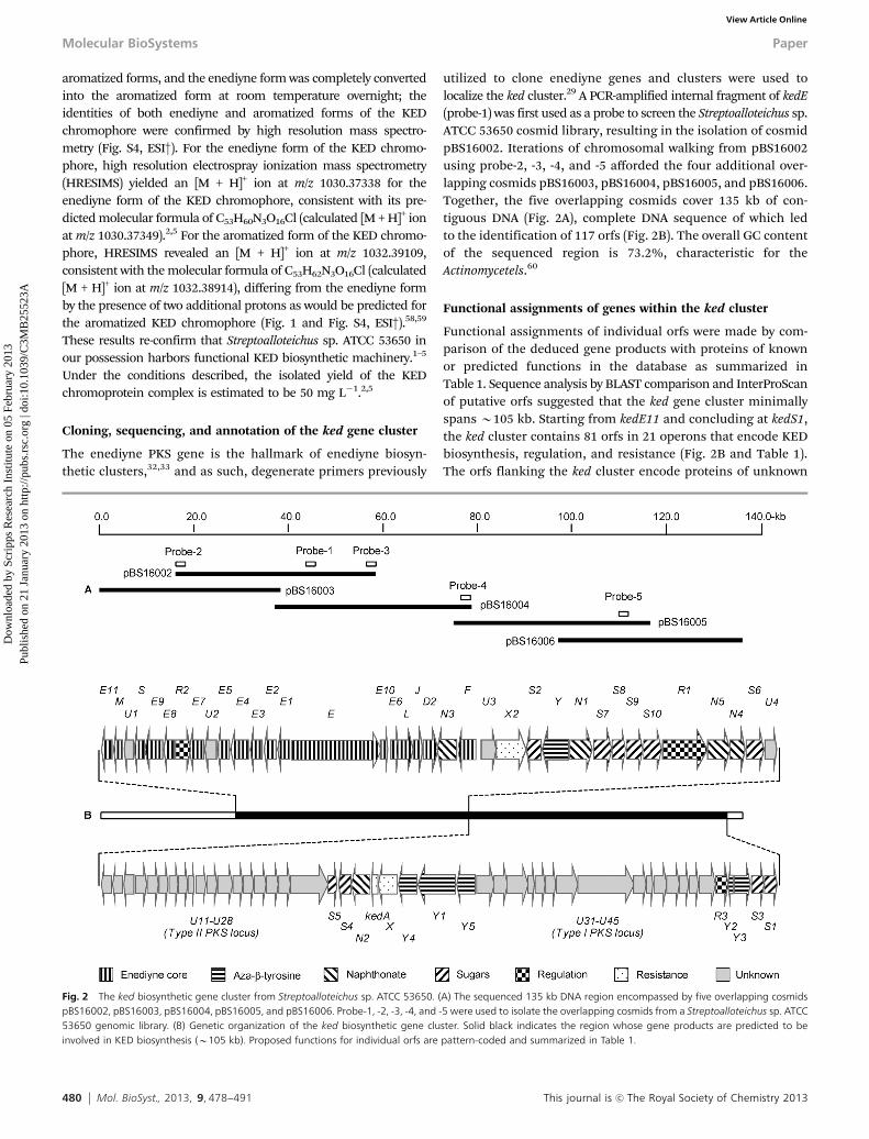
utilized to clone enediyne genes and clusters were used tolocalize the ked cluster.29 A PCR-amplified internal fragment of kedE(probe-1) was first used as a probe to screen the Streptoalloteichus sp.ATCC 53650 cosmid library, resulting in the isolation of cosmidpBS16002. Iterations of chromosomal walking from pBS16002using probe-2, -3, -4, and -5 afforded the four additional over-lapping cosmids pBS16003, pBS16004, pBS16005, and pBS16006.Together, the five overlapping cosmids cover 135 kb of con-tiguous DNA (Fig. 2A), complete DNA sequence of which ledto the identification of 117 orfs (Fig. 2B). The overall GC contentof the sequenced region is 73.2%, characteristic for theActinomycetels.60
Functional assignments of genes within the ked cluster
Functional assignments of individual orfs were made by com-parison of the deduced gene products with proteins of knownor predicted functions in the database as summarized inTable 1. Sequence analysis by BLAST comparison and InterProScanof putative orfs suggested that the ked gene cluster minimallyspans B105 kb. Starting from kedE11 and concluding at kedS1,the ked cluster contains 81 orfs in 21 operons that encode KEDbiosynthesis, regulation, and resistance (Fig. 2B and Table 1).The orfs flanking the ked cluster encode proteins of unknown
Fig. 2 The ked biosynthetic gene cluster from Streptoalloteichus sp. ATCC 53650. (A) The sequenced 135 kb DNA region encompassed by five overlapping cosmidspBS16002, pBS16003, pBS16004, pBS16005, and pBS16006. Probe-1, -2, -3, -4, and -5 were used to isolate the overlapping cosmids from a Streptoalloteichus sp. ATCC53650 genomic library. (B) Genetic organization of the ked biosynthetic gene cluster. Solid black indicates the region whose gene products are predicted to beinvolved in KED biosynthesis (B105 kb). Proposed functions for individual orfs are pattern-coded and summarized in Table 1.
Molecular BioSystems Paper
Dow
nloa
ded
by S
crip
ps R
esea
rch
Inst
itute
on
05 F
ebru
ary
2013
Publ
ishe
d on
21
Janu
ary
2013
on
http
://pu
bs.r
sc.o
rg |
doi:1
0.10
39/C
3MB
2552
3AView Article Online
This journal is c The Royal Society of Chemistry 2013 Mol. BioSyst., 2013, 9, 478--491 481
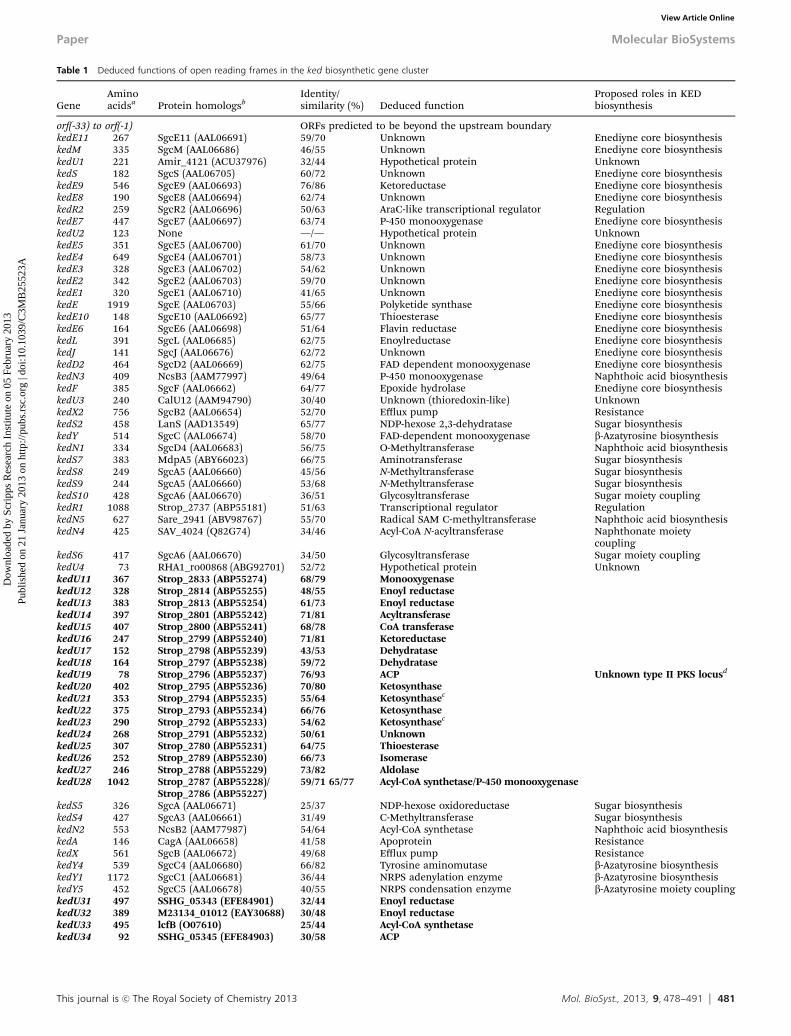
Table 1 Deduced functions of open reading frames in the ked biosynthetic gene cluster
GeneAminoacidsa Protein homologsb
Identity/similarity (%) Deduced function
Proposed roles in KEDbiosynthesis
orf(-33) to orf(-1) ORFs predicted to be beyond the upstream boundarykedE11 267 SgcE11 (AAL06691) 59/70 Unknown Enediyne core biosynthesiskedM 335 SgcM (AAL06686) 46/55 Unknown Enediyne core biosynthesiskedU1 221 Amir_4121 (ACU37976) 32/44 Hypothetical protein UnknownkedS 182 SgcS (AAL06705) 60/72 Unknown Enediyne core biosynthesiskedE9 546 SgcE9 (AAL06693) 76/86 Ketoreductase Enediyne core biosynthesiskedE8 190 SgcE8 (AAL06694) 62/74 Unknown Enediyne core biosynthesiskedR2 259 SgcR2 (AAL06696) 50/63 AraC-like transcriptional regulator RegulationkedE7 447 SgcE7 (AAL06697) 63/74 P-450 monooxygenase Enediyne core biosynthesiskedU2 123 None —/— Hypothetical protein UnknownkedE5 351 SgcE5 (AAL06700) 61/70 Unknown Enediyne core biosynthesiskedE4 649 SgcE4 (AAL06701) 58/73 Unknown Enediyne core biosynthesiskedE3 328 SgcE3 (AAL06702) 54/62 Unknown Enediyne core biosynthesiskedE2 342 SgcE2 (AAL06703) 59/70 Unknown Enediyne core biosynthesiskedE1 320 SgcE1 (AAL06710) 41/65 Unknown Enediyne core biosynthesiskedE 1919 SgcE (AAL06703) 55/66 Polyketide synthase Enediyne core biosynthesiskedE10 148 SgcE10 (AAL06692) 65/77 Thioesterase Enediyne core biosynthesiskedE6 164 SgcE6 (AAL06698) 51/64 Flavin reductase Enediyne core biosynthesiskedL 391 SgcL (AAL06685) 62/75 Enoylreductase Enediyne core biosynthesiskedJ 141 SgcJ (AAL06676) 62/72 Unknown Enediyne core biosynthesiskedD2 464 SgcD2 (AAL06669) 62/75 FAD dependent monooxygenase Enediyne core biosynthesiskedN3 409 NcsB3 (AAM77997) 49/64 P-450 monooxygenase Naphthoic acid biosynthesiskedF 385 SgcF (AAL06662) 64/77 Epoxide hydrolase Enediyne core biosynthesiskedU3 240 CalU12 (AAM94790) 30/40 Unknown (thioredoxin-like) UnknownkedX2 756 SgcB2 (AAL06654) 52/70 Efflux pump ResistancekedS2 458 LanS (AAD13549) 65/77 NDP-hexose 2,3-dehydratase Sugar biosynthesiskedY 514 SgcC (AAL06674) 58/70 FAD-dependent monooxygenase b-Azatyrosine biosynthesiskedN1 334 SgcD4 (AAL06683) 56/75 O-Methyltransferase Naphthoic acid biosynthesiskedS7 383 MdpA5 (ABY66023) 66/75 Aminotransferase Sugar biosynthesiskedS8 249 SgcA5 (AAL06660) 45/56 N-Methyltransferase Sugar biosynthesiskedS9 244 SgcA5 (AAL06660) 53/68 N-Methyltransferase Sugar biosynthesiskedS10 428 SgcA6 (AAL06670) 36/51 Glycosyltransferase Sugar moiety couplingkedR1 1088 Strop_2737 (ABP55181) 51/63 Transcriptional regulator RegulationkedN5 627 Sare_2941 (ABV98767) 55/70 Radical SAM C-methyltransferase Naphthoic acid biosynthesiskedN4 425 SAV_4024 (Q82G74) 34/46 Acyl-CoA N-acyltransferase Naphthonate moiety
couplingkedS6 417 SgcA6 (AAL06670) 34/50 Glycosyltransferase Sugar moiety couplingkedU4 73 RHA1_ro00868 (ABG92701) 52/72 Hypothetical protein UnknownkedU11 367 Strop_2833 (ABP55274) 68/79 MonooxygenasekedU12 328 Strop_2814 (ABP55255) 48/55 Enoyl reductasekedU13 383 Strop_2813 (ABP55254) 61/73 Enoyl reductasekedU14 397 Strop_2801 (ABP55242) 71/81 AcyltransferasekedU15 407 Strop_2800 (ABP55241) 68/78 CoA transferasekedU16 247 Strop_2799 (ABP55240) 71/81 KetoreductasekedU17 152 Strop_2798 (ABP55239) 43/53 DehydratasekedU18 164 Strop_2797 (ABP55238) 59/72 DehydratasekedU19 78 Strop_2796 (ABP55237) 76/93 ACP Unknown type II PKS locusd
kedU20 402 Strop_2795 (ABP55236) 70/80 KetosynthasekedU21 353 Strop_2794 (ABP55235) 55/64 Ketosynthasec
kedU22 375 Strop_2793 (ABP55234) 66/76 KetosynthasekedU23 290 Strop_2792 (ABP55233) 54/62 Ketosynthasec
kedU24 268 Strop_2791 (ABP55232) 50/61 UnknownkedU25 307 Strop_2780 (ABP55231) 64/75 ThioesterasekedU26 252 Strop_2789 (ABP55230) 66/73 IsomerasekedU27 246 Strop_2788 (ABP55229) 73/82 AldolasekedU28 1042 Strop_2787 (ABP55228)/
Strop_2786 (ABP55227)59/71 65/77 Acyl-CoA synthetase/P-450 monooxygenase
kedS5 326 SgcA (AAL06671) 25/37 NDP-hexose oxidoreductase Sugar biosynthesiskedS4 427 SgcA3 (AAL06661) 31/49 C-Methyltransferase Sugar biosynthesiskedN2 553 NcsB2 (AAM77987) 54/64 Acyl-CoA synthetase Naphthoic acid biosynthesiskedA 146 CagA (AAL06658) 41/58 Apoprotein ResistancekedX 561 SgcB (AAL06672) 49/68 Efflux pump ResistancekedY4 539 SgcC4 (AAL06680) 66/82 Tyrosine aminomutase b-Azatyrosine biosynthesiskedY1 1172 SgcC1 (AAL06681) 36/44 NRPS adenylation enzyme b-Azatyrosine biosynthesiskedY5 452 SgcC5 (AAL06678) 40/55 NRPS condensation enzyme b-Azatyrosine moiety couplingkedU31 497 SSHG_05343 (EFE84901) 32/44 Enoyl reductasekedU32 389 M23134_01012 (EAY30688) 30/48 Enoyl reductasekedU33 495 lcfB (O07610) 25/44 Acyl-CoA synthetasekedU34 92 SSHG_05345 (EFE84903) 30/58 ACP
Paper Molecular BioSystems
Dow
nloa
ded
by S
crip
ps R
esea
rch
Inst
itute
on
05 F
ebru
ary
2013
Publ
ishe
d on
21
Janu
ary
2013
on
http
://pu
bs.r
sc.o
rg |
doi:1
0.10
39/C
3MB
2552
3AView Article Online
482 Mol. BioSyst., 2013, 9, 478--491 This journal is c The Royal Society of Chemistry 2013
functions or with similarities to enzymes involved in aromaticamino acid metabolism.
Among the orfs identified within the ked cluster include:(i) one (kedE) encodes the enedyne PKS and 17 (kedE1 to kedE11,kedM, kedS, kedL, kedJ, kedD2, kedF) encodes accessory enzymesfor enediyne core biosynthesis, (ii) six (kedY and kedY1 to kedY5)encode enzymes for tailoring the (R)-2-aza-3-chloro-b-tyrosinemoiety and its coupling to the enediyne core, (iii) five (kedN1to kedN5) encode enzymes for modifying the 2-naphthonatemoiety and its coupling, via (R)-2-aza-3-chloro-b-tyrosine, to theenediyne core, (iv) ten (kedS1 to kedS10) encode enzymes forbiosynthesis of the two sugar moieties and their couplings tothe enediyne core, (v) three (kedR1, kedR2, kedR3) encodeproteins for pathway regulation, (vi) three (kedA, kedX, andkedX2) encode elements for resistance, and (vii) four (kedU1to kedU4) encode proteins of unknown function. In addition,there are two loci, termed type II PKS locus consisting of18 genes (kedU11 to kedU28) and type I PKS locus consistingof 15 gene (kedU31 to kedU45), inserted within the ked cluster,that are unprecedented among all enediyne clusters known todate. They serve as candidates encoding biosynthesis of thenascent precursors for the (R)-2-aza-3-chloro-b-tyrosine and2-naphthonate moieties (Fig. 3).
The KED apoprotein KedA
Bioinformatics analysis revealed a single gene, kedA, within theked cluster, for which the deduced gene product matched theisolated KedA apoprotein.2,3 The kedA gene is translated asa 145-amino acid protein, and SignalP analysis predicted aleader peptide that is cleaved between A31 and A32, resultingin a 114-amino acid protein with a predicted N-terminus of
Ala-Ser-Ala-Ala-Val (Fig. S1, ESI†). This is consistent with theamino acid sequence of the isolated mature KedA apoprotein.2,3
The two additional known variants of KedA, with a Ser-Ala-Ala-Valand an Ala-Ala-Val terminus, respectively, could be accounted forby either the promiscuity of the leader peptide cleavage site orpartial proteolysis of the N-terminus of the mature KedA duringisolation. The kedA gene has also been independently clonedrecently from Streptoalloteichus sp. ATCC 53650 and sequenced,overexpression of which in Streptoalloteichus sp. ATCC 53650resulted in a 2-fold enhancement of KED titer.61
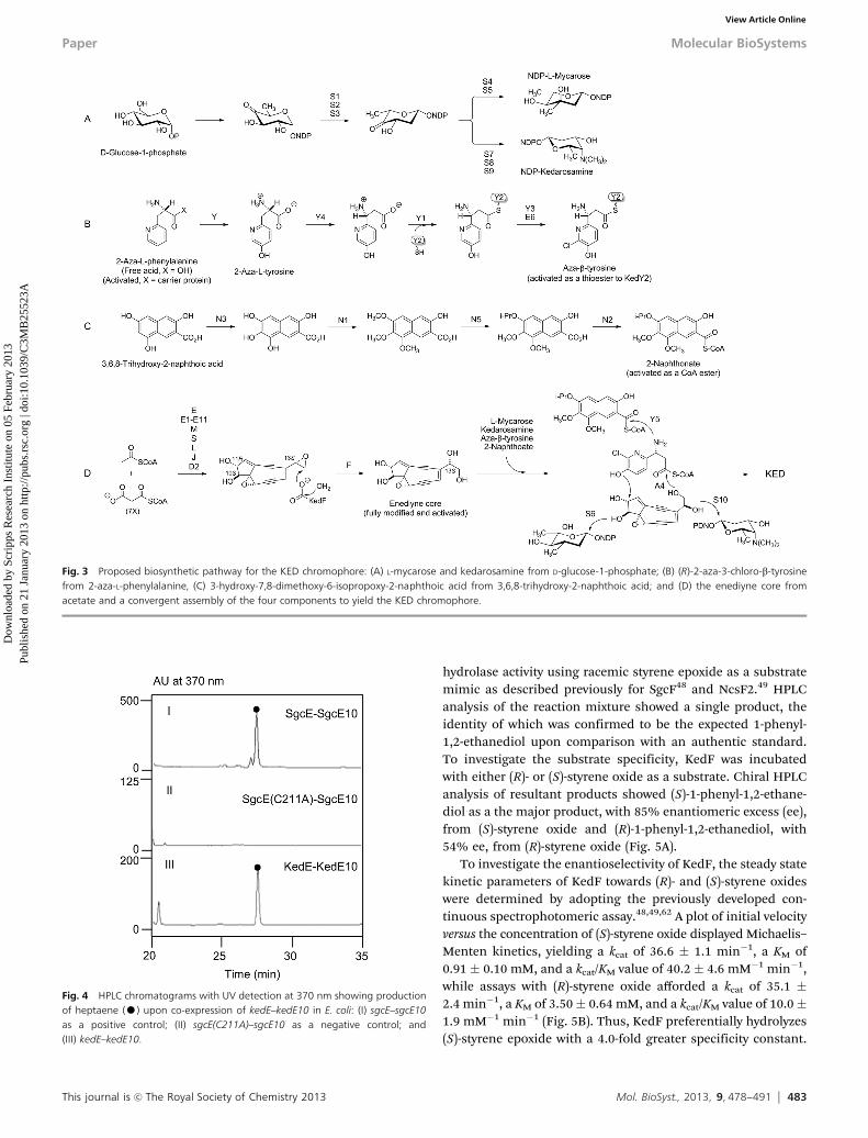
In vivo characterization of KedE–KedE10 as enediynePKS–thioesterase for heptaene production
We have previously shown the production of heptaene as ahallmark for enediyne biosynthesis, which has been detected fromall enediyne producers examined to date and can be produced uponco-expression of the enediyne pksE and associated thioesterase (TE)in either E. coli or Streptomyces lividans.32,33 Co-expression ofkedE–kedE10 in E. coli, with co-expressions of both sgcE–sgcE10as a positive control33 and sgcE(C211A)–sgcE10 as a negativecontrol,33 indeed resulted in the production of heptaene, theidentity of which was confirmed by HPLC analysis in comparisonwith an authentic standard (Fig. 4).
In vitro characterization of KedF as an epoxide hydrolase
The kedF gene was predicted to encode an epoxide hydrolase,and epoxide hydrolases, such as SgcF48 and NcsF2,49 have beenshown to play a critical role in enediyne biosynthesis, setting upthe stereochemistry of the enediyne core for appending theperipheral moieties. KedF was overproduced in E. coli, purifiedto homogeneity (Fig. S5, ESI†), and directly assayed for epoxide
Table 1 (continued )
GeneAminoacidsa Protein homologsb
Identity/similarity (%) Deduced function
Proposed roles in KEDbiosynthesis
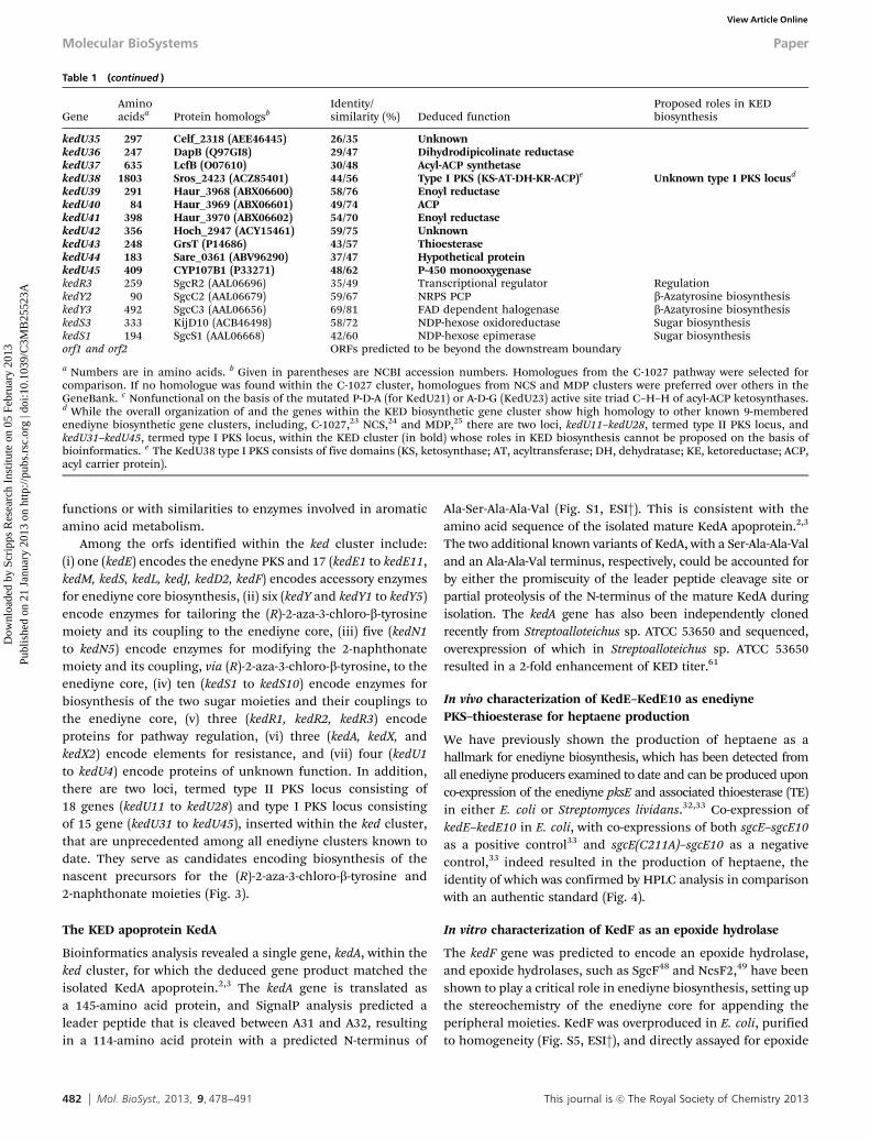
kedU35 297 Celf_2318 (AEE46445) 26/35 UnknownkedU36 247 DapB (Q97GI8) 29/47 Dihydrodipicolinate reductasekedU37 635 LcfB (O07610) 30/48 Acyl-ACP synthetasekedU38 1803 Sros_2423 (ACZ85401) 44/56 Type I PKS (KS-AT-DH-KR-ACP)e Unknown type I PKS locusd
kedU39 291 Haur_3968 (ABX06600) 58/76 Enoyl reductasekedU40 84 Haur_3969 (ABX06601) 49/74 ACPkedU41 398 Haur_3970 (ABX06602) 54/70 Enoyl reductasekedU42 356 Hoch_2947 (ACY15461) 59/75 UnknownkedU43 248 GrsT (P14686) 43/57 ThioesterasekedU44 183 Sare_0361 (ABV96290) 37/47 Hypothetical proteinkedU45 409 CYP107B1 (P33271) 48/62 P-450 monooxygenasekedR3 259 SgcR2 (AAL06696) 35/49 Transcriptional regulator RegulationkedY2 90 SgcC2 (AAL06679) 59/67 NRPS PCP b-Azatyrosine biosynthesiskedY3 492 SgcC3 (AAL06656) 69/81 FAD dependent halogenase b-Azatyrosine biosynthesiskedS3 333 KijD10 (ACB46498) 58/72 NDP-hexose oxidoreductase Sugar biosynthesiskedS1 194 SgcS1 (AAL06668) 42/60 NDP-hexose epimerase Sugar biosynthesisorf1 and orf2 ORFs predicted to be beyond the downstream boundary
a Numbers are in amino acids. b Given in parentheses are NCBI accession numbers. Homologues from the C-1027 pathway were selected forcomparison. If no homologue was found within the C-1027 cluster, homologues from NCS and MDP clusters were preferred over others in theGeneBank. c Nonfunctional on the basis of the mutated P-D-A (for KedU21) or A-D-G (KedU23) active site triad C–H–H of acyl-ACP ketosynthases.d While the overall organization of and the genes within the KED biosynthetic gene cluster show high homology to other known 9-memberedenediyne biosynthetic gene clusters, including, C-1027,23 NCS,24 and MDP,25 there are two loci, kedU11–kedU28, termed type II PKS locus, andkedU31–kedU45, termed type I PKS locus, within the KED cluster (in bold) whose roles in KED biosynthesis cannot be proposed on the basis ofbioinformatics. e The KedU38 type I PKS consists of five domains (KS, ketosynthase; AT, acyltransferase; DH, dehydratase; KE, ketoreductase; ACP,acyl carrier protein).
Molecular BioSystems Paper
Dow
nloa
ded
by S
crip
ps R
esea
rch
Inst
itute
on
05 F
ebru
ary
2013
Publ
ishe
d on
21
Janu
ary
2013
on
http
://pu
bs.r
sc.o
rg |
doi:1
0.10
39/C
3MB
2552
3AView Article Online
This journal is c The Royal Society of Chemistry 2013 Mol. BioSyst., 2013, 9, 478--491 483
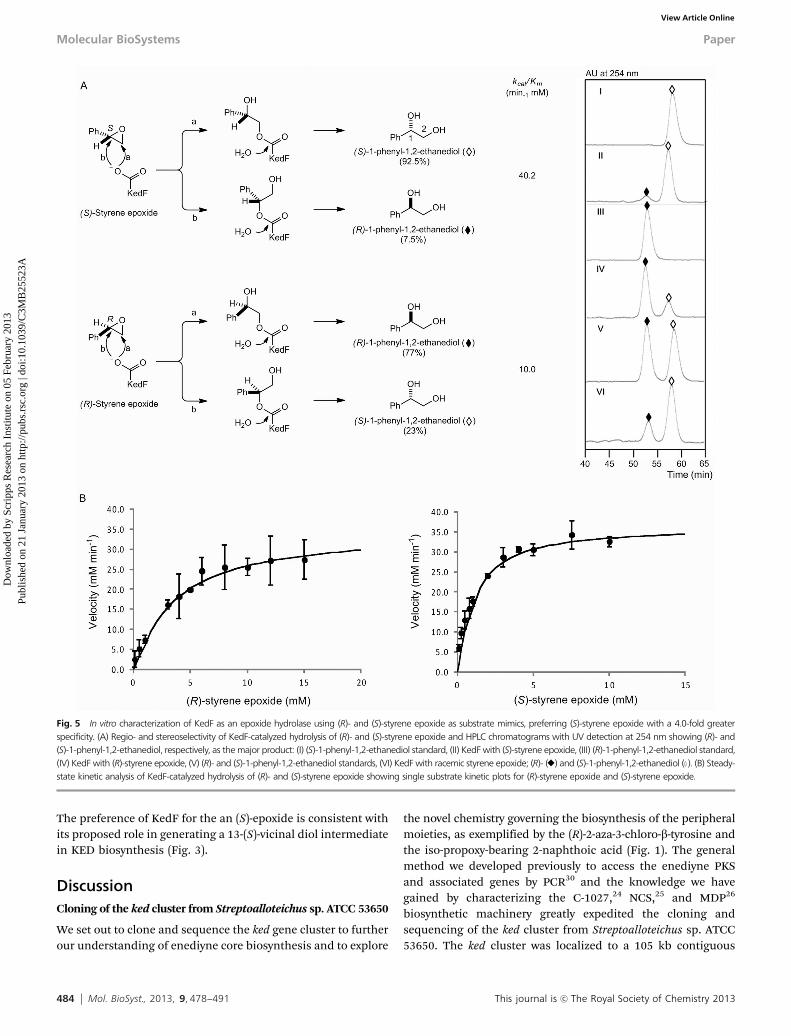
hydrolase activity using racemic styrene epoxide as a substratemimic as described previously for SgcF48 and NcsF2.49 HPLCanalysis of the reaction mixture showed a single product, theidentity of which was confirmed to be the expected 1-phenyl-1,2-ethanediol upon comparison with an authentic standard.To investigate the substrate specificity, KedF was incubatedwith either (R)- or (S)-styrene oxide as a substrate. Chiral HPLCanalysis of resultant products showed (S)-1-phenyl-1,2-ethane-diol as a the major product, with 85% enantiomeric excess (ee),from (S)-styrene oxide and (R)-1-phenyl-1,2-ethanediol, with54% ee, from (R)-styrene oxide (Fig. 5A).
To investigate the enantioselectivity of KedF, the steady statekinetic parameters of KedF towards (R)- and (S)-styrene oxideswere determined by adopting the previously developed con-tinuous spectrophotomeric assay.48,49,62 A plot of initial velocityversus the concentration of (S)-styrene oxide displayed Michaelis–Menten kinetics, yielding a kcat of 36.6 � 1.1 min�1, a KM of0.91 � 0.10 mM, and a kcat/KM value of 40.2 � 4.6 mM�1 min�1,while assays with (R)-styrene oxide afforded a kcat of 35.1 �2.4 min�1, a KM of 3.50� 0.64 mM, and a kcat/KM value of 10.0�1.9 mM�1 min�1 (Fig. 5B). Thus, KedF preferentially hydrolyzes(S)-styrene epoxide with a 4.0-fold greater specificity constant.
Fig. 3 Proposed biosynthetic pathway for the KED chromophore: (A) L-mycarose and kedarosamine from D-glucose-1-phosphate; (B) (R)-2-aza-3-chloro-b-tyrosinefrom 2-aza-L-phenylalanine, (C) 3-hydroxy-7,8-dimethoxy-6-isopropoxy-2-naphthoic acid from 3,6,8-trihydroxy-2-naphthoic acid; and (D) the enediyne core fromacetate and a convergent assembly of the four components to yield the KED chromophore.
Fig. 4 HPLC chromatograms with UV detection at 370 nm showing productionof heptaene (K) upon co-expression of kedE–kedE10 in E. coli: (I) sgcE–sgcE10as a positive control; (II) sgcE(C211A)–sgcE10 as a negative control; and(III) kedE–kedE10.
Paper Molecular BioSystems
Dow
nloa
ded
by S
crip
ps R
esea
rch
Inst
itute
on
05 F
ebru
ary
2013
Publ
ishe
d on
21
Janu
ary
2013
on
http
://pu
bs.r
sc.o
rg |
doi:1
0.10
39/C
3MB
2552
3AView Article Online
484 Mol. BioSyst., 2013, 9, 478--491 This journal is c The Royal Society of Chemistry 2013
The preference of KedF for the an (S)-epoxide is consistent withits proposed role in generating a 13-(S)-vicinal diol intermediatein KED biosynthesis (Fig. 3).
DiscussionCloning of the ked cluster from Streptoalloteichus sp. ATCC 53650
We set out to clone and sequence the ked gene cluster to furtherour understanding of enediyne core biosynthesis and to explore
the novel chemistry governing the biosynthesis of the peripheralmoieties, as exemplified by the (R)-2-aza-3-chloro-b-tyrosine andthe iso-propoxy-bearing 2-naphthoic acid (Fig. 1). The generalmethod we developed previously to access the enediyne PKSand associated genes by PCR30 and the knowledge we havegained by characterizing the C-1027,24 NCS,25 and MDP26
biosynthetic machinery greatly expedited the cloning andsequencing of the ked cluster from Streptoalloteichus sp. ATCC53650. The ked cluster was localized to a 105 kb contiguous
Fig. 5 In vitro characterization of KedF as an epoxide hydrolase using (R)- and (S)-styrene epoxide as substrate mimics, preferring (S)-styrene epoxide with a 4.0-fold greaterspecificity. (A) Regio- and stereoselectivity of KedF-catalyzed hydrolysis of (R)- and (S)-styrene epoxide and HPLC chromatograms with UV detection at 254 nm showing (R)- and(S)-1-phenyl-1,2-ethanediol, respectively, as the major product: (I) (S)-1-phenyl-1,2-ethanediol standard, (II) KedF with (S)-styrene epoxide, (III) (R)-1-phenyl-1,2-ethanediol standard,(IV) KedF with (R)-styrene epoxide, (V) (R)- and (S)-1-phenyl-1,2-ethanediol standards, (VI) KedF with racemic styrene epoxide; (R)- (E) and (S)-1-phenyl-1,2-ethanediol (}). (B) Steady-state kinetic analysis of KedF-catalyzed hydrolysis of (R)- and (S)-styrene epoxide showing single substrate kinetic plots for (R)-styrene epoxide and (S)-styrene epoxide.
Molecular BioSystems Paper
Dow
nloa
ded
by S
crip
ps R
esea
rch
Inst
itute
on
05 F
ebru
ary
2013
Publ
ishe
d on
21
Janu
ary
2013
on
http
://pu
bs.r
sc.o
rg |
doi:1
0.10
39/C
3MB
2552
3AView Article Online
This journal is c The Royal Society of Chemistry 2013 Mol. BioSyst., 2013, 9, 478--491 485
DNA region, consisting of 81 orfs that encode KED biosynthesis,resistance, and regulation (Fig. 2 and Table 1). The clusterboundaries were assigned on the basis of bioinformatics analysis,pending future experimental confirmation. The difficulty indeveloping a genetic system for Streptoalloteichus sp. ATCC53650, in spite of exhaustive effort, has also prevented us fromverifying the ked cluster directly by in vivo experiments. Never-theless, the identity of the cloned gene cluster to encode KEDbiosynthesis is supported by: (i) the finding of kedA within thecloned ked cluster that encodes the previously isolated KEDapoprotein,2,5 (ii) production of the signature heptaene productfor enediyne biosynthesis upon co-expression of kedE–kedE10in E. coli32,33,35 and (iii) in vitro characterization of KedF as anepoxide hydrolase using a substrate mimic that affords a vicinaldiol product with the regio- and absolute stereochemistry aswould be expected for the KED chromophore.6,7,48,49
Biosynthesis of the two deoxysugars and their incorporation
Identification of the ten sugar biosynthesis genes within theked cluster and their deduced functions supported a divergentpathway for biosynthesis of the two sugars from the commonprecursor D-glucose-1-phosphate (Fig. 2B and Table 1).35,63,64
Thus, as depicted in Fig. 3A, D-glucose-1-phosphate is first con-verted into the common intermediate NDP-2,6-dideoxy-4-keto-D-glucose, and three of the five enzymes needed are encoded withinthe ked cluster (KedS1, KedS2, and KedS3). The enzymes respon-sible for the first two steps, a D-glucopyranosyl-1-nucleotidyltrans-ferase and a NDP-glucose-4,6-dehydratase, are most likely providedby other biosynthetic pathways in Streptoalloteichus sp. ATCC53650, and biosynthetic crosstalk between sugar biosynthetic path-ways has been noted previously.65 NDP-2,6-dideoxy-4-keto-D-glucoseis then diverged by KedS4 and KedS5, affording NDP-L-mycarose,and by KedS7, KedS8, and kedS9, affording NDP-kedarosamine,respectively, both of which are finally coupled to the enediyne coreby the two glycosyltransferases, KedS6 and KedS10.63,64
Biosynthesis of the (R)-2-aza-3-chloro-b-tyrosine moiety and itsincorporation
2-Aza-b-tyrosine is not known as a natural product, nor has it beenfound as a part in any other natural product. 2-Aza-L-tyrosine hasbeen isolated from Streptomyces chibaensis SF-1346,66 but nothingis known about its biosynthesis. Therefore, we did not know apriori what candidate genes to look for that would encode for(R)-2-aza-3-chloro-b-tyrosine biosynthesis within the ked cluster.Remarkably, comparative analysis of the ked cluster with theC-1027 and MDP clusters unveiled a subset of six genes, kedY,kedY1 to kedY5, as well as kedE6, that are absolutely conservedamong the three gene clusters (Fig. S6, ESI†).24,25,56,57 It is thesefindings that inspired us to propose a pathway for (R)-2-aza-3-chloro-b-tyrosine biosynthesis starting from 2-aza-L-tyrosine, in amechanistic analogy to the biosynthesis, activation, and incor-poration of the L-tyrosine-derived moieties in C-1027 and MDP(Fig. S6, ESI†). Thus, as depicted in Fig. 3B, 2-aza-L-tyrosine is firstconverted to (R)-2-aza-b-tyrosine, catalyzed by KedY4, a 4-methyl-ideneimidazole-5-one (MIO) containing aminomutase.36–43,56
Loading of (R)-2-aza-b-tyrosine to the free standing peptidyl
carrier protein KedY2 by the discrete adenylation enzyme KedY1activates (R)-2-aza-b-tyrosine as the (R)-2-aza-b-tyrosyl-S-KedY2intermediate. The latter is chlorinated by KedY3, a FAD-dependenthalogenase requiring the KedE6 flavin reductase, and finallycoupled to the enediyne core via an ester linkage catalyzed bythe discrete condensation enzyme KedY5.45–49,54 The highsequence homology between KedY1 to KedY5, as well as KedE6,and their counterparts in the C-1027 and MDP biosyntheticmachinery supports the proposed pathway for (R)-2-aza-3-chloro-b-tyrosine in KED biosynthesis.56,57 The distinct substrate speci-ficity, as exemplified by KedY1 for 2-aza-L-tyrosine vs. SgcC1 forL-tyrosine, regiospecificity, as exemplified by KedY3 for C-6 chlori-nation of (R)-2-aza-b-tyrosyl-S-KedY2 vs. SgcC3 for C-3 chlorinationof (S)-b-tyrosyl-S-SgcC2, and enantiospecificity, as exemplified byKedY4 affording (R)-2-aza-b-tyrosine vs. SgcC4 affording (S)-b-tyrosine, provide outstanding opportunities to investigate structure-and-activity relationship of this set of fascinating enzymes.
Bioinformatics analysis, however, failed to yield clues for thebiosynthetic origin of 2-aza-L-tyrosine. In the absence of anyother apparent candidates, we now propose, based more onnecessity rather than on bioinformatics data, that the 18-genetype II PKS locus may play a role in 2-aza-L-tyrosine biosynthesis(Fig. 2B and Table 1). This locus has an identical geneticorganization and shares high sequence homology with a locusfrom Salinispora tropica (Table 1), which resides near the SPOenediyne cluster but its functions are unknown.27 There are twosets of ketosynthase a and b (KSa and KSb) within this locus. TheKSa of both sets lacks the canonical C–H–H/N active site motifsbut retain the active site residue cysteines (C-E-A for KedU20 andC-E-S for KedU22), while the KSb of both sets lacks the active siteresidue cysteine (P-D-A for KedU21 and, A-D-G for KedU23). KSswith noncanonical active site motifs are rare but known, and theyrepresent an emerging family of enzymes catalyzing a broad rangeof chemistry.67–69 On the assumption that this locus does play arole in 2-aza-L-tyrosine biosynthesis, one could envisage 2-aza-L-phenylalanine, either free or tethered to a carrier protein, as apenultimate intermediate of the pathway. Hydroxylation of 2-aza-L-phenylalanine, catalyzed by KedY, a FAD-dependent monooxy-genase requiring the KedE6 flavin reductase, finally affords2-aza-L-tyrosine.45,47 Although our attempt to express this type IIPKS locus, with or without kedY, in selected heterologous hostsfailed to produce detectable amount of 2-aza-L-phenylalanine or2-aza-L-tyrosine, this proposal now sets the stage to investigate2-aza-L-tyrosine biosynthesis in S. chibaensis SF-1346.66
Biosynthesis of the iso-propoxy bearing 2-naphthonate moietyand its incorporation
The 2-naphthonate moiety is most likely of polyketide origin,but the exact nature of the nascent linear polyketide intermediateand its subsequent folding pattern to afford the 2-naphthonatebackbone cannot be predicted in the absence of isotope label-ing experiments. Similar aromatic polyketide moieties havebeen found in other enediyne natural products, as exemplifiedby the benzoic acid moiety in MDP and the 1-naphthoic acidmoiety in NCS, and the biosynthesis of both moieties are cata-lyzed by the iterative type I PKSs, MdpB26,52 and NcsB,25,49–51
Paper Molecular BioSystems
Dow
nloa
ded
by S
crip
ps R
esea
rch
Inst
itute
on
05 F
ebru
ary
2013
Publ
ishe
d on
21
Janu
ary
2013
on
http
://pu
bs.r
sc.o
rg |
doi:1
0.10
39/C
3MB
2552
3AView Article Online
486 Mol. BioSyst., 2013, 9, 478--491 This journal is c The Royal Society of Chemistry 2013
respectively (Fig. S7, ESI†). Inspired by this biosynthetic pre-cedence, we took a close examination of the orfs within the kedcluster and identified, in addition to kedE that encodes theenediyne PKS, kedU38 that resides in the middle of the 15-genetype I PKS locus, encodes a type I PKS with a similar domainorganization as MdpB and NcsB (Fig. S7, ESI†).25,26 On the basisof these findings, we now propose that the type I PKS locus mayplay a role in the biosynthesis of the 2-naphthonate moiety.It could be imagined that KedU38 catalyzes the formation of anascent intermediate, which is further modified by the otheractivities within the type I PKS locus to yield 3,6,8-trihydroxy-2-naphthoic acid as a key intermediate (Fig. S7, ESI†). However,all attempts to express the type I PKS locus in selected hetero-logous failed to produce detectable amount of the proposed2-naphthoic acid intermediates, therefore this proposal awaitsexperimental verification.
Regardless the exact biosynthetic origin of the 2-naphthonatemoiety, comparative analysis of ked cluster to the MDP and NCSclusters further unveiled a subset of five genes, KedN1 to KedN5,with high sequence homology to the tailoring enzymes for the1-naphthonate moiety in NCS biosynthesis (Fig. S7, ESI†).25,26,49–51
These findings lend additional support to the intermediacy of3,6,8-trihydroxy-2-naphthoic acid in KED biosynthesis. Thus, asdepicted in Fig. 3C, 3,6,8-trihydroxy-2-naphthoic acid could beC-7 hydroxylated by the KedN3 P-450 monooxygenase, tripleO-methylated by the KedN1 O-methyltransferase, and tandemC-methylated to furnish the isopropoxy group by the KedN5radical SAM methyltransferase. The fully modified 2-naphthoicacid is finally activated by KedN2 as a naphthonyl CoAand coupled to the enediyne core via an amide linkage to the
(R)-2-aza-3-chloro-b-tyrosine moiety by the KedN4 acyltransferase.The KedN5-catalyzed tandem C-methylation of an O–CH3 groupis unusual for isopropoxy group biosynthesis. A similar mecha-nism has been proposed for CndI, which was identified toC-methylate an O–CH3 group to afford an ethoxy group forchondrochloren biosynthesis in Chondromyces crocatus Cmc5.70 The fact that KedN5 shows significant sequence homologyto CndI (24% identity/37% similarity) supports the proposedrole of KedN5 in KED biosynthesis.
The enediyne core biosynthesis and convergent biosynthesisfor the KED chromophore
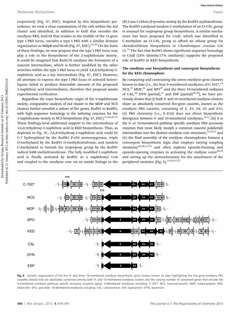
By comparing and contrasting the seven enediyne gene clustersknown to date [i.e., the four 9-membered enediynes of C-1027,24
NCS,25 MDP,26 and SPO27 and the three 10-membered endiynesof CAL,28 DYN (partial),31 and ESP (partial)29,30], we have pre-viously shown that (i) both 9- and 10-membered enediyne clustersshare an absolutely conserved five-gene cassette, known as theenediyne PKS cassette, consisting of E, E3, E4, E5 and E10,(ii) PKS chemistry (i.e., E–E10) does not direct biosyntheticdivergence between 9- and 10-membered enediynes,32,33 (iii) it isthe 9- or 10-membered pathway specific enediyne PKS accessoryenzymes that most likely morph a common nascent polyketideintermediate into the distinct enediyne core structures,32,33,53 and(iv) the final assembly of the enediyne chromophores features aconvergent biosynthetic logic that employs varying couplingchemistry44,46,53,54 and often exploits epoxide-forming andepoxide-opening enzymes in activating the endiyne cores48,49
and setting up the stereochemistry for the attachment of theperipheral moieties (Fig. 6).19,20,53,57
Fig. 6 Genetic organization of the five 9- and three 10-membered enediyne biosynthetic gene clusters known to date highlighting the five-gene enediyne PKScassettes (black) that are absolutely conserved among both 9- and 10-membered enediyne clusters and the varying number of conserved genes that encode the9-membered enediyne pathway specific accessory enzymes (gray). 9-Membered enediynes including: C-1027; NCS, neocarzinostatin; MDP, maduropeptin; KED,kedarcidin; SPO, sporolide. 10-Membered enediynes including: CAL, calicheamicin; ESP, esperamicin; DYN, dynemicin.
Molecular BioSystems Paper
Dow
nloa
ded
by S
crip
ps R
esea
rch
Inst
itute
on
05 F
ebru
ary
2013
Publ
ishe
d on
21
Janu
ary
2013
on
http
://pu
bs.r
sc.o
rg |
doi:1
0.10
39/C
3MB
2552
3AView Article Online
This journal is c The Royal Society of Chemistry 2013 Mol. BioSyst., 2013, 9, 478--491 487
The ked cluster now joins the growing list of enediynebiosynthetic machinery, supporting the emerging paradigmfor enediyne core biosynthesis19,20,53,56,57 but also revealingnew insights. Thus, the ked cluster also harbors the absolutelyconserved five-gene enediyne PKS cassette (Fig. 6), whose PKSchemistry is demonstrated by the production of the hallmarkheptaene product for enediyne biosynthesis upon co-expressionof kedE–kedE10 in E. coli (Fig. 4).32,33 Flanking the ked enediynePKS cassette are the highly conserved 13 genes, kedE1, kedE2,kedE6 to kedE9, kedE11, kedD2, kedF, kedJ, kedL, kedM, and kedS(Fig. 2B and Table 1), that are highly conserved among the five9-membered enediyne gene clusters known to date (Fig. 6).33,53
They encode the 9-membered enediyne pathway specific PKSaccessory enzymes for endiyne core biosynthesis, includingKedF whose epoxide hydrolase activity was demonstratedin vitro to afford a vicinal diol with the same regio- and absolutestereochemistry as would be for the KED enediyne core (Fig. 5).It should be noted that while the five genes consisting of theenediyne PKS cassette are typically clustered, the organizationof genes encoding the accessory enzymes is less conserved,scattering on either side of the enediyne PKS cassette withinthe gene cluster (Fig. 6). They nonetheless show significantsequence homology, ensuring their identification upon carefulbioinformatics analysis (Table 1). These observations shouldnow be taken into consideration in future effort to identify andannotate new enediyne biosynthetic gene clusters. Finally, thefully modified and activated KED enediyne core intermediate iscoupled with the two deoxysugars, the (R)-2-aza-3-chloro-b-tyrosine,and the 3-hydroxy-7,8-dimethoxy-6-isopropoxy-2-naphthonatemoiety, and KedS6, KedS10, KedN4, and KedY5 are proposed tocatalyze these coupling steps, respectively, the timing of which ispending future determination (Fig. 3D). It has long been specu-lated that the convergent molecular logic for enediyne biosynthesispresents outstanding opportunities to engineer new enediynenatural products by combinatorial strategies.12–14,19,20 The avail-ability of the ked cluster and the novel chemistry associated withthe KED biosynthetic machinery surely will enrich the enediynegenetic toolbox and facilitate such engineering effort.
KED biosynthesis and structural revisions
The structure of KED chromophore has been revised twicesince it was first published4–7 (Fig. S2, ESI†). The originalstructure had the (R)-2-aza-3-chloro-a-tyrosine moiety with the2-napthamide linked at the a-amino position.4,5 This structurewas subsequently revised to (R)-2-aza-3-chloro-b-tyrosine withthe 2-naphthamide linked at the b-amino position.6 Cloningand sequencing of the ked cluster in the current study supportsthis revision, as KedY4 is similar to SgcC4 and MdpC4, twoMIO-containing aminomutases that have been characterizedin vitro to catalyze the conversion of a-tyrosine to b-tyrosine56
(Fig. S6, ESI†), supporting the intermediacy of (R)-2-aza-3-chloro-b-tyrosine in KED biosynthesis (Fig. 3).
The second revision was of the stereochemistry of the KEDenediyne core, initially inverting the entire enediyne core intoits enantiomer and subsequently revising the C-10/C-11 disubsti-tution pattern from trans- to cis-configuration (Fig. S2, ESI†).4–7
The C-10/C-11 cis-disubstitution provides a a-glycosidic and anether linkage to the L-mycaroside and the (R)-2-aza-3-chloro-b-tyrosine moiety, respectively, in the KED chromophore (7).Intriguingly, similar glycosidic and ether/ester linkages todeoxysugar, tyrosine-derived moieties (C-1027 and MDP) andthe 1-naphthonate moiety (NCS) are also present in otherenediyne natural products, but the relative stereochemistry ofthese disubstitutions are in the trans-configuration, as exem-plified by the C-1027, MDP, and NCS chromophores (Fig. S3,ESI†). Comparative studies of KED biosynthesis to those ofC-1027, MDP, and NCS now provide opportunities to decipherthe mechanism, thereby controlling and exploiting the regio-and stereochemistry in appending the peripheral moieties toeach of the endiyne cores for enediyne biosynthesis and struc-tural diversity.12–14,19
Conclusion
Kedarcidin, a member of the enediyne family of antitumorantibiotics, features a novel molecular architecture. The kedarcidinbiosynthetic gene cluster is cloned from Streptoalloteichus sp.ATCC 53650 and sequenced and annotated. The identity of thecloned gene cluster to encode KED biosynthesis is supportedby: (i) finding the kedA gene within the cloned ked cluster thatencodes the previously isolated KED apoprotein, (ii) productionof the signature heptaene product for enediyne biosynthesisupon co-expression of kedE–kedE10, encoding the enediyne PKSand the associated type II TE, in E. coli, and (iii) in vitrocharacterization of KedF as an epoxide hydrolase using asubstrate mimic that affords a vicinal diol product with theregio- and absolute stereochemistry as would be expected forthe KED chromophore. Comparative analysis between ked andthe other cloned 9- and 10-membered enediyne gene clusterssupports a convergent biosynthetic pathway for the KED chromo-phore, an emerging paradigm for the enediyne family of naturalproducts, but the KED biosynthetic machinery is also predictedto feature much novel chemistry.
ExperimentalBacterial strains, plasmids, and sequence analysis
Streptoalloteichus sp. ATCC 53650, the KED producer, andM. luteus ATCC 9431, the test organism for assay of the anti-bacterial activity of KED, were from American Type CultureCollection (Rockville, MD). SuperCos1, Gigapack III XL andE. coli XL1-Blue MR cells (Stratagene, La Jolla, CA), pGEM-TEasy and pSP72 (Promega, Madison, WI), and pETDuet-1,pRSFDuet-1, and E. coli BL21(DE3) cells (Novagen, Madison, WI)were from commercial sources. pANT841,71 pBS1050,32 pBS1051,32
and pBS106532 were described previously. DIG-labeling kit and calfintestinal phosphatase (Roche, Indianapolis, IN), T4 DNA ligase(Promega), and restriction enzymes (New England Biolabs Ipswich,MA or Invitrogen, Carlsbad, CA) were from commercial sources.DNA sequencing was carried out at the University of Wisconsin-Madison Biotechnology Center (Madison, WI). Sequenceanalysis was carried out using BLASTN available from NCBI,
Paper Molecular BioSystems
Dow
nloa
ded
by S
crip
ps R
esea
rch
Inst
itute
on
05 F
ebru
ary
2013
Publ
ishe
d on
21
Janu
ary
2013
on
http
://pu
bs.r
sc.o
rg |
doi:1
0.10
39/C
3MB
2552
3AView Article Online
488 Mol. BioSyst., 2013, 9, 478--491 This journal is c The Royal Society of Chemistry 2013
and contiguous DNA was compiled using Lasergene (DNASTARInc., Madison, WI). Open reading frames (orfs) were predictedusing ORFfinder from NCBI and Genemark,72 and proteinsequences were analyzed using PSI-BLAST and InterProScan.73
All recombinant DNA manipulations were performed by followingstandard procedures60,74 or the manufacturers’ instructions.
Production, isolation, and analysis of KED
KED production, isolation, and analysis were carried outessentially by following the literature procedures.2,5 Thus,Streptoalloteichus sp. ATCC 5360 was grown on TSB agar plate60
for single colonies. Seed inoculum was prepared by introducingthe colony periphery of petri dish cultures into 250 mL flaskscontaining 50 mL of TSB medium,60 followed by shaking at250 rpm and 28 1C for two days. Production fermentation wascarried out by adding 3 mL of seed inoculum into each of theten 250 mL flasks containing 50 mL of production medium(3% glycerol, 1% pharmamedia, 1.5% distiller’s solubles extract,1% fish meal, 0.05% KH2PO4, and 0.6% CaCO3, pH 7.0), andshaking at 250 rpm and 28 1C for five days. The fermentationculture was centrifuged (8000 rpm, 4 1C, 35 min) and filtered toremove mycelia. The supernatant was slowly adjusted topH 5.0 with 2 N HCl while stirring, followed by centrifugation(12 000 rpm, 4 1C, 35 min) to remove precipitates. The resultingsupernatant was mixed with DEAE-cellulose resin equilibratedin buffer (0.05 M Tris-HCl, pH 5.6). The resulting DEAE-cellulose resin was washed with the same buffer twice andeluted with the same buffer containing 1 M NaCl. The eluatewas dialyzed against Milli-Q H2O at 4 1C overnight using a10 kDa molecular weight cutoff membrane. The dialyzedsolution was lyophilized, dissolved in 4 mL cold H2O, andapplied to a DEAE-cellulose column equilibrated in 0.05 MTris-HCl, pH 5.6. The column was washed with cold H2Oand eluted stepwise with 0.1 M, 0.2 M, and 0.3 M NaCl.Fractions were assayed against M. luteus,34 and the activefractions were combined and lyophilized to afford a yellowpowder. Further purification was achieved using SephadexG-75 chromatography eluting with cold H2O at 4 1C. Again,fractions were followed by assay against M. luteus, and activefractions were combined and lyophilized to give pure KEDchromoprotein.
To dissociate the KED chromophore from the apoprotein,5 mg of purified KED chromoprotein was dissolved in 0.2 mL of0.1 M potassium phosphate buffer, pH 4.3, and extracted twicewith 0.3 mL of EtOAc each at 4 1C. The combined EtOAc extractwas evaporated in vacuum, and the residue was subjectedto HRESIMS analysis on an IonSpec HiResMALDI FT massspectrometer with a 7 Tesla superconducting magnet. A portionof the EtOAc extract was also left at room temperature overnightand then similarly evaporated to dryness and analyzed byHRESIMS. The freshly prepared and the overnight EtOAcextracts were also subjected to HPLC analysis. HPLC wascarried out on a Varian HPLC system equipped with Prostar210 pumps, a photodiode array detector, and an Atima-C18column (5 mm, 4.6 mm � 250 mm, Grace Davison DiscoverySciences, Deerfield, IL). The column was developed at flow rate
of 1 mL min�1 with a linear gradient from 100% buffer A(0.01 M potassium phosphate, pH 6.8) to 20% buffer A/80%buffer B (80% CH3CN in 0.01 M potassium phosphate buffer,pH 6.8) in 35 min, monitored at 320 nm.
Cosmid DNA library construction, screening, and sequencing
A SuperCos1 cosmid library was constructed using partiallydigested (Sau3AI) Streptoalloteichus sp. ATCC 53650 chromo-somal DNA followed by dephosphorylation with calf intestinalphosphatase according to standard procedures.60,74 After anovernight ligation at 16 1C, the mixture was packaged usingGigapack III XL and used to transfect E. coli XL1 Blue MR cellsfollowing the manufacturer’s instructions (Stratagene). A 3.5 kbinternal fragment of kedE was PCR amplified from total geno-mic DNA using Platinum Taq DNA polymerase (Invitrogen,Carlsbad, CA) and the following pair of primers (forward50-GGCGGCGGVTACACSGTSGACGGMGCCTGC-30/reverse, 50-CCCATSCCGACSCCGGACCASACSGACCAYTCCA-3 0, where M = A orC; S = C or G; V = A, C, or G; Y = C or T) as describedpreviously.30 The PCR product was cloned into pGEM-T Easyto afford pBS16001, confirmed to encode an internal fragmentof an enediyne PKS gene by sequencing,29,39 and used toprepare the DIG-labeled probe (probe-1). Probe-1 was then usedto screen the cosmid library by colony hybridization, yieldingthree positive clones. One of the positive clones, pBS16002, wasend-sequenced using the following pair of primers (forward50-GGGAATAAGGGCGACACGGG-3 0/reverse 50-GCTTATCGATGATAAGCGGTC-3 0) and confirmed to encode a part of the kedcluster. Four additional rounds of chromosomal walking frompBS16002 were subsequently carried out using probe-2, -3, -4,and -5, respectively to isolate overlapping cosmids that coverthe entire ked cluster (Fig. 2A). Thus, probe-2 and probe-3 wereprepared by PCR from pBS16002 using the following pairs ofprimers (probe-2, forward 50-GGTACTACCTGCTGTGC-30/reverse50-GGTCTTGGTGAAGCTGC-3 0) and (probe-3, forward 50-CGATCAAGTCGATCCTGACC-3 0/reverse 50-GGTCGCTGGTGATGTCGTCG-30), respectively. Screening the cosmid library by colonyhybridization with probe-2 and probe-3, respectively, resultedin the isolation of pBS16003 and pBS16004. Similarly, probe-4was prepared by PCR from pBS16004 using the following pairsof primers (forward 50-GGAGGTCGAGGTGCGTGC-3 0/reverse50-GGTTCCACGTGATCAGC-3 0) and used to screen the cosmidlibrary to isolate pBS16005. Probe-5 was prepared by PCR frompBS16005 using the following pairs of primers (forward50-GCTGTGCCTGGTGGACCTGACC-3 0/reverse 50-GCAGCAGGTCGAGGTCG-3 0) and used to screen the cosmid library to isolatepBS16006. Finally, the five overlapping cosmids (i.e., pBS16002,pBS16003, pBS16004, pBS16005, and pBS16006) were similarlyend-sequenced to confirm their candidacy for completesequencing (Fig. 2A).
The five overlapping cosmids were used to generate sub-clone libraries for complete DNA sequence determination. Theresultant DNA sequences were compiled and assembled intocontigs, and gaps were filled in by primer walking or bysubcloning fragments covering the gaps and subsequentlysequencing the cloned fragments (Fig. 2B and Table 1).
Molecular BioSystems Paper
Dow
nloa
ded
by S
crip
ps R
esea
rch
Inst
itute
on
05 F
ebru
ary
2013
Publ
ishe
d on
21
Janu
ary
2013
on
http
://pu
bs.r
sc.o
rg |
doi:1
0.10
39/C
3MB
2552
3AView Article Online
This journal is c The Royal Society of Chemistry 2013 Mol. BioSyst., 2013, 9, 478--491 489
The kedE–kedE10 co-expression construct for E. coli expression
To construct the kedE–kedE10 co-expression plasmid, a 2400 bpSstI–MluI fragment, containing the 1.8 kb of the 30 region ofkedE together with kedE10 was first cloned from pBS16002 andligated into the same sites of pANT841 to afford pBS16007.A 4354 bp XmnI–SstI fragment containing the 50 region of kedEtogether with B400 bp of upstream sequence was next clonedfrom pBS16002 and ligated into the same sites of pSP72 toafford pBS16008. The 30 region of kedE together with kedE10was then recovered as an SstI–HindIII fragment from pBS16007and cloned into the same sites of pBS16008 to yield pBS16009,which contain the complete kedE–kedE10 cassette. This cassettewas moved as a BglII–HindIII fragment into the compatibleBamHI–HindIII sites of pETDuet-1 to afford final constructpBS16010 for co-expressing kedE–kedE10 in E. coli.
Co-expression of kedE–kedE10 in E. coli for heptaeneproduction
Co-expression of kedE–kedE10 in E. coli was carried out asdescribed previously.32,33,53 Thus, pBS16010 was transformedinto E. coli BL21(DE3) and cultured as described previously,with co-expressions of sgcE–sgcE10 (pBS1050–pBS1051) as apositive control and of sgcE(C211A)–sgcE10 (pBS1065–pBS1051)as a negative control.32,33 Briefly, E. coli recombinant strainscarrying the varying co-expression cassettes were cultured in50 mL LB medium supplemented with the appropriate anti-biotics for selection. The cultures were first grown at 37 1C to anoptical density at 600 nm (OD600) of B0.2 and then transfer to18 1C for continued incubation until they reached OD600 B0.4;upon induction with 0.1 mM IPTG, incubation continued for anadditional two days. The cultures were acidified to pH B3 andharvested by centrifuging to pellet the cells. The cell pellet wasextracted by vortexing with 20 mL of acetone. The acetoneextract was centrifuged, and the supernatant was concentratedby rotary evaporation to B1 mL, of which 100 mL was subjectedto HPLC analysis. The same HPLC system and Atima-C18column as described above were used. The column was developedat a flow rate of 1 mL min�1 with a linear gradient from 40%buffer A (0.1% trifluoroacetic acid in H2O)/60% buffer B (0.1%trifluoroacetic acid in CH3OH) to 100% buffer B in 35 min withUV detection at 370 nm. The identity of heptaene was confirmedby comparison with an authentic standard.32,33
Expression of kedF in E. coli and purification of KedF
The kedF gene was amplified by PCR from pBS16002 usingPlatinum Pfx polymerase (Invitrogen) and the following pair ofprimers (forward 50-AAAACCTCTATTTCCAGTCGATGCGCCGCTTCCGCATAGCCG-30/reverse 50-TACTTACTTAAATGTTATCAGGCCAGGGAGCGGGCGAACGC-3 0). The resultant product was gel-purified and cloned into pBS160011, a variant of pRSFDuet-1that contains both a TEV protease recognition site and ligationindependent cloning site, to afford the expression constructpBS16012. Under this construct, KedF was overproduced as anN-terminal His6-tagged fusion protein, whose His6-tag can beremoved upon TEV protease treatment. Introduction of pBS16012
into E. coli BL21 (DE3) for kedF expression and overproductionand purification of KedF by affinity chromatography using a5 mL HisTrap HP column (GE Healthcare, Piscataway, NJ) wereperformed at 4 1C following standard procedures. Immediatelyfollowing the affinity chromatography, the KedF fraction wasdiluted to 50 mL with buffer A (50 mM Tris-HCl, pH 8.0, 10 mMNaCl) and loaded on a MonoQ 10/100 column for anionexchange chromatography on an AKTA FPLC unit (GE Healthcare).The column was developed at a flow rate of 2 mL min�1 with alinear gradient from 85% buffer A/15% buffer B (50 mM Tris-HCl, pH 8.0, 1.0 M NaCl) to 40% buffer A/60% buffer B in40 min. The eluted KedF protein was concentrated with a 30 KMWCO Vivaspin ultrafiltration device (Sartorius, Edgewood, NY)and stored at �80 1C in 100 mL aliquots. The purified KedF wasanalyzed by SDS-PAGE on 12% gel. KedF concentration wasdetermined from the absorbance at 280 nm using a molarabsorptivity (e 76.89 mM�1 cm�1) calculated according to thededuced KedF amino acid sequence.
In vitro characterization of KedF
In vitro characterization of KedF as an epoxide hydrolase wascarried out as described previously, using styrene oxide as asubstrate mimic.48,49 Thus, HPLC-based assays were carried outin 200 mL reaction mixtures containing 2 mM racemic styreneoxide in 50 mM phosphate buffer, pH 8.0.48,49 The reaction wasinitiated by the addition of 50 mM KedF, incubated at 25 1C for1 h, and terminated by extracting the assay mixture with 200 mLof EtOAc for three times. Negative controls were carried outunder the identical conditions in the absence of KedF, whilepositive controls were carried out under the identical condi-tions with SgcF instead of KedF.48 The combined EtOAc extractswere concentrated in vacuum, and the resulting residue wasdissolved in 50 mL of CH3CN, 25 mL of which was subjected toHPLC analysis. HPLC was performed with an Alltech AppoloC18 column (5 mM, 4.6 � 250 mm, Grace Davison DiscoverySciences), developed at a flow rate of 1 mL min�1 with a lineargradient from 0 to 60% CH3CN in H2O in 20 min with UVdetection at 254 nm. The enantiomeric analysis of the vicinaldiol products was performed on a Waters HPLC systemequipped with 600 pumps, a 996 photodiode array detector, anda Chiralcel OD-H column (5 mM, 4.6 � 250 mm, Grace DavisonDiscovery Sciences). The column was eluted isocratically, at a flowrate of 0.7 mL min�1, with 2.5% isopropanol in hexane.
Determination of the steady-state kinetic parameters ofKedF-catalyzed hydrolysis of (R)- or (S)-styrene oxide followedthe continuous spectrophotometric assay62 previously adoptedfor the SgcF and NcsF2 epoxide hydrolase.48,49 Thus, the reac-tions were carried out in 1 mL reaction mixture containing10 mL of 300 mM sodium periodate in DMF, 20 mL of (R)- or(S)-styrene oxide in DMSO, with varying concentrations between0.1 mM and 15 mM, in 50 mM phosphate buffer, pH 8.0. Thereactions were initiated by the addition of 9.6 or 4.0 mM KedF, for(R)- or (S)-styrene oxide, respectively, and these reactions werecarried out in triplicate. The absorbance at 290 nm wasmonitored in a 1 mL quartz cuvette, thermostated at 25 1C,and the velocity was calculated based on the rate of change of
Paper Molecular BioSystems
Dow
nloa
ded
by S
crip
ps R
esea
rch
Inst
itute
on
05 F
ebru
ary
2013
Publ
ishe
d on
21
Janu
ary
2013
on
http
://pu
bs.r
sc.o
rg |
doi:1
0.10
39/C
3MB
2552
3AView Article Online
490 Mol. BioSyst., 2013, 9, 478--491 This journal is c The Royal Society of Chemistry 2013
absorbance over 5 to 30 s Michaelis–Menten equation was fittedto plots of velocity of 1-phenyl-1,2-ethanediol formation versussubstrate concentration to extract the Km and kcat values.
Nucleotide sequence accession number
The nucleotide sequence reported in this study is available inthe GenBank database under accession number JX679499.
Acknowledgements
We thank the Analytical Instrumentation Center of the Schoolof Pharmacy, University of Wisconsin-Madison for support inobtaining MS data. This work is supported in part by NIHgrants CA78747 and CA113297. G.P.H. is the recipient of anNSERC (Canada) postdoctoral fellowship.
Notes and references
1 K. S. Lam, G. A. Hesler, D. R. Gustavson, A. R. Crosswell,J. M. Veitch, S. Forenza and K. Tomita, J. Antibiot., 1991, 44,472–478.
2 S. J. Hofstead, J. A. Matson, A. R. Malacko andH. Marquardt, J. Antibiot., 1992, 45, 1250–1254.
3 K. L. Constantine, K. L. Colson, M. Wittekind, M. S. Friedrichs,N. Zein, J. Tuttle, D. R. Langley, J. E. Leet, D. R. Schroeder,K. S. Lam, B. T. Farmer II, W. J. Metzler, R. E. Bruccoler andL. Mueller, Biochemistry, 1994, 33, 11438–11452.
4 J. E. Leet, D. R. Schroeder, S. J. Hofstead, J. Golik,K. L. Colson, S. Huang, S. E. Klohr, T. W. Doyle andJ. A. Matson, J. Am. Chem. Soc., 1992, 114, 7946–7948.
5 J. E. Leet, D. R. Schroeder, D. R. Langley, K. L. Colson,S. Huang, S. E. Klohr, M. S. Lee, J. Golik, S. J. Hofstead,T. W. Doyle and J. A. Matson, J. Am. Chem. Soc., 1993, 115,8432–8443.
6 S. Kawata, S. Ashizawa and M. Hirama, J. Am. Chem. Soc.,1997, 119, 12012–12013.
7 F. Ren, P. C. Hogan, A. J. Anderson and A. G. Myers, J. Am.Chem. Soc., 2007, 129, 5381–5383.
8 K. C. Nicolaou and W.-M. Dai, Angew. Chem., Int. Ed. Engl.,1991, 30, 1387–1530.
9 Z. Xi and I. G. Goldberg, in Comprehensive Natural ProductsChemistry, ed. D. Barton, K. Nakanish and O. Meth-Cohn,Elsevier, New York, 1999, vol. 7, pp. 553–592.
10 U. Galm, M. H. Hager, S. G. Van Lanen, J. S. Thorson andB. Shen, Chem. Rev., 2005, 105, 739–758.
11 N. Zein, K. L. Colson, J. E. Leet, D. R. Schroeder,W. Solomon and T. W. Doyle, Proc. Natl. Acad. Sci. U. S. A.,1993, 90, 2822–2826.
12 D. R. Kennedy, L. S. Gawron, J. Ju, W. Liu, B. Shen andT. A. Beerman, Cancer Res., 2007, 67, 773–781.
13 D. R. Kennedy, J. Ju, B. Shen and T. A. Beerman, Proc. Natl.Acad. Sci. U. S. A., 2007, 104, 17632–17637.
14 T. A. Beerman, L. S. Gawron, S. Shin, B. Shen andM. M. McHugh, Cancer Res., 2009, 69, 593–598.
15 Enediyne antibiotics as antitumor agents, ed. T. W. Doyle andD. B. Borders, Marcel-Dekker, New York, 1995.
16 Neocarzinostatin: the past, present, and future of an anticancerdrug, ed. H. Maeda, K. Edo and, N. Ishida, Springer-Verlag,New York, 1997.
17 J. S. Thorson, B. Shen, R. E. Whitwam, W. Liu and Y. Li,Bioorg. Chem., 1999, 27, 172–188.
18 I. Brukner, Curr. Opin. Oncol., Endocr. Metab. Invest. Drugs,2000, 2, 344–352.
19 S. G. Van Lanen and B. Shen, Curr. Top. Med. Chem., 2008, 8,448–459.
20 Z.-X. Liang, Nat. Prod. Rep., 2010, 27, 499–528.21 K. Edo, M. Mizugaki, Y. Koide, H. Seto, K. Furihata, N. Otake
and N. Ishida, Tetrahedron Lett., 1985, 26, 331–340.22 M. D. Lee, T. S. Dunne, M. M. Siegel, C. C. Chang, G. O. Morton
and D. B. Borders, J. Am. Chem. Soc., 1987, 109, 3464–3466.23 S.-J. Nam, S. P. Gaudencio, C. A. Kauffman, P. R. Jensen,
T. P. Kondratyuk, L. E. Marler, J. M. Pezzuto and W. Fenical,J. Nat. Prod., 2010, 73, 1080–1086.
24 W. Liu, S. D. Christenson, S. Standage and B. Shen, Science,2002, 297, 1170–1173.
25 W. Liu, K. Nonaka, L. Nie, J. Zhang, S. D. Christenson,J. Bae, S. G. Van Lanen, E. Zazopoulos, C. M. Farnet,C. F. Yang and B. Shen, Chem. Biol., 2005, 12, 293–302.
26 S. G. Van Lanen, T.-J. Oh, W. Liu, E. Wendt-Pienkowski andB. Shen, J. Am. Chem. Soc., 2007, 129, 13082–13094.
27 R. P. McGlinchey, M. Nett and B. S. Moore, J. Am. Chem. Soc.,2008, 130, 2406–2407.
28 J. Ahlert, E. Shepard, N. Lomovskaya, E. Zazopoulos,A. Staffa, B. O. Bachmann, K. Huang, L. Fonstein,A. Czisny, R. E. Whitwam, C. M. Farnet and T. S. Thorson,Science, 2002, 297, 1173–1176.
29 E. Zazopoulos, K. Huang, A. Staffa, W. Liu, B. O. Bachmann,K. Nonaka, J. Ahlert, J. S. Thorson, B. Shen and C. M. Farnet,Nat. Biotechnol., 2003, 21, 187–190.
30 W. Liu, J. Ahlert, Q. Gao, E. Wendt-Pienkowski, B. Shen andJ. S. Thorson, Proc. Natl. Acad. Sci. U. S. A., 2003, 100,11959–11963.
31 Q. Gao and J. S. Thorson, FEMS Microbiol. Lett., 2008, 282,105–114.
32 J. Zhang, S. G. Van Lanen, J. Ju, W. Liu, P. C. Dorrestein,W. Li, N. L. Kelleher and B. Shen, Proc. Natl. Acad. Sci. U. S. A.,2008, 105, 1460–1465.
33 G. P. Horsman, Y. Chen, J. S. Thorson and B. Shen, Proc.Natl. Acad. Sci. U. S. A., 2010, 107, 11331–11335.
34 W. Liu and B. Shen, Antimicrob. Agents Chemother., 2000, 44,382–392.
35 J. M. Murrell, W. Liu and B. Shen, J. Nat. Prod., 2004, 67,206–213.
36 S. D. Christenson, W. Liu, M. D. Toney and B. Shen, J. Am.Chem. Soc., 2003, 125, 6062–6063.
37 S. D. Christenson, W. Wu, B. Shen and M. D. Toney,Biochemistry, 2003, 42, 12708–12718.
38 C. V. Christianson, T. J. Montavon, S. G. Van Lanen, B. Shenand S. D. Bruner, Biochemistry, 2007, 46, 7025–7214.
39 C. V. Christianson, T. J. Montavon, G. M. Festin,H. A. Cooke, B. Shen and S. D. Bruner, J. Am. Chem. Soc.,2007, 129, 15744–15745.
Molecular BioSystems Paper
Dow
nloa
ded
by S
crip
ps R
esea
rch
Inst
itute
on
05 F
ebru
ary
2013
Publ
ishe
d on
21
Janu
ary
2013
on
http
://pu
bs.r
sc.o
rg |
doi:1
0.10
39/C
3MB
2552
3AView Article Online
This journal is c The Royal Society of Chemistry 2013 Mol. BioSyst., 2013, 9, 478--491 491
40 T. J. Montavon, C. V. Christianson, G. M. Festin, B. Shen andS. D. Bruner, Bioorg. Med. Chem. Lett., 2008, 18, 3099–3102.
41 S. G. Van Lanen, P. C. Dorrestein, S. D. Christenson, W. Liu,J. Ju, N. L. Kelleher and B. Shen, J. Am. Chem. Soc., 2005, 127,11594–11595.
42 S. G. Van Lanen, S. Lin, P. C. Dorrestein, N. L. Kelleher andB. Shen, J. Biol. Chem., 2006, 281, 29633–29640.
43 S. Lin, S. G. Van Lanen and B. Shen, J. Am. Chem. Soc., 2007,129, 12432–12438.
44 S. G. Van Lanen, S. Lin and B. Shen, Proc. Natl. Acad. Sci.U. S. A., 2008, 105, 494–499.
45 S. Lin, S. G. Van Lanen and B. Shen, J. Am. Chem. Soc., 2008,130, 6616–6623.
46 S. Lin, S. G. Van Lanen and B. Shen, Proc. Natl. Acad. Sci.U. S. A., 2009, 106, 4183–4188.
47 S. G. Van Lanen, S. Lin, G. P. Horsman and B. Shen, FEMSMicrobiol. Lett., 2009, 300, 237–241.
48 S. Lin, G. P. Horsman and B. Shen, J. Am. Chem. Soc., 2009,131, 16410–16417.
49 S. Lin, G. P. Horsman and B. Shen, Org. Lett., 2010, 12,3816–3819.
50 H. A. Cooke, J. Zhang, M. A. Griffin, K. Nonaka, S. G.Van Lanen, B. Shen and S. D. Bruner, J. Am. Chem. Soc.,2007, 129, 7728–7729.
51 Y. Luo, S. Lin, J. Zhang, H. A. Cooke, S. D. Bruner andB. Shen, J. Biol. Chem., 2008, 283, 14694–14702.
52 H. A. Cooke, E. L. Guenther, Y. Luo, B. Shen andS. D. Bruner, Biochemistry, 2009, 48, 9590–9598.
53 G. P. Horsman, S. G. Van Lanen and B. Shen, MethodsEnzymol., 2009, 459, 97–112.
54 J. Ling, G. P. Horsman, S.-X. Huang, Y. Luo, S. Lin andB. Shen, J. Am. Chem. Soc., 2010, 132, 12534–12536.
55 S. Lin, T. Huang, G. P. Horsman, S.-X. Huang, X. Guo andB. Shen, Org. Lett., 2012, 14, 2300–2303.
56 J. R. Lohman and B. Shen, Methods Enzymol., 2012, 516, 299–319.57 S. Lin, T. Huang and B. Shen, Methods Enzymol., 2012, 516,
321–343.58 A. G. Myers, A. R. Hurd and P. C. Hogan, J. Am. Chem. Soc.,
2002, 124, 4583–4585.59 K. Ogawa, Y. Koyama, I. Ohashi, I. Sato and M. Hirama,
Angew. Chem., Int. Ed., 2009, 48, 1110–1113.
60 T. Kieser, M. J. Bibb, M. M. Buttner, K. F. Chater and D. A.Hopwood, Practical Streptomyces Genetics, The John InnesFoundation, Norwich, UK, 2000.
61 V. T. T. Hang, T. S. Kim, T.-J. Oh and K. Sohng, Biotechnol.Bioprocess Eng., 2011, 16, 462–469.
62 C. Mateo, A. Archelas and R. Furstoss, Anal. Biochem., 2003,314, 135–141.
63 C. J. Thibodeaux, C. E. Melancon III and H.-w. Liu, Nature.,2007, 446, 10081016.
64 C. J. Thibodeaux, C. E. Melancon III and H.-w. Liu, Angew.Chem., Int. Ed., 2008, 47, 9814–9858.
65 M. Tao, L. Wang, E. Wendt-Pienkowski, N. P. George,U. Galm, G. Zhang, J. M. Coughlin and B. Shen, Mol.BioSyst., 2007, 3, 60–74.
66 S. Inouye, T. Shomura, T. Tsuruoka, Y. Ogawa,H. Watanabe, J. Yoshida and T. Niida, Chem. Pharm. Bull.,1975, 23, 2669–2677.
67 H.-J. Kwon, W. C. Smith, A. J. Scharon, S. H.Hwang, M. J. Kurth and B. Shen, Science, 2002, 297,1327–1330.
68 B. Kusebauch, B. Busch, K. Scherlach, M. Roth andC. Hertweck, Angew. Chem., Int. Ed., 2009, 48, 5001–5004.
69 B. Shen, Curr. Opin. Chem. Biol., 2003, 7, 285–295.70 S. Rachid, M. Scharfe, H. Bloecker, K. J. Weissman and
R. Mueller, Chem. Biol., 2009, 16, 70–81.71 V. B. Rajgarhia and W. R. Strohl, J. Bacteriol., 1997, 179,
2690–2696.72 A. V. Lukashin and M. Borodovsky, Nucleic Acids Res., 1998,
26, 1107–1115.73 S. Hunter, R. Apweiler, T. K. Attwood, A. Bairoch,
A. Bateman, D. Binns, P. Bork, U. Das, L. Daugherty,L. Duquenne, R. D. Finn, J. Gough, D. Haft, N. Hulo,D. Kahn, E. Kelly, A. Laugraud, I. Letunic, D. Lonsdale,R. Lopez, M. Madera, J. Maslen, C. McAnulla, J. McDowall,J. Mistry, A. Mitchell, N. Mulder, D. Natale, C. Orengo,A. F. Quinn, J. D. Selengut, C. J. A. Sigrist, M. Thimma,P. D. Thomas, F. Valentin, D. Wilson, C. H. Wu and C. Yeats,Nucleic Acids Res., 2009, 37, D211–D215.
74 J. Sambrook, E. F. Fritsch and T. Maniatis, Molecular Cloning:A Laboratory Manual, Cold Spring Harbor Laboratory,Cold Spring Harbor, NY, 3rd edn, 2000.
Paper Molecular BioSystems
Dow
nloa
ded
by S
crip
ps R
esea
rch
Inst
itute
on
05 F
ebru
ary
2013
Publ
ishe
d on
21
Janu
ary
2013
on
http
://pu
bs.r
sc.o
rg |
doi:1
0.10
39/C
3MB
2552
3AView Article Online


![ATCC Bacterial Culture Guide[1]](https://img.pdfslide.us/doc/110x75/55cf856a550346484b8dccc1/atcc-bacterial-culture-guide1.jpg)