Embed Size (px)
Citation preview
International Journal of
Molecular Sciences
Article
rs657075 (CSF2) Is Associated with the DiseasePhenotype (BAS-G) of Ankylosing Spondylitis
Wei-Chiao Chen 1,†, James Cheng-Chung Wei 2,3,4,†, Hsing-Fang Lu 5,Henry Sung-Ching Wong 5,6, Peng Yeong Woon 7, Yu-Wen Hsu 8, Jin-Ding Huang 1,*and Wei-Chiao Chang 5,6,8,9,10,*
1 Institude of Clinical Pharmacy and Pharmaceutical Sciences, College of Medicine,National Cheng Kung University, Tainan 70101, Taiwan; [email protected]
2 Division of Allergy, Immunology and Rheumatology, Chung Shan Medical University Hospital,Taichung 40201, Taiwan; [email protected]
3 Institute of Medicine, Chung Shan Medical University, Taichung 40201, Taiwan4 Institute of Integrative Medicine, China Medical University, Taichung 40201, Taiwan5 Department of Clinical Pharmacy, School of Pharmacy, Taipei Medical University, Taipei 11014, Taiwan;
[email protected] (H.-F.L.); [email protected] (H.S.-C.W.)6 Master Program for Clinical Pharmacogenomics and Pharmacoproteomics, School of Pharmacy,
Taipei Medical University, Taipei 11014, Taiwan7 Department of Molecular Biology and Human Genetics, Tzu Chi University, Hualien 97004, Taiwan;
[email protected] The Ph.D. Program for Translational Medicine, College of Medical Science and Technology,
Taipei Medical University and Academia Sinica, Taipei 11031, Taiwan; [email protected] Center for Biomarkers and Biotech Drugs, Kaohsiung Medical University, Kaohsiung 80708, Taiwan10 Department of Pharmacy, Taipei Medical University-Wanfang Hospital, Taipei 116, Taiwan* Correspondence: [email protected] (J.-D.H.); [email protected] (W.-C.C.);
Tel.: +886-6-275-2536 (J.-D.H.); +886-2-2736-1661 (ext. 6187) (W.-C.C.)† These authors contributed equally to this study.
Academic Editors: Emil Alexov and William Chi-shing ChoReceived: 19 September 2016; Accepted: 23 December 2016; Published: 3 January 2017
Abstract: Ankylosing spondylitis (AS) is a systemic autoimmune disease mainly affecting the lumbarspine and sacroiliac joints, and exhibits peripheral inflammatory arthropathy. More than 25 loci havebeen identified as associated with AS. Because both AS and rheumatoid arthritis (RA) are autoimmunediseases that may share some common genetic factors, we therefore examined if the newly identifiedRA genetic polymorphisms were associated with AS in a Taiwanese population. In this study,we enrolled 475 AS patients and 11,301 healthy subjects from a Taiwanese biobank as controls.Although none of single-nucleotide polymorphisms (SNPs) were associated with the susceptibility toAS, the AS disease index Bath AS Global (BAS-G) clinical phenotype was observed as significantlycorrelated to the AA genotype of rs657075 (CSF2). The significance remains after gender/age/diseaseduration adjustment and after group categorization by human leukocyte antigen-B 27 (HLA-B27)genotype. We further investigated the possible functions of rs657075 through bioinformaticsapproaches. Results revealed that polymorphism of rs657075 is able to influence the expressionof acyl-CoA synthetase long-chain family member 6 (ACSL6). In conclusion, our study indicatedthat rs657075 (CSF2) is strongly associated with the AS disease index Bath AS Global (BAS-G)clinical phenotype.
Keywords: ankylosing spondylitis; CSF2; rs657075; Bath AS Global (BAS-G); single nucleotidepolymorphism
Int. J. Mol. Sci. 2017, 18, 83; doi:10.3390/ijms18010083 www.mdpi.com/journal/ijms
Int. J. Mol. Sci. 2017, 18, 83 2 of 13
1. Introduction
Ankylosing spondylitis (AS) is a systemic autoimmune disease, which is characterized byinflammation of the lumbar spine and sacroiliac joints, peripheral inflammatory arthropathy, andthe absence of rheumatoid factor [1,2]. The disease predominantly strikes men between the age of20 and 40 years, in their peak productive years, leading to significant loss of work productivity anda decreased quality of life [3]. Family and twin studies indicated that genetic factors contribute toover 90% to the overall AS susceptibility [4,5]. Human leukocyte antigen (HLA)-B 27 has been knownas the major AS-susceptibility gene for more than 40 years [6], but HLA-B27 accounts for only 16%of the genetic variability in AS [7]. The other HLA-B allele operative in AS susceptibility is HLA-B60,which was identified in studies of US and UK patients with AS [8,9]. In addition, HLA-B60 andHLA-B61 were associated with AS in HLA-B27-negative patients in Taiwan [10]. The susceptibility toAS can be enhanced when combining different patterns of HLA-B60 and HLA-B27 genotypes in Dutchand Taiwanese populations, implying genetic interaction mechanisms that may contribute to AS riskamong two genes [11,12]. The interleukin (IL)-1 and IL-23R genes were also proven to be importantin the pathogenesis of AS [13,14]. Genes involved in regulating peripheral tolerance were foundhave a combined effect on the development of AS, such as the PD-1 G-536A/PD-L1 A8923C/PD-L2C47103T [15] or PTPN22 G-1123C/CTLA-4 A49G [16], and the CTLA-4 +49A/G genotype associatedwith circulatory C-reactive protein (CRP) level [17]. Our previous studies reported that geneticpolymorphisms of ORAI1 (rs12313273 and rs7135617) and STIM1 (rs3750996) were associated with thepathogenesis of HLA-B27-positive AS patients [18,19].
Additionally, many non-major histocompatibility complex (MHC) regions were found to besignificantly associated with AS in genome-wide association studies (GWASs) [20–22]. Twelve lociwere previously confirmed to be associated with AS in Europeans [20,21,23], 2 loci were recentlyreported in Han Chinese [22], and an additional 13 new loci were identified in a recent global GWAS,bringing the total AS-associated loci to 43 [24]. Two studies confirmed the findings of previous ASstudies that ERAP1 and rs10865331 are risk factors for AS susceptibility [25,26]. However, somesusceptibility loci, such as EDIL3, HAPLN1, and ANO6, discovered in a Han Chinese GWAS were notassociated in a Taiwanese AS population [27].
Rheumatoid arthritis (RA) is a type of autoimmune arthritis, triggered by a faulty immune systemresulting in chronic synovitis and the destruction of localized cartilage and bone [28]. Previous twin andfamily-base studies indicate that the associations with major histocompatibility complex (MHC) regionsexplain around 12% of total heritability in the susceptibility of RA [29]. A recent GWAS meta-analysisindicated that nine new loci were associated with RA in a Japanese population [30]. Because AS andRA are autoimmune diseases that may share similar genetic factors as well as immune regulatorypathways, we therefore examined if the RA susceptibility polymorphisms from GWAS meta-analysisare also associated with the pathogenesis of AS. In this study, we selected eight single-nucleotidepolymorphisms (SNPs) from previous GWAS meta-analysis study. The AS activity index (Bath ASDisease Activity Index, BASDAI), Bath AS Functional Index (BASFI), and Bath AS Global (BAS-G), aswell as inflammatory biochemical parameters (immunoglobulin A, IgA, erythrocyte sedimentation rate,ESR, and CRP) were analyzed. We found that the AA genotype of rs657075 (CSF2) was significantlyassociated to the clinical phenotype Bath AS Global (BAS-G).
Int. J. Mol. Sci. 2017, 18, 83 3 of 13
2. Results
2.1. Association Study between RA Polymorphisms and Susceptibility to AS
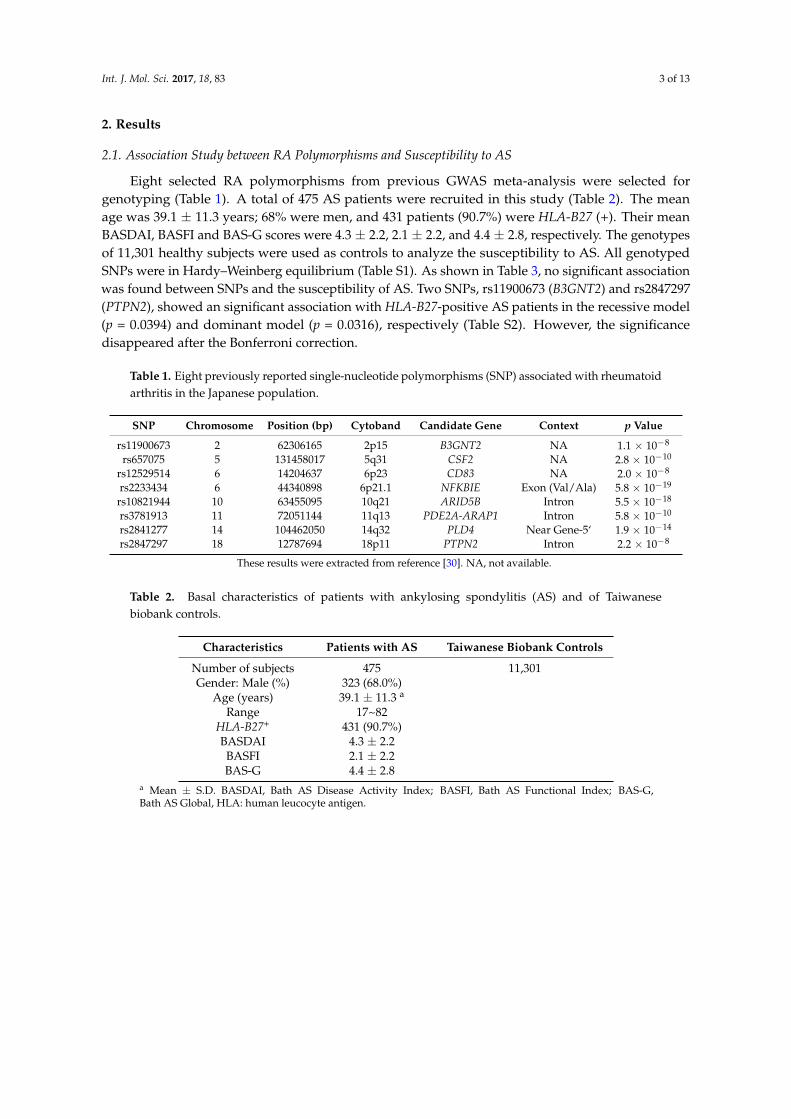
Eight selected RA polymorphisms from previous GWAS meta-analysis were selected forgenotyping (Table 1). A total of 475 AS patients were recruited in this study (Table 2). The meanage was 39.1 ± 11.3 years; 68% were men, and 431 patients (90.7%) were HLA-B27 (+). Their meanBASDAI, BASFI and BAS-G scores were 4.3 ± 2.2, 2.1 ± 2.2, and 4.4 ± 2.8, respectively. The genotypesof 11,301 healthy subjects were used as controls to analyze the susceptibility to AS. All genotypedSNPs were in Hardy–Weinberg equilibrium (Table S1). As shown in Table 3, no significant associationwas found between SNPs and the susceptibility of AS. Two SNPs, rs11900673 (B3GNT2) and rs2847297(PTPN2), showed an significant association with HLA-B27-positive AS patients in the recessive model(p = 0.0394) and dominant model (p = 0.0316), respectively (Table S2). However, the significancedisappeared after the Bonferroni correction.
Table 1. Eight previously reported single-nucleotide polymorphisms (SNP) associated with rheumatoidarthritis in the Japanese population.
SNP Chromosome Position (bp) Cytoband Candidate Gene Context p Value
rs11900673 2 62306165 2p15 B3GNT2 NA 1.1 × 10⁻⁸
rs657075 5 131458017 5q31 CSF2 NA 2.8 × 10⁻¹⁰
rs12529514 6 14204637 6p23 CD83 NA 2.0 × 10⁻⁸
rs2233434 6 44340898 6p21.1 NFKBIE Exon (Val/Ala) 5.8 × 10⁻¹⁹
rs10821944 10 63455095 10q21 ARID5B Intron 5.5 × 10⁻¹⁸
rs3781913 11 72051144 11q13 PDE2A-ARAP1 Intron 5.8 × 10⁻¹⁰
rs2841277 14 104462050 14q32 PLD4 Near Gene-5‘ 1.9 × 10⁻¹⁴
rs2847297 18 12787694 18p11 PTPN2 Intron 2.2 × 10⁻⁸
These results were extracted from reference [30]. NA, not available.
Table 2. Basal characteristics of patients with ankylosing spondylitis (AS) and of Taiwanesebiobank controls.
Characteristics Patients with AS Taiwanese Biobank Controls
Number of subjects 475 11,301Gender: Male (%) 323 (68.0%)
Age (years) 39.1 ± 11.3 a
Range 17~82HLA-B27+ 431 (90.7%)BASDAI 4.3 ± 2.2BASFI 2.1 ± 2.2BAS-G 4.4 ± 2.8
a Mean ± S.D. BASDAI, Bath AS Disease Activity Index; BASFI, Bath AS Functional Index; BAS-G,Bath AS Global, HLA: human leucocyte antigen.
Int. J. Mol. Sci. 2017, 18, 83 4 of 13
Table 3. Genotype and allelic frequencies in a Taiwanese biobank of controls and patients with ankylosing spondylitis.
Single-NucleotidePolymorphism Genotype Cases (%)
(n = 475)Control Subjects (%)
(n = 11,301) Allele Cases (%)(n = 475)
Control Subjects (%)(n = 11,301)
Dominantp Value
Recessivep Value
Allelicp Value
B3GNT2 TT 5 (1.3) 346 (3.0) T 118 (15.6) 4008 (17.7) 0.2993 0.0512 0.1191rs11900673 CT 108 (28.6) 3316 (29.4) C 638 (84.4) 18,574 (82.3)
CC 265 (70.1) 7629 (67.6)CSF2 AA 27 (7.1) 711 (6.3) A 185 (24.2) 5715 (25.7) 0.2272 0.5558 0.4579
rs657075 GA 131 (34.2) 4293 (38.1) G 581 (75.8) 16,481 (74.3)GG 225 (58.7) 6274 (55.6)
CD83 CC 28 (7.1) 613 (5.4) C 199 (25.3) 5319 (23.6) 0.5013 0.1514 0.2731rs12529514 TC 143 (36.3) 4093 (36.3) T 589 (74.7) 17,251 (76.5)
TT 223 (56.6) 6579 (58.3)NFKBIE CC 11 (2.5) 12 (2.6) C 135 (15.1) 151 (16.6) 0.3533 0.8665 0.3855
rs2233434 TC 113 (25.3) 127 (28.0) T 757 (84.9) 757 (83.4)TT 322 (72.2) 315 (69.4)
ARID5B GG 32 (7.9) 792 (7.0) G 209 (25.7) 5988 (26.5) 0.3120 0.5123 0.5901rs10821944 TG 145 (35.6) 4404 (39.0) T 605 (74.3) 16,588 (73.3)
TT 230 (56.5) 6092 (54.0)PDE2A-ARAP1 CC 47 (10.8) 44 (8.9) C 275 (31.5) 284 (28.6) 0.2362 0.3280 0.1731
rs3781913 AC 181 (41.4) 196 (39.4) A 599 (68.5) 710 (74.3)AA 209 (47.8) 257 (51.7)
PLD4 CC 82 (18.3) 1954 (17.3) C 379 (42.4) 9370 (41.5) 0.7506 0.5730 0.6012rs2841277 TC 215 (48.1) 5462 (48.4) T 515 (57.6) 13,200 (58.5)
TT 150 (33.6) 3869 (34.3)PTPN2 GG 40 (9.3) 1053 (9.2) G 273 (31.7) 6728 (29.8) 0.1447 0.9848 0.2640
rs2847297 AG 193 (44.8) 4658 (41.2) A 589 (68.3) 15,862 (70.2)AA 198 (45.9) 5602 (49.6)
Int. J. Mol. Sci. 2017, 18, 83 5 of 13
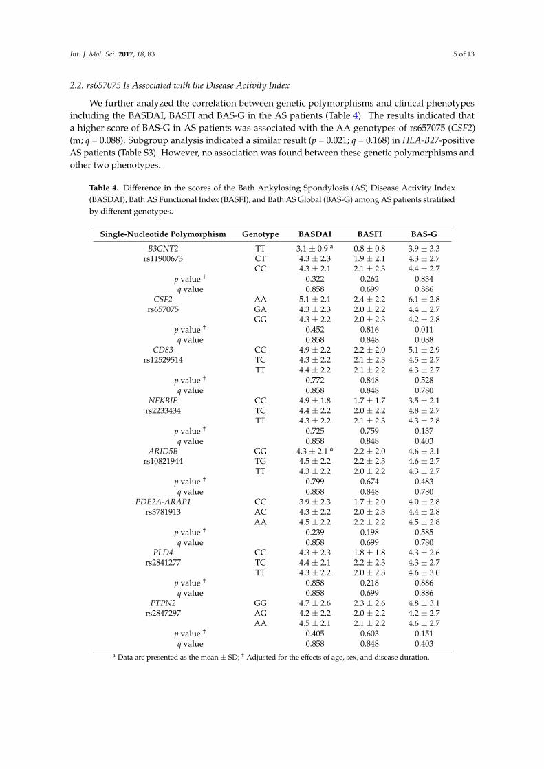
2.2. rs657075 Is Associated with the Disease Activity Index
We further analyzed the correlation between genetic polymorphisms and clinical phenotypesincluding the BASDAI, BASFI and BAS-G in the AS patients (Table 4). The results indicated thata higher score of BAS-G in AS patients was associated with the AA genotypes of rs657075 (CSF2)(m; q = 0.088). Subgroup analysis indicated a similar result (p = 0.021; q = 0.168) in HLA-B27-positiveAS patients (Table S3). However, no association was found between these genetic polymorphisms andother two phenotypes.
Table 4. Difference in the scores of the Bath Ankylosing Spondylosis (AS) Disease Activity Index(BASDAI), Bath AS Functional Index (BASFI), and Bath AS Global (BAS-G) among AS patients stratifiedby different genotypes.
Single-Nucleotide Polymorphism Genotype BASDAI BASFI BAS-G
B3GNT2 TT 3.1 ± 0.9 a 0.8 ± 0.8 3.9 ± 3.3rs11900673 CT 4.3 ± 2.3 1.9 ± 2.1 4.3 ± 2.7
CC 4.3 ± 2.1 2.1 ± 2.3 4.4 ± 2.7p value † 0.322 0.262 0.834q value 0.858 0.699 0.886
CSF2 AA 5.1 ± 2.1 2.4 ± 2.2 6.1 ± 2.8rs657075 GA 4.3 ± 2.3 2.0 ± 2.2 4.4 ± 2.7
GG 4.3 ± 2.2 2.0 ± 2.3 4.2 ± 2.8p value † 0.452 0.816 0.011q value 0.858 0.848 0.088
CD83 CC 4.9 ± 2.2 2.2 ± 2.0 5.1 ± 2.9rs12529514 TC 4.3 ± 2.2 2.1 ± 2.3 4.5 ± 2.7
TT 4.4 ± 2.2 2.1 ± 2.2 4.3 ± 2.7p value † 0.772 0.848 0.528q value 0.858 0.848 0.780
NFKBIE CC 4.9 ± 1.8 1.7 ± 1.7 3.5 ± 2.1rs2233434 TC 4.4 ± 2.2 2.0 ± 2.2 4.8 ± 2.7
TT 4.3 ± 2.2 2.1 ± 2.3 4.3 ± 2.8p value † 0.725 0.759 0.137q value 0.858 0.848 0.403
ARID5B GG 4.3 ± 2.1 a 2.2 ± 2.0 4.6 ± 3.1rs10821944 TG 4.5 ± 2.2 2.2 ± 2.3 4.6 ± 2.7
TT 4.3 ± 2.2 2.0 ± 2.2 4.3 ± 2.7p value † 0.799 0.674 0.483q value 0.858 0.848 0.780
PDE2A-ARAP1 CC 3.9 ± 2.3 1.7 ± 2.0 4.0 ± 2.8rs3781913 AC 4.3 ± 2.2 2.0 ± 2.3 4.4 ± 2.8
AA 4.5 ± 2.2 2.2 ± 2.2 4.5 ± 2.8p value † 0.239 0.198 0.585q value 0.858 0.699 0.780
PLD4 CC 4.3 ± 2.3 1.8 ± 1.8 4.3 ± 2.6rs2841277 TC 4.4 ± 2.1 2.2 ± 2.3 4.3 ± 2.7
TT 4.3 ± 2.2 2.0 ± 2.3 4.6 ± 3.0p value † 0.858 0.218 0.886q value 0.858 0.699 0.886
PTPN2 GG 4.7 ± 2.6 2.3 ± 2.6 4.8 ± 3.1rs2847297 AG 4.2 ± 2.2 2.0 ± 2.2 4.2 ± 2.7
AA 4.5 ± 2.1 2.1 ± 2.2 4.6 ± 2.7p value † 0.405 0.603 0.151q value 0.858 0.848 0.403
a Data are presented as the mean ± SD; † Adjusted for the effects of age, sex, and disease duration.
Int. J. Mol. Sci. 2017, 18, 83 6 of 13
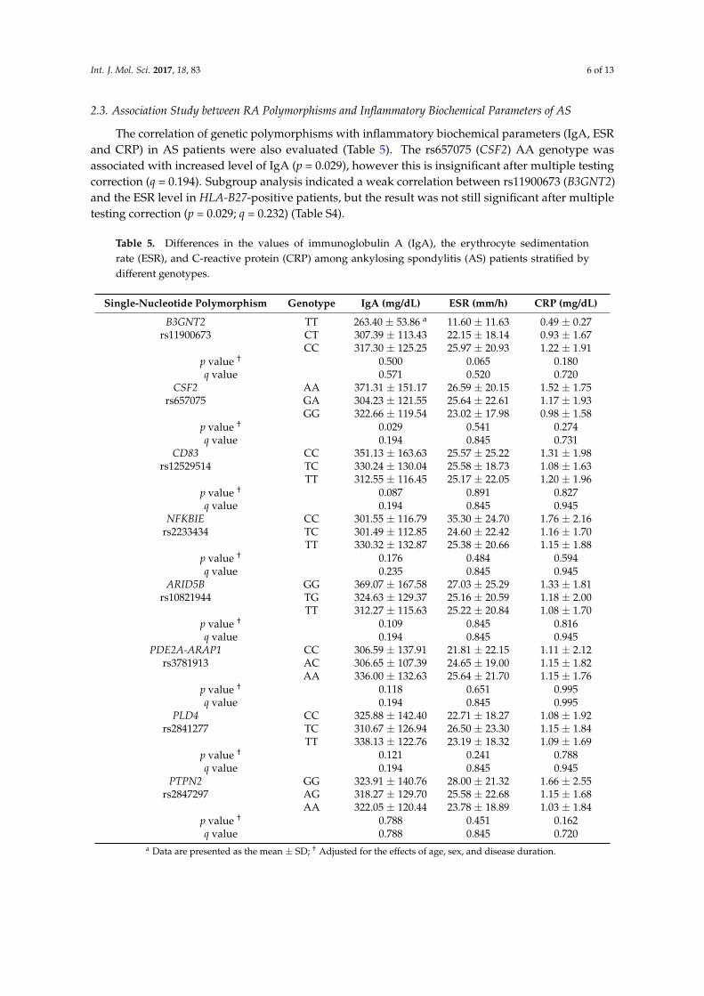
2.3. Association Study between RA Polymorphisms and Inflammatory Biochemical Parameters of AS
The correlation of genetic polymorphisms with inflammatory biochemical parameters (IgA, ESRand CRP) in AS patients were also evaluated (Table 5). The rs657075 (CSF2) AA genotype wasassociated with increased level of IgA (p = 0.029), however this is insignificant after multiple testingcorrection (q = 0.194). Subgroup analysis indicated a weak correlation between rs11900673 (B3GNT2)and the ESR level in HLA-B27-positive patients, but the result was not still significant after multipletesting correction (p = 0.029; q = 0.232) (Table S4).
Table 5. Differences in the values of immunoglobulin A (IgA), the erythrocyte sedimentationrate (ESR), and C-reactive protein (CRP) among ankylosing spondylitis (AS) patients stratified bydifferent genotypes.
Single-Nucleotide Polymorphism Genotype IgA (mg/dL) ESR (mm/h) CRP (mg/dL)
B3GNT2 TT 263.40 ± 53.86 a 11.60 ± 11.63 0.49 ± 0.27rs11900673 CT 307.39 ± 113.43 22.15 ± 18.14 0.93 ± 1.67
CC 317.30 ± 125.25 25.97 ± 20.93 1.22 ± 1.91p value † 0.500 0.065 0.180q value 0.571 0.520 0.720
CSF2 AA 371.31 ± 151.17 26.59 ± 20.15 1.52 ± 1.75rs657075 GA 304.23 ± 121.55 25.64 ± 22.61 1.17 ± 1.93
GG 322.66 ± 119.54 23.02 ± 17.98 0.98 ± 1.58p value † 0.029 0.541 0.274q value 0.194 0.845 0.731
CD83 CC 351.13 ± 163.63 25.57 ± 25.22 1.31 ± 1.98rs12529514 TC 330.24 ± 130.04 25.58 ± 18.73 1.08 ± 1.63
TT 312.55 ± 116.45 25.17 ± 22.05 1.20 ± 1.96p value † 0.087 0.891 0.827q value 0.194 0.845 0.945
NFKBIE CC 301.55 ± 116.79 35.30 ± 24.70 1.76 ± 2.16rs2233434 TC 301.49 ± 112.85 24.60 ± 22.42 1.16 ± 1.70
TT 330.32 ± 132.87 25.38 ± 20.66 1.15 ± 1.88p value † 0.176 0.484 0.594q value 0.235 0.845 0.945
ARID5B GG 369.07 ± 167.58 27.03 ± 25.29 1.33 ± 1.81rs10821944 TG 324.63 ± 129.37 25.16 ± 20.59 1.18 ± 2.00
TT 312.27 ± 115.63 25.22 ± 20.84 1.08 ± 1.70p value † 0.109 0.845 0.816q value 0.194 0.845 0.945
PDE2A-ARAP1 CC 306.59 ± 137.91 21.81 ± 22.15 1.11 ± 2.12rs3781913 AC 306.65 ± 107.39 24.65 ± 19.00 1.15 ± 1.82
AA 336.00 ± 132.63 25.64 ± 21.70 1.15 ± 1.76p value † 0.118 0.651 0.995q value 0.194 0.845 0.995
PLD4 CC 325.88 ± 142.40 22.71 ± 18.27 1.08 ± 1.92rs2841277 TC 310.67 ± 126.94 26.50 ± 23.30 1.15 ± 1.84
TT 338.13 ± 122.76 23.19 ± 18.32 1.09 ± 1.69p value † 0.121 0.241 0.788q value 0.194 0.845 0.945
PTPN2 GG 323.91 ± 140.76 28.00 ± 21.32 1.66 ± 2.55rs2847297 AG 318.27 ± 129.70 25.58 ± 22.68 1.15 ± 1.68
AA 322.05 ± 120.44 23.78 ± 18.89 1.03 ± 1.84p value † 0.788 0.451 0.162q value 0.788 0.845 0.720
a Data are presented as the mean ± SD; † Adjusted for the effects of age, sex, and disease duration.
Int. J. Mol. Sci. 2017, 18, 83 7 of 13
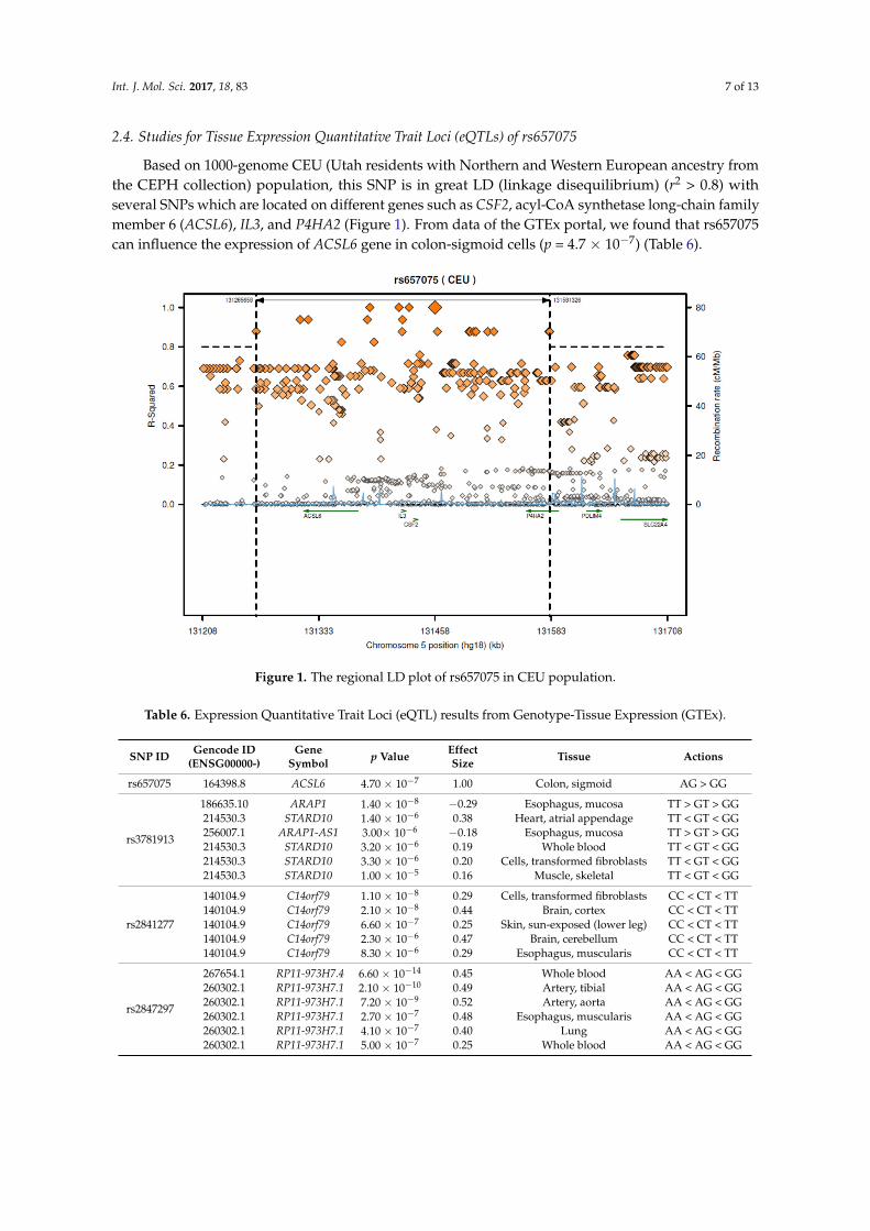
2.4. Studies for Tissue Expression Quantitative Trait Loci (eQTLs) of rs657075
Based on 1000-genome CEU (Utah residents with Northern and Western European ancestry fromthe CEPH collection) population, this SNP is in great LD (linkage disequilibrium) (r2 > 0.8) withseveral SNPs which are located on different genes such as CSF2, acyl-CoA synthetase long-chain familymember 6 (ACSL6), IL3, and P4HA2 (Figure 1). From data of the GTEx portal, we found that rs657075can influence the expression of ACSL6 gene in colon-sigmoid cells (p = 4.7 × 10⁻⁷) (Table 6).
Int. J. Mol. Sci. 2017, 18, 83 7 of 13
2.4. Studies for Tissue Expression Quantitative Trait Loci (eQTLs) of rs657075
Based on 1000-genome CEU (Utah residents with Northern and Western European ancestry from the CEPH collection) population, this SNP is in great LD (linkage disequilibrium) (r2 > 0.8) with several SNPs which are located on different genes such as CSF2, acyl-CoA synthetase long-chain family member 6 (ACSL6), IL3, and P4HA2 (Figure 1). From data of the GTEx portal, we found that rs657075 can influence the expression of ACSL6 gene in colon-sigmoid cells (p = 4.7 × 10⁻⁷) (Table 6).
Figure 1. The regional LD plot of rs657075 in CEU population.
Table 6. Expression Quantitative Trait Loci (eQTL) results from Genotype-Tissue Expression (GTEx).
SNP ID Gencode ID
(ENSG00000-) Gene
Symbol p Value Effect Size Tissue Actions
rs657075 164398.8 ACSL6 4.70 × 10⁻⁷ 1.00 Colon, sigmoid AG > GG
rs3781913
186635.10 ARAP1 1.40 × 10⁻⁸ −0.29 Esophagus, mucosa TT > GT > GG 214530.3 STARD10 1.40 × 10⁻⁶ 0.38 Heart, atrial appendage TT < GT < GG 256007.1 ARAP1-AS1 3.00× 10⁻⁶ −0.18 Esophagus, mucosa TT > GT > GG 214530.3 STARD10 3.20 × 10⁻⁶ 0.19 Whole blood TT < GT < GG 214530.3 STARD10 3.30 × 10⁻⁶ 0.20 Cells, transformed fibroblasts TT < GT < GG 214530.3 STARD10 1.00 × 10⁻⁵ 0.16 Muscle, skeletal TT < GT < GG
rs2841277
140104.9 C14orf79 1.10 × 10⁻⁸ 0.29 Cells, transformed fibroblasts CC < CT < TT 140104.9 C14orf79 2.10 × 10⁻⁸ 0.44 Brain, cortex CC < CT < TT 140104.9 C14orf79 6.60 × 10⁻⁷ 0.25 Skin, sun-exposed (lower leg) CC < CT < TT 140104.9 C14orf79 2.30 × 10⁻⁶ 0.47 Brain, cerebellum CC < CT < TT 140104.9 C14orf79 8.30 × 10⁻⁶ 0.29 Esophagus, muscularis CC < CT < TT
rs2847297
267654.1 RP11-973H7.4 6.60 × 10⁻¹⁴ 0.45 Whole blood AA < AG < GG 260302.1 RP11-973H7.1 2.10 × 10⁻¹⁰ 0.49 Artery, tibial AA < AG < GG 260302.1 RP11-973H7.1 7.20 × 10⁻⁹ 0.52 Artery, aorta AA < AG < GG 260302.1 RP11-973H7.1 2.70 × 10⁻⁷ 0.48 Esophagus, muscularis AA < AG < GG 260302.1 RP11-973H7.1 4.10 × 10⁻⁷ 0.40 Lung AA < AG < GG 260302.1 RP11-973H7.1 5.00 × 10⁻⁷ 0.25 Whole blood AA < AG < GG
Figure 1. The regional LD plot of rs657075 in CEU population.
Table 6. Expression Quantitative Trait Loci (eQTL) results from Genotype-Tissue Expression (GTEx).
SNP ID Gencode ID(ENSG00000-)
GeneSymbol p Value Effect
Size Tissue Actions
rs657075 164398.8 ACSL6 4.70 × 10⁻⁷ 1.00 Colon, sigmoid AG > GG
rs3781913
186635.10 ARAP1 1.40 × 10⁻⁸ −0.29 Esophagus, mucosa TT > GT > GG214530.3 STARD10 1.40 × 10⁻⁶ 0.38 Heart, atrial appendage TT < GT < GG256007.1 ARAP1-AS1 3.00× 10⁻⁶ −0.18 Esophagus, mucosa TT > GT > GG214530.3 STARD10 3.20 × 10⁻⁶ 0.19 Whole blood TT < GT < GG214530.3 STARD10 3.30 × 10⁻⁶ 0.20 Cells, transformed fibroblasts TT < GT < GG214530.3 STARD10 1.00 × 10⁻⁵ 0.16 Muscle, skeletal TT < GT < GG
rs2841277
140104.9 C14orf79 1.10 × 10⁻⁸ 0.29 Cells, transformed fibroblasts CC < CT < TT140104.9 C14orf79 2.10 × 10⁻⁸ 0.44 Brain, cortex CC < CT < TT140104.9 C14orf79 6.60 × 10⁻⁷ 0.25 Skin, sun-exposed (lower leg) CC < CT < TT140104.9 C14orf79 2.30 × 10⁻⁶ 0.47 Brain, cerebellum CC < CT < TT140104.9 C14orf79 8.30 × 10⁻⁶ 0.29 Esophagus, muscularis CC < CT < TT
rs2847297
267654.1 RP11-973H7.4 6.60 × 10⁻¹⁴ 0.45 Whole blood AA < AG < GG260302.1 RP11-973H7.1 2.10 × 10⁻¹⁰ 0.49 Artery, tibial AA < AG < GG260302.1 RP11-973H7.1 7.20 × 10⁻⁹ 0.52 Artery, aorta AA < AG < GG260302.1 RP11-973H7.1 2.70 × 10⁻⁷ 0.48 Esophagus, muscularis AA < AG < GG260302.1 RP11-973H7.1 4.10 × 10⁻⁷ 0.40 Lung AA < AG < GG260302.1 RP11-973H7.1 5.00 × 10⁻⁷ 0.25 Whole blood AA < AG < GG
Int. J. Mol. Sci. 2017, 18, 83 8 of 13
3. Discussion
AS and RA are autoimmune diseases with distinct phenotypes. RA is characterized as a chronicinflammatory joint disease with cartilage and bone damage, whereas spondyloarthropathies in ASare illustrated by entheses and subchondral bone marrow inflammation, particularly abnormalosteoproliferation at involved sites. Familial aggregations of AS and RA have been knownfor years [31,32]. In a 2008 Sweden study, Sundquist and coworkers analyzed 30 years ofhospitalizations (1973–2004) for concordant and discordant associations among RA, AS, and systemiclupus erythematosus (SLE). They reported that significant concordant measures with standardizedincidence ratios (SIRs) or sibling risks for RA and AS were 5.12 and 17.14, respectively. However, thediscordant association between RA and AS was not significant, and AS was only associated with ASwhen discordance was taken into account [31]. A further study in 2009 indicated that offspring ofparental probands with RA were significantly associated with AS with a SIR of 2.96 [32]. This laidthe foundation for the identification of common genetic components for RA and AS. Recently, someautoimmune disease loci were identified as being shared among multiple autoimmune diseases [33,34].Sirota and coworkers (2009) compared genetic variation profiles of six autoimmune disease and foundthat RA and AS displayed an autoimmune disease locus cluster that was distinct from the others.For instance, the G allele of rs2076530 in BTNL2 predisposed patients to RA, AS, and type 1 diabetes,but may play a protective role instead in multiple sclerosis and autoimmune thyroid disease.
To date, more than 43 AS risky loci [24] and 101 RA risky loci [35] have been identified.Most of the identified risk loci for autoimmune diseases are related to B-cell or T-cell activationpathways, differentiation, or innate immunity, or involved cytokine signaling or regulating peripheraltolerance. HLA-B27 has been known for years to be the major AS-susceptibility gene [6], butfewer than 5% of HLA-B27 carriers develop AS [24], suggesting that non-HLA-B27 alleles areimportant for AS susceptibility. Indeed, we recently provided evidence that genetic polymorphisms ofORAI1 (rs12313273 and rs7135617) and STIM1 (rs3750996) were associated with the pathogenesis ofHLA-B27-positive AS patients [18,19]. There is evidence that three of the SNPs (rs4552569, rs17095830,and rs13210693) which correspond to two newly identified AS risky loci (EDIL3-HAPLN1 and ANO6)in Han Chinese [22] were not replicated in Caucasian populations [20,21,23] and were negativelyassociated with Taiwanese AS subjects in a previous study of ours [27], eliciting the question ofwhether these two newly-identified loci associated with AS in Han Chinese [22] may have beenover-represented. Indeed the association of one SNP, rs13210693 at the 6q21 locus, did not reach ap value at the genome-wide significance level (p > 5 × 10⁻⁸) [22].
Bath ankylosing spondylitis global score (BAS-G) was a valuable quantitative measure which givesa global assessment of the well-being of the person with AS over a given time period as completedby the patient. It was first reported by Jones et al. in 1960 [36], and endorsed by Assessment ofSpondyloArthritis international Society (ASAS) [37]. It uses two horizontal visual analogue scales(10 cm) to measure the effect of AS on the respondent’s well-being, where none = 0 and severe = 10.The first estimates over the last week, and the second over the last six months. In this study, we firstlyreported that the genetic polymorphism rs657075 (CSF2) was associated with BAS-G.
CSF2 loci were previously reported to be associated with RA and exhibited high levels in jointsof RA patients [30]. In this study, for the first time we showed that CSF2 loci were not associatedwith AS development, but may be weakly associated with AS clinical phenotype BAS-G levels ina Taiwanese population. CSF2, also known as granulocyte-macrophage colony-stimulating factor,functions as a cytokine and is secreted by macrophages, T cells, mast cells, natural killer (NK) cells,endothelial cells, and fibroblasts. It stimulates stem cells to produce granulocytes and is part of theimmune/inflammatory cascade. On the other hand, the T helper Th17 cells require Csf2 to induceautoimmune encephalomyelitis or autoimmune neuroinflammation [38]. It is known that IL-23 andIL-23R are involved in pathogenic Th17 responses, and IL-23R is associated with many autoimmunediseases such as psoriasis, AS, and Crohn’s disease [24]. Perhaps levels of the weakly associated ASclinical phenotype BAS-G seen may have something to do with some of these gene–gene interactions.
Int. J. Mol. Sci. 2017, 18, 83 9 of 13
According to SNAP website, this SNP has high LD with several genes including CSF2 and ACSL6(Figure 1). Since the GTEx database includes limited cell types, rs657075 does not show significantlyassociated eQTL with the CSF2 gene in the current database. It is associated with expression ofACSL6 in sigmoid colon cells (Table 6). On searching HaploReg V4.1, rs657075 is involved in changingchromatin status in primary T cells. Therefore, further functional studies are needed to validate theimpact of rs657075 on the expression of CSF2.
Using a high-density immune-related loci platform (Immunochip), Cortes and coworkers reportedthat some of the AS risky loci overlapped with those of other immune diseases. Eleven of these ASrisky loci were positively associated with ulcerative colitis and 12 loci with Crohn’s disease. However,only two of the AS risky loci were marginally associated with RA; rs11065898 (SH2B3 or LNK) andrs2283790 (UBE2L3) displayed a concordant and discordant mode, respectively. As SH2B3 encoded anadaptor protein involving T cell signaling, it may be functionally related to the pathogenesis of AS.Interestingly, rs11065898 loci were indeed positively associated with CD4+ lymphocyte counts [24].
The modest correlation between RA risk SNPs and AS susceptibility that we observed in thisstudy may due to several reasons.
Firstly, the SNPs were identified from a RA GWAS study conducted in a Japanese cohort. In thisstudy, we assess their contribution to AS susceptibility and disease severity in a Taiwanese cohort.The nonsignificant results regarding susceptibility to Taiwanese AS patients may be attributed tothe heterogeneity of the phenotype (diagnostic criteria of the disease). In considering of phenotypicheterogeneity, we have noted different criteria of clinical diagnosis between RA (in the GWAS studyfrom a Japanese population) and AS (in this study based on a Taiwanese population).
Secondly, it is likely that different genetic backgrounds (variation in allele frequencies acrossdifferent ethnic population admixture) in two populations may play a role in the observed inconsistentresults. In particular, different genetic backgrounds between Japanese and Taiwanese populations mayhave an impact on the genetic influence of rs657075 in the two study populations, consequently, thismay result in the inconsistent association that we observed in these two studies.
Thirdly, the prevalences of AS between Taiwanese and Japanese populations are different.The prevalence in Japanese population is lower than in Taiwanese population (0.0065% vs.0.19%–0.54%) [39–41]. The distinct prevalence of AS may imply various genetic factors influencingdifferent underlying regulations in AS across population, which may partially explain thenonsignificant association observed in our study. For example, a previous study reported that threeSNPs (rs4552569, rs17095830, and rs13210693) were associated with AS in Han Chinese [22], but thiswas not replicated in Caucasian population [20,21,23], nor were these associated with Taiwanese ASsubjects [27].
Fourthly, it has been known that the genetic effect size of SNP may be different across populations.Since some important confounding factors are unmeasured and can not be adjusted in the analyses,this may have a certain degree of influence when determining the risk effect of those SNPs on AS.
Although no correlation between RA susceptible SNPs and AS risk was observed, we have shownthat these SNPs are good candidates for AS disease severity, i.e., BASDAI, BASFI, and BAS-G afteradjusting for the effects of gender, age, and disease duration. It would still be interesting to considerthose previously identified SNPs as good candidates for determining increased risk of AS. As such,further investigation would be merited to validate these SNPs identified from the Japanese AS GWASstudy. It will be also important to the authors to further examine the association between these SNPsand increasing AS risk when sample size allows in the future.
The small sample size in this study may have limited the statistical power for detecting sizeablechanges in AS risk assessments. Larger cohort studies and replication studies in different populationsare required to validate our findings in this study. In summary, although the SNPs (rs657075 on CSF2and rs11900673 on B3GNT2) are not the risk alleles for the susceptibility of AS, we provide supportiveevidence that these SNPs are important generic markers for clinical manifestations of AS patients in aTaiwanese population. Further investigation would be needed to validate the findings in this study.
Int. J. Mol. Sci. 2017, 18, 83 10 of 13
4. Materials and Methods
4.1. Study Subjects
Patients were sequentially recruited from the AS clinic at Chung Shan Medical University Hospital(Taichung, Taiwan). Our study conformed to the Declaration of Helsinki, and the design of the work andfinal report were performed with the approval of the institutional review board of Chung Shan MedicalUniversity Hospital (CSMUH No: CS12255). We received informed consent from all subjects beforeany data were collected. Patients with AS who met the selection criteria were asked to participate in thestudy. In total 475 AS patients were recruited from the Arthritis Clinic, and diagnosed by a qualifiedrheumatologist. The criteria included (i) patients being aged 17–82 years; (ii) an AS diagnosis havingbeen made by modified New York criteria developed in 1984 [42]; (iii) being fluent Chinese languagespeakers; and (iv) having cognitive performance that was not influenced by other disease such asdementia. Patients with sacroiliitis were confirmed by a qualified radiologist. The detailed clinicalhistory included age at initial symptoms, extra-spinal manifestations , and laboratory parametersof inflammation, i.e., the ESR, CRP, and IgA. The age of onset of AS symptom was defined as thetime when the first symptom (axial symptom, peripheral arthritis, uveitis, or enthesitis) was noted.The BASDAI, BASFI, and BAS-G were applied to evaluate the disease activity, physical functions,and global wellbeing, respectively. A family history of AS was also recorded. Peripheral arthritiswas defined as the presence of at least one swollen joint. These symptoms were determined by arheumatologist, ophthalmologist, and gastroenterologist.
4.2. Candidate Single-Nucleotide Polymorphisms (SNPs)
We included eight SNPs which showed a significant association with RA in previous GWASs ina Japanese population (Table 1) [30]. rs2867461 (ANXA3) was excluded from the statistical analysis,because many subjects were unsuccessfully genotyped due to failure of polymerase chain reaction(PCR) amplification.
4.3. DNA Extraction
Peripheral venous blood was collected during medical surveillance, and processed in the sameday. Blood was centrifuged to separate the serum and cells. DNA of blood cells was extracted by firsttreating them with 0.5% sodium dodecylsulfate lysis buffer and then protease K (1 mg/mL) to digestnuclear proteins for 4 h at 60 ◦C according to the manufacturer’s procedures. Total DNA was harvestedusing a Gentra (Qiagen, Valencia, CA, USA) extraction kit followed by 70% alcohol precipitationas recommended.
4.4. Genotyping
Genotyping for the eight SNPs was performed using the TaqMan Allelic Discrimination Assay(Applied Biosystems, Foster City, CA, USA). PCR was performed in the 96-well microplate andwith the ABI9700 Thermal Cycler (Applied Biosystems). After the PCR, fluorescence was measuredand analyzed using the StepOne software vers. 2.2.2 (Applied Biosystems, Foster City, CA, USA).HLA-B27 carriage was assessed by flow cytometry as previously described [43].
4.5. SNP Annotation Data Query
In order to understand to potential regulated genes by each SNP, we queried SNAP(https://www.broadinstitute.org/mpg/snap/index.php) to download the regional LD plot ofinterests for SNPs. The GTEx Portal (http://www.gtexportal.org/home/), which includes a variety oftissue expression quantitative trait loci (eQTLs), was used to investigate the association between geneexpression profiles and the SNPs.
Int. J. Mol. Sci. 2017, 18, 83 11 of 13
4.6. Statistical Analysis
SAS 9.3 (SAS Institute, Cary, NC, USA) for Windows was used for the statistical analysis.Hardy–Weinberg equilibrium (HWE) was evaluated to test the allelic frequencies of SNP’s usinga chi-squared test (χ2 test). Statistical differences between the patient and control group in genotypefrequencies were evaluated by a χ2 test. An analysis of variance (ANOVA) was used to compare themean of continuous variables (BASDAI, BASFI, BAS-F, IgA, ESR, and CRP) among different genotypesin AS patients. An analysis of covariance (ANCOVA) was applied to adjust for gender, age, and diseaseduration. The false discovery rate (FDR) was applied in order to correct for multiple testing errors.FDR q values of <0.05 was considered statistically significant.
Supplementary Materials: Supplementary materials can be found at www.mdpi.com/1422-0067/18/1/83/s1.
Acknowledgments: This work was supported by funding from an Excellence for Cancer Research Center grant,Department of Health, Executive Yuan, Taiwan, ROC (DOH102-TD-C-111-002) and grants from the NationalScience Council, Taiwan, ROC (MOST 104-2320-B-038-016) and from Taipei Medical University (12310-0223) andfrom Chung Shan Medical University Hospital (CSH-2011-C-004).
Author Contributions: Experimental design: Wei-Chiao Chen, James Cheng-Chung Wei, and Jin-Ding Huang;Implementation of experiments: Wei-Chiao Chen, Peng Yeong Woon, and Yu-Wen Hsu; Data analysis:Wei-Chiao Chen, Hsing-Fang Lu, Wei-Chiao Chang, James Cheng-Chung Wei, and Jin-Ding Huang; Contributionof reagents/materials/analysis tools: James Cheng-Chung Wei, Wei-Chiao Chang, and Jin-Ding Huang; Clinicalsamples and data collection: James Cheng-Chung Wei; Writing of the paper: Wei-Chiao Chen, Hsing-Fang Lu,Henry Sung-Ching Wong, and Wei-Chiao Chang.
Conflicts of Interest: The authors declare no conflict of interest.
References
1. Braun, J.; Sieper, J. Ankylosing spondylitis. Lancet 2007, 369, 1379–1390. [CrossRef]2. Pal, B. Ankylosing spondylitis, a seronegative spondarthritis. Practitioner 1987, 231, 785–793. [PubMed]3. Calin, A.; Brophy, S.; Blake, D. Impact of sex on inheritance of ankylosing spondylitis: A cohort study. Lancet
1999, 354, 1687–1690. [CrossRef]4. Brown, M.A.; Laval, S.H.; Brophy, S.; Calin, A. Recurrence risk modelling of the genetic susceptibility to
ankylosing spondylitis. Ann. Rheum. Dis. 2000, 59, 883–886. [CrossRef] [PubMed]5. Brown, M.A.; Kennedy, L.G.; MacGregor, A.J.; Darke, C.; Duncan, E.; Shatford, J.L.; Taylor, A.; Calin, A.;
Wordsworth, P. Susceptibility to ankylosing spondylitis in twins: The role of genes, HLA, and theenvironment. Arthritis Rheumatol. 1997, 40, 1823–1828. [CrossRef]
6. Brewerton, D.A.; Hart, F.D.; Nicholls, A.; Caffrey, M.; James, D.C.; Sturrock, R.D. Ankylosing spondylitis andHL-A 27. Lancet 1973, 1, 904–907. [CrossRef]
7. Khan, M.A.; Ball, E.J. Genetic aspects of ankylosing spondylitis. Best Pract. Res. Clin. Rheumatol. 2002, 16,675–690. [CrossRef]
8. Robinson, W.P.; Van der Linden, S.M.; Khan, M.A.; Rentsch, H.U.; Cats, A.; Russell, A.; Thomson, G.HLA-Bw60 increases susceptibility to ankylosing spondylitis in HLA-B27+ patients. Arthritis Rheumatol.1989, 32, 1135–1141. [CrossRef]
9. Brown, M.A.; Pile, K.D.; Kennedy, L.G.; Calin, A.; Darke, C.; Bell, J.; Wordsworth, B.P.; Cornelis, F. HLA classI associations of ankylosing spondylitis in the white population in the United Kingdom. Ann. Rheum. Dis.1996, 55, 268–270. [CrossRef] [PubMed]
10. Wei, J.C.; Tsai, W.C.; Lin, H.S.; Tsai, C.Y.; Chou, C.T. HLA-B60 and B61 are strongly associated with ankylosingspondylitis in HLA-B27-negative Taiwan Chinese patients. Rheumatology 2004, 43, 839–842. [CrossRef][PubMed]
11. Wei, J.C.; Sung-Ching, H.W.; Hsu, Y.W.; Wen, Y.F.; Wang, W.C.; Wong, R.H.; Lu, H.F.; Gaalen, F.A.; Chang, W.C.Interaction between HLA-B60 and HLA-B27 as a Better Predictor of Ankylosing Spondylitis in a TaiwanesePopulation. PLoS ONE 2015, 10, e0137189. [CrossRef] [PubMed]
12. Van Gaalen, F.A.; Verduijn, W.; Roelen, D.L.; Bohringer, S.; Huizinga, T.W.; Van der Heijde, D.M.; Toes, R.E.Epistasis between two HLA antigens defines a subset of individuals at a very high risk for ankylosingspondylitis. Ann. Rheum. Dis. 2013, 72, 974–978. [CrossRef] [PubMed]
Int. J. Mol. Sci. 2017, 18, 83 12 of 13
13. Guo, Z.S.; Li, C.; Lin, Z.M.; Huang, J.X.; Wei, Q.J.; Wang, X.W.; Xie, Y.Y.; Liao, Z.T.; Chao, S.Y.; Gu, J.R.Association of IL-1 gene complex members with ankylosing spondylitis in Chinese Han population.Int. J. Immunogenet. 2010, 37, 33–37. [CrossRef] [PubMed]
14. Safrany, E.; Pazar, B.; Csongei, V.; Jaromi, L.; Polgar, N.; Sipeky, C.; Horvath, I.F.; Zeher, M.; Poor, G.;Melegh, B. Variants of the IL23R gene are associated with ankylosing spondylitis but not with Sjogrensyndrome in Hungarian population samples. Scand. J. Immunol. 2009, 70, 68–74. [CrossRef] [PubMed]
15. Huang, C.H.; Wong, R.H.; Wei, J.C.; Tsay, M.D.; Chen, W.C.; Chen, H.Y.; Shih, W.T.; Chiou, S.P.; Tu, Y.C.;Lee, H.S. Effects of genetic polymorphisms of programmed cell death 1 and its ligands on the developmentof ankylosing spondylitis. Rheumatology 2011, 50, 1809–1813. [CrossRef] [PubMed]
16. Huang, C.H.; Wei, J.C.; Chen, C.C.; Chuang, C.S.; Chou, C.H.; Lin, Y.J.; Wang, M.F.; Wong, R.H. Associationsof the PTPN22 and CTLA-4 genetic polymorphisms with Taiwanese ankylosing spondylitis. Rheumatol. Int.2014, 34, 683–691. [CrossRef] [PubMed]
17. Lee, W.Y.; Chang, Y.H.; Lo, M.K.; Chang, C.P.; Yang, S.C.; Yang, T.P.; Ho, K.T.; Juan, C.W.; Shiau, M.Y.Polymorphisms of cytotoxic T lymphocyte-associated antigen-4 and cytokine genes in Taiwanese patientswith ankylosing spondylitis. Tissue Antigens 2010, 75, 119–126. [CrossRef] [PubMed]
18. Wei, J.C.; Yen, J.H.; Juo, S.H.; Chen, W.C.; Wang, Y.S.; Chiu, Y.C.; Hsieh, T.J.; Guo, Y.C.; Huang, C.H.;Wong, R.H.; et al. Association of ORAI1 haplotypes with the risk of HLA-B27 positive ankylosing spondylitis.PLoS ONE 2011, 6, e20426. [CrossRef] [PubMed]
19. Wei, J.C.; Hung, K.S.; Hsu, Y.W.; Wong, R.H.; Huang, C.H.; Jan, M.S.; Wu, S.J.; Juan, Y.S.; Chang, W.C.Genetic polymorphisms of stromal interaction molecule 1 associated with the erythrocyte sedimentationrate and C-reactive protein in HLA-B27 positive ankylosing spondylitis patients. PLoS ONE 2012, 7, e49698.[CrossRef] [PubMed]
20. Australo-Anglo-American Spondyloarthritis Consortium; Reveille, J.D.; Sims, A.M.; Danoy, P.; Evans, D.M.;Leo, P.; Pointon, J.J.; Jin, R.; Zhou, X.; Bradbury, L.A.; et al. Genome-wide association study of ankylosingspondylitis identifies non-MHC susceptibility loci. Nat. Genet. 2010, 42, 123–127.
21. Evans, D.M.; Spencer, C.C.; Pointon, J.J.; Su, Z.; Harvey, D.; Kochan, G.; Oppermann, U.; Dilthey, A.;Pirinen, M.; Stone, M.A.; et al. Interaction between ERAP1 and HLA-B27 in ankylosing spondylitis implicatespeptide handling in the mechanism for HLA-B27 in disease susceptibility. Nat. Genet. 2011, 43, 761–767.[CrossRef] [PubMed]
22. Lin, Z.; Bei, J.X.; Shen, M.; Li, Q.; Liao, Z.; Zhang, Y.; Lv, Q.; Wei, Q.; Low, H.Q.; Guo, Y.M.; et al.A genome-wide association study in Han Chinese identifies new susceptibility loci for ankylosing spondylitis.Nat. Genet. 2012, 44, 73–77. [CrossRef] [PubMed]
23. Wellcome Trust Case Control Consortium; Australo-Anglo-American Spondylitis Consortium; Burton, P.R.;Clayton, D.G.; Cardon, L.R.; Craddock, N.; Deloukas, P.; Duncanson, A.; Kwiatkowski, D.P.;McCarthy, M.I.; et al. Association scan of 14,500 nonsynonymous SNPs in four diseases identifiesautoimmunity variants. Nat. Genet. 2007, 39, 1329–1337.
24. International Genetics of Ankylosing Spondylitis Consortium; Cortes, A.; Hadler, J.; Pointon, J.P.;Robinson, P.C.; Karaderi, T.; Leo, P.; Cremin, K.; Pryce, K.; Harris, J.; et al. Identification of multiplerisk variants for ankylosing spondylitis through high-density genotyping of immune-related loci. Nat. Genet.2013, 45, 730–738.
25. Wang, C.M.; Ho, H.H.; Chang, S.W.; Wu, Y.J.; Lin, J.C.; Chang, P.Y.; Wu, J.; Chen, J.Y. ERAP1 geneticvariations associated with HLA-B27 interaction and disease severity of syndesmophytes formation inTaiwanese ankylosing spondylitis. Arthritis Res. Ther. 2012, 14, R125. [CrossRef] [PubMed]
26. Wen, Y.F.; Wei, J.C.; Hsu, Y.W.; Chiou, H.Y.; Wong, H.S.; Wong, R.H.; Ikegawa, S.; Chang, W.C.rs10865331 associated with susceptibility and disease severity of ankylosing spondylitis in a Taiwanesepopulation. PLoS ONE 2014, 9, e104525. [CrossRef] [PubMed]
27. Wei, J.C.; Hsu, Y.W.; Hung, K.S.; Wong, R.H.; Huang, C.H.; Liu, Y.T.; Guo, Y.C.; Ikegawa, S.; Chang, W.C.Association study of polymorphisms rs4552569 and rs17095830 and the risk of ankylosing spondylitis in aTaiwanese population. PLoS ONE 2013, 8, e52801. [CrossRef] [PubMed]
28. Firestein, G.S. Evolving concepts of rheumatoid arthritis. Nature 2003, 423, 356–361. [CrossRef] [PubMed]29. Terao, C.; Raychaudhuri, S.; Gregersen, P.K. Recent Advances in Defining the Genetic Basis of Rheumatoid
Arthritis. Annu. Rev. Genom. Hum. Genet. 2016, 17, 273–301. [CrossRef] [PubMed]
Int. J. Mol. Sci. 2017, 18, 83 13 of 13
30. Okada, Y.; Terao, C.; Ikari, K.; Kochi, Y.; Ohmura, K.; Suzuki, A.; Kawaguchi, T.; Stahl, E.A.; Kurreeman, F.A.;Nishida, N.; et al. Meta-analysis identifies nine new loci associated with rheumatoid arthritis in the Japanesepopulation. Nat. Genet. 2012, 44, 511–516. [CrossRef] [PubMed]
31. Sundquist, K.; Martineus, J.C.; Li, X.; Hemminki, K.; Sundquist, J. Concordant and discordant associationsbetween rheumatoid arthritis, systemic lupus erythematosus and ankylosing spondylitis based on allhospitalizations in Sweden between 1973 and 2004. Rheumatology 2008, 47, 1199–1202. [CrossRef] [PubMed]
32. Hemminki, K.; Li, X.; Sundquist, J.; Sundquist, K. Familial associations of rheumatoid arthritis withautoimmune diseases and related conditions. Arthritis Rheum. 2009, 60, 661–668. [CrossRef] [PubMed]
33. Sirota, M.; Schaub, M.A.; Batzoglou, S.; Robinson, W.H.; Butte, A.J. Autoimmune disease classification byinverse association with SNP alleles. PLoS Genet. 2009, 5, e1000792. [CrossRef] [PubMed]
34. Richard-Miceli, C.; Criswell, L.A. Emerging patterns of genetic overlap across autoimmune disorders.Genom. Med. 2012, 4, 6. [CrossRef] [PubMed]
35. Okada, Y.; Wu, D.; Trynka, G.; Raj, T.; Terao, C.; Ikari, K.; Kochi, Y.; Ohmura, K.; Suzuki, A.; Yoshida, S.; et al.Genetics of rheumatoid arthritis contributes to biology and drug discovery. Nature 2014, 506, 376–381.[CrossRef] [PubMed]
36. Jones, S.D.; Steiner, A.; Garrett, S.L.; Calin, A. The Bath Ankylosing Spondylitis Patient Global Score (BAS-G).Br. J. Rheumatol. 1996, 35, 66–71. [CrossRef] [PubMed]
37. Zochling, J. Measures of symptoms and disease status in ankylosing spondylitis: Ankylosing SpondylitisDisease Activity Score (ASDAS), Ankylosing Spondylitis Quality of Life Scale (ASQoL), Bath AnkylosingSpondylitis Disease Activity Index (BASDAI), Bath Ankylosing Spondylitis Functional Index (BASFI), BathAnkylosing Spondylitis Global Score (BAS-G), Bath Ankylosing Spondylitis Metrology Index (BASMI),Dougados Functional Index (DFI), and Health Assessment Questionnaire for the Spondylarthropathies(HAQ-S). Arthritis Care Res. 2011, 63, S47–S58.
38. Codarri, L.; Gyulveszi, G.; Tosevski, V.; Hesske, L.; Fontana, A.; Magnenat, L.; Suter, T.; Becher, B.RORgammat drives production of the cytokine GM-CSF in helper T cells, which is essential for the effectorphase of autoimmune neuroinflammation. Nat. Immunol. 2011, 12, 560–567. [CrossRef] [PubMed]
39. Hukuda, S.; Minami, M.; Saito, T.; Mitsui, H.; Matsui, N.; Komatsubara, Y.; Makino, H.; Shibata, T.; Shingu, M.;Sakou, T.; et al. Spondyloarthropathies in Japan: Nationwide questionnaire survey performed by the JapanAnkylosing Spondylitis Society. J. Rheumatol. 2001, 28, 554–559. [PubMed]
40. Chou, C.T.; Pei, L.; Chang, D.M.; Lee, C.F.; Schumacher, H.R.; Liang, M.H. Prevalence of rheumatic diseases inTaiwan: A population study of urban, suburban, rural differences. J. Rheumatol. 1994, 21, 302–306. [PubMed]
41. Chou, C.T.; Schumacher, H.R. Human leucocyte antigens (class I and II) in central Taiwan aborigines:Can these explain the observed differences in rheumatic disease patterns compared with Han Chinese?Rheumatology 2000, 39, 1297–1299. [CrossRef] [PubMed]
42. Van der Linden, S.; Valkenburg, H.A.; Cats, A. Evaluation of diagnostic criteria for ankylosing spondylitis.A proposal for modification of the New York criteria. Arthritis Rheum. 1984, 27, 361–368. [CrossRef][PubMed]
43. Chou, C.T.; Tsai, Y.F.; Liu, J.; Wei, J.C.; Liao, T.S.; Chen, M.L.; Liu, L.Y. The detection of the HLA-B27antigen by immunomagnetic separation and enzyme-linked immunosorbent assay-comparison with a flowcytometric procedure. J. Immunol. Methods 2001, 255, 15–22. [CrossRef]
© 2017 by the authors; licensee MDPI, Basel, Switzerland. This article is an open accessarticle distributed under the terms and conditions of the Creative Commons Attribution(CC-BY) license (http://creativecommons.org/licenses/by/4.0/).


![Distraction Histiogenesis for the Treatment of Lichtman ...web.csh.org.tw/web/csh/wp-content/uploads/2018/02/27.pdf · tiogenesis as used in Perthes disease in children[8, 9]. We](https://img.pdfslide.us/doc/110x75/5aeda4f97f8b9a90318ff8e9/distraction-histiogenesis-for-the-treatment-of-lichtman-webcshorgtwwebcshwp-contentuploads20180227pdftiogenesis.jpg)