Embed Size (px)
Citation preview

Anesthesiology, V 121 • No 1 149 July 2014
(R,S)-KETAMINE is a rapid and long-lasting antidepres-sant agent used in the treatment of major depressive disorder and bipolar depression.1–3 Data from studies in rats suggest that the rapid effect produced by (R,S)-ketamine is due to the increased phosphorylation of the mammalian target of rapamycin (mTOR) and corresponding increases in the phosphorylated forms of the extracellular signal–regulated kinases (pERK1/ERK2), protein kinase B (pAkt), eukaryotic initiation factor 4E binding protein (p4E-BP1), and p70S6 kinase (pp70S6K)4 and an increase in the number and func-tion of new spine synapses in the prefrontal cortex.4,5
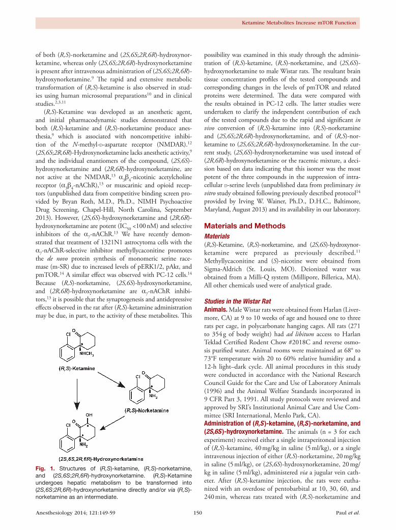
(R,S)-Ketamine is extensively transformed into the N-demethylated metabolite (R,S)-norketamine, two dia-stereomeric hydroxyketamines, a series of diastereomeric hydroxynorketamines, including (2S,6S;2R,6R)-hydroxynor-ketamine, and (R,S)-dehydronorketamine6–8; figure 1. In the rat, (R,S)-ketamine is rapidly converted to (R,S)-norketamine
and (2S,6S;2R,6R)-hydroxynorketamine, which occurs within 2 min after an intravenous administration.9 Similarly, administration of (R,S)-norketamine leads to the detection
What We Already Know about This Topic
• Anesthetic effects of ketamine are primarily due to (R,S)-ketamine;itsmetabolite(R,S)-norketaminecontributestothiseffect but its metabolite (2S,6S;2R,6R)-hydroxynorketaminedoesnot
What This Article Tells Us That Is New
• Antidepressant effects of subanesthetic doses of (R,S)-ketaminemaybedue to a combinationof interrelated ef-fectsat theα7-nicotinicacetylcholine receptor (α7-nAChR)producedby(R,S)-ketamineanditsmetabolites
• Oneeffectisincreasedproteinexpressionviathemammaliantarget of rapamycin signaling pathway,which is initiated byantagonismofα7-nAChRandisreflectedbyincreasedmono-mericserineracemaseexpression
Copyright © 2014, the American Society of Anesthesiologists, Inc. Lippincott Williams & Wilkins. Anesthesiology 2014; 121:149-59
ABSTRACT
Background: Subanesthetic doses of (R,S)-ketamine are used in the treatment of neuropathic pain and depression. In the rat, the antidepressant effects of (R,S)-ketamine are associated with increased activity and function of mammalian target of rapamycin (mTOR); however, (R,S)-ketamine is extensively metabolized and the contribution of its metabolites to increased mTOR signaling is unknown.Methods: Rats (n = 3 per time point) were given (R,S)-ketamine, (R,S)-norketamine, and (2S,6S)-hydroxynorketamine and their effect on the mTOR pathway determined after 20, 30, and 60 min. PC-12 pheochromocytoma cells (n = 3 per experi-ment) were treated with escalating concentrations of each compound and the impact on the mTOR pathway was determined.Results: The phosphorylation of mTOR and its downstream targets was significantly increased in rat prefrontal cortex tissue by more than ~2.5-, ~25-, and ~2-fold, respectively, in response to a 60-min postadministration of (R,S)-ketamine, (R,S)-norketamine, and (2S,6S)-hydroxynorketamine (P < 0.05, ANOVA analysis). In PC-12 pheochromocytoma cells, the test compounds activated the mTOR pathway in a concentration-dependent manner, which resulted in a significantly higher expression of serine racemase with ~2-fold increases at 0.05 nM (2S,6S)-hydroxynorketamine, 10 nM (R,S)-norketamine, and 1,000 nM (R,S)-ketamine. The potency of the effect reflected antagonistic activity of the test compounds at the α7-nicotinic acetylcholine receptor.Conclusions: The data demonstrate that (R,S)-norketamine and (2S,6S)-hydroxynorketamine have potent pharmacological activity both in vitro and in vivo and contribute to the molecular effects produced by subanesthetic doses of (R,S)-ketamine. The results suggest that the determination of the mechanisms underlying the antidepressant and analgesic effects of (R,S)-ketamine requires a full study of the parent compound and its metabolites. (Anesthesiology 2014; 121:149-59)
This article is featured in “This Month in Anesthesiology,” page 3A. Corresponding article on p. 4. Supplemental Digital Content is avail-able for this article. Direct URL citations appear in the printed text and are available in both the HTML and PDF versions of this article. Links to the digital files are provided in the HTML text of this article on the Journal’s Web site (www.anesthesiology.org). The first two authors contributed equally to this work (R.K.P. and N.S.S.).
Submitted for publication October 12, 2013. Accepted for publication March 28, 2014. From the Laboratory of Clinical Investigation (R.K.P., N.S.S., M.K., R.M., M.S., I.W.W.) and Translational Gerontology Branch (M.B.), National Institute on Aging, National Institutes of Health, Baltimore, Maryland; SRI Biosciences, SRI International, Menlo Park, California (C.E.G., K.O.); and Department of Anesthesiology, Cooper Medical School of Rowan University, Camden, New Jersey (M.C.T., I.W.W.).
(R,S)-Ketamine Metabolites (R,S)-norketamine and (2S,6S)-hydroxynorketamine Increase the Mammalian Target of Rapamycin Function
RajibK.Paul,Ph.D.,NagendraS.Singh,Ph.D.,MohammedKhadeer,Ph.D.,RuinMoaddel,Ph.D.,MiteshSanghvi,Ph.D.,CarolE.Green,Ph.D.,DABT,KathleenO’Loughlin,B.Sc.,MarcC.Torjman,Ph.D.,MichelBernier,Ph.D.,IrvingW.Wainer,Ph.D.,D.H.C.
Anesthesiology 2014; 121:149-59 150 Paul et al.
Ketamine Metabolites Increase mTOR Function
of both (R,S)-norketamine and (2S,6S;2R,6R)-hydroxynor-ketamine, whereas only (2S,6S;2R,6R)-hydroxynorketamine is present after intravenous administration of (2S,6S;2R,6R)-hydroxynorketamine.9 The rapid and extensive metabolic transformation of (R,S)-ketamine is also observed in stud-ies using human microsomal preparations10 and in clinical studies.2,3,11
(R,S)-Ketamine was developed as an anesthetic agent, and initial pharmacodynamic studies demonstrated that both (R,S)-ketamine and (R,S)-norketamine produce anes-thesia,9 which is associated with noncompetitive inhibi-tion of the N-methyl-D-aspartate receptor (NMDAR).12 (2S,6S;2R,6R)-Hydroxynorketamine lacks anesthetic activity,9 and the individual enantiomers of the compound, (2S,6S)-hydroxynorketamine and (2R,6R)-hydroxynorketamine, are not active at the NMDAR,13 α3β4-nicotinic acetylcholine receptor (α3β4-nAChR),13 or muscarinic and opioid recep-tors (unpublished data from competitive binding screen pro-vided by Bryan Roth, M.D., Ph.D., NIMH Psychoactive Drug Screening, Chapel-Hill, North Carolina, September 2013). However, (2S,6S)-hydroxynorketamine and (2R,6R)-hydroxynorketamine are potent (IC50 <100 nM) and selective inhibitors of the α7-nAChR.13 We have recently demon-strated that treatment of 1321N1 astrocytoma cells with the α7-nAChR-selective inhibitor methyllycaconitine promotes the de novo protein synthesis of monomeric serine race-mase (m-SR) due to increased levels of pERK1/2, pAkt, and pmTOR.14 A similar effect was observed with PC-12 cells.14 Because (R,S)-norketamine, (2S,6S)-hydroxynorketamine, and (2R,6R)-hydroxynorketamine are α7-nAChR inhibi-tors,13 it is possible that the synaptogenesis and antidepressive effects observed in the rat after (R,S)-ketamine administration may be due, in part, to the activity of these metabolites. This
possibility was examined in this study through the adminis-tration of (R,S)-ketamine, (R,S)-norketamine, and (2S,6S)-hydroxynorketamine to male Wistar rats. The resultant brain tissue concentration profiles of the tested compounds and corresponding changes in the levels of pmTOR and related proteins were determined. The data were compared with the results obtained in PC-12 cells. The latter studies were undertaken to clarify the independent contribution of each of the tested compounds due to the rapid and significant in vivo conversion of (R,S)-ketamine into (R,S)-norketamine and (2S,6S;2R,6R)-hydroxynorketamine, and of (R,S)-nor-ketamine to (2S,6S;2R,6R)-hydroxynorketamine. In the cur-rent study, (2S,6S)-hydroxynorketamine was used instead of (2R,6R)-hydroxynorketamine or the racemic mixture, a deci-sion based on data indicating that this isomer was the most potent of the three compounds in the suppression of intra-cellular D-serine levels (unpublished data from preliminary in vitro study obtained following previously described protocol14 provided by Irving W. Wainer, Ph.D., D.H.C., Baltimore, Maryland, August 2013) and its availability in our laboratory.
Materials and MethodsMaterials(R,S)-Ketamine, (R,S)-norketamine, and (2S,6S)-hydroxynor-ketamine were prepared as previously described.11 Methyllycaconitine and (S)-nicotine were obtained from Sigma-Aldrich (St. Louis, MO). Deionized water was obtained from a Milli-Q system (Millipore, Billerica, MA). All other chemicals used were of analytical grade.
Studies in the Wistar RatAnimals. Male Wistar rats were obtained from Harlan (Liver-more, CA) at 9 to 10 weeks of age and housed one to three rats per cage, in polycarbonate hanging cages. All rats (271 to 354 g of body weight) had ad libitum access to Harlan Teklad Certified Rodent Chow #2018C and reverse osmo-sis purified water. Animal rooms were maintained at 68° to 73°F temperature with 20 to 60% relative humidity and a 12-h light–dark cycle. All animal procedures in this study were conducted in accordance with the National Research Council Guide for the Care and Use of Laboratory Animals (1996) and the Animal Welfare Standards incorporated in 9 CFR Part 3, 1991. All study protocols were reviewed and approved by SRI’s Institutional Animal Care and Use Com-mittee (SRI International, Menlo Park, CA).Administration of (R,S )-ketamine, (R,S )-norketamine, and (2S,6S )-hydroxynorketamine. The animals (n = 3 for each experiment) received either a single intraperitoneal injection of (R,S)-ketamine, 40 mg/kg in saline (5 ml/kg), or a single intravenous injection of either (R,S)-norketamine, 20 mg/kg in saline (5 ml/kg), or (2S,6S)-hydroxynorketamine, 20 mg/kg in saline (5 ml/kg), administered via a jugular vein cath-eter. After (R,S)-ketamine injection, the rats were eutha-nized with an overdose of pentobarbital at 10, 30, 60, and 240 min, whereas rats treated with (R,S)-norketamine and
Fig. 1. Structures of (R,S)-ketamine, (R,S)-norketamine, and (2S,6S;2R,6R)-hydroxynorketamine. (R,S)-Ketamine undergoes hepatic metabolism to be transformed into (2S,6S;2R,6R)-hydroxynorketamine directly and/or via (R,S)-norketamine as an intermediate.
Anesthesiology 2014; 121:149-59 151 Paul et al.
PAIN MEDICINE
(2S,6S)-hydroxyketamine were euthanized with an overdose of pentobarbital at 10, 20, 60, and 240 min postadminis-tration. In all cases, whole brains were promptly collected, rinsed with phosphate-buffered saline, and stored frozen at −70°C until analysis.Preparation of Brain Tissue Samples. The frozen whole brains were thawed on ice and longitudinally dissected. One of the hemispheres was used for the determination of (R,S)-ketamine, (R,S)-norketamine, and (2S,6S)-hydroxynorket-amine concentrations. A portion of the prefrontal cortex of the other hemisphere was used for Western blotting. Brain tissue obtained from drug-free male Wistar rats was used as control tissue samples.Analysis of Brain Tissue Concentrations of (R,S )-ketamine, (R,S )-norketamine, and (2S,6S )-hydroxynorketamine. The brain tissue sample was weighed (average weight 900 mg) and suspended in 990 μl of water:methanol (3:2, v/v) and 10 μl of the internal standard 3,4,5,6-tetradeuterophenyl-(R,S)-ketamine·HCl (10 μg/ml in methanol) (Cerilliant, Round Rock, TX). The mixture was homogenized on ice with a polytron homogenizer, centrifuged at 21,000g for 30 min, and the supernatant was collected. The analytes were isolated using 1 ml Oasis HLB solid-phase extraction cartridges (Waters Corp., Waltham, MA). The cartridges were preconditioned with 1 ml of methanol, followed by 1 ml of water, and then 1 ml of ammonium acetate (10 mM, pH 9.5). The supernatants were added to the cartridges, fol-lowed by 1 ml of water and (R,S)-ketamine, (R,S)-norket-amine, and (2S,6S)-hydroxynorketamine were eluted with 1 ml of methanol. The eluent was transferred to an autos-ampler vial for analysis. The samples were assayed using a previously reported liquid chromatographic method using mass spectrometric detection, which had been validated for use with clinical samples.11 The method was cross-validated using whole brains obtained from drug-free Wistar rats and spiked drug concentrations ranging from 0.025 to 25 μM. The measured analyte concentrations were normalized using the weight of each tissue sample and reported as μM/g tis-sue. Based on 1 g of tissue, the lowest levels of quantifica-tion were 0.16 μM/g tissue for (R,S)-ketamine, 0.18 μM/g tissue for (R,S)-norketamine, and 0.16 μM/g tissue for (2S,6S)-hydroxynorketamine.Western Blotting. Cells and brain tissues (50 mg cortex) were lysed in radioimmunoprecipitation buffer contain-ing EDTA and EGTA (Boston BioProducts, Ashland, MA) and supplemented with a protease inhibitor cocktail (Sigma-Aldrich) and phosphatase inhibitor cocktail sets I and II (EMD Millipore, Billerica, MA). Brain tissues were homogenized using PRO200 (PRO Scientific Inc., Oxford, CT) hand homogenizer for 15 s on ice, and soluble extracts were collected after centrifugation at 14,000g for 20 min at 4°C. Protein concentrations were determined in the clarified lysates using the bicinchoninic acid reagent (Thermo Fisher Scientific, Waltham, MA). Proteins (20 μg per well) were separated on 4 to 12% precast gels (Invitrogen, Carlsbad,
CA) using sodium dodecyl sulfate-polyacrylamide gel elec-trophoresis under reducing conditions and then electropho-retically transferred onto polyvinylidene fluoride membranes (Invitrogen). Western blotting experiments were performed according to standard methods, which involved a block-ing step in 5% nonfat milk/0.1% Tween-20 in phosphate-buffered saline and incubation with the primary antibody of interest, followed by incubation with a secondary anti-body conjugated with the enzyme horseradish peroxidase. The detection of immunoreactive bands was performed by using the ECL Plus Western Blotting Detection Sys-tem (GE Healthcare, Piscataway, NJ). The quantification of bands was done by volume densitometry using ImageJ software (National Institutes of Health, Bethesda, MD) and normalization to β-actin. The primary antibodies for the phosphorylated forms of ERK1/2 (pERK1/2; Thr202/Tyr204; cat. # 4376S), Akt (pAkt; Ser473; cat. # 9271S), mTOR (pmTOR; Ser2448; cat. # 2971), 4E-BP1 (p4E-BP1; Thr37/46; cat. # 2855), p70S6K (pp70S6K; Thr389; cat. # 9234), and total forms of ERK1/2 (cat. # 9108S), Akt (cat. # 4685S), 4E-BP1 (cat. # 9452), p70S6K (cat. # 2708), and mTOR (cat. # 2972) were obtained from Cell Signal-ing Technology (Beverly, MA). The primary antibodies for the determination of SR (cat. # ab45434) and β-actin (cat. #ab6276) were purchased from Abcam, Inc. (Cambridge, MA). The antibodies were used at a dilution recommended by the manufacturers.
Studies in PC-12 CellsMaintenance of PC-12 Cells. The PC-12 pheochromocy-toma cell line derived from rat adrenal medulla was obtained from American Type Culture Collection (Manassas, VA). RPMI-1640, trypsin solution, phosphate-buffered saline, fetal bovine serum, sodium pyruvate (0.1 M), L-glutamine (0.2 M), and penicillin/streptomycin solution (containing 10,000 units/ml penicillin and 10,000 μg/ml streptomycin) were obtained from Quality Biological (Gaithersburg, MD), horse serum (heat inactivated) was purchased from Bio-source (Rockville, MD), and HEPES (4-(2-hydroxyethyl)-1-piperazineethanesulfonic acid) buffer (1 M, pH 7.4) was obtained from Mediatech, Inc. (Manassas, VA). The PC-12 cells were maintained in RPMI-1640 supplemented with 1 mM HEPES buffer, 10% horse serum, 5% fetal bovine serum, 1% sodium pyruvate, 1% L-glutamine, and 1% penicillin/streptomycin.Effect of (R,S )-ketamine, (R,S )-norketamine, (2S,6S)-hydroxynorketamine, Methyllycaconitine, and (S)-nicotine on Monomeric SR Expression in PC-12 Cells. The studies were carried out using a previously described approach.14 In brief, PC-12 cells were seeded on 100-mm tissue culture plates and maintained at 37°C under humidified 5% CO2 in air until they reached greater than 70% confluence. The original media was replaced with medium containing the test compounds and the plates were incubated for an additional 36 h. The medium was removed, and the cells were collected
Anesthesiology 2014; 121:149-59 152 Paul et al.
Ketamine Metabolites Increase mTOR Function
for analysis. The concentrations used for (R,S)-ketamine, (R,S)-norketamine, and (2S,6S)-hydroxynorketamine were as indicated below. In some experiments, PC-12 cells were preincubated with (S)-nicotine (2 μM) for 1 h followed by the addition of vehicle, (R,S)-ketamine (1 μM), (R,S)-nor-ketamine (10 nM), (2S,6S)-hydroxynorketamine (0.1 nM), or methyllycaconitine (50 nM) and the incubation continued for an additional 36 h.14 Determination of m-SR protein level was carried out by Western blot analysis on one set of dishes, and the same experiment was repeated on 3 separate days (n = 3).Effect of (R,S )-ketamine, (R,S )-norketamine, (2S,6S)-hydroxynorketamine, Methyllycaconitine, and (S )-nicotine on pmTOR, pAkt, pERK, p4E-BP1, and pp70S6K Levels in PC-12 Cells. PC-12 cells were seeded on 100-mm tissue cul-ture dishes and maintained at 37°C under humidified 5% CO2 in air until they reached greater than 70% confluence. The original media was replaced with serum-free medium and the plates were incubated for an additional 12 h, unless otherwise indicated. Then, the media were replaced with serum-free medium containing the test compounds and the plates were incubated for an additional 1 h. (R,S)-Ketamine was used at the concentrations of 0 to 10 μM; (R,S)-norket-amine at 0 to 1 μM, and (2S,6S)-hydroxynorketamine at 0 to 0.1 μM. The effect of (S)-nicotine on (R,S)-ketamine-, (R,S)-norketamine-, (2S,6S)-hydroxynorketamine-, and methyllycaconitine-induced changes in mTOR, Akt, ERK, 4E-BP1, and p70S6K phosphorylation levels was then inves-tigated in PC-12 cells. In this experiment, PC-12 cells were incubated for 1 h in the presence of (R,S)-ketamine (1 μM), (R,S)-norketamine (10 nM), (2S,6S)-hydroxynorketamine (0.1 nM), or methyllycaconitine (50 nM) alone and com-bined with (S)-nicotine (2 μM).14 Determination of the expression of total and phosphorylated forms of the signal-ing proteins was carried out on one set of dishes, and the same experiment was repeated on 3 separate days (n = 3).
Statistical AnalysisData are presented as “average relative change ± SD.” All sta-tistical analyses were performed using one-way ANOVA and the Dunnett test for post hoc multiple comparisons. Graph-pad Prism 4 software package (GraphPad Software, Inc., La Jolla, CA) was used to carry out statistical analyses. P values of 0.05 or less were considered statistically significant.
ResultsBrain Tissue Concentrations of ( R,S)-ketamine, ( R,S)-norketamine, and ( 2S,6S)-hydroxynorketamineAfter the intraperitoneal administration of (R,S)-ketamine, the brain tissue samples contained (R,S)-ketamine, (R,S)-norketamine, and (2S,6S;2R,6R)-hydroxynorketamine as well as four additional diastereomeric hydroxynorketamines. A representative chromatogram from a 60-min sample is presented in figure 2A. The brain tissue concentration of (R,S)-ketamine peaked at 137 ± 6 μM/g in the 10-min post-administration sample and then rapidly declined to 0.6 ± 0.1
μM/g in the 240-min sample, table 1. In these samples, the concentration of (2S,6S;2R,6R)-hydroxynorketamine exceeded that of (R,S)-norketamine at the 60- and 240-min time points (table 1).
After the intravenous administration of (R,S)-norket-amine, the brain tissue samples contained (R,S)-norketamine, (2S,6S;2R,6R)-hydroxynorketamine, and three additional dia-stereomeric hydroxynorketamines, figure 2B. (R,S)-Norket-amine concentrations peaked at 88 ± 8 μM/g in the 10-min postadministration sample and then rapidly declined to 1.0 ± 0.1 μM/g in the 240-min sample, table 1. (2S,6S;2R,6R)-Hydroxynorketamine was also present in the 10-min sample and the concentration of this metabolite exceeded that of (R,S)-norketamine in the 240-min sample, table 1.
After intravenous administration of (2S,6S)-hydroxynor-ketamine, the brain tissue samples contained only this compound and no additional diastereomeric hydroxynor-ketamines were observed (fig. 2C). The peak brain tissue concentration of (2S,6S)-hydroxynorketamine was 127 ± 4 μM/g in the 10-min sample and was essentially maintained in the 20-min sample, with the 240-min sample retaining ~10% of the peak concentration, table 1.
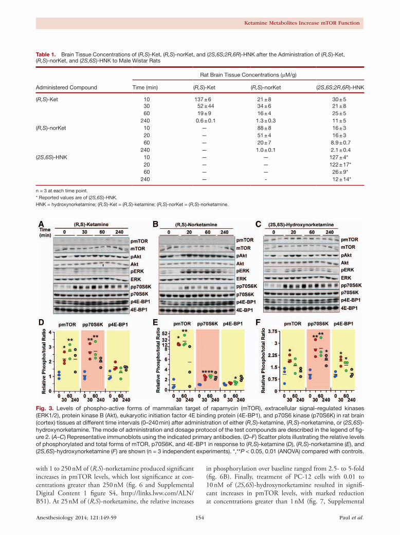
Effect of ( R,S)-ketamine, ( R,S)-norketamine, and ( 2S,6S)-hydroxynorketamine on the In Vivo Phosphorylation of mTOR and Related Proteins in Rat Brain TissueThe phosphorylation of mTOR and related proteins was determined in rat brain tissue obtained 30, 60, and 240 min after the administration of (R,S)-ketamine, but not in the 10-min postadministration samples as the amount of tis-sue was insufficient to carry out these studies. Western blot analysis was performed using primary antibodies specific for the phosphorylated forms of mTOR, 4E-BP1, p70S6K, ERK1/2, and Akt. The results demonstrated that (R,S)-ket-amine administration produced a time-dependent increase in the expression of pmTOR (~2.5-fold) and pp70S6K (~2.5-fold), which reached a maximum at the 30- and 60-min time points, followed by a gradual decline (figs. 3A and D). Similar findings were reported in a recent study where the increase in the phosphorylated forms of these proteins (~1.5-fold) peaked at 30 to 60 min postadministra-tion of (R,S)-ketamine in Wistar rats, before returning to baseline levels.4 In this study, ANOVA analysis showed that administration of (R,S)-ketamine for 30 and 60 min led to significant increases in pmTOR and pp70S6K levels (P < 0.05), whereas increases in the average expression of p4E-BP1 (~1.5-fold), pERK1/2 (~2-fold), and pAkt (~1.3-fold) did not reach statistical significance (figs. 3A and D, and Supplemental Digital Content 1 figure S1, http://links.lww.com/ALN/B51, which illustrates the ratios of phosphory-lated to total Akt and ERK protein levels at various incuba-tion periods with (R,S)-ketamine and its metabolites).
Administration of (R,S)-norketamine produced sig-nificant increases in the levels of phosphorylated forms of mTOR, pp70S6K, and p4E-BP1 in the 20- and 60-min

Anesthesiology 2014; 121:149-59 153 Paul et al.
PAIN MEDICINE
samples (figs. 3B and E). The 15- and 25-fold increases in the pmTOR levels were much higher than the ~2.5-fold increase observed after the administration of (R,S)-ketamine and may be a result of larger initial exposure to (R,S)-norketamine relative to the concentration of the compound produced by the administration of (R,S)-ketamine (figs. 2A and B, table 1). Although the relative pAkt levels were not affected, there was significant increase in the relative levels of pERK1/2 in response to (R,S)-norketamine administration (figs. 3B and E and Supplemental Digital Content 1 figure S1, http://links.lww.com/ALN/B51). The ~6-fold increase in pERK
level was also significantly greater than the ~2-fold (figs. 3A and D) and 1.5-fold4 increases produced by (R,S)-ketamine.
Administration of (2S,6S)-hydroxynorketamine produced time-dependent increases in pmTOR (~2.0-fold), p4E-BP1 (~2.0-fold), and pp70S6K (~2.5-fold) levels, which reached significance in the 20- and 60-min samples (figs. 3C and F). These effects were similar to that produced by (R,S)-ketamine (figs. 3A and D). Even though the relative levels of pERK1/2 and pAkt were increased in the 20- and 60-min samples, they did not reach statistical significance (Supplemental Digital Content 1 figure S1, http://links.lww.com/ALN/B51).
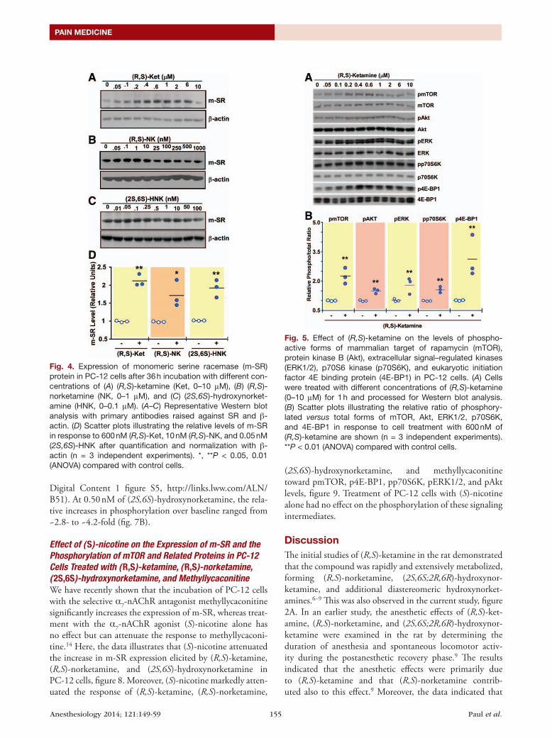
Effect of ( R,S)-ketamine, ( R,S)-norketamine, and ( 2S,6S)-hydroxynorketamine on the Expression of m-SR in PC-12 CellsThe treatment of PC-12 cells with (R,S)-ketamine (0 to 10,000 nM), (R,S)-norketamine (0 to 1,000 nM), and (2S,6S)-hydroxynorketamine (0 to 100 nM) produced concentration-dependent increases in the expression of the m-SR protein, with maximum at 600, 10, and 0.05 nM, respectively, and gradual decline at higher concentrations of the test compounds (fig. 4 and Supplemental Digital Con-tent 1 figure S2, http://links.lww.com/ALN/B51, which illustrates m-SR protein levels at various concentrations of (R,S)-ketamine and its metabolites). ANOVA analysis showed significant increases at 600 and 1,000 nM of (R,S)-ketamine, 10 and 25 nM of (R,S)-norketamine, and between 0.05 and 0.25 nM of (2S,6S)-hydroxynorketamine.
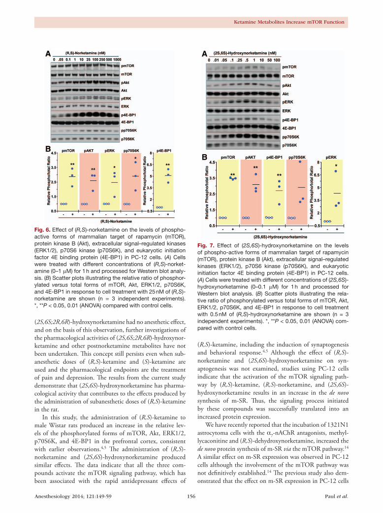
Effect of ( R,S)-ketamine, ( R,S)-norketamine, and ( 2S,6S)-hydroxynorketamine on the Phosphorylation of mTOR and Related Proteins in PC-12 CellsPC-12 cells were incubated for 60 min with a range of concen-trations of (R,S)-ketamine (50 to 10,000 nM), (R,S)-norket-amine (0.05 to 1,000 nM), and (2S,6S)-hydroxynorketamine (0.01 to 100 nM). The incubation time was chosen based on the data obtained in the in vivo studies in which significant increases in the levels of phosphorylated forms of mTOR, 4E-BP1, p70S6K, ERK1/2, and Akt were observed 60 min after administration of (R,S)-ketamine and its metabolites. All three compounds produced significant and concentra-tion-dependent increases in the phosphorylation of these signaling intermediates (figs. 5–7 and Supplemental Digital Content 1 figures S3–S5, http://links.lww.com/ALN/B51, which illustrate quantitative densitometric analyses of the relative phosphorylation levels of these signaling proteins at various concentrations of (R,S)-ketamine and its metabo-lites). Cell treatment with (R,S)-ketamine (400 to 600 nM) elicited significant increases in protein phosphorylation, which gradually lost significance at concentrations greater than 2,000 nM (fig. 5 and Supplemental Digital Content 1 figure S3, http://links.lww.com/ALN/B51). At 600 nM of (R,S)-ketamine, the relative increases in phosphorylation over baseline ranged from 1.5- to 3-fold (fig. 5B). Incubation
Fig. 2. A representative chromatogram from a 60-min brain sample obtained (A) after a single intraperitoneal injection of (R,S)-ketamine, 40 mg/kg in saline (5 ml/kg), (B) after a single intravenous injection of (R,S)-norketamine, 20 mg/kg in sa-line (5 ml/kg), and (C) after a single intravenous injection of (2S,6S)-hydroxynorketamine, 20 mg/kg in saline (5 ml/kg). The labeled peaks correspond to (R,S)-ketamine (1), (R,S)-norketamine (2), (2S,6S;2R,6R)-hydroxynorketamine (4a), (2S,6R;2R,6S)-hydroxynorketamine (4b), (2S,5S;2R,5R)-hy-droxynorketamine (4c), (2S,4S;2R,5R)-hydroxynorketamine (4d), and (2S,4R;2R,4S)-hydroxynorketamine (4f).
Anesthesiology 2014; 121:149-59 154 Paul et al.
Ketamine Metabolites Increase mTOR Function
with 1 to 250 nM of (R,S)-norketamine produced significant increases in pmTOR levels, which lost significance at con-centrations greater than 250 nM (fig. 6 and Supplemental Digital Content 1 figure S4, http://links.lww.com/ALN/B51). At 25 nM of (R,S)-norketamine, the relative increases
in phosphorylation over baseline ranged from 2.5- to 5-fold (fig. 6B). Finally, treatment of PC-12 cells with 0.01 to 10 nM of (2S,6S)-hydroxynorketamine resulted in signifi-cant increases in pmTOR levels, with marked reduction at concentrations greater than 1 nM (fig. 7, Supplemental
Table 1. Brain Tissue Concentrations of (R,S)-Ket, (R,S)-norKet, and (2S,6S;2R,6R)-HNK after the Administration of (R,S)-Ket, (R,S)-norKet, and (2S,6S)-HNK to Male Wistar Rats
Administered Compound
Rat Brain Tissue Concentrations (μM/g)
Time (min) (R,S)-Ket (R,S)-norKet (2S,6S;2R,6R)-HNK
(R,S)-Ket 10 137 ± 6 21 ± 8 30 ± 530 52 ± 44 34 ± 6 21 ± 860 19 ± 9 16 ± 4 25 ± 5
240 0.6 ± 0.1 1.3 ± 0.3 11 ± 5(R,S)-norKet 10 — 88 ± 8 16 ± 3
20 — 51 ± 4 16 ± 360 — 20 ± 7 8.9 ± 0.7
240 — 1.0 ± 0.1 2.1 ± 0.4(2S,6S)-HNK 10 — — 127 ± 4*
20 — — 122 ± 17*60 — — 26 ± 9*
240 — - 12 ± 14*
n = 3 at each time point.* Reported values are of (2S,6S)-HNK.HNK = hydroxynorketamine; (R,S)-Ket = (R,S)-ketamine; (R,S)-norKet = (R,S)-norketamine.
Fig. 3. Levels of phospho-active forms of mammalian target of rapamycin (mTOR), extracellular signal–regulated kinases (ERK1/2), protein kinase B (Akt), eukaryotic initiation factor 4E binding protein (4E-BP1), and p70S6 kinase (p70S6K) in rat brain (cortex) tissues at different time intervals (0–240 min) after administration of either (R,S)-ketamine, (R,S)-norketamine, or (2S,6S)-hydroxynorketamine. The mode of administration and dosage protocol of the test compounds are described in the legend of fig-ure 2. (A–C) Representative immunoblots using the indicated primary antibodies. (D–F) Scatter plots illustrating the relative levels of phosphorylated and total forms of mTOR, p70S6K, and 4E-BP1 in response to (R,S)-ketamine (D), (R,S)-norketamine (E), and (2S,6S)-hydroxynorketamine (F) are shown (n = 3 independent experiments). *,**P < 0.05, 0.01 (ANOVA) compared with controls.
Anesthesiology 2014; 121:149-59 155 Paul et al.
PAIN MEDICINE
Digital Content 1 figure S5, http://links.lww.com/ALN/B51). At 0.50 nM of (2S,6S)-hydroxynorketamine, the rela-tive increases in phosphorylation over baseline ranged from ~2.8- to ~4.2-fold (fig. 7B).
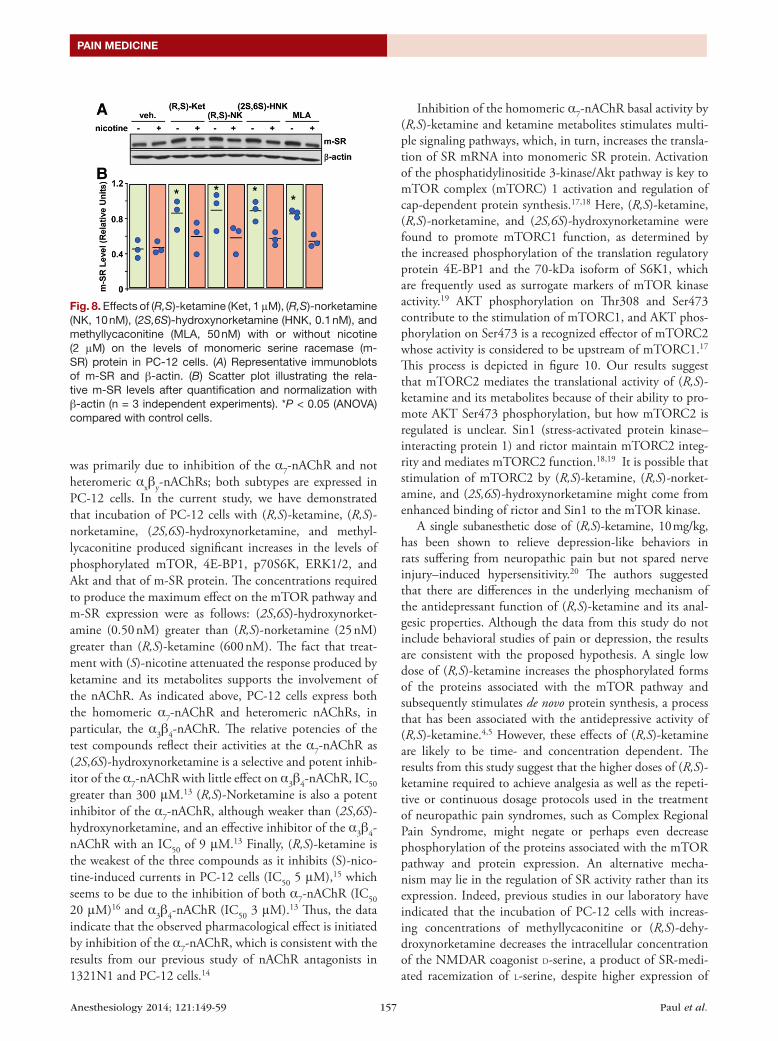
Effect of ( S)-nicotine on the Expression of m-SR and the Phosphorylation of mTOR and Related Proteins in PC-12 Cells Treated with ( R,S)-ketamine, ( R,S)-norketamine, ( 2S,6S)-hydroxynorketamine, and MethyllycaconitineWe have recently shown that the incubation of PC-12 cells with the selective α7-nAChR antagonist methyllycaconitine significantly increases the expression of m-SR, whereas treat-ment with the α7-nAChR agonist (S)-nicotine alone has no effect but can attenuate the response to methyllycaconi-tine.14 Here, the data illustrates that (S)-nicotine attenuated the increase in m-SR expression elicited by (R,S)-ketamine, (R,S)-norketamine, and (2S,6S)-hydroxynorketamine in PC-12 cells, figure 8. Moreover, (S)-nicotine markedly atten-uated the response of (R,S)-ketamine, (R,S)-norketamine,
(2S,6S)-hydroxynorketamine, and methyllycaconitine toward pmTOR, p4E-BP1, pp70S6K, pERK1/2, and pAkt levels, figure 9. Treatment of PC-12 cells with (S)-nicotine alone had no effect on the phosphorylation of these signaling intermediates.
DiscussionThe initial studies of (R,S)-ketamine in the rat demonstrated that the compound was rapidly and extensively metabolized, forming (R,S)-norketamine, (2S,6S;2R,6R)-hydroxynor-ketamine, and additional diastereomeric hydroxynorket-amines.6–9 This was also observed in the current study, figure 2A. In an earlier study, the anesthetic effects of (R,S)-ket-amine, (R,S)-norketamine, and (2S,6S;2R,6R)-hydroxynor-ketamine were examined in the rat by determining the duration of anesthesia and spontaneous locomotor activ-ity during the postanesthetic recovery phase.9 The results indicated that the anesthetic effects were primarily due to (R,S)-ketamine and that (R,S)-norketamine contrib-uted also to this effect.9 Moreover, the data indicated that
Fig. 4. Expression of monomeric serine racemase (m-SR) protein in PC-12 cells after 36 h incubation with different con-centrations of (A) (R,S)-ketamine (Ket, 0–10 μM), (B) (R,S)-norketamine (NK, 0–1 μM), and (C) (2S,6S)-hydroxynorket-amine (HNK, 0–0.1 μM). (A–C) Representative Western blot analysis with primary antibodies raised against SR and β-actin. (D) Scatter plots illustrating the relative levels of m-SR in response to 600 nM (R,S)-Ket, 10 nM (R,S)-NK, and 0.05 nM (2S,6S)-HNK after quantification and normalization with β-actin (n = 3 independent experiments). *, **P < 0.05, 0.01 (ANOVA) compared with control cells.
Fig. 5. Effect of (R,S)-ketamine on the levels of phospho-active forms of mammalian target of rapamycin (mTOR), protein kinase B (Akt), extracellular signal–regulated kinases (ERK1/2), p70S6 kinase (p70S6K), and eukaryotic initiation factor 4E binding protein (4E-BP1) in PC-12 cells. (A) Cells were treated with different concentrations of (R,S)-ketamine (0–10 μM) for 1 h and processed for Western blot analysis. (B) Scatter plots illustrating the relative ratio of phosphory-lated versus total forms of mTOR, Akt, ERK1/2, p70S6K, and 4E-BP1 in response to cell treatment with 600 nM of (R,S)-ketamine are shown (n = 3 independent experiments). **P < 0.01 (ANOVA) compared with control cells.
Anesthesiology 2014; 121:149-59 156 Paul et al.
Ketamine Metabolites Increase mTOR Function
(2S,6S;2R,6R)-hydroxynorketamine had no anesthetic effect, and on the basis of this observation, further investigations of the pharmacological activities of (2S,6S;2R,6R)-hydroxynor-ketamine and other postnorketamine metabolites have not been undertaken. This concept still persists even when sub-anesthetic doses of (R,S)-ketamine and (S)-ketamine are used and the pharmacological endpoints are the treatment of pain and depression. The results from the current study demonstrate that (2S,6S)-hydroxynorketamine has pharma-cological activity that contributes to the effects produced by the administration of subanesthetic doses of (R,S)-ketamine in the rat.
In this study, the administration of (R,S)-ketamine to male Wistar rats produced an increase in the relative lev-els of the phosphorylated forms of mTOR, Akt, ERK1/2, p70S6K, and 4E-BP1 in the prefrontal cortex, consistent with earlier observations.4,5 The administration of (R,S)-norketamine and (2S,6S)-hydroxynorketamine produced similar effects. The data indicate that all the three com-pounds activate the mTOR signaling pathway, which has been associated with the rapid antidepressant effects of
(R,S)-ketamine, including the induction of synaptogenesis and behavioral response.4,5 Although the effect of (R,S)-norketamine and (2S,6S)-hydroxynorketamine on syn-aptogenesis was not examined, studies using PC-12 cells indicate that the activation of the mTOR signaling path-way by (R,S)-ketamine, (R,S)-norketamine, and (2S,6S)-hydroxynorketamine results in an increase in the de novo synthesis of m-SR. Thus, the signaling process initiated by these compounds was successfully translated into an increased protein expression.
We have recently reported that the incubation of 1321N1 astrocytoma cells with the α7-nAChR antagonists, methyl-lycaconitine and (R,S)-dehydroxynorketamine, increased the de novo protein synthesis of m-SR via the mTOR pathway.14 A similar effect on m-SR expression was observed in PC-12 cells although the involvement of the mTOR pathway was not definitively established.14 The previous study also dem-onstrated that the effect on m-SR expression in PC-12 cells
Fig. 6. Effect of (R,S)-norketamine on the levels of phospho-active forms of mammalian target of rapamycin (mTOR), protein kinase B (Akt), extracellular signal–regulated kinases (ERK1/2), p70S6 kinase (p70S6K), and eukaryotic initiation factor 4E binding protein (4E-BP1) in PC-12 cells. (A) Cells were treated with different concentrations of (R,S)-norket-amine (0–1 μM) for 1 h and processed for Western blot analy-sis. (B) Scatter plots illustrating the relative ratio of phosphor-ylated versus total forms of mTOR, Akt, ERK1/2, p70S6K, and 4E-BP1 in response to cell treatment with 25 nM of (R,S)-norketamine are shown (n = 3 independent experiments). *, **P < 0.05, 0.01 (ANOVA) compared with control cells.
Fig. 7. Effect of (2S,6S)-hydroxynorketamine on the levels of phospho-active forms of mammalian target of rapamycin (mTOR), protein kinase B (Akt), extracellular signal–regulated kinases (ERK1/2), p70S6 kinase (p70S6K), and eukaryotic initiation factor 4E binding protein (4E-BP1) in PC-12 cells. (A) Cells were treated with different concentrations of (2S,6S)-hydroxynorketamine (0–0.1 μM) for 1 h and processed for Western blot analysis. (B) Scatter plots illustrating the rela-tive ratio of phosphorylated versus total forms of mTOR, Akt, ERK1/2, p70S6K, and 4E-BP1 in response to cell treatment with 0.5 nM of (R,S)-hydroxynorketamine are shown (n = 3 independent experiments). *, **P < 0.05, 0.01 (ANOVA) com-pared with control cells.
Anesthesiology 2014; 121:149-59 157 Paul et al.
PAIN MEDICINE
was primarily due to inhibition of the α7-nAChR and not heteromeric αxβy-nAChRs; both subtypes are expressed in PC-12 cells. In the current study, we have demonstrated that incubation of PC-12 cells with (R,S)-ketamine, (R,S)-norketamine, (2S,6S)-hydroxynorketamine, and methyl-lycaconitine produced significant increases in the levels of phosphorylated mTOR, 4E-BP1, p70S6K, ERK1/2, and Akt and that of m-SR protein. The concentrations required to produce the maximum effect on the mTOR pathway and m-SR expression were as follows: (2S,6S)-hydroxynorket-amine (0.50 nM) greater than (R,S)-norketamine (25 nM) greater than (R,S)-ketamine (600 nM). The fact that treat-ment with (S)-nicotine attenuated the response produced by ketamine and its metabolites supports the involvement of the nAChR. As indicated above, PC-12 cells express both the homomeric α7-nAChR and heteromeric nAChRs, in particular, the α3β4-nAChR. The relative potencies of the test compounds reflect their activities at the α7-nAChR as (2S,6S)-hydroxynorketamine is a selective and potent inhib-itor of the α7-nAChR with little effect on α3β4-nAChR, IC50 greater than 300 μM.13 (R,S)-Norketamine is also a potent inhibitor of the α7-nAChR, although weaker than (2S,6S)-hydroxynorketamine, and an effective inhibitor of the α3β4-nAChR with an IC50 of 9 μM.13 Finally, (R,S)-ketamine is the weakest of the three compounds as it inhibits (S)-nico-tine-induced currents in PC-12 cells (IC50 5 μM),15 which seems to be due to the inhibition of both α7-nAChR (IC50 20 μM)16 and α3β4-nAChR (IC50 3 μM).13 Thus, the data indicate that the observed pharmacological effect is initiated by inhibition of the α7-nAChR, which is consistent with the results from our previous study of nAChR antagonists in 1321N1 and PC-12 cells.14
Inhibition of the homomeric α7-nAChR basal activity by (R,S)-ketamine and ketamine metabolites stimulates multi-ple signaling pathways, which, in turn, increases the transla-tion of SR mRNA into monomeric SR protein. Activation of the phosphatidylinositide 3-kinase/Akt pathway is key to mTOR complex (mTORC) 1 activation and regulation of cap-dependent protein synthesis.17,18 Here, (R,S)-ketamine, (R,S)-norketamine, and (2S,6S)-hydroxynorketamine were found to promote mTORC1 function, as determined by the increased phosphorylation of the translation regulatory protein 4E-BP1 and the 70-kDa isoform of S6K1, which are frequently used as surrogate markers of mTOR kinase activity.19 AKT phosphorylation on Thr308 and Ser473 contribute to the stimulation of mTORC1, and AKT phos-phorylation on Ser473 is a recognized effector of mTORC2 whose activity is considered to be upstream of mTORC1.17 This process is depicted in figure 10. Our results suggest that mTORC2 mediates the translational activity of (R,S)-ketamine and its metabolites because of their ability to pro-mote AKT Ser473 phosphorylation, but how mTORC2 is regulated is unclear. Sin1 (stress-activated protein kinase–interacting protein 1) and rictor maintain mTORC2 integ-rity and mediates mTORC2 function.18,19 It is possible that stimulation of mTORC2 by (R,S)-ketamine, (R,S)-norket-amine, and (2S,6S)-hydroxynorketamine might come from enhanced binding of rictor and Sin1 to the mTOR kinase.
A single subanesthetic dose of (R,S)-ketamine, 10 mg/kg, has been shown to relieve depression-like behaviors in rats suffering from neuropathic pain but not spared nerve injury–induced hypersensitivity.20 The authors suggested that there are differences in the underlying mechanism of the antidepressant function of (R,S)-ketamine and its anal-gesic properties. Although the data from this study do not include behavioral studies of pain or depression, the results are consistent with the proposed hypothesis. A single low dose of (R,S)-ketamine increases the phosphorylated forms of the proteins associated with the mTOR pathway and subsequently stimulates de novo protein synthesis, a process that has been associated with the antidepressive activity of (R,S)-ketamine.4,5 However, these effects of (R,S)-ketamine are likely to be time- and concentration dependent. The results from this study suggest that the higher doses of (R,S)-ketamine required to achieve analgesia as well as the repeti-tive or continuous dosage protocols used in the treatment of neuropathic pain syndromes, such as Complex Regional Pain Syndrome, might negate or perhaps even decrease phosphorylation of the proteins associated with the mTOR pathway and protein expression. An alternative mecha-nism may lie in the regulation of SR activity rather than its expression. Indeed, previous studies in our laboratory have indicated that the incubation of PC-12 cells with increas-ing concentrations of methyllycaconitine or (R,S)-dehy-droxynorketamine decreases the intracellular concentration of the NMDAR coagonist D-serine, a product of SR-medi-ated racemization of L-serine, despite higher expression of
Fig. 8. Effects of (R,S)-ketamine (Ket, 1 μM), (R,S)-norketamine (NK, 10 nM), (2S,6S)-hydroxynorketamine (HNK, 0.1 nM), and methyllycaconitine (MLA, 50 nM) with or without nicotine (2 μM) on the levels of monomeric serine racemase (m-SR) protein in PC-12 cells. (A) Representative immunoblots of m-SR and β-actin. (B) Scatter plot illustrating the rela-tive m-SR levels after quantification and normalization with β-actin (n = 3 independent experiments). *P < 0.05 (ANOVA) compared with control cells.

Anesthesiology 2014; 121:149-59 158 Paul et al.
Ketamine Metabolites Increase mTOR Function
m-SR.14 A similar decrease in intracellular D-serine concen-trations was observed after incubation of PC-12 cells with the voltage-gated calcium channel α2δ inhibitors gabapen-tin and (S)-pregabalin,21 which are used in the treatment of neuropathic pain, without affecting m-SR protein level. The inhibition of SR activity has also been associated with a decrease in NMDAR activity, and SR inhibitors are being explored for use in the treatment of some central nervous system disorders.22,23
The results of the study suggest that the therapeutic effects produced by subanesthetic doses of (R,S)-ketamine may be the result of a combination of independent but inter-related pharmacological effects at the α7-nAChR produced by the parent drug and its metabolites. One of the effects is increased protein expression via the mTOR pathway, which is initiated by antagonism of α7-nAChR, and is reflected by the observed increase in m-SR expression. The second effect is an “indirect” inhibition of NMDAR activity resulting from a reduction in the intracellular Ca2+ flux. These two interconnected mechanisms are reflected in the previously observed and apparently contradictory effects produced by methyllycaconitine and (R,S)-dehydroxynorketamine in
Fig. 10. Schematic representation of the modulation of serine racemase (SR) expression functioning via mammalian target of rapamycin (mTOR) and extracellular signal–regulated kinase (ERK) pathways. HNK = (2S,6S)-hydroxynorketamine; Ket = (R,S)-ketamine; mTORC1 = mTOR complex 1; mTORC2 = mTOR complex 2; nAchR = nicotinic acetylcholine receptor; NorKet = (R,S)-norketamine.
Fig. 9. Effects of (R,S)-ketamine, (R,S)-norketamine (NK), (2S,6S)-hydroxynorketamine (HNK), and methyllycaconitine with or without nicotine on the levels of phospho-active forms of mammalian target of rapamycin (mTOR), protein kinase B (Akt), extra-cellular signal–regulated kinases (ERK1/2), p70S6 kinase (p70S6K), and eukaryotic initiation factor 4E binding protein (4E-BP1) in PC-12 cells. (A) Cells were treated with (R,S)- Ket (1 μM), (R,S)-NK (10 nM), (2S,6S)-HNK (0.1 nM), or methyllycaconitine (MLA, 50 nM) with or without nicotine (2 μM) for 1 h and then processed for Western blot analysis. (A) Representative immunoblots. (B–F) Scatter plots illustrating the relative ratio of phosphorylated versus total forms of mTOR (B), Akt (C), ERK1/2 (D), p70S6K (E), and 4E-BP1 (F) are shown (n = 3 independent experiments). *, **P < 0.05, 0.01 (ANOVA) compared with control cells.

Anesthesiology 2014; 121:149-59 159 Paul et al.
PAIN MEDICINE
1321N1 and PC-12 cells in which m-SR expression was increased, whereas the m-SR function, expressed as intracel-lular D-serine concentration, was reduced.14 The interrelation and importance of the effects produced by (R,S)-ketamine metabolites and the related mechanisms are under investiga-tion, and the results will be reported elsewhere.
AcknowledgmentsThis work was supported by funding from the Intramural Research Program of the National Institute on Aging, Na-tional Institutes of Health (Baltimore, Maryland) and by Na-tional Institute on Aging Contract No. HHSN271201000008I.
Competing InterestsDrs. Wainer, Moaddel, Bernier, and Torjman are listed as coinventors on a patent application for the use of ketamine metabolites in the treatment of bipolar disorder and major depression. They have assigned their rights in the patent to the U.S. government but will share a percentage of any royalties that may be received by the government. The other authors declare no competing interests.
CorrespondenceAddress correspondence to Dr. Wainer: Laboratory of Clini-cal Investigation, National Institute on Aging, National Insti-tutes of Health, Biomedical Research Center, 251 Bayview Boulevard, Suite 100, Baltimore, Maryland 21224. [email protected]. This article may be accessed for personal use at no charge through the Journal Web site, www.anes-thesiology.org.
References 1. Diazgranados N, Ibrahim L, Brutsche NE, Newberg
A, Kronstein P, Khalife S, Kammerer WA, Quezado Z, Luckenbaugh DA, Salvadore G, Machado-Vieira R, Manji HK, Zarate CA Jr: A randomized add-on trial of an N-methyl-D-aspartate antagonist in treatment-resistant bipolar depres-sion. Arch Gen Psychiatry 2010; 67:793–802
2. Zarate CA Jr, Brutsche N, Laje G, Luckenbaugh DA, Venkata SL, Ramamoorthy A, Moaddel R, Wainer IW: Relationship of ket-amine’s plasma metabolites with response, diagnosis, and side effects in major depression. Biol Psychiatry 2012; 72:331–8
3. Zhao X, Venkata SL, Moaddel R, Luckenbaugh DA, Brutsche NE, Ibrahim L, Zarate CA Jr, Mager DE, Wainer IW: Simultaneous population pharmacokinetic modelling of ket-amine and three major metabolites in patients with treat-ment-resistant bipolar depression. Br J Clin Pharmacol 2012; 74:304–14
4. Li N, Lee B, Liu RJ, Banasr M, Dwyer JM, Iwata M, Li XY, Aghajanian G, Duman RS: mTOR-dependent synapse for-mation underlies the rapid antidepressant effects of NMDA antagonists. Science 2010; 329:959–64
5. Dwyer JM, Duman RS: Activation of mammalian target of rapamycin and synaptogenesis: Role in the actions of rapid-acting antidepressants. Biol Psychiatry 2013; 73:1189–98
6. Cohen ML, Chan SL, Way WL, Trevor AJ: Distribution in the brain and metabolism of ketamine in the rat after intrave-nous administration. ANESTHESIOLOGY 1973; 39:370–6
7. Adams JD Jr, Baillie TA, Trevor AJ, Castagnoli N Jr: Studies on the biotransformation of ketamine. 1-Identification of
metabolites produced in vitro from rat liver microsomal preparations. Biomed Mass Spectrom 1981; 8:527–38
8. Woolf TF, Adams JD: Biotransformation of ketamine, (Z)-6-hydroxyketamine, and (E)-6-hydroxyketamine by rat, rabbit, and human liver microsomal preparations. Xenobiotica 1987; 17:839–47
9. Leung LY, Baillie TA: Comparative pharmacology in the rat of ketamine and its two principal metabolites, norket-amine and (Z)-6-hydroxynorketamine. J Med Chem 1986; 29: 2396–9
10. Desta Z, Moaddel R, Ogburn ET, Xu C, Ramamoorthy A, Venkata SL, Sanghvi M, Goldberg ME, Torjman MC, Wainer IW: Stereoselective and regiospecific hydroxyl-ation of ketamine and norketamine. Xenobiotica 2012; 42: 1076–87
11. Moaddel R, Venkata SL, Tanga MJ, Bupp JE, Green CE, Iyer L, Furimsky A, Goldberg ME, Torjman MC, Wainer IW: A parallel chiral-achiral liquid chromatographic method for the determination of the stereoisomers of ketamine and ketamine metabolites in the plasma and urine of patients with complex regional pain syndrome. Talanta 2010; 82: 1892–904
12. Ebert B, Mikkelsen S, Thorkildsen C, Borgbjerg FM: Norketamine, the main metabolite of ketamine, is a non-competitive NMDA receptor antagonist in the rat cortex and spinal cord. Eur J Pharmacol 1997; 333:99–104
13. Moaddel R, Abdrakhmanova G, Kozak J, Jozwiak K, Toll L, Jimenez L, Rosenberg A, Tran T, Xiao Y, Zarate CA, Wainer IW: Sub-anesthetic concentrations of (R,S)-ketamine metab-olites inhibit acetylcholine-evoked currents in α7 nicotinic acetylcholine receptors. Eur J Pharmacol 2013; 698:228–34
14. Singh NS, Paul RK, Ramamoorthy A, Torjman MC, Moaddel R, Bernier M, Wainer IW: Nicotinic acetylcholine receptor antag-onists alter the function and expression of serine racemase in PC-12 and 1321N1 cells. Cell Signal 2013; 25:2634–45
15. Sasaki T, Andoh T, Watanabe I, Kamiya Y, Itoh H, Higashi T, Matsuura T: Nonstereoselective inhibition of neuronal nico-tinic acetylcholine receptors by ketamine isomers. Anesth Analg 2000; 91:741–8
16. Coates KM, Flood P: Ketamine and its preservative benze-thonium chloride both inhibit human recombinant α7 and α4β2 neuronal nicotinic acetylcholine receptors in Xenopus oocytes. Br J Pharmacol 2001; 134:871–9
17. Foster KG, Fingar DC: Mammalian target of rapamycin (mTOR): Conducting the cellular signaling symphony. J Biol Chem 2010; 285:14071–7
18. Wullschleger S, Loewith R, Hall MN: TOR signaling in growth and metabolism. Cell 2006; 124:471–84
19. Frias MA, Thoreen CC, Jaffe JD, Schroder W, Sculley T, Carr SA, Sabatini DM: mSin1 is necessary for Akt/PKB phosphory-lation, and its isoforms define three distinct mTORC2s. Curr Biol 2006; 16:1865–70
20. Wang J, Goffer Y, Xu D, Tukey DS, Shamir DB, Eberle SE, Zou AH, Blanck TJ, Ziff EB: A single subanesthetic dose of ketamine relieves depression-like behaviors induced by neu-ropathic pain in rats. ANESTHESIOLOGY 2011; 115:812–21
21. Singh NS, Paul RK, Torjman MC, Wainer IW: Gabapentin and (S)-pregabalin decrease intracellular D-serine concentrations in PC-12 cells. Neurosci Lett 2013; 535:90–4
22. Sethuraman R, Lee TL, Tachibana S: D-serine regulation: A possible therapeutic approach for central nervous diseases and chronic pain. Mini Rev Med Chem 2009; 9:813–9
23. Jirásková-Vaníčková J, Ettrich R, Vorlová B, Hoffman HE, Lepšík M, Jansa P, Konvalinka J: Inhibition of human serine racemase, an emerging target for medicinal chemistry. Curr Drug Targets 2011; 12:1037–55