Embed Size (px)
Citation preview

RPC for Positron EmittionTomography

L. Litov RPC for PET NCPP, Primorsko, 2007
Discussion
� What is PET
� Why PET/MRI
� RPC as PET detectors
� Human PET
� Small animals PET
� RPCPET project
L. Litov RPC for PET NCPP, Primorsko, 2007
What is PET
What is and How PET works
Positron Emission Tomography (PET) is a radiotracer imaging
technique, in which tracer compounds labelled with positron emitting
radionuclides are injected into the subject of the study. These tracers
compounds can then be used to track biomedical and physiological
processes.
L. Litov RPC for PET NCPP, Primorsko, 2007
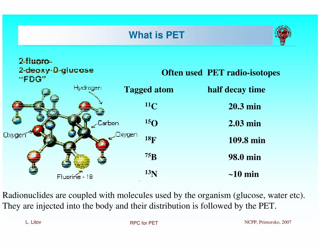
Often used PET radio-isotopes
Tagged atom half decay time
11C 20.3 min
15O 2.03 min
18F 109.8 min
75B 98.0 min
13N ~10 min
Radionuclides are coupled with molecules used by the organism (glucose, water etc).
They are injected into the body and their distribution is followed by the PET.
What is PET

L. Litov RPC for PET NCPP, Primorsko, 2007
What is PET

L. Litov RPC for PET NCPP, Primorsko, 2007

L. Litov RPC for PET NCPP, Primorsko, 2007
PET-CT

L. Litov RPC for PET NCPP, Primorsko, 2007
PET-CT

L. Litov RPC for PET NCPP, Primorsko, 2007
Problems

L. Litov RPC for PET NCPP, Primorsko, 2007
2D and 3D reconstruction
In order to do 3d reconstruction, it is extremely important to introduce
corrections for absorption and scattering of the γγγγ inside the bodyRequires precise MC, individual for the particular patient.

L. Litov RPC for PET NCPP, Primorsko, 2007
Scintillators
Commercially available PET use scintillating crystals to detect gammas
Gamma interactions – photo-effect and Compton scattering
Require materials with high Z and density

L. Litov RPC for PET NCPP, Primorsko, 2007
RPC

L. Litov RPC for PET NCPP, Primorsko, 2007
RPC for PET
Photo-effect
Compton scattering

L. Litov RPC for PET NCPP, Primorsko, 2007
Neutral radiation (photons)
No signalSignal
photon electron
intrconv εεε ⋅=

L. Litov RPC for PET NCPP, Primorsko, 2007
convεMonte Carlo simulation of
s thicknes photonsincident
electrons exitingvs.conv =ε
Electrode thickness
Linear growth: the
glass thickness is
smaller than the
average range of
emitted electrons.
No needs to make
thicker glasses
since only the last
layers of the
electrodes (faced to
the gap) contribute
to the signal

L. Litov RPC for PET NCPP, Primorsko, 2007
(Monte Carlo simulation – GEANT4)
By increasing the number of
gaps one can obtain one
order of magnitude increase in the efficiency w.r.t.
standard bakelite RPCs
Glass plates
Number of gaps
By increasing the number of
electrodes a photon has greater
probability to make an interaction
because it “sees” more material

L. Litov RPC for PET NCPP, Primorsko, 2007
Recipe
� Adequate thickness electrodes
� Increase the number of gaps
� Reduce also the gas thickness
(the probability of interaction of a photon in the gas is low)
� ���� Better time resolution

L. Litov RPC for PET NCPP, Primorsko, 2007
Multigap RPC
Gas gaps (300 µm)
(85% C2H2F4, 5% i-C4H10, 10% SF6)
Ground
High voltage

L. Litov RPC for PET NCPP, Primorsko, 2007
RPC

L. Litov RPC for PET NCPP, Primorsko, 2007
RPC vs Scintillator
Scintillator
•Expensive crystals
•Requirements:
•High efficiency
•Good time resolution
•Good energy resolution
•25-30 mm long crystals
•4x4 mm2 face
•The space resolution is limited
by the parallax
RPC
●Cheaper
●Able to work in strong magnetic
fields
●There is no parallax
●Easy to cover big surface
●(large FOV)
●Fast detectors
●Good time resolution (50 ps)
●Good space resolution (500 µm)

L. Litov RPC for PET NCPP, Primorsko, 2007
Comparison with crystals
Parameters:
1) Efficiency
2) Time resolution
3) Energy resolution
4) Cost

L. Litov RPC for PET NCPP, Primorsko, 2007
Efficiency - Possible coating materials
Other possibility is to use metal (heavy) electrode RPC

L. Litov RPC for PET NCPP, Primorsko, 2007
Typical time resolution
of BGOs is 3 ns ?
Less random coincidences and measurement of TOF
Time resolution
Better time resolution
means
�P.Fonte
52 ps with 1 gap
Alice
Fonte & Williams

L. Litov RPC for PET NCPP, Primorsko, 2007
%27/ totalkeV 511 ≈= NNr
Energy resolution (response function)
Energy spectrum of exiting photons
Real Scatter fraction 3D
PET up to 60%
Geometry for the simulation study on the gamma energy spectrum from annihilation

L. Litov RPC for PET NCPP, Primorsko, 2007
X 3
MRPC efficiency (1 gap) vs incident gamma energy
IN INDetected gamma
spectrum
Energy resolution (response function)

L. Litov RPC for PET NCPP, Primorsko, 2007
Energy (keV)
r = N511 / NTOT = 62%
Reduction of
coincidences
from scattering
Cost: The average cost per unit surface
would be at least 10 time less vs. crystals
Energy resolution (response function)

L. Litov RPC for PET NCPP, Primorsko, 2007
RPC for PET

L. Litov RPC for PET NCPP, Primorsko, 2007
Possible directions
� Human PET
� Requires
� High spatial resolution ~ 1 mm
� Very good time resolution (TOF)
� Cover big surface (full body PET)
� Fast data collection (to look at dynamics of the processes)
� Good simulation and correction for the scattering inside the body
� Small animals PET
� Better spatial resolution ( <1 mm)
� Good timing for suppression of the random coincidence
� Difference
� In human PET – measure time of flight of the two gammas
� In small animals – narrow coincidence gate - suppression of random
coincidences

L. Litov RPC for PET NCPP, Primorsko, 2007
RPC construction� Goals:
� Efficiency ~ 20%
� Spatial resolution – better 500 µm
� Time resolution - < 50 ps
� Construction
� The electrodes are made from glass (200 – 300 µm),
� one with special coating (<200 µm)
� Gas gap – 200 or 300 µm
� One electrode is glass,
� second metal (Bi, Cu, Pb, Au) - thickness 3 – 20 µm
� Multigap
Possible directions

L. Litov RPC for PET NCPP, Primorsko, 2007
Hybrid RPC

L. Litov RPC for PET NCPP, Primorsko, 2007
• High geometry acceptance > 90%.
• Fully 3D measurement of the interaction point of the photon => No parallax error.
• Sub-millimetre spatial resolution.
• High timing resolution ~ 0.3 ns FWHM*
• Moderate Efficiency.
• Compatible with Magnetic Resonance Imaging.
* Nucl. Instr. And Meth A, 443 (2003) 88-93
Small animal PET with RPCs
Resolution studies on a small animal RPC-PET prototype Alberto Blanco. LIP-Coimbra
Use the plates as a γγγγ
convertor

L. Litov RPC for PET NCPP, Primorsko, 2007
Charge-sensitive electronics allowing
interstrip position interpolation
Resolution studies on a small animal RPC-PET prototype Alberto Blanco. LIP-Coimbra
X
Z
....... .......
32
strips
16
plates
Transaxial
Depth of
interaction
16 stacked RPCs.
Aimed at verifying the concept and
show the viability of a
sub-millimetre spatial resolution.
2D measurement of the
photon interaction point.
3D measurement possible NIMA
508 (2003) 70–74

L. Litov RPC for PET NCPP, Primorsko, 2007
Resolution studies on a small animal RPC-PET prototype Alberto Blanco. LIP-Coimbra
• Copper (on a PCB) and glass electrodes.
• 32 1-mm wide X pickup strips.
• 0.3 mm Gap.
• Not optimized for high efficiency.
Active area 32 x 10 mm2
Copper electrode (cathode)Glass electrode (anode)
X
0.3 mm spacers
Transaxial coordinate Depth of interaction
Small animal PET

L. Litov RPC for PET NCPP, Primorsko, 2007
Resolution studies on a small animal RPC-PET prototype Alberto Blanco. LIP-Coimbra
-4 -2 0 2 410
-3
10-2
10-1
100
101
D (mm)
Pro
babili
ty
Point spread function- Annihilation photon non-collinearity.
N(x) = exp(-x2/2σ2), where FWHM = 2.35σ = 0.0022ds with ds the system diameter (mm).
-4 -2 0 2 410
-3
10-2
10-1
100
101
D (mm)
Pro
babili
ty
non Collinearity
J. Nucl. Med. 34 101 1993.
- Annihilation photon non-collinearity.N(x) = exp(-x2/2σ2), where FWHM = 2.35σ = 0.0022ds with ds the system diameter (mm).
- Positron range. P(x) = C1exp(-k1x) + (1-C1)exp(-k2x)
-4 -2 0 2 410
-3
10-2
10-1
100
101
D (mm)
Pro
babili
ty
non Collinearity
Positron range
IEEE TNS, vol 33, No 1, (1986), 565-569
- Annihilation photon non-collinearity.N(x) = exp(-x2/2σ2), where FWHM = 2.35σ = 0.0022ds with ds the system diameter (mm).
- Positron range. P(x) = C1exp(-k1x) + (1-C1)exp(-k2x)
- Source size.S(x)=sqrt(size2 - x2), where size is the source radio (mm).
-4 -2 0 2 410
-3
10-2
10-1
100
101
D (mm)
Pro
babili
ty
non Collinearity
Positron range
Source size
- Annihilation photon non-collinearity.N(x) = exp(-x2/2σ2), where FWHM = 2.35σ = 0.0022ds with ds the system diameter (mm).
- Positron range. P(x) = C1exp(-k1x) + (1-C1)exp(-k2x)
- Source size.S(x)=sqrt(size2 - x2), where size is the source radio (mm).
- Detector response.D(x) = exp(-x2/2σdet
2)
-4 -2 0 2 410
-3
10-2
10-1
100
101
D (mm)
Pro
babili
ty
non Collinearity
Positron range
Source size
Detector response
- Annihilation photon non-collinearity.N(x) = exp(-x2/2σ2), where FWHM = 2.35σ = 0.0022ds with ds the system diameter (mm).
- Positron range. P(x) = C1exp(-k1x) + (1-C1)exp(-k2x)
- Source size.S(x)=sqrt(size2 - x2), where size is the source radio (mm).
- Detector response.D(x) = exp(-x2/2σdet
2)- Scatter background.
SC(x) = exp(-k3x)
-4 -2 0 2 410
-3
10-2
10-1
100
101
D (mm)
Pro
babili
ty
non Collinearity
Positron range
Source size
Detector response
Scatter background
- Annihilation photon non-collinearity.N(x) = exp(-x2/2σ2), where FWHM = 2.35σ = 0.0022ds with ds the system diameter (mm).
- Positron range. P(x) = C1exp(-k1x) + (1-C1)exp(-k2x)
- Source size.S(x)=sqrt(size2 - x2), where size is the source radio (mm).
- Detector response.D(x) = exp(-x2/2σdet
2)- Scatter background.
SC(x) = exp(-k3x)
-4 -2 0 2 410
-3
10-2
10-1
100
101
D (mm)
Pro
babili
ty
520 µm FWHM
1550 µm FWTM
Calculated
Experimental
-4 -2 0 2 410
-3
10-2
10-1
100
101
D (mm)
Pro
babili
ty
non Collinearity
Positron range
Source size
Detector response
Scatter background
All contributions
- Annihilation photon non-collinearity.N(x) = exp(-x2/2σ2), where FWHM = 2.35σ = 0.0022ds with ds the system diameter (mm).
- Positron range. P(x) = C1exp(-k1x) + (1-C1)exp(-k2x)
- Source size.S(x)=sqrt(size2 - x2), where size is the source radio (mm).
- Detector response.D(x) = exp(-x2/2σdet
2)- Scatter background.
SC(x) = exp(-k3x)
R(x) = C2(N(X)⊗⊗⊗⊗ P(X)⊗⊗⊗⊗ D(X)⊗⊗⊗⊗ S(X))+(1-C2)SC(X)
520 µµµµm FWHM
1550 µµµµm FWTM
K2 = 3.75 mm-1
FWHMdet = 220 µm
C2 = 0.04, K3 = 0.32 mm-1
Phys. Med. Biol. 44 (1999) 781-799.
Intrinsic spatial resolution

L. Litov RPC for PET NCPP, Primorsko, 2007
Resolution studies on a small animal RPC-PET prototype Alberto Blanco. LIP-Coimbra
Filtered Back Projection FBP
~ 465 µµµµm FWHM
Maximum likelihood-expectation
maximization with resolution
modeling (ML-EM)
~ 305 µµµµm FWHM
-10 -5 -1 1 5 100
10
20
30
40
Distance (mm)
Counts
/100 µ
m
-10 -5 -1 1 5 10 0
10
20
30
40
Distance (mm)
Proceeding IEEE MIC (2004) M2-177
Image spatial resolution.
0.29
0.30
0.31
0.32
Homogeneous spatial resolution over the entire detector
0.450.47
0.460.48

L. Litov RPC for PET NCPP, Primorsko, 2007
Resolution studies on a small animal RPC-PET prototype Alberto Blanco. LIP-Coimbra
Scanner
Image spatial
Resolution,
FBP (mm)
Time
resolution (ns
FWHM)
FOV (mm Ø
x mm)
Central point absolute
sensitivity (cps/kBq)
Source
(mm Ø x mm)
Peak NEC
(Kcps)
microPET II®
[1],[2] 1.1 3 160 x 49 23 - 33
25 x 70 mouse size
235 at ~2.35 MBq/cm3
microPET Focus
F120 [6] 1.75 6 147 x 76 71 mouse size 809
at ~88 MBq
YAP-PET [3],[4] 1.6 2 40 x 40 18
at (Ø = 150 mm) - 90 (not peak)
at ~16.6 MBq
Quad HIDAC
(32 modules) [5] 0.95 - 170 x 280 18 - 100
at ~0.2MBq/cm3
RPC-PET 0.47 0.3 60 x 100 21 25 x 70
mouse size 318
at ~ 2.63 MBq/cm3
Comparison between different small animal PET parameters and the
expected parameters of the RPC-PET.
1. Yuan-Chuan Tai et al., “MicroPET II: design, development and initial performance of an improved MicroPET scanner for small-animal imaging”, Phys. Med. Biol. 48 (2003) 1519-1537.
2. Yongfeng Yang, et al., “Optimization and performance evaluation of the microPET II scanner for in vivo small-animal imaging”, Phys. Med. Biol. 49 (2004) 2527-2545.
3. A. del Guerra, G. Di Domenico, M. Scandola, G. Zavattini, “YAP-PET: first results of a small animal Positron Emission Tomograph based on YAP:Ce finger crystals”, IEEE Trans. Nucl. Sci., vol 45, No. 6 December 1998, 3105-3108.
4. G. Di Domenico et al., “Characterization of the Ferrera animal PET scanner”, Nucl. Instr. And Meth. A, 477 (2002) 505-508.
5. A.P. Jeavons, R.A. Chandler, C.A.R. Dettmar, “A 3D HIDAC-PET Camera with Sub-millimetre Resolution for Imaging Small Animals”, IEEE Trans. Nucl. Sci., vol. 46, No. 3, June 1999, 468-473.
6. Richard Laforest et al. “Performance Evaluation of the microPET – Focus F120”, presented at IEEE NSS/MIC Rome 2005.

L. Litov RPC for PET NCPP, Primorsko, 2007
University of Pavia

L. Litov RPC for PET NCPP, Primorsko, 2007
Hybrid RPC

L. Litov RPC for PET NCPP, Primorsko, 2007

L. Litov RPC for PET NCPP, Primorsko, 2007

L. Litov RPC for PET NCPP, Primorsko, 2007

L. Litov RPC for PET NCPP, Primorsko, 2007
RPCPET Project
� Understanding of the processes inside the body
� Phantoms – commercial or specially designed and build
� MC simulation (GEANT4, GATE)
� Measurement of the phantoms and comparison with simulations
� Goal –
� corrections for the reconstruction (scattering, absorption etc)
� Detector design and construction
� In parallel – several options (glass -glass, glass - metal, different readouts)
� Simulation of the detector response (GEANT + GARFIELD)
� Production of prototypes
� Measurements with point like sources and phantoms
� Tests in strong magnetic and high frequency electric fields (MRI)
� Goal –
� choose more suitable construction (can be different for human and animal PET)
� Proof RPC ability to work with MRI

L. Litov RPC for PET NCPP, Primorsko, 2007
� Electronics
� Analog – charge sensitive preamplifiers
� Measurement of the amplitude (if required) – ADC - (possible dependence of time from amplitude)
� Measurement of the time – TDC (possible dependence of time from amplitude)
� Trigger electronics (possibly using cathode information)
� Data storage
� Slow control system
� HV
� LV
� Gas system
� Temperature and pressure control
� Image reconstruction
� New detector requires specially designed reconstruction taking into account specific detector properties (different backgrounds, corrections, calibration, alignment etc)
� Combine reconstruction with MC simulations for every particular patient
RPCPET project

L. Litov RPC for PET NCPP, Primorsko, 2007
� Goal of the project
� Build full prototype – one ring
� Fully functioning system
� Test together with MRI
RPCPET project

L. Litov RPC for PET NCPP, Primorsko, 2007
Participants
� Bulgaria
� University of Sofia
� INRNE of BAS
� IPP of BAS
� Tokuda hospital
� LTD in Physics
� Italy
� INFN Pavia – Paolo Vitulo
� University Roma II “Tor Vergata” – R. Santonico
� GT
� Portugal
� LIP Coimbra – Paulo Fonte
CMS
ATLAS
ALICE
CMS ATLAS