Embed Size (px)
Citation preview
Vol. 53, No. 4MICROBIOLOGICAL REVIEWS, Dec. 1989, p. 410-4490146-0749/89/040410-40$02.00/0Copyright ©) 1989, American Society for Microbiology
Rotavirus Gene Structure and FunctionMARY K. ESTES'* AND JEAN COHEN2
Division of Molecular Virology, Baylor College of Medicine, One Baylor Plaza, Houston, Texas 77030,1 andInstitut National de la Recherche Agronomique, Station de Recherche de Virologie et d'Immunologie Moleculaires,
C.R.J. Domaine de Vilvert, 78350 Jouy-en-Josas, France2
INTRODUCTION ................................................................................ 410
ROTAVIRUS CHARACTERISTICS AND CLASSIFICATION.......................................................411
VIRION STRUCTURE ................................................................................ 412
GENOME STRUCTURE ................................................................................ 414
Sequences of Rotavirus RNA Segments ................................................................................ 415
General Primary Structure of Genome Segments ......................................................................415
Secondary Structure of Rotavirus Genome Segments or Viral Transcripts ......................................415
Evolution of the Rotavirus Genome ................................................................................ 416
Genome Analysis for Virus Detection ................................................................................ 416
Genome Rearrangements ................................................................................ 417
GENE-CODING ASSIGNMENTS ................................................................................ 417
STRUCTURE AND FUNCTION OF ROTAVIRUS PROTEINS......................................................418
Core and Inner Capsid Proteins ................................................................................ 418
................................................................................ 418
VP2................................................................................ 418
VP3................................................................................ 420
VP6................................................................................ 420
Outer Capsid Proteins ................................................................................ 421
VP4................................................................................ 421
VP7................................................................................ 423
Nonstructural Proteins ................................................................................ 429
NS53 ................................................................................ 429
NS35 ................................................................................ 430
NS34 ................................................................................ 430
NS28 ................................................................................ 430
NS26 ................................................................................ 430
GENE FUNCTIONS IN DIFFERENT STAGES OF VIRUS REPLICATION ..................................... 431
STUDY OF ROTAVIRUS PROTEINS TO PROBE EUCARYOTIC PROTEIN TARGETING ANDOLIGOSACCHARIDE PROCESSING ................................................................................434
GENETIC APPROACHES TO UNDERSTANDING ROTAVIRUS GENE FUNCTION ....................... 435
ROTAVIRUS GENES INVOLVED IN PATHOGENESIS AND IMMUNITY .....................................436
IMPACT OF MOLECULAR BIOLOGY ON VACCINE DEVELOPMENT.......................................438
Active Immunization ................................................................................ 438
Passive Immunization................................................................................ 439
CONCLUDING REMARKS ................................................................................ 439
ACKNOWLEDGMENTS ................................................................................ 439
LITERATURE CITED ................................................................................ 440
INTRODUCTION
Rotaviruses, which form one genus of the family Reovir-idae, are now recognized as the most important cause ofsevere viral gastroenteritis in humans and animals (seereferences 75, 100, 107, 147, 149, and 173 for reviews). Theseviral pathogens are more diverse than was originallythought, and this review highlights information learned dur-ing the past 5 years on the classification, biology, gene
expression, and pathogenesis of the rotaviruses. Historical,epidemiological, and clinical features of rotavirus infections,methods of virus detection, and details of the replicationcycle are considered only if they report significant new
* Corresponding author.
410
information relevant to the biology of these viruses. Thesetopics are covered extensively in the reviews noted aboveand in others (16, 96, 101, 314, 337; M. K. Estes, in B. N.Fields, ed., Virology, 2nd ed., in press).
This review also highlights the usefulness of studying therotaviruses as models to obtain basic information on proteinprocessing, ribonucleic acid (RNA) replication, and viralmorphogenesis in eucaryotic cells. Progress toward under-standing what influences the genetic and antigenic variabilityof the rotaviruses and the outcome of rotavirus infections ofthe gastrointestinal tract is emphasized. This review wasundertaken with the goal of identifying areas in which newresearch may be useful to further understand and helpcombat these viruses (and other members of the familyReoviridae) in the laboratory and in nature.
on March 16, 2020 by guest
http://mm
br.asm.org/
Dow
nloaded from

ROTAVIRUS GENE STRUCTURE AND FUNCTION 411
ROTAVIRUS CHARACTERISTICS ANDCLASSIFICATION
Morphologic and biochemical characteristics shared bymembers of the rotavirus genus include the following: (i)mature virus particles are nonenveloped and possess amultilayered icosahedral protein capsid, approximately 75nm in diameter, composed of an outer layer, an inner layer,and a core; (ii) the virus genome consists of 11 segments ofdouble-stranded RNA (dsRNA); (iii) particles contain anRNA-dependent RNA polymerase and other enzymes capa-ble of producing capped RNA transcripts; (iv) virus replica-tion occurs in the cytoplasm of infected cells; (v) the virusesare capable of genetic reassortment; (vi) virus cultivation invitro is facilitated by treatment with proteolytic enzymes,which enhances infectivity by cleavage of the outer capsidpolypeptide VP4; (vii) virus particles are formed by buddinginto the endoplasmic reticulum (ER), and enveloped parti-cles are evident transiently at this stage of morphogenesis;and (viii) mature particles are liberated from infected cells bycell lysis. Only a few of the numerous isolates included in therotavirus genus are known to possess all these characteris-tics. Instead, most isolates have been included in the genuson the basis of morphology, the presence of 11 segments ofdsRNA, or antigenic cross-reactivity.
Until 1980, all rotaviruses were thought to have commonantigens that were detectable by immunofluorescence, com-plement fixation, or enzyme-linked immunosorbent assays(ELISAs) (107, 353) and to fall into a limited number ofspecies-specific virus serotypes. Recent studies have shownthat neither of these early hypotheses is true. Instead, it isnow known that (i) many isolates do not share cross-reactingantigens with the rotaviruses originally shown to causegastroenteritis in the young (39a, 231, 249), (ii) many (at leastsix) human serotypes exist (Table 1), (iii) strains of animaland human origin occur within the same serotype (Table 1),and (iv) two genome segments encode neutralization anti-gens, and these segments can segregate (reassort) indepen-dently (153, 237).These developments have emphasized the need for and
importance of developing a serologic classification schemefor rotavirus isolates that allows for the presence of multiplegroups of rotaviruses and for the existence of serotypeswhich cross species. This need has been addressed by anumber of investigators (133, 154, 249, 280), but a uniformclassification system remains to be established.Although no classification system has been officially
adopted, rotaviruses are classified serologically first intogroups (or serogroups) containing viruses that share cross-reacting antigens detectable by serologic tests such as im-munofluorescence, ELISA, and immunoelectron micros-copy. Six distinct groups (A to F) of viruses have beendescribed (39a, 231, 249). Group A, B, and C rotaviruseshave been found in both humans and animals; group D, E,and F rotaviruses have been found only in animals (39a).Group A rotaviruses have clearly been established as caus-ing severe diarrheal disease in the young, and they maycause disease more frequently in the elderly than previouslyrecognized (155). Group B rotaviruses include viruses thathave been associated with annual epidemics of severe diar-rhea primarily in adults in China (56a, 160, 232, 319, 332).Group C viruses have been found in sporadic cases andoutbreaks of diarrhea in piglets and children, but the severityand number of such infections are unclear (39a, 40a, 309,342b). The clinical importance of the group B rotaviruses hasbegun to be studied owing to the recent establishment of
rapid diagnostic tests (46, 231, 356). Available evidenceindicates that infections with human group B rotaviruseshave not been widespread outside China, but group Binfections in animals are more common (40b, 232). Thedescription of the non-group A rotaviruses is important, andthe potential impact of these other groups of viruses toongoing vaccine programs with the group A rotaviruses mustbe assessed. Because only one non-group A rotavirus strainhas been successfully cultivated (a group C virus [292]), onlylimited information is available. Existing data suggest thatthe group B and C viruses have structural proteins similar tothose of the group A viruses reviewed here (39, 46, 92, 103).Lack of reassortment of genes between viruses in differentgroups may be a useful criterion for taxonomic differentia-tion of virus groups. Unless noted otherwise, this reviewfocuses on the group A rotaviruses.
Viruses within a serogroup are classified further intoserotypes. Serotypes are defined by plaque reduction orfluorescent-focus reduction neutralization assays by usingantisera to purified virus particles prepared in hyperim-munized animals. These assays measure the reactivity ofantibody with the two outer capsid proteins (VP4 and VP7),which induce antibodies with neutralizing activity (151, 153,237). In most cases the predominant antibody reactivity inhyperimmune serum is against the glycoprotein VP7. Thesimplest explanation for this is that VP7 makes up a greaterpercentage of the virion outer capsid of purified particles(see Table 4); alternatively, VP7 induces more specificantibodies than VP4 does following hyperimmunization reg-imens, possibly because VP4 is lost during preparation orstorage of virus. The serotypes of viruses, originally definedby reciprocal neutralization assays with hyperimmune anti-sera, have now been confirmed to represent types of VP7;this was done by using monoclonal antibodies (MAbs) tospecific epitopes on VP7 (26, 74, 120, 303, 330, 339, 340,342). Previous references to rotavirus serotypes, therefore,really refer to viruses with specific VP7 types. The ability tosuccessfully determine the VP7 type of viruses directly instools by using MAb-based ELISAs has greatly simplifiedvirus characterization.
In general, reactivities with VP4 are detected when twoviruses generate a one-way (but not reciprocal) cross-neu-tralization with hyperimmune antiserum. The presence ofhigher proportions of antibody to VP4 in convalescent-phaseantisera than in hyperimmune antisera and of a shared VP4type between viruses with different VP7 types may explainwhy convalescent-phase antisera distinguish virus serotypespoorly (132). Support for this hypothesis is provided byrecent estimates of protein-specific antibody levels in serumsamples of volunteers to whom rotaviruses were adminis-tered (22a, 109, 301). Reagents for distinguishing VP4 typesare not yet well characterized (45, 179, 304, 327, 328).Because serologic assays with hyperimmune antisera arepoor discriminators of VP4 types and because the produc-tion of MAbs to characterize VP4 types has been slow, otherapproaches have been used to classify types of VP4. Gene 4segments (which encode VP4) have been compared byhybridization and by nucleic acid sequencing. These com-parisons suggest that at least nine types of VP4 exist (ofwhich four occur in human viruses [Table 2]). Whether VP4types (distinguished by these nonserologic methods) actuallyreflect different antigenic types remains to be determined.
Different virus isolates also can be distinguished by non-neutralizing epitopes located on the major polypeptide of theinner shell (VP6). Two such epitopes, now called subgroupantigens, were first described when human viruses were
VOL. 53, 1989
on March 16, 2020 by guest
http://mm
br.asm.org/
Dow
nloaded from

412 ESTES AND COHEN
TABLE 1. Classification of group A rotaviruses basedon outer capsid protein VP7'
VP7 (G) Strain from following species of origin:serotype Human Animal'
1 Wa, KU, RV-4, K8, M37,D, S12, Mont
2 DS-1, S2, RV-5, RV-6,HN-126, 1076
3 Ito, Yo, P, M, Nemoto, Si/SA11, Si/RRV:i, Si/AU-1, RV-3, W178 RRV:2, Po/CRW-8, Po/
MDR-13, Po/AT/76, CalK9, Ca/CU-1, La/Ala,La/C-11, La/R-2, Mu/EB, Mu/EW, Eq/H-2,Eq/FI-14, Fe/Taka, Fe/2, Fe/3, Fe/22, Fe/97
4 Hochi, Hosakawa, St. Po/Gottfried, Po/SB-1A,Thomas-3, VA70, 57M Po/SB-2, Po/BEN144
5 Po/OSU, Po/TFR-41, Po/EE, Eq/H-1
6 Bo/NCDV, Bo/UK, Bo/486, Bo/Rf, Bo/WC3
7 Ch/2, Ty/I8 69M, B379 WI-61, F45, AU3210 Bo/22311 Po/YM
Updated from reference 95 (with information from references 3, 62, 140,150, 154, 180, 212, 214, 228, 229, 234a, 283, and 339).
" Abbreviations: Si, simian; Po, porcine; Bo, bovine; Eq, equine; Fe,feline; La, lapine; Ch, chicken (avian); Ty, turkey (avian); Ca, canine; Mu,murine. The year and country of origin are not indicated, because thisinformation is not available for all strains.
differentiated by ELISAs. These reactivities were originallyconfused with neutralizing epitopes and were thought to berecognizing serotypes, but ultimately these epitopes wereshown to be nonneutralizing and to be located on the innercapsid protein VP6 (174). Viruses can be characterized aspossessing one, both, or neither of these epitopes withsubgroup-specific (subgroup I or II) MAbs (137, 150, 323).Other epitopes not yet compared with the subgroup epitopeshave also been found on VP6 (189, 190, 261). Characteriza-tion of virus strains by epitopes on this independent antigencan be useful for epidemiologic studies, for monitoring virustransmission, and for identifying natural reassortants.
Other studies of the antigenic properties of rotavirusisolates have shown that group determinants, or commondeterminants, are found on most (if not all) of the structuralproteins of particles (95). This has been documented byshowing that antisera (and some MAbs) specific for individ-ual polypeptides cross-react with virus strains in all sero-types.A simple and effective classification of any rotavirus strain
seems possible by the use of a cryptogram (modified fromthat originally proposed by Rodger and Holmes [280]) thatconveys the following information: group/species of origin/place of origin/strain designation/year/subtype antigens (toindicate the independent antigens encoded on different genesegments). The use of a single letter would simplify thenotation. For example, the letter P (to denote the protease-sensitive outer capsid protein VP4 that is the hemagglutininin some strains) and the letter G (to denote the outer capsidglycoprotein VP7) were proposed at the International Con-gress of Virology, Edmonton, Alberta, 1987. The VP6 sub-group antigen would be abbreviated SI or S,,, as alreadyaccepted in the literature (352). An example of this scheme
applied to the best-characterized rotavirus strain, the simianrotavirus SAl1, is A/SI/S. Africa/SA11/58/G3,P2,S1. In ab-breviated form, this strain would be referred to as A/SI/SA11/G3,P2,51 or A/SA11/G3,P2,S1. This indicates thatSAil contains a type 3 VP7, a type 2 VP4, and a subgroup IVP6. Designation of the SAl1 VP4 as a type 2 is used as anarbitrary example. The designation of groups and numberingof subtype antigens (VP4 and VP7 types) must be standard-ized by an international committee of veterinary and medicalresearchers to facilitate scientific communication and thedevelopment and exchange of standard reagents.The preparation of polyclonal typing serum to classify the
distinct types of VP4 and VP7 (as has been accomplished forthe influenza virus H and N subtypes) would certainlyrepresent a major step in simplifying the characterization ofboth existing and future virus isolates. However, whethersuch antiserum can be produced and whether classificationby VP4 and VP7 is feasible and useful remains to bedemonstrated. Possible problems with this approach are thatit remains unknown whether infection with a virus withspecific P and G types correlates with protection from asecond challenge. In addition, certain combinations of VP4and VP7 may alter the antigenic structure of these individualproteins (56) and some epitopes on VP7 are shared byviruses with different VP7 types (73, 157). However, if suchchanges are subtle or detectable only with MAbs, they maynot significantly affect the typing with monospecific poly-clonal antisera.
VIRION STRUCTURE
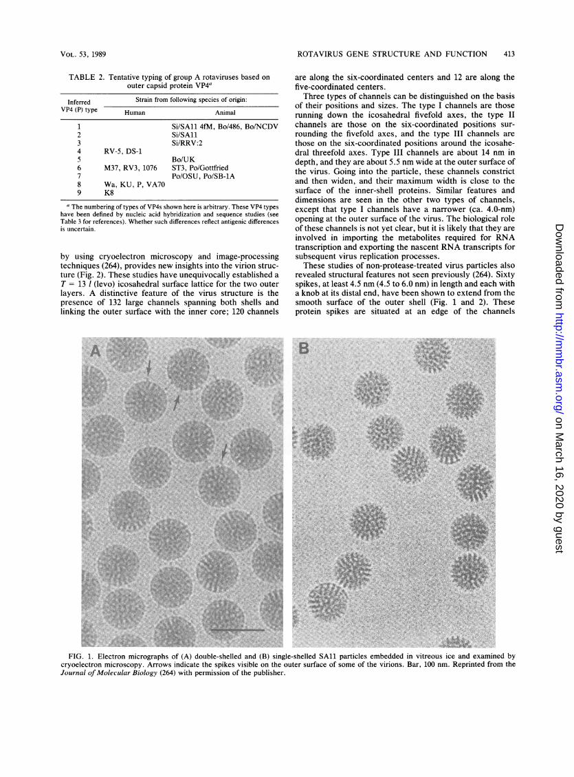
The morphologic appearance of rotavirus particles isdistinctive. Intact virus particles resemble a wheel, withshort spokes and a well-defined rim, when examined bynegative-stain electron microscopy (EM). The name rotavi-rus (from the Latin rota, meaning wheel) was suggested onthe basis of this characteristic (106). Three types of particles(double-shelled, single-shelled, and core) are often observedby EM (Fig. 1). SAl double-shelled particles are 76.5 nm indiameter, single-shelled particles are 70.5 nm in diameter,and cores are 50 nm in diameter. Single-shelled particles andcores can be produced by chemical disruption of double-shelled or single-shelled particles, respectively. It is un-known whether the single-shelled or core particles areidentical to subviral particles synthesized during virus repli-cation.
Early structural studies of rotavirus particles agreed thatthese particles possessed icosahedral symmetry, but theultrastructure of the virus was controversial. For example,the triangulation number (1), which designates the relation-ship between the neighboring fivefold axes of the icosahedralsurfaces, was initially reported to vary from 3 to 16 (90, 181,205, 281). The application of new techniques (which avoideddouble-sided images produced by negative staining) to thestudy of rotavirus structure has recently resolved theseearlier discrepancies. Roseto et al. (281) studied the struc-ture of single-shelled rotaviruses by using a freeze-dryingtechnique and reported the existence of 132 capsomeresarranged in a skew symmetry with T = 13. They also showedthat the outer layer contained small holes that correspondedone-to-one with holes in the inner capsid. This structure wassubsequently confirmed for the inner capsid by Ludert et al.(200), using the same technique and chemically disruptedparticles.The three-dimensional structure of double- and single-
shelled rotavirus particles, determined at a 4-nm resolution
MICROBIOL. REV.
on March 16, 2020 by guest
http://mm
br.asm.org/
Dow
nloaded from
ROTAVIRUS GENE STRUCTURE AND FUNCTION 413
TABLE 2. Tentative typing of group A rotaviruses based onouter capsid protein VP4a
Inferred Strain from following species of origin:VP4 (P) type Human Animal
1 Si/SA11 4fM, Bo/486, Bo/NCDV2 Si/SA113 Si/RRV:24 RV-5, DS-15 Bo/UK6 M37, RV3, 1076 ST3, Po/Gottfried7 Po/OSU, Po/SB-lA8 Wa, KU, P, VA709 K8
The numbering of types of VP4s shown here is arbitrary. These VP4 typeshave been defined by nucleic acid hybridization and sequence studies (seeTable 3 for references). Whether such differences reflect antigenic differencesis uncertain.
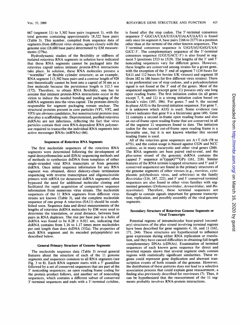
by using cryoelectron microscopy and image-processingtechniques (264), provides new insights into the virion struc-ture (Fig. 2). These studies have unequivocally established aT = 13 1 (levo) icosahedral surface lattice for the two outerlayers. A distinctive feature of the virus structure is thepresence of 132 large channels spanning both shells andlinking the outer surface with the inner core; 120 channels
are along the six-coordinated centers and 12 are along thefive-coordinated centers.Three types of channels can be distinguished on the basis
of their positions and sizes. The type I channels are thoserunning down the icosahedral fivefold axes, the type IIchannels are those on the six-coordinated positions sur-rounding the fivefold axes, and the type III channels arethose on the six-coordinated positions around the icosahe-dral threefold axes. Type III channels are about 14 nm indepth, and they are about 5.5 nm wide at the outer surface ofthe virus. Going into the particle, these channels constrictand then widen, and their maximum width is close to thesurface of the inner-shell proteins. Similar features anddimensions are seen in the other two types of channels,except that type I channels have a narrower (ca. 4.0-nm)opening at the outer surface of the virus. The biological roleof these channels is not yet clear, but it is likely that they areinvolved in importing the metabolites required for RNAtranscription and exporting the nascent RNA transcripts forsubsequent virus replication processes.These studies of non-protease-treated virus particles also
revealed structural features not seen previously (264). Sixtyspikes, at least 4.5 nm (4.5 to 6.0 nm) in length and each witha knob at its distal end, have been shown to extend from thesmooth surface of the outer shell (Fig. 1 and 2). Theseprotein spikes are situated at an edge of the channels
B
'5. *
*
FIG. 1. Electron micrographs of (A) double-shelled and (B) single-shelled SAl1 particles embedded in vitreous ice and examined bycryoelectron microscopy. Arrows indicate the spikes visible on the outer surface of some of the virions. Bar, 100 nm. Reprinted from theJournal of Molecular Biology (264) with permission of the publisher.
VOL. 53, 1989
.1 'k A, 4V. .4
.:lWA ;.A:
r', #I, . ,
R4
04
on March 16, 2020 by guest
http://mm
br.asm.org/
Dow
nloaded from

414 ESTES AND COHEN
FIG. 2. Surface representations of the three-dimensional structure of double- and single-shelled rotavirus particles. The particles areshown along the icosahedral fivefold axis (a and c) and along the icosahedral threefold axis (b and d). In panels b and d, a pair of neighboringfivefold axes (designated as 5) and six-coordinated positions (designated as 6) are indicated to illustrate T = 13 1. Three types of channels (I,II, and III in panels a and c, showing one of each type) are found at all five- and six-coordinated positions spanning the outer- and inner-shellproteins. One of the protein spikes situated at the edge of type II channels surrounding the fivefold positions in double-shelled particles ishighlighted with a dashed circle. Reprinted from the Journal of Molecular Biology (264) with permission of the publisher.
surrounding the fivefold icosahedral axes, and they appar-ently are composed of dimers of the hemagglutinin (VP4)known to be present on the outer capsid of the particles(B. V. V. Prasad, J. W. Burns, E. Marietta, M. K. Estes,and W. Chiu, submitted for publication). Examination of thespike structure in protease-treated particles will be of inter-est. It is noteworthy that new studies have shown thatreovirus particles have long fibers topped with knobs extend-ing from the surfaces of virions and that these fibers havebeen shown to be composed of the sigma 1 protein, which isthe hemagglutinin and a major determinant of virulence(115).The distinctive morphology of rotaviruses is easily seen
by conventional EM, and for this reason, EM remains thestandard against which new diagnostic techniques (ELISAs,latex agglutination, etc.) are compared. In addition, EM (inconjunction with either immunologic analyses or RNA anal-
yses) continues to be useful for detecting the non-group Arotavirus strains that do not share any antigenic cross-reactivities with previously identified strains (see above).Since EM remains so critical for diagnosis, it is important toknow that the detection of rotaviruses by EM can beinfluenced by the method used for staining. For example,negative staining with uranyl acetate or with phosphotung-stic acid at low pH (pH 4.5) allows detection of all rotavirusstrains; however, staining with phosphotungstic acid atneutral pH may result in removal of the outer capsid ofparticles and complete disintegration of virus particles,particularly with non-group A rotaviruses (233, 320).
GENOME STRUCTURE
The viral genome of 11 segments of dsRNA is containedwithin the virus core capsid. The segments range in size from
MICROBIOL. REV.
on March 16, 2020 by guest
http://mm
br.asm.org/
Dow
nloaded from
ROTAVIRUS GENE STRUCTURE AND FUNCTION 415
667 (segment 11) to 3,302 base pairs (segment 1), with thetotal genome containing approximately 18,522 base pairs(Table 3). This number, compiled from sequence data ofsegments from different virus strains, agrees closely with thegenome size (18,680 base pairs) determined by EM measure-ments (279a).Hydrodynamic studies of the flexibility or stiffness of
isolated rotavirus RNA segments in solution have indicatedthat these RNA segments cannot be packaged into therotavirus capsid unless intimate protein-RNA interactionstake place. In solution, these RNA molecules possess a"wormlike" or flexible cylinder structure; as an example,RNA segment 1 (3,302 base pairs and a contour length of 928nm) theoretically cannot be bent into a capsid of 50 nm as afree molecule because the persistence length is 112.5 nm(172). Therefore, to obtain RNA flexibility, one has toassume that intimate protein-RNA interactions occur in thevirion to induce the needed bending and packaging of thedsRNA segments into the virus capsid. The proteins directlyresponsible for segment packaging remain unclear. Thestructural proteins present in core particles (VP1, VP2, andVP3) are obvious candidates, but nonstructural proteins mayalso play a scaffolding role. Deproteinized, purified rotavirusdsRNAs are not infectious, reflecting the fact that virusparticles contain their own RNA-dependent RNA polymer-ase required to transcribe the individual RNA segments intoactive messenger RNAs (mRNAs) (66).
Sequences of Rotavirus RNA Segments
The first nucleotide sequences of the rotavirus RNAsegments were determined following the development ofrapid deoxyribonucleic acid (DNA) sequencing methods andof methods to synthesize dsDNA from templates of eithersingle-stranded viral RNA transcripts or from genomicdsRNA. Once initial sequence information for any RNAsegment was obtained, direct dideoxy-chain terminationsequencing with reverse transcriptase and oligonucleotideprimers with mRNA (or denatured dsRNA segments) oftenbypassed the need for additional molecular cloning andfacilitated the rapid acquisition of comparative sequenceinformation from numerous virus strains. The nucleotidesequences of the 11 RNA segments from different virusstrains are known (Table 3), and the complete nucleotidesequence of one group A rotavirus (SAl) should be estab-lished soon. Sequence data and direct measurements of thelengths of rotavirus dsRNA molecules by EM were used todetermine the translation, or axial distance, between basepairs in RNA duplexes. The rise per base pair in a helix ofdsRNA was found to be 0.28 ± 0.011 nm, indicating thatdsRNA contains from 1.16 to 1.17 times more nucleotidesper unit length than does dsDNA (332a). The properties ofeach RNA segment and its encoded polypeptide(s) aredescribed below.
General Primary Structure of Genome Segments
The nucleotide sequence data (Table 3) reveal generalfeatures about the structure of each of the 11 genomesegments and sequences common to all RNA segments (seeFig. 3 to 8). Each RNA segment starts with a 5' guanidinefollowed by a set of conserved sequences that are part of the5' noncoding sequences; an open reading frame coding forthe protein product follows, and another set of noncodingsequences, which contains a different subset of conserved3'-terminal sequences and ends with a 3'-terminal cytidine,
is found after the stop codon. The 5'-terminal consensussequence 5'-GGC(AIU)(AIU)U(AIU)A(AIU)(AIU) is foundonly once (in segment 4, base pairs 2166 to 2177) in a positionother than at the termini of the other known sequences. The3'-terminal consensus sequence is U(GIU)(UIG)(GIU)(AIG)CC-3'. The complementary sequence of the 3'-terminalconsensus sequence (UGUGACC-3') is also found in seg-ment 5 (positions 1513 to 1519). The lengths of the 3' and 5'noncoding sequences vary for different genes. However,these lengths are conserved among strains for a given gene,with the exception of the 3' end of segment 7 (134 bases forSAl and 112 bases for bovine UK viruses) and segment 10(from 182 to 186 bases for five different virus strains). Thereis no preferential use of stop codons, and a polyadenylationsignal is not found at the 3' end of the genes. Most of thesequenced segments (except gene 11) possess only one longopen reading frame. The first initiation codon (in all genesexcept 7, 9, and 11) is a strong initiation codon based onKozak's rules (185, 186). For genes 7 and 9, the secondin-phase AUG is the favored initiation sequence. For gene 7,it is unknown which AUG is used. Some evidence (seebelow) indicates that gene 9 is a bicistronic gene (49). Gene11 contains a second in-frame open reading frame and alsoan out-of-frame open reading frame that are conserved in allof the segment 11 sequences found to date. The initiationcodon for the second out-of-frame open reading frame is afavorable one, but it is not known whether this secondreading frame is used.
All of the rotavirus gene sequences are A+T rich (58 to67%), and the codon usage is biased against CGN and NCCcodons, as in many eucaryotic and other viral genes (260).The RNA segments are base paired end-to-end, and theplus-sense strand of the genomic dsRNA contains thecapped 5' sequence m7GpppG(m)GPy (161, 218). Similarfeatures of the RNA termini (capped structures and 5' and 3'conserved sequences) are found in the primary structures ofthe genome segments of other viruses (e.g., reovirus, cyto-plasmic polyhedrosis virus, and orbivirus) in the familyReoviridae (116, 187, 222), and 5' and 3' conserved terminalsequences are also found in other virus families with seg-mented genomes (Orthomyxoviridae, Arenaviridae, and Bu-nyaviridae). Therefore, these terminal sequences arethought to contain signals important for genome transcrip-tion, replication, and possibly assembly of the viral genomesegments.
Secondary Structure of Rotavirus Genome Segments orViral Transcripts
Potential regions of intramolecular base-paired (second-ary) structures of the plus strand involving terminal regionshave been described for gene segments 4, 10, and 11 (162,171, 244). These structures are hypothesized to influencegene expression during either RNA replication or transla-tion, and they have caused difficulties in obtaining full-lengthcomplementary DNAs (cDNAs). Examination of terminalsequences of each known gene sequence for direct andinverted repeats shows that several segment ends containregions with statistically significant similarities. These re-gions could represent gene duplication and aberrant tran-scription events of both strands of the genome. However,the distribution of these patterns does not lead to a selectiveassociation process that could explain gene reassortment, afinding also previously described for reoviruses (7). Thus, itcan be hypothesized that the reassortment of the 11 seg-ments probably involves RNA-protein interactions.
VOL. 53, 1989
on March 16, 2020 by guest
http://mm
br.asm.org/
Dow
nloaded from

416 ESTES AND COHEN
TABLE 3. Nucleotide sequences of rotavirus RNA segments"
RNA RNAsegment size Protein Strain(s) Reference(s)semn siz
1 3,302
2 2,687
3 2,591
VP1
VP2
VP3
A/Bo/Rf
A/Bo/Rf
A/Si/SAlI
4 2,362 VP4 A/Si/SAll 4fMA/Bo/486A/Hu/RV-5A/Si/RRVA/Hu/Wa, DS-1, P,VA70, M37, 1076,McN13, ST3
A/Si/SAl1A/Bo/NCDVA/Bo/UK
5 1,581 NS53
6 1,356 VP6
A/Bo/Rf
A/Si/SAilA/Hu/WaA/Bo/RfA/Hu/S2A/Hu/1076A/Eq/Fl-14A/Po/Gottfried
7 1,104 NS34 A/Si/SAilA/Bo/UKA/Po/OSU
8 1,059 NS35 A/Si/SAilA/Bo/UKA/Po/OSU
9 1,062 VP7 A/Si/SAilA/Bo/UKA/Hu/Hu5A/Hu/WaA/Hu/S2A/Bo/NCDVA/Bo/RfA/Po/OSUA/Si/RRVA/Hu/D, MO, M37,DS1, HN126, P,VA70, S73
A/Po/OSUA/Po/GottfriedA/Po/YMA/Hu/KUA/Hu/69M, W161A/La/Ala, R-2, CII
66a
188
Liu and Estes,in press
195, 196262170204126
225a,235235171
38
36, 993668148128128128
31346284
3180284
10, 328783206, 27614212254125135135
284130283326134234a
Evolution of the Rotavirus Genome
Comparisons of the sequences of individual segments ofdifferent group A rotaviruses have shown that changes occurthrough genetic shift (genome reassortment) and drift (se-quence changes within segments) via mechanisms analogousto those seen with the type A influenza viruses (2). Inaddition, changes apparently occur by another mechanism(rearrangements within a genome segment [described be-low]). A comparison of sequences of the gene encoding theneutralization glycoprotein VP7 of 27 human and animal
TABLE 3-Continued
RNA RNA Protein Strain(s) Reference(s)segment size
10 751 NS28 A/Si/SAil 35A/HuIWa 244A/Bo/UK 21, 345A/Bo/NCDV 263
11 667 NS26 A/Si/SAil 225, 349A/Hu/Wa 163A/Bo/UK 345
Compiled April, 1989. The gene-protein assignments are for SAil (seeTable 4), and sequences of the cognate gene in other strains are listed on thebasis of homology with the SAII gene. For some strains, this represents adifferent RNA segment based on electrophoretic migration on polyacrylamidegels. For example, the SAil genome segment 8, the bovine UK genomesegment 7, and the rhesus rotavirus genome segment 9 encode NS35, whereasSAII genome segment 9, UK genome segment 8, and RRV genome segment7 encode VP7 (80, 123, 124, 208, 217). Such shifts are most common forgenome segments 7, 8, and 9. The order of migration of segments 10 and 11 isalso changed between viruses with long and short RNA patterns. The size ofeach segment is given only for the first virus listed and may differ for the samesegment in other virus strains. Segment size differences have been reportedfor genome segments 4, 7, 10, and 11, and these differences may be due togenetic drift, rearrangements of sequences within a segment, or sequencingerrors (see text).
viruses which each have a type 3 VP7 suggests that there arespecies-specific sequences in this gene (234a), as well asserotype-specific regions (see below and Fig. 6). The identi-fication of species-specific, unique amino acid sequences inthis gene suggests that the VP7 gene is involved in interspe-cies reassortment with low frequency in nature (234a). Thebiologic properties and antigenic composition of particlesmay be altered by such genomic evolution, but this is onlybeginning to be understood. Interesting recent results indi-cate that the antigenic composition of particles may beinfluenced by the interactions of specific combinations of thetwo outer capsid proteins, by trypsin treatment of particles,or by oligosaccharide addition at different sites on the outercapsid glycoprotein (47, 56, 72, 203, 302).
Genome Analysis for Virus Detection
Rotaviruses are the only mammalian agents known tocontain 11 segments of dsRNA. In most cases, the genomeof the group A viruses is composed of four high-molecular-weight dsRNA segments (segments 1 to 4), five middle-sizedsegments (segments 5 to 9) including a distinctive triplet ofsegments (segments 7 to 9), and two smaller segments(segments 10 and 11). Analysis of the genome pattern ofvirus specimens is relatively easy and rapid; therefore, thistechnique has become a useful and popular procedure forvirus detection and for molecular epidemiology studies (seereferences 53 and 96 for reviews).Although in most cases the basic pattern described above
is seen, RNA analyses have shown that some rotaviruseshave distinct genome migration patterns. Viruses with suchdistinct patterns have been characterized as either non-group A viruses or group A viruses that contain rearrange-ments within individual genome segments (see below). Sincethese distinct RNA patterns can arise by different mecha-nisms, RNA profiles cannot be used as the sole criterion forclassification of a virus strain in a specific group (i.e., A toF). Instead, RNA profiles combined with group A ELISAreactivity should be used for preliminary characterization ofnon-group A rotaviruses (96, 249, 309). For viruses within aspecific group, RNA patterns cannot be used to classify
MICROBIOL. REV.
on March 16, 2020 by guest
http://mm
br.asm.org/
Dow
nloaded from
ROTAVIRUS GENE STRUCTURE AND FUNCTION 417
them into different serotypes or subgroups (1, 22, 41, 117,229, 298, 325, 333, 341). However, because RNA patternsusually remain constant for individual virus strains, RNAanalysis is useful for molecular epidemiology studies tomonitor virus outbreaks and transmission.The electrophoretic migration of the same gene in different
virus strains often shows heterogeneity. In contrast, thegene sequence data show that the same genes from differentstrains almost always contain the same number of nucleo-tides. This suggests that the heterogeneity in RNA segmentmobility, observed among the cognate RNA segments ofdifferent virus strains, is attributable to sequence differencesand secondary structure (which remain during electrophore-sis of the segments). Recent longitudinal surveillance studiessuggest that correlation of RNA patterns with epitopes onspecific proteins is possible within limits of time and loca-tion; confirmation of this awaits more extensive antigenicand RNA analyses of a large number of rotavirus strainsisolated from distinct locations and in different years (70,144, 230; D. 0. Matson and M. K. Estes, unpublished data).
Genome Rearrangements
Analyses of some group A rotaviruses with atypical RNAprofiles on gels have resulted in the recognition that rear-rangements within genome segments can occur. In viruseswith genome rearrangements, normal RNA segments aremissing in an electrophoretic profile, and these are replacedby additional, more slowly migrating bands of dsRNA.These new bands usually represent concatemeric forms ofdsRNA containing sequences specific for the missing RNAsegments (158, 215, 250, 256, 325). Viruses with such ge-nome rearrangements have been isolated from immunodefi-cient, chronically infected children (250), asymptomaticallyinfected immunocompetent children (24), and animals(calves [256], pigs [23], or rabbits [325, 335]). They have alsobeen obtained after serial in vitro passage of a tissue-culture-adapted bovine rotavirus at high multiplicity of in-fection (158). Isolates with rearrangements in segments 5, 6,8, 10, and 11 (27, 158, 159, 215, 250, 256, 325) have beencharacterized, with the greatest number having rearrange-ments in segment 11. In retrospect, it is possible that shortand super-short RNA patterns reported for human virusesreflect rearrangements in genome segment 11 (81). In fact,determination of gene 11 sequences from these strains hasshown that they contain insertions of A+T rich 3' noncodingregions which are not significantly homologous to eachother, to other parts of gene 11, or to other rotavirus genesthat have been sequenced (236a). These appear differentfrom rearrangements described below.
Characterization of viruses containing rearranged genomesegments has shown that these viruses are often not defec-tive and that the rearranged segments can reassort andreplace normal RNA segments structurally and functionally(4, 27, 131, 215). Biophysical characterization of such parti-cles has shown that up to 1,800 additional base pairs can bepackaged in particles without causing detectable changes inparticle diameter or apparent sedimentation values. Thedensity of particles containing rearranged genomes wasincreased, and the change in density was directly propor-tional to the number of additionally packaged base pairs(220). These results indicate that rotaviruses have consider-able capacity to package additional genomic RNA, and onewonders what the upper limit of this capacity might be.Although a total of 11 RNA segments (or rearranged bands)are invariantly packaged, there seems to be much less
constraint on the length of individual RNA segments assem-bled into the maturing virus particle.
In most cases, the profiles of virus-specific proteins incells infected with rotaviruses with rearranged genomes aresimilar to those seen in cells infected with standard rotavirusstrains (4, 27, 215, 256, 325). This indicates that the rear-rangement of the segment-specific sequences apparently hasleft the normal reading frames and their expression unal-tered.
This hypothesis has been confirmed by analyses of severalrearranged genome segment 11 sequences. In two cases, therearranged segment 11 consists of a partial duplication ofsegment 11-specific sequences, with maintenance of theopen reading frame for the protein product from the firstinitiation codon and with the duplicated sequences lackingthe initiation codon to reinitiate translation. In addition, theconserved 5'- and 3'-terminal sequences are present at theends of the rearranged segments, but these are not present inthe middle of the gene upstream from the duplicated se-quences (124a; M. A. McCrae, personal communication).Other cases of rearrangements have apparently led to theabolition or extension of the normal reading frame, with theconsequence that no protein or extended novel proteinproduct is made (158); the sequence of one of these RNAsshows a duplication that extends "in-frame" of the normalreading frame (M. A. McCrae, personal communication). Inanother case, partial duplications and deletions (with theappearance of a possible new open reading frame) in arearranged segment have been found (129a). Rearrange-ments leading to nonfunctional gene products probablywould not have been identified by present assays.One proposed mechanism for these rearrangements that is
compatible with the available data is that at various stages ofinitial transcription, the virion-associated RNA polymerasefalls back or jumps to a second site on its template toreiterate part of it in the transcript (124a, 129a, 159). Suchcopy choice mechanisms have been documented to explainproduction of deletion-defective particles of vesicular sto-matitis virus and coronavirus recombinants (192a, 204a). Forrotaviruses, these aberrant replicative events must be rareand the polymerase apparently maintains template speci-ficity, because the formation of mosaic gene structures hasnot been seen. However, such rearrangements have notbeen seen in mRNA synthesized in vitro with the endoge-nous RNA polymerase, possibly because the correct condi-tions to synthesize large amounts (or to detect few copies)have not yet been tested. Without such evidence, one mustalso consider that these rearrangements might occur duringreplication of the RNA (synthesis of the negative strand offthe mRNA template). Similar rearrangements have beendescribed for the orbiviruses (85, 272). Therefore, genomerearrangements may be operative to a much greater extentthan was previously envisaged as a mechanism of evolutionof rotaviruses and other dsRNA viruses. It remains un-known whether such rearrangements also affect antigenicdiversity (perhaps by altering the virion structure) or patho-genic properties of viruses (perhaps by altering the growthproperties of viruses).
GENE-CODING ASSIGNMENTS
The gene-coding assignments and known properties of theproteins encoded in each of the 11 genome segments are now
fairly well established (Table 4). These assignments havebeen determined by in vitro translation studies with mRNAor denatured dsRNA (81, 207, 208, 217, 307) and by analyses
VOL. 53, 1989
on March 16, 2020 by guest
http://mm
br.asm.org/
Dow
nloaded from

418 ESTES AND COHEN
of reassortant viruses (123, 168, 194, 237-239, 341). Theinformation on SAl serves as the basis for comparativestudies with other rotavirus strains. Such comparisons haveshown that the absolute order of migration of a gene codingfor a particular protein (cognate gene) may differ for differentvirus strains, and so gene assignments cannot be based onRNA patterns alone (see Table 3, footnote a). Instead,identification of cognate genes must be based on hybridiza-tion with gene-specific probes (22b, 79, 101), analysis ofreassortants, or protein identification based on biochemicalor immunologic identification of the protein translated in acell-free system programmed with mRNA specific to thegene. The ability to directly obtain sequence informationfrom dsRNAs or ssRNAs (18, 135, 136) and the accumulat-ing nucleic acid sequence data bases also make it possible toidentify cognate genes solely on the basis of sequencehomology.The rotavirus genes code for structural proteins found in
virus particles and for nonstructural proteins found in in-fected cells but not in mature particles. The consensus is thatthe protein products (VP1 to VP4, VP5*, VP6, VP7, VP8*)of six of the genome segments are structural proteins foundin virus particles and that the other five genome segmentscode for nonstructural proteins. It remains unknownwhether the additional open reading frames found in genomesegment 11 code for other proteins.
Early studies often contained seemingly conflicting con-clusions concerning the numbers and locations of the rota-virus proteins. Many of these were resolved, as reviewedelsewhere (100, 149), when it was recognized that posttrans-lational modifications (glycosylation, trimming of carbohy-drate residues, and proteolytic cleavages) occur followingpolypeptide synthesis. In addition, strain variations (such asthe presence of more than one glycosylation site on VP7 insome bovine and human rotavirus strains) have been clearlyshown (183, 184, 294) (see below), and these provide expla-nations for other discrepancies.The nomenclature of the viral proteins has recently been
changed (194). We originally proposed the designation of theSAl structural proteins as viral protein (VP) followed by anumber, with VP1 being the highest-molecular-weight pro-tein, and proteins generated by cleavage of a larger precur-sor were indicated by an asterisk (VP4 is cleaved to produceVP5* and VP8* [11, 97]). Our initial studies failed to identifya protein product from gene segment 3, and the proteinproduct of gene segment 4 was called VP3 (208). Whenrecent studies showed that the protein of gene segment 3 isa structural protein located in the inner core, for consistencywe called the gene 3 product VP3 and renamed the gene 4product VP4 (194). This new nomenclature is used through-out this review and will be used in all our future publications,but it should be realized that much of the literature on"VP3" up to and including 1988 refers to the gene 4 product.Our naming the gene 3 protein VP3 brings into agreement
our nomenclature system for SAl with that for the UKvirus for the products of genes 1 to 4 as designated byMcCrae and McCorquodale (217) and for several humanrotavirus strains (294). Our finding that the four largest RNAsegments code for structural proteins confirms the earlierreports of others (149, 307) and also requires changes inanother nomenclature system, in which the nonstructuralproteins were called NCVP1 to NCVP5 and the gene 3product was called NCVP1 (11, 33). We continue to desig-nate nonstructural proteins by NS followed by a numberindicating their apparent molecular weight (in thousands)
seen following electrophoresis in denaturing polyacrylamidegels.
STRUCTURE AND FUNCTION OF ROTAVIRUSPROTEINS
Core and Inner Capsid Proteins
VPI. VP1 is encoded by genome segment 1 in all virusesstudied to date, and it is one of three proteins (VP1, VP2,and VP3) that make up the rotavirus core particles. Infor-mation on these core proteins currently is limited. Thededuced amino acid sequence of VP1 of a bovine rotavirusstrain indicates that this protein migrates on sodium dodecylsulfate-polyacrylamide gel electrophoresis according to itscalculated molecular weight of 124,847 and that it is arelatively hydrophobic and slightly basic protein (66a). Asmall number of VP1 molecules (ca. 2% of the virion mass)are present in virions, suggesting that this protein does nothave an important structural function but, rather, that it maybe part of an enzymatic complex. Consistent with this idea,homology searches of the protein data base have revealedsimilarities with a family of RNA polymerases of RNAviruses (GDD consensus sequence in Fig. 3). This putativefunctional site and larger regions of amino acid similaritywith VP1 are also present in the reovirus lambda 3 protein(amino acids [aa] 585 to 739) and in the bluetongue virus P1protein (aa 667 to 769).One unusual property of VP1 is that it often fails to react
in immunoprecipitations or in immunoblots with many hy-perimmune antisera made to purified virus particles (88) orwith serum from naturally or experimentally infected chil-dren or animals (61, 69, 240). Other serum samples canprecipitate VP1 (324; J. Cohen, unpublished observation),and the successful production of a VP1-reactive antiserum(by immunoprecipitation, immunofluorescence, and immu-noblots) by immunizing animals with VP1 synthesized with abaculovirus expression system indicates that this protein isantigenic and immunogenic (66a). The different capacity ofsera to react with VP1 after natural infection or parenteralimmunization may be explained by the degree to which VP1is accessible to the immune system. VP1 was partiallyaccessible to iodination in single-shelled but not double-shelled particles (236). This suggests that either VP1 isexposed on single-shelled particles or the channels thatpenetrate into the inner core of particles are large enough foriodination reactions to occur inside the particles. Examina-tion of whether antibody to VP1 reacts with single-shelledparticles may help to answer this question.
VP2. VP2, encoded by genome segment 2, is the mostabundant structural protein found in core particles (25, 194)and is the third most abundant protein in double-shelledparticles. Lactoperoxidase-catalyzed iodination experimentssuggest that VP2 is partially exposed on single-shelled par-ticles (236), and this idea is supported by the demonstrationthat antibodies to VP2 are able to react by ELISA withsingle-shelled particles bound to plates (331). However,since direct binding of particles to plates can induce confor-mational changes in particles (45, 121), immunoelectronmicroscopic examination of whether these antibodies di-rectly bind to particles would help to interpret these results.VP2 is highly immunogenic, and serum antibodies to thisprotein are a good indicator of prior infection (69, 324).VP2 is the only structural protein shown to possess
nucleic acid (dsRNA, ssRNA, and dsDNA)-binding activitywhen evaluated by an RNA overlay-protein blot assay (37).
MICROBIOL. REV.
on March 16, 2020 by guest
http://mm
br.asm.org/
Dow
nloaded from

ROTAVIRUS GENE STRUCTURE AND FUNCTION 419
TABLE 4. Rotavirus genome RNA segments and protein products'
Number of Mol wt of Approx. S Temperature
Seg- noncoding Protein nascent poly- Mature protein (by wt) of sensitivement sequencesb product' peptided (no. of modified virion mutant Remarks-
equencesb amino acids) (no. protein groupf5' 3'
1 18 17 VP1 124,847 (1088) 2 C Inner-core protein,slightly basic
152 16 28 VP2 102,431 (880) Myristilated
3 49 34 VP3 98,120 (835)
4 9 22 VP4 86,782 (776) Cleaved VP5* (529)hVP8* (247)"
5 32 73 NS53 58,654 (491)
6 23 139 VP6 44,816 (397) Myristilated
7 25 131 NS34 34,600 (315)
F Inner-core protein, RNAbinding; leucine zipper(aa 536 to 559 and 665to 686)
0.5 B Inner-core protein, basic
1.5 A Surface protein, hemag-glutinin, protease-en-hanced infectivity, neu-tralization antigen,virulence, putative fu-sion region(aa 384 to 401)
NA Slightly basic, zinc fingers(aa 54 to 66 and 314 to327)
51
8 46 59 NS35 36,633 (317)
G Inner capsid protein, tri-mer, hydrophobic sub-group antigen, requiredfor transcription
NA Slightly acidic, RNA bind-ing
E Basic, role in RNA repli-cation?
9 48 33 VP7(1) 37,368 (326)135 33 VP7(2) 33,919 (297)
10 41 182 NS20
11 21 49 NS26
Cleaved signal sequence, high-mannose glycosylation andtrimming'
20,290 (175) NS29 -> NS28 uncleaved signalsequence, high-mannoseglycosylation and trimming
21,725 (198) NS28, phosphorylated, possiblesecond type of modification
30 NA RER integral membraneglycoprotein, cell at-tachment protein, neu-tralization antigen, twohydrophobic NH2-ter-minal regions, bicis-tronic gene?, putativeCa2+-binding site (aa127 to 157)
NA Nonstructural RER trans-membrane glycoprotein,two hydrophobic NH2-terminal regions, role inmorphogenesis, putativeCa2+-binding site
NA Nonstructural, slightlybasic; serine and threo-nine rich
a For A/Si/SA11 strain except genes 1, 2, and 5 for an A/Bo/Rf strain; see Table 3 for references.b Number of 5' noncoding sequences is up to the first AUG; number of 3' noncoding sequences does not include the termination codon.'Determined by biochemical and genetic approaches (see text). The size (in thousands) of the primary translation product is given for the nonstructural (NS)
proteins.d Molecular weights are calculated from the deduced amino acid sequences from nucleotide sequence data. The molecular weights are calculated from the
largest potential open reading frame.e Adapted from Liu et al. (194). These estimates are not totally consistent with estimates of the numbers of molecules of VP4, VP6, and VP7 (120, 780, and
780 molecules, respectively) made from structural analyses of particles (264; Prasad et al., submitted for publication).f From Gombold and Ramig (123, 124). NA indicates that none was assigned.g See text for references.h There are two trypsin cleavage sites in SAl 4fM VP4 at amino acid 241 and 247. The indicated mature products are those based on the use of only the
preferred second cleavage site (195, 196).i Mature cleaved VP7 contains 276 amino acids.
VOL. 53, 1989
on March 16, 2020 by guest
http://mm
br.asm.org/
Dow
nloaded from

420 ESTES AND COHEN
RF VP1 (gene 1)
GDD
1088173bp
c cc c _3302COOH
1 -iC-1NH2
c c c c N536-559 665-686
88028bp
COOH
SAl 1 VP6 (gene 6)
NH2L
high sequence
variation
zczI80 89
c1356
COOH
FIG. 3. Features of the core and inner capsid proteins VP1, VP2, and VP6. These schematics show features of each protein based on theamino acid sequence deduced from the nucleotide sequence. The total number of nucleotides in each gene, the length of the 5' and 3'noncoding regions, and the numbers of nucleotides or amino acids in each gene or deduced protein are shown. Cysteine residues, thepolymerase consensus (GDD) signal in VP1 (aa 630 to 632), and the leucine zipper (aa 536 to 559 and 665 to 686) in VP2 are also indicated.The box (E) shows a region of high sequence variation. Compiled from sequences of the genes which code for these proteins (see Table 3for references).
VP2 binds ssRNA in preference to dsRNA, but the bindingis not sequence specific. Analysis of the sequence of VP2shows that this protein contains leucines at every seventhresidue, starting at amino acids 536 and 665. Such a periodicarray of leucines (leucine zipper [192]) is hypothesized torepresent a part of a protein that molds it to interact with a
target site on DNA (and probably on dsRNA) (Fig. 3) (188).VP2 may be a nucleocapsid protein that is bound tightly tothe RNA segments. The apparent molecular weight (of94,000) of VP2 calculated from its mobility on sodiumdodecyl sulfate-polyacrylamide gel electrophoresis differsfrom the molecular weight (of 102,431) deduced from thenucleotide sequence. This may result from the high percent-age of predicted alpha helix in this protein. VP2 has beenfound to be myristilated, and although the functional signif-icance of this is unknown, such modification in other viralsystems has been found on scaffolding proteins important forthe formation of virus particles (59). Epitopes on VP2 thatcosegregate with subgroup specificity of VP6 also have beenreported (331), but other biologic properties of VP2 are notyet known. MAbs to VP2 should be useful to further probethe function of this protein in the assembly of rotavirusparticles or in RNA replication.VP3. VP3, encoded by genome segment 3, is a minor
structural protein that may comigrate with the outer capsidprotein VP4 in many gel systems (194). In early studies, thisgene product was reported to be a structural protein (307), anonstructural protein (11), or a structural protein that wastranslated poorly in vitro and was synthesized and processedrapidly (217). These early studies all approached analysis ofthe genome segment 3 protein by comparing the proteinssynthesized in infected cells with those translated in cell-freesystems. A reevaluation of this question for SAil involvingthe use of reassortant viruses and new methods of electro-phoresis of the proteins on polyacrylamide gels confirmedthat four proteins are located in single-shelled particles and
three proteins are located in core particles (194), as shownearlier by others (25, 307). The poor cell-free translation ofthis gene product has been confirmed and remains unex-
plained. The sequence of gene 3 indicates that this segmentcodes for a protein of 835 amino acids with a predictedmolecular weight of 98,120 (M. Liu and M. K. Estes, Nu-cleic Acids Res., in press). Analysis of the deduced aminoacid sequence indicates that this is a basic protein thatcontains multiple repeats of amino acids. Homology withother RNA polymerases from other viruses also suggeststhat this protein is involved in RNA replication (Liu andEstes, in press).VP6. VP6 is encoded by genome segment 6 and is the
major structural protein in virus particles located on theouter surface of single-shelled particles. Biochemical char-acterization of VP6 removed from particles revealed that it isa trimer (129, 288), and three-dimensional structural studiesof single-shelled particles have shown trimers to be presenton the surface of these particles (264). Trimerization andformation of tubules is an intrinsic property of this protein,since VP6 synthesized in the absence of other viral proteinsforms such structures (94). VP6 removed from particles willalso form tubules and viruslike particles at low pH, butwhether other proteins play a role in these reactions remainsunclear (273). These results and biochemical data showingthat VP6 is removed from inner-core particles prepared bytreating single-shelled particles with chaotropic agents (25,194, 293) counter the suggestion (based on iodination resultswith single-shelled particles) that VP6 is more internal thanVP2 in particles (236). Alternative interpretations of theseiodination results are that (i) the tyrosine residues requiredfor iodination are not accessible in trimeric VP6; (ii) thechannels leading into the inner particle are large enough topermit the iodination reactants (lactoperoxidase) to pene-trate the particles and therefore label VP2; and (iii) VP2,which is a core protein that may bind to the RNA segments,
1 18 bp E
NH2
RF VP2 (gene 2)
MICROBIOL. REV.
c
on March 16, 2020 by guest
http://mm
br.asm.org/
Dow
nloaded from

ROTAVIRUS GENE STRUCTURE AND FUNCTION 421
lines the channels and protrudes out of single-shelled parti-cles.
Biochemical and immunologic approaches have been usedto examine whether VP6 performs specific biologic functionsduring virus replication. Removal of VP6 from single-shelledparticles results in a loss of transcriptase activity of parti-cles. Readdition of VP6 to stripped single-shelled particlesrestores transcriptional activity, and addition of excess VP6to stripped single-shelled particles inhibits transcription (25,293). These experiments indicate that VP6 is required forpolymerase activity, but they do not directly prove that VP6itself is involved in transcription. VP6 may merely beimportant as a structural component of particles to maintainthe proper conformation or organization of the viral corestructure or transcriptional complex composed of one ormore of the core proteins. VP6 is myristilated, and thismodification may be important for formation of virus parti-cles or for targeting single-shelled particles to the ER mem-brane for budding (59) (see below).VP6 is both highly immunogenic and antigenic, and it is
the most frequently targeted protein in diagnostic assays todetect virus particles. VP6 contains common (cross-reac-tive) epitopes shared by other group A viruses, and it cancontain zero, one, or several forms of an antigen called thesubgroup antigen (137, 168, 174, 190, 323). Whether VP6plays a role in inducing protective immunity remains un-clear.Some MAbs to VP6 have been reported to possess low
levels of activity that neutralizes virus infectivity whenassayed in vitro (288). In addition, a synthetic peptidecorresponding to aa 40 to 60 of VP6 has been reported toinduce protective immunity in a mouse model of passiveprotection (111). These results contrast with most studies,which have failed to detect neutralization with anti-VP6MAbs (119, 139, 150, 282, 305, 312) or with polyclonalmonospecific serum raised to VP6 synthesized with a bacu-lovirus expression system (94). It is possible that neutraliza-tion activity observed with VP6 antiserum is not specific butis caused by nonspecific trapping of infectious double-shelled particles owing to the aggregation of single-shelledparticles present in most virus preparations. Neutralizationactivity detected with polyclonal monospecific serum madeto gel-purified VP6 (178, 287) is difficult to interpret becauseof questions of the specificity of these sera. Contaminationof the VP6 from gels with small amounts of the closelymigrating VP7 can result in a neutralizing antiserum as aresult of antibodies to VP7 (M. K. Estes, unpublished data).The subgroup epitopes on VP6 detected by MAbs (137)
have been used as an epidemiologic tool to monitor theantigenic properties of different virus strains. To date, at-tempts to map the location of the subgroup epitopes by usingbiochemical or molecular techniques have met with limitedsuccess because many of these epitopes are conformationaland appear to be located on the trimeric but not monomericstructures (128). Analysis of VP6 with a panel of MAbs hasindicated that VP6 possesses at least five nonoverlappingepitopes (261). Unfortunately, none of these MAbs or othersto VP6 (128, 189, 190) have been directly compared with theoriginal subgrouping MAbs to permit comparisons of results.Use of a rapid and simple method to map epitopes willfacilitate such needed comparisons (45).A rotavirus strain that was isolated from horses (FI-14)
and that possesses both subgroup I and II epitopes has beenidentified (150). Sequence analysis of gene 6 of this virussuggested that two VP6 proteins may be produced from twodifferent initiation codons (128). This study also determined
the sequence of VP6 from several new virus strains andconfirmed regions of sequence divergence recognized previ-ously (36, 68, 99, 148); unfortunately, it did not shed furtherlight on the map sites of the subgroup or common epitopeson VP6 (128). Comparison of the sequences of different virusstrains has identified one region of fairly high sequencevariation (Fig. 3), but no function has yet been mapped tothis or other sites. Additional virus strains which lacksubgroup I and II epitopes have been described (323), andsequence analyses of these strains may help define regionson VP6 which react with the subgroup MAbs. Use of MAbswhich react with either the monomeric or trimeric form ofVP6 should help us to understand the process of trimeriza-tion and the role of VP6 in the viral morphogenetic process(14; L. Svensson, personal communication).
Outer Capsid ProteinsVP4. VP4 is the protein product of genome segment 4, and
it is a nonglycosylated outer capsid protein and a hemagglu-tinin in many virus strains (167, 208). It is responsible for anumber of biologically important functions. In the presenceof trypsin, VP4 is cleaved into VP5* (molecular weightapproximately 60,000) and VP8* (molecular weight approx-imately 28,000), and this cleavage results in enhancement ofviral infectivity (91, 97). Cleavage of VP4 has been shown toenhance penetration (but not binding) of the virus into cells(64, 113, 169). VP4 is also associated with restriction of thegrowth of certain rotavirus strains in tissue culture cells (137)and in mice (238) and with protease-enhanced plaque forma-tion (167). Antibodies directed at VP4 neutralize rotavirus invitro (45, 71, 137, 139a, 153, 179, 329) and passively protectmice against heterologous rotavirus challenge in vivo (210,238). Further studies have suggested that VP4 inducesprotective immunity in animals (239) and is immunogenic inchildren and animals (69, 301, 324).These diverse properties of VP4 highlight the importance
of this protein in the biology of the rotaviruses. Figures 4 and5 show features of VP4 based on recent sequence data (Table3) and mapping of antigenic sites by sequencing escapemutants selected with MAbs that possess neutralizationactivity (204, 327). Direct amino acid sequence analysis ofVP4, VP5*, and VP8* of a strain called SAl 4fM deter-mined the site of trypsin cleavage (196). Both VP4 and VP8*had blocked NH2 termini, and the VP5* of SAl 4fM wasfound to be composed of two polypeptides with slightlydifferent amino acid sequences at their NH2 termini. Com-parison of these data with the deduced amino acid sequenceof SAl 4fM VP4 identified two trypsin cleavage sites(arginine 241 and arginine 247), with the latter position beingthe preferred cleavage site (196). Subsequent nucleotidesequencing across this region of gene 4 in other strainsrevealed that the two trypsin cleavage sites at arginines areconserved in every rotavirus VP4 sequence analyzed (Fig. 5)(197). Further sequencing has also shown that the SAl 4fMgene 4 sequence is identical to gene 4 from bovine virusesand different from SAl gene 4. The SAl 4fM virusapparently is a reassortant containing an SAl gene 9 and abovine gene 4 (Table 2) (171, 235).
In animal rotavirus strains, VP4 contains 776 aa. In humanrotavirus strains, the deduced VP4 is 775 instead of 776 aa(126, 170), with the difference being that the human strainshave lost one amino acid at residue 136 of the animal strains(Fig. 5) (170); VP8* of the human strains is, therefore, oneamino acid shorter.
In several (but not all) human rotavirus strains, an addi-tional potential trypsin cleavage site at either lysine or
VOL. 53, 1989
on March 16, 2020 by guest
http://mm
br.asm.org/
Dow
nloaded from

422 ESTES AND COHEN
arginine is found just before the 3'-proximal arginine site (aa246 in human strains, aa 247 in animal strains [126, 196]). Itis unknown whether these sequence differences at thetrypsin cleavage site affect the biologic properties of dif-ferent virus strains, but the additional trypsin cleavage site insome human viruses has been suggested to be correlatedwith virus virulence, because viruses with this extra sitewere originally isolated from children with symptomaticinfections (Fig. 5, lines RV-5, WA, P, and VA70) (126, 130).Ninety-one amino acids on VP4 are conserved among strainsfrom children with asymptomatic infections (Fig. 5, linesM37 and ST3), whereas a different amino acid is conservedat each of these sites among virulent rotaviruses (126). Inaddition, three of these amino acids are located within the6-aa connecting peptide between the two cleavage productsof VP4 (126). Some or all of the 91 differences in VP4 aminoacids between viruses from asymptomatic and symptomaticchildren have been postulated to be responsible for thedifference in virulence of these two groups of human rotavi-ruses, because none of the other 10 rotavirus gene segmentsare conserved among asymptomatic or virulent strains (108).The significance of these findings is unclear, because farfewer amino acids are conserved if one compares the con-served sequences in asymptomatic strains with those invirulent animal strains (Fig. 5) and there is no proof that thestrains from asymptomatic newborns are really avirulent.Other studies have suggested that host (rather than viral)factors may cause asymptomatic disease in newborns, be-cause virus strains that cause disease in older children oftencause asymptomatic infections in newborns (338, 344).The mechanism of cleavage activation of infectivity is not
clear from the sequence analyses alone. It is thought thatcleavage of VP4 activates an early step of replication whichmay be triggered by one or both of the terminal regionsgenerated by the cleavage or by a possible conformationalchange in cleaved VP4 molecules. Trypsin activation oforthomyxoviruses and paramyxoviruses generates a highlyconserved apolar amino terminus, and the infectivity ofthese activated viruses can be blocked by oligopeptides thatmimic this sequence (274, 275). The new terminus generatedby cleavage in the rotavirus VP4 is not hydrophobic; instead,it contains many polar amino acids (some of which might becharged at neutral pH). Therefore, the mechanism of activa-tion of rotavirus infectivity is apparently different from thatpostulated for other viruses. Since rotavirus infectivity hasbeen shown to be enhanced by trypsin, elastase, and anuncharacterized acid-stable enzyme in pancreatin (but notby chymotrypsin [132]), further examination of the sites ofnatural cleavage may be useful to understand activation. Italso remains unknown whether cleavage at both sites resultsin removal of the small intervening peptide and whether theminor and major cleavage sites shown to be used for SAl4fM are used identically for other virus strains.
Studies are just beginning to determine whether the cleav-age region (and the intervening peptide) possesses anyfunctions. The region (aa 228 to 241) adjacent to the cleavageregion has been shown to induce neutralizing antibodies byimmunization of rabbits with a synthetic peptide to thisregion. The antipeptide serum showed strain-specific low-level neutralizing activity, and it bound to virus particles inELISAs and aggregated particles by immunoelectron mi-croscopy (317). Neutralization titers were increased (from800 to 3,200) following assay of trypsin-treated virus-anti-peptide serum complexes. Trypsin treatment of virus parti-cles also has been shown to result in loss of reactivity of thevirus with a nonneutralizing antibody to the glycoprotein
VP7 (72). Knowing the location of this epitope might help inmapping possible sites of interaction between VP4 and VP7.A region in VP4 (aa 384 to 401) has been identified that
shares some similarity with an internal fusogenic hydropho-bic domain thought to be located about 80 aa from the Nterminus of the El glycoprotein of Sindbis and SemlikiForest viruses (118, 204). These similarities with the alpha-virus El protein are of interest, and it is tempting tospeculate that this region in VP4 is one which acts alone (orin cooperation with other hydrophobic regions of VP4 [as inEl]) to induce membrane fusion. This is an attractive hy-pothesis because cleavage of VP4 has been associated withinternalization of virus (perhaps by mediating direct mem-brane penetration) into cells (64, 113, 169) and acidificationof endosomes is not important for virus entry (114, 176, 201).Conservation of the sequences in this putative fusion regionof VP4 of different rotavirus strains lends credence to thesuggestion that it plays an important function in the replica-tion cycle. However, it should be noted that the exactlocation of this fusogenic domain in El is not proven, sinceexperiments with MAbs have shown that fusion activitiesare located at different sites in the El molecule (29, 50, 51,245, 296). In addition, it remains unknown whether thisputative fusion activity would act to mediate rotavirus entryinto cells or to mediate virus maturation that involvesbudding across the membrane of the ER (see below), orboth.
Analysis of the deduced amino acid sequence of VP4indicates that the amino-terminal 70% of the protein is ratherhydrophobic. The predicted amino acid sequence of VP4 hasa net negative charge at pH 7.0, and secondary-structurepredictions suggest that the amino terminus (prior to thetrypsin cleavage site) contains numerous random coils andturns, suggesting a globular structure. Four cysteines in VP4(aa 216, 318, 380, and 774) are conserved in all the rotavirusstrains sequenced to date (126, 170, 171, 204, 235). Anadditional cysteine is present at aa 266 in the human M37strain (126) and at aa 203 in SAl1, SAl 4fM, bovine 486,and RRV. It has been proposed that the trypsin cleavage siteis kept accessible through disulfide bonds at cysteines 203and 216 in VP8* and at cysteines 318 and 380 in VP5*; lackof Cys-203 was suggested to explain the poor growth ofhuman rotavirus strains in cell culture (204). This seemsunlikely since the bovine UK virus, which grows well invitro, has a serine at aa 203 (171). However, the regionsflanking the trypsin cleavage sites (aa 224 to 235 and 257 to271) are relatively highly conserved in all strains, and thesecould serve to hold the cleavage sites in the proper confor-mation for cleavage.
Sequencing of escape mutants selected with neutralizingMAbs has identified amino acids involved in homologousand heterologous neutralization (204, 327). VP8* containspredominantly type-specific sites, and five such sites (Fig. 4,sites 1 to 5) have been localized in VP8* (204, 304). Cross-reactive MAbs from two different laboratories have beenlocated on VP5* clustered in a relatively limited area at aa306, 388, 393, 434, and 440 (204, 327). These positions arebased on the numbering for the animal rotavirus strains (i.e.,aa 306 would be 305 in the human virus VP4). One MAb (site6) that neutralizes viruses in serotypes 3, 5, and 6 (based onVP7 reactivities) is located at aa 393 in the middle of theconserved hydrophobic region and the putative fusion re-gion. Other cross-reactive MAbs (epitope II) for humanviruses cause changes at aa 392 and 439 (393 and 440 ofanimal strains), suggesting that this site is conformational(327). Another cross-reactive site (epitope I) is sequential (or
MICROBIOL. REV.
on March 16, 2020 by guest
http://mm
br.asm.org/
Dow
nloaded from

ROTAVIRUS GENE STRUCTURE AND FUNCTION 423
proteolyticcleavageactivation
high sitessequence
1 variation A
NH2 2 2 2 27171 204 224 236 257 271
VP81247 aai
conservedhydrophobic
c c
776
]COOH
384 401FUSION REGION ?
1 -, 241
NH2AAA AA A1 23 4 5
247
1"1
VP5(529 aa) 776
1 COOH
A AtA A
6I It III
FIG. 4. Features of the outer capsid protein VP4. Schematic of structural and antigenic properties of VP4 based on the analyses ofnucleotide sequences of different virus strains and escape mutants selected with neutralizing MAbs. Symbols: F, regions of sequenceconservation among different virus strains; E, region of greatest sequence variation in VP8*; 0111, potential fusion region; 4 , sites ofcleavage by trypsin. Conserved cysteines (C) at aa 215, 317, 379, and 773 and the cysteine (C-) at aa 203 not present in some human rotavirusstrains are shown. The locations of neutralization epitopes defined by Mackow et al. (204) are shown by A and a number from 1 to 6; theepitopes defined by Taniguchi et al. (327) are indicated by A and a Roman numeral (I to III). The peptide possibly removed by trypsincleavage at aa 241 and 247 is shown ( a ). The numbering shown here for VP4 is based on a protein of 776 aa as determined for SAl1 4fM,RRV, and Bo/486. VP4 of human rotavirus strains contains 775 aa, lacking an amino acid at residue 136 (see Fig. 5). See text for references.
linear) on the basis of reactivity with a synthetic peptidefrom aa 296 to 313, and aa 305 of the human VP4 isapparently critical for this epitope (327). The cross-reactiveepitope I site and an epitope III site (at aa 433) have beenidentified only by one set of MAbs (327, 328). None of theseMAb-identified neutralization regions corresponds to thecleavage region shown to be immunogenic by injection ofsynthetic peptides from the cleavage site (317) (see above).This suggests that variability exists in the immunogenicity ofdistinct regions of VP4, and the relative immunogenicity ofthese epitopes remains to be clarified.Comparisons of the segment 4 sequences of viruses in
different serotypes to look for regions of diversity show thathomologies are not evenly distributed across VP4 (Fig. 5).The most distinct sequence variation is seen between aa 71and 204 in VP8* (Fig. 5). It is tempting to speculate thatVP8* is externally exposed on the virus particle, since VP8*contains the locations of greatest sequence diversity and ofbinding of the strain-specific MAbs (whereas cross-reactivesites have been mapped to VP5*) and since VP8* is pre-dicted to be globular. If readily exposed, the sequencediversity in VP8* could easily arise by immune selection. Nofunction or neutralization epitopes have yet been mapped tothis variable domain. A second region of sequence variationsis found between aa 578 and 608.Attempts to locate other functional domains on VP4 have
not yet been successful. Genetic analyses show that VP4 isthe hemagglutinin, and hemagglutination activity has beendemonstrated with VP4 molecules expressed by baculovirusrecombinants (202; S. E. Crawford and M. K. Estes, unpub-lished data). The site on VP4 that binds to erythrocytesremains unknown, but it may involve several regions of VP4because MAbs to both VP5* and VP8* can inhibit hemag-
glutination (45, 139a, 179, 304). However, some of theseMAbs may inhibit hemagglutination by steric hindrance.Site-specific mutagenesis and high-resolution structuralstudies of particles or of individual proteins are needed toanswer these questions.VP7. VP7 is the second most abundant capsid protein and
is encoded by genome segment 9 of SAl1, genome segment8 of the UK bovine virus, and genome segment 7 of rhesusrotavirus. This outer capsid glycoprotein is highly immuno-genic and induces neutralizing antibodies. Recognition thatthis neutralization antigen is a structural glycoprotein stim-ulated numerous studies of the structure, biosynthesis, andfunctions of VP7. Biochemical analyses rapidly determinedthat (i) VP7 is a glycoprotein that contains only N-linkedhigh-mannose oligosaccharide residues, which are addedcotranslationally as this protein is inserted into the mem-brane of the ER, and (ii) VP7 is an integral membrane proteinwith a luminal orientation whose oligosaccharides are mod-ified by trimming which occurs only in the ER (89, 165; seethe section on eucaryotic protein targeting and oligosaccha-ride processing).The nucleotide sequence of VP7 from virus strains in six
human serotypes and two animal serotypes and the deducedprotein amino acid sequence reveal several interesting andpossibly biologically relevant features of this gene and itsdeduced protein (Fig. 6 and 7). The nucleotide sequencepredicts an open reading frame of 326 amino acids beginningwith an initiation codon with a weak consensus sequence(186). A second, in-frame initiation codon with a strongconsensus sequence lies 30 codons downstream. Each ofthese first two initiation codons precedes a region of hydro-phobic amino acids (Hi and H2) which could act as a signalsequence to direct VP7 to the ER. A third in-frame initiation
VOL. 53, 1989
on March 16, 2020 by guest
http://mm
br.asm.org/
Dow
nloaded from

424 ESTES AND COHEN MICROBIOL. REV.
20SAl1 M A S L I Y R Q L L T N S Y T V D L S D E I Q E I
SA114fM -- A - - - - - - - - - - - - - E - - - - - - - -
B0486 - - - - - - - - - - - - - - - - E - - - - - - - -
RRV - - - - - - - - - - - - - - - - - - - - - - - -
RV-5: - - - - - - - - - - - - - - S - - - H - - - E -
M37 - - - - - - - - - - - - - - S - E - - - - - N T -ST3: - - - - - - - - - - - - - - - - E - - - - - N T -
WA: - - - - - - - - - - - - - - S - - - H - - - EP - - - - - - - - - - - - - - S - - - H - - - EQ-
VA70 :- - - - - - - - - - - - - - S - - - H - - - E
61 ,-------__ __ _ __ __ __ _
SAll: V E P L L D G P Y Q P:M T F N P P V D Y W M L L ASA114fM : V - - - - - - - T - - - - - - S - - - - - -
B0486: - - V - - - - - - -iT - - - - - - S - - - - - -
RRV:--- V - - - - - - -TS - - - - - - - - - - - -
RV-5:--- V - - - - - - -IT-- K - - N - - - F - I SM37: I- - V - - - - - - - T S - K - - S - - - I - - NST3: I- - V - - - - - - -T S - K - - S - - - I - - NWA:--- I - - - - - - - T - - T - - N - - - I - I N
PV - L I - - - - - - --'T - - T - - T- - - I - I NVA70 :L - - I - - - - - - -@ T_- -T - - N -- - - I -I N
40G S T K S Q N V T I N P G P F A Q T G Y A----T - - - - V - - - - - - - - N------T - - - - V - - - - - - - - N------T ------L-----------E -T -S --V - - - - - - - - R----E -T - - - - - - - - - - - - - N----E ----I - - - - - - - - - - N----E -T - - - - - - - S - - - --R----E -T - - - - V - - - - - - - - R----E -T - - - - - - - - - - - - - R--___ ~~~~~10O
P T T P G V I V E G--N A --V - - -
--N A --V D Q -
--A A --V - - -
S N - D --V Y -S- - D Q Q- VL- -- -NQQ-VL- -S N -N --V Y -SS N -N --V Y -SS N -N --V Y -S
P V N W G P G E I N D
s - - - -
- - - - -
- - - - -
- - V L E- - V L E5----v----V__--V__
_ _ _
60S T T
- - - T - - - - -
- - - T - - - - -
- - - T - - - - -
H H - - - - - -
S W- - - - - ---
-W -
-
- - ---H
H- - - -
H
H- - -
----T-------
------------I-L-V-------------TQ-Q---T N N T D R W L A T I L I E P N V Q S E N R T Y T----N - - - - - - - - - - - - - QV E - - - -
----N --------K ----QV E - - - -
- - - - - - - - - - - - V ----T --T -S-----N -F -T -V - A V --H -S Q T - - I--K --I -I -L L -V - - - - T N Q S- ---K --I -I -L L -V - - - - T N Q S ----S -F -T -V V A - - - H -N P V D -Q-L---S -F -T -V V A V --H -N P V D -Q-L---S - F -T -V V A - --H - N P V D-Q--
- -__ - I__ _I -_ ___- V - -__ ___ N___ __-
121 140 _ 160 180
:L ----V-V-- D - K - V L Q D Y:L - - Q - V - V - - - - D - - T K - - - V - L S - Q - Q D - N Y S - H - - - - - T ----G - - - - G G - I - - - N - E:L - - Q - V - V - - - - D - - T K - R - V - L S - Q - Q D - N Y S - H - P - - - T ----G - - --G G - I - - - N - E:L - - T - - - I - I A Y A - - T - - - - - - - - - - - Q - - - Y S - - - P - Q - T - - - - - - - - - - G - I - - - N - E:L - - E N K - F N - E - N - D K - - - F E M F - G S S Q - D F S N R R T - T - N N R - V G M L - Y G G R V W - F H - E:L - - E T K - I - - E - N T N .K - - - F E M F R K N V S A E F Q H K R T - T - D T - - A G F L - - Y N S V W - F H - E:L - - E T K - I - - E - N T N .K - - - F E H F R S S V S S E F Q H K R T - T - D T - - A G F L - - Y N S V W S F H - E:---E S K - F N - - - D - N .K - - - L E M F R S S S Q N E F Y N R R T - T - D T R F V G I L - Y G G R V W - F H - E:---E N K - F N - - - D - D .K - - - L E M F R S S S Q N E F Y N R R T - T - D T R - V G I L - Y G G R V W - F H - E- - -E S K - F N - - - N- T- N .K - - - F E M-F I S S S Q N E F- Y N R R T - T - D T R - V -G I L - Y G G R V W - F H - E
181 _200___ 2 v v I v 240__----------------- ---A---------- , I___ __
SAll: T P N A R T G H Y S T T N Y D S V N M T A F C DiF YSA114fM - - - - N - - Y - - - - - F - T - - - - - Y - -- -B0486:---- T - - Y - - - - - F - T - - - - - Y -RRV:--- V T - K Y - - - - - - - - - - - - - - - -
RV-5:--R -T - D S S N - A D L N N I S IM I H S E--M37:---- T - D Y S - - S - L S E C E T V I H V E- -
ST3:--H -T - D Y S - - S - L S E - E T - I H V E,- -
WA:-- R -T - D S S - - A - L N N I S I - I H S El- -P R - T - D S S N - A - L N N I S I - I H S El- -
VA70: R - T - D S S - - A - L N N I S I - I H S E,- --__- _- _- _ -----------------------------_-____
I I P R S E E S- - - L A Q - A- - - L A Q - A- - - - E - - -
- - S - - 00 _ _
- Q _ _- - - - 0 _ _
K C T
T - -- - N
E Y I N
- - - K
N G L P P I Q N T R'N V V P L S L T A- I - V I V S
- - - - - - - - - - - I - - V - I V S- - - - - - - - - - - I - - - A - S -
- - - - - S S
- V - - - - T - - - - - - - - - I - - V A - S S- V - - - - T - - - - M - - - - - I - - V A - S S- N - - - - - - - - - - - - - - - - - - - P - S S- N - - - - - - - - - - - - - - - - - - - - - S S- N - - - - - - - - - - - - - - - - - - - P - S S
241**:N-SAil : R D V I H Y R#A 0 A N E D I V I S K T S F W K E 0 Y N R D I
260 300T I R F K F A N T I I K S G G L G Y K W S E I S F K P A N
SA114fH: N I V Y T - - - P - Q - - - V - - - - L - - - - - - - - - - V - - - - - - - S - - - - - - - - - - - - - V - - - - - FBN0486: N I V Y T - --P - Q - M - V - - - - L - - - - - - - - - - V - - - - - - - S - - - - - - - - - - - - - V - - - - - -
RRV:- N I- S - ------- -V - - - - L - - - - - - - - - - - - - - - - - S S - V - - - - - - - - - - - - - - - - - -
RV-5:- S I QY - - - V - - - - T - - - - - L - - - - - - - - - - I - - - - - G - S V - - L- - - - - - - - - - - Y -A --
M37: S - T Y Q - --V - - - - I - - - - - L - - - - - C - - - - I - ----N - S - V - L - - - - - - - - - - - - - A - -
ST3: S - T Y Q - --V - - - - I - - - - - L - - - - - - - - - - I-----N - S - - - L - - - - - - - - - - - - - A - -
WA: S I Q YK - - - V - - - - I V - - - - L - - - - - - - - - - I-----G - S - V - M - - - - - - - - - - - Y - A - -
P: S I Q YK - - - V - - - - T - - - - - L - - - - - - - G - - I - - - - - G -S - - - P - - - - - - - - - - - - - A--VA70: S I Q Y K- - - V - - - - I V - - - - L - - - - - - Y - - - I-----G -S - V - V - - - - - - - - - - - Y - A--
301 X * m2 No3 360SA11 Y Q Y T Y T R D G E E V T A H T T C S V N G V N D F S F N G G S L P T D F V V S K F E V I K E N S Y V Y I D Y W D D S Q
SA114fM:---- - - - - - - - - - - - - - - - - - - N Y - - - - - - - - - - I - - Y - - - - - - - F - - - - - - - - - -
FO486: - - - - - - - - - - - - - - - - - - - - - - I - - - N Y - - - - - - - - - - I - - Y - - - - - - - F - - - - - - - - - -
RRV: - - - - - - - - - - D - - - - - - - - - - - M - - - N - - - - - - - - - - I I - R Y - - - - - - - - - - V - - - - S D -
RV-5: S - S - - - - Q - - - - - - - - - - - - - N - - Y - - - - - - - - - S I - R Y - - S - - - - - - - - - - - - - - KM37: N - L - - - - Q - - - - - - - - - - - - - N - - Y - - - - - - - - - S - - R Y - - - - - - - - - - v - - - - - - -
ST3: N - L - - - - Q - - - - - - - - - - - - - N - - Y - - - L - - - H - S - - R Y - - - - - - - - - - V N - - - - - -
WA: N - L - - - - Q- - - - - - - - - - - - - N - - Y - - - - - - - - - G I - R Y - - - - - - - - - - V - - - - - - KP:---N -L - - - - Q- - - - - - - - - - - I - N --Y - - - Y - - - - - S - L R Y - - - - - - - - - - V - - - - - - K
VA70: N -L - - - - Q - - - - - - - - - - - - - N --Y - - - - - - - - - G I - R Y - - - - - - - - - - V - - - - - - K
3"~~~~~~~~~n X v X v 4M
ml37:----T-------V--------L----------V-L-A--0
SAll A F R N V M Y V R S L A A N L N S V M C T G GVS Y N F S L P V G W P V L T G G A S LIH S A G V T L S T F T D F V SSA114fM: M V - - - - - - - D - - - - - - - - - D - S - A - - - - N Y - - M - - - - - - -,- - - - - - - - - - - - - - - -
B0486: M V - - - - - - - D - - - - - - - - -,D - S - A I - - - N Y - - M - - - - - - - - - - - - - - - - - - - - - - -
RRV :----M V - - - - - - - - - - - - I----D - S - A - - - - - - - - M - - - - - - - *- - - - - - - - - - - - - - - -
RV-5: M V - - - - - - - - - - - - K - - - - I --D - R - - - - G - - I M N - - - - - -- F - - - - - - - - - - - - - -
M37: M V - - - - - - - - - - - - K - S - -IN - - - Q----A - - - M S - - - - - -- F - - - - - - - - - - - - - -
ST3: L - - M V - - - - - - - - - - - - K - S - -IN - - - Q----A - - - M S - - - - - -- F - - - - - - - K - - - - - -
WA: M V - - - - - - - - - - - - K - - - -,---- I - - - A - - - M N - - - - - -- F - - - - - - - - - - - - - -
P: M V - - - - - - - - - - - - K - - - --- D - - I - - - A - - - M N - - - - - -- F - - - - - - - - - - - - - -
VA70 :----M V - - - - - - - - - - - - K - - - - *--D - - I - - - A - - - M N - - - - - - $-F D - - - - - - - - - - - - -L-T--------N-L-----Q--N----------___--R-V
SAl 1SA114fM
B0486RRV
RV-5M37ST3WAp
VA70
on March 16, 2020 by guest
http://mm
br.asm.org/
Dow
nloaded from

ROTAVIRUS GENE STRUCTURE AND FUNCTION 425
421 4v 40L N S L R F R F R L A V E E P H F K L T R T:- - - - - - - - - - S ----P -S I L--
- - - - -- - - - - P- S I L--:F- - -- -- -T ----S -S I ---:-- - -- -S -T -D --S -S I I--:-- - -- -S -T -W-- P -S I L--:-- - -- -S -T - - - - P -S I L--:-- - -- -S -T -D --P -S I L--:-- - -- -S -T -D --S -S I L--:---- -- -L S -T -D --S -S I M
481 V NOO:Q T P I A N S V T V R Q D L E R Q L G E L R:----M - - - - - - - - - - - - - - - - -
:----I - - - - - - - - - - - - - - - - -
T- - --T - - - - - - - - - - - - -
:----M - - - - - - - - - - - - - N D--:----M - - - - - - - - - - - - - - D--:----M - - I - - - - - - - - - - - D--:----M - - - - - - - - - - - - - T D--:----M - - - - - - - - - - - - - T D--:----M - - - - - - - - - - - - - T D--
T N V M
- S - -
- S - -
- K - -
- S - -
- S - -
- S - -
mK K F K K S G L A N S V S T L T D S
-R ----S - - - - - - - - - - -
- - - - - - - - - - - - - - - -
_K__KSCLANSVSTLTDS
---R - - K - - T -I -E M -N-----R - - - - T -I -E --G-----R - - - - T -I -E --R----R --K --T -I -E M -N----R --K --T - - - E M -N----R --K --T - I - E M -N-
:I S S S V S S V S T Q T S T I:-N V T T --I- - - - - - -
:-N V T T --I- - - - - - -
:V T D --N D - - - - - - - -
:S -D - - - N I - - - M - A-:S -D -- R N I - - - - - A-:V T N -L N D I - - - - - - -
:V T D -- N D - - - - - - - -
:V A D -L N D - - - - - - - -
v v 40 40R L D R L Y G L P A A D P N N G K E Y Y E I A G R F S L I S L V P S N D D Y-V S G - - - - - - - K -- - S Q - - - - - - - - - - - - - - - - L - -
-V S G - - - - - - - K ---S Q - - - - - - - - - - - - - - - - - - - - -
-V G G - - - - - - - Y - - - - - - - - - V - --L - - - - - - - - - - - -
-T I N - - - - - - - N ----N - - - - M S - - - - - - - - - L T - - - -
-V S G - - - - - - F N --S -H - - - - - - - - - - - - - - - - T S - - -
-V S G - - - - - - S N --S -H - - - - - - - - - - - - - - - - - - - - -
-T V N - - - - - - - N ----N - - - --S - - - - - - Y - --T----T V N - - - - - - - N ----N - - - - - S - - - - - - - - - - T----
-I V N - F - F - - - N - - - - N - - - --S - - - - - - - - - - T----
520 vL S QE I A M S Q L I D L A L L P L D M F S M F S
-- - - - -T - - - - - - - - - - - - - - - - -
------I T - - - - - - - - - - - - - - - - -
--- - -A - - - - - - - - - - - - - - - - -
- - - - - - --
- - - - - - - - --
- - - - - - -
A S
L S D A A S S I S R G S S I R S I G S S A S A W T- - - - - - - - - - S A - V - - V S - T - - - - -
- - - - - - - - - - S A - V - - V S - T - - - - -
- - - - - - - - - - - A - - - - V - - - - - - - -
-- - -- -A --S A - - - - N L - T I - N - -
--N -------S - - - --N I - - I - V - -
-- - -- -V - - S - - - - - N I - - I - E - -
-- - -- -A - - N V - - - - N L - A I - N - -
- -- - -A - - S V - V - - N I - A I - N - -
- -- - -A - - N V - - - - N L - A I - N - -
540G I K S T I D A
.HL-~~~~L
NT - TVS
N T V N
N-_ -DDV-SN
--ND AS
N N D V S N
N D D V S N
N --N D V S N
60I S A A V L K T K I D K S T Q I S P N T- - - - - - - - - - - - - - - L N T--- - - - - - - - - - - - - - - L N T--- -- - - - - _ - R - - - - - ---
- - - - - - - - - - - M ----G K - -
- - - - - - - - - - - R --N - - - D-- - - - - - - - - - - R --H - R - D-- - - - - - - - - - - M - - - - G K--- - - - - - - - - - - - - - - - R K--- - - - - - - - - - - M - - --G K--
1 v G0 700SAl1 T E A S E K F I P N R A Y R V I N N D D V F E A G I D G K F F A Y K V D T F E E I P
SA114fM: - - - - - - - - - - - - - - - - K D - E - L - - S - - - - Y - - - - - E- - - - - -
B0486: - - - - - - - - - - - - - - - - K D - E - L - - T T - - - Y - - - - - E- I L K R FRRV: - - - - - - - - - - - - - - - - - - - E - - - - - T - - R Y - - - R - E - - D - - -
RV-5: - - - - - - - - - K - S - - - LK D N E - M - I N T E - - - - - - - - - - L N - - -
M37:-- S - K - - - - K - - - - - L K D - E - M - - D V - - - - - - - - - - - - - - V -
ST3 S - - - - - - K - - - - - L K D - E - M - - D V - - - - - - - - - - - - - - V -
WA: - - - - - - - - - K - S --I L K D - E - M - I N T E - - - - - - - I N --D -V -P: E - - - - - - - - K -S --I L K D -E -M -I N T E - - - - - - - I - -L N -V-
VA70 - - - - - - - - - K -S --I L K D -E -M -I N T E - - - - - - - I N --D -V-
21 740I I D F K T L K N L N D N Y G I T K Q Q A F N L L- - - - - - - - - - - - - - - - S R - - - L---- - - - - - - - - - - - - - - - S R - - - L---- - - - - - - - - - - - - - - - S R - - - - - - -
- - - - - - - - - - - - - - - - - R I E - - - - I- - - - - - - - - - - - - - - - - R S --L D -I- - - - - - - - - - - - - - - - - R S --L D -I- - - - - - - - - - - - - - - - - R T E -L - -I- - - - - - - - - - - - - - - - - R T E - - - -I- - - - - - - - - - - - - - - - - R T E - - - -I
ILLLLLLLLL
VF D V Q K F A D L V T D SP
H S M Y - - - - - - - - - -
--I N - - - E - - - - - -
---D --V - - - - - - -
---D --V - - - - - - -
---N - - - E - - - - - -
---N --T E - - - N T-N- -- - -______
owP D I V- E - -- E - -- - - -
- - -
_ - - M- I
-
- -K- -
- K - -
720V I S A- - - -
- - - -
- - -
-
v 760y 776R S D P R V L R E F I N Q D N P I I R N R I E Q L I M Q LRL
-D_____----N------------
-D_____--------- ---- S
K-N-N --- N ---- N ------------ L--K-K-N--TN- -- -ND - - - - N-K - - - - - - - L - - - -
----- -- -D - - - - N ----K - - - - - - - L - ---K -N -K M --N - - - - N H - - - - - - - - - - - L - - - -
K -N -N - - - N - - - - N H - - - - - - - - - - - L - - - -
K -N -T M - - N - - - - N H - - - - - - - - - - - L - - - -
FIG. 5. Comparisons of the deduced amino acid sequences of VP4. The sequences of six strains of human rotaviruses and four strains ofanimal rotaviruses are shown with cysteine (X) and proline (V) residues conserved in all strains being indicated. The arrows highlight thetrypsin cleavage site(s) known to be used in SAil at aa 241 and 247 and another potential site at aa 231. The additional potential trypsincleavage site found in some human rotavirus strains is boxed, and dots (0) show the amino acid missing in human strains at residue 136 ofthe animal strains. The region of high sequence variation in VP8* (aa 71 to 204) and the relatively conserved hydrophobic region in VP5* (aa385 to 404) which contains the putative fusion region are highlighted by dashed boxes. The sites of amino acid changes of neutralization escapemutants are marked by a bar. Altered sites found in neutralization escape mutants with MAbs that react with viruses in more than oneserotype are marked by an X; X indicates site of amino acid changes of neutralization escape mutants, as well as an altered site that reactswith viruses in more than one serotype. The sequences shown are for SA1l (235), SAil 4fM (195, 196), bovine 486 (262), rhesus RRV M18006(204), human RV-5 (170), and human M37, ST3, WA, P, and VA70 (126). The human viruses RV-5, WA, P, and VA70 were isolated fromchildren with symptomatic infections; the human viruses M37 and ST3 were isolated from children with asymptomatic infections (126). Thesimilarity of the SAil 4fM to the bovine 486 protein is striking. The sequence of the SAil gene 4 from other laboratories is distinct (171, 235;Estes and Crawford, unpublished), suggesting that gene 4 of SAil 4fM is a bovine gene derived by reassortment (see text). Only amino acidswhich differ from the sequence of SAil are shown.
VOL. 53, 1989
SA1lSA114fM
B0486RRV
Rv-5M37ST3NAp
VA70
SAl 1SAl14fM
B0486RRV
RV-5M37ST3WAp
VA70
SAl 1SA1l14M
B0486RRV
RV-5M137ST3WAp
VA70
SAl 1SAil 4fM
B0486RRV
RV-5M37ST3WAp
VA70
541: A K S M A: - - - - -
: -- - - -
: -- - - -
: T - - - -
: - - - - -
: -- - - -
: T -- - -
: T - - - -
: T - - - -
S R R L R L K E M A T Q T E G M N F D D- - - - - - - - - - - - - D - - - - - -
- - S - - - - - - - - - - D - - - - - -
- KX - - - R -- I - - - - - - S ---------R -I T - - - - - - - - - -
------R -I T - - - - - - - -I--K K - - - - - - I- - - - - - S----K K - - - - - - I- - - - - - S----K K - - - - - - I- - - - - - S---
SAl1 :SA114fM:B0486:RRV:RV-5:M137:ST3:WA:P:
VA70:
riI
on March 16, 2020 by guest
http://mm
br.asm.org/
Dow
nloaded from

426 ESTES AND COHEN
codon, found 42 nucleotides downstream from the secondhydrophobic domain, is not present in all strains. The simianrotavirus SAIl strain contains one potential N-linked glyco-sylation site at aa 69 which is utilized; this site is found in allother viruses, including the bovine UK strain, but it ismissing in the bovine NCDV and RF strains. These bovineand other strains contain other potential glycosylation sitesat aa 146, 238, and 318 (54, 122, 135, 142), confirmingbiochemical studies indicating that some strains contain twoglycosylation sites in VP7 (183, 294). The location of thesecond site of glycosylation is not known for any strain. Aregion (aa 133 to 146) that is well conserved in rotavirusstrains examined so far (except ST3) shares similarities withCa2'-binding sites of other proteins. This region is betweentwo predicted alpha helices and contains six amino acidswith oxygen atoms on their side chains that may coordinatethe interaction with calcium. The sequence divergence inthis putative region for VP7 of the human ST3 strain suggeststhat differences in the calcium-binding capacity of distinctstrains may exist. The presence of this Ca2+-binding sitemay explain why VP7 can be removed from double-shelledparticles by chelation of calcium ions (67). Although nodirect proof of calcium binding to VP7 has yet been shown,calcium has been shown to bind to double-shelled and not tosingle-shelled particles (299).The biosynthesis of VP7 from SAl has been studied in
some detail. Because the two in-frame initiation codons areconserved in all rotavirus strains examined to date, this geneis potentially bicistronic. Furthermore, since translationfrom such multiple in-phase initiation codons has biologicsignificance in other viral systems (186, 234, 251), it is ofinterest to know whether the second in-phase initiationcodon is used. Two bands of VP7 are detectable when theproteins in preparations of purified SAl are analyzed onpolyacrylamide gels (49, 316), and similar bands also werenoted in infected cells by others (82). On the basis ofbiochemical studies and cell-free translation of VP7 in wheatgerm lysates, it was suggested that these bands may resultfrom the initiation of translation of two primary gene prod-ucts from the first two in-phase initiation codons, withsubsequent distinctive processing of these proteins (49).These studies also hypothesized that the final protein prod-ucts would have different amino termini owing to differentprocessing of signal peptides of the two primary products(49).
Studies designed to determine the site of cleavage of thesignal peptide in VP7 initially were unsuccessful becauseviral VP7 has a blocked amino terminus (10, 316). However,reevaluation of this question by using gene constructs con-taining altered initiation codons showed that proteins trans-lated from either initiation codon are processed in vitro inrabbit reticulocyte lysates to yield products identical in size(316). Therefore, the primary translation products appear tobe cleaved at the same site. Amino-terminal sequence anal-ysis identified Gln-51 as the amino-terminal residue of atleast some molecules of VP7 from purified virus. Gln-51 isconserved in all strains, and pyroglutamic acid has beenidentified as the blocking group (316). Since these studiesindicate that the processed products translated from eitherthe first or second AUG are the same, it would seem that thefirst AUG is not needed to produce VP7. This raises thequestion of why this first initiation codon is conserved in allVP7 genes. Possibly this codon is used in vivo to producelow levels of a protein with a subtly different property or theconserved RNA sequences in that region have a specificfunction. Further studies are required to resolve this issue.
Experiments with bovine rotavirus and human rotavirusesindicate that VP7 is the cell attachment protein (113, 213,287). Initial studies showed that a polyclonal monospecificantibody, made to native VP7 purified by isoelectric focusingin glycerol gradients, blocked the adsorption of radiolabeledvirus to MA104 cells (213). Later studies reported thatsoluble VP7 in infected cells binds to monolayers of intacttarget cells. Binding of soluble VP7 is blocked by virusparticles, and binding of radiolabeled virus is blocked byanti-VP7 (but not anti-VP4) MAbs (113, 287). Adsorption ofrotavirus to tissue culture cells has also been shown to beindependent of trypsin treatment of virions normally neededfor virus infectivity (64, 113).
In studies to identify and characterize the domain on VP7responsible for binding to cells, a 14,000-molecular-weightfragment of VP7 (the 14K fragment) and a peptide containingaa 275 to 295 blocked adsorption of the virus to cells. MAbsto this peptide also blocked adsorption (111, 287). The 14Kfragment of VP7 was reported to be an immunodominantdomain on VP7 which induces neutralizing antibodies (286).MAbs that reacted with the 14K fragment and the peptidecontaining aa 275 to 295 also reacted with other serotypes,suggesting that this region may be conserved among rotavi-ruses (111, 287). Glycosylation was not found to be essentialfor the reactivity of the 14K fragment with antibodies andwith cell surface receptors; in contrast, disulfide bridgingwas essential for binding (287), although, surprisingly, thepeptide that blocked binding did not contain any disulfidebonds (111). This peptide was also reported to induceprotection against virus challenge in the murine model ofpassive protection (111).
This proposed neutralization, cell-binding, and protectivedomain on VP7 remains unconfirmed, and it is accepted withreservation because of the following seemingly conflictingresults. Sequence analyses of different virus strains indicatethat although this region is hydrophilic, it is not absolutelyconserved as might be expected from the data summarizedabove (Fig. 7) (54, 135, 206). Other investigators also usedthe same VP7 peptide (aa 275 to 295) from SAl virus toimmunize rabbits (142). The SAl peptide was immuno-genic, and antiserum produced to it reacted with denaturedVP7 on immunoblots, but this serum failed to bind to orneutralize virus (142). Finally, others have reported that thesoluble protein in infected-cell lysates that adheres to cells isNS35, not VP7 (D. M. Bass, E. R. Mackow, and H. B.Greenberg, Gastroenterology 96(5, part 2):A30, 1989). Thereason for these very different results is presently unclear.One possible explanation for the conflicting results (de-
scribed above) and a major problem in evaluating the immu-nogenicity of peptides or expressed rotavirus proteins (seebelow) is the difficulty of obtaining seronegative animals forimmunization. Rotaviruses are ubiquitous, and many inves-tigators have shown that sera from "normal" animals (in-cluding prebleeds and negative controls) contain antibodiesto rotavirus proteins (241, 286, 318). Therefore, all animalsmust be carefully screened to prove that they are seronega-tive, and conclusions from studies of animals with preexist-ing antibodies must be interpreted carefully, because bio-logic activities in sera from primed and boosted animals maynot accurately reflect the immunogenicity of the inoculum.Conformation maintained by disulfide bonds has been
shown to be important in maintaining neutralizing epitopeson VP7. This was first suggested by the inability to producehyperimmune antiserum with neutralizing activity againstVP7 purified from denaturing polyacrylamide gels (20, 95,287). The inability of many neutralizing MAbs to react with
MICROBIOL. REV.
on March 16, 2020 by guest
http://mm
br.asm.org/
Dow
nloaded from

ROTAVIRUS GENE STRUCTURE AND FUNCTION 427
Hi H2 y1 F | M F l
NH 2
6 23 30 33 48 70 (147)
- .
326
ZCOOH(238) (319)
Signal peptide cleavage
Gln
NH2 c cCCC C C
*51 87 101 120 130 142 152 208 221 237 242A
topes: A(87-99) (145
ST-SPECIFIC CROSS-REACTIVE94, 96, 97, 99 94, 96, 98, 99
B C;-150) (208-221)
ST-SPECIFIC211, 213
FIG. 6. Features of the outer capsid protein VP7. The figure shows a schematic of structural and antigenic properties of VP7 based on theanalyses of nucleotide sequences of different virus strains and escape mutants selected with neutralizing MAbs. The location of the twoamino-terminal conserved hydrophobic regions ( A ), the second in-phase methionine (M) at a preferred initiation codon (see text), the one
glycosylation site known to be used in SAl1 ( y ), additional potential glycosylation sites found in other virus strains ("' ), conserved
cysteine (C) residues, and the one known site of signal peptide cleavage are shown. The lower diagram illustrates the regions of sequencevariation (E), the proposed cell attachment domain (aa 275 to 295), and the locations of amino acid changes in escape mutants selectedfollowing reactivity with serotype-specific and cross-reactive neutralizing MAbs. See text for references.
denatured VP7 in Western immunoblots or by immunopre-cipitation (71, 121, 144, 329, 330) and the failure of linearsynthetic peptides derived from different regions of VP7 toelicit neutralizing antibodies (142) confirm these conclu-sions.
Studies of escape mutants made with neutralizing MAbsand comparisons of the deduced amino acid sequences ofVP7 of viruses with different serotypes have identifiedregions on this protein that are involved in neutralization(Fig. 6 and 7). Six regions are evident on the basis of thedivergence of nucleic acid sequences between viruses withdifferent serotypes (Fig. 7). However, only five of these areapparently present in mature VP7, and only three of theseregions (A, B, and C), located at aa 87 to 101, 142 to 152, and208 to 221, respectively, have been confirmed to be involvedin neutralization, based on mapping the sequence changes inneutralization escape mutants (84, 203, 326). The mapping ofsuch mutations (in escape mutants made with neutralizingMAbs that are strain specific or that cross-react betweenviruses in different serotypes) also has allowed locations ofcross-reactive (in region A) and strain-specific (in regions Aand C) epitopes to be determined (Fig. 7) (203, 326). Theseresults, together with competition results of the neutralizingMAbs, indicate that the three-dimensional folding of thenative protein brings regions A and C in close proximity (84).Region C appears to be the most important, since a mutantcontaining an amino acid substitution within this region was
found to have a markedly increased resistance to hyperim-mune antisera (84). On the basis of these results, region Chas been suggested to be an immunodominant antigenic site.The study of viruses with different numbers of glycosyl-
ation sites (from zero to two) on VP7 is beginning to showthat glycosylation can modulate the biologic and antigenicproperties of VP7. The observation that an SAil variantwith a nonglycosylated VP7 is infectious and possesses thebiologic properties of hemagglutination and serotype speci-ficity indicates that glycosylation is not essential for thesefunctions (254; Estes, unpublished). Selection and charac-
terization of neutralization escape mutants of this nongly-cosylated variant of SAil found that some mutants possessedan amino acid substitution at residue 238 of VP7, whereasmutants of wild-type SAil (selected with the same antibody)contained an amino acid substitution at residue 211 (in the Cantigenic region). In both cases, the mutations produced newpotential glycosylation sites, and these were found to beutilized. These mutations led to gross antigenic changes,which were found to be reversible upon removal of theattached carbohydrate (47). Andrew et al. (6) also postulatedthat the presence of carbohydrate at aa 146 in serotype 2viruses blocked cross-reactivity in immunoblots with an
anti-SAIl VP7 antibody that reacted with viruses withserotypes 1, 3, and 4. Finally, the presence of a potentialglycosylation site at aa 238 in the Saint Thomas type 4 VP7(which is not present in the VA70 type 4 VP7) may explainwhy these two type 4 VP7 proteins can be differentiated byserologic assays and some MAbs (120, 135). These resultsshow that carbohydrates can have an important role ininfluencing the exposure of antigenic determinants on VP7.However, this is not a universal phenomenon, since theaddition of a new carbohydrate side chain at residue 99 inanother neutralization escape mutant did not dramaticallyaffect antibody binding to either the A or C region (203).
Identification of the location of specific glycosylation siteson VP7 in strains with several potential sites may helpinterpret existing and future studies, including anticipatedhigh-resolution structural studies of VP7. It will be particu-larly interesting to see whether the potential glycosylationsite at aa 318 is used, since this carboxy-terminal region ofVP7 should be exposed, based on the reactivity of virusparticles with an antiserum made to the carboxy-terminal 8aa of SAil VP7 (318). Since this serum also reacts with thehuman serotype 1 Wa virus and with a serotype 4 (Hochi)virus, this site is probably not used or the attached carbo-hydrates do not interfere with the immunoreactivity of the 7aa closest to the carboxy terminal.The effects of glycosylation on antigenicity are not sur-
Epit
26
|COOH
275 295cell attachmentdomain (?)
VOL. 53, 1989
I
32
on March 16, 2020 by guest
http://mm
br.asm.org/
Dow
nloaded from

Hi H2
SAil314Y0 K YT T L TF L S II L N YI LKSLTNS14DFIIY IFL FSSVI LS F RA'Q NYGINLPITSAlla 3 -L RIMD IIYR-6
p 3 -- - ------------V- V-V-------------- -------
NA S - - - - -- ------------L-T-A -FALT-L-1437 1 - - - - -- - - - L TS2 2 - ------- T - T - - F L LI :RVS5 2 - -- - - - - - ------------ I- T - - F L LI V T:ST 4 ----------FY- --FV-VS- TII --Y--- IA V V -VL MOSU S - - - - -----------LVFV------V-T------- LV--V A LIKI-MCDV6- - - - ----S-S--~T -T- .----I--M4 y---- L-V-- -A T ISMIN V-----Nf 6 - - - - - - - I- T- T- - -1-M.- -- . Y---- L-V- - -A T I 5K- - VUK 6 - - - - - --- S- T-T------------- L-V-V -AT4M IN .V -
6914 6 ----T-----LLV--------y- L-M':- LF ---VT --VNS'WI61 9.-- S A --'-_T
61 80. V.A S x xioo0 120SAI 1 3 0GSMHDISAY AN ST0EKEKT FL T ST LC L YYPIT K A A T K NDN K DTL S Q LF L T KG0WNP TG S VY F KKSAlSa 3 - - T - . P .I--- - --------------------------
NNV 3 T- - -------
1437 1 - -T I---- -R- I01.---- S-o S-o GE .. S:....-S2 2 -L-AV- --- G S A- N
NV-S5 2 - L-AV-T----K--S-D--N- -IDOSUS ----T
~ --S -P --A --K N --S - DK
- E N-:------- I------ST3 4 - -T- DNN--V- .---- -PQ S -TE-MI-
NCDV 6 - -T- --D---s-p-. . '---- V-SN A TE ---Rf6L--T -I--N~AKUK 6 - -T D- P - VIE
WI561 9NPP----- ---j--D----I-V-TK---------------------L I
SASS.-1~121140 ir---B-- ---- 160 * 180Y NS SF VDP LY DY VVLIM K Y D A T L 0 L D M415 K L A D L S L N K N L C N P M14 LYYYSAlla 3..---I- -I--NNV 3 - D - - ----------
NA 3V--DLSAM37 I------N-- LNA 5 -~~S--VD- LNE.I---- ----V------------so-S2 2 -MD-TT--MN----'------N---FN-SIE--V:------------------s------NS-NV-S 2 NDNT- --N'- -I!.J .A --- -5T3 4 -S-VLE--I-I-K---:-R-V-OKK--IIR--- 0-OSU S -AD-K-' -- GM -G-E4MCDV 6 -AD--A- - -El--K ----L - S-EONf 6 -AD--A- --E'--------LS KUK 6 D--D A-EIE ------L - S-O ---K -----------6914 --D-T---.S-MM-K-N5619 --D . 5I.S----- K-----
1812
SASS3AMKMIS MOSS CT 1KV C L N TOT LOS ~~~~~~~~~~~~~~~Y -f240SAll3ANKWISMGSSCTIKVCPLNT0TLG TITGDCAL TFKE ATR MIEK L VI T D V V DG0VN NKK17 DVSASSa 3 -I .-pNNV 3 ----------------------------TAI -
NA S S-----------V- ---OM-- VDS M-1I-K IEA VI 51
S2 2 5 . TD-- K---V ---I--S-.N . II NSL
5T3 4-VMCDV 6 ---- T-----V-S------M---- PD- --T- - -Ml---------------NNf 6 ---- T-----V-I-M------- PD--T-T: -UK 6.------ V.S-M---- PD- --T-- TOI. - - -- - - N-6914 N .--- D----T--------Y-- D[JWI161 9 -A --O----- ------T NTA - AS*
SASS ~~~~241 * * 260WV8SAl3 TC IINMCKKLGPNKNVAVIOVGGSDILDIT
NNV 3 --1 -----------------------p 3 -NI.--v m
WA S -T1 -------------NV - NVM3 1 -TI ----------------M---------V-N .V----N---S2 2 5IS------- --------M . -I-PNA . V- O SNV-S5 2 II- - -----N-- PMA-V---V--O-P.ST3 4 ST- ----.M ----S- M-NMCDV 6-AMV - T ---R----
UK 6---------- ANVN561 9 T-- ---N-----------I . V
301 320 326SASS1 3 D YV D 0II 0VM1S5KRNSRNS LNMSAA F YYRNVSAlla 3 -
NNV 3 --N-- --A--------------N-S3 --N-V-A-
NA S -- M-V-1437 5 -IN- V-S2 2 - IN. DT- .S--NV-S 2 --N-V-----------DA----IST3 4 -IN--V----------D-SS-----OSU S -- N--V----------D -------
MCDV 6 --N----T- --- -- -- -Fi-------Ntf 6 --- -- --T- - -- - - - -- -SI- .---UK 6 - N- ---T---------- ------
6914 S -N- ---T----------SWI161 9 I N4- -V-
428
on March 16, 2020 by guest
http://mm
br.asm.org/
Dow
nloaded from

ROTAVIRUS GENE STRUCTURE AND FUNCTION 429
prising, owing to earlier studies with influenza virus. Forexample, the presence of a new carbohydrate moiety wasclearly shown to be essential in preventing a MAb frombinding to a mutant virus, and antibody binding reappearedwhen carbohydrate attachment was inhibited by growingvirus in the presence of the glycosylation inhibitor tunicamy-cin (306). The rotavirus results, however, differ from theinfluenza virus results, because with SAl1 an effect onreactivity with polyclonal hyperimmune antibody (notmerely MAb) was demonstrated, indicating that a majorantigenic site is involved.
Other biologic differences between rotavirus strains(which may be due to differences in glycosylation) are worthmentioning. Although isolation of the infectious SAl vari-ant with a nonglycosylated VP7 indicates that the presenceof oligosaccharides at the one site (Asn-69) in the SAl1 VP7is not essential for cell binding or virus infectivity, thepresence of oligosaccharides at other sites in VP7 may havepreviously unrecognized effects. In the KUN human sero-type 2 rotavirus, two VP7 proteins of molecular weight37,000 and 35,000 (37K and 35K proteins) were detected ininfected cells (294), and only the faster-migrating 35K bandpossessed cell-binding activity (113). Sequence analysesshow that serotype 2 viruses possess additional potentialglycosylation sites at residues 146 and 238 (135). Since thetwo VP7 bands in this strain reportedly reflect differences inglycosylation, the presence of oligosaccharide at a secondsite in the more slowly migrating 37K VP7 may prevent VP7from binding to cells. If the cell attachment site (describedabove) at aa 275 to 295 is correct, and if glycosylation at asecond distinct site (aa 146 or 238) can prevent binding tocells, one must propose that addition of oligosaccharide tothis site alters the overall conformation of VP7. This remainsspeculative, but possible, on the basis of recent data showingthe importance of conformation and oligosaccharide sites inantigenicity (see above). This observation suggests the inter-esting possibility that the two VP7 glycoproteins in the KUNvirus strain are functionally distinct.A significant reduction in outer-shell assembly has been
reported when bovine and human rotavirus strains (whichcontain two sites of glycosylation of VP7) are grown in thepresence of tunicamycin (285, 322). This contrasts with theobservation that double-shelled SAl is made under similarconditions (254). The outer capsid of human rotaviruses alsois easily removed during purification (329). These resultsmay indicate that assembly or stability of the outer capsid ofviruses with more than one carbohydrate moiety may bemore complex than for viruses which contain no glycosyl-ation sites or only one glycosylation site in VP7. Continuedstudies with a variant of SAl (which apparently possesses avery stable outer capsid and a VP4 with distinct properties)may help clarify how interactions between VP4 and VP7molecules can influence the biologic and antigenic propertiesof both outer capsid proteins (44, 56).Many MAbs and polyclonal antibodies to VP7 possess
hemagglutination inhibition activity. This activity is thought
to be due to steric hindrance because VP4, and not VP7, isthe hemagglutinin. This also suggests that VP4 and VP7interact closely, and it seems likely that such interactionswill ultimately be shown to influence many more biologicproperties of these viruses than are currently recognized.
Nonstructural Proteins
Our current knowledge of the functions of the nonstruc-tural proteins is generally less extensive than that of thecapsid proteins. However, sequence data have begun toyield some information on general protein structure andpossible clues about function (Fig. 8). Ongoing studies ofexpressed nonstructural proteins are expected to quicklyprovide more specific information on the roles of theseproteins in the replication cycle.NS53. NS53 is a nonstructural protein encoded by SAl1
genome segment 5. This protein has been found in infectedcells but not in virus particles by most (11, 88, 149) but notall (217) investigators. The kinetics of polypeptide synthesisin rotavirus-infected cells show that NS53 is made and isdetectable predominantly at early times after infection (88,236). The synthesis of this protein is apparently controlled,because in infected cells the levels of NS53 produced are lowrelative to those of the other viral proteins, whereas incell-free systems, NS53 is synthesized in large amountsrelative to the other viral proteins (88, 208). However,nothing is known about the mechanism(s) that regulates thesynthesis of this protein in infected cells. NS53 is a basicprotein with a net positive charge of 9 at pH 7.0 on the basisof the deduced amino acid sequence (38).More recent analyses show that NS53 contains predicted
zinc-binding sites between aa 54 to 66 and 314 to 327.Despite extensive sequence diversity between simian andbovine genes, these predicted metal-binding sites are con-served (D. B. Mitchell and G. W. Both, personal communi-cation). These sites also appear to be functional on the basisof preliminary direct binding of zinc (Fig. 8) (J. Cohen, M.Bremont, and M. K. Estes, unpublished data). Zinc bindingis a characteristic of proteins that bind to nucleic acid (102,295), but this was not demonstrated when the direct bindingof radiolabeled nucleic acids to rotavirus proteins (immobi-lized on nitrocellulose filters) was evaluated (37). Smallamounts of NS53 have been detected in complexes isolatedfrom infected cells associated with replicase activity (246),and NS53 has been detected in protein complexes (in asso-ciation with NS34 and RNA) that form following cell-freetranslation of mRNAs (K.-S. Au and M. K. Estes, unpub-lished data). However, it is unknown whether these com-plexes are associated with replicase activity. Further workto directly show whether NS53 is involved in RNA replica-tion should be facilitated by the recent successful synthesisof this protein in Escherichia coli and with the baculovirusexpression system (38; J. Cohen and M. K. Estes, unpub-lished data). Interestingly, NS53 is the first rotavirus proteinto be successfully expressed in E. coli without problems of
FIG. 7. Comparisons of the deduced amino acid sequences of VP7. The sequences of eight strains of human and of animal rotaviruses areshown, and conserved cysteine (A) and proline (V) residues are highlighted as in Fig. 5; conserved hydrophilic (---) regions are also shown.The two amino-terminal hydrophobic regions (Hi and H2), the methionine at the initiation codon used primarily in cells (*), the known siteof cleavage of the signal peptide (arrow), the sites of potential glycosylation (boxes), the sites of sequence variation (dashed boxes), and thelocations of known neutralization epitopes (A, B, and C) are also shown. The VP7 sequences of SAl1 as reported by Both et al. (34) and Ariaset al. (10) (called SAlla in the figure) are shown for comparison. Three changes from the original SAII sequence by Both et al. (34), C-32to F, L-37 to F, and T-65 to I, were reported for a separate clone studied by Stirzaker et al. (316). These are included in the SAl1 sequence.Only amino acids which differ from the sequence of SAl are shown.
VOL.S53, 1989
on March 16, 2020 by guest
http://mm
br.asm.org/
Dow
nloaded from
430 ESTES AND COHEN
degradation or toxicity necessitating the engineering of afusion protein to express other proteins (9, 38, 219).Genome segment 5 possesses other unusual properties.
Nucleic acid hybridization studies indicate that the nucleo-tide sequence of this gene may be the least conserved (101,297). In addition, genome segment 5 shows nonrandomreassortment when the distribution of individual segmentswhich reassort in animals or in cells infected in vitro withtwo different rotavirus strains have been examined (4, 123).The basis for this is currently unknown, but it suggests thatcertain gene 5 segments may be more favorable for replica-tion. In addition, gene 5 has often shown genome rearrange-ments (158). Taken together, these data suggest that NS53may interact with genome segments during RNA replicationor genome assortment.A recent paper described an empty capsid-specific protein
with a molecular weight of 55,000 (42). Although the origin ofthis protein was not identified, the possibility that it is NS53and that it is present only in empty capsids is an intriguingone. Perhaps NS53 is involved in packaging genomic RNAsegments. It will also be interesting to determine whetherany of the unusual properties of this gene are related to itscontaining the complementary sequence of the 3'-terminalconsensus sequence internally.NS35. NS35 is encoded by genome segment 7, 8, or 9,
depending on the virus strain. For example, SAIl genomesegment 8, RRV genome segment 9, and bovine UK virusgenome segment 7 each encode this protein (123, 208, 217).The deduced amino acid sequence of this protein indicatesthat it is basic, and immunocytochemistry experiments havelocalized this protein in association with viroplasms ininfected cells (255). These data and the fact that SAl groupE temperature-sensitive mutants with mutations that map tothis gene have an RNA-negative phenotype (267, 268) andproduce a large proportion of empty particles (271) suggestthat NS35 is involved in the replication of RNA or thepackaging of ssRNA into subviral particles. The presence ofa helix-turn-helix motif between residues 160 and 218 in thisprotein is consistent with possible interactions with nucleicacid. It will be of interest to determine whether thesesequences are altered in the group E temperature-sensitivemutants. NS35 has been detected in subviral particles iso-lated from infected cells and characterized to possess repli-case activity (145). However, NS35 denatured and immobi-lized on nitrocellulose membranes did not bind toradiolabeled nucleic acids (37). Characterization of theRNA-binding and replicase activity of NS35 produced byusing expression systems will help define these and otherpossible functions of NS35. One MAb for this protein hasbeen identified (255; H. B. Greenberg, unpublished data). Areport suggesting that this protein (and not VP7) is thecellular attachment protein raises the question of whetherthis is really a nonstructural protein (Bass et al., Gastroen-terology 96(5, part 2): A30, 1989).
NS34. NS34, encoded by SAl genome segment 7, ispredicted to be a slightly acidic protein (31), and it is the onenonstructural protein that reacts with radiolabeled nucleicacids (RNA or DNA) on Western blots (37). NS34 hasconsistently been found in complexes isolated from infectedcells that contain replicase activity and in complexes formedin vitro in association with NS53 (Au and Estes, unpub-lished). Again, these data suggest that NS34 may be acomponent of the viral replicase (145, 247), and, like NS35,further studies are required to define its precise role in thereplication process.
NS28. The SAIl genome segment 10 product, NS28, is a
nonstructural protein synthesized as a primary translationproduct of apparent molecular weight 20,000 and then co-translationally glycosylated (88, 165). The mature glycopro-tein is an integral membrane protein of the ER. The aminoterminus of this protein contains two sites where N-linkedhigh-mannose oligosaccharides are added (Fig. 8). The loca-tions of these sites of glycosylation and biochemical analysesof the biosynthesis of this protein indicate that NS28 con-tains a noncleavable signal sequence (35, 88, 165). Studies ofthe topography of NS28 indicate that the amino terminus ofthe molecule is maintained in the membrane and the carboxyterminus extends into the cytoplasm of infected cells (23a,48).The cytoplasmic domain of NS28 has been proposed to be
involved in the morphogenesis of virus particles that matureby budding through the membrane of the ER (89, 252). NS28has been proposed to act as a receptor to bind subviralparticles (possibly single-shelled particles) and mediate theirbudding into the lumen of the ER (89, 166). Glycosylation ofNS28 is required for removal of the transient envelope frombudding particles, because enveloped particles accumulate ifinfected cells are treated with tunicamycin (89, 149, 252).The accumulation of such enveloped particles in cells treatedwith tunicamycin and infected with the variant containingthe nonglycosylated VP7 proves that glycosylation of NS28is critical for removal of the envelope (252). Kinetic exper-iments have suggested that single-shelled particles are theprecursors to double-shelled particles (166), and a receptorrole for NS28 is confirmed by the demonstration that purifiedsingle-shelled particles bind specifically to ER membranescontaining only NS28 (13, 14, 222a). Further studies of thefunction of this protein should be helpful to our understand-ing of the process of virus envelopment in general.NS26. The identity of the protein product of genome
segment 11 as a structural or nonstructural protein hasremained unclear until recently. The primary translationproduct of the SAl gene 11 is a protein with an apparentmolecular weight of 26,000 (88); the predicted molecularweight calculated from the gene sequence is 21,560 for theWa virus (163, 345) and 21,725 for the SAIl virus (225, 349).This protein was initially given a tentative assignment as astructural protein because it comigrated with a low-molecu-lar-weight protein found in virus particles; however, lack ofa high-titer antibody precluded direct proof of this assign-ment (88, 217). Support for this assignment came fromstudies with reassortant viruses, which concluded that seg-ment 11 codes for a minor neutralizing antigen (211), andfrom the finding that the deduced amino acid sequence ofthis gene displays high sequence variation as expected forouter capsid structural proteins (345). Others concluded thisprotein was nonstructural (11).The gene 11 protein is unusual in that it has a very high
content of serine and threonine, with these amino acidsbeing present in runs of three or four consecutive residues;these features share similarities with glycophorin (345).Reexamination of the sequence of gene 11 shows that asecond out-of-phase open reading frame is present that couldcode for a protein of 92 amino acids with a predictedmolecular weight of 11,012 (225, 349). No evidence exists yeton whether this second protein is produced, but conserva-tion of its start and stop codons and predicted amino acidsequences in all strains sequenced to date are consistent withits being made.
Pulse-chase experiments showed that the gene 11 proteinundergoes posttranslational modification following its syn-thesis (81, 88, 217). Owing to the high content of serine and
MICROBIOL. REV.
on March 16, 2020 by guest
http://mm
br.asm.org/
Dow
nloaded from

ROTAVIRUS GENE STRUCTURE AND FUNCTION 431
RF NS53 (gene 5)
zinc binding1 finger
323b1 i CDCf ca
NH2 54 66
zinc bindingfinger
c cc c cc Cl c
314 327
49173 -73bp 18
COH1581D COOH
SAl 1 NS34 (gene 7)
3151104
COOH
SA 1 NS35 (gene 8)
3175 1059
'COOH2 30
SA1l NS28 (gene 10) H1 H2 H3
F,/ I 11S /
I I ER membrane l l
175
k\ I COOH131 141
hydrophilic cytoplasmic domain
SAl1 NS26 (genel1)
121 bp[
NH2C 6497bp 667--tCOOHc
112 124 132 140
FIG. 8. Features of the nonstructural proteins. These schematics show features as described in the legends to Fig. 3, 4, and 6. In addition,the zinc-binding finger in NS53 (aa 54 to 56 and 314 to 327), known glycosylation sites for NS28 ( y ), potential locations of the ERmembrane ( ?Z ), and regions of extremely high (1) and relatively high (1) sequence variation in NS28 and NS26 are shown. Compiled fromsequences for the genes which code for these proteins (see Table 3 for references).
threonine in this protein, it has been speculated that it mightbe modified by 0-linked glycosylation. However, attemptsto directly confirm this hypothesis have not been successful(349).
Synthesis of the gene 11 protein by using the baculovirusexpression system has allowed the production of a high-titerantiserum, which has proved useful in experiments to read-dress how the protein is modified and whether it is present invirus particles (349). Definite demonstration that this proteinis not a third outer capsid protein is important for investiga-tors interested in making a subunit vaccine. Antibody to thebaculovirus-expressed protein does not possess neutralizingactivity, and it fails to react with virus particles as shown byimmunoelectron microscopy, immunoblots, or immunopre-cipitation. Instead, these studies indicate that the gene 11protein is a phosphorylated nonstructural protein and thatphosphorylation is not responsible for the mobility shift seen
on polyacrylamide gels. The antibody to the baculovirus-expressed protein cross-reacts with viruses of all serotypes,showing discrete foci (viroplasms) in the cytoplasm of in-fected cells (112a, 349; E. Calomeni and M. K. Estes,unpublished data). This distinct staining could be used as a
marker of virus replication.
The gene 11 segment is one of the segments often found tocontain rearrangements (215, 250) (see above). Understand-ing the function of this protein might help us to understandwhy some segments (usually those encoding nonstructuralproteins) undergo rearrangements during the replicationcycle (see above). Reevaluation of the deduced amino acidsequences has shown that the high variation among thesequences of different viruses is due to a frameshift in thesequence of the Wa rotavirus at residues 123 to 132. Sincethis protein is nonstructural, the observed variability be-tween the SAil and UK sequences in the region suggeststhat this domain is not essential for functioning of this gene.The presence of a potential mononucleotide-binding se-
quence (between Gly-31 and Ser-37) in gene 11 and itslocalization to viroplasms suggest that NS26 may be in-volved in RNA replication or gene reassortment.
GENE FUNCTIONS IN DIFFERENT STAGES OF VIRUSREPLICATION
The information on the structure of the proteins encodedby the viral genome provides a foundation for understandingthe role of each gene in the replication cycle and in virus-
NM M
NH2
2 bp 751I "I X'\ I
VOL. 53, 1989
on March 16, 2020 by guest
http://mm
br.asm.org/
Dow
nloaded from
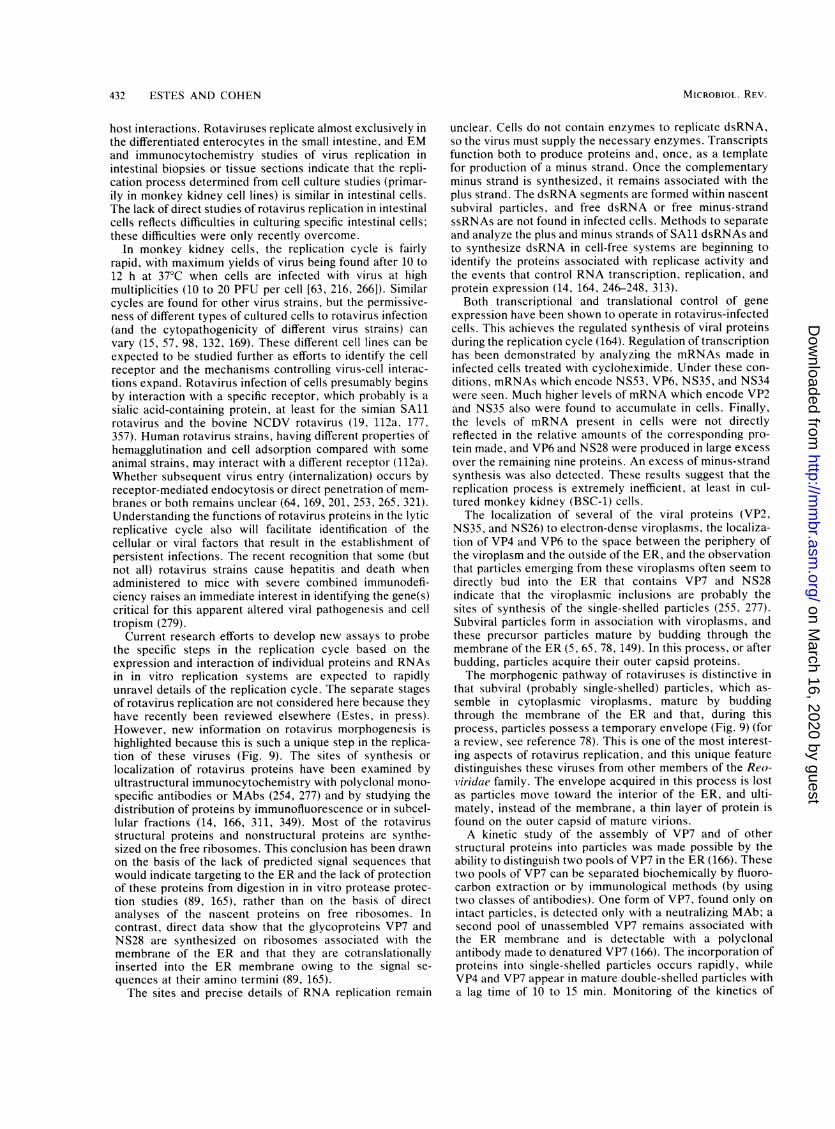
432 ESTES AND COHEN
host interactions. Rotaviruses replicate almost exclusively inthe differentiated enterocytes in the small intestine, and EMand immunocytochemistry studies of virus replication inintestinal biopsies or tissue sections indicate that the repli-cation process determined from cell culture studies (primar-ily in monkey kidney cell lines) is similar in intestinal cells.The lack of direct studies of rotavirus replication in intestinalcells reflects difficulties in culturing specific intestinal cells;these difficulties were only recently overcome.
In monkey kidney cells, the replication cycle is fairlyrapid, with maximum yields of virus being found after 10 to12 h at 37°C when cells are infected with virus at highmultiplicities (10 to 20 PFU per cell [63, 216, 266]). Similarcycles are found for other virus strains, but the permissive-ness of different types of cultured cells to rotavirus infection(and the cytopathogenicity of different virus strains) canvary (15, 57, 98, 132, 169). These different cell lines can beexpected to be studied further as efforts to identify the cellreceptor and the mechanisms controlling virus-cell interac-tions expand. Rotavirus infection of cells presumably beginsby interaction with a specific receptor, which probably is asialic acid-containing protein, at least for the simian SAlrotavirus and the bovine NCDV rotavirus (19, 112a, 177,357). Human rotavirus strains, having different properties ofhemagglutination and cell adsorption compared with someanimal strains, may interact with a different receptor (112a).Whether subsequent virus entry (internalization) occurs byreceptor-mediated endocytosis or direct penetration of mem-branes or both remains unclear (64, 169, 201, 253, 265, 321).Understanding the functions of rotavirus proteins in the lyticreplicative cycle also will facilitate identification of thecellular or viral factors that result in the establishment ofpersistent infections. The recent recognition that some (butnot all) rotavirus strains cause hepatitis and death whenadministered to mice with severe combined immunodefi-ciency raises an immediate interest in identifying the gene(s)critical for this apparent altered viral pathogenesis and celltropism (279).
Current research efforts to develop new assays to probethe specific steps in the replication cycle based on theexpression and interaction of individual proteins and RNAsin in vitro replication systems are expected to rapidlyunravel details of the replication cycle. The separate stagesof rotavirus replication are not considered here because theyhave recently been reviewed elsewhere (Estes, in press).However, new information on rotavirus morphogenesis ishighlighted because this is such a unique step in the replica-tion of these viruses (Fig. 9). The sites of synthesis orlocalization of rotavirus proteins have been examined byultrastructural immunocytochemistry with polyclonal mono-specific antibodies or MAbs (254, 277) and by studying thedistribution of proteins by immunofluorescence or in subcel-lular fractions (14, 166, 311, 349). Most of the rotavirusstructural proteins and nonstructural proteins are synthe-sized on the free ribosomes. This conclusion has been drawnon the basis of the lack of predicted signal sequences thatwould indicate targeting to the ER and the lack of protectionof these proteins from digestion in in vitro protease protec-tion studies (89, 165), rather than on the basis of directanalyses of the nascent proteins on free ribosomes. Incontrast, direct data show that the glycoproteins VP7 andNS28 are synthesized on ribosomes associated with themembrane of the ER and that they are cotranslationallyinserted into the ER membrane owing to the signal se-quences at their amino termini (89, 165).The sites and precise details of RNA replication remain
unclear. Cells do not contain enzymes to replicate dsRNA,so the virus must supply the necessary enzymes. Transcriptsfunction both to produce proteins and, once, as a templatefor production of a minus strand. Once the complementaryminus strand is synthesized, it remains associated with theplus strand. The dsRNA segments are formed within nascentsubviral particles, and free dsRNA or free minus-strandssRNAs are not found in infected cells. Methods to separateand analyze the plus and minus strands of SAIl dsRNAs andto synthesize dsRNA in cell-free systems are beginning toidentify the proteins associated with replicase activity andthe events that control RNA transcription, replication, andprotein expression (14, 164, 246-248, 313).
Both transcriptional and translational control of geneexpression have been shown to operate in rotavirus-infectedcells. This achieves the regulated synthesis of viral proteinsduring the replication cycle (164). Regulation of transcriptionhas been demonstrated by analyzing the mRNAs made ininfected cells treated with cycloheximide. Under these con-ditions, mRNAs which encode NS53, VP6, NS35, and NS34were seen. Much higher levels of mRNA which encode VP2and NS35 also were found to accumulate in cells. Finally,the levels of mRNA present in cells were not directlyreflected in the relative amounts of the corresponding pro-tein made, and VP6 and NS28 were produced in large excessover the remaining nine proteins. An excess of minus-strandsynthesis was also detected. These results suggest that thereplication process is extremely inefficient, at least in cul-tured monkey kidney (BSC-1) cells.The localization of several of the viral proteins (VP2,
NS35, and NS26) to electron-dense viroplasms, the localiza-tion of VP4 and VP6 to the space between the periphery ofthe viroplasm and the outside of the ER, and the observationthat particles emerging from these viroplasms often seem todirectly bud into the ER that contains VP7 and NS28indicate that the viroplasmic inclusions are probably thesites of synthesis of the single-shelled particles (255, 277).Subviral particles form in association with viroplasms, andthese precursor particles mature by budding through themembrane of the ER (5, 65, 78, 149). In this process, or afterbudding, particles acquire their outer capsid proteins.The morphogenic pathway of rotaviruses is distinctive in
that subviral (probably single-shelled) particles, which as-semble in cytoplasmic viroplasms, mature by buddingthrough the membrane of the ER and that, during thisprocess, particles possess a temporary envelope (Fig. 9) (fora review, see reference 78). This is one of the most interest-ing aspects of rotavirus replication, and this unique featuredistinguishes these viruses from other members of the Reo-viridae family. The envelope acquired in this process is lostas particles move toward the interior of the ER, and ulti-mately, instead of the membrane, a thin layer of protein isfound on the outer capsid of mature virions.A kinetic study of the assembly of VP7 and of other
structural proteins into particles was made possible by theability to distinguish two pools of VP7 in the ER (166). Thesetwo pools of VP7 can be separated biochemically by fluoro-carbon extraction or by immunological methods (by usingtwo classes of antibodies). One form of VP7, found only onintact particles, is detected only with a neutralizing MAb; asecond pool of unassembled VP7 remains associated withthe ER membrane and is detectable with a polyclonalantibody made to denatured VP7 (166). The incorporation ofproteins into single-shelled particles occurs rapidly, whileVP4 and VP7 appear in mature double-shelled particles witha lag time of 10 to 15 min. Monitoring of the kinetics of
MICROBIOL. REV.
on March 16, 2020 by guest
http://mm
br.asm.org/
Dow
nloaded from

ROTAVIRUS GENE STRUCTURE AND FUNCTION 433
*h4*... . ,. ''#
r, 5 ''
;. . p F . t~~~~~#
^ - x..e ft sL.. _,., g . n .
..,¢*,¢t.Z*. <+...,. ,,X' .~f>; t #;1':#4
t * ..... _:~~i4 4_,*:.?:. _~~~~~~~~~~~~~~f
*... .. ' *. ;:>r j ~~~~~4''.: ,# ,e,ti; __*~~~~~~9
,
9" S
*
#4S.
'Vt*s
ft *
#4i
A4
#4e '4.
>...
@:~~~~~~~~~~#:f'fSrt
3j # p*inp. .x.s~f
.:e.
nit... e.
FIG. 9. Unique features of rotavirus morphogenesis. EM of a thin section of SAil-infected MA104 cells. Dense viroplasmic inclusions (V)are abundant in the cytoplasm, often adjacent to the nucleus (N) and in close proximity to regions of the ER. In these structures, inner-coreparticles (small arrowheads) are assembled. Single-shelled particles located at the periphery of the viroplasm bud into the lumen of the ER,resulting in transient membrane-encapsidated intermediates (white arrows). These membranes are later lost, giving rise to the double-shelledmature virion (large arrowheads). Virus particles are never seen budding in the Golgi apparatus (G) or from the plasma membrane. Bar, 0.5,um.
processing of the oligosaccharides on each form of VP7 hasshown that the virus-associated VP7 oligosaccharides are
processed with a 15-min lag compared with that of themembrane-associated form, suggesting that the latter is theprecursor to virion VP7. This lag appears to represent thetime required for virus budding and outer capsid assembly(166).
Rotavirus maturation is apparently a calcium-dependentprocess, since it was observed that virus yields are de-creased when produced in cells maintained in calcium-depleted medium (300). In the absence of calcium, viruses
produced are reportedly exclusively single shelled and bud-ding of virus particles is not observed (299). Reduced levelsof only VP7 were observed owing to the preferential degra-dation (not to the impaired synthesis) of VP7 (299). Aninteresting result in these studies was that unglycosylatedVP7 made in the presence of tunicamycin (but not matureVP7) was relatively stable in a calcium-free environment.Unfortunately, the role of calcium in morphogenesis was notcompletely revealed by these studies. It is possible thatcalcium stabilizes or modulates folding or compartmental-ization of the newly glycosylated VP7 for subsequent assem-
VOL. 53, 1989
4Al
on March 16, 2020 by guest
http://mm
br.asm.org/
Dow
nloaded from

434 ESTES AND COHEN
bly into particles or perhaps that calcium deprivation desta-bilizes the ER or other ER proteins required for the stableassociation of glycosylated VP7 with the membrane.The infectious cycle ends with the release of progeny virus
by cell lysis, as observed by thin-section EM (5, 55, 221).Extensive cytolysis during infection has been confirmed bybiochemical analyses of infected cells, and alterations in thepermeability of the plasma membrane of infected cells mayresult in release of cellular and viral proteins (227). Despitecell lysis, all single-shelled particles and many double-shelled particles remain cell associated, suggesting that theseparticles interact with some structures within cells. Interac-tions with cell membranes and the cell cytoskeleton havebeen demonstrated (227). Whether the cytoskeleton pro-vides a means of transport of viral proteins and particles todiscrete sites in the cell for assembly or acts as a stabilizingelement at the assembly site for newly budded virionsremains to be determined.
STUDY OF ROTAVIRUS PROTEINS TO PROBEEUCARYOTIC PROTEIN TARGETING AND
OLIGOSACCHARIDE PROCESSING
The exclusive rough endoplasmic reticulum (RER) matu-ration site of the rotaviruses has been exploited to probe thebehavior of membrane glycoproteins which have the RER astheir final destination in the cell. The ER contains a mixtureof proteins with multiple destinations. Some remain in theER as permanent residents, whereas others are exported tothe Golgi apparatus for subsequent distribution to the cellsurface, lysosomes, or (in many cell types) secretory storagevesicles. The site of maturation of rotaviruses in the ER andthe fact that no complex carbohydrate is detectable on viralVP7 show that these proteins are not transported to theGolgi apparatus. Studies of the processing of these glyco-proteins have been useful in finding how protein targeting iscontrolled and in determining whether specific signals directprotein retention in the ER.The first evidence suggesting that specific signals mediate
protein retention in the ER came from studies of the expres-sion of a series of deletion mutants of VP7 in COS cells (259).Deletions in aa 47 to 61 in VP7 (in the second H2 hydropho-bic domain) resulted in secretion of a truncated membraneprotein into the medium, and this protein obtained newmodifications (the addition of complex carbohydrate, basedon its becoming resistant to endoglycosidase H). Othervariants lacking aa 42 to 61 or 43 to 61 were similarlyglycosylated and secreted. This was interpreted to mean thatthe second hydrophobic domain contains a positive signalfor ER location and a membrane anchor function. However,an alternative interpretation would be that the truncatedprotein is secreted simply because it is in the aqueous phaseand is not membrane bound.Subsequent studies with additional deletion mutants that
contained either the first or the second hydrophobic regionshowed that either of the hydrophobic regions can direct thetransport of VP7 across the ER membrane, but the nature ofthe anchor and targeting signal in VP7 was not elucidated(350). Identification of the site of cleavage in VP7 as occur-ring after residue 50 indicated that both amino-terminalhydrophobic domains are removed from the mature protein(257, 316). Since VP7 normally remains anchored afterprocessing, these results implied that residues distal toresidue 50 (i.e., the region from aa 51 to 61 that was presentin wild-type VP7 but missing from the secreted mutantlacking aa 47 to 61) must be involved in retaining VP7 in the
ER. The initial conclusion that the H2 domain itself con-tained the ER retention information thus required modifica-tion.
Instead, these seemingly conflicting results are consistentwith the idea that aa 47 to 61 might be necessary, but notsufficient, for ER retention. This idea has been testeddirectly by studying the expression and secretion of chimericgenes (257, 315). Poruchynsky and Atkinson constructedchimeric genes by fusing the first 63 codons of wild-type VP7or of constructs with deletions of residues 1 to 14 or residues51 to 61 to the mouse salivary o-amylase gene, a secretoryprotein. The fusion junction in these constructs was locatedat the exact mature terminus of amylase, and the chimericproteins from all three of these constructs were secreted,based on their being present in the media. One weakness ofthese studies was that there was no way to determinewhether the presence of the protein in the media was due toactive transport or to cell lysis, because the chimeras werenot glycosylated (257).A study of a second series of VP7-amylase chimeras in
which the VP7 contribution extended to aa 111 resulted inretention of amylase within the ER, and this protein retainedenzymatic activity. These results, therefore, suggested thattwo regions of VP7 mediate its retention in the ER. The firstlies within the region spanning aa 51 to 61, and the secondlies in the region spanning aa 62 to 111, the latter containingthe single glycosylation site for VP7. Both regions areapparently necessary for retention, although neither by itselfis sufficient for retention (257).
Different conclusions on the residues of VP7 needed forER retention were reached in an independent study in whichamino acid residues 30 to 64 of VP7 were exchanged with thesignal peptide from the influenza virus hemagglutinin (315).These constructs, which maintained the usual cleavage sitefor VP7, caused secretion of VP7. Because these HAF VP7constructs contained both regions (aa 51 to 61 and 62 to 111)proposed (as described above and in reference 257) to benecessary for VP7 retention, these regions alone are clearlynot sufficient for VP7 retention. Studies with other chimericproteins indicate that interactions between the cleaved signalpeptide (the H2 domain) and other downstream sequences inVP7 are required, directly or indirectly, for retention of thisprotein in the ER membrane (315). This is the first demon-stration of a function for a cleaved signal peptide.The method by which VP7 is retained in the ER remains
an interesting question. VP7 does not contain the discretepeptide Lys-Asp-Glu-Leu, which was recently found toconfer retention of other proteins to the ER (226). Thetopography of VP7 in the membrane also is not well under-stood. Based on its resistance to digestion with proteolyticenzymes, VP7 is not a membrane-spanning protein (89, 165).However, VP7 is membrane associated since it remainsmembrane bound after high-salt treatment and release of itsmicrosomal contents at high pH by sodium carbonate treat-ment (165). It is possible that VP7 is retained in the ER byconformational properties that mediate membrane attach-ment or by oligomerization with itself or other specificcellular proteins.These analyses of processing of VP7 with gene 9 mutants
produced by site-directed mutagenesis show the power ofthis approach to the correlation of gene structure and func-tion. These studies of COS cells transfected with mutantscontaining either both initiation codons or only the secondinitiation codon all clearly indicate that the first AUG isprobably not translated and that the mature VP7 lacks bothhydrophobic domains and contains an amino-terminal Gln-
MICROBIOL. REV.
on March 16, 2020 by guest
http://mm
br.asm.org/
Dow
nloaded from

ROTAVIRUS GENE STRUCTURE AND FUNCTION 435
51 (257, 316). These results are of interest for investigatorsusing strategies to produce authentic VP7 in expressionvectors for testing as potential vaccines (see below), andthey also reemphasize the puzzling question of why bothinitiation codons that each precede a hydrophobic domainare retained in all VP7 genes sequenced so far. Comparisonsof the amino acid sequences of VP7 are striking when theconserved and variable amino acids found upstream of thecleavage site prior to Gln-51 are examined. The first 8 aa ofVP7, both initiation codons, leucine 23 and lysine 25 at theend of the Hi domain, and glutamine 51 following the H2domain (and shown to be the major site of cleavage of themature protein) are strictly conserved. These conservedregions are consistent with the idea that both initiationcodons are used. In contrast, extensive variation is seen inthe H2 domain (Fig. 6). One wonders what factors causesuch variation in this domain. Perhaps these changes com-pensate for changes downstream in other amino acids (be-tween aa 62 and 111) with which H2 may interact (257, 315).
Analysis of the synthesis and expression of a series ofaltered gene 10 cDNAs has been used to localize thesequences involved in translocation of the nonstructuralglycoprotein NS28. A series of mutations in the codingregions for the three hydrophobic domains (Hi, H2, and H3)of this protein were constructed and analyzed by in vitrotranslation and by transfection in vivo. In vitro, all threehydrophobic domains associated with microsomes. How-ever, glycosylation and proteolysis of wild-type and mutantforms of NS28 indicate that the wild-type protein is anchoredin the membrane only by the second hydrophobic domain,leaving approximately 131 residues exposed on the cytoplas-mic side of the membrane (23a). These studies extendedinitial studies on the topology of NS28 in microsomal mem-branes (48) by suggesting that the cytoplasmic domain islarger than originally thought and by proposing a model inwhich the second (H2) hydrophobic domain is the mem-brane-spanning domain rather than the third (H3) as pro-posed earlier. Although both papers confirm that the H3domain is protected from digestion with various proteases,analyses of the mutant molecules indicate that protease sitesupstream of the H3 domain are available for cleavage. It istherefore concluded that H3 is merely resistant to cleavageby being embedded or protected by interactions with thecytoplasmic side of the membrane (23a). Site-directed mod-ification of the cytoplasmic domain of the protein will nodoubt help to better define the domains of NS28 involved inreceptor-ligand interactions.
Studies of the processing of the oligosaccharides on therotavirus glycoproteins have also been useful in probing thetypes of oQ-mannosidases specific to the RER. Initial pulse-chase experiments showed that the oligosaccharides on therotavirus glycoproteins are trimmed (89, 165); Man8GlcNAc2and Man6GlcNAc2 were found to be the predominant intra-cellular species detected after a 5-min pulse with[3H]mannose and a 90-min chase, and VP7 on mature viruswas processed to Man5GlcNAc2 (165). Although removal ofthe outer glucoses and at least one mannose residue had beenshown to occur on nascent chains of vesicular stomatitis virusG protein in the RER (12), the location, linkage specificity,and total number of the different o2-mannosidases actingduring the remaining processing reactions were not previ-ously clear. Evidence had been presented for both RER andGolgi apparatus activities (156). In contrast, the subsequentsteps of carbohydrate modification, including the addition ofN-acetylglucosamine residue, removal of cxl,3- and a.1,6-mannoses, and transfer of terminal sugars, were known to be
localized to the Golgi complex (104). Studies of the processingof the rotavirus glycoproteins provide evidence that cx1,2-mannosidase activities responsible for the formation ofMan8GlcNAc2 and Man6GlcNAc2 on VP7 and Man8GlcNAc2on NS28 reside in the ER (165, 258). Trimming of glycosy-lated VP7 (but not of vesicular stomatitis virus G protein) wasalso able to continue in the presence of the energy inhibitorcarbonyl cyanide m-chlorophenylhydrazone (CCCP). Theseresults suggest that the Man8-forming enzyme is in a function-ally distinct compartment to the Man8- and Man6-formingenzymes. In addition, it suggests that an energy-sensitivetranslocation step within the ER may mark the divergence ofthe processing pathways of glycoproteins which have distinctfinal destinations in the cell (258).
GENETIC APPROACHES TO UNDERSTANDINGROTAVIRUS GENE FUNCTION
Studies with rotavirus mutants and reassortants have onlybegun to contribute to our understanding of rotavirus genestructure and function. Temperature-sensitive mutant col-lections have been isolated from several rotavirus strains,including SAl1, UK, and RRV (105, 138, 266, 268). Themost extensive mutant collection is for SAll, for which 10 ofthe expected 11 reassortment groups have been identified,and 6 of these have been assigned to genome segments (124).The phenotypes of some of these mutants are consistent withfunctions inferred from biochemical data or recent sequenceinformation (reviewed above). For example, mutant groupsB and C, which mark genome segments 3 and 1, respec-tively, are both defective in the synthesis of ssRNA anddsRNA (124). Since these proteins are suspected to be partof the transcriptase, biochemical and sequence analyses ofthese mutants may now permit dissection and localization ofspecific enzymatic domains on VP1 and VP3. The availabil-ity of full-length cloned DNAs for the rotavirus genes hasstimulated the desire to learn how to selectively insert genesinto rotavirus-infected cells and rescue this information in aninfectious particle. Success in these experiments will have amajor impact on rotavirus research. One approach to thisidea is to coinfect cells with an mRNA (or a cDNA that canexpress an mRNA) and a temperature-sensitive mutant andto then select particles from the progeny that would containthe wild-type gene. Information on the genetic interactionsbetween temperature-sensitive mutants (or two wild-typeviruses) is available that is relevant when trying to producereassortants or to determine whether a specific gene can berescued.The genetic interactions between pairs of temperature-
sensitive mutants occur in an "all-or-none" fashion, asexpected for recombination by a mechanism of reassortmentof genome segments (266). Analysis of the factors that affectgenetic interactions has shown that recombinational fre-quency is affected by the multiplicity of infection and thetime of infection (269). The maximal or near-maximal recom-bination frequency is obtained at the earliest times whensignificant recombination can be detected; this suggests thatrecombination occurs early in the infectious cycle and thatthere is a single round of mating. Recombination is efficientat nonpermissive temperatures, and mutants from all recom-bination groups can interfere with the growth of wild-typevirus at both permissive and nonpermissive temperatures.An independent study of the factors that may affect reassor-tant formation found that no selection of genomic RNAsegments was detectable before or during virus particleassembly in coinfected cells. In addition, reproducible selec-
VOL. 53, 1989
on March 16, 2020 by guest
http://mm
br.asm.org/
Dow
nloaded from

436 ESTES AND COHEN
tion of reassortants was observed with specific pairs ofparental viruses. Therefore, selection of specific reassor-tants following coinfection is apparently due to differences inthe infectivities of progeny viruses and not in their assembly.This implies that reassortant infectivity is a function of theparental origin of specific genomic segments and that selec-tion of surviving reassortants appears to occur becausecertain reassortants grow better than other reassortants (123,348).Reassortment of genome segments among the progeny of
in vitro mixed infections has been exploited to derive reas-sortant viruses with a desired subset of genome segmentsfrom each parent. Such reassortants have been used tocorrelate functions with specific genome segments. In addi-tion to allowing the assignment of temperature-sensitivemutants to a specific genome segment, this powerful ap-proach has identified a number of biologic properties thatcosegregate with specific genome segments (reviewed aboveand below). This genetic approach to probing gene functionwill continue to be used to map phenotypic differences thatare identified between two parental viruses in the future.
Characterization of the progeny of cells coinfected withtwo strains of rotaviruses has shown that phenotypicallymixed viruses (pseudotypes and mosaics of the capsid pro-tein VP7) readily form. This was demonstrated by showingthat the progeny were able to be neutralized by MAbsspecific for the VP7 of each of the coinfecting strains (347).This result has practical implications, because it suggeststhat selection of reassortants (containing specific VP4 andVP7 proteins) by using neutralizing antibodies may be oflittle value unless these antibodies are added only to theoverlay agar during plaque formation.
Recent studies in which reassortants were used to mapbiologic properties of rotavirus genome segments haveshown that the criteria needed for firm conclusions fromstudies with reassortants are more rigorous than formerlyrealized. For example, early studies which examined thegene segments responsible for inducing neutralizing anti-body identified only one genome segment (8 or 9, dependingon the virus strain) with this property, but in those studies noreassortants containing a gene segment 4 from a human viruswas able to be cultivated and characterized (138). Subse-quent studies revealed that genome segment 4 also possessesthis property, and this highlights the importance of analyzingreciprocal reassortants when using the genetic approach forphenotypic mapping.
In addition, unknown common properties of parentalviruses may influence the interpretation of genetic studies ofreassortant viruses. For example, two bovine viruses (UKand NCDV) were eventually found to possess related VP7antigens, but their VP4 antigens are distinct (139a, 153).Therefore, properties related only to the distinct genomesegment 4 of each of these two parental viruses would beable to be dissected by reassortant analyses. Since thecommon properties of parental viruses may not be known inadvance, it is useful to confirm results by using differentparental strains. The identity of naturally occurring inter-typic rotavirus strains (152, 153, 214, 224) and the directdemonstration that rotavirus genes reassort at high fre-quency in vivo (123) and in vitro (131, 138, 266) highlight thefact that the genetic background of any rotavirus isolatecannot be assumed. This point has been further illustrated inrecent analyses of reassortants which indicate that thephenotype of specific reassortants is altered depending onthe parental strain used for analysis (56).
ROTAVIRUS GENES INVOLVED IN PATHOGENESISAND IMMUNITY
Details of the current information on the genes involved inpathogenesis and immunity have been reviewed recently(209), so we shall highlight only the areas where confusionexists. Most of the direct information on the genes involvedin pathogenesis and immunity has been obtained by using awide variety of animal models and a number of differentrotavirus strains. Studies with these animal models must bedistinguished in terms of whether they evaluate the role ofactive (rabbits, gnotobiotic piglets to 3 months of age, calvesto 3 weeks of age) or passive (mice, lambs, and calves)immunity and limited (abortive) or extensive viral replica-tion. Results with all these models and conclusions fromstudies of natural infections in children indicate that protec-tive immunity does develop after rotavirus disease; lessinformation is available on the duration of this immunity,whether protection from reinfection is possible, and how tobest induce protective immunity.
Virus virulence has been attributed primarily to propertiesof genome segment 4. Offit et al. (238) made reassortantsbetween two heterologous rotaviruses (SAl and NCDV)and mapped differences in the 50% diarrhea dose in theneonatal mouse model of gastroenteritis exclusively to genesegment 4. Understanding the nature of heterologous host-virus infections was of interest since viruses of animal(heterologous) origin are currently being explored as vac-cines for use in humans. In the mouse model, replication ofthese heterologous viruses is limited and usually abortive(270), and disease can be evaluated only in neonatal animals(<14 days of age). Because of this and because the differencein diarrheal dose of the parental strains used to map thediarrheal gene was only 50-fold (238), further studies withmore virulent homologous viruses in this and other animalmodels are warranted. Such studies are needed to confirmwhether additional genes influence disease induction andother properties of virulence, such as replication or virustransmission. The recent ability to cultivate and quantitatemurine rotavirus strains will facilitate such studies withneonatal mice (140), and studies with other older animals(rabbits, calves, and gnotobiotic piglets) are also possiblewith several cultivatable virus strains (69, 151, 334, 355).Genome segment 4 has also been implicated to be impor-
tant in virulence on the basis of comparisons of the aminoacid sequences of viruses originally isolated from newbornbabies with asymptomatic infections and older babies withsymptomatic infections (126, 127). Further studies are re-quired to determine the validity of this hypothesis and todetermine the relative roles of host and viral factors indetermining whether infections of newborns (and older chil-dren) result in symptomatic or asymptomatic illness. Newevidence shows that symptomatic infections do occur innewborns, that asymptomatic infections may occur muchmore frequently than previously realized, and that the ap-pearance of new virus types may cause unexpectedly severeillness in populations generally thought to be nonsusceptible(17, 77, 112; D. 0. Matson, M. K. Estes, J. W. Burns, H. B.Greenberg, K. Taniguchi, and S. Urasawa, submitted forpublication). These new findings emphasize the currentlimitations of our understanding of the virulence propertiesof the rotaviruses and their epidemiology.
Analysis of the sequences of genome segment 4 of twovirulent calf viruses and of the avirulent RIT 4237 vaccinestrain has recently been completed (235). Comparisons ofthese sequences show no changes in the trypsin cleavage
MICROBIOL. REV.
on March 16, 2020 by guest
http://mm
br.asm.org/
Dow
nloaded from

ROTAVIRUS GENE STRUCTURE AND FUNCTION 437
area, and only one amino acid difference (a change ofglutamine 23 to lysine) between the virulent Cody strain ofNCDV and the avirulent RIT vaccine strain was found.These results indicate that either aa 23 (outside the cleavageregion) plays an important role in virulence or, more likely,a gene (or genes) other than genome segment 4 containsmutations responsible for attenuation of the RIT strain ofNCDV. Further studies on the gene(s) responsible for viru-lence are clearly warranted.As mentioned above, protective immunity to rotavirus
gastroenteritis can be induced. However, what constitutesthe protective immune response and exactly how to induce ithave not been completely defined. Difficulties in interpretingthe existing data on immunity include the following: (i) aninability in studies with children to know the previousexposure to rotavirus and problems with dissecting theeffects from maternally acquired antibody from those ofactive primary or secondary infections; (ii) a lack of studiesof active immunity in animal models which mimic infectionsin children on the basis of virologic and immunologic param-eters; (iii) seeming conflicting results on which in vitroparameter is the best to use to measure or determinehomotypic or heterotypic protective immunity in vivo; (iv)lack of knowledge or systematic consideration of how dif-ferent virus strains, different virus dosages, and differenthost factors may influence results; and (v) difficulty inobtaining antibody-negative animals and failure to performstudies in proven antibody-negative animals. Fortunately,the recent expansion of our knowledge of the antigenic andmolecular properties of the rotaviruses and new expandingsources of molecular probes, reassortants, and immunologicreagents will help dissect the immune response in futurestudies.The proteins involved in inducing a protective immune
response have been studied with reassortants containingdifferent types of VP4 and VP7. Mouse dams orally hyper-immunized with a reassortant rotavirus containing the VP4of one serotype and the VP7 of another serotype confer totheir progeny passive protection against challenge witheither parental rotavirus (239). Similar results were foundfollowing primary infection of 4- to 5-day-old gnotobioticpiglets with reassortant viruses containing distinct types ofVP4 and VP7 and challenge with two serotypically distinctvirulent viruses 3 weeks later (151). These results indicatethat antibodies to VP4 and to VP7 are each associated withresistance to diarrhea. These results are of general impor-tance, because they suggest that a reassortant (or selectedvirus strains with a particular gene constellation) may beable to induce broader protection than either of the twoparental virus strains. In addition, these reassortants wereable to induce heterotypic immunity, although this was notseen after immunization with either of the parental virusstrains. The general conclusions of these studies have beenconfirmed by showing that passive administration of neutral-izing MAbs to VP4 and VP7 also can provide protectionagainst challenge in the mouse model (209, 243).
Several studies have raised doubts about the importancein protection of the differences in neutralization antigensmeasured by in vitro plaque reduction assays, and mecha-nisms other than neutralizing antibody have been proposedto be important in protection. Initial studies failed to showheterotypic protection in gnotobiotic calves (354). However,more recently, protection between different serotypes hasbeen seen, but this was dependent on the virus strain usedfor immunization. Unexpectedly, the plaque reduction neu-tralization titers of antibody in serum and coproantibody did
not predict a state of protection against the challenge virus(355). The question of whether an infection with an avirulentrotavirus strain can protect against subsequent rotavirusdisease when the avirulent virus is related (or only poorlyrelated) to the challenge virus by neutralization has also beenasked. Results with calves indicate that one avirulent viruscan provide protection from disease without inducing highlevels of neutralizing antibody in serum (40). Such studiessuggest that our knowledge of the immune response torotavirus infections is clearly incomplete. Efforts to evaluatethe role of cell-mediated immune responses and nonimmunefactors in the resistance to infection and disease may helpclarify this current confusion (182, 242, 278, 336).
Other approaches, examining the immune responses tospecific proteins, are directed toward finding the proteinsimportant in protection. A method to measure immuneresponses to specific epitopes on rotavirus proteins has beendeveloped that measures the ability of a serum to block thebinding of a specific MAb to virus (22a, 301). This andsimilar competitive assays show great promise in being ableto determine what responses are standard and whether theresponse to a particular epitope can be associated withprotection following an infection in an individual. However,more data are needed on the sensitivity and specificity of thisassay. For example, it will be important to know thatantibody responses to distinct epitopes situated closely onVP4 or VP7 do not compete with the specific binding of theMAb being used in the assay. Alternative assays to measureantibody responses to specific proteins are using proteinsproduced in expression vectors (see below).
Electrophoretic analysis of viral proteins recognized inimmunoprecipitation reactions is a third method to deter-mine the immune response to specific proteins (69, 324). Thismethod has the advantage of allowing responses to proteinsother than structural proteins to be easily recognized, andcorrelation of primary responses with the time when animalsbecome protected from challenge may help dissect whichresponses are protective. Studies analyzing the serum andmucosal antibody responses are only beginning to be per-formed (141, 199, 291). Interestingly, both children (respond-ing to natural infections) and rabbits (responding to a knownprimary infection) develop antibodies to a number of pro-teins, but serum samples with neutralizing antibody do notnecessarily possess detectable antibody to VP7 (324; M. E.Conner, M. Gilger, D. Y. Graham, and M. K. Estes, manu-script in preparation). Strong reactivities are detected toseveral of the nonstructural polypeptides, indicating thatthese are good markers to document virus replication in thegastrointestinal tract; these responses to nonstructural pro-teins raise the question of whether they also play a role inprotection. Finally, differences in responses of hyperim-mune and postinfection serum have been noted (302), andthese demonstrate that studies comparing the immune re-sponse to parenteral immunization and intestinal infectionwill be worthwhile. Such studies may help us learn how toinduce responses to different proteins or domains withinproteins, as is being learned for responses to poliovirusvaccination (358).
Additional analyses of parental and reassortant viruseswill continue to be useful to dissect the genes involved invirulence, to determine the mechanism by which VP4 (andpossibly other gene products) affects virulence, and todetermine the relative importance of specific and nonspecificimmune and nonimmune factors in mediating protectionfrom infection and disease. Such information will havepractical potential until an effective rotavirus vaccine is
VOL. 53, 1989
on March 16, 2020 by guest
http://mm
br.asm.org/
Dow
nloaded from

438 ESTES AND COHEN
developed and will be of basic interest in expanding ourunderstanding of the mechanisms controlling viral replica-tion in the gastrointestinal tract.
IMPACT OF MOLECULAR BIOLOGY ON VACCINEDEVELOPMENT
Active Immunization
Epidemiologic studies showing the significant mortalityand morbidity caused by rotavirus disease in individuals indeveloping and developed countries, respectively, focus theneed for an effective vaccine. Since life-threatening andclinically significant disease occurs in children less than 2years of age, current vaccines are aimed at preventingclinically significant diarrhea (and not infections) in thistarget population. The first vaccines to be tested in childrenhave been live vaccines, because they are expected toinduce more effective local mucosal immunity and greaterduration of immunity than killed vaccines (52, 86, 175, 343).The initial live candidate vaccines to be developed have
been from animal strains. Three candidate vaccines (RIT4237, a bovine serotype 6 virus; RRV MMU 18006, a rhesusmonkey serotype 3 virus; and WC3, a bovine serotype 6virus) have progressed to field trials. These vaccines are notrepresentative of the most common serotypes found inhumans, but they were developed with the assumption thatheterotypic immunity would provide protection from diseasewith human viruses (175). The bovine strain (RIT 4237)showed good efficacy when tested initially in developedcountries, but failed to provide protection when tested indeveloping countries; it has now been removed from furthertesting (351). A subsequent field trial of RIT 4237 in L ma,Peru, showed that this vaccine afforded 58 to 75% protectionagainst the more severe rotavirus illness if given in morethan one dose, and better protection was found againstdisease caused by serotype 1 viruses than by serotype 2viruses (191). This vaccine trial provided important insightsby defining the requirements for performing a successfulvaccine trial in a developing country (191). It also highlightedthe need to provide protection against multiple rotavirusserotypes and the possible need for multiple doses to obtainadequate protection.
Surprisingly, most vaccine trials have been performed inareas where the natural challenge viruses were of one or twoserotypes. Increasing information on the geographical distri-bution and changes in the prevalence of rotaviruses indicatesthat it will not be possible to predict which serotype(s) willbe more common at a given time in a given geographicallocation (342a; Matson et al., submitted). Because of this,none of the current vaccine candidates have really beengiven a thorough evaluation, and possibly a live polyvalentvaccine will be necessary to account for expected changes inserotype distribution.The RRV MMU 18006 vaccine has undergone fairly
extensive testing. It has been shown to be immunogenic andslightly reactogenic, and it has shown efficacy in field trialsto natural infections with serotype 3 challenge but notserotype 1 challenge (52). These results, together with theprevious studies of the RIT 4237 vaccine, suggest thatserotype-specific immunity may be required for satisfactoryprotection against rotavirus diarrhea. This conclusion agreeswith epidemiologic data, which showed that natural protec-tion against rotavirus gastroenteritis is related to concentra-tions of serum antibody against homotypic virus and that thelevels of heterotypic antibodies are generally below the
threshold level needed for protection (58). Studies of theactive immune response in known seronegative animals alsoindicate that the primary neutralizing immune response ishomotypic (93, 310). This suggests that demonstration ofheterotypic responses in vaccinated children may reflectmeasuring responses in children who had been previouslyprimed by unrecognized infections (93, 309) or who areinfected by a virus strain whose outer capsid proteinscontain epitopes shared by several other virus strains (73).
Preliminary efficacy testing of the bovine WC3 vaccineshowed that this virus strain provided protection againstdisease with heterologous human serotype 1 virus infectionswhen tested in the United States (60). These results arepromising (as were the initial RIT 4237 trials), and onlyadditional field trials will determine whether this vaccine willprove efficacious in other settings, including developingcountries. If it does, further characterization of this bovinevirus strain will be of interest to determine the mechanism bywhich it induces heterotypic protection.
Other approaches to developing candidate vaccines areunder way. Reassortants containing a single gene whichcodes for the outer capsid glycoprotein from human virusserotypes on a rhesus rotavirus background have beenproduced as possible candidate vaccines to induce homo-typic immunity to the four human serotypes (223). Thesestrains are now in phase I trials to evaluate their reactoge-nicity and immunogenicity and to determine whether admin-istration of a polyvalent vaccine will affect "takes" ofindividual strains (109, 143).The possibility that viruses that have become endemic in
neonatal nurseries are attenuated, because they causeasymptomatic infections, and that they might be useful ashuman virus vaccine strains is also being considered. Thisidea is based on an epidemiologic study showing that chil-dren infected with these strains as neonates had no severedisease with subsequent rotavirus infections during their first2 years of life (28). Therefore, neonatal vaccination withsuch strains may induce protective clinical immunity. How-ever, there is currently no direct evidence from studies inanimal models or volunteers that these viruses are actuallyattenuated. This idea needs further confirmation, since otherrecent studies suggest that reinfections may give rise to anequally or even more severe illness (193).The availability of cDNA copies of many of the rotavirus
genes has opened the possibility of using live expressionvectors or subunit vaccines of individual proteins synthe-sized from these clones as recombinant vaccines. Possibleapproaches have been discussed elsewhere (33, 95). Therotavirus outer capsid proteins (VP4, VP7) have been ex-pressed alone or as fusion products using bacterial expres-sion systems (8, 9, 110, 219). Expression of about 45% of theSAl VP4 in a thermoinducible expression system resultedin the synthesis of hybrid MS2 polymerase-VP4 proteinwhich was antigenic on the basis of immunoblot reactivitywith hyperimmune guinea pig anti-SAl serum. Immuniza-tion of mice with this fusion protein induced antibodieswhich neutralized virus with a mean titer of 1:1,200. Al-though these titers were at least 20 times lower than thoseobtained following immunization with complete virus parti-cles, the hemagglutination inhibition titers in each type ofserum were similar. Heterotypic neutralization titers werenot reported. The synthesized protein was aggregated, and itis not known whether this had an effect on the immunoge-nicity of this protein. VP4 expressed from baculovirusrecombinants also is antigenic and immunogenic (202; Estes,inpublished).
MICROBIOL. REV.
on March 16, 2020 by guest
http://mm
br.asm.org/
Dow
nloaded from

ROTAVIRUS GENE STRUCTURE AND FUNCTION 439
Several different constructs of VP7 have been expressedin bacteria (8, 110, 219). VP7 expressed alone was toxic(219), whereas VP7 expressed as a fusion protein with3-galactosidase was not. Neutralizing antibody of relatively
low titer (1:100 to 1:1,600 for SAIl VP7 [8]; 1:300 for UKbovine VP7 [219]) has been induced, but antibody to ex-pressed NCDV VP7 did not possess neutralizing activity(110). Examination of the immunogenicity of VP7 producedby using a vaccinia virus expression system indicated thatneutralizing antibodies with low titers (1:40 to 1:640) wereinduced and that these were serotype specific when tested inplaque reduction assays or by ELISA (6). In immunoblotsthese sera reacted with denatured VP7 from serotypes 1, 3,and 4, and the lack of reactivity with serotype 2 wassuggested to be due to blocking of a possible cross-reactivesite at aa 149 by carbohydrate in these viruses.A high-yielding baculovirus expression system has also
been used to successfully produce 9 of the 11 rotavirus geneproducts (94, 202; Cohen and Estes, unpublished). Theproteins synthesized in this system possess native confor-mation on the basis of reactivities with available MAbs andpolyclonal antibodies and biochemical comparisons withtheir authentic counterparts made in mammalian cells. VP6expressed alone forms trimers and capsomerlike structures(94). Neutralizing antibody is induced by immunization ofguinea pigs with VP7 (Estes, unpublished). Future effortswill determine whether an effective subunit vaccine can beproduced by immunization with these antigens singly and incombination. Since none of the expressed proteins induceneutralizing antibodies with equivalent titers to those pro-duced by immunization with viral particles, it is possible thatcombinations of proteins will be required to achieve properconformation and the best vaccination results.
Successful expression of the rotavirus proteins is only thefirst step toward making an effective subunit vaccine toprotect against rotavirus gastroenteritis. The following chal-lenging questions must now be considered. (i) Will it bepossible to induce a protective mucosal immunity by oralvaccination with a nonreplicating antigen? (ii) Can noveldelivery systems or adjuvants be established to ensureproper antigen presentation in the gastrointestinal tract? (iii)Can long-lasting immunity be induced? (iv) Can subunitvaccines safely and effectively immunize malnourished and/or immunocompromised children who normally may de-velop persistent infections?The potential for improving the immunogenicity of ex-
pressed rotavirus peptides has already been demonstratedby including peptide determinants known to induce T helpercells in mice (30). Knowledge of the specific rotavirusproteins that induce intestinal cytotoxic T cells and stimulatethe uptake of viral antigen by Peyer's patches and knowl-edge of the role of cell-mediated immunity in protectionagainst, recovery from, or pathogenesis of rotavirus diseasemay permit the stimulation of more rapid recovery frominfection. Studies addressing these questions have beeninitiated (76, 242, 278). Future studies addressing thesequestions and the testing of live expression vectors to deliverantigens directly to the gastrointestinal tract are expected tobe fruitful.
Passive Immunization
Alternative approaches to active vaccination to preventrotavirus gastroenteritis are also being considered. In ani-mals in which the young receive immunoglobulins fromcolostrum and milk instead of by transplacental transfer,
vaccination of dams can raise colostral antibody levels tosufficiently high titers that the young animals are passivelyprotected through the time of maximum sensitivity to infec-tion (289). Success has been achieved with this approach incalves (22a, 290, 308). It has clearly been shown that in theseanimals, vaccination with live or inactivated vaccines booststhe immune response in naturally primed animals, and theresulting heterotypic lactogenic immune response has beenshown to be dependent on the preexposure history of theanimals (43, 309). Because of the expected lower cost ofproduction and safety testing of subunit vaccines and theability to use inactivated and heterologous viruses to boostlactogenic immunity, testing of the available expressed pro-teins in such systems may be worthwhile.The possibility of ameliorating rotavirus infections in
children by the administration of cow's milk containing suchheterotypic antibodies is also being evaluated. Althoughsuch treatments will not be recommended in developingcountries, where breast-feeding needs to be encouraged,they have been used to stop prolonged rotavirus excretion inimmunocompromised and normal children (146, 198). Al-though it remains unclear whether such therapy will signifi-cantly decrease the incidence of diarrhea in immunocompe-tent children, it is possible that such treatment will be usefulin certain settings (e.g., day-care centers) where diarrhea isa continuing problem and decreased shedding might helpprevent transmission.
CONCLUDING REMARKS
The new information learned about the structure andfunction of rotavirus genes and their proteins in the last 5years is impressive in its amount and its significance inhelping to understand the epidemiology, pathogenesis, rep-lication, and molecular biology of these viruses. Futurestudies to understand how trypsin cleavage mediates theselective internalization of the viral capsid into cells and howthe nonstructural proteins function in the replication cycleshould elucidate the mechanisms by which the virus repli-cates and encapsidates one of each of its 11 genome seg-ments into nascent particles. This information, in turn, mayallow the manipulation and encapsidation of any RNAsegment. Further studies of rotavirus morphogenesis shouldhelp clarify the virus-budding process in general. With thefundamental information on the structure of the genes andtheir proteins reviewed here, research efforts can be directedtoward unraveling the biology of these viruses and theirinteractions with the hosts they infect. Studies to identify thecellular receptor, to dissect the immune or nonimmuneresponses to specific proteins that are important for aprotective immune response, and to learn how to inducethese responses can be expected. Finally, further studies ofthe properties of viruses that circulate over time or thatreinfect children should shed light on how these virusesevolve and help determine whether this evolution will com-plicate needed vaccination programs.
ACKNOWLEDGMENTS
Work in our laboratories is supported in part by Public HealthService grants DK30144, A120649, and A124649 and training grantCA09197 from the National Institutes of Health (M.K.E.) and byEuropean Economic Community grant BAP 156 F (J.C.).Computer analyses were performed with the programs and kind
staff assistance of the Molecular Biology Information Resource,Department of Cell Biology, Baylor College of Medicine, andMolecular Biology Programs, Institut National de la Recherche
VOL. 53, 1989
on March 16, 2020 by guest
http://mm
br.asm.org/
Dow
nloaded from

440 ESTES AND COHEN
Agronomique. We thank our many colleagues who contributedmanuscripts prior to publication and who generously read andcritiqued this review. We also thank our students, postdoctoralfellows, and technicians whose creativity and experimental exper-tise continue to stimulate our work. Finally, we thank Katie Piersonand Caroyl Robinson for patient and skillful preparation of thismanuscript.
LITERATURE CITED1. Aboudy, Y., I. Shif, I. Zilberstein, and T. Gotlieb-Stematsky.
1988. Use of polyclonal and monoclonal antibodies and analy-sis of viral RNA in the detection of unusual group A humanrotaviruses. J. Med. Virol. 25:351-359.
2. Air, G. M., W. G. Laver, and R. G. Webster. 1987. Antigenicvariation in influenza viruses. Contrib. Microbiol. Immunol.8:20-59.
3. Albert, M. J., L. E. Unicomb, S. R. Tzipori, and R. F. Bishop.1987. Isolation and serotyping of animal rotaviruses and anti-genic comparison with human rotaviruses. Arch. Virol. 93:123-130.
4. Allen, A. M., and U. Desselberger. 1985. Reassortment ofhuman rotaviruses carrying rearranged genomes with bovinerotavirus. J. Gen. Virol. 66:2703-2714.
5. Altenburg, B. C., D. Y. Graham, and M. K. Estes. 1980.Ultrastructural study of rotavirus replication in cultured cells.J. Gen. Virol. 46:75-85.
6. Andrew, M. E., D. B. Boyle, B. E. H. Coupar, P. L. Whitfeld,G. W. Both, and A. R. Bellamy. 1987. Vaccinia virus recom-binants expressing the SAII rotavirus VP7 glycoprotein geneinduce serotype-specific neutralizing antibodies. J. Virol. 61:1054-1060.
7. Antczak, J. B., R. Chmelo, D. J. Pickup, and W. K. Joklik.1982. Sequences at both termini of the 10 genes of reovirusserotype 3 (strain Dearing). Virology 121:307-319.
8. Arias, C. F., T. Ballado, and M. Plebanski. 1986. Synthesis ofthe outer-capsid glycoprotein of the simian rotavirus SAl inEscherichia coli. Gene 47:211-219.
9. Arias, C. F., M. Lizano, and S. Lopez. 1987. Synthesis inEscherichia coli and immunological characterization of a poly-peptide containing the cleavage sites associated with trypsinenhancement of rotavirus SAIl infectivity. J. Gen. Virol.68:633-642.
10. Arias, C. F., S. Lopez, J. R. Bell, and J. H., Strauss. 1984.Primary structure of the neutralization antigen of simian rota-virus SAl as deduced from cDNA sequence. J. Virol. 50:657-661.
11. Arias, C. F., S. Lopez, and R. T. Espejo. 1982. Gene proteinproducts of SAIl simian rotavirus genome. J. Virol. 41:42-50.
12. Atkinson, P. H., and J. T. Lee. 1984. Co-translational excisionof oa-glucose and ox-mannose in nascent vesicular stomatitisvirus G protein. J. Cell Biol. 98:2245-2249.
13. Au, K.-S., W.-K. Chan, J. W. Burns, and M. K. Estes. 1989.Receptor activity of rotavirus nonstructural glycoproteinNS28. J. Virol. 63:4553-4562.
14. Au, K.-S., W.-K. Chan, and M. K. Estes. 1989. Rotavirusmorphogenesis involves an endoplasmic reticulum transmem-brane glycoprotein, p. 257-267. In R. Compans, A. Helenius,and M. Oldstone (ed.), Cell biology of virus entry, replicationand pathogenesis. Alan R. Liss, Inc., New York.
15. Babiuk, L. A., and V. Misra. 1979. Persistent infection ofBSC-1 cells with rotavirus, p. 91-94. In F. Bricout, T. H.Flewett, M. Pensaert, and R. Scherrer (ed.), Viral enteritis inhumans and animals. Institut National de la Sante et de laRecherche Medicale, Paris.
16. Bartlett, A. V., A. J. Bednarz-Prashad, H. L. DuPont, andL. K. Pickering. 1987. Rotavirus gastroenteritis. Annu. Rev.Med. 38:399-415.
17. Bartlett, A. V., R. R. Reves, and L. K. Pickering. 1988.Rotavirus in infant-toddler day care centers; epidemiologyrelevant to disease control strategies. J. Pediatr. 113:435-441.
18. Bassel-Duby, R., D. R. Spriggs, K. L. Tyler, and B. N. Fields.1986. Identification of attenuating mutations on the reovirustype 3 SI double-stranded RNA segment with a rapid sequenc-
ing technique. J. Virol. 60:64-67.19. Bastardo, J. W., and I. H. Holmes. 1980. Attachment of SA-11
rotavirus to erythrocyte receptors. Infect. Immun. 29:1134-1140.
20. Bastardo, J. W., J. L. McKimm-Breschkin, S. Sonza, L. D.Mercer, and I. H. Holmes. 1981. Preparation and characteriza-tion of antisera to electrophoretically purified SAl viruspolypeptides. Infect. Immun. 34:641-647.
21. Baybutt, H. N., and M. A. McCrae. 1984. The molecularbiology of rotaviruses. VII. Detailed structural analysis of gene10 of bovine rotavirus. Virus Res. 1:533-541.
22. Beards, G. M. 1982. Polymorphism of genomic RNAs withinrotavirus serotypes and subgroups. Arch. Virol. 74:65-70.
22a.Beards, G. M., and U. Desselberger. 1989. Determination ofrotavirus serotype-specific antibodies in sera by competitiveenhanced enzyme immunoassay. J. Virol. Methods 24:103-110.
22b.Bellinzoni, R., X. Jiang, T. N. Tanaka, E. Scodeller, and M. K.Estes. 1989. Rotavirus gene detection with biotinylated single-stranded RNA probes. Mol. Cell. Probes 3:233-244.
22c.Bellinzoni, R. C., J. Blackhall, N. Baro, N. Auza, N. Mattion,A. Casaro, J. L. La Torre, and E. A. Scodeller. 1989. Efficacyof an inactivated oil-adjuvanted rotavirus vaccine in the con-trol of calf diarrhoea in beef herds in Argentina. Vaccine 7:263-268.
23. Bellinzoni, R. C., N. M. Mattion, 0. Burrone, A. Gonzalez,J. L. La Torre, and E. A. Scodeller. 1987. Isolation of group Aswine rotaviruses displaying atypical electropherotypes. J.Clin. Microbiol. 25:952-954.
23a.Bergmann, C. C., D. Maass, M. S. Poruchynsky, P. H. Atkin-son, and R. Bellamy. 1989. Topology of the non-structuralrotavirus receptor glycoprotein NS28 in the rough endoplasmicreticulum. EMBO J. 8:1695-1703.
24. Besselaar, T. G., A. Rosenblatt, and A. H. Kidd. 1986. Atypicalrotavirus from South African neonates. Arch. Virol. 87:327-330.
25. Bican, P., J. Cohen, A. Charpilienne, and R. Scherrer. 1982.Purification and characterization of bovine rotavirus cores. J.Virol. 43:1113-1117.
26. Birch, C. J., R. L. Heath, and I. D. Gust. 1988. Use ofserotype-specific monoclonal antibodies to study the epidemi-ology of rotavirus infection. J. Med. Virol. 24:45-53.
27. Biryahwaho, B., F. Hundley, and U. Desselberger. 1987. Bovinerotavirus with rearranged genome reassorts with human rota-virus. Arch. Virol. 96:257-264.
28. Bishop, R. F., G. L. Barnes, E. Cipriani, and J. S. Lund. 1983.Clinical immunity after neonatal rotavirus infection. A pro-spective longitudinal study in young children. N. Engl. J. Med.309:72-76.
29. Boere, W. A. M., T. Harmsen, J. Vinje, B. J. Benaissa-Trouw,C. A. Kraaijeveld, and H. Snippe. 1984. ldentification ofdistinct antigenic determinants on Semliki Forest virus byusing monoclonal antibodies with different antiviral activities.J. Virol. 52:575-582.
30. Borras-Cuesta, F., A. Petit-Camurdan, and Y. Fedon. 1987.Engineering of immunogenic peptides by co-linear synthesis ofdeterminants recognized by B and T cells. Eur. J. Immunol.17:1213-1215.
31. Both, G. W., A. R. Bellamy, and L. J. Siegman. 1984. Nucle-otide sequence of the ds RNA genomic segment 7 of simian 11rotavirus. Nucleic Acids Res. 12:1621-1626.
32. Both, G. W., A. R. Bellamy, J. E. Street, and L. J. Siegman.1982. A general strategy for cloning double-stranded RNA:nucleotide sequence of the simian-11 rotavirus gene 8. NucleicAcids Res. 10:7075-7088.
33. Both, G. W., and P. A. Jennings. 1987. Gene cloning to studyviral antigens: prospects for novel influenza and rotavirusvaccines. Prog. Vet. Microbiol. Immunol. 3:179-213.
34. Both, G. W., J. S. Mattick, and A. R. Bellamy. 1983. Serotype-specific glycoprotein of simian 11 rotavirus: coding assignmentand gene sequence. Proc. Natl. Acad. Sci. USA 80:3091-3095.
35. Both, G. W., L. J. Siegman, A. R. Bellamy, and P. H. Atkinson.1983. Coding assignment and nucleotide sequence of simian
MICROBIOL. REV.
on March 16, 2020 by guest
http://mm
br.asm.org/
Dow
nloaded from

ROTAVIRUS GENE STRUCTURE AND FUNCTION 441
rotavirus SAl gene segment 10: location of glycosylation sitessuggests that the signal peptide is not cleaved. J. Virol.48:335-339.
36. Both, G. W., L. J. Siegman, A. R. Bellamy, N. Ikegami, A. J.Shatkin, and Y. Furuichi. 1984. Comparative sequence analysisof rotavirus genomic segment 6-the gene specifying viralsubgroups 1 and 2. J. Virol. 51:97-101.
37. Boyle, J. F., and K. V. Holmes. 1986. RNA-binding proteins ofbovine rotavirus. J. Virol. 58:561-568.
38. Bremont, M., A. Charpilienne, D. Chabanne, and J. Cohen.1987. Nucleotide sequence and expression in Escherichia coliof the gene encoding the nonstructural protein NCVP2 ofbovine rotavirus. Virology 161:138-144.
39. Bremont, M., J. Cohen, and M. A. McCrae. 1988. Analysis ofthe structural polypeptides of a porcine group C rotavirus. J.Virol. 62:2183-2185.
39a.Bridger, J. C. 1987. Novel rotaviruses in animals and man.CIBA Found. Symp. 128:6-23.
40. Bridger, J. C., and G. Oldham. 1987. Avirulent rotavirusinfections protect calves from disease with and without induc-ing high levels of neutralizing antibody. J. Gen. Virol. 68:2311-2317.
40a.Bridger, J. C., S. Pedley, and M. A. McCrae. 1986. Group Crotaviruses in humans. J. Clin. Microbiol. 23:760-763.
40b.Brown, D. W. G., G. M. Beards, G.-M. Chen, and T. H.Flewett. 1987. Prevalence of antibody to group B (atypical)rotavirus in humans and animals. J. Clin. Microbiol. 25:316-319.
41. Brown, D. W. G., M. M. Mathan, M. Mathew, R. Martin,G. M. Beards, and V. I. Mathan. 1988. Rotavirus epidemiologyin Vellore, South India: group, subgroup, serotype, and elec-tropherotype. J. Clin. Microbiol. 26:2410-2414.
42. Brussow, H., S. Marc-Martin, W. Eichhorn, J. Sidoti, and V.Fryder. 1987. Characterization of a second bovine rotavirusserotype. Arch. Virol. 94:29-41.
43. Brussow, H., I. Walther, V. Fryder, J. Sidoti, and A. Bruttin.1988. Cross-neutralizing antibodies induced by single serotypevaccination of cows with rotavirus. J. Gen. Virol. 69:1647-1658.
44. Burns, J. W., D. Chen, M. K. Estes, and R. F. Ramig. 1989.Biological and immunological characterization of a simianrotavirus SAl variant with an altered genome segment 4.Virology 169:427-435.
45. Burns, J. W., H. B. Greenberg, R. D. Shaw, and M. K. Estes.1988. Functional and topographical analyses of epitopes on thehemagglutinin (VP4) of the simian rotavirus SAl. J. Virol.62:2164-2172.
46. Burns, J. W., S. K. W. Welch, S. Nakata, and M. K. Estes.1989. Characterization of monoclonal antibodies to humangroup B rotavirus and their use in an antigen-detection en-zyme-linked immunosorbent assay. J. Clin. Microbiol. 27:245-250.
47. Caust, J., M. L. Dyall-Smith, I. Lazdins, and I. H. Holmes.1987. Glycosylation, an important modifier of rotavirus antige-nicity. Arch. Virol. 96:123-134.
48. Chan, W.-K., K.-S. Au, and M. K. Estes. 1988. Topography ofthe simian rotavirus nonstructural glycoprotein (NS28) in theendoplasmic reticulum membrane. Virology 164:435-442.
49. Chan, W.-K., M. E. Penaranda, S. E. Crawford, and M. K.Estes. 1986. Two glycoproteins are produced from the rotavi-rus neutralization gene. Virology 151:243-253.
50. Chanas, A. C., D. S. Ellis, S. Stamford, and E. A. Gould. 1982.Interaction of monoclonal antibodies directed against envelopeglycoprotein E1 of Sindbis virus with virus infected cells.Antiviral Res. 2:191-201.
51. Chanas, A. C., E. A. Gould, J. C. S. Clegg, and M. G. R.Varma. 1982. Monoclonal antibodies to Sindbis virus glyco-protein E1 can neutralize, enhance infectivity and indepen-dently inhibit haemagglutination or hemolysis. J. Gen. Virol.58:37-46.
52. Chanock, R. M., B. R. Murphy, P. L. Collins, K. V. W.Coelingh, R. A. Olmsted, M. H. Snyder, M. K. Spriggs, G. A.Prince, B. Moss, J. Flores, M. Gorziglia, and A. Z. Kapikian.
1988. Live viral vaccines for respiratory and enteric tractdiseases. Vaccine 6:129-133.
53. Chanock, S. J., E. A. Wenske, and B. N. Fields. 1983. Humanrotaviruses and genomic RNA. J. Infect. Dis. 148:49-50.
54. Charpilienne, A., F. Borras, L. D'Auriol, F. Galibert, and J.Cohen. 1986. Sequence of the gene encoding the outer glyco-protein of the bovine rotavirus (RF strain) and comparisonwith homologous genes from four bovine, simian and humanrotaviruses. Ann. Inst. Pasteur/Virol. 137E:71-77.
55. Chasey, D. 1977. Different particle types in tissue culture andintestinal epithelium infected with rotavirus. J. Gen. Virol.37:443-451.
56. Chen, D., J. W. Burns, M. K. Estes, and R. F. Ramig. 1989.The phenotypes of rotavirus reassortants depend upon therecipient genetic background. Proc. Natl. Acad. Sci. USA86:3743-3747.
56a.Chen, G.-M., T. Hung, J. C. Bridger, and M. A. McCrae.1985. Chinese adult rotavirus is a group B rotavirus. Lancetii:1123-1124.
57. Chiarini, A., S. Arista, A. Giammanco, and A. Sinatra. 1983.Rotavirus persistence in cell cultures: selection of resistantcells in the presence of foetal calf serum. J. Gen. Virol.64:1101-1110.
58. Chiba, S., T. Yokoyama, S. Nakata, Y. Morita, T. Urasawa, K.Taniguchi, S. Urasawa, and T. Nakao. 1986. Protective effect ofnaturally acquired homotypic and heterotypic rotavirus anti-bodies. Lancet ii:417-421.
59. Clark, B., and U. Desselberger. 1988. Myristylation of rotavirusproteins. J. Gen. Virol. 69:2681-2686.
60. Clark, H. F., F. E. Borian, L. M. Bell, K. Modesto, V. Gouvea,and S. A. Plotkin. 1988. Protective effect of WC3 vaccineagainst rotavirus diarrhea in infants during a predominantlyserotype 1 rotavirus season. J. Infect. Dis. 158:570-587.
61. Clark, H. F., K. T. Dolan, P. Horton-Slight, J. Palmer, andS. A. Plotkin. 1985. Diverse serologic response to rotavirusinfection of infants in a single epidemic. Ped. Infect. Dis. J.4:626-631.
62. Clark, H. F., Y. Hoshino, L. M. Bell, J. Groff, G. Hess, P.Bachman, and P. A. Offit. 1987. Rotavirus isolate W161 repre-senting a presumptive new human serotype. J. Clin. Microbiol.25:1757-1762.
63. Clark, S. M., B. B. Barnett, and R. S. Spendlove. 1979.Production of high-titer bovine rotavirus with trypsin. J. Clin.Microbiol. 9:413-417.
64. Clark, S. M., J. R. Roth, M. L. Clark, B. B. Barnett, and R. S.Spendlove. 1981. Trypsin enhancement of rotavirus infectivity:mechanism of enhancement. J. Virol. 39:816-822.
65. Clark, S. M., R. S. Spendlove, and B. B. Barnett. 1980. Role oftwo particle types in bovine rotavirus morphogenesis. J. Virol.34:272-276.
66. Cohen, J. 1977. Ribonucleic acid polymerase activity associ-ated with purified calf rotavirus. J. Gen. Virol. 36:395-402.
66a.Cohen, J., A. Charpilienne, S. Chilmonczyk, and M. K. Estes.1989. Nucleotide sequence of bovine rotavirus gene 1 andexpression of the gene product in baculovirus. Virology 171:131-140.
67. Cohen, J., J. Laporte, A. Charpilienne, and R. Scherrer. 1979.Activation of rotavirus RNA polymerase by calcium chelation.Arch. Virol. 60:177-186.
68. Cohen, J., F. Lefevre, M. K. Estes, and M. Bremont. 1984.Cloning of bovine rotavirus (RF strain): nucleotide sequence ofthe gene coding for the major capsid protein. Virology 138:178-182.
69. Conner, M. E., M. K. Estes, and D. Y. Graham. 1988. Rabbitmodel of rotavirus infection. J. Virol. 62:1625-1633.
70. Coulson, B. S. 1987. Variation in neutralization epitopes ofhuman rotaviruses in relation to genomic RNA polymorphism.Virology 159:209-216.
71. Coulson, B. S., K. J. Fowler, R. F. Bishop, and R. G. H. Cotton.1985. Neutralizing monoclonal antibodies to human rotavirusand indications of antigenic drift among strains from neonates.J. Virol. 54:14-20.
72. Coulson, B. S., K. J. Fowler, J. R. White, and R. G. H. Cotton.
VOL. 53, 1989
on March 16, 2020 by guest
http://mm
br.asm.org/
Dow
nloaded from

442 ESTES AND COHEN
1987. Non-neutralizing monoclonal antibodies to a trypsin-sensitive site on the major glycoprotein of rotavirus whichdiscriminate between virus serotypes. Arch. Virol. 93:199-121.
73. Coulson, B. S., J. M. Tursi, W. J. McAdam, and R. F. Bishop.1986. Derivation of neutralizing monoclonal antibodies tohuman rotaviruses and evidence that an immunodominantneutralization site is shared between serotypes 1 and 3. Virol-ogy 154:301-312.
74. Coulson, B. S., L. E. Unicomb, G. A. Pitson, and R. F. Bishop.1987. Simple and specific enzyme immunoassay using mono-clonal antibodies for serotyping human rotaviruses. J. Clin.Microbiol. 25:509-515.
75. Cukor, G., and N. R. Blacklow. 1984. Human viral gastroen-teritis. Microbiol. Rev. 48:157-179.
76. Dharakul, T., M. Riepenhoff-Talty, B. Albini, and P. L. Ogra.1988. Distribution of rotavirus antigen in intestinal lymphoidtissues: potential development of the mucosal immune re-sponse to rotavirus. Clin. Exp. Immunol. 74:14-19.
77. di Matteo, A., A. Sarasini, M. S. Scotta, M. Parea, G. Licardi,and G. Gerna. 1989. Nosocomial outbreak of infant rotavirusdiarrhea due to the appearance of a new serotype 4 strain. J.Med. Virol. 27:100-104.
78. Dubois-Dalcq, M., K. V. Holmes, and B. Rentier (ed.). 1984.Assembly of enveloped RNA viruses. Springer-Verlag, NewYork.
79. Dyall-Smith, M. L., A. A. Azad, and I. H. Holmes. 1983. Genemapping of rotavirus double-stranded RNA segments byNorthern blot hybridization: application to segments 7, 8, and9. J. Virol. 46:317-320.
80. Dyall-Smith, M. L., T. C. Elleman, P. A. Hoyne, I. H. Holmes,and A. A. Azad. 1983. Cloning and sequence of UK bovinerotavirus gene segment 7: marked sequence homology withsimian rotavirus gene segment 8. Nucleic Acids Res. 11:3351-3362.
81. Dyall-Smith, M. L., and I. H. Holmes. 1981. Gene-codingassignments of rotavirus double-stranded RNA segments 10and 11. J. Virol. 38:1099-1103.
82. Dyall-Smith, M. L., and I. H. Holmes. 1981. Comparisons ofrotavirus polypeptides by limited proteolysis: close similarityof certain polypeptides of different strains. J. Virol. 40:720-728.
83. Dyall-Smith, M. L., and I. H. Holmes. 1984. Sequence homol-ogy between human and animal rotavirus serotype-specificglycoproteins. Nucleic Acids Res. 12:3973-3982.
84. Dyall-Smith, M. L., I. Lazdins, G. W. Tregear, and I. H.Holmes. 1986. Location of the major antigenic sites involved inrotavirus serotype-specific neutralization. Proc. Natl. Acad.Sci. USA 83:3465-3468.
85. Eaton, B. T., and A. R. Gould. 1987. Isolation and character-ization of orbivirus genotypic variants. Virus Res. 6:363-382.
86. Edelman, R. 1987. Perspective on the development and deploy-ment of rotavirus vaccines. Pediatr. Infect. Dis. J. 6:704-710.
87. Elleman, T. C., P. A. Hoyne, M. L. Dyall-Smith, I. H. Holmes,and A. A. Azad. 1983. Nucleotide sequence of the geneencoding the serotype-specific glycoprotein of UK bovinerotavirus. Nucleic Acids Res. 11:4689-4701.
88. Ericson, B. L., D. Y. Graham, B. B. Mason, and M. K. Estes.1982. Identification, synthesis, and modifications of simianrotavirus SAl polypeptides in infected cells. J. Virol. 42:825-839.
89. Ericson, B. L., D. Y. Graham, B. B. Mason, H. H. Hanssen, andM. K. Estes. 1983. Two types of glycoprotein precursors areproduced by the simian rotavirus SAl. Virology 127:320-332.
90. Esparza, J., and F. Gil. 1978. A study on the ultrastructure ofhuman rotavirus. Virology 91:141-150.
91. Espejo, R. T., S. Lopez, and C. F. Arias. 1981. Structuralpolypeptides of simian rotavirus SAil and the effect of trypsin.J. Virol. 37:156-160.
92. Espejo, R. T., F. Puerto, C. Soler, and N. Gonzalez. 1984.Characterization of a human pararotavirus. Infect. Immun.44:112-116.
93. Estes, M. K., M. E. Conner, M. A. Gilger, and D. Y. Graham.1989. Molecular biology and immunology of rotavirus infec-
tions. Immunol. Invest. 18:571-581.94. Estes, M. K., S. E. Crawford, M. E. Penaranda, B. L. Petrie,
J. W. Burns, W.-K. Chan, B. Ericson, G. E. Smith, and M. D.Summers. 1987. Synthesis and immunogenicity of the rotavirusmajor capsid antigen using a baculovirus expression system. J.Virol. 61:1488-1494.
95. Estes, M. K., and D. Y. Graham. 1985. Rotavirus antigens.Adv. Exp. Med. Biol. 185:201-214.
96. Estes, M. K., D. Y. Graham, and D. H. Dimitrov. 1984. Themolecular epidemiology of rotavirus gastroenteritis, p. 1-22. InJ. L. Melnick (ed.), Progress in medical virology. S. Karger,Basel.
97. Estes, M. K., D. Y. Graham, and B. B. Mason. 1981. Proteo-lytic enhancement of rotavirus infectivity: molecular mecha-nisms. J. Virol. 39:879-888.
98. Estes, M. K., D. Y. Graham, E. M. Smith, and C. P. Gerba.1979. Rotavirus stability and inactivation. J. Gen. Virol. 43:403-409.
99. Estes, M. K., B. B. Mason, S. Crawford, and J. Cohen. 1984.Cloning and nucleotide sequence of the simian rotavirus gene 6that codes for the major inner capsid protein. Nucleic AcidsRes. 12:1875-1887.
100. Estes, M. K., E. L. Palmer, and J. F. Obijeski. 1983. Rotavi-ruses: a review. Curr. Top. Microbiol. Immunol. 105:123-184.
101. Estes, M. K., and T. Tanaka. 1989. Nucleic acid probes forrotavirus detection and characterization, p. 79-100. In F.Tenover (ed.), DNA probes for infectious diseases. CRCPress, Inc., Boca Raton, Fla.
102. Evans, R. M., and S. M. Hollenberg. 1988. Zinc fingers: gilt byassociation. Cell 52:1-3.
103. Fang, Z.-Y., R. I. Glass, M. Penaranda, H. Dong, S. S. Monroe,L. Wen, M. K. Estes, J. Eiden, R. H. Yolken, L. Saif, V.Gouvea, and T. Hung. 1989. Purification and characterizationof adult diarrhea rotavirus: identification of viral structuralproteins. J. Virol. 63:2191-2197.
104. Farquhar, M. G., and G. E. Palade. 1981. The Golgi apparatus(complex)-(1954-1981)-from artifact to center stage. J. CellBiol. 91:77s-103s.
105. Faulkner-Valle, G. P., A. V. Clayton, and M. A. McCrae. 1982.Molecular biology of rotaviruses. lll. Isolation and character-ization of temperature-sensitive mutants of bovine rotavirus. J.Virol. 42:669-677.
106. Flewett, T. H., A. S. Bryden, H. Davies, G. N. Woode, J. C.Bridger, and J. M. Derrick. 1974. Relation between virusesfrom acute gastroenteritis of children and newborn calves.Lancet ii:61-63.
107. Flewett, T. H., and G. N. Woode. 1978. The rotaviruses. Briefreview. Arch. Virol. 57:1-23.
108. Flores, J., K. Midthun, Y. Hoshino, K. Green, M. Gorziglia,A. Z. Kapikian, and R. M. Chanock. 1986. Conservation of thefourth gene among rotaviruses recovered from asymptomaticnewborn infants and its possible role in attenuation. J. Virol.60:972-979.
109. Flores, J., I. Perez-Schael, M. Blanco, M. Vilar, D. Garcia, M.Perez, N. Daoud, K. Midthun, and A. Z. Kapikian. 1989.Reactions to and antigenicity of two human-rhesus rotavirusreassortant vaccine candidates of serotypes 1 and 2 in Vene-zuelan infants. J. Clin. Microbiol. 27:512-518.
110. Francavilla, M., P. Miranda, A. DiMatteo, A. Sarasini, G.Gerna, and G. Milanesi. 1987. Expression of bovine rotavirusneutralization antigen in Escherichia coli. J. Gen. Virol. 68:2975-2980.
111. Frenchick, P., M. I. Sabara, M. K. Ijaz, and L. A. Babiuk.1988. Immune responses to synthetic peptide vaccines ofveterinary importance, p. 141-151. In E. Kurstak, R. G.Marusyk, F. A. Murphy, and M. H. V. Van Regenmortel (ed.),Applied virology research, vol. 1. New vaccines and chemo-therapy. Plenum Publishing Corp., New York.
112. Friedman, M. G., A. Galil, B. Sarov, M. Margalith, G. Katzir,K. Midthun, K. Taniguchi, S. Urasawa, A. Z. Kapikian, R.Edelman, and I. Sarov. 1988. Two sequential outbreaks ofrotavirus gastroenteritis: evidence for symptomatic andasymptomatic reinfections. J. Infect. Dis. 158:814-822.
MICROBIOL. REV.
on March 16, 2020 by guest
http://mm
br.asm.org/
Dow
nloaded from

ROTAVIRUS GENE STRUCTURE AND FUNCTION 443
112a.Fukudome, K., 0. Yoshie, and T. Konno. 1989. Comparison ofhuman, simian, and bovine rotaviruses for requirement ofsialic acid in hemagglutination and cell adsorption. Virology172:196-205.
113. Fukuhara, N., 0. Yoshie, S. Kitaoka, and T. Konno. 1988. Roleof VP3 in human rotavirus internalization after target cellattachment via VP7. J. Virol. 62:2209-2218.
114. Fukuhara, N., 0. Yoshie, S. Kitaoka, T. Konno, and N. Ishida.1987. Evidence for endocytosis-independent infection by hu-man rotavirus. Arch. Virol. 97:93-100.
115. Furlong, D. B., M. L. Nibert, and B. N. Fields. 1988. Sigma 1protein of mammalian reoviruses extends from the surfaces ofviral particles. J. Virol. 62:246-256.
116. Furuichi, Y., M. Morgan, S. Muthukrishnan, and A. J. Shat-kin. 1975. Reovirus messenger RNA contains a methylated,blocked 5'-terminal structure: m7G(5')ppp(5')GmpCp-. Proc.Natl. Acad. Sci. USA 72:362-366.
117. Garbarg-Chenon, A., F. Bricout, and J.-C. Nicolas. 1986.Serological characterization of human reassortant rotaviruses.J. Virol. 59:510-513.
118. Garoff, H., A.-M. Frischauf, K. Simons, H. Lehrach, and H.Delius. 1980. Nucleotide sequence ofcDNA coding for SemlikiForest virus membrane glycoproteins. Nature (London) 288:236-241.
119. Gary, G. W., Jr., D. R. Black, and E. Palmer. 1982. Monoclo-nal IgG to the inner capsid of human rotavirus. Brief report.Arch. Virol. 72:223-227.
120. Gerna, G., A. Sarasini, A. di Matteo, M. Parea, P. Orsolini, andM. Battaglia. 1988. Identification of two subtypes of serotype 4human rotavirus by using VP7-specific neutralizing monoclo-nal antibodies. J. Clin. Microbiol. 26:1388-1392.
121. Gerna, G., A. Sarasini, A. di Matteo, N. Passarani, V.Gagliardi, G. Milanesi, G. C. B. A. Ricotti, and M. Battaglia.1988. The outer capsid glycoprotein VP7 of simian rotavirusSAl1 contains two distinct neutralization epitopes. J. Gen.Virol. 69:937-944.
122. Glass, R. I., J. Keith, 0. Nakagomi, T. Nakagomi, J. Askaa,A. Z. Kapikian, R. M. Chanock, and J. Flores. 1985. Nucleo-tide sequence of the structural glycoprotein VP7 gene ofNebraska calf diarrhea virus rotavirus: comparison with ho-mologous genes from four strains of human and animal rotavi-ruses. Virology 141:292-298.
123. Gombold, J. L., and R. F. Ramig. 1986. Analysis of reassort-ment of genome segments in mice mixedly infected withrotaviruses SAl1 and RRV. J. Virol. 57:110-116.
124. Gombold, J. L., and R. F. Ramig. 1987. Assignment of simianrotavirus SAl1 temperature-sensitive mutant groups A, C, F,and G to genome segments. Virology 161:463-473.
124a.Gonzailez, S. A., N. M. Mattion, R. Bellinzoni, and 0. R.Burrone. 1989. Structure of rearranged genome segment 11 intwo different rotavirus strains generated by a similar mecha-nism. J. Gen. Virol. 70:1329-1336.
125. Gorziglia, M., Y. Aguirre, Y. Hoshino, J. Esparza, I. Blumen-tal, J. Askaa, M. Thompson, R. I. Glass, A. Z. Kapikian, andR. M. Chanock. 1986. VP7 serotype-specific glycoprotein ofOSU porcine rotavirus: coding assignment and gene sequence.J. Gen. Virol. 67:2445-2454.
126. Gorziglia, M., K. Green, K. Nishikawa, K. Taniguchi, R. Jones,A. Z. Kapikian, and R. M. Chanock. 1988. Sequence of thefourth gene of human rotaviruses recovered from asympto-matic or symptomatic infections. J. Virol. 62:2978-2984.
127. Gorziglia, M., Y. Hoshino, A. Buckler-White, I. Blumentals, R.Glass, J. Flores, A. Z. Kapikian, and R. M. Chanock. 1986.Conservation of amino acid sequence of VP8 and cleavageregion of 84-kDa outer capsid protein among rotaviruses re-covered from asymptomatic neonatal infection. Proc. Natl.Acad. Sci. USA 83:7039-7043.
128. Gorziglia, M., Y. Hoshino, K. Nishikawa, W. L. Maloy, R. W.Jones, A. Z. Kapikian, and R. M. Chanock. 1988. Comparativesequence analysis of the genomic segment 6 of four rotaviruseseach with a different subgroup specificity. J. Gen. Virol.69:1659-1669.
129. Gorziglia, M., C. Lanea, F. Liprandi, and J. Esparza. 1985.
Biochemical evidence for the oligomeric (possibly trimeric)structure of the major inner capsid polypeptide (45K) ofrotavirus. J. Gen. Virol. 66:1889-1900.
129a.Gorziglia, M., K. Nishikawa, and N. Fukuhara. 1989. Evidenceof duplication and deletion in super short segment 11 of rabbitrotavirus Alabama strain. Virology 170:587-590.
130. Gorziglia, M., K. Nishikawa, K. Green, and K. Taniguchi.1988. Gene sequence of the VP7 serotype specific glycoproteinof Gottfried porcine rotavirus. Nucleic Acids Res. 16:775.
131. Graham, A., G. Kudesia, A. M. Allen, and U. Desselberger.1987. Reassortment of human rotavirus possessing genomerearrangements with bovine rotavirus: evidence for host cellselection. J. Gen. Virol. 68:115-122.
132. Graham, D. Y., and M. K. Estes. 1980. Proteolytic enhance-ment of rotavirus infectivity: biologic mechanisms. Virology101:432-439.
133. Graham, D. Y., and M. K. Estes. 1985. Proposed workingserologic classification system for rotaviruses. Ann. Inst. Pas-teur/Virol. 136E:5-12.
134. Green, K. Y., Y. Hoshino, and N. Ikegami. 1989. Sequenceanalysis of the gene encoding the serotype-specific glycopro-tein (VP7) of two new human rotavirus serotypes. Virology168:429-433.
135. Green, K. Y., K. Midthun, M. Gorziglia, Y. Hoshino, A. Z.Kapikian, R. M. Chanock, and J. Flores. 1987. Comparison ofthe amino acid sequences of the major neutralization protein offour human rotavirus serotypes. Virology 161:153-159.
136. Green, K. Y., J. F. Sears, K. Taniguchi, K. Midthun, Y.Hoshino, M. Gorziglia, K. Nishikawa, S. Urasawa, A. Z.Kapikian, R. M. Chanock, and J. Flores. 1988. Prediction ofhuman rotavirus serotype by nucleotide sequence analysis ofthe VP7 protein gene. J. Virol. 62:1819-1823.
137. Greenberg, H. B., J. Flores, A. R. Kalica, R. G. Wyatt, and R.Jones. 1983. Gene coding assignments for growth restriction,neutralization and subgroup specificities of the W and DS-1strains of human rotavirus. J. Gen. Virol. 64:313-320.
138. Greenberg, H. B., A. R. Kalica, R. W. Wyatt, R. D. Jones,A. Z. Kapikian, and R. M. Chanock. 1981. Rescue of noncul-tivatable human rotavirus by gene reassortment during mixedinfection with ts mutants of a cultivatable bovine rotavirus.Proc. NatI. Acad. Sci. USA 78:420-424.
139. Greenberg, H. B., V. McAuliffe, J. Valdesuso, R. Wyatt, J.Flores, A. Kalica, Y. Hoshino, and N. Singh. 1982. Serologicalanalysis of the subgroup protein of rotavirus, using monoclonalantibodies. Infect. Immun. 39:91-99.
139a.Greenberg, H. B., J. Valdesuso, K. van Wyke, K. Midthun, M.Walsh, V. McAuliffe, R. G. Wyatt, A. R. Kalica, J. Flores, andY. Hoshino. 1983. Production and preliminary characterizationof monoclonal antibodies directed at two surface proteins ofrhesus rotavirus. J. Virol. 47:267-275.
140. Greenberg, H. B., P. T. Vo, and R. Jones. 1986. Cultivation andcharacterization of three strains of murine rotavirus. J. Virol.57:585-590.
141. Grimwood, K., J. C. S. Lund, B. S. Coulson, I. L. Hudson,R. F. Bishop, and G. L. Barnes. 1988. Comparison of serumand mucosal antibody responses following severe acute rota-virus gastroenteritis in young children. J. Clin. Microbiol.26:732-738.
142. Gunn, P. R., F. Sato, K. F. H. Powell, A. R. Bellamy, J. R.Napier, D. R. K. Harding, W. S. Hancock, L. J. Siegman, andG. W. Both. 1985. Rotavirus neutralizing protein VP7: anti-genic determinants investigated by sequence analysis andpeptide synthesis. J. Virol. 54:791-797.
143. Halsey, N. A., E. L. Anderson, S. D. Sears, M. Steinhoff, M.Wilson, R. B. Belshe, K. Midthun, A. Z. Kapikian, B. Burns,and M. L. Clements. 1988. Human-rhesus reassortant rotavirusvaccines: safety and immunogenicity in adults, infants, andchildren. J. Infect. Dis. 158:1261-1267.
144. Heath, R., C. Birch, and I. Gust. 1986. Antigenic analysis ofrotavirus isolates using monoclonal antibodies specific forhuman serotypes 1, 2, 3 and 4, and SAl1. J. Gen. Virol.67:2455-2466.
145. Helmberger-Jones, M., and J. T. Patton. 1986. Characteriza-
VOL. 53, 1989
on March 16, 2020 by guest
http://mm
br.asm.org/
Dow
nloaded from

444 ESTES AND COHEN
tion of subviral particles in cells infected with simian rotavirusSAIl. Virology 155:655-665.
146. Hilpert, H., H. Brussow, C. Mietens, J. Sidoti, L. Lerner, andH. Werchau. 1987. Use of bovine milk concentrate containingantibody to rotavirus to treat rotavirus gastroenteritis in in-fants. J. Infect. Dis. 156:158-166.
147. Ho, M.-S., R. I. Glass, P. F. Pinsky, and L. J. Anderson. 1988.Rotavirus as a cause of diarrheal morbidity and mortality in theUnited States. J. Infect. Dis. 158:1112-1116.
148. Hofer, J. M. I., F. Sato, J. E. Street, and A. R. Bellamy. 1987.Nucleotide sequence for gene 6 of rotavirus strain S2. NucleicAcids Res. 15:7175.
149. Holmes, I. H. 1983. Rotaviruses, p. 359-423. In W. K. Joklik(ed.), The Reoviridae. Plenum Publishing Corp., New York.
150. Hoshino, Y., M. Gorziglia, J. Valdesuso, J. Askaa, R. I. Glass,and A. Z. Kapikian. 1987. An equine rotavirus (FI-14) whichbears both subgroup I and subgroup II specificities on its VP6.Virology 157:488-496.
151. Hoshino, Y., L. J. Saif, M. M. Sereno, R. M. Chanock, andA. Z. Kapikian. 1988. Infection immunity of piglets to eitherVP3 or VP7 outer capsid protein confers resistance to chal-lenge with a virulent rotavirus bearing the correspondingantigen. J. Virol. 62:744-748.
152. Hoshino, Y., M. M. Sereno, K. Midthun, J. Flores, R. M.Chanock, and A. Z. Kapikian. 1987. Analysis by plaque reduc-tion neutralization assay of intertypic rotaviruses suggests thatgene reassortment occurs in vivo. J. Clin. Microbiol. 25:290-294.
153. Hoshino, Y., M. M. Sereno, K. Midthun, J. Flores, A. Z.Kapikian, and R. M. Chanock. 1985. Independent segregationof two antigenic specificities (VP3 and VP7) involved inneutralization of rotavirus infectivity. Proc. Natl. Acad. Sci.USA 82:8701-8704.
154. Hoshino, Y., R. G. Wyatt, H. B. Greenberg, J. Flores, and A. Z.Kapikian. 1984. Serotypic similarity and diversity of rotavi-ruses of mammalian and avian origin as studied by plaque-reduction neutralization. J. Infect. Dis. 149:694-702.
155. Hrdy, D. B. 1987. Epidemiology of rotaviral infection in adults.Rev. Infect. Dis. 9:461-469.
156. Hubbard, S. C., and R. J. Ivatt. 1981. Synthesis and processingof asparagine-linked oligosaccharides. Annu. Rev. Biochem.50:555-583.
157. Hum, C. P., M. L. Dyall-Smith, and I. H. Holmes. 1989. TheVP7 gene of a new G serotype of human rotavirus (B37) issimilar to G3 proteins in the antigenic C region. Virology170:55-61.
158. Hundley, F., B. Biryahwaho, M. Gow, and U. Desselberger.1985. Genome rearrangements of bovine rotavirus after serialpassage at high multiplicity of infection. Virology 143:88-103.
159. Hundley, F., M. McIntyre, B. Clark, G. Beards, D. Wood, I.Chrystie, and U. Desselberger. 1987. Heterogeneity of genomerearrangements in rotaviruses isolated from a chronically in-fected immunodeficient child. J. Virol. 61:3365-3372.
160. Hung, T., C. Wang, Z. Fang, Z. Chou, X. Chang, X. Liong, G.Chen, H. Yao, T. Chao, W. Ye, S. Den, and W. Chang. 1984.Waterborne outbreak of rotavirus diarrhea in adults in Chinacaused by a novel rotavirus. Lancet i:1139-1142.
161. Imai, M., K. Akatani, N. Ikegami, and Y. Furuichi. 1983.Capped and conserved terminal structures in human rotavirusgenome double-stranded RNA segments. J. Virol. 47:125-136.
162. Imai, M., M. A. Richardson, N. Ikegami, A. J. Shatkin, and Y.Furuichi. 1982. Cloning and nucleotide sequencing of Reovir-idae genomes, p. 15-26. In R. W. Compans and D. H. L.Bishop (ed.), Double-stranded RNA viruses. Elsevier Biomed-ical Press, New York.
163. Imai, M., M. A. Richardson, N. Ikegami, A. J. Shatkin, and Y.Furuichi. 1983. Molecular cloning of double-stranded RNAvirus genomes. Proc. Natl. Acad. Sci. USA 80:373-377.
164. Johnson, M. A., and M. A. McCrae. 1989. Molecular biology ofrotaviruses. VIII. Quantitative analysis of regulation of geneexpression during virus replication. J. Virol. 63:2048-2055.
165. Kabcenell, A. K., and P. H. Atkinson. 1985. Processing of therough endoplasmic reticulum membrane glycoproteins of rota-
virus SAl1. J. Cell Biol. 101:1270-1280.166. Kabcenell, A. K., M. S. Poruchynsky, A. R. Bellamy, H. B.
Greenberg, and P. H. Atkinson. 1988. Two forms of VP7 areinvolved in assembly of SAl1 rotavirus in endoplasmic retic-ulum. J. Virol. 62:2929-2941.
167. Kalica, A. R., J. Flores, and H. B. Greenberg. 1983. Identifi-cation of the rotaviral gene that codes for the hemagglutininand protease-enhanced plaque formation. Virology 125:194-205.
168. Kalica, A. R., H. B. Greenberg, R. G. Wyatt, J. Flores, M. M.Sereno, A. Z. Kapikian, and R. M. Chanock. 1981. Genes ofhuman (strain Wa) and bovine (strain UK) rotaviruses thatcode for neutralization and subgroup antigens. Virology 112:385-390.
169. Kaljot, K. T., R. D. Shaw, D. H. Rubin, and H. B. Greenberg.1988. Infectious rotavirus enters cells by direct cell membranepenetration, not by endocytosis. J. Virol. 62:1136-1144.
170. Kantharidis, P., M. L. Dyall-Smith, and I. H. Holmes. 1987.Marked sequence variation between segment 4 genes of humanRV-5 and simian SAl rotaviruses. Arch. Virol. 93:111-121.
171. Kantharidis, P., M. L. Dyall-Smith, G. W. Tregear, and I. H.Holmes. 1988. Nucleotide sequence of UK bovine rotavirussegment 4: possible host restriction of VP3 genes. Virology166:308-315.
172. Kapahnke, R., W. Rappold, U. Desselberger, and D. Riesner.1986. The stiffness of dsRNA: hydrodynamic studies on fluo-rescence-labelled RNA segments of bovine rotavirus. NucleicAcids Res. 14:3215-3228.
173. Kapikian, A. Z., and R. M. Chanock. 1985. Rotaviruses, p.863-906. In B. N. Fields (ed.), Virology. Raven Press, NewYork.
174. Kapikian, A. Z., W. L. Cline, H. B. Greenberg, R. G. Wyatt,A. R. Kalica, C. E. Banks, H. D. James, J. Flores, and R. M.Chanock. 1981. Antigenic characterization of human and ani-mal rotaviruses by immune adherence hemagglutination assay(IAHA): evidence for distinctness of IAHA and neutralizationantigens. Infect. Immun. 33:415-425.
175. Kapikian, A. Z., J. Flores, Y. Hoshino, R. 1. Glass, K. Midthun,M. Gorziglia, and R. M. Chanock. 1986. Rotavirus: the majoretiologic agent of severe infantile diarrhea may by controllableby a "Jennerian" approach to vaccination. J. Infect. Dis.153:815-822.
176. Keljo, D. J., M. Kuhn, and A. Smith. 1988. Acidification ofendosomes is not important for the entry of rotavirus into thecell. J. Pediatr. Gastroenterol. Nutr. 7:257-263.
177. Keljo, D. J., and A. K. Smith. 1988. Characterization of bindingof simian rotavirus SA-11 to cultured epithelial cells. J. Pedi-atr. Gastroenterol. Nutr. 7:249-256.
178. Killen, H. M., and N. J. Dimmock. 1982. Identification of aneutralization-specific antigen of a calf rotavirus. J. Gen. Virol.62:297-311.
179. Kitaoka, S., N. Fukuhara, F. Tazawa, H. Suzuki, T. Sato, T.Konno, T. Ebino, and N. Ishida. 1986. Characterization ofmonoclonal antibodies against human rotavirus hemagglutinin.J. Med. Virol. 19:313-323.
180. Kitaoka, S., T. Nakagomi, N. Fukuhara, Y. Hoshino, H.Suzuki, 0. Nakagomi, A. Z. Kapikian, T. Ebina, T. Konno, andN. Ishida. 1987. Serologic characteristics of human rotavirusisolate, AU-1, which has a "long" RNA pattern and subgroupI specificity. J. Med. Virol. 23:351-357.
181. Kogasaka, R., M. Akihara, K. Horino, S. Chiba, and T. Nakao.1979. A morphological study of human rotavirus. Arch. Virol.61:41-48.
182. Kohl, S., M. W. Harmon, and Y. P. Tang. 1983. Cytokine-stimulated human natural killer cytotoxicity: response to rota-virus-infected cells. Pediatr. Res. 17:868-872.
183. Kouvelos, K., M. Petric, and P. J. Middleton. 1984. Oligosac-charide composition of calf rotavirus. J. Gen. Virol. 65:1159-1164.
184. Kouvelos, K., M. Petric, and P. J. Middleton. 1984. Compari-son of bovine, simian and human rotavirus structural glyco-proteins. J. Gen. Virol. 65:1211-1214.
185. Kozak, M. 1981. Possible role of flanking nucleotides in recog-
MICROBIOL. REV.
on March 16, 2020 by guest
http://mm
br.asm.org/
Dow
nloaded from

ROTAVIRUS GENE STRUCTURE AND FUNCTION 445
nition of the AUG initiator codon by eukaryotic ribosomes.Nucleic Acids Res. 9:5233-5250.
186. Kozak, M. 1986. Bifunctional messenger RNAs in eukaryotes.Cell 47:481-483.
187. Kuchino, Y., S. Nishimura, R. E. Smith, and Y. Furuichi. 1982.Homologous terminal sequences in the double-stranded RNAgenome segments of cytoplasmic polyhedrosis virus of thesilkworm Bombyx mori. J. Virol. 44:538-543.
188. Kumar, A., A. Charpilienne, and J. Cohen. 1989. Nucleotidesequence of the gene encoding for the RNA binding protein(VP2) of RF bovine rotavirus. Nucleic Acids Res. 17:2126.
189. Lambert, J.-P., P. Marbehant, D. Marissens, and G. Zissis.1984. Monoclonal antibodies directed against different anti-genic determinants of rotavirus. J. Virol. 51:47-51.
190. Lambert, J.-P., D. Marissens, P. Marbehant, and G. Zissis.1983. Prevalence of subgroup 1, 2 and 3 rotaviruses in Belgianchildren suffering from acute diarrhea (1979-1981). J. Med.Virol. 11:31-38.
191. Lanata, C. F., R. E. Black, R. del Aguila, A. Gil, H. Verastegui,G. Gerna, J. Flores, A. Z. Kapikian, and F. E. Andre. 1989.Protection of Peruvian children against rotavirus diarrhea ofspecific serotypes by one, two, or three doses of the RIT 4237attenuated bovine rotavirus vaccine. J. Infect. Dis. 159:452-459.
192. Landschulz, W. H., P. F. Johnson, and S. L. McKnight. 1988.The leucine zipper: a hypothetical structure common to a newclass of DNA binding proteins. Science 240:1759-1764.
192a.Lazzarini, R. A., J. D. Keene, and M. Schubert. 1981. Theorigins of defective interfering particles of the negative-strandRNA viruses. Cell 26:145-154.
193. Linhares, A. C., Y. B. Gabbay, J. D. P. Mascarenhas, R. B.Freitas, T. H. Flewett, and G. M. Beards. 1988. Epidemiologyof rotavirus subgroups and serotypes in Belem, Brazil: athree-year study. Ann. Inst. Pasteur/Virol. 139:89-99.
194. Liu, M., P. A. Offit, and M. K. Estes. 1988. Identification of thesimian rotavirus SAl genome segment 3 product. Virology163:26-32.
195. Lopez, S., and C. F. Arias. 1987. The nucleotide sequence ofthe 5' and 3' ends of rotavirus SAl1 gene 4. Nucleic Acids Res.15:4691.
196. Lopez, S., C. F. Arias, J. R. Bell, J. H. Strauss, and R. T.Espejo. 1985. Primary structure of the cleavage site associatedwith trypsin enhancement of rotavirus SAl infectivity. Virol-ogy 144:11-19.
197. Lopez, S., C. F. Arias, E. Mendez, and R. T. Espejo. 1986.Conservation in rotaviruses of the protein region containingthe two sites associated with trypsin enhancement of infectiv-ity. Virology 154:224-227.
198. Losonsky, G. A., J. P. Johnson, J. A. Winkelstein, and R. H.Yolken. 1985. Oral administration of human serum immuno-globulin in immunodeficient patients with viral gastroenteritis:a pharmacokinetic and functional analysis. J. Clin. Invest.76:2362-2367.
199. Losonsky, G. A., M. B. Rennels, Y. Lim, G. Krall, A. Z.Kapikian, and M. M. Levin. 1988. Systemic and mucosalimmune responses to rhesus rotavirus vaccine MMU 18006.Pediatr. Infect. Dis. J. 7:388-393.
200. Ludert, J. E., F. Gil, F. Liprandi, and J. Esparza. 1986. Thestructure of the rotavirus inner capsid studied by electronmicroscopy of chemically disrupted particles. J. Gen. Virol.67:1721-1725.
201. Ludert, J. E., F. Michelangeli, F. Gil, F. Liprandi, and J.Esparza. 1987. Penetration and uncoating of rotaviruses incultured cells. Intervirology 27:95-101.
202. Mackow, E. R., J. W. Barnett, H. Chan, and H. B. Greenberg.1989. The rhesus rotavirus outer capsid protein NP4 functionsas a hemagglutinin and is antigenically conserved when ex-pressed by a baculovirus recombinant. J. Virol. 63:1661-1668.
203. Mackow, E. R., R. D. Shaw, S. M. Matsui, P. T. Vo, D. A.Benfield, and H. B. Greenberg. 1988. Characterization ofhomotypic and heterotypic VP7 neutralization sites of rhesusrotavirus. Virology 165:511-517.
204. Mackow, E. R., R. D. Shaw, S. M. Matsui, P. T. Vo, M. N.
Dang, and H. B. Greenberg. 1988. Characterization of therhesus rotavirus VP3 gene: location of amino acids involved inhomologous and heterologous rotavirus neutralization andidentification of a putative fusion region. Proc. Natl. Acad. Sci.USA 85:645-649.
204a.Makino, S., J. G. Keck, S. A. Stohlman, and M. M. C. Lai.1986. High-frequency RNA recombination of murine corona-viruses. J. Virol. 57:729-737.
205. Martin, M. L., E. L. Palmer, and P. J. Middleton. 1975.Ultrastructure of infantile gastroenteritis virus. Virology 68:146-153.
206. Mason, B. B., S. K. Dheer, C.-L. Hsiao, G. Zandle, B. Kostek,E. I. Rosanoff, P. P. Hung, and A. R. Davis. 1985. Sequence ofthe serotype-specific glycoprotein of the human rotavirus Wastrain and comparison with other human rotavirus serotypes.Virus Res. 2:291-299.
207. Mason, B. B., D. Y. Graham, and M. K. Estes. 1980. In vitrotranscription and translation of simian rotavirus SAl1 geneproducts. J. Virol. 33:1111-1121.
208. Mason, B. B., D. Y. Graham, and M. K. Estes. 1983. Biochem-ical mapping of the simian rotavirus SAl1 genome. J. Virol.46:413-423.
209. Matsui, S. M., E. R. Mackow, and H. B. Greenberg. 1989.Molecular determinants of rotavirus neutralization and protec-tion. Adv. Virus Res. 36:181-214.
210. Matsui, S. M., P. A. Offit, P. T. Vo, E. R. Mackow, D. A.Benfield, R. D. Shaw, L. Padilla-Noriega, and H. B. Greenberg.1989. Passive protection against rotavirus-induced diarrhea bymonoclonal antibodies to the heterotypic neutralization do-main of VP7 and the VP8 fragment of VP4. J. Clin. Microbiol.27:780-782.
211. Matsuno, S., A. Hasegawa, A. R. Kalica, and R. Kono. 1980.Isolation of a recombinant between simian and bovine rotavi-ruses. J. Gen. Virol. 48:253-256.
212. Matsuno, S., A. Hasegawa, A. Mukoyama, and S. Inouye. 1985.A candidate for a new serotype of human rotavirus. J. Virol.54:623-624.
213. Matsuno, S., and S. Inouye. 1983. Purification of an outercapsid glycoprotein in neonatal calf diarrhea virus and prepa-ration of its antisera. Infect. Immun. 39:155-158.
214. Matsuno, S., A. Mukoyama, A. Hasegawa, K. Taniguchi, and S.Inouye. 1988. Characterization of a human rotavirus strainwhich is possibly a naturally-occurring reassortant virus. VirusRes. 10:167-175.
215. Mattion, N., S. A. Gonzalez, 0. Burrone, R. Bellinzoni, J. L. LaTorre, and E. A. Scodeller. 1988. Rearrangement of genomicsegment 11 in two swine rotavirus strains. J. Gen. Virol.69:695-698.
216. McCrae, M. A., and G. P. Faulkner-Valle. 1981. Molecularbiology of rotaviruses. 1. Characterization of basic growthparameters and pattern of macromolecular synthesis. J. Virol.39:490-496.
217. McCrae, M. A., and J. G. McCorquodale. 1982. Molecularbiology of rotaviruses. 11. Identification of the protein-codingassignments of calf rotavirus genome RNA species. Virology117:435-443.
218. McCrae, M. A., and J. G. McCorquodale. 1983. Molecularbiology of rotaviruses. V. Terminal structure of viral RNAspecies. Virology 126:204-212.
219. McCrae, M. A., and J. G. McCorquodale. 1987. Expression ofa major bovine rotavirus neutralization antigen (VP7c) in
Escherichia coli. Gene 55:9-18.220. McIntyre, M., V. Rosenbaum, W. Rappold, M. Desselberger,
D. Wood, and U. Desselberger. 1987. Biophysical characteriza-tion of rotavirus particles containing rearranged genomes. J.Gen. Virol. 68:2961-2966.
221. McNulty, M. S., W. L. Curran, and J. B. McFerran. 1976. Themorphogenesis of a cytopathic bovine rotavirus in Madin-Darby bovine kidney cells. J. Gen. Virol. 33:503-508.
222. Mertens, P. P. C., and D. V. Sangar. 1985. Analysis of theterminal sequences of the genome segments of four orbiviruses,p. 371-387. In T. L. Barber and M. M. Jochim (ed.), Bluetongueand related orbiviruses. Alan R. Liss, Inc., New York.
VOL. 53, 1989
on March 16, 2020 by guest
http://mm
br.asm.org/
Dow
nloaded from

446 ESTES AND COHEN
222a.Meyer, J. C., C. C. Bergmann, and A. R. Bellamy. 1989.Interaction of rotavirus cores with the non-structural glycopro-tein NS28. Virology 171:98-107.
223. Midthun, K., H. B. Greenberg, Y. Hoshino, A. Z. Kapikian,R. G. Wyatt, and R. M. Chanock. 1985. Reassortant rotavi-ruses as potential live rotavirus vaccine candidates. J. Virol.53:949-954.
224. Midthun, K., J. Valdesuso, Y. Hoshino, J. Flores, A. Z.Kapikian, and R. M. Chanock. 1987. Analysis by RNA-RNAhybridization assay of intertypic rotaviruses suggests that genereassortment occurs in vivo. J. Clin. Microbiol. 25:295-300.
225. Mitchell, D. B., and G. W. Both. 1988. Simian rotavirus SAIlsegment 11 contains overlapping reading frames. Nucleic Ac-ids Res. 16:6244.
225a.Mitchell, D. B., and G. W. Both. 1989. Complete nucleotidesequence of the simian rotavirus SAIl VP4 gene. NucleicAcids Res. 17:2122.
226. Munro, S., and H. R. B. Pelham. 1987. A C-terminal signalprevents secretion of luminal ER proteins. Cell 48:899-907.
227. Musalem, C., and R. T. Espejo. 1985. Release of progeny virusfrom cells infected with simian rotavirus SAl1. J. Gen. Virol.66:2715-2724.
228. Nagesha, H. S., and I. H. Holmes. 1988. New porcine rotavirusserotype antigenically related to human rotavirus serotype 3. J.Clin. Microbiol. 26:171-174.
229. Nakagomi, O., T. Nakagomi, Y. Hoshino, J. Flores, and A. Z.Kapikian. 1987. Genetic analysis of a human rotavirus thatbelongs to subgroup I but has an RNA pattern typical ofsubgroup II human rotaviruses. J. Clin. Microbiol. 25:1159-1164.
230. Nakagomi, T., K. Akatani, N. Ikegami, N. Katsushima, and 0.Nakagomi. 1988. Occurrence of changes in human rotavirusserotypes with concurrent changes in genomic RNA electro-pherotypes. J. Clin. Microbiol. 26:2586-2592.
231. Nakata, S., M. K. Estes, D. Y. Graham, R. Loosle, T. Hung, S.Wang, L. J. Saif, and J. L. Melnick. 1986. Human group Brotavirus: antigenic characterization and ELISA detection ofadult diarrhea rotaviruses. J. Infect. Dis. 154:448-455.
232. Nakata, S., M. K. Estes, D. Y. Graham, S. Wang, G. W. Gary,and J. L. Melnick. 1987. Detection of antibody to group B adultdiarrhea rotaviruses in humans. J. Clin. Microbiol. 25:812-818.
233. Nakata, S., B. L. Petrie, E. P. Calomeni, and M. K. Estes. 1987.Electron microscopy procedure influences detection of rotavi-ruses. J. Clin. Microbiol. 25:1902-1906.
234. Neurath, A. R., and S. B. H. Kent. 1988. The pre-S region ofhepadnavirus envelope proteins. Adv. Virus Res. 34:65-142.
234a.Nishikawa, K., Y. Hoshino, K. Taniguchi, K. Y. Green, H. B.Greenberg, A. Z. Kapikian, R. M. Chanock, and M. Gorziglia.1989. Rotavirus VP7 neutralization epitopes of serotype 3strains. Virology 171:503-515.
235. Nishikawa, K., K. Taniguchi, A. Torres, Y. Hoshino, K. Green,A. Z. Kapikian, R. M. Chanock, and M. Gorziglia. 1988.Comparative analysis of the VP3 gene of divergent strains ofrotaviruses simian SAl and bovine Nebraska calf diarrheavirus. J. Virol. 62:4022-4026.
236. Novo, E., and J. Esparza. 1981. Composition and topographyof structural polypeptides of bovine rotavirus. J. Gen. Virol.56:325-335.
236a.Nuttall, S. D., C. P. Hum, I. H. Holmes, and M. L. Dyall-Smith.1989. Sequences of VP9 genes from short and supershortrotavirus strains. Virology 171:453-457.
237. Offit, P. A., and G. Blavat. 1986. Identification of the tworotavirus genes determining neutralization specificities. J. Vi-rol. 57:376-378.
238. Offit, P. A., G. Blavat, H. B. Greenberg, and H. F. Clark. 1986.Molecular basis of rotavirus virulence: role of gene segment 4.J. Virol. 57:46-49.
239. Offit, P. A., H. F. Clark, G. Blavat, and H. B. Greenberg. 1986.Reassortant rotaviruses containing structural proteins vp3 andvp7 from different parents induce antibodies protective againsteach parental serotype. J. Virol. 60:491-496.
240. Offit, P. A., H. F. Clark, and S. A. Plotkin. 1983. Response ofmice to rotaviruses of bovine or primate origin assessed by
radioimmunoassay, radioimmunoprecipitation, and plaque re-duction neutralization. Infect. Immun. 42:293-300.
241. Offit, P. A., H. F. Clark, A. H. Taylor, R. G. Hess, P. A.Bachmann, and S. A. Plotkin. 1984. Rotavirus-specific antibod-ies in fetal bovine serum and commercial preparations of serumalbumin. J. Clin. Microbiol. 20:266-270.
242. Offit, P. A., and K. I. Dudzik. 1988. Rotavirus-specific cyto-toxic T lymphocytes cross-react with target cells infected withdifferent rotavirus serotypes. J. Virol. 62:127-131.
243. Offit, P. A., R. D. Shaw, and H. B. Greenberg. 1986. Passiveprotection against rotavirus-induced diarrhea by monoclonalantibodies to surface proteins vp3 and vp7. J. Virol. 58:700-703.
244. Okada, Y., M. A. Richardson, N. Ikegami, A. Nomoto, and Y.Furuichi. 1984. Nucleotide sequence of human rotavirus ge-nome segment 10, an RNA encoding a glycosylated virusprotein. J. Virol. 51:856-859.
245. Omar, A., and H. Koblet. 1988. Semliki Forest virus particlescontaining only the E1 envelope glycoprotein are infectious andcan induce cell-cell fusion. Virology 166:17-23.
246. Patton, J. T. 1986. Synthesis of simian rotavirus SAl1 double-stranded RNA in a cell-free system. Virus Res. 6:217-233.
247. Patton, J. T., and C. 0. Gallegos. 1988. Structure and proteincomposition of the rotavirus replicase particle. Virology 166:358-365.
248. Patton, J. T., and S. Stacy-Phipps. 1986. Electrophoretic sep-aration of the plus and minus strands of rotavirus SAl1double-stranded RNAs. J. Virol. Methods 13:185-190.
249. Pedley, S., J. C. Bridger, J. F. Brown, and M. A. McCrae.1983. Molecular characterization of rotaviruses with distinctgroup antigens. J. Gen. Virol. 64:2093-2101.
250. Pedley, S., F. Hundley, I. Chrystie, M. A. McCrae, and U.Desselberger. 1984. The genomes of rotaviruses isolated fromchronically infected immunodeficient children. J. Gen. Virol.65:1141-1150.
251. Persing, D. H., H. E. Varmus, and D. Ganem. 1985. Aframeshift mutation in the pre-S region of the human hepatitisB virus genome allows production of surface antigen particlesbut eliminates binding to polymerized albumin. Proc. Natl.Acad. Sci. USA 82:3440-3444.
252. Petrie, B. L., M. K. Estes, and D. Y. Graham. 1983. Effects oftunicamycin on rotavirus morphogenesis and infectivity. J.Virol. 46:270-274.
253. Petrie, B. L., D. Y. Graham, and M. K. Estes. 1981. Identifi-cation of rotavirus particle types. Intervirology 16:20-28.
254. Petrie, B. L., D. Y. Graham, H. Hanssen, and M. K. Estes.1982. Localization of rotavirus antigens in infected cells byultrastructural immunocytochemistry. J. Gen. Virol. 63:457-467.
255. Petrie, B. L., H. B. Greenberg, D. Y. Graham, and M. K. Estes.1984. Ultrastructural localization of rotavirus antigens usingcolloidal gold. Virus Res. 1:133-152.
256. Pocock, D. H. 1987. Isolation and characterization of two groupA rotaviruses with unusual genome profiles. J. Gen. Virol.68:653-660.
257. Poruchynsky, M. S., and P. H. Atkinson. 1988. Primary se-quence domains required for the retention of rotavirus VP7 inthe endoplasmic reticulum. J. Cell Biol. 107:1697-1706.
258. Poruchynsky, M. S., A. K. Kabcenell, and P. H. Atkinson. 1989.Sequence requirements for retention of rotavirus VP7 in theendoplasmic reticulum, p. 243-255. In R. Compans, A. Hele-nius, and M. Oldstone (ed.), Cell biology of virus entry,replication and pathogenesis. Alan R. Liss, Inc., New York.
259. Poruchynsky, M. S., C. Tyndall, G. W. Both, F. Sato, A. R.Bellamy, and P. H. Atkinson. 1985. Deletion into an NH2-terminal hydrophobic domain results in secretion of rotavirusVP7, a resident endoplasmic reticulum membrane glycopro-tein. J. Cell Biol. 101:2199-2209.
260. Porter, A. G., C. Barber, N. H. Carey, R. A. Hallewell, G.Threlfall, and J. S. Emtage. 1979. Complete nucleotide se-quence of an influenza virus haemagglutinin gene from clonedDNA. Nature (London) 282:471-477.
261. Pothier, P., E. Kohli, E. Drouet, and S. Ghim. 1987. Analysis of
MICROBIOL. REV.
on March 16, 2020 by guest
http://mm
br.asm.org/
Dow
nloaded from

ROTAVIRUS GENE STRUCTURE AND FUNCTION 447
the antigenic sites on the major inner capsid protein (VP6) ofrotaviruses using monoclonal antibodies. Ann. Inst. Pasteur/Virol. 138:285-295.
262. Potter, A. A., G. Cox, M. Parker, and L. A. Babiuk. 1987. Thecomplete nucleotide sequence of bovine rotavirus C486 gene 4cDNA. Nucleic Acids Res. 15:4361.
263. Powell, K. F. H., P. R. Gunn, and A. R. Bellamy. 1988.Nucleotide sequence of bovine rotavirus genomic segment 10:an RNA encoding the viral non-structural glycoprotein. Nu-cleic Acids Res. 16:763.
264. Prasad, B. V. V., G. J. Wang, J. P. M. Clerx, and W. Chiu.1988. Three-dimensional structure of rotavirus. J. Mol. Biol.199:269-275.
265. Quan, C. M., and F. W. Doane. 1983. Ultrastructural evidencefor the cellular uptake of rotavirus by endocytosis. Intervirol-ogy 20:223-231.
266. Ramig, R. F. 1982. Isolation and genetic characterization oftemperature-sensitive mutants of simian rotavirus SAl1. Virol-ogy 120:93-105.
267. Ramig, R. F. 1983. Genetic studies with simian rotavirus SAil,p. 321-327. In R. W. Compans and D. H. L. Bishop (ed.),Double-stranded RNA viruses. Elsevier Science Publishing,Inc., New York.
268. Ramig, R. F. 1983. Isolation and genetic characterization oftemperature-sensitive mutants that define five additional re-combination groups in simian rotavirus SAl1. Virology 130:464-473.
269. Ramig, R. F. 1983. Factors that affect genetic interactionduring mixed infection with temperature-sensitive mutants ofsimian rotavirus SAl1. Virology 127:91-99.
270. Ramig, R. F. 1988. The effects of host age, virus dose, andvirus strain on heterologous rotavirus infection of sucklingmice. Microb. Pathog. 4:189-202.
271. Ramig, R. F., and B. L. Petrie. 1984. Characterization oftemperature-sensitive mutants of simian rotavirus SAl1: pro-tein synthesis and morphogenesis. J. Virol. 49:665-673.
272. Ramig, R. F., S. K. Samal, and S. McConnell. 1985. GenomeRNAs of virulent and attenuated strains of bluetongue virusserotypes 10, 11, 13 and 17. Prog. Clin. Biol. Res. 178:389-396.
273. Ready, K. F. M., and M. Sabara. 1987. In vitro assembly ofbovine rotavirus nucleocapsid protein. Virology 157:189-198.
274. Richardson, C. D., and P. W. Choppin. 1983. Oligopeptidesthat specifically inhibit membrane fusion by paramyxoviruses:studies on the site of action. Virology 131:518-532.
275. Richardson, C. D., A. Scheid, and P. W. Choppin. 1980.Specific inhibition of paramyxovirus and myxovirus replicationby oligopeptides with amino acid sequences similar to those atthe N-termini of the F1 or HA2 viral polypeptides. Virology105:205-222.
276. Richardson, M. A., A. Iwamoto, N. Ikegami, A. Nomoto, and Y.Furuichi. 1984. Nucleotide sequence of the gene encoding theserotype-specific antigen of human (Wa) rotavirus: comparisonwith the homologous genes from simian SAl1 and UK bovinerotaviruses. J. Virol. 51:860-862.
277. Richardson, S. C., L. E. Mercer, S. Sonza, and I. H. Holmes.1986. Intracellular localization of rotaviral proteins. Arch.Virol. 88:251-264.
278. Riepenhoff-Talty, M., T. Dharakul, E. Kowalski, S. Michalak,and P. L. Ogra. 1987. Persistent rotavirus infection in micewith severe combined immunodeficiency. J. Virol. 61:3345-3348.
279. Riepenhoff-Talty, M., I. Uhnoo, P. Chegas, and P. Ogra. 1989.Effect of nutritional deprivation on mucosal viral infections.Immunol. Invest. 18(1-4):127-139.
279a.Rixon, F., P. Taylor, and U. Desselberger. 1984. RotavirusRNA segments sized by electron microscopy. J. Gen. Virol.65:233-239.
280. Rodger, S. M., and I. H. Holmes. 1979. Comparison of thegenomes of simian, bovine, and human rotaviruses by gelelectrophoresis and detection of genomic variation amongbovine isolates. J. Virol. 30:839-846.
281. Roseto, A., J. Esgaig, E. Delain, J. Cohen, and R. Scherrer.1979. Structure of rotaviruses as studied by the freeze-drying
technique. Virology 98:471-475.282. Roseto, A., R. Scherrer, J. Cohen, M. C. Guillemin, A.
Charpilienne, C. Feynerol, and J. Peries. 1983. Isolation andcharacterization of anti-rotavirus immunoglobulins secreted bycloned hybridoma cell lines. J. Gen. Virol. 64:237-240.
283. Ruiz, A. M., I. V. Lopez, S. Lopez, R. T. Espejo, and C. F.Arias. 1988. Molecular and antigenic characterization of por-cine rotavirus YM, a possible new rotavirus serotype. J. Virol.62:4331-4336.
284. Rushlow, K., A. McNab, K. Olson, F. Maxwell, I. Maxwell, andG. Stiegler. 1988. Nucleotide sequence of porcine rotavirus(OSU strain) gene segments 7, 8, and 9. Nucleic Acids Res.16:367.
285. Sabara, M., L. A. Babiuk, J. Gilchrist, and V. Misra. 1982.Effect of tunicamycin on rotavirus assembly and infectivity. J.Virol. 43:1082-1090.
286. Sabara, M., A. Barrington, and L. A. Babiuk. 1985. Immuno-genicity of a bovine rotavirus glycoprotein fragment. J. Virol.56:1037-1040.
287. Sabara, M., J. E. Gilchrist, G. R. Hudson, and L. A. Babiuk.1985. Preliminary characterization of an epitope involved inneutralization and cell attachment that is located on the majorbovine rotavirus glycoprotein. J. Virol. 53:58-66.
288. Sabara, M., K. F. M. Ready, P. J. Frenchick, and L. A. Babiuk.1987. Biochemical evidence for the oligomeric arrangement ofbovine rotavirus nucleocapsid protein and its possible signifi-cance in the immunogenicity of this protein. J. Gen. Virol.68:123-133.
289. Saif, L. J. 1985. Passive immunity to coronavirus and rotavirusinfections in swine and cattle: enhancement by maternal vac-cination, p. 456-467. In S. Tzipori (ed.), Infectious diarrhoea inthe young. Elsevier Biomedical Press, Amsterdam.
290. Saif, L. J., K. L. Smith, B. J. Landmeier, E. H. Bohl, K. W.Theil, and D. A. Todhunter. 1984. Immune response of preg-nant cows to bovine rotavirus immunization. Am. J. Vet. Res.45:49-58.
291. Saif, L. J., P. Weilnau, K. Miller, and L. Stitzlein. 1987.Isotypes of intestinal and systemic antibodies in colostrum-fedand colostrum-deprived calves challenged with rotavirus. Adv.Exp. Med. Biol. 216B:1815-1823.
292. Saif, L. J., L. A. Terrett, K. L. Miller, and R. F. Cross. 1988.Serial propagation of porcine group C rotavirus (pararotavirus)in a continuous cell line and characterization of the passagedvirus. J. Clin. Microbiol. 26:1277-1282.
293. Sandino, A. M., M. Jashes, G. Faundez, and E. Spencer. 1986.Role of the inner protein capsid on in vitro human rotavirustranscription. J. Virol. 60:797-802.
294. Sato, T., H. Suzuki, S. Kitaoka, T. Konno, and N. Ishida. 1986.Patterns of polypeptide synthesis in human rotavirus infectedcells. Arch. Virol. 90:29-40.
295. Schiff, L. A., M. L. Nibert, M. S. Co, E. G. Brown, and B. N.Fields. 1988. Distinct binding sites for zinc and double-strandedRNA in the reovirus outer capsid protein r3. Mol. Cell. Biol.8:273-283.
296. Schmaljohn, A. L., K. M. Kokubun, and G. A. Cole. 1983.Protective monoclonal antibodies define maturational and pH-dependent antigenic changes in Sindbis virus E1 glycoprotein.Virology 130:144-154.
297. Schroeder, B. A., J. E. Street, J. Kalmakoff, and A. R. Bellamy.1982. Sequence relationships between the genome segments ofhuman and animal rotavirus strains. J. Virol. 43:379-385.
298. Sethi, S. K., D. M. Olive, 0. 0. Strannegard, and W. Al-Nakib.1988. Molecular epidemiology of human rotavirus infectionsbased on genome segment variations in viral strains. J. Med.Virol. 26:249-259.
299. Shahrabadi, M. S., L. A. Babiuk, and P. W. K. Lee. 1987.Further analysis of the role of calcium in rotavirus morphogen-esis. Virology 158:103-111.
300. Shahrabadi, M. S., and P. W. K. Lee. 1986. Bovine rotavirusmaturation is a calcium-dependent process. Virology 152:298-307.
301. Shaw, R. D., K. J. Fong, G. A. Losonsky, M. M. Levine, Y.Maldonado, R. Yolken, J. Flores, A. Z. Kapikian, P. T. Vo, and
VOL. 53, 1989
on March 16, 2020 by guest
http://mm
br.asm.org/
Dow
nloaded from

448 ESTES AND COHEN
H. B. Greenberg. 1987. Epitope-specific immune responses torotavirus vaccination. Gastroenterology 93:941-950.
302. Shaw, R. D., E. M. Mackow, M. L. Dyall-Smith, I. Lazdins,I. H. Holmes, and H. B. Greenberg. 1988. Serotypic analysis ofVP3 and VP7 neutralization escape mutants of rhesus rotavi-rus. J. Virol. 62:3509-3512.
303. Shaw, R. D., D. L. Stoner-Ma, M. K. Estes, and H. B.Greenberg. 1985. Specific enzyme-linked immunoassay forrotavirus serotypes 1 and 3. J. Clin. Microbiol. 22:286-291.
304. Shaw, R. D., P. T. Vo, P. A. Offit, B. S. Coulson, and H. B.Greenberg. 1986. Antigenic mapping of the surface proteins ofrhesus rotavirus. Virology 155:434-451.
305. Singh, N., M. M. Sereno, J. Flores, and A. Z. Kapikian. 1983.Monoclonal antibodies to subgroup 1 rotavirus. Infect. Immun.42:835-837.
306. Skehel, J. J., D. J. Stevens, R. S. Daniels, A. R. Douglas, M.Knossow, and I. A. Wilson. 1984. A carbohydrate side-chain onhemagglutinin of the Hong Kong influenza viruses inhibitsrecognition by a monoclonal antibody. Proc. Natl. Acad. Sci.USA 81:1779-1783.
307. Smith, M. L., I. Lazdins, and I. H. Holmes. 1980. Codingassignments of double-stranded RNA segments of SAl rota-virus established by in vitro translation. J. Virol. 33:976-982.
308. Snodgrass, D. R., K. J. Fahey, P. W. Wells, I. Campbell, and A.Whitelaw. 1980. Passive immunity in calf rotavirus infections:maternal vaccination increases and prolongs immunoglobulinGl antibody secretion in milk. Infect. Immun. 28:344-349.
309. Snodgrass, D. R., A. J. Herring, I. Campbell, J. M. Inglis, andF. D. Hargreaves. 1984. Comparison of atypical rotavirusesfrom calves, piglets, lambs and man. J. Gen. Virol. 65:909-914.
310. Snodgrass, D. R., C. K. Ojey, I. Campbell, and A. J. Herring.1984. Bovine rotavirus serotypes and their significance forimmunization. J. Clin. Microbiol. 20:342-346.
311. Soler, C., C. Musalem, M. Lorono, and R. T. Espejo. 1982.Association of viral particles and viral proteins with mem-branes in SAl1-infected cells. J. Virol. 44:983-992.
312. Sonza, S., A. M. Breschkin, and I. H. Holmes. 1983. Derivationof neutralizing monoclonal antibodies against rotavirus. J.Virol. 45:1143-1146.
313. Stacy-Phipps, S., and J. T. Patton. 1987. Synthesis of plus- andminus-strand RNA in rotavirus-infected cells. J. Virol. 61:3479-3484.
314. Steinhoff, M. C. 1980. Rotavirus: the first five years. J. Pediatr.96:611-622.
315. Stirzaker, S. C., and G. W. Both. 1989. The signal peptide ofthe rotavirus glycoprotein is essential for its retention in theER as an integral membrane protein. Cell 56:741-747.
316. Stirzaker, S. C., P. L. Whitfeld, D. L. Christie, A. R. Bellamy,and G. W. Both. 1987. Processing of rotavirus glycoproteinVP7: implications for the retention of the protein in theendoplasmic reticulum. J. Cell Biol. 105:2897-2903.
317. Streckert, H.-J., H. Brussow, and H. Werchau. 1988. A syn-thetic peptide corresponding to the cleavage region of VP3from rotavirus SAl induces neutralizing antibodies. J. Virol.62:4265-4269.
318. Streckert, H.-J., B. Grunert, and H. Werchau. 1986. Antibod-ies specific for the carboxy-terminal region of the major surfaceglycoprotein of simian rotavirus (SAl) and human rotavirus(Wa). J. Cell. Biochem. 30:41-49.
319. Su, C., Y. Wu, H. Shen, D. Wang, Y. Chen, D. Wu, L. He, andZ. Yang. 1986. An outbreak of epidemic diarrhoea in adultscaused by a new rotavirus in Anhui province of China in thesummer of 1983. J. Med. Virol. 19:167-173.
320. Suzuki, H., G.-M. Chen, T. Hung, G. M. Beards, D. W. G.Brown, and T. H. Flewett. 1987. Effects of two negativestaining methods on the Chinese atypical rotavirus. Arch.Virol. 94:305-308.
321. Suzuki, H., S. Kitaoka, T. Konno, T. Sato, and N. Ishida. 1985.Two modes of human rotavirus entry into MA104 cells. Arch.Virol. 85:25-34.
322. Suzuki, H., T. Sato, T. Konno, S. Kitaoka, T. Ebina, and N.Ishida. 1984. Effect of tunicamycin on human rotavirus mor-phogenesis and infectivity. Arch. Virol. 81:363-369.
323. Svensson, L., L. Grahnquist, C.-A. Pettersson, M. Grandien, G.Stintzing, and H. B. Greenberg. 1988. Detection of humanrotaviruses which do not react with subgroup I- and Il-specificmonoclonal antibodies. J. Clin. Microbiol. 26:1238-1240.
324. Svensson, L., H. Sheshberadaran, S. Vene, E. Norrby, M.Grandein, and G. Wadell. 1987. Serum antibody responses toindividual viral polypeptides in human rotavirus infections. J.Gen. Virol. 68:643-651.
325. Tanaka, T. N., M. E. Conner, D. Y. Graham, and M. K. Estes.1988. Molecular characterization of three rabbit rotavirusstrains. Arch. Virol. 98:253-265.
326. Taniguchi, K., Y. Hoshino, K. Nishikawa, K. Y. Green, W. L.Maloy, Y. Morita, S. Urasawa, A. Z. Kapikian, R. M. Chanock,and M. Gorziglia. 1988. Cross-reactive and serotype-specificneutralization epitopes on VP7 of human rotavirus: nucleotidesequence analysis of antigenic mutants selected with monoclo-nal antibodies. J. Virol. 62:1870-1874.
327. Taniguchi, K., W. L. Maloy, K. Nishikawa, K. Y. Green, Y.Hoshino, S. Urasawa, A. Z. Kapikian, R. M. Chanock, and M.Gorziglia. 1988. Identification of cross-reactive and serotypespecific neutralization epitopes on VP3 of human rotavirus. J.Virol. 62:2421-2426.
328. Taniguchi, K., Y. Morita, T. Urasawa, and S. Urasawa. 1987.Cross-reactive neutralization epitopes on VP3 of human rota-virus: analysis with monoclonal antibodies and antigenic vari-ants. J. Virol. 61:1726-1730.
329. Taniguchi, K., S. Urasawa, and T. Urasawa. 1985. Preparationand characterization of neutralizing monoclonal antibodieswith different reactivity patterns to human rotaviruses. J. Gen.Virol. 66:1045-1053.
330. Taniguchi, K., T. Urasawa, Y. Morita, H. B. Greenberg, and S.Urasawa. 1987. Direct serotyping of human rotavirus in stoolsby an enzyme-linked immunosorbent assay using serotype 1-,2-, 3-, and 4-specific monoclonal antibodies to VP7. J. Infect.Dis. 155:1159-1166.
331. Taniguchi, K., T. Urasawa, and S. Urasawa. 1986. Reactivitypatterns to human rotavirus strains of a monoclonal antibodyagainst VP2, a component of the inner capsid of rotavirus.Arch. Virol. 87:135-141.
332. Tao, H. 1988. Rotavirus and adult diarrhea. Adv. Virus Res.35:193-218.
332a.Taylor, P., F. Rixon, and U. Desselberger. 1985. Rise per basepair in helices of double-stranded rotavirus RNA determinedby electron microscopy. Virus Res. 2:175-182.
333. Theil, K. W., and C. M. McCloskey. 1988. Partial characteri-zation of a bovine group A rotavirus with a short genomeelectropherotype. J. Clin. Microbiol. 26:1094-1099.
334. Thouless, M. E., R. F. DiGiacomo, B. J. Deeb, and H. Howard.1988. Pathogenicity of rotavirus in rabbits. J. Clin. Microbiol.26:943-947.
335. Thouless, M. E., R. F. DiGiacomo, and D. S. Neuman. 1986.Isolation of two lapine rotaviruses: characterization of theirsubgroup, serotype and RNA electropherotypes. Arch. Virol.89:161-170.
336. Totterdell, B. M., J. E. Banatvala, I. L. Chrystie, G. Ball, andW. D. Cubitt. 1988. Systemic lymphoproliferative responses torotavirus. J. Med. Virol. 25:37-44.
337. Treanor, J., and R. Dolin. 1984. Viral infections of the gastro-intestinal tract, p. 369-397. In G. J. Gallasso, T. C. Merigan,and R. A. Buchanan (ed.), Antiviral agents and viral diseasesof man. Raven Press, New York.
338. Tufvesson, B., S. Polberger, L. Svanberg, and T. Sveger. 1986.A prospective study of rotavirus infections in neonatal andmaternity wards. Acta Pediatr. Scand. 75:211-215.
339. Tursi, J. M., M. J. Albert, and R. F. Bishop. 1987. Productionand characterization of neutralizing monoclonal antibody to ahuman rotavirus strain with a "super-short" RNA pattern. J.Clin. Microbiol. 25:2426-2427.
340. Unicomb, L. E., B. S. Coulson, and R. F. Bishop. 1989.Experience with an enzyme immunoassay for serotyping hu-man group A rotaviruses. J. Clin. Microbiol. 27:586-588.
341. Urasawa, S., T. Urasawa, and K. Taniguchi. 1986. Geneticreassortment between two human rotaviruses having different
MICROBIOL. REV.
on March 16, 2020 by guest
http://mm
br.asm.org/
Dow
nloaded from

ROTAVIRUS GENE STRUCTURE AND FUNCTION 449
serotype and subgroup specificities. J. Gen. Virol. 67:1551-1559.
342. Urasawa, S., T. Urasawa, K. Taniguchi, Y. Morita, N.Sakurada, Y. Saeki, 0. Morita, and S. Hasegawa. 1988. Valid-ity of an enzyme-linked immunosorbent assay with serotypespecific monoclonal antibodies for serotyping human rotavirusin stool specimens. Microbiol. Immunol. 32:699-708.
342a.Urasawa, S., T. Urasawa, K. Taniguchi, F. Wakasugi, N.Kobayashi, S. Chiba, N. Sakurada, M. Morita, 0. Morita, M.Tokieda, H. Kawamoto, Y. Minekawa, and M. Ohseto. 1989.Survey of human rotavirus serotypes in different locales inJapan by enzyme-linked immunosorbent assay with monoclo-nal antibodies. J. Infect. Dis. 160:44-51.
342b.Ushijima, H., H. Honma, A. Mukoyama, T. Shinozaki, Y.Fujita, M. Kobayashi, M. Ohseto, S. Morikawa, and T. Kita-mura. 1989. Detection of group C rotaviruses in Tokyo. J.Med. Virol. 27:299-303.
343. Vesikari, T. 1985. Progress in rotavirus vaccination. Pediatr.Infect. Dis. J. 4:612-614.
344. Vial, P. A., K. L. Kotloff, and G. A. Losonsky. 1988. Molecularepidemiology of rotavirus infection in a room for convalescingnewborns. J. Infect. Dis. 157:668-673.
345. Ward, C. W., A. A. Azad, and M. L. Dyall-Smith. 1985.Structural homologies between RNA gene segments 10 and 11from UK bovine, simian SAl1, and human Wa rotaviruses.Virology 144:328-336.
346. Ward, C. W., T. C. Elleman, A. A. Azad, and M. L. Dyall-Smith. 1984. Nucleotide sequence of gene segment 9 encodinga nonstructural protein of UK bovine rotavirus. Virology134:249-253.
347. Ward, R. L., D. R. Knowlton, and H. B. Greenberg. 1988.Phenotypic mixing during coinfection of cells with two strainsof human rotavirus. J. Virol. 62:4358-4361.
348. Ward, R. L., D. R. Knowlton, and P. L. Hurst. 1988. Reassor-tant formation and selection following coinfection of culturedcells with subgroup 2 human rotaviruses. J. Gen. Virol. 69:149-162.
349. Welch, S.-K. W., S. E. Crawford, and M. K. Estes. 1989. The
rotavirus SAl1 genome segment 11 protein is a nonstructuralphosphoprotein. J. Virol. 63:3974-3982.
350. Whitfeld, P. L., C. Tyndall, S. C. Stirzaker, A. R. Bellamy, andG. W. Both. 1987. Location of sequences within rotavirusSAl1 glycoprotein VP7 which direct it to the endoplasmicreticulum. Mol. Cell. Biol. 7:2491-2497.
351. W.H.O. Programme for Control of Diarrhoeal Diseases. 1987.Sixth Programme Report, 1986-1987. World Health Organiza-tion, Geneva.
352. W.H.O. Steering Committee of the Scientific Working Group onViral Diarrhoeas. 1984. Nomenclature of human rotaviruses:designation of subgroups and serotypes. Bull. W.H.O. 62:501-503.
353. Woode, G. N., J. Bridger, G. A. Hall, J. M. Jones, and G.Jackson. 1976. The isolation of reovirus-like agents (rotavirus-es) from acute gastroenteritis of piglets. J. Med. Microbiol.9:203-209.
354. Woode, G. N., N. E. Kelso, T. F. Simpson, S. K. Gaul, L. E.Evans, and L. Babiuk. 1983. Antigenic relationships amongsome bovine rotaviruses: serum neutralization and cross-pro-tection in gnotobiotic calves. J. Clin. Microbiol. 18:358-364.
355. Woode, G. N., S. Zheng, B. I. Rosen, N. Knight, N. E. K.Gourley, and R. F. Ramig. 1987. Protection between differentserotypes of bovine rotavirus in gnotobiotic calves: specificityof serum antibody and coproantibody responses. J. Clin.Microbiol. 25:1052-1058.
356. Yolken, R. H., S. B. Wee, J. Eiden, J. Kinney, and S.Vonderfecht. 1988. Identification of a group-reactive epitope ofgroup B rotaviruses recognized by monoclonal antibody andapplication to the development of a sensitive immunoassay forviral characterization. J. Clin. Microbiol. 26:1853-1858.
357. Yolken, R. H., R. Willoughby, S.-B. Wee, R. Miskuff, and S.Vonderfecht. 1987. Sialic acid glycoproteins inhibit in vitro andin vivo replication of rotaviruses. J. Clin. Invest. 79:148-154.
358. Zhaori, G., M. Sun, and P. L. Ogra. 1988. Characteristics ofthe immune response to poliovirus virion polypeptides afterimmunization with live or inactivated polio vaccines. J. Infect.Dis. 158:160-165.
VOL. 53, 1989
on March 16, 2020 by guest
http://mm
br.asm.org/
Dow
nloaded from