Embed Size (px)
Citation preview

Rotavirus Disrupts Calcium Homeostasis by NSP4 Viroporin Activity
Joseph M. Hyser,a Matthew R. Collinson-Pautz,a,b Budi Utama,a and Mary K. Estesa
Department of Molecular Virology and Microbiology, Baylor College of Medicine, Houston, Texas, USA,a and Texas Medical Center Summer Research Internship Program,Augustana College, Rock Island, Illinois, USAb
ABSTRACT Many viruses alter intracellular calcium homeostasis. The rotavirus nonstructural protein 4 (NSP4), an endoplasmicreticulum (ER) transmembrane glycoprotein, increases intracellular levels of cytoplasmic Ca2� ([Ca2�]cyto) through a phospho-lipase C-independent pathway, which is required for virus replication and morphogenesis. However, the NSP4 domain andmechanism that increases [Ca2�]cyto are unknown. We identified an NSP4 domain (amino acids [aa] 47 to 90) that inserts intomembranes and has structural characteristics of viroporins, a class of small hydrophobic viral proteins that disrupt membraneintegrity and ion homeostasis to facilitate virus entry, assembly, or release. Mutational analysis showed that NSP4 viroporin ac-tivity was mediated by an amphipathic �-helical domain downstream of a conserved lysine cluster. The lysine cluster directedintegral membrane insertion of the viroporin domain and was critical for viroporin activity. In epithelial cells, expression ofwild-type NSP4 increased the levels of free cytoplasmic Ca2� by 3.7-fold, but NSP4 viroporin mutants maintained low levels of[Ca2�]cyto, were retained in the ER, and failed to form cytoplasmic vesicular structures, called puncta, which surround viral rep-lication and assembly sites in rotavirus-infected cells. When [Ca2�]cyto was increased pharmacologically with thapsigargin, vi-roporin mutants formed puncta, showing that elevation of calcium levels and puncta formation are distinct functions of NSP4and indicating that NSP4 directly or indirectly responds to elevated cytoplasmic calcium levels. NSP4 viroporin activity estab-lishes the mechanism for NSP4-mediated elevation of [Ca2�]cyto, a critical event that regulates rotavirus replication and virionassembly.
IMPORTANCE Rotavirus is the leading cause of viral gastroenteritis in children and young animals. Rotavirus infection and ex-pression of nonstructural protein 4 (NSP4) alone dramatically increase cytosolic calcium, which is essential for replication andassembly of infectious virions. This work identifies the intracellular mechanism by which NSP4 disrupts calcium homeostasis byshowing that NSP4 is a viroporin, a class of virus-encoded transmembrane pores. Mutational analyses identified residues criticalfor viroporin activity. Viroporin mutants did not elevate the levels of cytoplasmic calcium in mammalian cells and were main-tained in the endoplasmic reticulum rather than forming punctate vesicular structures that are critical for virus replication andmorphogenesis. Pharmacological elevation of cytoplasmic calcium levels rescued puncta formation in viroporin mutants, dem-onstrating that elevation of calcium levels and puncta formation are distinct NSP4 functions. While viroporins typically func-tion in virus entry or release, elevation of calcium levels by NSP4 viroporin activity may serve as a regulatory function to facili-tate virus replication and assembly.
Received 13 October 2010 Accepted 27 October 2010 Published 30 November 2010
Citation Hyser, J. M., M. Collinson-Pautz, B. Utama, and M. K. Estes. 2010. Rotavirus disrupts calcium homeostasis by NSP4 viroporin activity. mBio 1(5):e00265-10. doi:10.1128/mBio.00265-10.
Invited Editor John Patton, National Institute of Allergy and Infectious Diseases Editor Terence Dermody, Vanderbilt University Medical Center
Copyright © 2010 Hyser et al. This is an open-access article distributed under the terms of the Creative Commons Attribution-Noncommercial-Share Alike 3.0 UnportedLicense, which permits unrestricted noncommercial use, distribution, and reproduction in any medium, provided the original author and source are credited.
Address correspondence to Mary K. Estes, [email protected].
Maintenance of ion gradients across membranes is crucial forcell viability and involves the coordinated function of a myr-
iad of channels and transporters (1). Several viruses alter intracel-lular ion concentrations by producing pore-forming proteins thatinsert into intracellular or plasma membranes to disrupt the nor-mal electrochemical ion gradients. These viral pore-forming pro-teins, classified as viroporins, are found in a wide variety of DNA(2–4) and RNA virus families (5–7).
Viroporins are small, hydrophobic proteins that oligomerizeto create a transmembrane aqueous pore. While viroporins targetdifferent intracellular compartments and ions, this class of pro-teins shares some common structural motifs, such as a hydropho-bic domain that forms an amphipathic �-helix and a cluster ofbasic (positively charged) residues that electrostatically interact
with negatively charged phospholipids to aid in membrane inser-tion (6). While some viroporins, such as HIV Vpu, have a singletransmembrane helix, others, such as hepatitis C virus (HCV) p7,form a two-helix transmembrane hairpin (8). Viroporins fulfill arange of functions for different viruses, such as being structuralproteins that facilitate virus entry (influenza M2) (9) or release(HIV Vpu, HCV p7, or polyomavirus VP4/agnoprotein) (4, 10,11), block apoptosis (respiratory syncytial virus [RSV] SH) (12),or activate transcription factors to promote latency (human T-cellleukemia virus type 1 [HTLV-1] p12I) (13).
Chief among biologically relevant ions is calcium, a universalsecondary messenger involved in most cellular processes. Calciumconcentrations are strictly regulated in different cellular compart-ments, and viruses have developed a wide-range of strategies to
RESEARCH ARTICLE
November/December 2010 Volume 1 Issue 5 e00265-10 mbio.asm.org 1
on May 11, 2019 by guest
http://mbio.asm
.org/D
ownloaded from

disrupt calcium homeostasis in ways that favor virus replication,assembly, and/or release (14, 15). The best-characterized viro-porins that disrupt calcium homeostasis are the enterovirus 2Bproteins (16). Biochemical analysis indicates that 2B forms atransmembrane hairpin structure that inserts into the endoplas-mic reticulum (ER), Golgi, and plasma membranes (17). The abil-ity of 2B to increase levels of cytoplasmic Ca2� ([Ca2�]cyto) de-pends on the cationic and amphipathic nature of the pore-forming domain and is essential for virus release (18).
Rotaviruses (RV) are triple-layered nonenveloped viruses,have genomes composed of 11 segments of double-stranded RNA,and cause life-threatening viral gastroenteritis in children world-wide (19, 20). RV infection alters cellular calcium homeostasis byincreasing [Ca2�]cyto by 2- to 4-fold, increasing cellular uptake of45Ca2� by 2-fold, depleting agonist-releasable ER calcium stores,and substantially increasing plasma membrane cation permeabil-ity (21–23). Elevated cytoplasmic and ER luminal calcium levelsare crucial for virus replication and morphogenesis, since virusyields are decreased if [Ca2�]cyto is reduced by chelating extracel-lular calcium with EDTA, buffering [Ca2�]cyto with 1,2-bis(o-aminophenoxy)ethane-N,N,N=,N=-tetraacetic acid tetra(ace-toxymethyl) ester (BAPTA-AM), blocking plasma membraneL-type calcium channels with methoxyverapamil, or blockingsarco/endoplasmic reticulum calcium ATPase (SERCA) pumpswith thapsigargin (TG) (21, 22, 24, 25).
All of these changes in calcium homeostasis are recapitulatedby expressing the rotavirus nonstructural protein 4 (NSP4) alonein insect and mammalian cells (26–28). NSP4 is an ER transmem-brane glycoprotein that acts through a phospholipase C (PLC)-independent pathway to elevate [Ca2�]cyto, suggesting that itmay disrupt calcium homeostasis independent of the ER-
associated inositol 1,4,5-trisphosphate receptor (IP3R) calciumrelease channel. The current studies sought to identify the domainof NSP4 that elicits the PLC-independent elevation of cytoplasmiccalcium levels, to identify the mechanism by which that domainfunctions, and to understand whether calcium release by NSP4regulates later steps in the RV replication cycle.
RESULTSThe NSP4 membrane-destabilizing domain has structural sim-ilarities to known viroporins. One goal of these studies was toidentify the NSP4 domain that induces ER calcium permeabilityand the PLC-independent elevation of [Ca2�]cyto. Previously, anER-proximal polybasic domain (amino acids [aa] 48 to 91) wasshown to permeabilize both mammalian and Escherichia colimembranes (29, 30). In BL21(DE3)pLysS E. coli, NSP4 expressiondisrupts inner membrane integrity, leading to T7 lysozyme-mediated cell lysis (29). Since other viroporins cause lysis in thisassay (31–33), we hypothesized that this domain of NSP4 functionedas a viroporin. We analyzed NSP4 for the following structural motifscommon to viroporins: oligomerization domains, lysine- orarginine-rich basic regions, and amphipathic �-helices (6, 16). Previ-ous studies showed that NSP4 oligomerizes through a coiled-coil do-main (CCD; aa 95 to 137) (34). Next, based on the helical propensityof NSP4 aa 48 to 91 (NSP448-91), helical wheel models of the simianagent 11 (SA11) NSP4 putative viroporin domain were generated.The N-terminal helix is predicted to be amphipathic and contains acluster of lysine residues on one face of the helix. The C-terminal helixis also predicted to be amphipathic, with distinct polar and nonpolarsurfaces (Fig. 1A). These subdomains were named the pentalysinedomain (PD) and amphipathic domain (AD).
To determine if the PD, AD, and CCD were necessary for
FIG 1 The amphipathic domain mediates membrane permeabilization. (A) Linear schematic of NSP4 and the primary sequence of the viroporin domain highlightingthe five conserved lysines (blue) and two conserved cysteines (arrowheads). H1 and H2, hydrophobic domains 1 and 2, respectively; CCD, coiled-coil domain; DLP-R,double-layered particle receptor domain. Helical wheel representations of the pentalysine domain (PD) and amphipathic domain (AD). Black, hydrophobic residues;green, polar uncharged residues; blue, basic residues; red, acidic residues. (B) Schematic of the NSP4 deletions tested. The membrane-destabilizing activity (MDA) issummarized at the right (�, activity; �, no activity). (C) The OD600 of uninduced (�IPTG, black line) or NSP4-expressing cultures were determined at 10-min intervalsfor 90 minutes and presented as the percent optical density relative to the optical density at the time of induction. (D) Immunoblot analysis of NSP4 expression. Fivefoldmore lysate was loaded in the bottom immunoblot to detect NSP447-90. (E) Oligomerization of partially purified NSP447-90 was analyzed by immunoblot analysis afterSDS-PAGE in the absence or presence of �-mercaptoethanol. m, monomer; d, dimer; o, oligomer.
Hyser et al.
2 mbio.asm.org November/December 2010 Volume 1 Issue 5 e00265-10
on May 11, 2019 by guest
http://mbio.asm
.org/D
ownloaded from

membrane permeabilization, SA11 NSP4 truncation and deletionconstructs were tested for membrane-destabilizing activity(MDA) using the bacterial lysis assay (Fig. 1B). In the absence ofIPTG (isopropyl-�-D-thiogalactopyranoside), bacteria bearing anNSP447-146 expression vector continued to grow (Fig. 1C, blackline), but induction of NSP4 expression led to rapid cell lysis (redline). Cell lysis was not directly caused by NSP4 because the opticaldensity of BL21(DE3) E. coli (lacking lysozyme) expressingNSP447-146 remained stable (see Fig. S1 in the supplemental ma-terial). Expression of NSP447-90, which includes only the PD andAD, slowed the kinetics but not the extent of cell lysis (Fig. 1C,orange line). Deletion of the PD had no effect on MDA (Fig. 1C,
blue line), but deletion of the AD abro-gated lysis (green line). Immunoblotanalysis showed that all four constructswere expressed, though detection ofNSP447-90 required that 5-fold more celllysate had to be analyzed, reflecting alower level of expression (Fig. 1D).
The MDA of NSP447-90 suggested thatthe putative viroporin domain could oli-gomerize independently of the CCD. Totest this, partially purified NSP447-90 wasanalyzed by immunoblotting followingreducing or nonreducing sodium dodecylsulfate-polyacrylamide gel electrophore-sis (SDS-PAGE) (Fig. 1E). In the absenceof �-mercaptoethanol (BME), the majorspecies detected had the fastest-migratingband of ~18 kDa, and slower-migratingspecies were also detected, with their mo-lecular masses increasing in ~12-kDa in-crements. In the presence of BME, thefastest-migrating band was ~10 kDa, andslower-migrating species increased inmolecular masses of ~6-kDa increments.These data indicate that NSP447-90 formsdisulfide-stabilized dimers that furtheroligomerize into higher-molecular-weightspecies that are stabilized by the lipid-mimetic SDS in the running buffer. Theputative viroporin domain contains twoconserved cysteine residues (Fig. 1A, ar-rowheads) that could support disulfidebond formation, and SDS-stabilized oli-gomers have been observed for other viro-porins (35–37). Together, these data showthat the viroporin domain oligomerizes in-dependently of the CCD and is sufficientfor NSP4 MDA and that this activity is me-diated by the AD amphipathic �-helix.
PD lysine residues 62, 66, and 69 pro-mote NSP4 MDA. The above-describedresults suggested that the PD is dispens-able for MDA when this domain is de-leted from NSP4, leaving the AD with afree amino terminus. However, clusteredlysine residues are important for the ac-tivity of other viroporins (38, 39), so wenext sought to determine whether the five
clustered lysine residues were important for NSP4 MDA in thecontext of the full viroporin domain. First, using site-directed mu-tagenesis, all five positively charged lysine residues (aa 55, 59, 62,66, and 69) in the SA11 NSP447-146 truncation were replaced byglutamic acid, a negatively charged residue that would not disruptthe helical propensity. This mutation blocked MDA, indicatingthat one or more lysine residues were crucial for NSP4-mediatedmembrane disruption (data not shown). To determine whichlysine residues were important, each lysine was replaced by glu-tamic acid individually or in combinations of two or three withinthe context of SA11 NSP454-146. This slightly shorter construct wasused to make the generation of multiple mutations easier, and no
FIG 2 Positive charges of amino acids 62, 66, and 69 are crucial for NSP4 viroporin activity. (A, top)Lysine-to-glutamic acid mutants were tested in the E. coli lysis assay. The OD600 of uninduced (�IPTG,black line) or NSP4-expressing cultures was determined at 10-min intervals for 90 minutes and pre-sented as the percent optical density relative to the optical density at the time of induction. (Bottom)Immunoblot of NSP4 expression using monoclonal antibody B4-2/55. (B, top) Lysine-to-alanine andlysine-to-histidine mutants were tested in the E. coli lysis assay, as described above. (Bottom) Immu-noblot of NSP4 expression using monoclonal antibody B4-2/55.
NSP4 Is a Viroporin
November/December 2010 Volume 1 Issue 5 e00265-10 mbio.asm.org 3
on May 11, 2019 by guest
http://mbio.asm
.org/D
ownloaded from

difference in the rate or extent of bacterial cell lysis was observedbetween NSP447-146 and NSP454-146 (compare red lines of Fig. 1C and2A). The MDA of wild-type (WT) NSP454-146 (red line) was equiva-lent to that of the single lysine-to-glutamic acid mutant K69E(Fig. 2A, orange line) and the other single mutants (data not shown).Double or triple mutations of any combination of K55, K59, and K62to glutamic acid also showed wild-type MDA (data not shown). Dou-ble or triple mutations of any combination of K62, K66, and K69 toglutamic acid (K62,66,69E) reduced NSP454-146 MDA (Fig. 2A), withthe triple K62,66,69E mutation (gray line) showing continuedgrowth similar to that of the uninduced negative control. Immuno-blot analysis of total cell lysates showed expression of each mutant(Fig. 2A, bottom). These data indicate that the C-terminal lysine res-
idues (aa 62, 66, and 69) within the PD wereimportant for NSP4 MDA.
Lysine is uniquely suited for mem-brane interaction because the positivelycharged �-amine can form an electro-static interaction with the negativelycharged phosphate head group, bringinga protein in close proximity to the mem-brane (40). Positively charged residues(lysine or arginine) are highly conservedwithin the PD among all serogroup A ro-tavirus NSP4 sequences (41), suggestingthat the charge may be functionally rele-vant. To determine whether the chargesof residues 62, 66, and 69 were crucial forNSP4 MDA, we constructed lysine-to-alanine (a nonpolar residue) and lysine-to-histidine (an aromatic basic residue)mutants and tested them in the bacteriallysis assay (Fig. 2B). While both the dou-ble (K66,69H) (Fig. 2B, maroon line) andtriple (K62,66,69H) (blue line) lysine-to-histidine mutants showed wild-typeMDA, the double lysine-to-alanine mu-tant (K66,69A) (Fig. 2B, gray line)showed impaired activity, and the triplelysine-to-alanine mutant (K62,66,69A)(purple line) had no MDA, similar to thatof the K62,66,69E mutant (Fig. 2B). Im-munoblot analysis confirmed similar ex-pression of the constructs (Fig. 2B, bot-tom). These data indicate that mutationsthat disrupt the positive charge of the PDalso disrupt NSP4 MDA.
Amphipathicity of the viroporin do-main is necessary for MDA. In viro-porins, the amphipathic �-helix formsthe pore lumen upon oligomerization ofthe protein. Thus, disruption of NSP4 ADamphipathicity would be predicted toabolish MDA. To test this prediction, asix-residue mutant was constructed thatchanged aa 75 to 80 from IFNTLL toASDASA and substantially decreased theamphipathic moment of the AD (seeFig. S2 in the supplemental material). Asexpected, the ASDASA mutant had no
MDA (Fig. 3A, gray line). To identify residues on the polar andnonpolar sides of the AD crucial for MDA, we made point muta-tions in the context of SA11 NSP454-146. Residues on the nonpolarside were replaced by either serine or asparagine (small polar res-idues), whereas residues on the polar side were replaced by eitheralanine or leucine (small and large nonpolar residues). The MDA ofthe AD mutants clustered into the following three groups: wild-typeMDA (C71S, F76S, N77A, T78A, L80S, K81L, and N77A T78A)(Fig. 3A, red line), impaired MDA (V73S, I75S, T78L, L79S, andK81A) (Fig. 3A, blue line), and no MDA (I72N, F76S L79S, N77AK81A, N77L T78L K81L, and ASDASA) (Fig. 3A, black line; see alsoTable S1 in the supplemental material). Single mutation of nonpolarresidues I72 and L79 abolished (Fig. 3A, orange line) or impaired
FIG 3 The amphipathic �-helix is crucial for NSP4 viroporin activity. (A) Mutations of the nonpolarsurface of the amphipathic domain were tested in the E. coli lysis assay. (Top) The OD600 of uninduced(�IPTG, black line) or NSP4-expressing cultures was determined at 10-min intervals for 90 minutesand presented as the percent optical density relative to the optical density at the time of induction.(Bottom) Immunoblot of NSP4 expression using monoclonal antibody B4-2/55. (B, top) Single andmultiple mutations of the polar surface of the amphipathic domain were tested in the E. coli lysis assay,as described above. (Bottom) Immunoblot of NSP4 expression using monoclonal antibody B4-2/55.
Hyser et al.
4 mbio.asm.org November/December 2010 Volume 1 Issue 5 e00265-10
on May 11, 2019 by guest
http://mbio.asm
.org/D
ownloaded from
(blue line) MDA. Single mutation of F76S had wild-type MDA(Fig. 3A, green line), but the F76S L79S double mutant had no MDA(Fig. 3A, purple line). Thus, the combined F76S L79S mutation ex-erted a synergistic blocking effect on membrane permeabilization.No single mutation of polar face residues abolished MDA. MutantsT78L (Fig. 3B, purple line) and K81A (light blue line) showed im-paired activity, though protein expression of T78L was slightly de-layed (Fig. 3B, bottom). Mutation K81A impaired NSP4 MDA(Fig. 3B, purple line), but mutation K81L (dark blue line) had wild-type MDA, indicating that residue choice affected the observed MDA.Finally, the triple-leucine mutation N77L T78L K81L (NTK) abol-ished MDA, again showing that multiple mutations have synergisticblocking effects (Fig. 3B, gray line). Together, these data indicate thatthe overall amphipathic nature of the AD is essential to supportMDA.
The importance of �-helical secondary structure was investi-gated by inserting a proline-glycine dipeptide (PG) at variousplaces within the PD or AD (see Fig. S3 in the supplemental ma-terial) within the NSP454-146 construct. Since PG disrupts helices,if the structure at the site of insertion is important, then the MDAwould be reduced. PG insertion after lysine 59, which is not im-portant for MDA, had wild-type MDA (see Fig. S3, orange line, inthe supplemental material). PG insertion after residues 62 or 67reduced the rate, but not the extent, of MDA (see Fig. S3, greenand blue lines). PG insertion after residues 71 and 76 (within theAD) significantly impaired and abolished MDA, respectively (seeFig. S3, dark blue and gray lines, respectively). Thus, the propen-sity of the viroporin domain to form a helical structure is impor-tant, though to differing degrees depending on the location.
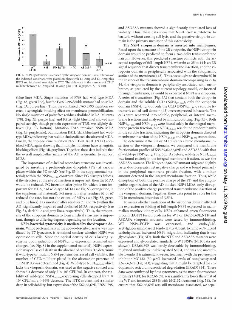
NSP4 bacterial cytotoxicity is mediated by the viroporin do-main. While bacterial lysis in the above-described assays was me-diated by T7 lysozyme, it remained unclear whether NSP4 wascytotoxic to cells. Since the optical density of cells lacking ly-sozyme upon induction of NSP447-146 expression remained un-changed (see Fig. S1 in the supplemental material), NSP4 expres-sion may cause cell death in the absence of cell lysis. To determineif wild-type or mutant NSP4 proteins decreased cell viability, thenumber of CFU/milliliter plated in the absence or presence of1 mM IPTG was determined (Fig. 4). Wild-type NSP495-146, whichlacks the viroporin domain, was used as the negative control andshowed a decrease of only 2 � 108 CFU/ml. In contrast, the via-bility of wild-type NSP454-146-expressing cells dropped by 7 �108 CFU/ml, a �99% decrease. The NTK mutant had a similardrop in cell viability, but expression of the K62,66,69E, F76S L79S,
and ASDASA mutants showed a significantly attenuated loss ofviability. Thus, these data show that NSP4 itself is cytotoxic tobacteria without causing cell lysis, and the putative viroporin do-main is the primary mediator of this cytotoxicity.
The NSP4 viroporin domain is inserted into membranes.Based upon the structure of the 2B viroporin, the NSP4 viroporindomain would be predicted to form a two-helix transmembranehairpin. However, this predicted structure conflicts with the ac-cepted topology of full-length NSP4, wherein aa 25 to 44 is an ERsignal sequence that directs transmembrane insertion, and the vi-roporin domain is peripherally associated with the cytoplasmicsurface of the membrane (42). Thus, we sought to determine if, inthe absence of the transmembrane domain encompassing aa 25 to44, the viroporin domain is peripherally associated with mem-branes, as predicted by the current topology model, or insertedthrough membranes, as would be expected if NSP4 is a viroporin.A series of truncations (Fig. 5A) that contain both the viroporindomain and the soluble CCD (NSP447-146), only the viroporindomain (NSP447-90), or only the CCD (NSP495-146), a soluble te-trameric coiled-coil domain (43), were expressed in bacteria. Thecells were separated into soluble, peripheral, or integral mem-brane fractions and analyzed by immunoblotting (Fig. 5B). BothNSP447-146 and NSP447-90 were found solely in the integral mem-brane protein fraction, but NSP495-146 was found predominantlyin the soluble fraction, indicating the viroporin domain directedmembrane insertion of the NSP447-146 and NSP447-90 truncations.
To determine if the PD or AD mutants altered membrane in-sertion of the viroporin domain, we compared the membranefractionation profiles of K55,59,62,66,69E and ASDASA with thatof wild-type NSP447-146 (Fig. 5C). As before, wild-type NSP447-146
was found entirely in the integral membrane fraction, as was theASDASA mutant. The K55,59,62,66,69E mutant migrated slightlyfaster due to a greater net negative charge and was found primarilyin the peripheral membrane protein fraction, with a minoramount detected in the integral membrane fraction. Thus, whiledisruption of both the positive charge of the PD and the amphi-pathic organization of the AD blocked NSP4 MDA, only disrup-tion of the positive charge prevented transmembrane insertion ofthe viroporin domain. Together, these data point to a role for thePD in membrane insertion of NSP4.
To assess whether mutations of the viroporin domain affectedthe expression or folding of full-length NSP4 expressed in mam-malian monkey kidney cells, NSP4-enhanced green fluorescentprotein (EGFP) fusion proteins for WT or K62,66,69E,NTK andASDASA viroporin mutants were tested by immunoblotting.WT NSP4-EGFP was expressed, and endo-�-N-acetylglucosaminidase H (endo H) treatment, to remove N-linkedcarbohydrates, increased NSP4 migration, indicating that it wasglycosylated (Fig. 5D). Both the NTK and ASDASA mutants wereexpressed and glycosylated similarly to WT NSP4 (NTK data notshown). K62,66,69E was barely detectable by immunoblotting,migrated similarly to unglycosylated NSP4, and was not suscepti-ble to endo H treatment; however, treatment with the proteosomeinhibitor MG132 (50 �M) increased levels of nonglycosylatedK62,66,69E (Fig. 5D), suggesting that it might be targeted for en-doplasmic reticulum-associated degradation (ERAD) (44). Thesedata were confirmed by flow cytometry, as the mean fluorescenceintensity (MFI) for K62,66,69E was significantly lower than that ofthe WT and increased 288% with MG132 treatment (Fig. 5E). Toensure that K62,66,69E was still membrane associated, we sepa-
FIG 4 NSP4 cytotoxicity is mediated by the viroporin domain. Serial dilutions ofthe indicated constructs were plated on plates with LB-Amp and LB-Amp plusIPTG and incubated overnight at 37°C. The difference in the numbers of CFU/milliliter between LB-Amp and LB-Amp plus IPTG is graphed. *, P � 0.01.
NSP4 Is a Viroporin
November/December 2010 Volume 1 Issue 5 e00265-10 mbio.asm.org 5
on May 11, 2019 by guest
http://mbio.asm
.org/D
ownloaded from

rated cells into soluble, peripheral, and integral membrane frac-tions, marked by immunoblot detection of GFP, GM130, andSTIM-1, respectively (see Fig. S4 in the supplemental material). Inthe absence of MG132, the WT and ASDASA were detected exclu-sively in the integral membrane fraction. In the presence ofMG132, K62,66,69E was found primarily in the integral mem-brane fraction, but some protein was detected in the soluble andperipheral membrane protein fractions (Fig. 5F). Together, thesedata suggest that NSP4 is initially targeted to the ER membrane viathe noncleaved signal sequence (aa 28 to 44), but the lysine clusterdirected translocation of the viroporin domain (aa 47 to 90) intothe membrane. Altering the charge of the lysine cluster blockedmembrane insertion of the viroporin domain in E. coli and tar-geted NSP4 for ERAD-mediated degradation in mammalian cells.Thus, the lysines facilitate proper NSP4 folding by directing viro-porin domain insertion into the membrane.
Viroporin mutants fail to elevate in-tracellular calcium levels in mammaliancells. Intracellular expression of full-length NSP4 in eukaryotic cells leads toincreased ER permeability and a signifi-cant increase in [Ca2�]cyto (26, 28). Wetested the mutants characterized usingthe E. coli lysis assay to determine if theviroporin domain is responsible for theincreased [Ca2�]cyto using the fluores-cent calcium indicator Indo-1. Usingflow cytometry, we measured the mean[Ca2�]cyto of cells expressing EGFP, theWT, or viroporin mutant NSP4-EGFP(Fig. 6). Expression of WT NSP4-EGFPincreased the ratio of calcium-bound tocalcium-free Indo-1 fluorescence, shift-ing the EGFP-positive population to theright (Fig. 6A, blue), but histograms forcells expressing NSP4-EGFP K62,66,69Eor ASDASA remained clustered to the left(Fig. 6A, red and green, respectively). Cy-toplasmic calcium levels (Fig. 5B) were3.7-fold higher in cells expressing WTNSP4-EGFP (119.6 � 33.8 nM) than incells expressing the EGFP negative con-trol (25.3 � 11.3 nM). NTK viroporinmutant-expressing cells have a similar el-evation in free calcium levels (111.6 �22.9 nM), but cells expressing theK62,66,69E or ASDASA viroporin mu-tants had significantly lower levels of freecalcium (40.0 � 4.0 nM and 47.4 �5.0 nM, respectively) than cells express-ing WT NSP4-EGFP (P � 0.05). To-gether, these data show that the NSP4 vi-roporin domain is responsible forelevating [Ca2�]cyto, and mutations thatabrogated MDA and E. coli cytotoxicitycorrelated with the inability to elevate[Ca2�]cyto.
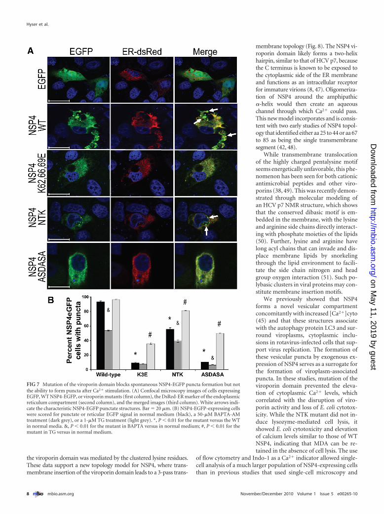
NSP4 viroporin mutants are ER lo-calized and require exogenous calciumstimulation to form vesicular puncta.
Previous studies demonstrated that three pools of NSP4 existwithin mammalian cells that localize to the (i) rough ER, (ii) ER-Golgi intermediate compartment (ERGIC), and (iii) puncta con-taining the autophagy marker LC3. NSP4 puncta formation iscalcium dependent, such that NSP4 is localized to the ER if[Ca2�]cyto remains low but an increase in the [Ca2�]cyto causesrapid formation of the puncta, and in rotavirus-infected cells,NSP4 colocalizes with LC3 in these puncta that surround viro-plasms, cytoplasmic inclusions where genome replication andprogeny virus assembly occur (45). Thus, assessment of NSP4puncta formation acts as a surrogate for testing the ability of viro-porin mutants to form the viroplasm-associated puncta. The sub-cellular localization and distribution of WT or mutant NSP4-EGFP fusion proteins were analyzed in normal medium (1.8 mMCaCl2), which supports spontaneous puncta formation for wild-type NSP4. The extent of ER localization for EGFP, WT NSP4-
FIG 5 The pentalysine motif mediates integral membrane insertion of the viroporin domain. (A) Sche-matic of the NSP4 constructs tested. (B and C) Immunoblot analysis of NSP4 in total cell lysate (T), solubleprotein (S), peripheral membrane protein (P), and integral membrane protein (I) fractions. M, molecularweight marker. (D) Immunoblot analysis of MA104 cell lysates for WT NSP4-EGFP, K62,66,69E, and AS-DASA in the absence or presence of MG132. Lysates were mock treated or Endo H treated to demonstrate theglycosylation of NSP4 (gly) by shifting to the unglycosylated form (ungly). (E) Flow cytometry analysis of themean fluorescence intensity (MFI) of MA104 cells expressing WT NSP4-EGFP or K62,66,69E in the absence(dark gray) or presence (light gray) of MG132. *, P � 0.01 for K62,66,69E versus WT in the absence ofMG132; #, P � 0.01 for K62,66,69E absence versus the presence of MG132. AU, arbitrary units. (F) Immu-noblot analysis of MA104 fractionation, as described above, for WT, K62,66,69E, and ASDASA.
Hyser et al.
6 mbio.asm.org November/December 2010 Volume 1 Issue 5 e00265-10
on May 11, 2019 by guest
http://mbio.asm
.org/D
ownloaded from

EGFP, and the three NSP4-EGFP viroporin mutants (K62,66,69E,NTK, and ASDASA) was determined by colocalization with anER-targeted DsRed2 fluorescent protein using confocal micros-copy (Fig. 7A). EGFP was found throughout the cell and did notlocalize to the ER or form puncta (Fig. 7A, first row). As seenpreviously, WT NSP4-EGFP localized partially to the ER com-partment and to distinct puncta that did not contain theDsRed-ER marker (Fig. 7A, second row). In contrast, NSP4-EGFPviroporin mutants were localized primarily in the ER, with cellsexpressing K62,66,69E and ASDASA lacking puncta (Fig. 7A,third and fifth rows, respectively), with a few small discrete punctathat did not contain DsRed-ER being observed in cells expressingNTK.
Next, we tested if changes in [Ca2�]cyto directly regulatedpuncta formation by quantitating the number of NSP4-EGFP-expressing cells containing puncta (i) in normal DMEM, (ii) aftertreatment with the intracellular calcium chelator BAPTA-AM, or(iii) after treatment with TG, a SERCA pump inhibitor that ele-vates cytoplasmic calcium levels. This experiment was designed todetermine if BAPTA treatment to buffer the elevated [Ca2�]cytocaused by the viroporin activity of WT NSP4 would decreasepuncta formation and if TG treatment to pharmacologically ele-vate [Ca2�]cyto would induce puncta formation of NSP4viroporin-deficient mutants. In normal DMEM, WT NSP4-EGFPformed discrete puncta in 93.3% of cells, but the viroporin mu-tants formed significantly fewer puncta, with rates of 9.4% forK62,66,69E, 55.5% for NTK, and 10.7% for ASDASA (Fig. 7B,
black bars) (P � 0.01). BAPTA-AM treatment of WT NSP4-EGFP-expressing cells reduced puncta formation to 53.7%(Fig. 7B) (P � 0.01). An overall decrease in the number of puncta-containing cells was also observed in BAPTA-treated viroporinmutant-expressing cells (Fig. 7B) (P of �0.01 for NTK and AS-DASA). Finally, cytoplasmic calcium levels were elevated pharma-cologically with TG to determine if exogenous calcium stimula-tion would induce puncta formation of the viroporin NSP4-EGFPmutants. TG treatment did not significantly increase the numberof puncta-containing WT NSP4-EGFP-expressing cells (Fig. 7B).In contrast, TG treatment increased puncta formation by 28.4%for K62,66,69E, 25.7% for NTK, and 39.5% for ASDASA (Fig. 7B,light gray). Thus, mutation of the viroporin domain decreasedspontaneous puncta formation; however, pharmacological eleva-tion of [Ca2�]cyto induced puncta formation of the mutant pro-teins. Together, these data show (i) that the viroporin mutantswere specifically deficient in the elevation of [Ca2�]cyto but not inthe ability to form puncta, (ii) that elevated [Ca2�]cyto triggersthe trafficking of NSP4 out of the ER and into cytoplasmic puncta,and (iii) that elevation of [Ca2�]cyto and formation of puncta areseparable steps within this process.
DISCUSSION
Seeking to define the mechanism for the PLC-independent in-crease in [Ca2�]cyto, we investigated a previously described NSP4domain (aa 48 to 91) with membrane-destabilizing activity thatmediates cytotoxic effects in both E. coli and mammalian cells (29,30). This study reports a comprehensive biochemical and mecha-nistic characterization of a viroporin domain from a viral non-structural protein that alters cellular calcium homeostasis to reg-ulate the progression of virus replication and assembly. The majornew findings of this study are as follows: (i) NSP4 aa 47 to 90 werestructurally similar to those of the enterovirus 2B protein andfunctionally consistent with the defining characteristics of viro-porins, (ii) the NSP4 PD mediated integral membrane insertion ofthe viroporin domain, (iii) NSP4 viroporin mutants that failed toinduce E. coli lysis and cytotoxicity also failed to elevate[Ca2�]cyto in mammalian cells, and (iv) elevation of [Ca2�]cytoregulates the subcellular distribution of NSP4 by triggering themovement of NSP4 out of the ER and into cytoplasmic puncta.
Using the E. coli lysis assay, we were able to show that the PDand AD are functionally distinct motifs within the viroporin do-main. The PD functioned as a membrane insertion motif, but thePD alone did not support viroporin activity. In contrast, viroporinactivity was mediated by the AD (aa 70 to 85), and mutation of thismotif blocked viroporin activity but not membrane insertion. TheAD also plays a role in NSP4 oligomerization by promoting theformation of high-molecular-weight NSP4 multimers that werelost by disrupting this domain (46). We confirmed this observa-tion, since expression of the viroporin domain alone (aa 47 to 90)had viroporin activity and formed disulfide-bonded dimers andSDS-stable oligomers (Fig. 1E), which have been seen with otherviroporins (35, 36). Therefore, oligomerization of the viroporindomain can occur in the absence of the CCD. Though direct evi-dence that this domain forms a pore is needed, this can be dem-onstrated only by an atomic structure of NSP4, which is hamperedby the necessity to use detergent to extract NSP4 during purifica-tion.
While full-length NSP4 is targeted to the ER membrane by anuncleaved signal sequence (aa 25 to 44) (42), membrane insertion of
FIG 6 NSP4 viroporin mutants do not elevate cytoplasmic calcium levels. (A)HEK293T cells expressing EGFP, WT NSP4-EGFP, or the indicated viroporinmutant NSP4-EGFP were loaded with 1.8 �M Indo-1 and analyzed by flowcytometry to measure the levels of cytoplasmic calcium. RFU, relative fluores-cence units. (B) The calcium-bound/calcium-free Indo-1 ratio (R) was deter-mined for 10,000 EGFP-positive cells, and the [Ca2�]cyto was calculated. Atotal of 3 independent experiments were performed, and error bars indicatethe standard deviations of the means. *, P � 0.05.
NSP4 Is a Viroporin
November/December 2010 Volume 1 Issue 5 e00265-10 mbio.asm.org 7
on May 11, 2019 by guest
http://mbio.asm
.org/D
ownloaded from
the viroporin domain was mediated by the clustered lysine residues.These data support a new topology model for NSP4, where trans-membrane insertion of the viroporin domain leads to a 3-pass trans-
membrane topology (Fig. 8). The NSP4 vi-roporin domain likely forms a two-helixhairpin, similar to that of HCV p7, becausethe C terminus is known to be exposed tothe cytoplasmic side of the ER membraneand functions as an intracellular receptorfor immature virions (8, 47). Oligomeriza-tion of NSP4 around the amphipathic�-helix would then create an aqueouschannel through which Ca2� could pass.This new model incorporates and is consis-tent with two early studies of NSP4 topol-ogy that identified either aa 25 to 44 or aa 67to 85 as being the single transmembranesegment (42, 48).
While transmembrane translocationof the highly charged pentalysine motifseems energetically unfavorable, this phe-nomenon has been seen for both cationicantimicrobial peptides and other viro-porins (38, 49). This was recently demon-strated through molecular modeling ofan HCV p7 NMR structure, which showsthat the conserved dibasic motif is em-bedded in the membrane, with the lysineand arginine side chains directly interact-ing with phosphate moieties of the lipids(50). Further, lysine and arginine havelong acyl chains that can invade and dis-place membrane lipids by snorkelingthrough the lipid environment to facili-tate the side chain nitrogen and headgroup oxygen interaction (51). Such po-lybasic clusters in viral proteins may con-stitute membrane insertion motifs.
We previously showed that NSP4forms a novel vesicular compartmentconcomitantly with increased [Ca2�]cyto(45) and that these structures associatewith the autophagy protein LC3 and sur-round viroplasms, cytoplasmic inclu-sions in rotavirus-infected cells that sup-port virus replication. The formation ofthese vesicular puncta by exogenous ex-pression of NSP4 serves as a surrogate forthe formation of viroplasm-associatedpuncta. In these studies, mutation of theviroporin domain prevented the eleva-tion of cytoplasmic Ca2� levels, whichcorrelated with the disruption of viro-porin activity and loss of E. coli cytotox-icity. While the NTK mutant did not in-duce lysozyme-mediated cell lysis, itshowed E. coli cytotoxicity and elevationof calcium levels similar to those of WTNSP4, indicating that MDA can be re-tained in the absence of cell lysis. The use
of flow cytometry and Indo-1 as a Ca2� indicator allowed single-cell analysis of a much larger population of NSP4-expressing cellsthan in previous studies that used single-cell microscopy and
FIG 7 Mutation of the viroporin domain blocks spontaneous NSP4-EGFP puncta formation but notthe ability to form puncta after Ca2� stimulation. (A) Confocal microscopy images of cells expressingEGFP, WT NSP4-EGFP, or viroporin mutants (first column), the DsRed-ER marker of the endoplasmicreticulum compartment (second column), and the merged images (third column). White arrows indi-cate the characteristic NSP4-EGFP punctate structures. Bar � 20 �m. (B) NSP4-EGFP-expressing cellswere scored for punctate or reticular EGFP signal in normal medium (black), a 50-�M BAPTA-AMtreatment (dark grey), or a 1-�M TG treatment (light grey). *, P � 0.01 for the mutant versus the WTin normal media. &, P � 0.01 for the mutant in BAPTA versus in normal medium; #, P � 0.01 for themutant in TG versus in normal medium.
Hyser et al.
8 mbio.asm.org November/December 2010 Volume 1 Issue 5 e00265-10
on May 11, 2019 by guest
http://mbio.asm
.org/D
ownloaded from

Fura-2 (27, 28). Additionally, Indo-1 is less sensitive to compart-mentalization into Ca2�-storage organelles, allowing more accu-rate measurements of [Ca2�]cyto (52).
NSP4 viroporin activity could trigger the elevation of cytoplas-mic Ca2� levels in several ways that are not necessarily mutuallyexclusive. In the ER, progressive depletion of the ER Ca2� storesby ER-associated NSP4 could activate store-operated Ca2� entry(SOCE) and indirectly increase plasma membrane permeabilityby opening cellular Ca2� entry channels (26, 53). Additionally,expression of NSP4 increases plasma membrane permeability tomono- and divalent cations (28). Since NSP4 traffics to the plasmamembrane in RV-infected cells, it is possible that NSP4 viroporinactivity could directly increase plasma membrane permeability toCa2� and possibly other ions (28, 54). Both mechanisms rely onNSP4 viroporin activity; however, a detailed analysis of how NSP4affects Ca2� at both the ER and plasma membrane will be neces-sary to determine the relative importance that either direct per-meabilization or SOCE activation, or both, has on the elevation ofintracellular Ca2� levels during a rotavirus infection.
Previous studies suggested that the movement of NSP4 fromthe ER into the punctate NSP4/LC3 vesicles was regulated by[Ca2�]cyto, but since the mechanism of NSP4-mediated ER cal-cium release was unknown, the dependence on calcium for thisprocess could not be characterized further (45). We demonstratedthat failure of the viroporin mutants to spontaneously formpuncta was a direct consequence of their inability to elevate cyto-plasmic Ca2� levels by measuring puncta formation after buffer-ing (BAPTA-AM) or elevating (TG) [Ca2�]cyto. Under normalconditions, puncta formation by NSP4 occurs spontaneously andrapidly; however, mutants of either the pentalysine domain(K62,66,69E) or amphipathic domain (ASDASA) were unable toform puncta. TG stimulation of viroporin mutant NSP4 punctaformation demonstrated that the mutations specifically blockedthe elevation of [Ca2�]cyto but not the ability to form puncta.Thus, disruption of cellular Ca2� homeostasis and puncta forma-tion are separable events, and NSP4 not only increases [Ca2�]cytoby viroporin activity but also appears to be a sensor for changes in[Ca2�]cyto. The NSP4 viroporin mutants developed in thesestudies will be useful tools to determine the precise [Ca2�]cytothat triggers NSP4 puncta formation.
These studies demonstrate that NSP4viroporin activity is responsible for the ele-vation of [Ca2�]cyto in rotavirus-infectedcells, which was first reported nearly20 years ago (21), and appears to regulateseveral changes in the subcellular distribu-tion of other RV proteins. First, elevation ofcytoplasmic Ca2� levels regulates the for-mation of viroplasms, the RV replicationcomplex. Nonstructural protein 5 (NSP5),a component of viroplasms, has twopseudo-EF-hand Ca2� binding sites and el-evated levels of cytoplasmic Ca2�, andCa2� binding triggers the aggregation ofsoluble NSP5 into a viroplasm-like struc-ture (55). Second, in response to elevatedlevels of Ca2�, NSP4 traffics out of the ERand into puncta that surround viroplasms.Third, the assembly of the RV outer capsidprotein VP7 onto virions requires highCa2� levels inside the ER (24). Since RNA
interference (RNAi)-mediated knockdown of NSP4 prevents theproper assembly of viroplasms and causes the mislocalization of sev-eral other RV proteins (56, 57), it appears that NSP4 viroporin activ-ity functionally regulates the progression of RV infection and assem-bly by altering the cytoplasmic Ca2� levels.
The regulatory function fulfilled by NSP4 viroporin activity isunique among viroporins, which function primarily in virus entry(influenza M2) (9), virus release (HCV p7, HIV Vpu, coronavirusE, and polyomavirus VP4/agnoprotein) (3, 10, 11, 58), or apopto-sis (RSV SH) (12). While picornavirus 2B elevates Ca2� levels ininfected cells, the role that elevated Ca2� levels plays in the repli-cation cycle for these viruses is not well characterized (39). Thus,as is shown here for rotavirus NSP4, it is possible that the use ofviroporins to modulate processes important for replication com-plex assembly, genome replication, and virus assembly is a mech-anism utilized by more viruses than is currently appreciated.
MATERIALS AND METHODSExpression vectors. E. coli expression constructs were generated by ligation-independent cloning (LIC) using the pET46Ek/LIC system (EMD Biosci-ences, San Diego, CA). Wild-type NSP4-EGFP was constructed by insertingthe SA11 NSP4 (GenBank accession no. AF087678.1) coding region intopEGFP-N1 (Clontech). Internal deletions and mutations were generated byusing the QuikChange mutagenesis kit (Stratagene, La Jolla, CA) or encodingthe desired mutation in the forward primer, and all constructs were se-quenced (Lone Star Laboratories, Houston, TX).
E. coli lysis assay. Assessment of NSP4 viroporin activity in E. coli wasperformed essentially as described previously (31). Overnight cultureswere diluted 1:100 into fresh LB and grown until the optical density at 600nm (OD600) was 0.4 to 0.6, and 1 mM IPTG was added to induce proteinexpression. OD600 measurements of each culture were taken before IPTGinduction and at 10-min intervals postinduction for 90 min using a mul-tiwell plate spectrophotometer.
Immunoblot analysis. Samples were mixed with sample buffer,boiled for 5 minutes, run on 4 to 20% Tris-glycine or 10 to 20% Tris-Tricine polyacryamide gradient gels (Bio-Rad, Hercules, CA), and trans-ferred onto a nitrocellulose membrane (GE Healthcare Bio-Sciences Cor-poration, Piscataway, NJ) as previously described (59). Bacteriallyexpressed NSP447-90–His was partially purified using Ni2�-nitrilotriaceticacid (NTA) beads (GE Healthcare Bio-Sciences) as previously described(59) and separated by SDS-PAGE as described above, except sample buf-
FIG 8 Model of the NSP4 viroporin as a three-pass transmembrane protein. (Left) Initial insertion ofNSP4 into the ER membrane (gray) occurs through the uncleaved signal sequence in the H2 domain.Lysine residues interact with ER membrane phospholipids and promote insertion of the viroporindomain as an anti-parallel �-helical hairpin. (Center) Insertion of the viroporin domain generates athree-pass transmembrane topology. (Right) Oligomerization of NSP4 around the amphipathic �-helixcreates an aqueous pore through the membrane and allows the release of ER Ca2�.
NSP4 Is a Viroporin
November/December 2010 Volume 1 Issue 5 e00265-10 mbio.asm.org 9
on May 11, 2019 by guest
http://mbio.asm
.org/D
ownloaded from

fer lacking BME was used under nonreducing conditions. Antibodies usedwere NSP4 MAb B4-2/55 ascites, anti-Penta-His antibody (Qiagen, Va-lencia, CA), anti-EGFP monoclonal antibody (Clontech), anti-GM130monoclonal antibody (BD Transduction Laboratories, San Jose, CA), andanti-STIM- 1 antibody (Sigma-Aldrich, St. Louis, MO).
E. coli viability assay. Stationary-phase cultures of E. coli BL21(DE3)for the indicated NSP4 constructs were serially diluted in LB (withoutampicillin), and 100 �l of each dilution was plated on LB-ampicillin platesin the absence or presence of 1 mM IPTG. The plates were incubatedovernight at 37°C, and the number of CFU per milliliter was calculated.
Membrane protein fractionation. BL21(DE3)pLysS broth cultureswere grown to an OD600 of 0.5 to 0.6, and protein expression was inducedwith 1 mM IPTG and cultured for 1 h. The cells were pelleted by centrif-ugation (21,000 � g, 1 h) and resuspended in 5 ml ice-cold phosphate-buffered saline (PBS) (total protein lysate fraction [T]). A 1-ml aliquotwas sonicated in PBS using a probe sonicator (soluble protein fraction[S]). The membranes were pelleted by centrifugation (100,000 � g, 1 h)and resuspended in 1 ml 100 mM sodium carbonate for 30 min on ice(peripheral membrane protein fraction [P]). The membranes were againpelleted and resuspended in 1 ml 1% SDS-PBS (integral membrane pro-tein fraction [I]). Equal buffer volumes were used to maintain the samerelative protein concentration as that of the starting material. Equivalentamounts of each fraction were analyzed by SDS-PAGE.
Cells and transfection. African green monkey MA104 kidney cells andhuman embryonic kidney (HEK) 293T cells were maintained and transfectedas previously described (45). In experiments using N-(benzyloxycarbonyl)leucinylleucinylleucinal-Z-Leu-Leu-Leu-al (MG132), at 4 h posttransfection,the medium was replaced with fresh Opti-MEM containing 50 �M MG132.In all cases, cells were incubated overnight at 37°C.
Confocal microscopy. MA104 cells were fixed with 4% paraformal-dehyde and permeabilized with 0.5% Triton X-100 for 10 min. Cells werestained with TO-PRO-3 (Invitrogen), and coverslips were mounted ontoslides using ProLong gold antifade reagents (Molecular Probes, Eugene,OR). Mounted slides were observed using a Carl Zeiss LSM 510 Metaconfocal microscope with a 63� immersion oil objective (Carl Zeiss, Ger-many). The pinhole was set to 1, and pixel time was set at 3.20 �s for 16scanning averages per track on each slice, and Z-stack slices were set to1 �m. The collected images were processed using LSM 510 image software(Carl Zeiss, Inc., Thornwood, NY).
Indo-1 calcium measurements. Indo-1 (50 �g; Molecular Probes)was resuspended in 50 �l 20% F-127 and 50 �l fetal bovine serum (FBS) at37°C. The Indo-1 loading buffer used was Hanks’ balanced salt solution(HBSS; Invitrogen) supplemented with 1% bovine serum albumin (BSA)(HBSS-BSA) and 1.8 �M Indo-1. At approximately 28 h posttransfection,the cells were gently washed with HBSS-BSA, and Indo- 1 loading bufferwas added for 30 min at 37°C. Cells were pelleted, resuspended in alpha-MEM (no phenol red) plus 10% FBS plus 10 mM HEPES, and maintainedat 37°C until analyzed. Flow cytometry analysis was performed using anLSRII system running FACSDiva software (BD Biosciences, FranklinLakes, NJ). Indo-1 fluorescence was excited by a UV laser (355 nm), andCa2�-free and Ca2�-bound emissions were split using a 505LP dichroicfilter. Ca2�-free emission was collected with a 525/50-nm band-pass filter,and the Ca2�-bound emission was collected with a 405/20-nm band-passfilter. EGFP fluorescence was excited by the argon laser (488 nm), andemission was collected with a 520/20-nm band-pass filter. The meanbound Ca2�-to-free Ca2� fluorescence ratio (R) was determined for eachsample. The concentration of calcium was calculated by using the follow-ing equation: Ca2� (nM) �Kd (R � Rmin)Sf2/(Rmax � R)Sb2. Treatment ofcells with 10 mM EDTA and 5 �M ionomycin was used to determine thefluorescence ratios at zero (Rmin) and saturated (Rmax) calcium, respec-tively. Sf2 and Sb2 are the fluorescence intensities of the calcium-free and-bound dyes, respectively. The dissociation constant of Indo-1 is 250 nM.Experiments were performed in triplicate, and results are presented at themean calculated calcium level.
Puncta formation assay. MA104 cells were either loaded with 50 �MBAPTA-AM for 1 h at 37°C at 4 h posttransfection or maintained innormal medium. At approximately 20 h posttransfection, a subset of thetransfected cells were treated with 1 �M thapsigargin (TG) for 3 h, andthen, all the cells were fixed in 4% paraformaldehyde. The number ofNSP4-EGFP-expressing cells containing diffuse rather than punctateNSP4-EGFP was counted in 25 random fields per well using the 40� lensobjective on an Olympus IX70 inverted epifluorescence microscope. Cellswith a completely uniform reticular NSP4-EGFP distribution were scoredas having no puncta; however, cells with the presence of even one punctatestructure were scored as having puncta.
Statistical analysis. Statistical differences between groups were deter-mined using a two-tailed Student’s t test. P values of �0.05 were consid-ered significant.
ACKNOWLEDGMENTS
This work was supported in part by NIH grant R01AI080656, NIH Re-search Training in Pediatric Gastroenterology grant T32DK007664, anNIH Training Grant in Molecular Virology, T32AI007471, and PublicHealth Service grant P30DK56338, which funds the Texas Medical CenterDigestive Diseases Center.
Scoring of cells was performed by J.M.H., and categorization of cellsinto groups with puncta/no puncta was validated by M.C.-P. and B.U.
SUPPLEMENTAL MATERIALSupplemental material for this article may be found at http://mbio.asm.org/lookup/suppl/doi:10.1128/mBio.00265-10/-/DCSupplemental.
Figure S1, TIF file, 3.902 MB.Figure S2, TIF file, 5.443 MB.Figure S3, TIF file, 5.300 MB.Figure S4, TIF file, 3.468 MB.Table S1, PDF file, 0.098 MB.
REFERENCES1. Dubyak, G. R. 2004. Ion homeostasis, channels, and transporters: an
update on cellular mechanisms. Adv. Physiol. Educ. 28:143–154.2. Kang, M., Moroni, A., Gazzarrini, S., DiFrancesco, D., Thiel, G., Sev-
erino, M., and Van Etten, J. L.. 2004. Small potassium ion channelproteins encoded by chlorella viruses. Proc. Natl. Acad. Sci. U. S. A. 101:5318 –5324.
3. Daniels, R., Sadowicz, D., and Hebert, D. N.. 2007. A very late viralprotein triggers the lytic release of SV40. PLoS Pathog. 3:e98.
4. Suzuki, T., Orba, Y., Okada, Y., Sunden, Y., Kimura, T., Tanaka, S.,Nagashima, K., Hall, W. W., and Sawa, H.. 2010. The human polyomaJC virus agnoprotein acts as a viroporin. PLoS Pathog. 6:e1000801.
5. Liao, Y., Tam, J. P., and Liu, D. X.. 2006. Viroporin activity of SARS-CoV E protein. Adv. Exp. Med. Biol. 581:199 –202.
6. Gonzalez, M. E., and Carrasco, L.. 2003. Viroporins. FEBS Lett. 552:28–34.7. Perez, M., Garcia-Barreno, B., Melero, J. A., Carrasco, L., and Guinea,
R.. 1997. Membrane permeability changes induced in Escherichia coli bythe SH protein of human respiratory syncytial virus. Virology 235:342–351.
8. Cook, G. A., Zhang, H., Park, S. H., Wang, Y., and Opella, S. J.. Compar-ative NMR studies demonstrate profound differences between twoviroporins: p7 of HCV and Vpu of HIV-1. Biochim. Biophys. Acta, in press.
9. Pielak, R. M., and Chou, J. J.. Influenza M2 proton channels. Biochim.Biophys. Acta, in press.
10. Ruiz, A., Guatelli, J. C., and Stephens, E. B.. 2010. The Vpu protein: newconcepts in virus release and CD4 down-modulation. Curr. HIV Res.8:240–252.
11. Steinmann, E., Penin, F., Kallis, S., Patel, A. H., Bartenschlager, R., andPietschmann, T.. 2007. Hepatitis C virus p7 protein is crucial for assemblyand release of infectious virions. PLoS Pathog. 3:e103.
12. Fuentes, S., Tran, K. C., Luthra, P., Teng, M. N., and He, B.. 2007.Function of the respiratory syncytial virus small hydrophobic protein. J.Virol. 81:8361– 8366.
13. Ding, W., Albrecht, B., Kelley, R. E., Muthusamy, N., Kim, S. J.,Altschuld, R. A., and Lairmore, M. D.. 2002. Human T-cell lymphotro-pic virus type 1 p12(I) expression increases cytoplasmic calcium to en-
Hyser et al.
10 mbio.asm.org November/December 2010 Volume 1 Issue 5 e00265-10
on May 11, 2019 by guest
http://mbio.asm
.org/D
ownloaded from

hance the activation of nuclear factor of activated T cells. J. Virol. 76:10374 –10382.
14. Zhou, Y., Frey, T. K., and Yang, J. J.. 2009. Viral calciomics: interplaysbetween Ca2� and virus. Cell Calcium 46:1–17.
15. Chami, M., Oules, B., and Paterlini-Brechot, P.. 2006. Cytobiologicalconsequences of calcium-signaling alterations induced by human viralproteins. Biochim. Biophys. Acta 1763:1344 –1362.
16. van Kuppeveld, F. J., de Jong, A. S., Melchers, W. J., and Willems, P. H..2005. Enterovirus protein 2B po(u)res out the calcium: a viral strategy tosurvive? Trends Microbiol. 13:41– 44.
17. Sanchez-Martinez, S., Huarte, N., Maeso, R., Madan, V., Carrasco, L.,and Nieva, J. L.. 2008. Functional and structural characterization of 2Bviroporin membranolytic domains. Biochemistry 47:10731–10739.
18. van Kuppeveld, F. J., Galama, J. M., Zoll, J., van den Hurk, P. J., andMelchers, W. J.. 1996. Coxsackie B3 virus protein 2B contains cationic am-phipathic helix that is required for viral RNA replication. J. Virol. 70:3876–3886.
19. Hyser, J. M., and Estes, M. K.. 2009. Rotavirus vaccines andpathogenesis: 2008. Curr. Opin. Gastroenterol. 25:36 – 43.
20. Jiang, V., Jiang, B., Tate, J., Parashar, U. D., and Patel, M. M.. Perfor-mance of rotavirus vaccines in developed and developing countries. Hum.Vaccin., in press.
21. Michelangeli, F., Ruiz, M. C., del Castillo, J. R., Ludert, J. E., andLiprandi, F.. 1991. Effect of rotavirus infection on intracellular calciumhomeostasis in cultured cells. Virology 181:520 –527.
22. Perez, J. F., Chemello, M. E., Liprandi, F., Ruiz, M. C., and Michelangeli,F.. 1998. Oncosis in MA104 cells is induced by rotavirus infection through anincrease in intracellular Ca2� concentration. Virology 252:17–27.
23. Zambrano, J. L., Diaz, Y., Pena, F., Vizzi, E., Ruiz, M. C., Michelangeli,F., Liprandi, F., and Ludert, J. E.. 2008. Silencing of rotavirus NSP4 orVP7 expression reduces alterations in Ca2� homeostasis induced by infec-tion of cultured cells. J. Virol. 82:5815–5824.
24. Ruiz, M. C., Aristimuno, O. C., Diaz, Y., Pena, F., Chemello, M. E.,Rojas, H., Ludert, J. E., and Michelangeli, F.. 2007. Intracellular disas-sembly of infectious rotavirus particles by depletion of Ca2� sequesteredin the endoplasmic reticulum at the end of virus cycle. Virus Res. 130:140 –150.
25. Michelangeli, F., Liprandi, F., Chemello, M. E., Ciarlet, M., and Ruiz,M. C.. 1995. Selective depletion of stored calcium by thapsigargin blocksrotavirus maturation but not the cytopathic effect. J. Virol. 69:3838 –3847.
26. Tian, P., Estes, M. K., Hu, Y., Ball, J. M., Zeng, C. Q., and Schilling,W. P.. 1995. The rotavirus nonstructural glycoprotein NSP4 mobilizesCa2� from the endoplasmic reticulum. J. Virol. 69:5763–5772.
27. Berkova, Z., Morris, A. P., and Estes, M. K.. 2003. Cytoplasmic calciummeasurement in rotavirus enterotoxin-enhanced green fluorescent protein(NSP4-EGFP) expressing cells loaded with Fura-2. Cell Calcium 34:55–68.
28. Diaz, Y., Chemello, M. E., Pena, F., Aristimuno, O. C., Zambrano, J. L.,Rojas, H., Bartoli, F., Salazar, L., Chwetzoff, S., Sapin, C., Trugnan, G.,Michelangeli, F., and Ruiz, M. C.. 2008. Expression of nonstructuralrotavirus protein NSP4 mimics Ca2� homeostasis changes induced byrotavirus infection in cultured cells. J. Virol. 82:11331–11343.
29. Browne, E. P., Bellamy, A. R., and Taylor, J. A.. 2000. Membrane-destabilizing activity of rotavirus NSP4 is mediated by a membrane-proximal amphipathic domain. J. Gen. Virol. 81:1955–1959.
30. Newton, K., Meyer, J. C., Bellamy, A. R., and Taylor, J. A.. 1997.Rotavirus nonstructural glycoprotein NSP4 alters plasma membrane per-meability in mammalian cells. J. Virol. 71:9458 –9465.
31. Lama, J., and Carrasco, L.. 1992. Expression of poliovirus nonstructuralproteins in Escherichia coli cells. Modification of membrane permeabilityinduced by 2B and 3A. J. Biol. Chem. 267:15932–15937.
32. Gonzalez, M. E., and Carrasco, L.. 1998. The human immunodeficiencyvirus type 1 Vpu protein enhances membrane permeability. Biochemistry37:13710 –13719.
33. Guinea, R., and Carrasco, L.. 1994. Influenza virus M2 protein modifiesmembrane permeability in E. coli cells. FEBS Lett. 343:242–246.
34. Bowman, G. D., Nodelman, I. M., Levy, O., Lin, S. L., Tian, P., Zamb,T. J., Udem, S. A., Venkataraghavan, B., and Schutt, C. E.. 2000. Crystalstructure of the oligomerization domain of NSP4 from rotavirus reveals acore metal-binding site. J. Mol. Biol. 304:861– 871.
35. Agirre, A., Lorizate, M., Nir, S., and Nieva, J. L.. 2008. Poliovirus 2binsertion into lipid monolayers and pore formation in vesicles modulatedby anionic phospholipids. Biochim. Biophys. Acta 1778:2621–2626.
36. Perez-Berna, A. J., Guillen, J., Moreno, M. R., Bernabeu, A., Pabst, G.,
Laggner, P., and Villalain, J.. 2008. Identification of the membrane-activeregions of hepatitis C virus p7 protein: biophysical characterization of theloop region. J. Biol. Chem. 283:8089 – 8101.
37. Carter, S. D., Dent, K. C., Atkins, E., Foster, T. L., Verow, M., Gorny,P., Harris, M., Hiscox, J. A., Ranson, N. A., Griffin, S., and Barr, J. N..2010. Direct visualization of the small hydrophobic protein of humanrespiratory syncytial virus reveals the structural basis for membrane per-meability. FEBS Lett. 584:2786 –2790.
38. StGelais, C., Foster, T. L., Verow, M., Atkins, E., Fishwick, C. W.,Rowlands, D., Harris, M., and Griffin, S.. 2009. Determinants of hepa-titis C virus p7 ion channel function and drug sensitivity identified invitro. J. Virol. 83:7970 –7981.
39. van Kuppeveld, F. J., Hoenderop, J. G., Smeets, R. L., Willems, P. H.,Dijkman, H. B., Galama, J. M., and Melchers, W. J.. 1997. Coxsacki-evirus protein 2B modifies endoplasmic reticulum membrane and plasmamembrane permeability and facilitates virus release. EMBO J. 16:3519 –3532.
40. Epand, R. M., and Lim, W.. 1995. Mechanism of liposome destabilizationby polycationic amino acids. Biosci. Rep. 15:151–160.
41. Lin, S. L., and Tian, P.. 2003. Detailed computational analysis of a compre-hensive set of group A rotavirus NSP4 proteins. Virus Genes 26:271–282.
42. Bergmann, C. C., Maass, D., Poruchynsky, M. S., Atkinson, P. H., andBellamy, A. R.. 1989. Topology of the non-structural rotavirus receptorglycoprotein NS28 in the rough endoplasmic reticulum. EMBO J.8:1695–1703.
43. Deepa, R., Durga, R. C., and Suguna, K.. 2007. Structure of the extendeddiarrhea-inducing domain of rotavirus enterotoxigenic protein NSP4.Arch. Virol. 152:847– 859.
44. Vembar, S. S., and Brodsky, J. L.. 2008. One step at a time: endoplasmicreticulum-associated degradation. Nat. Rev. Mol. Cell Biol. 9:944 –957.
45. Berkova, Z., Crawford, S. E., Trugnan, G., Yoshimori, T., Morris, A. P.,and Estes, M. K.. 2006. Rotavirus NSP4 induces a novel vesicular com-partment regulated by calcium and associated with viroplasms. J. Virol.80:6061– 6071.
46. Jagannath, M. R., Kesavulu, M. M., Deepa, R., Sastri, P. N., Kumar,S. S., Suguna, K., and Rao, C. D.. 2006. N- and C-terminal cooperationin rotavirus enterotoxin: novel mechanism of modulation of the proper-ties of a multifunctional protein by a structurally and functionally over-lapping conformational domain. J. Virol. 80:412– 425.
47. Au, K. S., Chan, W. K., Burns, J. W., and Estes, M. K.. 1989. Receptoractivity of rotavirus nonstructural glycoprotein NS28. J. Virol. 63:4553–4562.
48. Chan, W. K., Au, K. S., and Estes, M. K.. 1988. Topography of the simianrotavirus nonstructural glycoprotein (NS28) in the endoplasmic reticu-lum membrane. Virology 164:435– 442.
49. Zakharov, S. D., Rokitskaya, T. I., Shapovalov, V. L., Antonenko, Y. N.,and Cramer, W. A.. 2002. Tuning the membrane surface potential forefficient toxin import. Proc. Natl. Acad. Sci. U. S. A. 99:8654 – 8659.
50. Montserret, R., Saint, N., Vanbelle, C., Salvay, A. G., Simorre, J. P.,Ebel, C., Sapay, N., Renisio, J. G., Bockmann, A., Steinmann, E.,Pietschmann, T., Dubuisson, J., Chipot, C., and Penin, F.. 2010. NMRstructure and ion channel activity of the p7 protein from hepatitis C virus.J. Biol. Chem. 285:31446 –31461.
51. Kandasamy, S. K., and Larson, R. G.. 2006. Molecular dynamics simu-lations of model trans-membrane peptides in lipid bilayers: a systematicinvestigation of hydrophobic mismatch. Biophys. J. 90:2326 –2343.
52. Wahl, M., Lucherini, M. J., and Gruenstein, E.. 1990. Intracellular Ca2�
measurement with Indo-1 in substrate-attached cells: advantages and spe-cial considerations. Cell Calcium 11:487–500.
53. Cahalan, M. D. 2009. STIMulating store-operated Ca(2�) entry. Nat.Cell Biol. 11:669 – 677.
54. Ruiz, M. C., Diaz, Y., Pena, F., Aristimuno, O. C., Chemello, M. E., andMichelangeli, F.. 2005. Ca2� permeability of the plasma membrane in-duced by rotavirus infection in cultured cells is inhibited by tunicamycinand brefeldin A. Virology 333:54 – 65.
55. Sen, A., Sen, N., and Mackow, E. R.. 2007. The formation of viroplasm-like structures by the rotavirus NSP5 protein is calcium regulated anddirected by a C-terminal helical domain. J. Virol. 81:11758 –11767.
56. Silvestri, L. S., Tortorici, M. A., Vasquez-Del Carpio, R., and Patton,J. T.. 2005. Rotavirus glycoprotein NSP4 is a modulator of viral transcrip-tion in the infected cell. J. Virol. 79:15165–15174.
57. Lopez, T., Camacho, M., Zayas, M., Najera, R., Sanchez, R., Arias, C. F.,and Lopez, S.. 2005. Silencing the morphogenesis of rotavirus. J. Virol. 79:184–192.
NSP4 Is a Viroporin
November/December 2010 Volume 1 Issue 5 e00265-10 mbio.asm.org 11
on May 11, 2019 by guest
http://mbio.asm
.org/D
ownloaded from
58. Ye, Y., and Hogue, B. G.. 2007. Role of the coronavirus E viroporinprotein transmembrane domain in virus assembly. J. Virol. 81:3597–3607.
59. Hyser, J. M., Zeng, C. Q., Beharry, Z., Palzkill, T., and Estes, M. K..
2008. Epitope mapping and use of epitope-specific antisera to characterizethe VP5* binding site in rotavirus SA11 NSP4. Virology 373:211–228.
Hyser et al.
12 mbio.asm.org November/December 2010 Volume 1 Issue 5 e00265-10
on May 11, 2019 by guest
http://mbio.asm
.org/D
ownloaded from


![Amyloid-Beta Disrupts Calcium and Redox Homeostasis in ... · Amyloid-Beta Disrupts Calcium and Redox Homeostasis in Brain Endothelial Cells ... (SERCA) [39]. Although low ... Homeostasis](https://img.pdfslide.us/doc/110x75/5c76b20d09d3f2d3778bffa9/amyloid-beta-disrupts-calcium-and-redox-homeostasis-in-amyloid-beta-disrupts.jpg)