Embed Size (px)
Citation preview

7/21/2019 Rotary Negger
http://slidepdf.com/reader/full/rotary-negger 1/28
Coordinated control of eye and handmovements in dynamic reaching
S.F.W. Neggers a,c,*, H. Bekkering b,c
a Department of Psychonomics, Helmholtz Institute, Utrecht University, Heidelberglaan 2, 3584 CS Utrecht,
The Netherlandsb Department of Experimental and Work Psychology, University of Groningen, Grote Kruisstraat 2/1,
9712 TS Groningen, The Netherlandsc Department of Cognition and Action, Max-Planck-Institute for Psychological Research,
Amalienstraße 33, 80802 Munich, Germany
Abstract
In the present study, we integrated two recent, at first sight contradictory findings regardingthe question whether saccadic eye movements can be generated to a newly presented target
during an ongoing hand movement. Saccades were measured during so-called adaptive and
sustained pointing conditions. In the adapted pointing condition, subjects had to direct both
their gaze and arm movements to a displaced target location. The results showed that the eyes
could fixate the new target during pointing. In addition, a temporal coupling of these correc-
tive saccades was found with changes in arm movement trajectories when reaching to the new
target. In the sustained pointing condition, however, the same subjects had to point to the ini-
tial target, while trying to deviate their gaze to a new target that appeared during pointing. It
was found that the eyes could not fixate the new target before the hand reached the initial tar-
get location. Together, the results indicate that ocular gaze is always forced to follow the target
intended by a manual arm movement. A neural mechanism is proposed that couples oculargaze to the target of an arm movement. Specifically, the mechanism includes a reach neuron
layer besides the well-known saccadic layer in the primate superior colliculus. Such a tight,
sub-cortical coupling of ocular gaze to the target of a reaching movement can explain the con-
trasting behavior of the eyes in dependency of whether the eye and hand share the same target
position or attempt to move to different locations.
2002 Elsevier Science B.V. All rights reserved.
Human Movement Science 21 (2002) 349–376
www.elsevier.com/locate/humov
*Corresponding author. Address: Department of Psychonomics, Helmholtz Institute, Utrecht
University, Heidelberglaan 2, 3584 CS Utrecht, The Netherlands. Tel.: +31-30-253-4582; fax: +31-30-
253-4511.
E-mail address: [email protected] (S.F.W. Neggers).
0167-9457/02/$ - see front matter 2002 Elsevier Science B.V. All rights reserved.
PII: S 0 1 6 7 - 9 4 5 7 ( 0 2 ) 0 0 1 2 0 - 3

7/21/2019 Rotary Negger
http://slidepdf.com/reader/full/rotary-negger 2/28
PsycINFO classification: 2300; 2330; 2520
Keywords: Saccade; Eye–hand coordination; Superior colliculus; Neural control
1. Introduction
In order to interact effectively with the everyday world and to manipulate objects
in our workspace, humans often make goal-directed movements with the head, body,
eyes and arms. It is well known that such goal-directed movements are not indepen-
dent, but are coordinated in order to reach a certain common goal (e.g., bringing a
cup of coffee to the mouth). The present study involves a detailed analysis on the
coupling between saccadic eye movements and arm movements.
Typically, when pointing with the arm towards a position within reach, a saccadiceye movement is made to this position shortly before the pointing movement is ini-
tiated (Prablanc, Echallier, Komilis, & Jeannerod, 1979). The reaction times of coor-
dinated saccadic and manual movements are known to be moderately correlated on
a trial-by-trial basis for visual targets (Bekkering, Adam, van den Aarssen, Kingma,
& Whiting, 1995; Frens & Erkelens, 1991; Gielen, van den Heuvel, & van Gisbergen,
1984) and somewhat stronger so for somatosensory (Neggers & Bekkering, 1999)
and auditory targets (Mather & Fisk, 1985). Furthermore, the accuracy of a pointing
movement decreases when participants do not foveate the target (Bekkering et al.,
1995; Neggers & Bekkering, 1999; Prablanc et al., 1979; Vercher, Magenes, Prablanc,
& Gauthier, 1994). Also, eye and arm usually move to the same target (Gielen et al.,
1984) when multiple targets are present. The central nervous system (CNS) appar-
ently enforces a co-alignment of the ocular and manual motor systems in space
and time. Whereas coordinative behavior of saccades and pointing initiation has
been studied extensively, the behavior of the saccadic system during pointing has
not yet received much attention. We recently reported initial evidence that the eyes
cannot saccade away from a pointing target. That is, saccades to new targets, flashed
at pointing peak velocity, could not be made before the hand had reached the initial
target location that was already fixated by the eyes (Neggers & Bekkering, 2000).
Here, saccades were postponed until shortly after the hand reached the fixated target.Apparently the CNS enforces ocular fixation of a pointing target until pointing is
completed, an effect that we will refer to as gaze anchoring. In a second study (Neg-
gers & Bekkering, 2001) it was reported that ocular gaze was coupled to the target of
a pointing movement during the entire movement, and that gaze anchoring did not
depend on visibility of the moving hand. The delay of saccades during pointing de-
pended on the remaining duration (time between the onset of the new saccadic target
and pointing offset) of the pointing movements in question. Saccades did not occur
until shortly after pointing ended.
Interestingly, there have been some other reports of reaching movements to tar-
gets that were displaced in position in which the eye position was recorded (Goodale& Milner, 1992; Goodale, Pelisson, & Prablanc, 1986; Prablanc & Martin, 1992).
Participants had to point and saccade to a first target. The target displacement
350 S.F.W. Neggers, H. Bekkering / Human Movement Science 21 (2002) 349–376

7/21/2019 Rotary Negger
http://slidepdf.com/reader/full/rotary-negger 3/28
occurred during the saccade to the first target, and hence remained unnoticed. The
time of the displacement corresponded roughly to the onset time of the arm move-
ment. Participants had to change the course of their reaching movement to the new
target location. It was found that a moving hand could smoothly change course inflight, even though the displacement remained unnoticed. It appeared that eye move-
ments did occur during reaching, although no reports were made of the time of their
occurrence. A more recent study showed that there was a slight (14 ms) delay for sac-
cades to a displaced target in an eye–hand aiming task compared to saccades made in
isolation (Lunenburger, Kutz, & Hoffmann, 2000). The displacement now occurred
at fixed times during reaching, and was noticeable. Importantly, saccades in this
study occurred well before the movement ended. Unfortunately, no report was made
of a temporal correlation between the onset of the corrective movements of the arm
and the second saccade.
The results mentioned above seem to be in conflict with each other, with saccadesthat were prevented during pointing (Neggers & Bekkering, 2000, 2001), and sac-
cades that occurred well before pointing ends (Goodale & Milner, 1992; Lunen-
burger et al., 2000; Prablanc & Martin, 1992; Prablanc, Pelisson, & Goodale,
1986). However, the results could be different manifestations of the same coordina-
tive mechanism, when the different task settings are taken into account. In the ex-
periments where subjects had to continue pointing (Neggers & Bekkering, 2000,
2001) to an initial target and deviate their gaze to a new target, which will be re-
ferred to in this report as sustained pointing tasks, the target of the ongoing point-
ing movement remained fixed. If one assumes that, during reaching, an internal
representation of the pointing target in space is actively imposed by the manual
motor system onto the ocular motor system, the pointing target could be an active at-
tractor for ocular gaze during arm movements. It can then be expected that gaze
remains coupled to this initial target during the course of the arm movement in
the sustained pointing task. In contrast, if the intended target of the pointing move-
ment alters, for example by instructing participants to move their hand to a new tar-
get that appears during the pointing movement (an adapted pointing task), ocular
gaze will be attracted by the new target. As a consequence, as soon as internal re-
targeting of the manual motor system occurs, a saccade will follow to the new target
during the movement of the hand, as has been observed in several experiments(Goodale & Milner, 1992; Lunenburger et al., 2000; Prablanc & Martin, 1992; Pra-
blanc et al., 1986).
The purpose of the present study was to examine whether the seemingly contra-
dictory findings concerning saccades during arm movements are different manifesta-
tions of the same mechanism, and can simply be attributed to the different reaching
tasks used in the cited studies. In the present experiment each participant performed
the two reaching tasks described above (adapted and sustained) under exactly the
same temporal and spatial circumstances.
An interesting additional prediction for adapted pointing tasks can be made. If
during reaching to an initial target (that the eyes will usually fixate) suddenly andunpredictably a new target appears at another position, then it will of course take
some time for the new reaching goal to become internally represented and thus
S.F.W. Neggers, H. Bekkering / Human Movement Science 21 (2002) 349–376 351

7/21/2019 Rotary Negger
http://slidepdf.com/reader/full/rotary-negger 4/28
for the movement to change direction. Assuming that the eye–hand coupling mech-
anism maps reach-target coordinates on the saccadic system nearly instantaneously,
one would predict that around the time a detectable change in movement kinematics
occurs, a saccade to this new target will also occur. As a result, the occurrences of thechange in arm movement kinematics and the eye movement to the new target posi-
tion should correlate in time.
Thus, in the present study, sustained and adapted reaching movements were com-
pared, and the accompanying eye movements were measured and analyzed. In both
tasks, the subjects were instructed to point with their index finger and to look for
targets appearing peripherally. Then, a second target appeared related to different
dynamic states of the arm movement (at movement onset, peak velocity or move-
ment offset). In the sustained pointing task, subjects had to continue pointing to
the initial target, while attempting to direct their gaze to the new target. The sus-
tained pointing task we used was an exact replication of the experimental task usedin previous studies in which it was observed that under these circumstances ocular
gaze remained anchored to this initial pointing target, and saccades occurred only
after pointing had terminated (Neggers & Bekkering, 2000, 2001). In a second task,
the adapted pointing task, subjects had to change the course of their hand movement
and point to the location of the second target as soon as it appeared. It was hypoth-
esized that in the adapted pointing task the eyes will move to the new target during
pointing. Moreover, it was expected that such corrective eye movements be tempo-
rally correlated with the occurrence of the correction of the arm movement to the
new target location.
2. Methods
2.1. Apparatus
Pointing and saccadic targets were produced by illuminating light emitting diodes
(LEDs) attached on a tablets surface (see Fig. 1(a)). The LED board contained a
matrix of 12 8 two-colored LEDs. In the horizontal position, the surface of the
LED-board was 79 cm above floor level and could be tilted from the horizontalplane along the x-axis (parallel to the long side of the board). In order to establish
a frontal view, the LED board surface was tilted 20 relative to the horizontal plane.
The boards position was recorded and taken into account for calibrating the hand
movement data for analysis. A black sheet of paper covered the LED matrix, with 2
mm wide circular holes above each LED. The LEDs radiated red (660 nm) or green
(565 nm) light with a luminance of 80 and 55 cd/m2, respectively. The movements of
both eyes were tracked at a rate of 250 Hz with the head-mounted EyeLink system
(Sensomotoric Instruments) using infrared (IR) cameras. An additional camera
mounted on the headband of the EyeLink helmet measured the head position by
tracking the position of four IR LEDS that were attached to the corners of theLED board (see Fig. 1(a)). Head position and the angles of the eyes in the head al-
lowed for the calculation of the ocular gaze coordinates on the LED board in milli-
352 S.F.W. Neggers, H. Bekkering / Human Movement Science 21 (2002) 349–376

7/21/2019 Rotary Negger
http://slidepdf.com/reader/full/rotary-negger 5/28
meters. The viewing distance of the LED board relative to the head was approxi-
mately 600 mm, but was not restrained. Two Pentium 133 MHz PCs, which where
synchronized via an ethernet card, controlled both the LEDs and the EyeLink sys-
tem. The software controlling the measurements and communications with the OP-
TOTRAK PCs was partially homemade and partially commercial (SMI) and written
in Borland Cþþ 4.0 and ran under a DOS–OS.A tube with a narrow elongated tip was placed over the participant s index finger
and served as a pointing device (see Fig. 1b). Three IR markers were attached to the
pointing device and tracked at 50 Hz by the OPTOTRAK system (Northern Digital).
A Pentium II 233 MHz PC controlled the OPTOTRAK system, which was synchro-
nized with the other two PCs by means of a parallel interface (LPT). The software
controlling the measurements and communications with the SMI Eye Link PCs
was homemade and written in Borland Cþþ 4.0 and ran under a DOS–OS.
For spatial analyses, the 3D coordinates of the pointing device s tip could be cal-
culated by using the measured 3D positions of the three markers attached to the
tube. The position of the tip with respect to the three markers attached to it was cal-ibrated before the experiments. For each frame of 3D position measurements of the
markers attached to the pointing device, the tip position with respect to the board
Fig. 1. (a) The experimental setup with the LED table and the IR tracking devices OPTOTRAK from
Northern Digital for measuring 3D arm movements and the EyeLink System from SMI for measuringeye movements. (b) A pointing device (a narrow tube with a sharp tip) with three OPTOTRAK IR mark-
ers (IREDs) attached to it was placed over the participants index finger.
S.F.W. Neggers, H. Bekkering / Human Movement Science 21 (2002) 349–376 353

7/21/2019 Rotary Negger
http://slidepdf.com/reader/full/rotary-negger 6/28
was calculated by multiplying the Jacobian matrix (expressing the transformation
between two coordinate systems, in this case the pointing device and the board
space) with these stored tip coordinates. In order to be able to measure the orienta-
tion of the LED board in space, three OPTOTRAK IR markers were attached to thefront of the board. The corners of the board could be calculated from them in a sim-
ilar way as the tip of the pointing tube from its three markers. Both ocular gaze and
hand position could therefore be measured in a coordinate frame fixed to the LED
board, with the origin at the lower left corner. A dimmed spotlight attached to the
ceiling illuminated the room. The luminance reflecting from the table surface as mea-
sured from the participants point of view was approximately 2 cd/m2.
2.2. Participants
Ten healthy, right-handed participants took part in the experiment. All had nor-mal or corrected-to-normal vision, and were mostly college or doctoral students.
Most of the participants had prior experience participating in behavioral experi-
ments. All participants were selected from the age range 17–30 years. Participants
were informed beforehand about the experimental procedures, and were paid 6.14
EURO/h. Subjects gave their informed consent, and the procedures were approved
by the Max–Planck-Society.
2.3. Procedure
Participants completed two different pointing tasks: the sustained pointing task,
similar to the task used in previous studies (Neggers & Bekkering, 2000, 2001),
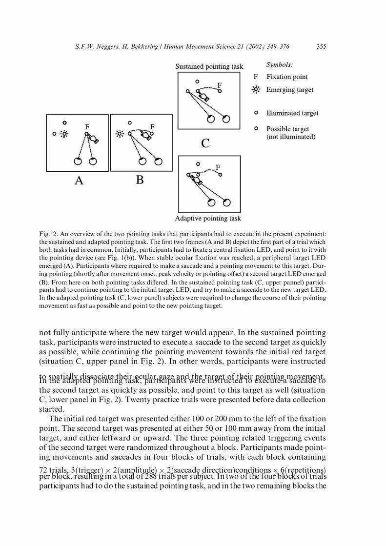
and a new task, the adapted pointing task. See Fig. 2 for a schematic illustration
of both tasks.
In both conditions, a trial started when the participants fixated and touched the
central fixation point that was situated at a central location on the board, approxi-
mately (since neither head nor trunk were fixated) at the body midline. After the eyes
had fixated the central fixation point for 2300 ms, a red target appeared (situation A
in Fig. 2), either 100 or 200 mm to its left, and participants had to initiate a sac-
cade and a pointing movement to that target as fast as possible. After participantsvisually fixated the red target and initiated the pointing movement, a second (green)
target appeared (situation B in Fig. 2) on each trial, except when pointing was too
slow (see the real-time data analysis paragraph below). The appearance of the sec-
ond target was triggered either by the onset, the peak velocity, or the offset of the
hand movement. The latter onset times were only selected in order to fully cover
the movement time of the arm. They were not selected in order to investigate specific
characteristics of kinematic triggering of eye movements during different stages of
the hand pointing movement. The primary objective was to investigate whether sac-
cades were locked to the target of a pointing movement during the entire movement
time.The second target appeared either 100 mm to the left or 100 mm above the first
target fixated by the eyes and approached by the hand, thus participants could
354 S.F.W. Neggers, H. Bekkering / Human Movement Science 21 (2002) 349–376
7/21/2019 Rotary Negger
http://slidepdf.com/reader/full/rotary-negger 7/28
not fully anticipate where the new target would appear. In the sustained pointing
task, participants were instructed to execute a saccade to the second target as quickly
as possible, while continuing the pointing movement towards the initial red target
(situation C, upper panel in Fig. 2). In other words, participants were instructed
to spatially dissociate their ocular gaze and the target of their pointing movement.In the adapted pointing task, participants were instructed to execute a saccade to
the second target as quickly as possible, and point to this target as well (situation
C, lower panel in Fig. 2). Twenty practice trials were presented before data collection
started.
The initial red target was presented either 100 or 200 mm to the left of the fixation
point. The second target was presented at either 50 or 100 mm away from the initial
target, and either leftward or upward. The three pointing related triggering events
of the second target were randomized throughout a block. Participants made point-
ing movements and saccades in four blocks of trials, with each block containing
72 trials, 3ðtriggerÞ 2ðamplitudeÞ 2ðsaccade directionÞconditions 6ðrepetitionsÞper block, resulting in a total of 288 trials per subject. In two of the four blocks of trials
participants had to do the sustained pointing task, and in the two remaining blocks the
Fig. 2. An overview of the two pointing tasks that participants had to execute in the present experiment:
the sustained and adapted pointing task. The first two frames (A and B) depict the first part of a trial which
both tasks had in common. Initially, participants had to fixate a central fixation LED, and point to it with
the pointing device (see Fig. 1(b)). When stable ocular fixation was reached, a peripheral target LED
emerged (A). Participants where required to make a saccade and a pointing movement to this target. Dur-
ing pointing (shortly after movement onset, peak velocity or pointing offset) a second target LED emerged
(B). From here on both pointing tasks differed. In the sustained pointing task (C, upper pannel) partici-pants had to continue pointing to the initial target LED, and try to make a saccade to the new target LED.
In the adapted pointing task (C, lower panel) subjects were required to change the course of their pointing
movement as fast as possible and point to the new pointing target.
S.F.W. Neggers, H. Bekkering / Human Movement Science 21 (2002) 349–376 355

7/21/2019 Rotary Negger
http://slidepdf.com/reader/full/rotary-negger 8/28
adapted pointing tasks. The blocks were ordered according to an ABAB or BABA de-
sign, and this was counterbalanced across subjects.
2.4. Real time data analysis
In the present study and two previous reports (Neggers & Bekkering, 2000, 2001),
the occurrence of a kinematic parameter triggered the onset of the second target.
Onset times of the second target were: movement onset (on), maximum velocity
(vmax), and movement offset (off). The online procedure resulted in approximate
estimates of these kinematic moments (see first paragraph of the results section of
one of our previous studies (Neggers & Bekkering, 2001)), since due to buffering re-
quirements in real time movement processing, the sample frequency of our OPTOT-
RAK system could not exceed 50 Hz for the current experiments. The procedure
used to determine movement kinematics in real-time, that is, during the online re-cording of the pointing movement, is described in detail in a previous study that used
the same setup (Neggers & Bekkering, 2001) with five kinematic moments in time:
the three kinematic moments mentioned above and maximum and minimum decel-
eration. Summarized, a regression line was fitted through the four most recent x-axis
positions and time samples of the finger tip in memory (x, t coordinate pairs). The
slope of this line is an estimate of the current velocity (slightly delayed with at least
40 ms, because we took the last four samples). The same procedure was used to de-
termine current acceleration from the latest four velocity samples. Movement onset
was determined when the current velocity exceeded 0.05 m/s for the first time after
target onset, whereas peak velocity was determined when the velocity exceeded
0.25 m/s and deceleration was determined between )0.01 m/s2 and þ0.01 m/s2 (rever-
sal point). Movement offset was defined as the moment when current velocity
dropped below 0.05 m/s again.
2.5. Off-line data analysis
To analyze performance, a number of movement parameters were calculated off-
line using algorithms, which were developed in our lab (MATLAB 6, R12 scripts).
The tangential hand movement velocity was calculated along the three-dimensionaltrajectory of the tip of the pointing device (Fig. 1(b)) and filtered with a 5th order
recursive 15 Hz Butterworth filter. A velocity threshold of 0.05 m/s was used to de-
tect hand movement onset and offset. Movement onset was defined as the first sam-
ple time at which the tangential velocity exceeded the threshold after stimulus onset,
while movement offset was defined as the first sample time after movement onset at
which the velocity was lower than the threshold. For the analysis of the onset of cor-
rective movements in response to a target change, the velocity was calculated along
the x- and y-axes separately, and also filtered with a 5th order recursive 15 Hz But-
terworth filter (see subsections 3.4 and 3.5). Two different onset thresholds had to be
used for the detection of initial movement onset and corrective movement onset, 0.05m/s for the initial horizontal movement and 0.1 m/s for the vertical corrective com-
ponent. See the Sections 3.4 and 3.5 for more details. Saccadic onset was calculated
356 S.F.W. Neggers, H. Bekkering / Human Movement Science 21 (2002) 349–376

7/21/2019 Rotary Negger
http://slidepdf.com/reader/full/rotary-negger 9/28
as the sample time at which the ocular velocity, measured in degrees relative to the
straight-ahead gaze (in head-referenced coordinates), exceeded 35/s and the ocular
acceleration exceeded 9500/s2. Saccadic offset was defined as the sample time at
which the saccadic velocity and deceleration dropped below these thresholds. The la-tencies of saccades were defined as the time between the second target presentation
and onset of the second saccade. Accordingly, the remaining pointing time (the time
the second target was present during pointing) was defined as the time between 2nd
target onset and pointing movement offset. On average, the real kinematic moment
(onset, peak velocity, etc.) preceded the moments that triggered target onset (as de-
termined by the real-time trigger procedure) by 90 ms (see also Neggers & Bekkering,
2001).
Statistical analyses were performed on the movement parameters using a three-
way analysis of variance with repeated measures with the factors TRIG (the three
different moments of triggering the second target by the hand movement), TASK(sustained or adapted) and AMP (the 100 and 200 amplitude between fixation and
the first target). Trials were excluded from analysis in which latencies of saccades
(to the initial target) or manual responses were less than 100 ms or larger than
700 ms.
3. Results
3.1. Typical trials
In Fig. 3, eye and hand position data of six single trials is plotted against time.
The data displayed here is representative for the data of all trials for all participants.
Fig. 3. Eye and hand position data of six single trials is plotted with respect to time. The upper three pan-
els (A, B, C), show data in the sustained pointing task, for trials where the 2nd target appeared around
hand movement onset, peak velocity and offset (from left to right). The lower three panels (D, E, F) show
movement data from the adapted pointing task, for the same trigger types.
S.F.W. Neggers, H. Bekkering / Human Movement Science 21 (2002) 349–376 357

7/21/2019 Rotary Negger
http://slidepdf.com/reader/full/rotary-negger 10/28
The upper three panels (A, B, C) show data from the sustained pointing task, for tri-
als in which the appearance of the second target was triggered by hand movement
onset, peak velocity and offset, respectively. It is clearly visible that saccades were
delayed until the pointing movement had finished. The lower three panels (D, E,F) show similar data but now from the adapted pointing task. It can be clearly seen
that under these circumstances saccades were present with close-to-normal reaction
times during hand movements that changed their course on-line ().
3.2. Saccadic reaction times
In general, saccades in the sustained pointing condition were delayed during
pointing to a target fixated with the eyes until the arm movement ended. That is,
saccadic RTs increased the earlier the second target is presented during pointing
(TRIG: F ð18; 2Þ ¼ 281:3, p < 0:01). As can be seen in Fig. 4, the saccadic RTs ob-served in the adapted pointing task (right panels) differed from those observed
in the sustained pointing task (left panels) with respect to the time during which
the saccade was evoked during pointing. Interestingly, in the adapted pointing task,
Fig. 4. Average (10 participants) reaction times of saccades to the 2nd target (filled squares) and the re-
maining pointing time (open squares, the time between the appearance of the second target and pointing
movement offset) for both pointing tasks. Data from the adapted pointing task is depicted in the right pan-
els, the sustained pointing task in the left panels. Upper panels show data for large pointing amplitudes
(200 mm), lower panels for small amplitudes (100 mm). Error bars denote 2 standard deviation from
top to bottom.
358 S.F.W. Neggers, H. Bekkering / Human Movement Science 21 (2002) 349–376

7/21/2019 Rotary Negger
http://slidepdf.com/reader/full/rotary-negger 11/28
the saccades for 2nd target trigger times at movement onset and peak velocity were
delayed (with respect to saccades triggered by targets offered after movement offset,
considered normal saccades), with 83 and 57 ms for 200 mm pointing amplitudes,
respectively (60 and 23 ms for 100 mm pointing amplitudes). In the sustained point-ing task, however, the saccades for second-target trigger times at movement onset
and peak velocity had significantly larger delays (with respect to saccades to targets
triggered at movement offset), namely 298 and 166 ms for trials with 200 mm point-
ing amplitude, respectively (233 and 107 ms for trials with 100 mm pointing ampli-
tude). On average saccades to the 2nd target did not occur until pointing ended, and
the saccadic delay was proportional to the time the 2nd target was presented during
pointing. The latter was confirmed statistically in a previous paper aimed at testing
that hypothesis (Neggers & Bekkering, 2000), as a significant correlation between re-
maining pointing time (after the occurrence of the 2nd target) and saccadic delay,
on a trial by trial basis. Separate ANOVAs on data obtained in the sustained andadapted pointing task confirmed that in the sustained pointing task saccadic RTs
were significantly different for 2nd targets offered at different times during pointing
(TRIG: F ð18; 2Þ ¼ 313; 4, p < 0:01) as well as in the adapted pointing task (TRIG:
F ð18; 2Þ ¼ 24:3, p < 0:01). The observation mentioned above that saccades to the
2nd target were delayed much longer is in the sustained pointing condition as com-
pared to the adapted condition was statistically confirmed by the interaction between
trigger time of the 2nd target and pointing task (TRIGTASK: F ð18; 2Þ ¼ 93:04,
p < 0:01).
For larger pointing amplitudes in the sustained pointing task (200 mm com-
pared to 100 mm), the slowing of saccades during pointing increased (TRIGAMP:
F ð18; 2Þ ¼ 6:65, p < 0:01). The two separate ANOVAs revealed that this was only
the case in the sustained pointing task (TRIGAMP: F ð18; 2Þ ¼ 5:67, p < 0:02)
and not in the adapted pointing task (TRIGAMP: F ð18; 2Þ ¼ 1:38, p ¼ 0:28).
3.3. Pointing movement time
There was a main effect on pointing movement duration of the time that the 2nd
target was presented during pointing (TRIG: F ð18; 2Þ ¼ 58:4, p < 0:01). See Fig. 5for the average movement durations in both tasks. In Fig. 6(a) and (b), the mean ve-
locity profile for hand movements is plotted for both the sustained and the adapted
pointing task, aligned ðt ¼ 0Þ at hand movement onset, for trials where the 2nd target
was offered at hand movement onset (Fig. 6(a)) and peak velocity (Fig. 6(b)), respec-
tively. Hand movement duration was prolonged in the adapted compared to the
sustained pointing condition, probably because movement corrections had to take
place in the adapted movement condition, resulting in larger pointing amplitudes
(TRIGTASK: F ð18; 2Þ ¼ 52:3, p < 0:01). A separate ANOVA was conducted on
the data of the sustained pointing task and the adapted pointing task. In the sustained
pointing task, no such influence of 2nd target presentation on pointing duration wasobserved (TRIG: F ð18; 2Þ < 1) as had been reported earlier (Neggers & Bekkering,
2000, 2001). However, in the adapted pointing task there was a substantial slowing
S.F.W. Neggers, H. Bekkering / Human Movement Science 21 (2002) 349–376 359

7/21/2019 Rotary Negger
http://slidepdf.com/reader/full/rotary-negger 12/28
of the pointing movements when the 2nd target for the pointing movement was
offered earlier during the movements (TRIG: F ð18; 2Þ ¼ 57:4, p < 0:01).
3.4. Movement corrections in the adapted pointing task
In Fig. 6(a), the average velocity profiles along the x-axis for hand movements
in the adapted (filled circles) and the sustained pointing task (open circles) are plot-
ted against time for saccadic targets offered at hand movement onset, for each sub-
ject separately. Single trial traces were aligned at pointing movement onset time
ðt ¼ 0Þ in order to obtain the averages plotted in Fig. 6(a). In order to be able to
compare the effect on movement corrections of the displacement in pointing target
position in the adapted movement task, only the 200 mm pointing amplitudes were
analyzed, with pointing corrections in the same direction as the initial pointing
movement (to the left). It is clearly visible that in the adapted pointing condition,
most subjects were able to correct their pointing movement in flight, i.e. with a ve-locity that never dropped to zero before the hand movement reached the initial target
location, when the 2nd target appeared around pointing movement onset.
Fig. 5. Average (10 participants) durations of the complete pointing movements in both pointing tasks.Data from the adapted pointing task is depicted in the right panels, the sustained pointing task in the left
panels. Upper panels show data for large pointing amplitudes (200 mm), lower panels for small amplitudes
(100 mm). Error bars denote 2 standard deviation from top to bottom.
360 S.F.W. Neggers, H. Bekkering / Human Movement Science 21 (2002) 349–376

7/21/2019 Rotary Negger
http://slidepdf.com/reader/full/rotary-negger 13/28
The data are plotted for all subjects separately, since different strategies were ob-
served in how subjects correct pointing movements to the new target position. Cor-
rections could be observed after movement onset and were best indicated in the
velocity profiles, which showed a plateau or dip before going up again. The plateau
was either preceded by a velocity increase, when the correction takes place before
peak velocity, or by a decrease, when occurring after peak velocity. This plateau
was sometimes visible before peak velocity, but mostly after peak velocity. Obvi-
ously, most subjects corrected relatively late during pointing (sa, b2, me1, sn, me2,
a3, rt and ri), that is, after peak velocity occurred, although some subjects were ableto correct fairly early during the pointing movement (m2, p2), even before peak
velocity occurred. Some subjects decreased their velocity more during a correction
Fig. 6. (a) For trials where the 2nd target appearance (leftward only, pointing amplitude 200 mm) was
triggered by hand movement onset, the hand movement velocity component along the x-axis was averaged
over single trials for each participant and pointing condition separately. Averaging was done after aligning
at hand movement onset ðt ¼ 0Þ. The velocity traces are plotted against time in a separate panel for each
subject. The sustained pointing task average velocity is plotted with open circles and the adapted point-
ing task average velocity with closed circles. Vertical solid and dashed lines denote the average onset
time of the 2nd saccade in the adapted and the sustained pointing task, respectively. (b) Same plots as
in (a) for trials where 2nd targets were offered around pointing peak velocity. The letters in the upper right
corner of each panel denote the participant whose data was depicted in that panel (anonymous abbrevi-
ation).
S.F.W. Neggers, H. Bekkering / Human Movement Science 21 (2002) 349–376 361

7/21/2019 Rotary Negger
http://slidepdf.com/reader/full/rotary-negger 14/28
(b2, rt, me2), while other subjects altered the course of their pointing movement
more smoothly and at a higher velocity.
In addition, as can be seen in Fig. 6(a) and (b), it appeared that the average ve-
locity in the adapted pointing conditions for some subjects (most notably m2 and
sn) was lower than in the sustained pointing task. Interestingly, this effect was al-
ready present in the first half of the movement even when the target changed position
later during the movement. 1
No quantitative analysis for horizontal movement corrections on the time of
pointing movements was performed, in view of the different criteria needed to detect
these plateaus in each subject. For vertical movement corrections a quantitative
comparison could be performed more easily, because, in this case, the velocity com-
ponent along the axis along which the correction took place (the y-axis) could be an-
alyzed separately from the initial movement (x-axis). For 2nd target presentation
Fig. 6 (continued )
1 A possible explanation for this finding might be that participants anticipate to some degree that a
displacement will occur in that particular block (pointing conditions were blocked), and, since they could
not know where the target would be displaced, decided to move slower in order to be better able to correct
the movements end location.
362 S.F.W. Neggers, H. Bekkering / Human Movement Science 21 (2002) 349–376

7/21/2019 Rotary Negger
http://slidepdf.com/reader/full/rotary-negger 15/28
times at peak velocity and movement offset subjects often were not able to correct
on-line, they first halted close to the initial target and then corrected to the 2nd tar-
get. For the 2nd targets offered around pointing movement offset it is of course triv-
ial that two separate movements were observed. When 2nd targets were offeredaround pointing peak velocity, the planned movement was apparently unfolded to
such an extent that correction could not take place any more in the remaining move-
ment time in most subjects, although some subjects (m2, p2 and sn) managed even
here to alter the course of their hand movements without the velocity dropping to
zero.
3.5. Relationship between time of pointing movement correction and saccadic RT
3.5.1. Horizontal target displacements
In Fig. 7(a), the same hand velocity data as in Fig. 6(a) is plotted, but now alignedat the time of saccade onset, for all trials with 2nd target presentation (triggered by
hand movement onset, pointing amplitude 200 mm, saccades to the left). The eye po-
sition is plotted in the background as thin gray lines. As a result, a clearer plateau or
dip was observed in the mean hand movement traces, around t ¼ 0, in Fig. 7. For
example, compare the subjects me1, sn, rt and ri, where a shallow dip or a plateau
in Fig. 6(a) changes in a clear velocity dip in Fig. 7(a).
For all participants, the plateau or dip in hand movement velocity seems to occur
around the time the saccade is made, although the corrections took place at different
moments during the reaching movements for different participants. The latter is an
argument in favor of a coupling of ocular gaze to the target of a (changing) pointing
movement.
3.5.2. Vertical target displacements
For vertical target displacements in the adapted pointing condition, first the same
analysis as for the horizontal target displacement was performed. Arm movement ve-
locity along the y-axis (since in that dimension the correction can be expected for ver-
tical target displacements) was calculated, and averaged aligned to the onset of the
saccade to the 2nd target (see Fig. 8).
It can be clearly seen that the component along the y-axis of the velocity of theinitial movement began to increase shortly after the saccade to the new target had
occurred. The correction movements of the arm were analyzed on a single trial basis.
First, for each trial the onset of the initial movement, t(on)HOR, was calculated with a
velocity threshold of 0.05 m/s for the velocity along the x-axis. Then, the onset of the
corrective arm movement, t(on)VERT, was calculated with a threshold of 0.10 m/s for
the velocity along the y-axis (perpendicular to the initial movement). By doing so, the
onset for horizontal and vertical movement components could be found separately
and for each individual trial. As a second criterion the velocity had to reach 0.2
m/s within 140 ms, in order to rule out short supra-threshold movements along
the in the y-axis that were sometimes made during the initial reaching movement,and find the real corrective movement as an answer to the target displacement. Fi-
nally the time of the saccade to the 2nd target, t(on)SACC, was calculated. The time of
S.F.W. Neggers, H. Bekkering / Human Movement Science 21 (2002) 349–376 363

7/21/2019 Rotary Negger
http://slidepdf.com/reader/full/rotary-negger 16/28
the 2nd saccade to the new target position with respect to the onset of the initial arm
movement t(on)HOR was correlated with the onset time of the vertical correction
component t(on)VERT (also with respect to t(on)HOR) on a trial-by-trial basis. The
ft ðonÞSACC t ðonÞHORg ft ðonÞVERT t ðonÞHORg data pairs are shown in Fig. 9(a)
for all trials with the initial target at 200 ms to the left and 2nd targets with vertical
displacements that were triggered at movement onset. A clear correlation can be ob-
served for most subjects (significant for 6 out of 10 subjects, see Fig. 9(a)), the mean
Fisher-Z corrected correlation was 0.85. The latter statistics confirmed the hypothe-
sis that on-line correction of the hand (with respect to movement onset) correlates in
time with the saccade to the new target. In Fig. 9(b) the data pairs for all trials of allparticipants are plotted, also showing a clear co-variation of the onset of the move-
ment correction in the vertical direction and the onset of the 2nd saccade.
Fig. 7. (a) For each participant separately, the hand movement velocity component along the x-axis in the
adapted pointing task is plotted for single trials (thin black lines), aligned ðt ¼ 0Þ at the time the 2nd saccade
was initiated. The single trials were then averaged (line with closed circles). Also the x-coordinates of the
position of the left eye are plotted in the same graph, also aligned to saccade onset (thin gray lines). Only
data is shown for trials where the 2nd target was offered around hand movement onset at the left of the ini-
tial target position, for 200 mm initial pointing amplitudes. (b) Same plots as in (a) for trials where 2nd tar-
gets were offered around pointing peak velocity. The letters in the upper right corner of each panel denote
the participant whose data was depicted in that panel (anonymous abbreviation). Note that the movement
correction dips are more profound as in graph 6, where data was aligned at hand movement onset.
364 S.F.W. Neggers, H. Bekkering / Human Movement Science 21 (2002) 349–376

7/21/2019 Rotary Negger
http://slidepdf.com/reader/full/rotary-negger 17/28
4. Discussion
In the present study, we integrated two recent, at first sight contradictory findings,
regarding the question whether saccadic eye movements can be generated to a newly
presented target during an ongoing hand movement. That is, in the adapted pointing
condition, when subjects had to direct both their gaze and arm movements to a dis-placed target location, it was found that the eyes could fixate the new target during
the arm movement. In addition, a temporal coupling of these corrective saccades was
found with changes in arm movement trajectories when reaching to the new target.
In the sustained pointing condition, however, when the same subjects had to point to
the initial target, while trying to deviate their gaze to a new target that appeared dur-
ing pointing, it was found that the eyes could not fixate the new target before the
hand has reached the initial target location. Together, these results indicate that
ocular gaze is always forced to follow the target intended by a manual arm movement.
Importantly, the results show that gaze anchoring during pointing is not the result of
a general inability in generating saccades during any arm movement, but more spe-cifically resulting from a mechanism coupling ocular gaze to a pointing target, as pre-
viously suggested (Neggers & Bekkering, 2000, 2001). Such a mechanism delays
Fig. 7 (continued )
S.F.W. Neggers, H. Bekkering / Human Movement Science 21 (2002) 349–376 365

7/21/2019 Rotary Negger
http://slidepdf.com/reader/full/rotary-negger 18/28
saccades to targets that appear during pointing, when the pointing movement main-
tains its course towards the initial target stimulus (sustained pointing condition).
Moreover, a mechanism as proposed above can trigger saccades to a new target po-sition appearing during pointing when the arm is required to change its course in
flight and move to the new target (adapted pointing condition). In other words,
ocular gaze is always forced to follow the target intended by a manual movement,
either when that target position dynamically changes during the arm movement or
stays fixed.
In addition, pointing movements in the adapted pointing condition were pro-
longed as a result of the movement correction that almost always occurred on-line
when the target was displaced at movement onset. The latter was concluded from an-
alyzing the velocity profiles along the x-axis of trials requiring arm movement cor-
rections in the same direction as the initial movement. When the second targetappeared around peak velocity, participants did not always succeed in changing
the pointing movement on-line and continued to point to the old target stimulus.
Fig. 8. For each participant separately, the hand movement velocity component along the y-axis in the
adapted pointing task is plotted for single trials (thin black lines), aligned ðt ¼ 0Þ at the time the 2nd sac-
cade was initiated. The single trials were then averaged (line with closed circles). Also the y-coordinates of
the position of the left eye are plotted in the same graph, also aligned to saccade onset (thin gray lines).
Only data is shown for trials where the 2nd target was offered around hand movement onset at the left of
the initial target position, for 200 mm initial pointing amplitudes.
366 S.F.W. Neggers, H. Bekkering / Human Movement Science 21 (2002) 349–376

7/21/2019 Rotary Negger
http://slidepdf.com/reader/full/rotary-negger 19/28
Fig. 9. (a) The onset time of the 2nd saccade to the new target position, t(on)SACC, with respect to the onset
of the initial arm movement, t(on)HOR, was correlated with the onset time of the vertical correction com-
ponent t(on)VERT (also with respect to t(on)HOR) on a trial-by-trial basis. For each participant separately,
the (t ðonÞSACC t ðonÞHOR, t ðonÞVERT t ðonÞHOR) data pairs are shown as dots for all trials with the initial
target at 200 ms to the left and 2nd targets with vertical displacements that were triggered at movement
onset. The Pearsons product moment correlation coefficient r was calculated for each subject separately,
and plotted in the lower right corner of the graph of that participant. When the alternative hypothesis
r > 0 (zero hypothesis r ¼ 0) could be confirmed on a 0.05 confidence interval, a was plotted after the
correlation coefficient for that participant. The average r (Fisher-Z corrected) was 0.84. (b) An overview
of the data pairs pooled for all trials and participants shown in (a). A clear increase of the onset times of saccades to the 2nd target with the onset time of the hand movement corrections 0 can be observed.
S.F.W. Neggers, H. Bekkering / Human Movement Science 21 (2002) 349–376 367

7/21/2019 Rotary Negger
http://slidepdf.com/reader/full/rotary-negger 20/28
The movement corrections, defined as dips or plateaus in the velocity profile, were
spaced differently throughout the movement for different subjects. Some subjects
changed the course of their movements shortly after the displacement, before peak
velocity. Others changed the course of the pointing movement only when the handwas already decelerating. Interestingly, when 2nd targets were offered around point-
ing movement onset, saccades were generally made around the time the hand move-
ment corrected its course on-line, as becomes visible when aligning the arm
movement velocity profiles to 2nd saccade onset (Fig. 9(a)). A clear dip in hand
movement velocity was observed around the time of 2nd saccade onset. Further-
more, when analyzing movement corrections perpendicular (here: vertical) to the ini-
tial movement direction (here: horizontal), the moment of onset of the corrective
hand movement can be determined separately from the initial movement onset.
The time of occurrence of the 2nd saccade appeared to be correlated on a trial-
by-trial basis with the time the movement correction was observed. The latter obser-vation strongly supports a direct interaction between hand motor control processes
and saccade execution processes, probably using a coupling of the movement targets
of both systems.
4.1. Neurophysiology of eye and arm movements
In order to further specify a possible implementation of the observed coupling be-
tween ocular gaze and arm movements, we examined the neural substrates involved
in controlling eye and arm movements. A neural mechanism coupling ocular gaze
and aiming movements should combine a non-visual dynamic (i.e., activity co-vary-
ing with the process of moving the arm) internal representation, or neural map, of
the target of an arm movement with a map representing the target of an eye move-
ment. The map containing the reach target should be dynamic since the coupling
process is mainly observed shortly before and during an arm movement (Neggers
& Bekkering, 2000, 2001; Prablanc et al., 1979; Prablanc & Martin, 1992). A neural
map containing instantaneous hand or limb position instead of target position (i.e.,
the time-varying position of the moving hand) would not be suitable, since imposing
it on the saccadic system would cause subjects to look at their moving hand during
aiming movements, which has not been observed. Furthermore, in order to be ableto interact directly, both maps should have the same reference frame. Finally, the sig-
nal coded in the neural map of reach targets should be of non-visual nature, since the
coupling of gaze to the reach target is also present when no visual information of the
arm or hand is available (Lunenburger et al., 2000; Neggers & Bekkering, 2001).
From the latter it cannot be concluded whether the source for the reach signal is
an afferent or efferent signal related to arm movement control.
Various complete descriptions of oculomotor maps have been obtained by means
of single cell recordings in primate cortical and subcortical areas. Some of these maps
turned out to be coded in an oculocentric reference frame (representing the eye-move-
ment target with respect to current gaze, a signal often referred to as the motor error):the supplementary eye fields (SEF) (Schlag & Schlag-Rey, 1985), the frontal eyefields
(FEF) (Bruce & Goldberg, 1985; Bruce, Goldberg, Bushnell, & Stanton, 1985) and the
368 S.F.W. Neggers, H. Bekkering / Human Movement Science 21 (2002) 349–376
7/21/2019 Rotary Negger
http://slidepdf.com/reader/full/rotary-negger 21/28
midbrain superior colliculus (SC) (Robinson, 1972; Schiller & Koerner, 1971; Sparks,
1986; Wurtz & Goldberg, 1971, 1972). The lateral intraparietal sulcus (LIP) has also
been reported to be involved in the programming of saccades (Andersen, Essick, &
Siegel, 1987; Colby, Duhamel, & Goldberg, 1996). However, recent studies concludedthat it is more likely that LIP neurons signal the occurrence of visual events and atten-
tional shifts as well as perisaccadic remapping of space throughout saccades (Colby,
Duhamel, & Goldberg, 1995; Powell & Goldberg, 2000), than being directly included
in motor control processes.
Hand movement related modulation of neurons has been observed in three of the
aforementioned oculomotor centers, the SEF (Mushiake, Fujii, & Tanji, 1996) and
the SC (Stuphorn, Bauswein, & Hoffmann, 2000; Werner, Dannenberg, & Hoffmann,
1997; Werner, Hoffmann, & Dannenberg, 1997) and to a smaller extent in LIP where
activity of some neurons is related to the planning phase of arm movements (Snyder,
Batista, & Andersen, 1997). The activity of saccade-related neuronal discharge of SEF neurons is modulated by the occurrence of an accompanying hand movement
(some neurons do only discharge when an additional arm movement is made, others
only for saccades in isolation (Tanji, Shima, & Mushiake, 1996)). In the SC, however,
a more specific pattern of hand movement related activity has been observed. So
called reach-neurons have been observed, that discharge during the movement pro-
portional with the arm muscles EMG activity (Werner et al., 1997; Werner et al.,
1997). In more recent work (Stuphorn et al., 2000) it was shown that the discharge
of SC reach neurons is coded in an oculocentric reference frame (i.e. with respect to
the actual gaze position) for 40% of SC reach-related neurons, mainly lying in inter-
mediate SC layers intermingled with the saccadic neurons. The neurons had a pre-
ferred direction (i.e. they discharged maximally) for movements with a specific
direction and amplitude with respect to the current gaze. Neurons deeper in the
SC (the remaining 60% of reach neurons) seemed to discharge in relation to arm
movements irrespective of ocular gaze.
Interestingly, reach neurons in SC are probably activated by motor-related areas,
via projections from the (pre)motor cortex (Fries, 1984, 1985; Werner et al., 1997;
Werner et al., 1997), a signal which is probably of a non-visual nature and could re-
flect an efference copy of signals related to arm movement control. The observed
gaze anchoring during pointing is also internally driven, that is, independent of whether the moving hand could be seen or not, as was shown previously (Neggers
& Bekkering, 2001). In sum, like the saccadic SC neurons, the SC reach neurons
fire in an oculocentric reference frame, and are driven by some non-visual sig-
nal, complying with all the conditions necessary for coupling the gaze to the tar-
get of a reach. The SC might therefore be well suited to implement the coupled
behavior of eyes and arm during arm movements, when the saccadic and reach-
related SC neurons interact. Some evidence has been reported that show that SC
intermediate-layer neurons are interconnected by inhibitory interneurons (Meredith
& Ramoa, 1998).
Interestingly, recent additional evidence has been reported suggesting the exis-tence of a direct coupling between arm movement control signals and saccadic con-
trol. That is, saccadic peak velocity was found to be larger when an accompanying
S.F.W. Neggers, H. Bekkering / Human Movement Science 21 (2002) 349–376 369
7/21/2019 Rotary Negger
http://slidepdf.com/reader/full/rotary-negger 22/28
arm movement was made, as compared with saccades that were made in isolation
(Snyder, Calton, Dickinson, & Lawrence, 2002). Also, this study showed that saccade
dynamics deviates from the well-known main sequence function in saccade-arm
movement tasks. The main sequence function describes a fixed relationship betweenpeak velocity and amplitude for saccades made in isolation (Bahill, Clark, & Stark,
1975; Fuchs, Kaneko, & Scudder, 1985). Interestingly, the deviation from the main
sequence as observed for saccades accompanied by arm movements by Snyder et al.
(2002) was only present for saccades and arm movements to the same goals, and not
when the eye and arm moved to different regions. The latter coupling also suggests a
correspondence of reference frames of the interacting eye–arm circuitry in structures
close to motor control, and the authors also render it likely that the SC is involved in
the observed behavior.
4.2. A possible coupling mechanism
At present, there exist several (system theoretical) quantitative models of primate
SC and brainstem that produce accurate simulations of saccades in response to vi-
sual stimuli. Most of these models exploit the original idea of a feedback mechanism
minimizing motor error, the distance between present gaze and desired target (Van
Gisbergen, Robinson, & Gielen, 1981). Also, distributed models (with some kind
of a neuronal map), including the SC itself in the feedback loop, have appeared that
translate the SC saccadic motor map (a set of neurons that form a map) into a one-
dimensional signal (Van Opstal & Kappen, 1993). Consequently, these models were
able to simulate known saccadic dynamics for saccades to a single target. Neverthe-
less, it remains an open question whether the SC is indeed included in the feedback
loop for saccadic control, or that it merely supplies the input signal to it, although
recent evidence seems to support the former probability (Soetedjo, Kaneko, & Fuchs,
2002). However, the lateral (inhibitory) interactions within the SC motor map (Mer-
edith & Ramoa, 1998) are not taken into account in the latter model, and without
such an interaction, saccades to one out of multiple competing targets are hard to
predict. Other models have been proposed that do take these lateral interactions into
account, and can hence simulate behavior in multiple target configurations (Das,
Keller, & Arai, 1996; Massone, 1994a,b).The latter model would be relevant for the type of mechanism presented here,
where in addition to visual input, a reach target could be imposed onto the saccadic
map, in order to obtain competition between the reach target and other (instructed)
target regions. In the scheme below (Fig. 10) an existing distributed SC-model with
lateral interactions (Das et al., 1996) was altered in that reach activity (in oculocen-
tric coordinates) is added to the SC motor layer (of course also coding for saccades
in oculocentric coordinates, as is well known).
The activity of a SC reach neuron, when a reach is made into its movement field,
is assumed to be proportional to the arm movement velocity, and shifted in time with
d ms, where d is in the order of 40 ms. The latter is a rough approximation of the SCreach neuron activity, which is known to be correlated to arm muscle EMG (Stup-
horn et al., 2000; Stuphorn, Hoffmann, & Miller, 1999; Werner et al., 1997). In a
370 S.F.W. Neggers, H. Bekkering / Human Movement Science 21 (2002) 349–376

7/21/2019 Rotary Negger
http://slidepdf.com/reader/full/rotary-negger 23/28
cross-correlational analysis, it was shown that SC reach activity on average precedes
arm muscle EMG with 40 ms (Stuphorn et al., 1999). Also, it is assumed that the
neurons are ordered in an anatomical map, where neighboring neurons code for
reaches to neighboring locations in space. The latter hypothesis has not been con-
firmed so far in single cell studies, in fact no clear ordering has been observed (Stup-
horn et al., 2000). Nevertheless, when the projection matrix between the proposed
reach layer in Fig. 10 and the SC motor layer is not the unity matrix (i.e., not map-ping the reach neuron map onto the SC motor map in a one to one fashion), this
problem may be overcome. At least the mapping between the reach layer and
Fig. 10. A schematic drawing of the model approach as used by Das et al. (1996), which successfully gen-erated saccades to visual targets in simulations, with an additional reach layer. The figure is redrawn from
Das et al. (1996, Fig. 1). A reach layer with neurons that code the target of a reach in oculocentric coor-
dinates, as have been observed in primate SC (see Section 4), directly imposes its signal onto the SC motor
layer, known to generate saccades. It is assumed that the temporal activity profile of a reach neuron, when
a reach is made into its movement field, roughly follows the hand movement velocity profile, slightly
shifted forward in time with d ms (d 40 ms), which has also been observed in primate SC. Horz, Vert
BG: model of burst generators, a premotor brainstem structure driving oculomotor plant, existing of sep-
arate systems for horizontal and vertical eye movements. OPN: omnipause neurons, active during fixa-
tions, are though to prevent BG from executing saccades. mt, mh and mv: driving motor signals for
omnipause neurons, horizontal and vertical horizontal burst generators. E h and E v: horizontal and vertical
eye position. I: general inhibitory signal, when inactive it releases SC to generate an eye movement. For
details on the model, see Das et al. (1996).
S.F.W. Neggers, H. Bekkering / Human Movement Science 21 (2002) 349–376 371

7/21/2019 Rotary Negger
http://slidepdf.com/reader/full/rotary-negger 24/28
saccade layer should result in overlapping movement fields (neurons with a preferred
movement vector of an arm movement goal with respect to gaze projects to saccadic
neurons coding for the same movement vector with respect to gaze).
The resulting model should be able to reproduce the effects found in the presentstudy, as well as findings from several other eye–hand coordination studies. First,
when the eyes fixate a target that is being reached to, the reach target in oculocentric
coordinates is close to the 0-vector (reach target is foveal). This should put extra ac-
tivity on the so-called SC fixation neurons. Such fixation neurons can be represented
by a 0-vector in the SC motor-map, representing a neuron firing for a saccade with
an amplitude of 0, or fixation. Such neurons have indeed been observed in the rostral
zone of the macaque SC (Munoz & Wurtz, 1993a). Stimulating SC fixation neurons
with a microelectrode is known to delay or even interrupt a saccade (Munoz &
Wurtz, 1993b). Adding the extra activity for reach neurons coding a 0-vector to
the SC fixation zone would then predict that saccades during reaching to a target fix-ated with the eyes are suppressed, as has been observed in previous studies (Neggers
& Bekkering, 2000, 2001).
Second, whenever retargeting in the reach layer would take place as a result of a
hand movement triggered target displacement, the reach target in oculocentric coor-
dinates is not the 0-vector anymore (i.e. now the reach target is not fixated with the
eyes). Hence, the reach layer would impose activity on the SC motor map at a loca-
tion triggering exactly the saccade to the new reach target position, as soon as the
retargeting in the reach layer occurred. The latter behavior has been observed in
the adapted pointing task in the present study.
Third, when intending to start a single reach to a single target, rising reach activity
in oculocentric coordinates in the models reach layer would also trigger a saccade to
that target. This situation is drawn in Fig. 10 as the current situation, the eyes are
directed at a starting location, the arm is about to start moving to the target region.
The reach target in oculocentric coordinates is drawn as a black dot. Any intended
saccade to another region (painted gray) would be suppressed. Since we assumed
that SC reach activity starts rising shortly before arm movement initiation, this
would imply a saccade to the target shortly before an arm movement to that target,
where eye and hand initiation times are correlated. The latter has in fact been ob-
served in many single target eye–hand movement studies in the past (Frens & Erke-lens, 1991; Gielen et al., 1984; Neggers & Bekkering, 1999; Prablanc et al., 1979).
Finally, when assuming that the SC is placed in the feedback loop controlling sacc-
adic eye movements (Soetedjo et al., 2002), the schematic model presented above can
also explain why saccades deviate from the mean sequence when a concurrent arm
movement is made (Snyder et al., 2002). When an SC reach neuron, becoming active
shortly before an arm movement, adds activity to an SC saccade neuron (which is
only true for the above model when the targets for both movements coincide), the
usual burst of activity going in the feedback controller would be higher, possibly re-
sulting in a larger peak velocity. Interestingly, it was indeed observed (Snyder et al.,
2002) that the deviation of saccadic peak velocity from the main sequence is onlypresent for eye and arm movements to the same target, as the above model would
predict.
372 S.F.W. Neggers, H. Bekkering / Human Movement Science 21 (2002) 349–376

7/21/2019 Rotary Negger
http://slidepdf.com/reader/full/rotary-negger 25/28
In the model proposed here, basically the eyes would be forced to go to the loca-
tion that is the current target for the manual system, whatever that is, by means of a
copy of reach activity presumably from (pre)motor regions to the SC in the proper
reference frame. The oculocentric reach neurons in the SC would therefore sub serveeye–hand coordination, and not arm movement control itself.
It has to be noted that there are various other input signals to the SC, for example
directly from the FEF (Hanes & Wurtz, 2001; Sommer & Wurtz, 1998, 2000) or by
means of inhibitory inputs from the substantia nigra (Hikosaka & Wurtz, 1983). It is
thought that such inputs can suppress the execution of reflexive visually guided sac-
cades through the SC, or enhance volitional saccades to targets that are not visually
salient (Guitton, Buchtel, & Douglas, 1985; Schiller, Sandell, & Maunsell, 1987;
Schiller, True, & Conway, 1980). Taking the latter into account, it can never be ex-
cluded that the coupling of ocular gaze to a reaching target will be broken up in com-
plex environments where other regions compete with the target of an arm movementfor the attraction of ocular gaze. Nevertheless, by assuming an interaction of reach-
target signals within the SC motor map, the target of a reaching movement gains in
importance as an attractor for the oculomotor system.
In general, the present experiment and movement analyses support the existence
of a coupling between reaching movements and saccades on the level of target rep-
resentations relative to gaze. Fields representing such targets are known to exist in
the primate SC, which makes it a primary site for the implementation of such a neu-
ral mechanism. Therefore, further research on this site and other relevant brain
structures is required to investigate whether the assumptions of the proposed model
are neurobiologically plausible. Furthermore, a quantitative implementation of the
qualitative model presented here is required.
Acknowledgements
The authors thank Julian Garbotz for his assistance in conducting the experi-
ments, Fiorello Banci, Karl-Heinz Honsberg and Henryk Milewski for technical sup-
port. We are also grateful to Peter Beek, Piet van Wieringen and two anonymous
reviewers for helpful comments on earlier versions of this paper. This study was sup-ported by grant no. BE 1873/2 of the German Research Foundation (DFG), as part
of the interdisciplinary project ‘‘Sensorimotor Integration’’, and by grant no. 440-20-
000 from the Dutch Organization for Fundamental Research (NWO).
References
Andersen, R. A., Essick, G. K., & Siegel, R. M. (1987). Neurons of area 7 activated by both visual stimuli
and oculomotor behavior. Experimental Brain Research, 67 , 316–322.
Bahill, A. T., Clark, M. R., & Stark, L. (1975). The main sequence: a tool for studying human eye
movements. Mathematical Biosciences, 24, 191–204.
Bekkering, H., Adam, J. J., van den Aarssen, A., Kingma, H., & Whiting, H. T. (1995). Interference
between saccadic eye and goal-directed hand movements. Experimental Brain Research, 106 , 475–484.
S.F.W. Neggers, H. Bekkering / Human Movement Science 21 (2002) 349–376 373
7/21/2019 Rotary Negger
http://slidepdf.com/reader/full/rotary-negger 26/28
Bruce, C. J., & Goldberg, M. E. (1985). Primate frontal eye fields. I. Single neurons discharging before
saccades. Journal of Neurophysiology, 53, 603–635.
Bruce, C. J., Goldberg, M. E., Bushnell, M. C., & Stanton, G. B. (1985). Primate frontal eye fields. II.
Physiological and anatomical correlates of electrically evoked eye movements. Journal of Neurophy-
siology, 54, 714–734.
Colby, C. L., Duhamel, J. R., & Goldberg, M. E. (1995). Oculocentric spatial representation in parietal
cortex. Cerebral Cortex, 5, 470–481.
Colby, C. L., Duhamel, J.-R., & Goldberg, M. E. (1996). Visual, presaccadic, and cognitive activation
of single neurons in monkey lateral intraparietal area. Journal of Neurophysiology, 76 , 2841–
2852.
Das, S., Keller, E. L., & Arai, K. (1996). A distributed model of the saccadic system: the effects of internal
noise. Neurocomputing , 245–269.
Frens, M. A., & Erkelens, C. J. (1991). Coordination of hand movements and saccades: evidence for a
common and a separate pathway. Experimental Brain Research, 85, 682–690.
Fries, W. (1984). Cortical projections to the superior colliculus in the macaque monkey: a retrograde study
using horseradish peroxidase. Journal of Comparative Neurology, 230, 55–76.Fries, W. (1985). Inputs from motor and premotor cortex to the superior colliculus of the macaque
monkey. Behavioral Brain Research, 18, 95–105.
Fuchs, A. F., Kaneko, C. R., & Scudder, C. A. (1985). Brainstem control of saccadic eye movements.
Annual Review of Neuroscience, 8, 307–337.
Gielen, C. C., van den Heuvel, P. J., & van Gisbergen, J. A. (1984). Coordination of fast eye and arm
movements in a tracking task. Experimental Brain Research, 56 , 154–161.
Goodale, M. A., & Milner, A. D. (1992). Separate visual pathways for perception and action. Trends in
Neuroscience, 15, 20–25.
Goodale, M. A., Pelisson, D., & Prablanc, C. (1986). Large adjustments in visually guided reaching do not
depend on vision of the hand or perception of target displacement. Nature, 320, 748–750.
Guitton, D., Buchtel, H. A., & Douglas, R. M. (1985). Frontal lobe lesions in man cause difficulties in
suppressing reflexive glances and in generating goal-directed saccades. Experimental Brain Research,58, 455–472.
Hanes, D. P., & Wurtz, R. H. (2001). Interaction of the frontal eye field and superior colliculus for saccade
generation. Journal of Neurophysiology, 85, 804–815.
Hikosaka, O., & Wurtz, R. H. (1983). Visual and oculomotor functions of monkey substantia nigra pars
reticulata. IV. Relation of substantia nigra to superior colliculus. Journal of Neurophysiology, 49, 1285–
1301.
Lunenburger, L., Kutz, D. F., & Hoffmann, K. P. (2000). Influence of arm movements on saccades in
humans. European Journal of Neuroscience, 12(11), 4107–4116.
Massone, L. L. (1994a). Local dynamic interactions in the collicular motor map: a neural network model.
Network, 6 , 1–18.
Massone, L. L. (1994b). A neural-network system for control of eye movements: basic mechanisms.
Biological Cybernetics, 71, 293–305.
Mather, J. A., & Fisk, J. D. (1985). Orienting to targets by looking and pointing: parallels and interactions
in ocular and manual performance. Quarterly Journal of Experimental Psychology Section A, Human
Experimental Psychology, 37A, 315–338.
Meredith, M. A., & Ramoa, A. S. (1998). Intrinsic circuitry of the superior colliculus: pharmacophys-
iological identification of horizontally oriented inhibitory interneurons. Journal of Neurophysiology, 79,
1597–1602.
Munoz, D. P., & Wurtz, R. H. (1993a). Fixation cells in monkey superior colliculus. I. Characteristics of
cell discharge. Journal of Neurophysiology, 70, 559–575.
Munoz, D. P., & Wurtz, R. H. (1993b). Fixation cells in monkey superior colliculus. II. Reversible
activation and deactivation. Journal of Neurophysiology, 70, 576–589.
Mushiake, H., Fujii, N., & Tanji, J. (1996). Visually guided saccade versus eye–hand reach: contrastingneuronal activity in the cortical supplementary and frontal eye fields. Journal of Neurophysiology, 75,
2187–2191.
374 S.F.W. Neggers, H. Bekkering / Human Movement Science 21 (2002) 349–376
7/21/2019 Rotary Negger
http://slidepdf.com/reader/full/rotary-negger 27/28
Neggers, S. F., & Bekkering, H. (1999). Integration of visual and somatosensory target information in
goal-directed eye and arm movements. Experimental Brain Research, 125, 97–107.
Neggers, S. F., & Bekkering, H. (2000). Ocular gaze is anchored to the target of an ongoing pointing
movement. Journal of Neurophysiology, 83, 639–651.
Neggers, S. F. W., & Bekkering, H. (2001). Gaze anchoring to a pointing target is present during the entire
pointing movement and is driven by a non-visual signal. Journal of Neurophysiology, 86 , 961–970.
Powell, K. D., & Goldberg, M. E. (2000). Response of neurons in the lateral intraparietal area to a
distractor flashed during the delay period of a memory-guided saccade. Journal of Neurophysiology, 84,
301–310.
Prablanc, C., Echallier, J. F., Komilis, E., & Jeannerod, M. (1979). Optimal response of eye and hand
motor systems in pointing at a visual target. I. Spatio-temporal characteristics of eye and hand
movements and their relationships when varying the amount of visual information. Biological
Cybernetics, 35, 113–124.
Prablanc, C., & Martin, O. (1992). Automatic control during hand reaching at undetected two-
dimensional target displacements. Journal of Neurophysiology, 67 , 455–469.
Prablanc, C., Pelisson, D., & Goodale, M. A. (1986). Visual control of reaching movements without visionof the limb.I. Role of retinal feedback of target position in guiding the hand. Experimental Brain
Research, 62, 293–302.
Robinson, D. A. (1972). Eye movements evoked by collicular stimulation in the alert monkey. Vision
Research, 12, 1795–1808.
Schiller, P. H., & Koerner, F. (1971). Discharge characteristics of single units in superior colliculus of the
alert rhesus monkey. Journal of Neurophysiology, 34, 920–936.
Schiller, P. H., Sandell, J. H., & Maunsell, J. H. (1987). The effect of frontal eye field and superior
colliculus lesions on saccadic latencies in the rhesus monkey. Journal of Neurophysiology, 57 , 1033–
1049.
Schiller, P. H., True, S. D., & Conway, J. L. (1980). Deficits in eye movements following frontal eye-field
and superior colliculus ablations. Journal of Neurophysiology, 44, 1175–1189.
Schlag, J., & Schlag-Rey, M. (1985). Unit activity related to spontaneous saccades in frontal dorsomedialcortex of monkey. Experimental Brain Research, 58, 208–211.
Snyder, L. H., Batista, A. P., & Andersen, R. A. (1997). Coding of intention in the posterior parietal
cortex. Nature, 386 , 167–170.
Snyder, L. H., Calton, J. L., Dickinson, A. R., & Lawrence, B. M. (2002). Eye–hand coordination:
saccades are faster when accompanied by a coordinated arm movement. Journal of Neurophysiology,
87 , 2279–2286.
Soetedjo, R., Kaneko, C. R., & Fuchs, A. F. (2002). Evidence that the superior colliculus participates in
the feedback control of saccadic eye movements. Journal of Neurophysiology, 87 , 679–695.
Sommer, M. A., & Wurtz, R. H. (1998). Frontal eye field neurons orthodromically activated from the
superior colliculus. Journal of Neurophysiology, 80, 3331–3335.
Sommer, M. A., & Wurtz, R. H. (2000). Composition and topographic organization of signals sent from
the frontal eye field to the superior colliculus. Journal of Neurophysiology, 83, 1979–2001.
Sparks, D. L. (1986). Translation of sensory signals into commands for control of saccadic eye move-
ments: role of primate superior colliculus. Physiological Review, 66 , 118–171.
Stuphorn, V., Bauswein, E., & Hoffmann, K. P. (2000). Neurons in the primate superior colliculus coding
for arm movements in gaze-related coordinates. Journal of Neurophysiology, 83, 1283–1299.
Stuphorn, V., Hoffmann, K. P., & Miller, L. E. (1999). Correlation of primate superior colliculus and
reticular formation discharge with proximal limb muscle activity. Journal of Neurophysiology, 81,
1978–1982.
Tanji, J., Shima, K., & Mushiake, H. (1996). Multiple cortical motor areas and temporal sequencing of
movements. Cognitive Brain Research, 5, 117–122.
Van Gisbergen, J. A., Robinson, D. A., & Gielen, S. (1981). A quantitative analysis of generation of
saccadic eye movements by burst neurons. Journal of Neurophysiology, 45, 417–442.Van Opstal, A. J., & Kappen, H. (1993). A two-dimensional ensemble coding model for spatial–temporal
transformation of saccades in monkey superior colliculus. Network , 19–38.
S.F.W. Neggers, H. Bekkering / Human Movement Science 21 (2002) 349–376 375

7/21/2019 Rotary Negger
http://slidepdf.com/reader/full/rotary-negger 28/28
Vercher, J. L., Magenes, G., Prablanc, C., & Gauthier, G. M. (1994). Eye–head–hand coordination in
pointing at visual targets: spatial and temporal analysis. Experimental Brain Research, 99, 507–523.
Werner, W., Dannenberg, S., & Hoffmann, K. P. (1997). Arm-movement-related neurons in the primate
superior colliculus and underlying reticular formation: comparison of neuronal activity with EMGs of
muscles of the shoulder, arm and trunk during reaching. Experimental Brain Research, 115, 191–205.
Werner, W., Hoffmann, K. P., & Dannenberg, S. (1997). Anatomical distribution of arm-movement-
related neurons in the primate superior colliculus and underlying reticular formation in comparison
with visual and saccadic cells. Experimental Brain Research, 115, 206–216.
Wurtz, R. H., & Goldberg, M. E. (1971). Superior colliculus cell responses related to eye movements in
awake monkeys. Science, 171, 82–84.
Wurtz, R. H., & Goldberg, M. E. (1972). Activity of superior colliculus in behaving monkey. 3. Cells
discharging before eye movements. Journal of Neurophysiology, 35, 575–586.
376 S.F.W. Neggers, H. Bekkering / Human Movement Science 21 (2002) 349–376