Embed Size (px)
Citation preview
Reactive oxygen species (ROS) is a collective term that describes the chemical species that are formed upon incomplete reduction of oxygen and includes the superoxide anion (O2
–), hydrogen peroxide (H2O2) and the hydroxyl radical (HO•). ROS are thought to mediate the toxicity of oxygen because of their greater chemical reactivity with regard to oxygen. They also operate as intracellular signalling molecules, a function that has been widely documented but is still controversial. This scepticism stems from the apparent paradox between the specificity that is required for signalling and the reactive nature of ROS that renders them indiscrimi-nate and potentially lethal oxidants. Specificity in signalling is achieved through the non-covalent binding of a ligand to its cognate receptor by virtue of the complementarity of macromolecular shapes. By contrast, ROS operate in signalling through chemical reactions with specific atoms of target proteins that lead to covalent protein modifications1. Therefore, ROS molecular recognition occurs at the atomic and not at the macromolecular level, which necessarily expands the potential number of ROS-specific receptors because the atomic targets of ROS are the amino-acid building blocks of numerous proteins. So, how is specificity achieved in ROS signalling?
To provide answers to this question, we consider particular ROS signalling pathways that control ROS intracellular homeostasis. Studies of these pathways have provided (and are still providing) the fundamental molecular principles of ROS-based redox regulation that constitute solid demonstrations of high specificity in ROS signalling. These pathways generally make
use of ROS sensors that ‘measure’ the intracellular concentration of ROS by a redox-based mechanism and proportionally set the expression of ROS-specific scavengers, thereby maintaining the concentration of ROS below a toxic threshold. These pathways regulate a physiological response that is fitted to a ROS signal, in which the ROS signal is the agonist and the sensor is a ROS-specific receptor.
In this Review, we discuss the redox mechanisms of such pathways in microbial models and their potential equivalents in mammals. We focus on pathway speci-ficity and on the mechanistic features that generate specificity. Finally, we compare these mechanisms with other types of ROS signalling pathways.
ROS chemistry sets target specificityChemical reactivity distinguishes ROS from other signalling molecules (BOX 1; BOX 2). ROS have distinct biological properties, which include chemical reactivity, half-life and lipid solubility2. HO• has indiscriminate reactivity towards biological molecules, whereas O2
– and H2O2 each have preferred biological targets. This preference is best exemplified by the striking early observation of the use of the redox-sensitive transcription factors SoxR and OxyR, which enable Escherichia coli to discriminate and regulate distinct responses towards ROS (see below). Such chemical dis-crimination is a hallmark of the high atomic reactivity of O2
– with iron–sulphur clusters ([Fe–S] clusters), which constitute the SoxR redox centre, and of the reactivity of H2O2 with Cys residues, which constitute the OxyR redox centre.
CEA, IBITECS, SBIGEM, Laboratoire Stress Oxydants et Cancer, Batiment 142, Commissariat à l’Energie Atomique-Saclay, 91191 Gif-sur-Yvette, France. Correspondence to M.B.T. e-mail [email protected]:10.1038/nrm2256Published online 12 September 2007
Iron–sulphur clustersIron–sulphur clusters are metal centres that consist of sulphide (S2–) and iron. The most common structures are the diamond [2Fe–2S] and the cubane [4Fe–4S] clusters. Iron–sulphur clusters are usually coordinated by Cys or His residues at the iron atoms.
ROS as signalling molecules: mechanisms that generate specificity in ROS homeostasisBenoît D’Autréaux and Michel B. Toledano
Abstract | Reactive oxygen species (ROS) have been shown to be toxic but also function as signalling molecules. This biological paradox underlies mechanisms that are important for the integrity and fitness of living organisms and their ageing. The pathways that regulate ROS homeostasis are crucial for mitigating the toxicity of ROS and provide strong evidence about specificity in ROS signalling. By taking advantage of the chemistry of ROS, highly specific mechanisms have evolved that form the basis of oxidant scavenging and ROS signalling systems.
R E V I E W S
naTuRe RevIeWS | molecular cell biology vOlume 8 | OCTOBeR 2007 | 813
© 2007 Nature Publishing Group
Nature Reviews | Molecular Cell Biology
[Fe–S] cluster (oxidation)
[Fe–S] cluster (Fe release)Radical (NO•, semiquinones)
[Fe–S] cluster (Fe release)Metallo-enzyme (oxidation)Cys, Met (oxidation)
Lipid (peroxidation)DNA (oxidation)Amino acid (oxidation)
H2O2 HO•
H2OO2–
O2
e–
CI–
e– 4e–
e–
e–
Flavin, quinoneNADPH oxidase
Fenton reaction(Fe2+, Cu+)
Flavin, quinoneSOD[Fe–S] cluster
Respiration
+ 0.94 V
– 0.16 V
+ 2.33 V
+ 0.46 V
HOCI
Myeloperoxidase
ROS originsROS targets
Reactive nitrogen speciesThese are derived from the reaction of nitric oxide (NO) with oxygen or superoxide and include nitrogen trioxide (N2O3), peroxinitrite (ONOO–) and nitrogen dioxide (NO2).
The avidity of O2– for [Fe–S] clusters is a consequence
of its high electrostatic attraction, which is not seen with H2O2 because it is uncharged. Indeed, [Fe–S] clusters are the main cellular target of O2
–-mediated toxicity3 (BOX 1). These clusters are also sensitive to other molecules; they constitute the redox centre of regulators that respond to iron, oxygen and reactive nitrogen species (RnS). In contrast to SoxR, none of these regulators are specific O2
– sensors. The molecular mechanisms that generate the specificity of [Fe–S] clusters towards a given signal are not understood.
The Cys residue is ideally suited for reacting with H2O2; it constitutes the catalytic centre of the thiol per-oxidase class of H2O2 scavengers and the main regulatory target of H2O2. With the exception of the transcription factor peroxide operon regulator (PerR; a member of the Fur family), which uses an iron centre, all characterized H2O2-specific regulatory mechanisms involve the oxida-tion of unique Cys residues by H2O2. Two unique chemical features endow the Cys residue with its redox-regulatory properties: the exquisite H2O2 chemical reactivity it can reach (depending on its protein context), and its ability to cycle between different stable redox forms (BOX 2). Cys residues are not equal in their ability to undergo redox modifications4–6, which provides the basis for selectiv-ity and specificity7–9. For example, it is often overlooked that the low molecular weight thiol glutathione (GSH) cannot react with ROS in vivo5; it only participates in peroxide scavenging by assisting glutathione peroxi-dases (GPXs) or by forming S-glutathionylated adducts with protein-sulphenic acids (SOHs). Selective reactivity explains why the Cys residue is not a main target of H2O2 toxicity (BOX 1). Indeed, proteome-wide identification of oxidized protein thiols shows that H2O2 does not cause random protein thiol oxidation10.
Cys can also serve in redox regulation by coordinating zinc, which provides a redox control of metal binding and metal control of Cys redox reactivity11–13. as a lewis acid, zinc lowers the pKa of its coordinating thiol and potentially modifies its reactivity. Reciprocally, Cys oxi-dation leads to zinc release, which results in a change of conformation that alters protein function.
met residues are another H2O2 target and can be oxidi-zed at the sulphur atom to form methionine sulphoxide; however, the slow reaction rates are not compatible with an in vivo regulatory function (BOX 1). By contrast, selenocysteine, which is more reactive with H2O2 than Cys, may operate in H2O2-based redox regulation; for example, the thiol oxidase function of the selenothiol-based hydroperoxide glutathione peroxidase GPX4 (ReFS 14,15).
Prokaryotic ROS homeostatic pathwaysProkaryotic pathways of ROS signalling constitute pre-cise, fast and highly dynamic adjustment mechanisms of intracellular ROS homeostasis (FIG. 1). a key feature is feedback regulation: this originates from the nature of the pathway outputs, namely, ROS scavengers that extin-guish ROS input signals. Tracking pathway outputs and physiological function is key to establishing the exact nature of the chemical(s) that are sensed, the knowledge of which is essential to understand the specificity of a redox mechanism. accordingly, not all redox-regulatory mechanisms fit this definition of a ROS homeostatic pathway.
Sensing superoxide through [Fe–S] clusters. The SoxR transcription factor is an O2
–-specific sensor16 that also has a minor role in the response to RnS17 (FIG. 1a). Its specificity for O2
– is attested by its target genes, the gene products of which are involved in O2
– catabolism (for example, manganese-superoxide dismutase) and
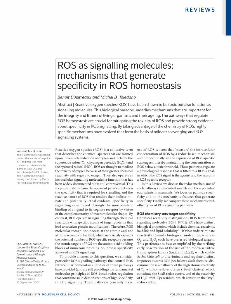
Box 1 | ROS sources and biochemical properties
Although molecular oxygen is a di-radical and rather unreactive, its univalent reduction leads to the formation of chemically more reactive species (known as reactive oxygen species; ROS). These are the superoxide anion (O2
–), hydrogen peroxide (H2O2) and the hydroxyl radical (HO•). Because of their intrinsic chemical properties, each ROS reacts with preferred biological targets (coloured boxes; see figure). As a rule, ROS reactivity dictates toxicity while decreasing signalling ability.
O2– is a by-product of respiration and is produced by NADPH oxidases. Due to high
attraction, O2– oxidizes iron–sulphur ([Fe–S]) clusters at a rate that is almost diffusion-
limited, and releases iron. O2– can react with thiols in vitro, but the slow reaction rates
mean that this cannot occur in vivo5. In Escherichia coli, the steady-state concentration of O2
– is very low (~10–11 M)2, which reflects its instability; this is not only due to its reaction with the [Fe–S] cluster, but also to spontaneous and superoxide-dismutase-mediated O2
– dismutation to H2O2. The instability of O2– and its inability to
diffuse through membranes because of its negative charge make this ROS a poor signalling molecule.
H2O2 toxicity is essentially the consequence of its reduction to HO• by metal-catalysed Fenton chemistry3. H2O2 is actually a poor oxidant and reacts mildly with [Fe–S] (rate constant of 102–103 M–1.s–1) and loosely bound metals (103–104 M–1.s–1), and very slowly with glutathione and free Cys (2–20 M–1.s–1) and with Met residues (10–2 M–1.s–1)3,5. By contrast, its reactivity towards Cys residues can significantly increase to 10–106 M–1.s–1 depending on the protein environment (BOX 2). As a corollary, H2O2 is relatively stable (cellular half-life ~1 ms, steady-state levels ~10–7 M). Diffusion of H2O2 might be modulated by changes in membrane permeability or by transport through aquaporins114. Its selective reactivity and diffusibility makes H2O2 fit for signalling.
The highly toxic HO• has high indiscriminate reactivity, which limits its diffusion to sites of production (half-life 10–9 s)2. Despite this, HO• seems to operate in H2O2 sensing.
R E V I E W S
814 | OCTOBeR 2007 | vOlume 8 www.nature.com/reviews/molcellbio
© 2007 Nature Publishing Group

Nature Reviews | Molecular Cell Biology
R-SH R-SOH
RSNR′R′′ RSSR′
R-SO2H R-SO3HH2O2 H2O2 H2O2
RS(O)SR′ RS(O)2SR′H2O2 H2O2
NHR′R′′ R′SH
PeroxidasePeroxide-reducing enzymes that function by heterolytic cleavage of the O–O bond. Peroxidases fall into different classes depending on the nature of their catalytic site or according to the mechanisms that regenerate the active form.
pKa
The equilibrium constant of proton (H+) exchange reactions between acids and bases according to the Brönsted theory. It reflects the strength of an acid to donate its proton as pKa decreases.
protection or repair of [Fe–S] clusters (for example, oxidation-resistant [2Fe–2S]-containing fumarase C and [Fe–S]-repair ferredoxin:naDPH oxidoreductase)18. Oxidation of the SoxR [2Fe–2S] cluster by O2
– causes a change of SoxR conformation that alters the structure of the SoxR-bound Dna operator, resulting in gene activation.
The search for a SoxR reduction mechanism led to the discovery of the rsxABCDGE operon and rseC gene product, which together form a putative multi-component naDPH-dependent reducing system that is associated with the membrane19. The stronger and more prolonged SoxR activation in strains that are impaired in naDP+ reduction can be explained by defective SoxR reduction, which indicates that SoxR is not only linked to O2
– levels but also to the ratio of naD(P)H compared with naD(P)+ (ReF. 18). Fridovich argued that SoxR may exclusively sense the naD(P)H/naD(P)+ ratio, because paraquat and menadione — redox-cycling drugs that are used to activate SoxR — generate O2
– while depleting cellular naD(P)H18. nevertheless, the fact that the SoxR output pathway specifies O2
– catabolism indicates the O2
– specificity of SoxR.
Sensing oxygen, iron and ROS through [Fe–S] clusters. Being chemically versatile, [Fe–S] clusters can sense other signals. The oxygen sensor FnR, which activates anaerobic gene transcription20,21, requires a [4Fe–4S]2+ cluster. This cluster is oxidized by oxygen, resulting in the formation of an unstable [3Fe–4S]+ cluster and release of Fe2+ and O2
–. H2O2 and O2– can both dis-
assemble the cluster; however, oxygen is thought to be the specific FnR physiological signal because it reaches a much higher concentration than H2O2 and O2
– during the aerobic switch20.
The [Fe–S] cluster-containing transcription factor IscR senses both the [Fe–S] cluster biosynthesis status and H2O2, and exemplifies the coordinated regulation of iron and ROS metabolism, which is important for mitigating the synergistic toxicity of these compounds. When the [2Fe–2S] cluster is intact, IscR represses the transcription of genes involved in [Fe–S] cluster biosynthesis22. H2O2 derepresses IscR gene expression by disassembling the IscR [2Fe–2S] cluster and activates OxyR-dependent expression of an alternate [Fe–S] assembly pathway, which is more robust towards oxidative stress23.
The coordination of iron and ROS responses is also carried out by the mammalian Rna-binding iron-response protein-1 (IRP1)24. IRP1 primarily senses iron through the assembly or disassembly of its [4Fe–4S] cluster (which is dependent on the cellular iron status) that regulates its Rna-binding activity. The Rna bind-ing of IRP1 is also regulated by O2
–, possibly by cluster disassembly, thereby decreasing iron availability and mitigating the potential toxicity of O2
–. The biological importance of the IRP1 oxidative stress response is controversial.
Active Cys or iron centres for peroxide sensing. OxyR25, PerR26 and OhrR27 are bona fide peroxide sensors, and each uses a distinct sensing mechanism (FIG. 1b). OxyR and PerR sense both H2O2 and organic peroxides, whereas OhrR only senses organic peroxides. PerR and OxyR are functional orthologues; each sensor is present in organisms that lack the other and they regulate many common target genes that specify peroxide scavenging and iron metabolism28. These genes include catalases, the peroxiredoxin ahpC and Fur — a transcriptional regulator of ferric uptake. OxyR also has a role in thiol redox control by regulating glutathione reductase, glutaredoxin (Grx) and thioredoxin (Trx). OhrR only regulates the organic peroxide-specific thiol-based peroxidase Ohra27,29.
OxyR fulfils the definition of a peroxide receptor; it is activated by H2O2 at concentrations that just exceed cellular physiological levels (20 nm) and that are below the threshold toxic level (estimated at >1 µm)30. Based on these characteristics, it is used as a faithful monitor of intracellular H2O2 levels31. OxyR reacts with H2O2 through a unique Cys residue (Cys199) that presum-ably oxidizes to a SOH, which then condenses with Cys208 to form an intramolecular disulphide25,32. OxyR is deactivated by reduction by Grx1, which, together with ahpC and catalase, contributes to OxyR auto-regulation. The reaction rate for SOH formation is rapid
Box 2 | Cys, the archetypal redox-regulatory amino acid
H2o2 cys oxidation productsSulphur-mediated nucleophilic attack of the peroxide O–O bond by the Cys thiol group (-SH) leads to H2O release and formation of sulphenic acid (-SOH). The -SOH is highly reactive, its stability being influenced by the availability of a proximal -SH with which it condenses to form a disulphide bond, or by availability of a proximal nitrogen to form a sulphenamide115,116 or by the presence of H2O2, which further oxidizes it to form sulphinic (-SO2H) or sulphonic (-SO3H) acids (see figure).
cys residues are not equally reactiveThe reactivity of Cys residues is dictated by their solvent-exposed localization and ionization state; thiolate anions (-S¯) are far more nucleophilic than their protonated counterpart4. When its pKa is less than or equal to intracellular pH (6.8–7.2), a protein thiol will be deprotonated in vivo by >50%. Neighbouring positively charged residues stabilize -S¯ by electrostatic interaction, decreasing the pKa of free Cys (8.3)7. However, there is an optimal value of pKa for Cys reactivity. Indeed, as pKa decreases, the stability of the -S¯
increases, decreasing negative charge availability and thus reactivity4,6. Reaction rates of H2O2 with thiols vary widely (10–106 M–1.s–1)4,5,117,118, which provides an extended reactivity range that is exploitable in vivo for selectivity.
reversal of cys oxidationOnly the forms of Cys that can be reversibly oxidized (-SOH, disulphide bond, sulphenamide, and occasionally -SO2H) operate in redox signalling. Reduction of -SOH proceeds by condensation with a donor -SH to form a disulphide. Disulphides are reduced by either thioredoxin (TRX) or glutaredoxin (GRX), which themselves are reduced by NADPH-dependent TRX- and glutathione reductases. TRX and GRX catalyse fast and reversible thiol disulphide exchanges between their CXXC active-site residues and half-cysteines of disulphide substrates. Glutathione (GSH) can reduce the R-SOH that emanate from peroxide oxidation by formation of an S-glutathionylation adduct, which is then reduced by GRX. S‑glutathionylation does not seem to derive from thiol-disulphide exchange between an R-SH and oxidized GSH. S-glutathionylation is important in protecting R-SOH from irreversible oxidation and might have protein-regulatory functions119.
R E V I E W S
naTuRe RevIeWS | molecular cell biology vOlume 8 | OCTOBeR 2007 | 815
© 2007 Nature Publishing Group
Nature Reviews | Molecular Cell Biology
Inactive
Inactive
Transcriptionallyactive
O2–
NADPH-dependentferredoxin(Rsx)
DNA-binding activity
Grx
H2O2
Repression
Sacrificialoxidation
H2O2
Derepression
Fe2+
Fe2+
N
H
NH + H2O2 + H2OHN
O
NH
a
bS
S
Hsp33SH: monomerInactive chaperone
Hsp33SS: dimerActive chaperone
Heat+H2O2/HOCI
Zn2+
Diamide
Zn2+
Zn
S-Cys
S-CysCys-SCys-S
RsrA-σR complexσR DNA-binding inhibition
S
S
S
S
S S
N-His
HS-Cys
RsrA-σR complexσR DNA-bindingactivation
c
S
SZn
S-CysCys-SCys-S
N-His
σR
σR
SoxR[2Fe–2S]+
SoxR[2Fe–2S]2+
OxyROxyRSH
SH
N-HisN-His
Fe2-oxo-His2-oxo-His
S-nitrosylationA modification to the –SNO form of the thiol moiety of a Cys residue, caused by reaction with peroxinitrite (ONOO¯) or nitrogen trioxide (N2O3).
(~1.1 × 10⁵ m–1.s–1) and reflects the high H2O2 reactivity of Cys199 (ReF. 33). Oxidation changes the conforma-tion of dimeric OxyR, which triggers the activation of OxyR site-specific binding to Dna promoters34. X-ray crystallography and time-resolved structural changes that are associated with oxidation indicate a rapid extrusion of Cys199-SOH from its hydrophobic pocket, which brings Cys199 close to Cys208 — these residues are 17 Å away in reduced protein — followed by a slower second change that results from disulphide-bond formation32,33. These changes lead to alteration of the dimer geometry, which is thought to be the key to OxyR Dna-binding activation. another study con-firmed the H2O2 sensory role of Cys199 but questioned this model; it showed that an OxyR Cys208 substitution mutant did not have a strong phenotype, and identified SOH and S-glutathionylated forms of Cys199 instead of the Cys199–Cys208 disulphide in proteins that were reacted with H2O2 and oxidized GSH, respectively35.
The thiol-specific oxidant diamide also activates OxyR, but the high concentrations that are required suggest an indirect effect either through inhibition of ROS scavengers, which increases H2O2 levels, and/or through GSH depletion, which decreases OxyR reduc-tion. Similarly, constitutive activation of OxyR following
inactivation of thiol-redox control pathways probably results from defective OxyR reduction rather than from OxyR redox equilibration with the oxidized cytoplasm of these cells36. accordingly, OxyR might not sense disulphide stress per se as proposed30,37 but, instead, might remain active under this condition owing to defective reduction. OxyR is also activated by nitro-sothiols through S-nitrosylation of Cys199 (R-SnO)38, but it has a minor role in the nitrosative stress response17.
The transcriptional repressor OhrR senses per-oxides using a unique reactive Cys residue that oxidizes to R-SOH39. In some prokaryotic species, the nascent OhrR-SOH forms a disulphide with a Cys of another subunit40, which causes the conformation change that inhibits Dna binding. By contrast, OhrR has a unique Cys residue in Bacillus subtilis41. The OhrR-SOH form retains Dna-binding activity, but its reaction to form a mixed disulphide with either free Cys, coenzyme a or a novel 394-Da thiol, or to form a sulphenyl amide with a proximal nitrogen, leads to derepression. The structure of OhrR provides clues for its preference towards organic peroxides; a long strip of hydrophobic and aromatic residues surrounds its reactive Cys and could provide a docking site for the hydrophobic tail of organic peroxides42.
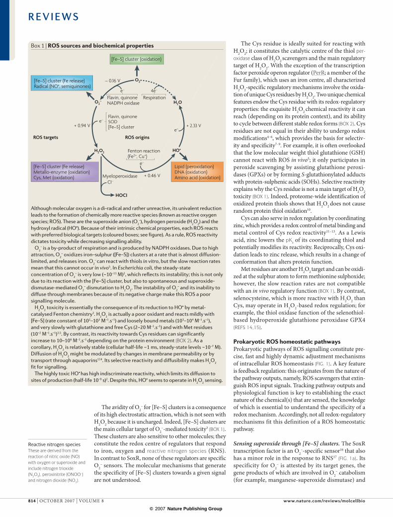
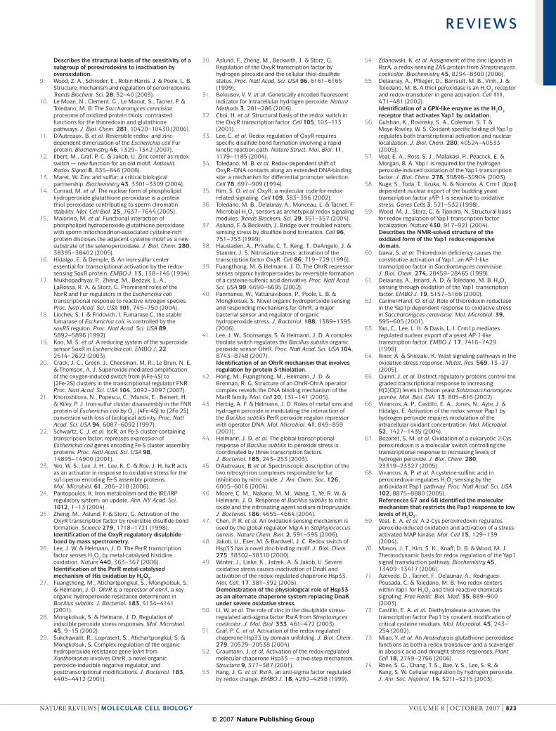
Figure 1 | microbial roS receptors. a | The SoxR transcription factor regulates the Escherichia coli response to the superoxide anion O2
– (ReF. 16). Oxidation of the SoxR [2Fe–2S] cluster by O2– causes a change in SoxR conformation,
which alters the SoxR-bound operator DNA structure and results in gene activation. SoxR is reduced by NADPH-dependent ferredoxin (Rsx). b | The OxyR transcription factor regulates the H2O2 E. coli response. H2O2 oxidizes OxyR Cys199 to an R-SOH that reacts with Cys208 in an intramolecular disulphide bond25. The resulting allosteric change activates OxyR DNA binding. OxyR is reduced by glutaredoxin-1 (Grx). PerR is an OxyR functional orthologue that senses H2O2 through oxidation of two His residues to 2-oxo-His by HO•, which is produced by iron-catalysed H2O2 reduction by the PerR iron centre. Oxidation releases iron and causes the loss of PerR DNA binding26. As no oxo-His reduction mechanism is currently known, PerR is viewed as a sacrificial regulator. c | Hsp33 is an E. coli redox-regulated chaperone that contains a Cys–zinc redox centre. Heat and H2O2 or hypochlorite (HOCl) lead to the formation of two disulphide bonds and zinc release, which together activate the Hsp33 chaperone12. RsrA is an anti-sigma factor with a Cys–zinc centre that negatively regulates sigma R (σR)53. Diamide-induced Cys oxidation and zinc release lead to the formation of two degenerate disulphide bonds, which causes the loss of σR binding by RsrA50.
R E V I E W S
816 | OCTOBeR 2007 | vOlume 8 www.nature.com/reviews/molcellbio
© 2007 Nature Publishing Group
Nature Reviews | Molecular Cell Biology
PRXSH
SH
PRXS
SPRX
SOH
SH
TRXS
S
TRXSH
SHH2O2
H2OAtypical2-Cys PRX
PRXSHHS
PRXSHHS
PRXSHHOS
PRXSOHHS
PRXSHHO2S
PRXSO2HHS
PRXSS
PRXSS
TRXS
S
TRXSH
SHH2O2
H2OTypical2-Cys PRX
H2O2
SRX
The PerR repressor sensing mechanism is unprec-edented. PerR uses a non-haem iron centre28 and does not use a reactive Cys residue26. either or both of the two His residues that contribute to iron coordination become oxidized to 2-oxo-His, presumably by HO• that is produced in situ by iron-catalysed H2O2 reduction. Oxidation releases iron and causes the loss of PerR Dna binding. Full inactivation only occurs in iron-containing medium43, which indicates that PerR not only senses H2O2 but also senses its toxic potential, which is a function of iron-catalysed Fenton chemistry (BOX 1). PerR transient inactivation44 might indicate an unknown mechanism that reduces PerR 2-oxo-His or causes degradation of its oxidized form. PerR target genes are not all peroxide-inducible; some of them are differentially regulated by iron and manganese, which indicates a further degree of complexity in PerR control28. The PerR mechanism pro-vides an absolute peroxide specificity because only these compounds can assist iron-catalysed His oxidation. as another manifestation of specificity, the PerR structural homologue Fur does not sense H2O2 but instead senses nitric oxide (nO) by dinitrosylation of the iron centre, which regulates a nitrosative stress response17,45. PerR can also respond to nO — not through the 2-oxo-His-based mechanism, but possibly through the mechanism described for Fur46.
Other Cys-based redox-regulatory models exist in prokaryotes but have been omitted here because of space constraints. However, it is worth mentioning the Staphylococcus aureus mgra virulence factor, which carries a unique Cys. The oxidation of mgra by per-oxide to an R-SOH leads to derepression of antibiotic resistance47.
Cys–zinc redox switches. Cys coordination of zinc constitutes the redox centres of heat-shock protein-33 (Hsp33) (ReFS 12,48) and the anti-sigma factor Rsra in prokaryotes (FIG. 1c) and probably of Kelch-like eCH-associated protein-1 (KeaP1) in mammals (KeaP1 is part of a putative ROS receptor complex in mammals; see below). These regulators differ from the aforemen-tioned ROS receptors by their response to high levels of redox signals. Hsp33 is apparently exclusively activated by simultaneous heat and elevated H2O2 concentration or hypochlorite, but not by either stress alone49. Rsra is activated by diamide at elevated concentrations. The low reactivity is reflected by the reaction rates that are predicted from calculation of the half life (t1/2) of zinc expulsion upon oxidation of Hsp33 by H2O2 at 43°C (ReF. 48) and for oxidation of Rsra by diamide50; these rates are 105–106 below the reaction rates of OxyR33 and PerR towards H2O2 (ReF. 26).
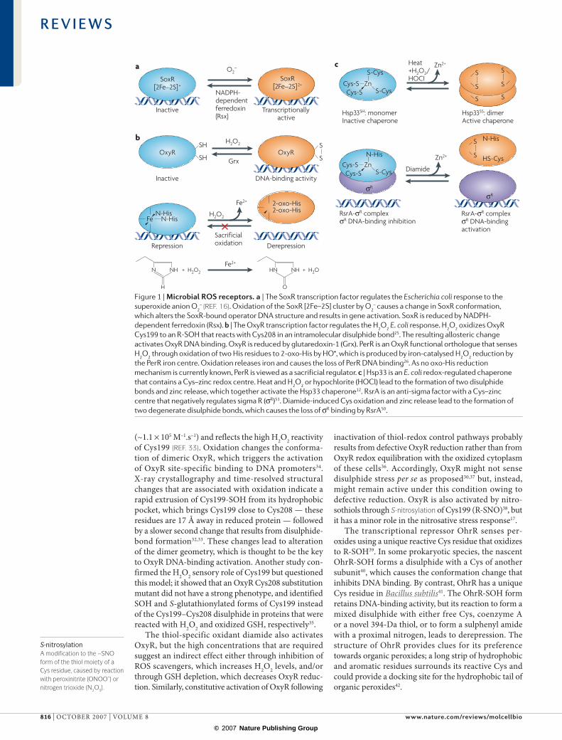
Box 3 | Cys peroxiredoxins
2-cys PrXs detect low levels of H2o2
2-Cys peroxiredoxins (PRXs) are a subclass of PRXs that provide unique examples of exquisite peroxide reactivity. Although they have poor catalytic constants (kcat = 1–80 s–1) compared to catalases (104–105 s–1), they are good catalysts (kcat/Km ~104–107 M–1.s–1 for PRX versus 106–107 M–1.s–1 for catalase) because of their much higher affinity (reactivity) for substrates (PRX Km = 1–103 µM versus catalase Km = 103–105 µM) (see Supplementary information S1 (table)). Therefore, PRXs are efficient when H2O2 levels are low, whereas catalases are efficient when these levels are high120.
The 2-cys PrX cycleTypical 2-Cys PRXs are homodimers that reduce H2O2 with a peroxidatic (catalytic) Cys that oxidizes to a -SOH. The -SOH then condenses with the resolving Cys of the other subunit to form a disulphide, reduced by thioredoxin (TRX). The resolving Cys is non-reactive due to shielding, but becomes solvent-exposed upon oxidation, which facilitates reduction. Homodimers form penta-homodimers that further increase the reactivity of the peroxidatic Cys by a conformational effect121. In atypical 2-Cys PRXs such as glutathione peroxidase (GPX)-like enzymes, the catalytic -SOH condenses with the resolving Cys on the same subunit to form an intramolecular disulphide (see figure).
regulation of 2-cys PrX by inactivationPRXs exhibit a fascinating but puzzling redox twist; they undergo H2O2-mediated inactivation by overoxidation of their catalytic Cys to a sulphinic acid (R-SO2H). This inactivation is unique to eukaryotic PRXs and is reversible by ATP-dependent reduction of the PRX Cys-SO2H by sulphiredoxin (SRX) and sestrins8,122–125, which suggests that this is an acquired gain-of-function that has been selected for regulatory purposes4,8. Inactivation is due to an additional C-terminal helix, which is absent in inactivation-insensitive PRXs, and slows down the rate of peroxidatic Cys-SOH condensation with the resolving Cys. This kinetic pause allows further oxidation of the R-SOH by H2O2 (ReF. 8.) Overoxidation only occurs during enzymatic cycling and is proportional to the amount of substrate at non-saturating conditions. Excess substrate also increases the rate of overoxidation by increasing the likelihood of collision of the Cys-SOH with H2O2
(ReF. 126).
R E V I E W S
naTuRe RevIeWS | molecular cell biology vOlume 8 | OCTOBeR 2007 | 817
© 2007 Nature Publishing Group
Nature Reviews | Molecular Cell Biology
SH
SHYap1
Ybp1
S598
S303
SH
SHYap1
S598
S303S310
S629
Yap1
Ybp1
Ybp1
S598 S36
SH303
HSSH
SH
Yap1 Orp1
Ybp1
SOH
SHOrp1
SH
SHOrp1
36
SOH
SHOrp1
SH
SHOrp1
Orp1
SH
SH
H2O2
H2O
H2O
TrxS
STrx
SR598SR620
SR629Yap1
Elec
trop
hile
s/m
etal
loid
s
Signalling Peroxidatic cycle
S
S
SH
SH
Hsp33 is a redox-regulated chaperone that substitutes for the aTP-dependent DnaK chaperone machinery, which loses activity under severe oxidative stress due to aTP depletion49. In Hsp33, tetrahedral zinc is coordinated by a C-terminal four-Cys zinc-binding motif, CXCX(27–32)CX2C, which stabilizes Hsp33 in an inactive conformation while keeping Cys residues in the thiolate form. The mechanism involves H2O2-promoted Cys oxidation with formation of two disulphide bonds between adjacent Cys residues and concomitant zinc release, resulting in unfolding of the C-terminal protein
portion51. unfolding unmasks the substrate-binding site and dimerization interface that promotes formation of the high-affinity dimer of the active chaperone52. Whether zinc release or Cys oxidation is the primary event that triggers Hsp33 is not established. Cys–zinc coordination carries the following theoretical paradox: Zn2+ is believed to keep Cys residues in the thiolate form poised to react with H2O2, but is also thought to decrease sulphur nucleophilicity by attracting sulphur negative charges. Furthermore, the high affinity of Zn2+ for Hsp33 Cys residues also prevents reaction of these resi-dues, which might explain the need for the combination of heat and harsh oxidative stress for activation.
Rsra negatively regulates the sigma R (σR) transcrip-tion factor53. Rsra activation by diamide is consistent with the requirement of Rsra for diamide tolerance and for regulation of σR-dependent expression of the thioredoxin operon. Rsra is poorly activated by H2O2, in keeping with reaction rates that are one order of magnitude slower for this oxidant50. Zinc binds to a CX25HX3CX2C motif with very high affinity, which sug-gests a structural rather than a redox-regulated motif, and protects the Cys residue from reaction with mild oxidants54. Diamide-induced Cys oxidation and zinc release lead to the formation of a degenerate disulphide bond between Cys11 and either Cys41 or Cys44, causing loss of σR-binding by Rsra50. elucidation of the natural signal mimicked by diamide should help us to under-stand the mechanism and function of this Cys–zinc redox switch.
Regulatory thiol peroxidases in yeastThiol peroxidases have unique H2O2 signalling functions and they contribute to the most important yeast H2O2-regulated pathways: namely, the Saccharomyces cerevisiae Yap1 and Schizosaccharomyces pombe Pap1 and Sty1 pathways. Thiol peroxidases consist of the peroxiredoxin (PRX)9 and GPX-like enzyme families, and represent an important subgroup of non-haem peroxide-scavenging enzymes15 (BOX 3). Thiol peroxidases function as both H2O2 receptors and redox transducers by uniquely combining high H2O2 reactivity and the ability to oxidize protein thiols other than their physiological reducing system36, which is usually thioredoxin. among these enzymes, 2-Cys PRXs can undergo reversible substrate-mediated inactivation, which supplies a second redox-switch that regulates both their H2O2 receptor and their scavenging functions. Yeast have other H2O2-signalling pathways, such as the msn2–msn4 transcription factors in budding yeast, but the corresponding receptors are not known.
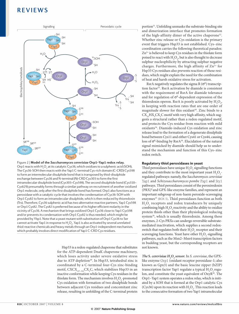
The S. cerevisiae H2O2 sensor. In S. cerevisiae, the GPX-like enzyme Orp1 (oxidant receptor peroxidase-1; also known as Gpx3) and the basic leucine zipper (bZIP) transcription factor Yap1 regulate a typical H2O2 regu-lon, and constitute the yeast equivalent of OxyR36. The Orp1–Yap1 system operates a redox relay, which is initi-ated by a SOH that is formed at the Orp1 catalytic Cys (Cys36) upon its reaction with H2O2. This reaction leads to the consecutive formation of two Yap1 intramolecular
Figure 2 | model of the Saccharomyces cerevisiae orp1–yap1 redox relay. Orp1 reacts with H2O2 at its catalytic Cys36, which oxidizes to a sulphenic acid (SOH). The Cys36-SOH then reacts with the Yap1 C-terminal Cys-rich domain (C-CRD) Cys598 to form an intermolecular disulphide bond that is transposed by thiol-disulphide exchange between Cys36 and N-terminal (N)-CRD Cys303 to form the first intramolecular disulphide bond (Cys303–Cys598). The second disulphide bond (Cys310–Cys629) presumably forms through a similar pathway on recruitment of another oxidized Orp1 molecule, only after the first disulphide bond has formed. Orp1 also functions as a peroxidase with a catalytic cycle that involves the condensation of Cys36-SOH with Orp1 Cys82 to form an intramolecular disulphide, which is then reduced by thioredoxin (Trx). Therefore, Cys36 sulphenic acid has two alternative reactive partners, Yap1 Cys598 or Orp1 Cys82. The Cys82 is preferred because of its higher efficient molarity in the vicinity of Cys36. A mechanism that brings oxidized Orp1 Cys36 close to Yap1 Cys598 and/or prevents its condensation with Orp1 Cys82 is thus needed, which might be provided by Ybp1. Note that a yeast mutant with substitution of Orp1 Cys36 to Ser cannot activate Yap1 in response to H2O2. Yap1 is also activated by several unrelated thiol-reactive chemicals and heavy metals through an Orp1-independent mechanism, which probably involves direct modification of Yap1 C-CRD Cys residues.
R E V I E W S
818 | OCTOBeR 2007 | vOlume 8 www.nature.com/reviews/molcellbio
© 2007 Nature Publishing Group
S
S
Nature Reviews | Molecular Cell Biology
Mak1 Mak3Mak2
Mpr1
Mcs4
Wak1 Win1 Tpx1
SH
SHTpx1
SOH
SHTpx1
Wis1
Sty1/Spc1
P
P
H2O2
H2O2
H2O2
H2O
SO2HTpx1
Atf1 Pap1
gpx1 ctt1
srx1
trx2
Srx1
His
phos
phor
elay
MA
PK m
odul
ebZ
IP t
rans
crip
tion
fact
ors
Targ
etge
nes
S
S
disulphides between n- and C-terminal Cys-rich domains (n- and C-CRD)55 (FIG. 2). Ybp1, a protein of as-yet-undefined non-redox molecular function, is vital for the oxidation of Yap1 by Orp1 (ReFS 56,57). Disulphide bond formation, by inhibiting Crm1 (also known as exportin-1)-dependent nuclear export of Yap1, promotes Yap1 nuclear accumulation and Yap1-dependent gene activation58. Disulphide bond form-ation produces a conformational change that conceals a C-CRD Crm1-cognate nuclear export signal (neS) within a hydrophobic core that is formed by the inter-action of n- and C-CRD59. Reduction of disulphide bonds, probably by thioredoxin60–62, exposes the neS and reactivates Yap1 nuclear export58,63. This oxida-tion-induced Yap1 allosteric change also seems to be important for transcriptional activation56.
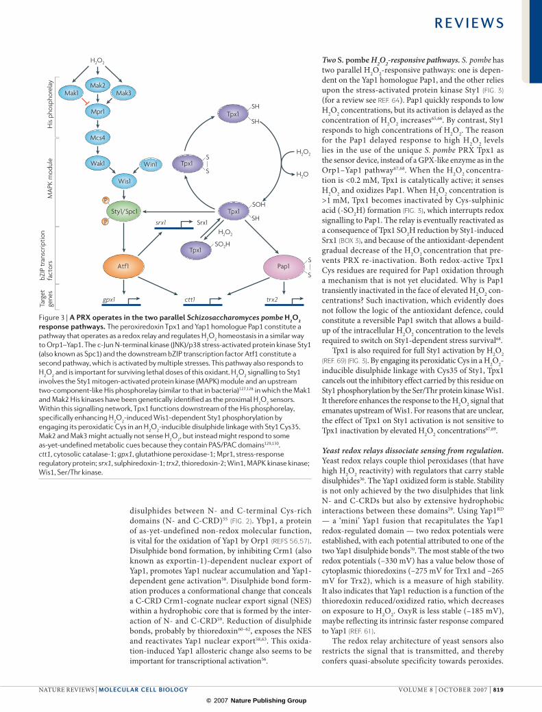
Two S. pombe H2O2-responsive pathways. S. pombe has two parallel H2O2-responsive pathways: one is depen-dent on the Yap1 homologue Pap1, and the other relies upon the stress-activated protein kinase Sty1 (FIG. 3) (for a review see ReF. 64). Pap1 quickly responds to low H2O2 concentrations, but its activation is delayed as the concentration of H2O2 increases65,66. By contrast, Sty1 responds to high concentrations of H2O2. The reason for the Pap1 delayed response to high H2O2 levels lies in the use of the unique S. pombe PRX Tpx1 as the sensor device, instead of a GPX-like enzyme as in the Orp1–Yap1 pathway67,68. When the H2O2 concentra-tion is <0.2 mm, Tpx1 is catalytically active; it senses H2O2 and oxidizes Pap1. When H2O2 concentration is >1 mm, Tpx1 becomes inactivated by Cys-sulphinic acid (-SO2H) formation (FIG. 3), which interrupts redox signalling to Pap1. The relay is eventually reactivated as a consequence of Tpx1 SO2H reduction by Sty1-induced Srx1 (BOX 3), and because of the antioxidant-dependent gradual decrease of the H2O2 concentration that pre-vents PRX re-inactivation. Both redox-active Tpx1 Cys residues are required for Pap1 oxidation through a mechanism that is not yet elucidated. Why is Pap1 transiently inactivated in the face of elevated H2O2 con-centrations? Such inactivation, which evidently does not follow the logic of the antioxidant defence, could constitute a reversible Pap1 switch that allows a build-up of the intracellular H2O2 concentration to the levels required to switch on Sty1-dependent stress survival68.
Tpx1 is also required for full Sty1 activation by H2O2 (ReF. 69) (FIG. 3). By engaging its peroxidatic Cys in a H2O2-inducible disulphide linkage with Cys35 of Sty1, Tpx1 cancels out the inhibitory effect carried by this residue on Sty1 phosphorylation by the Ser/Thr protein kinase Wis1. It therefore enhances the response to the H2O2 signal that emanates upstream of Wis1. For reasons that are unclear, the effect of Tpx1 on Sty1 activation is not sensitive to Tpx1 inactivation by elevated H2O2 concentrations67,69.
Yeast redox relays dissociate sensing from regulation. Yeast redox relays couple thiol peroxidases (that have high H2O2 reactivity) with regulators that carry stable disulphides36. The Yap1 oxidized form is stable. Stability is not only achieved by the two disulphides that link n- and C-CRDs but also by extensive hydrophobic interactions between these domains59. using Yap1RD — a ‘mini’ Yap1 fusion that recapitulates the Yap1 redox-regulated domain — two redox potentials were established, with each potential attributed to one of the two Yap1 disulphide bonds70. The most stable of the two redox potentials (–330 mv) has a value below those of cytoplasmic thioredoxins (–275 mv for Trx1 and –265 mv for Trx2), which is a measure of high stability. It also indicates that Yap1 reduction is a function of the thioredoxin reduced/oxidized ratio, which decreases on exposure to H2O2. OxyR is less stable (–185 mv), maybe reflecting its intrinsic faster response compared to Yap1 (ReF. 61).
The redox relay architecture of yeast sensors also restricts the signal that is transmitted, and thereby confers quasi-absolute specificity towards peroxides.
Figure 3 | a PrX operates in the two parallel Schizosaccharomyces pombe H2o2 response pathways. The peroxiredoxin Tpx1 and Yap1 homologue Pap1 constitute a pathway that operates as a redox relay and regulates H2O2 homeostasis in a similar way to Orp1–Yap1. The c-Jun N-terminal kinase (JNK)/p38 stress-activated protein kinase Sty1 (also known as Spc1) and the downstream bZIP transcription factor Atf1 constitute a second pathway, which is activated by multiple stresses. This pathway also responds to H2O2 and is important for surviving lethal doses of this oxidant. H2O2 signalling to Sty1 involves the Sty1 mitogen-activated protein kinase (MAPK) module and an upstream two-component-like His phosphorelay (similar to that in bacteria)127,128 in which the Mak1 and Mak2 His kinases have been genetically identified as the proximal H2O2 sensors. Within this signalling network, Tpx1 functions downstream of the His phosphorelay, specifically enhancing H2O2-induced Wis1-dependent Sty1 phosphorylation by engaging its peroxidatic Cys in an H2O2-inducible disulphide linkage with Sty1 Cys35. Mak2 and Mak3 might actually not sense H2O2, but instead might respond to some as-yet-undefined metabolic cues because they contain PAS/PAC domains129,130. ctt1, cytosolic catalase-1; gpx1, glutathione peroxidase-1; Mpr1, stress-response regulatory protein; srx1, sulphiredoxin-1; trx2, thioredoxin-2; Win1, MAPK kinase kinase; Wis1, Ser/Thr kinase.
R E V I E W S
naTuRe RevIeWS | molecular cell biology vOlume 8 | OCTOBeR 2007 | 819
© 2007 Nature Publishing Group
In S. cerevisiae, only the Orp1-SOH, which is exclusively generated by reaction with peroxides, can promote Yap1 oxidation. In fact, both Yap1 and Pap1 can respond to other thiol-reactive chemicals and heavy metals by altera-tion of their neS by direct, thiol-peroxidase-independent modification of C-CRD Cys residues71,72.
Redox sensors in higher eukaryotesROS homeostasis in single-cell and multicellular organ-isms cannot be strictly compared because of differences in the external environments of the cells. mammalian cells within their host habitat may not need the fast and highly dynamic ROS concentration-adjustment mechanisms that are crucial for the survival of single-cell organisms. Instead, mammalian cells respond to oxidative stress using either long-lasting protective responses that are usually part of global differentiation programmes or cell-death mechanisms.
Thiol-peroxidase-initiated H2O2 signalling. In multi-cellular organisms, thiol peroxidases affect several H2O2 signalling pathways; however, in contrast to yeast, none of these mechanisms regulate ROS homeostasis. Genetic data indicate that Arabidopsis thaliana aTGPX3, an enzyme similar to S. cerevisiae Orp1, functions as a trans-ducer that relays a H2O2 signal in the abscisic acid (aBa) pathway of stomatal guard cells that controls drought stress tolerance73. In these cells, aTGPX3 is required to inhibit the ABA INSENSITIVE-2 (ABI2)-encoded protein phosphatase type-2C, possibly by oxidation.
In mammals, although a wealth of data suggest the involvement of PRXs and GPX in signalling74, these factors are not known to operate in the same way as in yeast; however, a redox-transducing function might be suggested for peroxiredoxin-1 (PRX1) on the basis of its requirement for the activation of the p38 mitogen-activated protein kinase (maPK) by H2O2 and oxidized lipids75. nevertheless, a thiol oxidase function has been clearly established for the selenothiol-based GPX4. GPX4 does not signal H2O2 but promotes polymerization of the sperm protective mitochondrial capsule by catalysing disulphide bond formation between structural keratin-like proteins and other proteins15. GPX4 also contributes to the condensation of chromatin in sperm by oxidation of protamine, which replaces histones during matura-tion14. GPX4-catalysed disulphide bond formation is presumably triggered by oxidation of GPX4 by peroxide, and requires a decrease in the concentration of the GPX4-physiological reducing system GSH, which occurs during sperm maturation.
another mechanism by which thiol peroxidases may affect H2O2 signalling is by controlling the flux and concentration of the H2O2 that is produced upon growth-factor or cytokine-induced cellular signalling, especially within specific subcellular compartments8,74. For exam-ple, the most abundant mitochondrial peroxidase, PRX3, regulates apoptosis signalling by scavenging mitochon-drial H2O2 (ReF. 76). another example of a process that also imparts specificity to the messenger function of H2O2 is the site-specific negative regulation of platelet-derived growth factor receptor (PDGFR) phosphorylation by
peroxiredoxin-2 (PRX2), as established by studies of Prx2-knockout mice77. upon PDGF stimulation, PRX2 associates with PDGFR, thereby possibly restricting the action radius of the H2O2 produced upon this stimula-tion, presumably by a neighbouring naDPH oxidase complex. This effect of PRX2 might ultimately lead to an alteration of the redox state of a putative PDGFR-cognate protein tyrosine phosphatase as the mechanism by which PRX2 specifically affects site-specific PDGFR phosphorylation.
The post-translational regulation of thiol peroxidases might add another layer of control to the signalling flux of H2O2 (ReF. 9). In addition to being reversibly inactivated by their substrate (BOX 3), PRXs can be regulated by phosphorylation; PRX1 is reversibly inactivated during mitosis by phosphorylation by the CDC2-dependent kinase78. Similarly, GPX1 (ReF. 79) and also catalase80 are both activated by phosphorylation by c-aBl and arg non-receptor tyrosine kinases.
ROS-inducible pathways in search of ROS receptors. Several signal circuitries regulate ROS-inducible oxida-tive stress-protective responses, but the corresponding upstream ROS receptors are still unknown. These path-ways seem to differ from those of microbial organisms because they participate in large metabolic programmes, respond to many other physiological cues and provide either long-term oxidative stress protection or trigger apoptosis as a clearance mechanism for oxidatively damaged cells.
The main physiological function of the p53 tumour suppressor is to prevent the transmission of mutations to daughter cells either by restricting the proliferation of damaged cells and enhancing Dna repair or by induc-ing apoptosis. another important component of the p53 tumour-suppressor function relies on a newly discovered antioxidant function. p53 prevents the accumulation of damage that could be caused by the continuous endo-genous production of H2O2 by regulating GPX1 and ses-trins, which are sulphiredoxin functional homologues81. Reminiscent of the distinct S. pombe responses to vary-ing H2O2 concentration, low levels of H2O2 activate the p53 antioxidant response, whereas high levels trigger p53-dependent apoptosis through the induction of pro-oxidant activities.
The peroxisome proliferator-activated receptor-g (PPaRg) coactivator-1α (PGC1α), which stimulates mitochondrial biogenesis in response to increased energy demand, also regulates an H2O2-inducible antioxidant transcriptional programme that includes superoxide dismutases, catalase and GPX1 (ReF. 82). PGC1α thus establishes an important link between the control of mitochondrial respiration that generates ROS and an anti-ROS programme.
Similarly, the oncogene c-myc increases ROS tolerance by activating transcription of GSH biosynthesis genes in response to H2O2 (ReF. 83). Transcription factors of the class O forkhead box (FOXO) family are activated by H2O2 and induce either cell death or a quiescent cell state that is characterized by improved tolerance to oxidative stress. This state is characterized by SOD1 expression84.
R E V I E W S
820 | OCTOBeR 2007 | vOlume 8 www.nature.com/reviews/molcellbio
© 2007 Nature Publishing Group

Nature Reviews | Molecular Cell Biology
Ser40NRF2 CNC-bZIPUb
UbUb
UbUb
UbUb
UbUb
K K K
Cys288 -S
Cys273 -S
Zn
Cys288 -S
Cys273 -S
Zn
L
L
E2
Ub
Cys151 Cys151
CUL3
BTB
IVR
DGR
KEAP1 modifications:Cys oxidations (Cys151 Cys273, Cys288)Zn releasePhosphorylation by PKC, PERK, P13K (Ser40)
NRF2 ubiquitylation arrest
KEAP1–CUL3–NRF2 complex
NRF2 proteasomal degradation
NRF2 stabilization and nuclear accumulation
ARE gene activation• Antioxidants• Electrophile detoxification• GSH• NADPH• Proteasome function
KEAP1
ROSElectrophilesER stress
AlkylationA chemical modification that involves the transfer of a carbon chain to any other atom.
This FOXO ROS-protective function is important for stem-cell survival85. Several mechanisms have been shown to transduce ROS signals to FOXO. The stress-activated Jun n-terminal kinase (JnK)86 activates H2O2-induced FOXO protective functions, whereas the sterile-like kinase mST1 mediates H2O2-induced FOXO activation that leads to cell death87. FOXO activity is also regulated in response to H2O2 through acetylation by the cyclic-amP responsive element binding (CReB)-binding protein (CBP)88 and deacetylation by the sirtuin SIRT189.
The KEAP1–NRF2 pathway. The KeaP1–nRF2 complex constitutes the closest fit to a ROS receptor in mammals, and regulates environmental and xenobiotic stress-protective responses. nRF2 (nuclear factor (erythroid-derived-2)- like-2) is a CnC (cap ‘n’ collar)-bZIP transcription factor, whereas BTB (broad complex–tramtrack–bric-a-brac)-Kelch KeaP1 (ReF. 90) functions as the receptor of the pathway and the nRF2-cognate adaptor of a cullin-3 (Cul3)–ring-box-1 (RBX1) ubiquitin-ligase complex that marks nRF2 for proteosomal degradation91–94. The func-tions of KeaP1–nRF2 in oxidative and environmental stress tolerance are attested by the nature of its inducers; these include H2O2, lipid oxidation products, nitric oxide, heavy metals and arsenicals and a multitude of natural and synthetic electrophilic compounds (for a detailed review see ReFS 95,96). The function of this pathway in oxidative and environmental stress tolerance is also
reflected by the nRF2 target genes, which comprise phase II xenobiotic enzymes and antioxidants. moreover, Nrf2-null mice are characterized by susceptibility to sev-eral stresses, including acetaminophen, diesel exhaust fumes, butylated hydroxytoluene, hyperoxia, ultraviolet irradiation and carcinogens. natural and synthetic nRF2 inducers are used in cancer chemoprevention based on the premise that higher nRF2 activity should help to detoxify carcinogens.
Chemical inducers, by modifying KeaP1, decrease ubiquitylation and proteosomal degradation of nRF2 and promote its nuclear accumulation95,96. KeaP1 sensing of nRF2 inducers involves reactive Cys residues97–99, but a more complex mechanism that involves a Cys–zinc redox centre was suggested following the observation that KeaP1 binds to zinc with high affinity at stoichiometric amounts in vitro95. KeaP1 Cys273 and Cys288, which are among the few reactive Cys residues that have been identified in this protein, are indeed crucial for both zinc coordin-ation and nRF2 degradation96,98,99. By contrast, KeaP1 Cys151 is not required for zinc binding, but this residue is reactive and important for derepression of nRF2 (ReF. 99). modification of these Cys residues and possibly others by alkylation, oxidation or disulphide bond formation100 might displace zinc, thus switching KeaP1 to a confor-mation that is non-permissive for ubiquitylation. Zinc coordination by Cys residues in KeaP1 is reminiscent of Hsp33 and Rsra, suggesting that, in a similar way to
Figure 4 | Schematic of the speculative two-site interaction model of KeaP1–NrF2. The dimeric Kelch-like ECH-associated protein-1 (KEAP1) receptor contacts one NRF2 (nuclear factor (erythroid-derived-2)-like-2) molecule at two distinct N-terminal sites103,131. The KEAP1–cullin-3 (CUL3) complex constantly targets NRF2 for proteosomal degradation through ubiquitylation of NRF2 Lys residues that are located at its N terminus, between the two KEAP1 interaction sites. Electrophiles and signals from reactive oxygen species (ROS) modify KEAP1 Cys residues, leading to zinc release and a conformation that is non-permissible for ubiquitylation. An alternative speculative model proposes that two NRF2 molecules are each contacted by the Kelch domain of dimeric KEAP1 (ReF. 104). Phosphorylation of Ser40 by phospha-tidylinositol 3-kinase (PI3K), protein kinase RNA (PKR)-like endoplasmic reticulum (ER) kinase (PERK) or protein kinase C (PKC) might also lead to ubiquitylation arrest. KEAP1 dimerizes through the broad complex–tramtrack–bric-a-brac (BTB) domain and interacts with NRF2 through the Kelch domain. The KEAP1 intervening region (IVR) that carries reactive Cys residues is located between the BTB and Kelch domains. ARE, antioxidant response element; CNC-bZIP, cap ‘n’ collar-basic leucine zipper; DGR, double glycine repeat; GSH, glutathione.
R E V I E W S
naTuRe RevIeWS | molecular cell biology vOlume 8 | OCTOBeR 2007 | 821
© 2007 Nature Publishing Group
these regulators, KeaP1 is not suited for detection of the low peroxide concentration that is characteristic of instant intracellular peroxide homeostasis.
How KeaP1 affects nRF2 ubiquitylation remains unclear. Chemical inducers might either disrupt the interaction of KeaP1 with nRF2 (ReFS 94,96,101) or with Cul3 (ReFS 94,102) or displace nRF2 within the KeaP1–Cul3 complex in a position that is not permis-sive for ubiquitylation103. a two-site interaction model of dimeric KeaP1 that contacts one nRF2 molecule at two distinct n-terminal sites of different affinities provides a basis for modularity in the position of nRF2 within the KeaP1–Cul3 complex103 (FIG. 4), which is in keeping with the model that proposes displacement of nRF2 within the KeaP1–Cul3 complex. alternatively, dimeric KeaP1 might contact two nRF2 molecules, with each KeaP1 monomer contacting one nRF2 molecule104. The KeaP1–nRF2 complex might also be controlled at the level of its subcellular localization; it undergoes nucleocytoplasmic shuttling by virtue of an nRF2 nuclear localization signal and a CRm1-cognate KeaP1 neS sequence105,106. The KeaP1 neS, which is close to Cys273 and Cys288, might also be altered by either zinc release and/or modification of Cys residues106, leading to KeaP1–nRF2 nuclear accumulation in a manner that is similar to Yap1 and Pap1.
ConclusionsThe ROS receptors that regulate ROS homeostasis define a distinct category of ROS signalling in which the ROS signal is the exclusive agonist that interacts with high specificity with its cognate receptor. Specificity is built up on the exceptionally high reactivity of agonists for their receptors, which is imprinted in the unique chemistry of the particular ROS and its atomic target within regulatory proteins. The ROS receptor function of thiol peroxidases provides a striking example of how nature has used the high H2O2 reactivity of these H2O2 scavengers to operate H2O2 homeostatic signalling. Surprisingly, these path-ways, which are a hallmark of microbial organisms, have not yet been found in mammals.
a microarray analysis of the H2O2 response of human cells identified a large genomic response that includes genes involved in the cell cycle, cellular repair and death, many of which are targets of p53 (ReF. 107). However, this study also showed the absence of fast and coordinated antioxidant transcriptional responses similar to those of microbes, leading to the hypothesis that mammalian ROS homeostasis is constitutive107. mammalian ROS-specific responses, such as those regulated by FOXO, p53, c-myc and PGC1α, seem to be part of global long-term dif-ferentiation programmes that integrate ROS protection together with multiple metabolic responses. In this view,
the KeaP1–nRF2 pathway (which is devoted to xeno-biotic and oxidant elimination) would be an exception to the rule. The lack of instant ROS homeostatic control in higher eukaryotes might be an inescapable consequence of ROS being used as diffusible signals that modulate multiple intracellular signalling pathways.
In multicellular organisms, ROS signals have been diverted from the primitive function of ROS homeostasis. These ROS signals originate from endogenous sources, such as the membrane naDPH oxidases or mitochon-dria, through poorly understood control mechanisms. They modulate a multitude of redox-sensitive signalling pathways, such as those downstream of growth-factor receptors74,108. Well-characterized targets of these endo-genous ROS signals are the redox-sensitive catalytic Cys residues of Tyr and maPK phosphatases, the oxidation of which reversibly abolishes enzymatic activity109. These ROS signals are generally not primary agonists but are superimposed co-signals that may allow the integration of cellular activities by recruiting, timing, and tuning growth-factor signalling pathways in accordance with the metabolic state of the cell. Specificity also exists in such situations, and is at least partly achieved by the highly compartmentalized production of H2O2 within the cell110–112, and maybe also by the local modulation of H2O2 flux by peroxide scavengers8,77. However, the ubiquitous effects of these ROS signals, which potentially affect many pathways simultaneously, suggest a different kind of specificity that is less stringent than that of ROS receptors (as proposed by nathan1). an instant ROS homeostatic control would mute ROS endogenous signals by elimination. In this view, inactivation of con-stitutively expressed PRX enzymes by either reversible overoxidation or phosphorylation might be an important mechanism in H2O2 signalling77.
ROS homeostasis and ROS signalling are important in the ageing process and associated diseases, but their exact roles still remain puzzling. The study of ROS biology is made difficult because of the labile nature of ROS as well as their numerous cellular effects. The biochemistry of protein thiol oxidation is beginning to be mastered but rig-orous methods of monitoring ROS cellular concentration are still vitally needed113. mammalian ROS homeostatic pathways need to be more accurately described, espe-cially regarding their genetic control and the biochemical mechanisms that operate ROS signal sensing. a genetic description of the cellular and organismal mechanisms of ROS tolerance is also needed. Further elucidation of the sources, cellular location, controlled production and regulatory targets of ROS as endogenous signalling molecules should provide the conceptual framework to understand the interplay between ROS signalling and ROS homeostasis.
1. Nathan, C. Specificity of a third kind: reactive oxygen and nitrogen intermediates in cell signaling. J. Clin. Invest. 111, 769–778 (2003).An opinion paper that discusses the physiological and molecular basis of specificity in ROS and RNS signalling.
2. Halliwell, B. & Gutteridge, J. M. C. Free Radicals in Biology and Medicine (Oxford University Press, Oxford, 1999).
3. Imlay, J. A. Pathways of oxidative damage. Annu. Rev. Microbiol. 57, 395–418 (2003).
4. Poole, L. B., Karplus, P. A. & Claiborne, A. Protein sulfenic acids in redox signaling. Annu. Rev. Pharmacol. Toxicol. 44, 325–347 (2004).
5. Winterbourn, C. C. & Metodiewa, D. Reactivity of biologically important thiol compounds with superoxide and hydrogen peroxide. Free Radic. Biol. Med. 27, 322–328 (1999).
6. Gilbert, H. F. Molecular and cellular aspects of thiol-disulfide exchange. Adv. Enzymol. Relat. Areas Mol. Biol. 63, 69–172 (1990).
7. Hofmann, B., Hecht, H. J. & Flohe, L. Peroxiredoxins. Biol. Chem. 383, 347–364 (2002).
8. Wood, Z. A., Poole, L. B. & Karplus, P. A. Peroxiredoxin evolution and the regulation of hydrogen peroxide signaling. Science 300, 650–653 (2003).
R E V I E W S
822 | OCTOBeR 2007 | vOlume 8 www.nature.com/reviews/molcellbio
© 2007 Nature Publishing Group
Describes the structural basis of the sensitivity of a subgroup of peroxiredoxins to inactivation by overoxidation.
9. Wood, Z. A., Schroder, E., Robin Harris, J. & Poole, L. B. Structure, mechanism and regulation of peroxiredoxins. Trends Biochem. Sci. 28, 32–40 (2003).
10. Le Moan, N., Clement, G., Le Maout, S., Tacnet, F. & Toledano, M. B. The Saccharomyces cerevisiae proteome of oxidized protein thiols: contrasted functions for the thioredoxin and glutathione pathways. J. Biol. Chem. 281, 10420–10430 (2006).
11. D’Autreaux, B. et al. Reversible redox- and zinc-dependent dimerization of the Escherichia coli Fur protein. Biochemistry 46, 1329–1342 (2007).
12. Ilbert, M., Graf, P. C. & Jakob, U. Zinc center as redox switch — new function for an old motif. Antioxid. Redox Signal 8, 835–846 (2006).
13. Maret, W. Zinc and sulfur: a critical biological partnership. Biochemistry 43, 3301–3309 (2004).
14. Conrad, M. et al. The nuclear form of phospholipid hydroperoxide glutathione peroxidase is a protein thiol peroxidase contributing to sperm chromatin stability. Mol. Cell Biol. 25, 7637–7644 (2005).
15. Maiorino, M. et al. Functional interaction of phospholipid hydroperoxide glutathione peroxidase with sperm mitochondrion-associated cysteine-rich protein discloses the adjacent cysteine motif as a new substrate of the selenoperoxidase. J. Biol. Chem. 280, 38395–38402 (2005).
16. Hidalgo, E. & Demple, B. An iron-sulfur center essential for transcriptional activation by the redox-sensing SoxR protein. EMBO J. 13, 138–146 (1994).
17. Mukhopadhyay, P., Zheng, M., Bedzyk, L. A., LaRossa, R. A. & Storz, G. Prominent roles of the NorR and Fur regulators in the Escherichia coli transcriptional response to reactive nitrogen species. Proc. Natl Acad. Sci. USA 101, 745–750 (2004).
18. Liochev, S. I. & Fridovich, I. Fumarase C, the stable fumarase of Escherichia coli, is controlled by the soxRS regulon. Proc. Natl Acad. Sci. USA 89, 5892–5896 (1992).
19. Koo, M. S. et al. A reducing system of the superoxide sensor SoxR in Escherichia coli. EMBO J. 22, 2614–2622 (2003).
20. Crack, J. C., Green, J., Cheesman, M. R., Le Brun, N. E. & Thomson, A. J. Superoxide-mediated amplification of the oxygen-induced switch from [4Fe-4S] to [2Fe-2S] clusters in the transcriptional regulator FNR. Proc. Natl Acad. Sci. USA 104, 2092–2097 (2007).
21. Khoroshilova, N., Popescu, C., Munck, E., Beinert, H. & Kiley, P. J. Iron-sulfur cluster disassembly in the FNR protein of Escherichia coli by O2: [4Fe-4S] to [2Fe-2S] conversion with loss of biological activity. Proc. Natl Acad. Sci. USA 94, 6087–6092 (1997).
22. Schwartz, C. J. et al. IscR, an Fe-S cluster-containing transcription factor, represses expression of Escherichia coli genes encoding Fe-S cluster assembly proteins. Proc. Natl Acad. Sci. USA 98, 14895–14900 (2001).
23. Yeo, W. S., Lee, J. H., Lee, K. C. & Roe, J. H. IscR acts as an activator in response to oxidative stress for the suf operon encoding Fe-S assembly proteins. Mol. Microbiol. 61, 206–218 (2006).
24. Pantopoulos, K. Iron metabolism and the IRE/IRP regulatory system: an update. Ann. NY Acad. Sci. 1012, 1–13 (2004).
25. Zheng, M., Aslund, F. & Storz, G. Activation of the OxyR transcription factor by reversible disulfide bond formation. Science 279, 1718–1721 (1998).Identification of the OxyR regulatory disulphide bond by mass spectrometry.
26. Lee, J. W. & Helmann, J. D. The PerR transcription factor senses H2O2 by metal-catalysed histidine oxidation. Nature 440, 363–367 (2006).Identification of the PerR metal-catalysed mechanism of His oxidation by H2O2.
27. Fuangthong, M., Atichartpongkul, S., Mongkolsuk, S. & Helmann, J. D. OhrR is a repressor of ohrA, a key organic hydroperoxide resistance determinant in Bacillus subtilis. J. Bacteriol. 183, 4134–4141 (2001).
28. Mongkolsuk, S. & Helmann, J. D. Regulation of inducible peroxide stress responses. Mol. Microbiol. 45, 9–15 (2002).
29. Sukchawalit, R., Loprasert, S., Atichartpongkul, S. & Mongkolsuk, S. Complex regulation of the organic hydroperoxide resistance gene (ohr) from Xanthomonas involves OhrR, a novel organic peroxide-inducible negative regulator, and posttranscriptional modifications. J. Bacteriol. 183, 4405–4412 (2001).
30. Aslund, F., Zheng, M., Beckwith, J. & Storz, G. Regulation of the OxyR transcription factor by hydrogen peroxide and the cellular thiol-disulfide status. Proc. Natl Acad. Sci. USA 96, 6161–6165 (1999).
31. Belousov, V. V. et al. Genetically encoded fluorescent indicator for intracellular hydrogen peroxide. Nature Methods 3, 281–286 (2006).
32. Choi, H. et al. Structural basis of the redox switch in the OxyR transcription factor. Cell 105, 103–113 (2001).
33. Lee, C. et al. Redox regulation of OxyR requires specific disulfide bond formation involving a rapid kinetic reaction path. Nature Struct. Mol. Biol. 11, 1179–1185 (2004).
34. Toledano, M. B. et al. Redox-dependent shift of OxyR–DNA contacts along an extended DNA-binding site: a mechanism for differential promoter selection. Cell 78, 897–909 (1994).
35. Kim, S. O. et al. OxyR: a molecular code for redox-related signaling. Cell 109, 383–396 (2002).
36. Toledano, M. B., Delaunay, A., Monceau, L. & Tacnet, F. Microbial H2O2 sensors as archetypical redox signaling modules. Trends Biochem. Sci. 29, 351–357 (2004).
37. Aslund, F. & Beckwith, J. Bridge over troubled waters: sensing stress by disulfide bond formation. Cell 96, 751–753 (1999).
38. Hausladen, A., Privalle, C. T., Keng, T., DeAngelo, J. & Stamler, J. S. Nitrosative stress: activation of the transcription factor OxyR. Cell 86, 719–729 (1996).
39. Fuangthong, M. & Helmann, J. D. The OhrR repressor senses organic hydroperoxides by reversible formation of a cysteine-sulfenic acid derivative. Proc. Natl Acad. Sci. USA 99, 6690–6695 (2002).
40. Panmanee, W., Vattanaviboon, P., Poole, L. B. & Mongkolsuk, S. Novel organic hydroperoxide-sensing and responding mechanisms for OhrR, a major bacterial sensor and regulator of organic hydroperoxide stress. J. Bacteriol. 188, 1389–1395 (2006).
41. Lee, J. W., Soonsanga, S. & Helmann, J. D. A complex thiolate switch regulates the Bacillus subtilis organic peroxide sensor OhrR. Proc. Natl Acad. Sci. USA 104, 8743–8748 (2007).Identification of an OhrR mechanism that involves regulation by protein S-thiolation.
42. Hong, M., Fuangthong, M., Helmann, J. D. & Brennan, R. G. Structure of an OhrR-OhrA operator complex reveals the DNA binding mechanism of the MarR family. Mol. Cell 20, 131–141 (2005).
43. Herbig, A. F. & Helmann, J. D. Roles of metal ions and hydrogen peroxide in modulating the interaction of the Bacillus subtilis PerR peroxide regulon repressor with operator DNA. Mol. Microbiol. 41, 849–859 (2001).
44. Helmann, J. D. et al. The global transcriptional response of Bacillus subtilis to peroxide stress is coordinated by three transcription factors. J. Bacteriol. 185, 243–253 (2003).
45. D’Autreaux, B. et al. Spectroscopic description of the two nitrosyl-iron complexes responsible for fur inhibition by nitric oxide. J. Am. Chem. Soc. 126, 6005–6016 (2004).
46. Moore, C. M., Nakano, M. M., Wang, T., Ye, R. W. & Helmann, J. D. Response of Bacillus subtilis to nitric oxide and the nitrosating agent sodium nitroprusside. J. Bacteriol. 186, 4655–4664 (2004).
47. Chen, P. R. et al. An oxidation-sensing mechanism is used by the global regulator MgrA in Staphylococcus aureus. Nature Chem. Biol. 2, 591–595 (2006).
48. Jakob, U., Eser, M. & Bardwell, J. C. Redox switch of Hsp33 has a novel zinc-binding motif. J. Biol. Chem. 275, 38302–38310 (2000).
49. Winter, J., Linke, K., Jatzek, A. & Jakob, U. Severe oxidative stress causes inactivation of DnaK and activation of the redox-regulated chaperone Hsp33. Mol. Cell. 17, 381–392 (2005).Demonstration of the physiological role of Hsp33 as an alternate chaperone system replacing DnaK under severe oxidative stress.
50. Li, W. et al. The role of zinc in the disulphide stress-regulated anti-sigma factor RsrA from Streptomyces coelicolor. J. Mol. Biol. 333, 461–472 (2003).
51. Graf, P. C. et al. Activation of the redox-regulated chaperone Hsp33 by domain unfolding. J. Biol. Chem. 279, 20529–20538 (2004).
52. Graumann, J. et al. Activation of the redox-regulated molecular chaperone Hsp33 — a two-step mechanism. Structure 9, 377–387 (2001).
53. Kang, J. G. et al. RsrA, an anti-sigma factor regulated by redox change. EMBO J. 18, 4292–4298 (1999).
54. Zdanowski, K. et al. Assignment of the zinc ligands in RsrA, a redox-sensing ZAS protein from Streptomyces coelicolor. Biochemistry 45, 8294–8300 (2006).
55. Delaunay, A., Pflieger, D., Barrault, M. B., Vinh, J. & Toledano, M. B. A thiol peroxidase is an H2O2 receptor and redox-transducer in gene activation. Cell 111, 471–481 (2002).Identification of a GPX-like enzyme as the H2O2 receptor that activates Yap1 by oxidation.
56. Gulshan, K., Rovinsky, S. A., Coleman, S. T. & Moye-Rowley, W. S. Oxidant-specific folding of Yap1p regulates both transcriptional activation and nuclear localization. J. Biol. Chem. 280, 40524–40533 (2005).
57. Veal, E. A., Ross, S. J., Malakasi, P., Peacock, E. & Morgan, B. A. Ybp1 is required for the hydrogen peroxide-induced oxidation of the Yap1 transcription factor. J. Biol. Chem. 278, 30896–30904 (2003).
58. Kuge, S., Toda, T., Iizuka, N. & Nomoto, A. Crm1 (XpoI) dependent nuclear export of the budding yeast transcription factor yAP-1 is sensitive to oxidative stress. Genes Cells 3, 521–532 (1998).
59. Wood, M. J., Storz, G. & Tjandra, N. Structural basis for redox regulation of Yap1 transcription factor localization. Nature 430, 917–921 (2004).Describes the NMR-solved structure of the oxidized form of the Yap1 redox-responsive domain.
60. Izawa, S. et al. Thioredoxin deficiency causes the constitutive activation of Yap1, an AP-1-like transcription factor in Saccharomyces cerevisiae. J. Biol. Chem. 274, 28459–28465 (1999).
61. Delaunay, A., Isnard, A. D. & Toledano, M. B. H2O2 sensing through oxidation of the Yap1 transcription factor. EMBO J. 19, 5157–5166 (2000).
62. Carmel-Harel, O. et al. Role of thioredoxin reductase in the Yap1p-dependent response to oxidative stress in Saccharomyces cerevisiae. Mol. Microbiol. 39, 595–605 (2001).
63. Yan, C., Lee, L. H. & Davis, L. I. Crm1p mediates regulated nuclear export of a yeast AP-1-like transcription factor. EMBO J. 17, 7416–7429 (1998).
64. Ikner, A. & Shiozaki, K. Yeast signaling pathways in the oxidative stress response. Mutat. Res. 569, 13–27 (2005).
65. Quinn, J. et al. Distinct regulatory proteins control the graded transcriptional response to increasing H(2)O(2) levels in fission yeast Schizosaccharomyces pombe. Mol. Biol. Cell. 13, 805–816 (2002).
66. Vivancos, A. P., Castillo, E. A., Jones, N., Ayte, J. & Hidalgo, E. Activation of the redox sensor Pap1 by hydrogen peroxide requires modulation of the intracellular oxidant concentration. Mol. Microbiol. 52, 1427–1435 (2004).
67. Bozonet, S. M. et al. Oxidation of a eukaryotic 2-Cys peroxiredoxin is a molecular switch controlling the transcriptional response to increasing levels of hydrogen peroxide. J. Biol. Chem. 280, 23319–23327 (2005).
68. Vivancos, A. P. et al. A cysteine-sulfinic acid in peroxiredoxin regulates H2O2-sensing by the antioxidant Pap1 pathway. Proc. Natl Acad. Sci. USA 102, 8875–8880 (2005).References 67 and 68 identified the molecular mechanism that restricts the Pap1 response to low levels of H2O2.
69. Veal, E. A. et al. A 2-Cys peroxiredoxin regulates peroxide-induced oxidation and activation of a stress-activated MAP kinase. Mol. Cell 15, 129–139 (2004).
70. Mason, J. T., Kim, S. K., Knaff, D. B. & Wood, M. J. Thermodynamic basis for redox regulation of the Yap1 signal transduction pathway. Biochemistry 45, 13409–13417 (2006).
71. Azevedo, D., Tacnet, F., Delaunay, A., Rodrigues-Pousada, C. & Toledano, M. B. Two redox centers within Yap1 for H2O2 and thiol-reactive chemicals signaling. Free Radic. Biol. Med. 35, 889–900 (2003).
72. Castillo, E. A. et al. Diethylmaleate activates the transcription factor Pap1 by covalent modification of critical cysteine residues. Mol. Microbiol. 45, 243–254 (2002).
73. Miao, Y. et al. An Arabidopsis glutathione peroxidase functions as both a redox transducer and a scavenger in abscisic acid and drought stress responses. Plant Cell 18, 2749–2766 (2006).
74. Rhee, S. G., Chang, T. S., Bae, Y. S., Lee, S. R. & Kang, S. W. Cellular regulation by hydrogen peroxide. J. Am. Soc. Nephrol. 14, S211–S215 (2003).
R E V I E W S
naTuRe RevIeWS | molecular cell biology vOlume 8 | OCTOBeR 2007 | 823
© 2007 Nature Publishing Group
Hurt
Supplementary information S1
S1
Toledano
Supplementary information S1
S1
75. Conway, J. P. & Kinter, M. Dual role of peroxiredoxin I in macrophage-derived foam cells. J. Biol. Chem. 281, 27991–28001 (2006).
76. Chang, T. S. et al. Peroxiredoxin III, a mitochondrion-specific peroxidase, regulates apoptotic signaling by mitochondria. J. Biol. Chem. 279, 41975–41984 (2004).
77. Choi, M. H. et al. Regulation of PDGF signalling and vascular remodelling by peroxiredoxin II. Nature 435, 347–353 (2005).Genetic demonstration of the role of peroxiredoxin-II in modulating signalling downstream of the PDGF receptor.
78. Chang, T. S. et al. Regulation of peroxiredoxin I activity by Cdc2-mediated phosphorylation. J. Biol. Chem. 277, 25370–25376 (2002).
79. Cao, C., Leng, Y., Huang, W., Liu, X. & Kufe, D. Glutathione peroxidase 1 is regulated by the c-Abl and Arg tyrosine kinases. J. Biol. Chem. 278, 39609–39614 (2003).
80. Cao, C., Leng, Y. & Kufe, D. Catalase activity is regulated by c-Abl and Arg in the oxidative stress response. J. Biol. Chem. 278, 29667–29675 (2003).
81. Sablina, A. A. et al. The antioxidant function of the p53 tumor suppressor. Nature Med. 11, 1306–1313 (2005).Documentation of the antioxidant function of p53.
82. St-Pierre, J. et al. Suppression of reactive oxygen species and neurodegeneration by the PGC-1 transcriptional coactivators. Cell 127, 397–408 (2006).
83. Benassi, B. et al. c-Myc phosphorylation is required for cellular response to oxidative stress. Mol. Cell 21, 509–519 (2006).
84. Burgering, B. M. & Kops, G. J. Cell cycle and death control: long live Forkheads. Trends Biochem. Sci. 27, 352–360 (2002).
85. Tothova, Z. et al. FoxOs are critical mediators of hematopoietic stem cell resistance to physiologic oxidative stress. Cell 128, 325–339 (2007).
86. Essers, M. A. et al. FOXO transcription factor activation by oxidative stress mediated by the small GTPase Ral and JNK. EMBO J. 23, 4802–4812 (2004).
87. Lehtinen, M. K. et al. A conserved MST–FOXO signaling pathway mediates oxidative-stress responses and extends life span. Cell 125, 987–1001 (2006).
88. van der Horst A. et al. FOXO4 is acetylated upon peroxide stress and deacetylated by the longevity protein hSir2(SIRT1). J. Biol. Chem. 279, 28873–28879 (2004).
89. Brunet, A. et al. Stress-dependent regulation of FOXO transcription factors by the SIRT1 deacetylase. Science 303, 2011–2015 (2004).
90. Itoh, K. et al. Keap1 represses nuclear activation of antioxidant responsive elements by Nrf2 through binding to the amino-terminal Neh2 domain. Genes Dev. 13, 76–86 (1999).The identification of KEAP1 as a negative regulator of NRF2.
91. Cullinan, S. B., Gordan, J. D., Jin, J., Harper, J. W. & Diehl, J. A. The Keap1-BTB protein is an adaptor that bridges Nrf2 to a Cul3-based E3 ligase: oxidative stress sensing by a Cul3-Keap1 ligase. Mol. Cell Biol. 24, 8477–8486 (2004).
92. Furukawa, M. & Xiong, Y. BTB protein Keap1 targets antioxidant transcription factor Nrf2 for ubiquitination by the Cullin 3–Roc1 ligase. Mol. Cell Biol. 25, 162–171 (2005).
93. Kobayashi, A. et al. Oxidative stress sensor Keap1 functions as an adaptor for Cul3-based E3 ligase to regulate proteasomal degradation of Nrf2. Mol. Cell Biol. 24, 7130–7139 (2004).
94. Zhang, D. D., Lo, S. C., Cross, J. V., Templeton, D. J. & Hannink, M. Keap1 is a redox-regulated substrate adaptor protein for a Cul3-dependent ubiquitin ligase complex. Mol. Cell. Biol. 24, 10941–10953 (2004).References 91–94 identified KEAP1 as an adaptor of a CUL3-based ubiquitin ligase.
95. Dinkova-Kostova, A. T., Holtzclaw, W. D. & Kensler, T. W. The role of Keap1 in cellular protective responses. Chem. Res. Toxicol. 18, 1779–1791 (2005).
96. Kobayashi, M. & Yamamoto, M. Nrf2-Keap1 regulation of cellular defense mechanisms against electrophiles and reactive oxygen species. Adv. Enzyme Regul. 46, 113–140 (2006).
97. Dinkova-Kostova, A. T. et al. Direct evidence that sulfhydryl groups of Keap1 are the sensors regulating induction of phase 2 enzymes that protect against carcinogens and oxidants. Proc. Natl Acad. Sci. USA 99, 11908–11913 (2002).The first documentation of the role of Cys residues in the regulation of KEAP1.
98. Levonen, A. L. et al. Cellular mechanisms of redox cell signalling: role of cysteine modification in controlling antioxidant defences in response to electrophilic lipid oxidation products. Biochem. J. 378, 373–382 (2004).
99. Zhang, D. D. & Hannink, M. Distinct cysteine residues in Keap1 are required for Keap1-dependent ubiquitination of Nrf2 and for stabilization of Nrf2 by chemopreventive agents and oxidative stress. Mol. Cell. Biol. 23, 8137–8151 (2003).
100. Wakabayashi, N. et al. Keap1-null mutation leads to postnatal lethality due to constitutive Nrf2 activation. Nature Genet. 35, 238–245 (2003).
101. Eggler, A. L., Liu, G., Pezzuto, J. M., van Breemen, R. B. & Mesecar, A. D. Modifying specific cysteines of the electrophile-sensing human Keap1 protein is insufficient to disrupt binding to the Nrf2 domain Neh2. Proc. Natl Acad. Sci. USA 102, 10070–10075 (2005).
102. Gao, L. et al. Novel n-3 fatty acid oxidation products activate Nrf2 by destabilizing the association between Keap1 and Cullin3. J. Biol. Chem. 282, 2529–2537 (2007).
103. Tong, K. I. et al. Keap1 recruits Neh2 through binding to ETGE and DLG motifs: characterization of the two-site molecular recognition model. Mol. Cell. Biol. 26, 2887–2900 (2006).
104. Lo, S. C., Li, X., Henzl, M. T., Beamer, L. J. & Hannink, M. Structure of the Keap1:Nrf2 interface provides mechanistic insight into Nrf2 signaling. EMBO J. 25, 3605–3617 (2006).
105. Nguyen, T., Sherratt, P. J., Nioi, P., Yang, C. S. & Pickett, C. B. Nrf2 controls constitutive and inducible expression of ARE-driven genes through a dynamic pathway involving nucleocytoplasmic shuttling by Keap1. J. Biol. Chem. 280, 32485–32492 (2005).
106. Velichkova, M. & Hasson, T. Keap1 regulates the oxidation-sensitive shuttling of Nrf2 into and out of the nucleus via a Crm1-dependent nuclear export mechanism. Mol. Cell. Biol. 25, 4501–4513 (2005).
107. Desaint, S., Luriau, S., Aude, J. C., Rousselet, G. & Toledano, M. B. Mammalian antioxidant defenses are not inducible by H2O2. J. Biol. Chem. 279, 31157–31163 (2004).
108. Finkel, T. Oxidant signals and oxidative stress. Curr. Opin. Cell. Biol. 15, 247–254 (2003).
109. Tonks, N. K. Redox redux: revisiting PTPs and the control of cell signaling. Cell 121, 667–670 (2005).
110. Li, Q. et al. Nox2 and Rac1 regulate H2O2-dependent recruitment of TRAF6 to endosomal interleukin-1 receptor complexes. Mol. Cell Biol. 26, 140–154 (2006).
111. Ushio-Fukai, M. Localizing NADPH oxidase-derived ROS. Sci. STKE 349, re8 (2006).
112. Vilhardt, F. & van Deurs, B. The phagocyte NADPH oxidase depends on cholesterol-enriched membrane microdomains for assembly. EMBO J. 23, 739–748 (2004).
113. Miller, E. W., Tulyanthan, O., Isacoff, E. Y. & Chang, C. J. Molecular imaging of hydrogen peroxide produced for cell signaling. Nature Chem. Biol. 3, 263–267 (2007).
114. Bienert, G. P., Schjoerring, J. K. & Jahn, T. P. Membrane transport of hydrogen peroxide. Biochim. Biophys. Acta 1758, 994–1003 (2006).
115. Salmeen, A. et al. Redox regulation of protein tyrosine phosphatase 1B involves a sulphenyl-amide intermediate. Nature 423, 769–773 (2003).
116. van Montfort, R. L., Congreve, M., Tisi, D., Carr, R. & Jhoti, H. Oxidation state of the active-site cysteine in protein tyrosine phosphatase 1B. Nature 423, 773–777 (2003).
117. Denu, J. M. & Tanner, K. G. Specific and reversible inactivation of protein tyrosine phosphatases by hydrogen peroxide: evidence for a sulfenic acid intermediate and implications for redox regulation. Biochemistry 37, 5633–5642 (1998).
118. Baker, L. M. & Poole, L. B. Catalytic mechanism of thiol peroxidase from Escherichia coli. Sulfenic acid formation and overoxidation of essential Cys61. J. Biol. Chem. 278, 9203–9211 (2003).
119. Ghezzi, P. Regulation of protein function by glutathionylation. Free Radic. Res. 39, 573–580 (2005).
120. Seaver, L. C. & Imlay, J. A. Alkyl hydroperoxide reductase is the primary scavenger of endogenous hydrogen peroxide in Escherichia coli. J. Bacteriol. 183, 7173–7181 (2001).
121. Parsonage, D. et al. Analysis of the link between enzymatic activity and oligomeric state in AhpC, a bacterial peroxiredoxin. Biochemistry 44, 10583–10592 (2005).Demonstration that peroxiredoxin oligomerization changes catalytic Cys reactivity and enzymatic activity.
122. Woo, H. A. et al. Reversing the inactivation of peroxiredoxins caused by cysteine sulfinic acid formation. Science 300, 653–656 (2003).Documentation of the reversibility of the Cys sulphinic acid form of peroxiredoxin.
123. Rabilloud, T. et al. Proteomics analysis of cellular response to oxidative stress. Evidence for in vivo overoxidation of peroxiredoxins at their active site. J. Biol. Chem. 277, 19396–19401 (2002).
124. Budanov, A. V., Sablina, A. A., Feinstein, E., Koonin, E. V. & Chumakov, P. M. Regeneration of peroxiredoxins by p53-regulated sestrins, homologs of bacterial AhpD. Science 304, 596–600 (2004).
125. Biteau, B., Labarre, J. & Toledano, M. B. ATP-dependent reduction of cysteine-sulphinic acid by S. cerevisiae sulphiredoxin. Nature 425, 980–984 (2003).Identification of the enzyme that catalyses Cys sulphinic-acid reduction in peroxiredoxins.
126. Yang, K. S. et al. Inactivation of human peroxiredoxin I during catalysis as the result of the oxidation of the catalytic site cysteine to cysteine-sulfinic acid. J. Biol. Chem. 277, 38029–38036 (2002).
127. Buck, V. et al. Peroxide sensors for the fission yeast stress-activated mitogen-activated protein kinase pathway. Mol. Biol. Cell 12, 407–419 (2001).
128. Nguyen, A. N., Lee, A., Place, W. & Shiozaki, K. Multistep phosphorelay proteins transmit oxidative stress signals to the fission yeast stress-activated protein kinase. Mol. Biol. Cell 11, 1169–1181 (2000).
129. Galperin, M. Y., Nikolskaya, A. N. & Koonin, E. V. Novel domains of the prokaryotic two-component signal transduction systems. FEMS Microbiol. Lett. 203, 11–21 (2001).
130. Gilles-Gonzalez, M. A. & Gonzalez, G. Heme-based sensors: defining characteristics, recent developments, and regulatory hypotheses. J. Inorg. Biochem. 99, 1–22 (2005).
131. Padmanabhan, B. et al. Structural basis for defects of Keap1 activity provoked by its point mutations in lung cancer. Mol. Cell. 21, 689–700 (2006).
AcknowledgementsWe acknowledge members of the Toledano laboratory for discussions, C. Mann for critical reading of the manuscript, and grants from the Agence Nationale pour la Recherche (ANR), Association de Recherche pour le Cancer (ARC) and the CEA ToxNuc programme to M.B.T. We regret that we were unable to cite much interesting work in this area because of space limitations.
Competing interests statementThe authors declare no competing financial interests.
DATABASESEntrez Genome Project: http://www.ncbi.nlm.nih.gov/sites/entrez?db=genomeprjBacillus subtilis | Staphylococcus aureusUniProtKB: http://ca.expasy.org/sprotCUL3 | Hsp33 | KEAP1 | NRF2 | Orp1 | OxyR | Pap1 | PerR | RsrA | SoxR | Sty1 | Yap1
FURTHER INFORMATIONMichel B. Toledano’s homepage: http://www-dsv.cea.fr/sbigem
SUPPLEMENTARY INFORMATIONSee online article: S1 (table)
all liNKS are acTive iN THe oNliNe PdF
R E V I E W S
824 | OCTOBeR 2007 | vOlume 8 www.nature.com/reviews/molcellbio
© 2007 Nature Publishing Group