Embed Size (px)
Citation preview
HAL Id: hal-01997403https://hal-amu.archives-ouvertes.fr/hal-01997403
Submitted on 29 Jan 2019
HAL is a multi-disciplinary open accessarchive for the deposit and dissemination of sci-entific research documents, whether they are pub-lished or not. The documents may come fromteaching and research institutions in France orabroad, or from public or private research centers.
L’archive ouverte pluridisciplinaire HAL, estdestinée au dépôt et à la diffusion de documentsscientifiques de niveau recherche, publiés ou non,émanant des établissements d’enseignement et derecherche français ou étrangers, des laboratoirespublics ou privés.
Distributed under a Creative Commons Attribution| 4.0 International License
It doesn’t matter what you say: FMRI correlatesof voice learning and recognition independent of speech
contentRomi Zäske, Bashar Awwad Shiekh Hasan, Pascal Belin
To cite this version:Romi Zäske, Bashar Awwad Shiekh Hasan, Pascal Belin. It doesn’t matter what you say: FMRIcorrelates of voice learning and recognition independent of speech content. Cortex, Elsevier, 2017,94, pp.100-112. �10.1016/j.cortex.2017.06.005�. �hal-01997403�
www.sciencedirect.com
c o r t e x 9 4 ( 2 0 1 7 ) 1 0 0e1 1 2
Available online at
ScienceDirect
Journal homepage: www.elsevier.com/locate/cortex
Research report
It doesn't matter what you say: FMRI correlatesof voice learning and recognition independentof speech content
Romi Z€aske a,b,*,1, Bashar Awwad Shiekh Hasan c,1 and Pascal Belin d,e,f
a Department of Otorhinolaryngology, Jena University Hospital, Jena, Germanyb Department for General Psychology and Cognitive Neuroscience, Institute of Psychology,
Friedrich Schiller University of Jena, Jena, Germanyc Institute of Neuroscience, Newcastle University, Newcastle Upon Tyne, UKd Aix Marseille Univ, CNRS, INT, Inst Neurosci Timone, Marseille, Francee Institute of Neuroscience and Psychology, University of Glasgow, Glasgow, Scotland, UKf D�epartement de Psychologie, Universit�e de Montr�eal, Montr�eal, QC, Canada
a r t i c l e i n f o
Article history:
Received 29 August 2016
Reviewed 8 January 2017
Revised 6 March 2017
Accepted 11 June 2017
Action editor Stefano Cappa
Published online 27 June 2017
Keywords:
Voice memory
fMRI
Learning and recognition
Speech
TVA
* Corresponding author. Department of OtorE-mail address: [email protected]
1 Romi Z€aske and Bashar Awwad Shiekh Hhttp://dx.doi.org/10.1016/j.cortex.2017.06.0050010-9452/© 2017 The Authors. Published byorg/licenses/by/4.0/).
a b s t r a c t
Listeners can recognize newly learned voices from previously unheard utterances, sug-
gesting the acquisition of high-level speech-invariant voice representations during
learning. Using functional magnetic resonance imaging (fMRI) we investigated the
anatomical basis underlying the acquisition of voice representations for unfamiliar
speakers independent of speech, and their subsequent recognition among novel voices.
Specifically, listeners studied voices of unfamiliar speakers uttering short sentences and
subsequently classified studied and novel voices as “old” or “new” in a recognition test. To
investigate “pure” voice learning, i.e., independent of sentence meaning, we presented
German sentence stimuli to non-German speaking listeners. To disentangle stimulus-
invariant and stimulus-dependent learning, during the test phase we contrasted a “same
sentence” condition in which listeners heard speakers repeating the sentences from the
preceding study phase, with a “different sentence” condition. Voice recognition perfor-
mance was above chance in both conditions although, as expected, performance was
higher for same than for different sentences. During study phases activity in the left
inferior frontal gyrus (IFG) was related to subsequent voice recognition performance and
same versus different sentence condition, suggesting an involvement of the left IFG in the
interactive processing of speaker and speech information during learning. Importantly, at
test reduced activation for voices correctly classified as “old” compared to “new” emerged
in a network of brain areas including temporal voice areas (TVAs) of the right posterior
superior temporal gyrus (pSTG), as well as the right inferior/middle frontal gyrus (IFG/MFG),
the right medial frontal gyrus, and the left caudate. This effect of voice novelty did not
hinolaryngology, Jena University Hospital, Am Klinikum 1, 07747, Jena, Germany.a.de (R. Z€aske).asan contributed equally to the work and are joint first authors.
Elsevier Ltd. This is an open access article under the CC BY license (http://creativecommons.
c o r t e x 9 4 ( 2 0 1 7 ) 1 0 0e1 1 2 101
interact with sentence condition, suggesting a role of temporal voice-selective areas and
extra-temporal areas in the explicit recognition of learned voice identity, independent of
speech content.
© 2017 The Authors. Published by Elsevier Ltd. This is an open access article under the CC
BY license (http://creativecommons.org/licenses/by/4.0/).
1. Introduction
In daily social interactions we easily recognize familiar people
from their voices across various utterances (Skuk &
Schweinberger, 2013). Importantly, listeners can recognize
newly learned voices from previously unheard utterances
suggesting the acquisition of high-level speech-invariant
voice representations (Z€aske, Volberg, Kovacs, &
Schweinberger, 2014). Although it has been suggested that
the processing of unfamiliar and familiar voices can be
selectively impaired and relies on partially distinct cortical
areas (Blank, Wieland, & von Kriegstein, 2014; Van Lancker &
Kreiman, 1987; von Kriegstein & Giraud, 2004), the neural
substrates underlying the transition from unfamiliar to
familiar voices are elusive.
According to a recent meta-analysis (Blank et al., 2014)
voice identity processing recruits predominantly right middle
and anterior portions of the superior temporal sulcus/gyrus
(STS/STG) and the inferior frontal gyrus (IFG). Specifically,
functional magnetic resonance imaging (fMRI) research sug-
gests that following low-level analysis in temporal primary
auditory cortices, voices are structurally encoded and
compared to long-term voice representations in bilateral
temporal voice areas (TVAs) predominantly of the right STS
(Belin, Zatorre, Lafaille, Ahad, & Pike, 2000; Pernet et al., 2015).
This is in line with hierarchical models of voice processing
(Belin, Fecteau, & Bedard, 2004; Belin et al. 2011). TVAs are
thought to code acoustic-based voice information (Charest,
Pernet, Latinus, Crabbe, & Belin, 2013; Latinus, McAleer,
Bestelmeyer, & Belin, 2013) despite changes in speech (Belin
& Zatorre, 2003), and irrespective of voice familiarity
(Latinus, Crabbe, & Belin, 2011; von Kriegstein & Giraud, 2004)
and perceived identity (Andics, McQueen, & Petersson, 2013).
The right inferior frontal cortex (IFC), by contrast, has been
implicated in the perception of voice identity following
learning irrespective of voice-acoustic properties (Andics
et al., 2013; Latinus et al., 2011). This is in line with recent
findings that the inferior prefrontal cortex is part of a broader
network of voice-sensitive areas (Pernet et al., 2015). However,
while previous studies have used various tasks and levels of
voice familiarity to identify the neural correlates of voice
identity processing, the neural mechanisms mediating the
acquisition of high-level (invariant) voice representations
during learning and subsequent recognition remain poorly
explored.
Using a recognition memory paradigm we recently
showed that voice learning results in substantial recognition
of studied voices even when the test involved previously
unheard utterances (Z€aske et al., 2014). This supports the
notion that listeners acquire relatively abstract voice
representations (Belin et al., 2011) that allow for speaker
recognition despite low-level variations between study and
test, similar to findings in the face domain (Kaufmann,
Schweinberger, & Burton, 2009; Yovel & Belin, 2013;
Zimmermann & Eimer, 2013). Importantly, Z€aske et al.
(2014) found that study voices later remembered versus
forgotten elicited a larger parietal positivity (~250e
1400 msec) in event-related potentials (ERPs). This difference
due to memory (Dm) effect was independent of whether or
not test speakers uttered the same sentence as during study
and may thus reflect the acquisition of speech-invariant
high-level voice representations. At test we observed OLD/
NEWeffects, i.e., a larger parietal positivity for old versus new
voices (300e700 msec), only when test voices were recog-
nized from the same sentence as heard during study.
Crucially, an effect of voice learning irrespective of speech
content was found in a reduction of beta band oscillations for
old versus new voices (16e17 Hz, 290e370 msec) at central
and right temporal sites. Thus, while the ERP OLD/NEW effect
may reflect speech-dependent retrieval of specific voice
samples from episodic memory, beta band modulations may
reflect activation of speech-invariant identity representa-
tions. However, due to the lack of imaging data, the precise
neural substrates of these effects are currently unknown.
By contrast, areas mediating the encoding and explicit
retrieval of study items from episodic memory for various
other stimulus domains. For instance, Dm effects, with
stronger activation to study items subsequently remembered
versus forgotten have been reported for words, visual scenes
and objects including faces (Kelley et al., 1998; McDermott,
Buckner, Petersen, Kelley, & Sanders, 1999; reviewed in;
Paller & Wagner, 2002), and musical sounds (Klostermann,
Loui, & Shimamura, 2009). Essentially, this research suggests
a role of inferior prefrontal and medial temporal regions for
the successful encoding of visual items with laterality
depending on the stimulus domain. For musical stimuli Dm
effects were found in right superior temporal lobes, posterior
parietal cortices and bilateral frontal regions (Klostermann
et al., 2009). Similarly OLD/NEW effects for test items indi-
cated successful retrieval of various visual and auditory
stimuli (Klostermann, Kane, & Shimamura, 2008;
Klostermann et al. 2009; McDermott et al., 1999; reviewed in;
Wagner, Shannon, Kahn, & Buckner, 2005). These studies
suggest greater activation for correctly recognized studied
versus novel items in parietal and/or prefrontal areas with
stimulus-dependent laterality.
As from the above research it is unclear which brain areas
might mediate learning and explicit recognition of voices we
addressed this issue using fMRI. Specifically, we sought to
disentangle speech-dependent and speech-invariant recog-
nition by using either the same or a different sentence than
c o r t e x 9 4 ( 2 0 1 7 ) 1 0 0e1 1 2102
presented at study in a recognition memory paradigm analo-
gous to Z€aske et al. (2014). Unlike previous fMRI research
which focused on neural correlates of voice recognition for
voices which were already familiar or have been familiarized
prior to scanning (e.g., Latinus et al., 2011; Schall, Kiebel,
Maess, & von Kriegstein, 2015; von Kriegstein & Giraud, 2004)
we investigate the brain responses during both learning of
unfamiliar voices and recognition in a subsequent test. To use
ecologically valid and complex speech stimuli, and yet to
prevent interactive processing of the speaker's voice with the
semantic content of speech, we presented German sentence
stimuli to listeners who were unable to understand German.
Specifically, the use of an unintelligible natural language
should make it less likely for participants to engage extra-
neous top-down strategies (such as imagery) in the same
sentence condition.
Based on the above research, a range of distributed brain
areasmaybe involved in the learningandrecognitionofnewly-
learned voices. Based on literature on subsequentmemory,we
considered that during study, voices would differentially
engage inferior prefrontal regions as well as temporal and
posterior parietal regions depending on subsequent recogni-
tion performance (e.g., Kelley et al., 1998; Klostermann et al.,
2009; McDermott et al., 1999). At test, studied compared to
novel voices might increase parietal and/or (right) prefrontal
areas, as suggested by research on newly-learned voices
(Latinus et al., 2011) and research on OLD/NEW effects in
episodic memory (Klostermann et al., 2008; Klostermann et al.
2009; McDermott et al., 1999; reviewed in; Wagner et al., 2005).
Furthermore, studiedcomparedtonovelvoicesmightdecrease
activity in (right) TVAs in line with findings on voice repetition
(Belin & Zatorre, 2003). Specifically, while parietal OLD/NEW
effects may be expected to be stimulus-dependent as these
reflect episodic memory for a specific study item (Cabeza,
Ciaramelli, & Moscovitch, 2012; Z€aske et al., 2014), sensitivity
to voice novelty in voice sensitive areas of the right TVAs and
the IFC should be independent of speech content (Belin &
Zatorre, 2003; Formisano, De Martino, Bonte, & Goebel, 2008;
Latinus et al., 2011; Z€aske et al., 2014).
2 Voice selection was based on vowel stimuli. However, for the
2. Methods
2.1. Participants
Twenty-four student participants at the University of Glas-
gow, UK (12 female, all right-handed and unable to under-
stand German, mean age ¼ 21.6 yrs, range ¼ 19e30 yrs)
contributed data. None reported hearing problems, learning
difficulties or prior familiarity with any of the voices used in
the experiment. Data from two additional participants were
excluded because one participant ended the scan prematurely
and another understood German. Participants received a
payment of £12. All gave written informed consent. The study
was conducted in accordancewith the Declaration of Helsinki,
and was approved by the ethics committee of the College of
Science and Engineering of the University of Glasgow.
2.2. Stimuli
Stimuli were voice recordings from 60 adult native speakers of
German (30 female) aged 18e25 yrs (mean age ¼ 21.9 yrs).
Female speakers (mean age ¼ 22.0 yrs) and male speakers
(mean age ¼ 21.9 yrs) did not significantly differ in age (t
[58]¼ .231, p¼ .818). All speakers uttered 16 German sentences
(8 of which started with the article “Der” and “Die”, respec-
tively) resulting in 960 different stimuli. All sentences had the
same syntactic structure and consisted of 7 or 8 syllables, e.g.,
“Der Fahrer lenkt denWagen.” (The driver steers the car.), “Die
Kundin kennt den Laden.” (The customer knows the shop.) cf.
Supplementary material for transcripts and translations of
German sentence stimuli. Speakers were asked to intonate
sentences as emotionally neutral as possible. In order to
standardize intonation and sentence duration and to keep
regional accents to a minimum, speakers were encouraged to
mimic as closely as possible a pre-recorded model speaker
(first author) presented via loudspeakers. Each sentence was
recorded 4e5 times in a quiet and semi-anechoic room by
means of a Sennheiser MD 421-II microphone with a pop
protection and a Zoom H4n audio interface (16-bit resolution,
48 kHz sampling rate, stereo). The best recordings were cho-
sen as stimuli (no artifacts nor background noise, clear pro-
nunciation). Using PRAAT software (Boersma & Weenink,
2001) voice recordings were cut to contain one sentence
starting exactly at plosive onset of “Der”/“Die”. Voice re-
cordings were then resampled to 44.1 kHz, converted to mono
and RMS normalized to 70 dB. Mean sentence duration was
1,697 msec (SD ¼ 175 msec, range 1,278e2,227 msec). Study
and test voices were chosen based on distinctiveness ratings
performed by an independent group of 12 German listeners (6
female, M ¼ 22.4 yrs, range ¼ 19e28 yrs). In this study, raters
were presented with 64 voices (32 female) each uttering 5
sustained vowels (1.5 sec of the stable portion of [a:], [e:], [i:],
[o:] and [u:]). They performed “voice in the crowd” distinc-
tiveness ratings on a 6-point rating scale (1 ¼ ‘non-distinctive’
to 6 ¼ ’very distinctive’). In analogy to the “face in the crowd”
task (e.g., Valentine & Bruce, 1986; Valentine & Ferrara, 1991)
raters were instructed to imagine a crowded place with many
people speaking simultaneously. Voices that would pop-out
from the crowd should be considered distinctive. Sixty of
those voices were used for the experiment. As voice distinc-
tiveness affects voice recognition (Mullennix et al., 2011; Skuk
& Schweinberger, 2013), we chose 6 female and 6 male study
voiceswith intermediate levels ofmean distinctiveness across
vowels (i.e., values between the lower and upper quartile of
the female and male distribution respectively).2 Mean
distinctiveness did not differ between the female (M ¼ 3.2,
SD ¼ .08) and the male (M ¼ 3.2, SD ¼ .07) study set (t[10] ¼�.080, p¼ .938). The remaining voiceswere used as test voices.
As before distinctiveness did not differ between female (M
¼ 3.2, SD ¼ .29) and male (M ¼ 3.3, SD ¼ .25) test voices (t
[46] ¼ �.607, p ¼ .547).
main experiment we used sentence stimuli assuming that theperception of voice distinctiveness should be correlated betweendifferent samples of speech.

c o r t e x 9 4 ( 2 0 1 7 ) 1 0 0e1 1 2 103
As practice stimuli, we used voices of 8 additional speakers
(4 female) uttering 2 sentences not used in the main experi-
ment. Stimuli were presented diotically via headphones
(Sensimetrics-MRI-Compatible Insert Earphones, S14) with an
approximate peak intensity of 65 dB(A) as determined with a
Bruel & Kjær Precision Sound Level Meter Type 2206.
2.3. Procedure
Participants were familiarized with the task outside the
scanner bymeans of 4 study trials and 8 test trials for practice.
Instructions were delivered orally according to a standardized
protocol. For the main experiment and the subsequent voice
localizer scan participants were asked to keep their eyes shut.
The experiment was divided in two functional runs, one male
and one female, in which participants learned 6 voices,
respectively. Each run comprised 8 study-test cycles each
consisting of a study phase with 6 voices and a subsequent
test phase with 12 voices of the same sex, all presented in
random order (cf. Fig. 1 for experimental procedures).
Scanning started with a silent interval of 60 sec before the
beginning of the first study phase. Study phases were
announcedwith a beep (500msec) followed by 6 sec of silence.
Participants were instructed to listen carefully to the 6 voices
in order to recognize them in a subsequent test. In each of six
study trials a different speaker was presented with two
identical voice samples. The ISI between the two samples and
between trials were jittered at 1.5e2.5 sec and 3.5e4.5 sec
respectively. No responses were required on study trials.
Test phases were announced by two beeps (500 msec) fol-
lowed by 6 sec of silence. Participants performed old/new
classifications for the voices of 12 speaker identities. Six (old)
Fig. 1 e (A) Six male and six female study voices were presented
in which study speakers were repeated and subsequently tested
12 voices (6 old/6 new). Half of the old and new speakers repea
sentence condition), the other half uttered a different sentence
study-test cycle. During the study phase each speaker uttered th
two trials for the “different sentence condition”: one with an “old
trials were presented in random order.
test voices had been studied before and 6 (new) voices had not.
For a given test phase, half of the test voices (3 old/3 new
voices) said the same sentence as during the preceding study
phase; the other half said a different sentence. Assignment of
speakers to sentence conditions was randomly determined
for each participant and remained constant throughout the
experiment. Because each of two test sentenceswithin a given
test phase was always uttered by both “old” and “new” voices,
sentence content could not serve as a cue for voice recogni-
tion. Sentence conditions (same/different) and test voice
conditions (old/new) varied randomly between trials. Test
trials consisted of one presentation of each test voice followed
by a jittered ISI of 3.5se4.5 sec within which responses were
collected. Importantly, participants were instructed to
respond as correctly as possible to speaker identity, i.e.,
regardless of sentence content, after voice offset. Responses
were entered using the right index and middle finger placed
on the upper two keys of a 4-key response box. Assignment of
keys to “old” or “new” responses was counterbalanced be-
tween participants.
In order to improve learning the same set of 6 study voices
was repeated across the 8 study-test cycles of each run, while
new test voices were randomly chosen for each test phase
among the 24 remaining speakers of the respective sex. With
24 speakers available for the 48 “new” test trials (6 new test
voices for each of 8 test phases), “new” test voices were pre-
sented twice. Therefore, in order to minimize spurious
recognition of “new” voices, these voices were never repeated
within the same test phase and never with the same voice
sample. After the first 8 cycles (first run) a new set of 6 study
voices was used in the remaining 8 cycles (second run). With
16 sentences available overall, each run comprised a different
in separate runs. Each run consisted of 8 study-test cycles
. At test, participants performed old/new classifications for
ted the sentence from the preceding study phase (same
(different sentence condition). (B) Trial procedure for one
e same sentence twice in succession. The example shows
” test voice and one with a “new” test voice. Study and test

c o r t e x 9 4 ( 2 0 1 7 ) 1 0 0e1 1 2104
set of 8 sentences. Voice gender and sentence sets (“Die”/”Der”
sentences) were counterbalanced across participants.
Assignment of study and test sentences to cycles varied
randomly between participants. Notably, across all study-test
cycles of each run the same 8 sentenceswere used both for the
same and different sentence conditions respectively. Thus,
overall phonetic variability of sentences was identical across
both sentence conditions. This was to ensure that potential
effects of sentence condition on voice learning could not be
explained by differences in phonetic variability of the speech
material (Bricker& Pruzansky, 1966; Pollack, Pickett,& Sumby,
1954). Accordingly, while in the “same sentence condition” a
given test sentence had always occurred in the directly pre-
ceding study phase, test sentences in the “different sentence
condition” occurred as study sentences in another cycle of the
respective run.
Taken together there were 96 study trials (2 runs � 8
cycles � 6 trials) and 192 test trials (2 runs � 8 cycles � 12
trials). Breaks of 20 sec were allowed after every cycle. In total,
the experimental runs lasted about 50 min.
2.4. Image acquisition
Functional images covering the whole brain (field of view
[FOV]: 192mm, 31 slices, voxel size 33 mm) were acquired on a
3-T Tim Trio Scanner (Siemens) using an echoplanar imaging
(EPI) continuous sequence (interleaved, time repetition [TR]:
2.0 sec, multiecho [iPAT¼ 4] with 5 time echoes [TE]: 9.4 msec,
18.4 msec, 27.4 msec, 36.5 msec, and 45.5 msec, flip angle: 77�,matrix size: 642). Two runs of ~25 min (~750 volumes) were
acquired; 10 volumes were recorded with no stimulation at
the end of a run to create a baseline. Between the two exper-
imental runs, high-resolution T1-weighted images (anatom-
ical scan) were obtained (FOV: 256 mm, 192 slices, voxel size:
13 mm, flip angle: 9�, TR: 2.3 sec, TE: 2.96 msec, matrix size:
2562) for ~10 min. After the second run, voice-selective areas
were localized using a ‘‘voice localizer’’ scan in order to allow
region of interest (ROI) analyses: 8 sec blocks of auditory
stimuli containing either vocal or non-vocal sounds (Belin
et al., 2000 e available online: http://vnl.psy.gla.ac.uk/
resources_main.php); the voice localizer (FOV: 210 mm, 32
slices, voxel size: 33, flip angle: 77�, TR: 2s, TE: 30 msec, matrix
size: 702).
2.5. Data analyses
2.5.1. Behavioral dataBehavioral data were collapsed across the male and female
runs andwere submitted to analyses of variance (ANOVA) and
t-tests using SPSS 19. Where appropriate, Epsilon corrections
for heterogeneity of covariances (Huynh & Feldt, 1976) were
performed throughout. Errors of omission were excluded (.9%
of responses). We analyzed recognition performance in signal
detection parameters (Stanislaw & Todorov, 1999) d-prime (d0)and response bias (C), as well as in accuracy data.
2.5.2. FMRI dataData were analyzed using SPM8 software (Welcome Depart-
ment of Imaging Neuroscience; http://www.fil.ion.ucl.ac.uk/
spm). During preprocessing anatomical images were aligned
to the anterior and posterior commissure (ACePC) and the
orientation change was applied to all functional images, i.e.,
images from both the experimental runs and voice localizer
scan. Functional scans were corrected for head motion by
aligning all volumes of the five echo series to the first volume
of the first run of the first echo series and, subsequently, to the
mean volume. The anatomical scan (T1) was co-registered to
the mean volume and segmented. Following the combination
of the five echo series for the experimental runs, all functional
scans (including voice localizer scan) and the T1 image were
transformed to Montreal Neurological Institute (MNI) space
using the parameters obtained from the segmentation. We
kept the original voxel resolution of 1 mm3 for T1, and
resampled the voxel resolution to 23 mm for all functional
scans (including voice localizer). All functional images were
then spatially smoothed by applying a Gaussian kernel of
6 mm full-width at half mean (FWHM).
Functional data of the experimental runs were collapsed
across the male and female runs and analyses were per-
formed separately for study and test trials using the general
linear model (GLM) implemented in SPM8. Each trial was
treated as one single event in the design matrix. Thus, for
study trials, one event consisted of two consecutive pre-
sentations of the same voice sample of a given speaker. For
test trials, each event consisted of one voice sample of a given
speaker. We performed both whole-brain and ROI-based an-
alyses. A whole brain analysis is important as previous
research suggested widespread candidate areas sensitive to
voice learning and recognition (cf. Introduction). However,
ROI-based analyses are still important to provide potential
effects within TVAs.
First-level analyses for each participant involved the
comparison (t-tests) of 14 conditions to the implicit baseline of
SPM in order to determine cerebral regions which are more
activated relative to baseline. Accordingly, study trials were
sorted based on sentence condition (same sentence [same] vs
different sentence [diff]) and subsequent recognition at test
(hit vs miss) resulting in 4 conditions: same-hit, same-miss,
diff-hit, and diff-miss. Test trials were sorted based on sen-
tence condition (same vs diff) and voice recognition perfor-
mance (hit, correct rejection [CR], miss and false alarm [FA])
resulting in 8 conditions: same-hit, same-CR, same-miss,
same-FA, diff-hit, diff-CR, diff-miss, diff-FA. Trials with
missing responses in study and test phases were sorted into 2
further conditions. Thus, the design matrix for the GLM con-
tained a regressor for each of 14 conditions and 6 motion
regressors.
Group-level ANOVAs were performed on individual con-
trasts across the brain volumes using three full (2 � 2) factorial
designs: (1) Difference due to memory (Dm) effects were
assessed for study trials in 2 sentence conditions (same/diff)� 2
subsequent recognition conditions (hit/miss), (2) OLD/NEW ef-
fectswereassessed for correct test trials in2 sentenceconditions
(same/diff)�2voicenovelty conditions (old/new), andfinally (3)
errorswereanalyzedanalogous to the secondANOVA,however
including only incorrect test trials (2 sentence conditions [same/
diff] � 2 voice novelty conditions [old/new]). Statistical signifi-
cancewas assessed at the peak levelwith a threshold of p< .001
(uncorrected) and with significant results reported for the
cluster level atanextent thresholdof100voxels,withp< .05and
c o r t e x 9 4 ( 2 0 1 7 ) 1 0 0e1 1 2 105
Family-Wise Error (FWE) correction. Brain areaswere identified
using the xjView toolbox (http://www.alivelearn.net/xjview)
andMNI-Talairach converter of Yale Universitywebsite (http://
sprout022.sprout.yale.edu/mni2tal/mni2tal.html). Brain maps
were generated using MRIcron (http://people.cas.sc.edu/
rorden/mricron/index.html).
For the TVA Localizer analysis, a univariate analysis was
carried out as described in Belin et al. (2000). The design ma-
trix for the GLM contained a voice and a non-voice regressor
and 6 motion regressors. A contrast image of vocal versus
non-vocal (t-test) was then generated per participant.
Contrast images were then entered into a second-level
random effects analysis (p < .05, FWE corrected). The result-
ing image was used as an explicit mask to the three group
level ANOVAs described above.
3. Results
3.1. Performance
ANOVAs on signal detection parameters were performedwith
repeated measures on two sentence conditions (same/
different) and four cycle pairs (1&2/3&4/5&6/7&8), in order to
test the progression of learning throughout the experiment.
For this analysis, 2 consecutive cycles were collapsed due to
the otherwise low number of test trials per cycle (6 studied/6
novel voices per sentence condition, i.e., after merging male
and female cycles). ANOVAs for accuracies were performed
with the additional within-subjects factor voice novelty (old/
new). Performance data are summarized in Table 1.
3.1.1. Response criterionResponses in the same sentence condition were more liberal
than in the different sentence condition (F[1,23] ¼ 20.16,
p < .001, hp2 ¼ .467). No further effects were observed.
3.1.2. SensitivityWe obtained higher d0 when voices were tested with the same
sentence as in the study phase than with a different sentence
(F[1,23] ¼ 26.07, p < .001, hp2 ¼ .531). Voice recognition perfor-
mance was unaffected by cycle pairs (F[3,69] ¼ 1.27, p ¼ .293),
but substantially above-chance (d0 > 0) in all conditions
(3.48 < ts[23] < 6.94, ps � .016, determined with one-sample
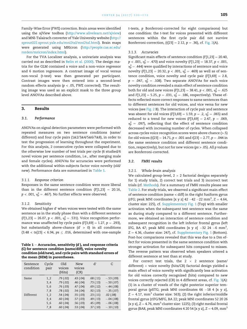
Table 1 e Accuracies, sensitivity (d′), and response criteria(C) for sentence condition (same/diff), voice noveltycondition (old/new), and cycle pairswith standard errors ofthe mean (SEM) in parentheses.
Sentencecondition
Cyclepair
Oldvoices(Hits)
Newvoices(CR)
d’ C
Same 1_2 .79 (.02) .43 (.04) .68 (.11) �.53 (.09)
3_4 .79 (.02) .46 (.04) .73 (.13) �.50 (.07)
5_6 .76 (.03) .47 (.04) .69 (.12) �.44 (.08)
7_8 .78 (.02) .54 (.04) .92 (.13) �.35 (.07)
Different 1_2 .54 (.04) .55 (.03) .23 (.11) .02 (.08)
3_4 .60 (.04) .57 (.03) .49 (.13) �.04 (.08)
5_6 .60 (.04) .56 (.03) .45 (.09) �.06 (.08)
7_8 .60 (.04) .53 (.04) .37 (.10) �.10 (.10)
t-tests, p Bonferroni-corrected for eight comparisons) but
one condition: the t-test for voices presented with different
sentences within the first cycle pair did not survive
Bonferroni-correction, (t[23] ¼ 2.12, p ¼ .36), cf. Fig. 2(A).
3.1.3. AccuraciesSignificant main effects of sentence condition (F[1,23] ¼ 20.40,
p < .001, hp2 ¼ .470) and voice novelty (F[1,23] ¼ 18.37, p < .001,
hp2 ¼ .444) were qualified by interactions of sentence and voice
novelty (F[1,23] ¼ 20.32, p < .001, hp2 ¼ .469) as well as of sen-
tence condition, voice novelty and cycle pair (F[3,69] ¼ 2.8,
p ¼ .047, hp2 ¼ .108). Two separate ANOVAs for each voice
novelty condition revealed amain effect of sentence condition
both for old and new voices (F[1,23] ¼ 38.41, p < .001, hp2 ¼ .625
and F[1,23] ¼ 5.25, p ¼ .031, hp2 ¼ .186, respectively). These ef-
fects reflectedmore correct responses to same sentences than
to different sentences for old voices, and vice versa for new
voices (see Fig. 2 B). The interaction of cycle pair and sentence
was absent for old voices (F[3,69] ¼ 1.59, p ¼ .2, hp2 ¼ .065) and
reduced to a trend for new voices (F[3,69] ¼ 2.47, p ¼ .069,
hp2 ¼ .097), reflecting that the effect of sentence condition
decreased with increasing number of cycles. When collapsed
across cycles voice recognition scores were above chance (>.5)for old voices (t[23] ¼ 14.71, p < .001 and t[23] ¼ 2.77, p¼ .044 in
the same sentence condition and different sentence condi-
tion, respectively), but not for new voices (ps > .05). All p values
are Bonferroni-corrected.
3.2. FMRI results
3.2.1. Whole-brain analysisWe calculated group-level, 2 � 2 factorial designs separately
for 1) study trials, 2) correct test trials and 3) incorrect test
trials (cf. Methods). For a summary of FMRI results please see
Table 2. For study trials, we observed a significant main effect
of sentence condition (same < diff) in the right fusiform gyrus
(rFG; peak MNI coordinates [x y z] 42 �42 �22 mm3, Z ¼ 4.64,
cluster size: 225), cf. Supplementary Fig. 1 (Top) with smaller
activation when the subsequent test sentence was the same
as during study compared to a different sentence. Further-
more, we obtained an interaction of sentence condition and
subsequent recognition in the left inferior frontal gyrus (left
IFG, BA 47; peak MNI coordinates [x y z] �32 24 �6 mm3,
Z ¼ 4.36, cluster size: 247), cf. Supplementary Fig. 1 (Bottom).
Post-hoc comparisons revealed that this was due to a Dm ef-
fect for voices presented in the same sentence condition with
stronger activation for subsequent hits compared to misses.
The reverse pattern was observed when speakers uttered a
different sentence at test than at study.
For correct test trials, the 2 � 2 sentence (same/
different) � voice novelty (hits/CR) factorial design yielded a
main effect of voice novelty with significantly less activation
for old voices correctly recognized (hits) compared to new
voices correctly rejected (CR) in 4 different areas, cf. Fig. 3(A):
(1) in a cluster of voxels of the right posterior superior tem-
poral gyrus (pSTG; peak MNI coordinates 66 �18 4 [x y z],
Z ¼ 4.17, mm3 cluster size: 563); (2) the right inferior/middle
frontal gyrus (rIFG/MFG, BA 22; peak MNI coordinates 52 20 26
[x y z], Z¼ 4.76, mm3 cluster size: 1225); (3) rightmedial frontal
gyrus (BA8, peak MNI coordinates 4 20 54 [x y z], Z ¼ 4.09, mm3
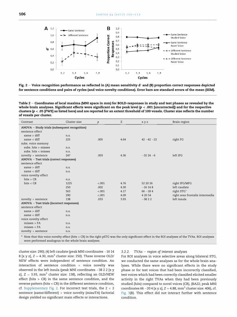
Fig. 2 e Voice recognition performance as reflected in (A) mean sensitivity d′ and (B) proportion correct responses depicted
for sentence conditions and pairs of cycles (and voice novelty conditions). Error bars are standard errors of the mean (SEM).
Table 2 e Coordinates of local maxima (MNI space in mm) for BOLD-responses in study and test phases as revealed by thewhole brain analyses. Significant effects were significant on the peak level (p < .001 [uncorrected]) and for the respectiveclusters (p < .05 [FWE] as listed here) and are reported for an extent threshold of 100 voxels. Cluster size reflects the numberof voxels per cluster.
Contrast Cluster size p Z x y z Brain region
ANOVA e Study trials (subsequent recognition)
sentence effect
same > diff n.s.
same < diff 225 .005 4.64 42 �42 �22 right FG
subs. voice memory
subs. hits > misses n.s.
subs. hits < misses n.s.
novelty � sentence 247 .003 4.36 �32 24 �6 left IFG
ANOVA e Test trials (correct responses)
sentence effect
same > diff n.s.
same < diff n.s.
voice novelty effect
hits > CR n.s.
hits < CR 1225 <.001 4.76 52 20 26 right IFG/MFG
250 .002 4.30 �16 14 8 left caudate
563 <.001 4.17 66 �18 4 right STGa
290 <.001 4.09 4 20 54 right area frontalis intermedia
novelty � sentence 138 .033 3.93 �38 2 2 left insula
ANOVA e Test trials (incorrect responses)
sentence effect
same > diff n.s.
same < diff n.s.
voice novelty effect
misses > FA n.s.
misses < FA n.s.
novelty � sentence n.s.
a Note that this voice novelty effect (hits < CR) in the right pSTG was the only significant effect in the ROI analyses of the TVAs. ROI analyses
were performed analogous to the whole brain analyses.
c o r t e x 9 4 ( 2 0 1 7 ) 1 0 0e1 1 2106
cluster size: 290); (4) left caudate (peakMNI coordinates�16 14
8 [x y z], Z ¼ 4.30, mm3 cluster size: 250). These inverse OLD/
NEW effects were independent of sentence condition. An
interaction of sentence condition � voice novelty was
observed in the left insula (peak MNI coordinates �38 2 2 [x y
z], Z ¼ 3.93, mm3 cluster size: 138), reflecting an OLD/NEW
effect (hits > CR) in the same sentence condition, and the
reverse pattern (hits < CR) in the different sentence condition,
cf. Supplementary Fig. 2. For incorrect test trials, the 2 � 2
sentence (same/different) � voice novelty (miss/FA) factorial
design yielded no significant main effects or interactions.
3.2.2. TVAs e region of interest analysesFor ROI analyses in voice selective areas along bilateral STG,
we conducted the same analyses as for the whole brain ana-
lyses. While there were no significant effects in the study
phase or for test voices that had been incorrectly classified,
test voices which had been correctly classified elicited smaller
activity in the right TVAs when they had been previously
studied (hits) compared to novel voices (CR), (BA22; peak MNI
coordinates 66�20 4 [x y z], Z ¼ 4.88, mm3 cluster size: 406), cf.
Fig. 3(B). This effect did not interact further with sentence
condition.

Fig. 3 e (A) Whole brain analysis of test phases. Brain areas sensitive to voice novelty (hits < CR) irrespective of sentence
condition in the right STG, right IFG/MFG, right medial frontal gyrus, and the left caudate. (B) ROI analysis of test phases in
bilateral voice-sensitive areas. Reduced activity to studied voices (hits) compared to novel voices (CR) independent of speech
content were observed in the right STG with no effect of sentence condition.
c o r t e x 9 4 ( 2 0 1 7 ) 1 0 0e1 1 2 107
4. Discussion
Here we report the first evidence that successful voice recog-
nition following learning of unfamiliar voices engages a
network of brain areas including right posterior temporal
voice areas (TVAs), the right inferior/middle frontal gyrus (IFG/
MFG) and medial frontal gyrus, as well as the left caudate
nucleus. Furthermore, in the study phase we observed brain
activity in the left IFG which was related to subsequent voice
recognition performance.
4.1. Recognition performance
As a replication of earlier findings we show that voice learning
with a few brief sentences results in above-chance voice
c o r t e x 9 4 ( 2 0 1 7 ) 1 0 0e1 1 2108
recognition that generalizes to new speech samples (e.g.,
Legge, Grosmann, & Pieper, 1984; Sheffert, Pisoni, Fellowes, &
Remez, 2002; Z€aske et al., 2014). This suggests that listeners
have acquired voice representations which store idiosyncratic
voice properties independent of speech content (see also
Z€aske et al., 2014). Notably, the present findingswere obtained
for listeners (mostly British) who were unfamiliar with the
speakers' language (German). This is remarkable in light of
research showing substantial impairments for the discrimi-
nation of unfamiliar speakers (Fleming, Giordano, Caldara, &
Belin, 2014) and speaker identification following voice
learning (Perrachione & Wong, 2007) for foreign versus native
language samples of speech. Language familiarity effects
likely arise from a lack of listeners' linguistic proficiency in the
foreign language which impedes the use of phonetic idio-
syncrasies for speaker identification. Note, however, that a
direct comparison between studies is limited by the fact that
discrimination of unfamiliar voices and the identification of
individual speakers by name may invoke partly different
cognitive mechanisms than the present task of old/new voice
recognition (Hanley & Turner, 2000; Schweinberger, Herholz,
& Sommer, 1997a; Van Lancker & Kreiman, 1987).
Although above-chance voice recognition (d0) was achieved
in both sentence conditions, performance was highest in the
same sentence condition, i.e., when study samples were
repeated at test. This is consistent with previous research
(e.g., Schweinberger, Herholz,& Stief, 1997b; Z€aske et al., 2014)
and reflects some interdependence of speech and speaker
perception (see also Perrachione & Wong, 2007; Perrachione,
Del Tufo, & Gabrieli, 2011; Remez, Fellowes, & Nagel, 2007).
In terms of accuracies, however, the same sentence condition
elicited both the highest and lowest performance, i.e., for old
and for new voices, respectively. Accordingly, whenever test
speakers repeated the study sentences, listeners tended to
perceive their voices as old. This is also reflected in a more
liberal response criterion in the same as compared to the
different sentence condition. Note, that we obtained these
results although it was pointed out to all participants prior to
the experiment that sentence content was not a valid cue to
speaker identity and that the task was voice recognition, not
sentence recognition.
4.2. Neural correlates of voice recognition followinglearning
Our fMRI data revealed reduced activation for old voices
correctly recognized as old compared to new voices correctly
rejected as new in the right posterior TVAs as well as pre-
frontal and subcortical areas (right IFG/MFG and medial
frontal gyrus as well as the left caudate nucleus). Crucially,
these effects of voice novelty were unaffected by whether or
not speakers repeated the study sentences at test suggesting
that activity in these areas is related to genuine voice identity
processing, i.e., independent of low-level speech-based acoustic
variability. This finding parallels our recent report of electro-
physiological correlates of voice recognition independent of
speech content (Z€aske et al., 2014). Essentially, Z€aske and
colleagues showed that successful voice recognition inde-
pendent of speech was accompanied by a reduction of beta
band oscillations (16e17 Hz, 290e370msec) for old versus new
test voices at central and right temporal sites. Note that this
right-lateralized topography of ERP recognition effects is
overall in linewith the present finding of predominantly right-
hemispheric involvement in voice recognition. However,
although Z€aske et al. used the same task and an almost
identical design and stimulus set, the main difference is that
in the present study, we investigated foreign-language voice
recognition rather than native-language voice recognition.
While the underlying neural processes may therefore not be
completely comparable (Perrachione, Pierrehumbert,&Wong,
2009), it remains possible that the electrophysiological voice
recognition effect (Z€aske et al., 2014) and the present effect in
BOLD responses are related to a similar mechanism. Specif-
ically, we suggest that the reduction in activity in the above
network of brain areas reflects access to speech-independent
high-level voice representations acquired during learning.
With respect to the TVA and the rIFC, our findings converge
well with previous reports that both are voice sensitive areas
(Belin et al., 2000; Blank et al., 2014) which respond to acoustic
voice properties and perceived identity information, respec-
tively (Andics et al., 2013; Latinus et al., 2011). Furthermore,
the rIFC has been associated with the processing of vocal
attractiveness (Bestelmeyer et al., 2012) and emotional pros-
ody (Fruehholz, Ceravolo, & Grandjean, 2012) as well as the
representation of voice gender (Charest et al., 2013). Although
there is no consensus as yet on the exact function of the rIFC
in voice processing, two recent studies suggest that it codes
perceived identity of voices in a prototype-referencedmanner
independently of voice-acoustic properties (Andics et al., 2013;
Latinus et al., 2011). Specifically, Andics et al. (2013) showed
that repeating prototypical versus less prototypical voice
samples of newly-learned speakers leads to an adaptation-
induced reduction of activity in the rIFC. Based on the above
studies, the present response reduction could in part reflect
neural adaptation to old voices relative to new voices.
Alternatively, the present effects may be related to the
explicit recognition of studied voices. To consider this possi-
bility, we have analyzed incorrect trials analogous to correct
trials. Specifically, we reasoned that if voice novelty modu-
lates activity in the same or in overlapping brain areas for both
types of trials, this may be indicative of implicit repetition-
related effects, rather than explicit recognition. Since no sig-
nificant effects emerged from these analyses we therefore
favor the view that the present novelty effects reflect explicit
recognition of learned voice identity. Thiswould be in line also
with neuroimaging research demonstrating that explicit
recognition of a target voice among other voices activates
bilateral frontal cortices compared to a task requiring the
recognition of speech content (von Kriegstein & Giraud, 2004).
In that study, bilateral frontal cortices were less activated
during attention to voices of (personally) familiar speakers
compared to unfamiliar speakers. This is similar to the pre-
sent study where with increasing voice familiarity, respon-
siveness of the rIFG/MFG decreases (old < new voices).
Additionally, von Kriegstein and Giraud showed that TVAs in
the right STS functionally interacted with the right inferior
parietal cortex and the right dorsolateral prefrontal cortex
during the recognition of unfamiliar voices. The latter finding
was attributed to increased difficulty of recognizing unfamil-
iar voices. Note, however, that unfamiliar voices in that study
c o r t e x 9 4 ( 2 0 1 7 ) 1 0 0e1 1 2 109
were not completely unfamiliar. Instead, and similar to the
present study, von Kriegstein and Giraud had briefly famil-
iarized participants with all voices and sentences prior to the
testing session. Therefore, rather than indicating task diffi-
culty, functional connections of TVAs with prefrontal cortex
in that study may alternatively reflect explicit recognition of
newly-learned voice identities, similar to the present study.
In addition, voice novelty was also found to modulate ac-
tivity in the right medial frontal gyrus (BA8) and the left
caudate nucleus. Although BA8 has been related to a number
of cognitive functions, in the context of the present study,
reduced responses in this area for old compared to new voices
could reflect relatively higher response certainty (Volz,
Schubotz, & von Cramon, 2005) for studied voices. Caudate
nuclei have been suggested to mediate stimulus-response
learning (Seger & Cincotta, 2005) and response inhibition
(Aron et al., 2003). Accordingly, the present modulation in this
area may be related to response selection processes during
voice classification.
At variancewith previous research on episodicmemory for
other classes of stimuli (reviewed in Cabeza et al., 2012;
Cabeza, Ciaramelli, Olson, & Moscovitch, 2008), we did not
find classical OLD/NEW effects (hits > CR) for voices in parietal
cortex areas. This may be due to the present analysis
approach which either targeted the whole brain with low
statistical power, or regions of interest (TVAs) outside the
parietal lobe.
Interestingly, the left insula was also sensitive to voice
novelty, however, with voice novelty effects depending on
sentence condition. When speakers repeated the study sen-
tences at test, old voices enhanced left insula activity relative
to new voices. The reverse pattern emerged when test
speakers uttered a different sentence. The insula has been
implicated in many tasks and has been discussed as a general
neural correlate of awareness (reviewed in Craig, 2009). In the
context of auditory research, the right insula has been sug-
gested to play a role in the processing of conspecific
communication sounds in primates (Remedios, Logothetis, &
Kayser, 2009). In humans, the left insula has been associated
with the processing of pitch patterns in speech (Wong,
Parsons, Martinez, & Diehl, 2004) and motor planning of
speech (Dronkers, 1996). It is further sensitive to non-
linguistic vocal information including emotional expressions
(Morris, Scott, & Dolan, 1999) and voice naturalness (Tamura,
Kuriki, & Nakano, 2015). In general, the insulae have been
found to respond more strongly to stimuli of negative valence
(reviewed in Phillips, Drevets, Rauch, & Lane, 2003) including
negative affective voices (Ethofer et al., 2009). Furthermore,
previous research suggest that insula activity may reflect
subjective familiarity with stronger responses in a network of
brain areas including the insular cortex for (perceived as) new
stimuli compared to familiar or repeated stimuli (e.g., Downar,
Crawley, Mikulis, & Davis, 2002; Linden et al., 1999; Plailly,
Tillmann, & Royet, 2007).
In the present study, the strongest responses in the left
insula have emerged for test samples that were either iden-
tical to studied voice samples, i.e., old voices which repeated
the study sentence at test, or which were maximally different
from the studied samples, i.e., new voices uttering a different
sentence at test. Based on the above research one could
speculate that two mechanisms underlie the present activity
pattern: while the recognition of stimulus-specific prosody in
the same sentence condition may have enhanced insula ac-
tivity for old relative to new voices, a particularly pronounced
feeling of “unfamiliarity” for different test sentences when
uttered by new voices relative to old voices may have
increased insula activity.
4.3. Neural correlates of subsequent voice memory
Here we show for the first time that activity in the left IFG
(BA47) interacts with subsequent voice memory, thereby
extending the episodic memory literature by an important
new class of auditory stimuli. Interestingly, the Dm effect for
voice memory depended on sentence condition: 1) study voi-
ces subsequently remembered elicited stronger responses
than study voices subsequently forgotten when speakers
uttered the same sentences at study and at test (classic Dm); 2)
conversely, voices subsequently remembered elicited weaker
responses than study voices subsequently forgotten when
speakers uttered different sentences at study and at test (in-
verse Dm). The first finding is in line with previous reports of
Dm effects for various stimuli as observed in a network of
areas including left and/or right inferior prefrontal regions for
identical study and test items (e.g., Kelley et al., 1998;
Klostermann et al., 2009; McDermott et al., 1999; Ranganath,
Johnson, & D'Esposito, 2003).The second effect, i.e., inverse Dm, has previously been
related to unsuccessful encoding of study items into memory,
however with effects typically located in ventral parietal and
posteromedial cortex (Cabeza et al., 2012; Huijbers et al., 2013)
rather than in the IFG. This inconsistency may be resolved
when considering that the present effects may reflect com-
bined effects of voice encoding and semantic retrieval pro-
cesses. The left IFG, and BA47 in particular, has been
repeatedly associated with language processing (e.g., Demb
et al., 1995; Lehtonen et al., 2005; Sahin, Pinker, & Halgren,
2006; Wong et al., 2002). For instance, Wong and colleagues
observed activations in the left BA47 in response to backward
speech, but not for meaningful forward speech suggesting
that this area is involved in the effortful attempt to retrieve
semantic information in (meaningless) backward speech.
Similarly, although our participants were unable to under-
stand the semantic content of the German utterances, the
present Dm effects may reflect the attempt to nevertheless
assign meaning to unintelligible speech. Depending on sen-
tence condition, this process might have elicited different
response patterns in the left IFG: while the association of se-
mantic meaning with study voices provided a beneficial
retrieval cue for the same stimuli at test (classic Dm), it may
have compromised voice recognition from different test sen-
tences (inverse Dm) which had not been previously associated
with the speaker.
An unexpected finding was that responses in the right
fusiform gyrus (FG) were decreased when study voices were
subsequently tested with the same sentence relative to
different sentences. The right FG is part of the face perception
network and hosts the fusiform face area (FFA; Kanwisher,
McDermott, & Chun, 1997). Although functional and
anatomical coupling of the FFA and TVA have been reported

c o r t e x 9 4 ( 2 0 1 7 ) 1 0 0e1 1 2110
for familiar speakers (Blank, Anwander, & von Kriegstein,
2011; von Kriegstein, Kleinschmidt, Sterzer, & Giraud, 2005),
it is difficult to reconcile these findings with the present sen-
tence effect. As a possible mechanism, learning unfamiliar
voices may have triggered facial imagery as mediated by the
right FG. However, it remains to be explored why this effect
was stronger in the same compared to the different sentence
condition.
5. Conclusions
In conclusion, the present study reports brain areas involved
in the learning and recognition of unfamiliar voices. This
relatively widespread network may serve several sub-
functions: During voice learning brain activity in the left IFG
was related to subsequent voice recognition performance
which further interacted with speech content. This suggests
that the left IFGmediates the interactive processing of speaker
and speech information while new voice representations are
being built. During voice recognition, correct recognition of
studied compared to novel voices was associated with
decreased activation in voice-selective areas of the right pSTG
and IFG/MFG, medial frontal gyrus, as well as the left caudate
nucleus. Importantly, these effects were independent of
speech content. We therefore suggest that these areas sub-
serve the access to speech-invariant high-level voice repre-
sentations for successful voice recognition following learning.
Specifically, while the right pSTG and IFG/MFG may process
idiosyncratic information about voice identity, the medial
frontal gyrus and left caudate may be involved in more gen-
eral mechanisms related to response certainty and response
selection.
In view of other research pointing to differential voice
processing depending on whether listeners are familiar with
the speakers' language (Perrachione & Wong, 2007;
Perrachione et al., 2009), the precise role of comprehensible
speech for neuroimaging correlates of voice learning will be
an interesting question for future research. Since we obtained
the present findings with listeners who were unfamiliar with
the speaker's language, the present findings arguably reflect a
rather general mechanism of voice learning that is largely
devoid of speech-related semantic processes.
Acknowledgments
This research was funded by the Deutsche For-
schungsgemeinschaft (DFG), grant ZA 745/1-1 and ZA 745/1-2
to RZ, and by grants BB/E003958/1 from BBSRC (UK), large
grant RES-060-25-0010 by ESRC/MRC, and grant AJE201214 by
the Fondation pour la Recherche M�edicale (France) to P.B. We
thank Leonie Fresz, Achim H€otzel, Christoph Klebl, Katrin
Lehmann, Carolin Leistner, Constanze Muhl, Finn Pauls,
Marie-Christin Perlich, Johannes Pfund,Mathias Riedel, Saskia
Rudat and Meike Wilken for stimulus acquisition and editing.
We are also thankful to David Fleming and Emilie Salvia for
Matlab support, Francis Crabbe for assistance in fMRI scan-
ning, and Stefan Schweinberger as well as Marlena Itz for
helpful comments on earlier drafts of thismanuscript. Finally,
we thank two anonymous reviewers for helpful comments.
Supplementary data
Supplementary data related to this article can be found at
http://dx.doi.org/10.1016/j.cortex.2017.06.005.
r e f e r e n c e s
Andics, A., McQueen, J. M., & Petersson, K. M. (2013). Mean-basedneural coding of voices. NeuroImage, 79, 351e360.
Aron, A. R., Schlaghecken, F., Fletcher, P. C., Bullmore, E. T.,Eimer, M., Barker, R., et al. (2003). Inhibition of subliminallyprimed responses is mediated by the caudate and thalamus:Evidence from functional mri and huntington's disease. Brain:a Journal of Neurology, 126, 713e723.
Belin, P., Bestelmeyer, P. E., Latinus, M., & Watson, R. (2011).Understanding voice perception. British Journal of Psychology,102, 711e725.
Belin, P., Fecteau, S., & Bedard, C. (2004). Thinking the voice:Neural correlates of voice perception. Trends in CognitiveSciences, 8(3), 129e135.
Belin, P., & Zatorre, R. J. (2003). Adaptation to speaker's voice inright anterior temporal lobe. NeuroReport, 14(16), 2105e2109.
Belin, P., Zatorre, R. J., Lafaille, P., Ahad, P., & Pike, B. (2000). Voice-selective areas in human auditory cortex. Nature, 403(6767),309e312.
Bestelmeyer, P. E. G., Latinus, M., Bruckert, L., Rouger, J.,Crabbe, F., & Belin, P. (2012). Implicitly perceived vocalattractiveness modulates prefrontal cortex activity. CerebralCortex, 22(6), 1263e1270.
Blank, H., Anwander, A., & von Kriegstein, K. (2011). Directstructural connections between voice- and face-recognitionareas. The Journal of Neuroscience: the Official Journal of the Societyfor Neuroscience, 31(36), 12906e12915.
Blank, H., Wieland, N., & von Kriegstein, K. (2014). Personrecognition and the brain: Merging evidence from patientsand healthy individuals. Neuroscience and Biobehavioral Reviews,47, 17.
Boersma, P., & Weenink, D. (2001). Praat, a system for doingphonetics by computer. Glot International, 5(9/10), 341e345.
Bricker, P. D., & Pruzansky, S. (1966). Effects of stimulus contentand duration on talker identification. Journal of the AcousticalSociety of America, 40(6), 1441.
Cabeza, R., Ciaramelli, E., & Moscovitch, M. (2012). Cognitivecontributions of the ventral parietal cortex: An integrativetheoretical account. Trends in Cognitive Sciences, 16(6),338e352.
Cabeza, R., Ciaramelli, E., Olson, I. R., & Moscovitch, M. (2008). Theparietal cortex and episodic memory: An attentional account.Nature Reviews Neuroscience, 9(8), 613e625.
Charest, I., Pernet, C., Latinus, M., Crabbe, F., & Belin, P. (2013).Cerebral processing of voice gender studied using acontinuous carryover fmri design. Cerebral Cortex, 23(4),958e966.
Craig, A. D. (2009). How do you feel - Now? The anterior insulaand human awareness. Nature Reviews Neuroscience, 10(1),59e70.
Demb, J. B., Desmond, J. E., Wagner, A. D., Vaidya, C. J.,Glover, G. H., & Gabrieli, J. D. E. (1995). Semantic encoding andretrieval in the left inferior prefrontal cortex - A functional mristudy of task-difficulty and process specificity. Journal ofNeuroscience, 15(9), 5870e5878.
c o r t e x 9 4 ( 2 0 1 7 ) 1 0 0e1 1 2 111
Downar, J., Crawley, A. P., Mikulis, D. J., & Davis, K. D. (2002). Acortical network sensitive to stimulus salience in a neutralbehavioral context across multiple sensory modalities. Journalof Neurophysiology, 87(1), 615e620.
Dronkers, N. F. (1996). A new brain region for coordinating speecharticulation. Nature, 384(6605), 159e161.
Ethofer, T., Kreifelts, B., Wiethoff, S., Wolf, J., Grodd, W.,Vuilleumier, P., et al. (2009). Differential influences of emotion,task, and novelty on brain regions underlying the processingof speech melody. Journal of Cognitive Neuroscience, 21(7),1255e1268.
Fleming, D., Giordano, B. L., Caldara, R., & Belin, P. (2014). Alanguage-familiarity effect for speaker discrimination withoutcomprehension. Proceedings of the National Academy of Sciencesof the United States of America, 111(38), 13795e13798.
Formisano, E., De Martino, F., Bonte, M., & Goebel, R. (2008).“Who” is saying “what”? Brain-based decoding of human voiceand speech. Science, 322(5903), 970e973.
Fruehholz, S., Ceravolo, L., & Grandjean, D. (2012). Specific brainnetworks during explicit and implicit decoding of emotionalprosody. Cerebral Cortex, 22(5), 1107e1117.
Hanley, J. R., & Turner, J. M. (2000). Why are familiar-onlyexperiences more frequent for voices than for faces? QuarterlyJournal of Experimental Psychology A Human ExperimentalPsychology, 53(4), 1105e1116.
Huijbers, W., Schultz, A. P., Vannini, P., McLaren, D. G.,Wigman, S. E., Ward, A. M., et al. (2013). The encoding/retrievalflip: Interactions between memory performance and memorystage and relationship to intrinsic cortical networks. Journal ofCognitive Neuroscience, 25(7), 1163e1179.
Huynh, H., & Feldt, L. S. (1976). Estimation of the box correctionfor degrees of freedom from sample data in randomized blockand split block designs. Journal of Educational Statistics, 1,69e82.
Kanwisher, N., McDermott, J., & Chun, M. M. (1997). The fusiformface area: A module in human extrastriate cortex specializedfor face perception. Journal of Neuroscience, 17(11), 4302e4311.
Kaufmann, J. M., Schweinberger, S. R., & Burton, A. (2009). N250erp correlates of the acquisition of face representations acrossdifferent images. Journal of Cognitive Neuroscience, 21(4),625e641.
Kelley, W. M., Miezin, F. M., McDermott, K. B., Buckner, R. L.,Raichle, M. E., Cohen, N. J., et al. (1998). Hemisphericspecialization in human dorsal frontal cortex and medialtemporal lobe for verbal and nonverbal memory encoding.Neuron, 20(5), 927e936.
Klostermann, E. C., Kane, A. J. M., & Shimamura, A. P. (2008).Parietal activation during retrieval of abstract and concreteauditory information. NeuroImage, 40(2), 896e901.
Klostermann, E. C., Loui, P., & Shimamura, A. P. (2009). Activationof right parietal cortex during memory retrieval ofnonlinguistic auditory stimuli. Cognitive Affective & BehavioralNeuroscience, 9(3), 242e248.
von Kriegstein, K., & Giraud, A. L. (2004). Distinct functionalsubstrates along the right superior temporal sulcus for theprocessing of voices. NeuroImage, 22(2), 948e955.
von Kriegstein, K., Kleinschmidt, A., Sterzer, P., & Giraud, A. L.(2005). Interaction of face and voice areas during speakerrecognition. Journal of Cognitive Neuroscience, 17(3), 367e376.
Latinus, M., Crabbe, F., & Belin, P. (2011). Learning-inducedchanges in the cerebral processing of voice identity. CerebralCortex, 21(12), 2820e2828.
Latinus, M., McAleer, P., Bestelmeyer, P. E., & Belin, P. (2013).Norm-based coding of voice identity in human auditorycortex. Current Biology, 23(12), 1075e1080.
Legge, G. E., Grosmann, C., & Pieper, C. M. (1984). Learningunfamiliar voices. Journal of Experimental Psychology LearningMemory and Cognition, 10(2), 298e303.
Lehtonen, M. H., Laine, M., Niemi, J., Thomsen, T., Vorobyev, V. A.,& Hugdahl, K. (2005). Brain correlates of sentence translationin Finnish-norwegian bilinguals. NeuroReport, 16(6), 607e610.
Linden, D. E. J., Prvulovic, D., Formisano, E., Vollinger, M.,Zanella, F. E., Goebel, R., et al. (1999). The functionalneuroanatomy of target detection: An fmri study of visual andauditory oddball tasks. Cerebral Cortex, 9(8), 815e823.
McDermott, K. B., Buckner, R. L., Petersen, S. E., Kelley, W. M., &Sanders, A. L. (1999). Set- and code-specific activation in thefrontal cortex: An fmri study of encoding and retrieval offaces and words. Journal of Cognitive Neuroscience, 11(6),631e640.
Morris, J. S., Scott, S. K., & Dolan, R. J. (1999). Saying it with feeling:Neural responses to emotional vocalizations. Neuropsychologia,37(10), 1155e1163.
Mullennix, J., Ross, A., Smith, C., Kuykendall, K., Conard, J., &Barb, S. (2011). Typicality effects on memory for voice:Implications for earwitness testimony. Applied CognitivePsychology, 25(1), 29e34.
Paller, K. A., & Wagner, A. D. (2002). Observing the transformationof experience into memory. Trends in Cognitive Sciences, 6(2),93e102.
Pernet, C. R., McAleer, P., Latinus, M., Gorgolewski, K. J.,Charest, I., Bestelmeyer, P. E. G., et al. (2015). The human voiceareas: Spatial organization and inter-individual variability intemporal and extra-temporal cortices. NeuroImage, 119,164e174.
Perrachione, T. K., Del Tufo, S. N., & Gabrieli, J. D. (2011). Humanvoice recognition depends on language ability. Science,333(6042), 595.
Perrachione, T. K., Pierrehumbert, J. B., & Wong, P. C. M. (2009).Differential neural contributions to native- and foreign-language talker identification. Journal of Experimental PsychologyHuman Perception and Performance, 35(6), 1950e1960.
Perrachione, T. K., & Wong, P. C. M. (2007). Learning to recognizespeakers of a non-native language: Implications for thefunctional organization of human auditory cortex.Neuropsychologia, 45(8), 1899e1910.
Phillips, M. L., Drevets, W. C., Rauch, S. L., & Lane, R. (2003).Neurobiology of emotion perception I: The neural basis ofnormal emotion perception. Biological Psychiatry, 54(5),504e514.
Plailly, J., Tillmann, B., & Royet, J. P. (2007). The feeling offamiliarity of music and odors: The same neural signature?Cerebral Cortex, 17(11), 2650e2658.
Pollack, I., Pickett, J. M., & Sumby, W. H. (1954). On theidentification of speakers by voice. Journal of the AcousticalSociety of America, 26(3), 403e406.
Ranganath, C., Johnson, M. K., & D'Esposito, M. (2003). Prefrontalactivity associated with working memory and episodic long-term memory. Neuropsychologia, 41(3), 378e389.
Remedios, R., Logothetis, N. K., & Kayser, C. (2009). An auditoryregion in the primate insular cortex responding preferentiallyto vocal communication sounds. Journal of Neuroscience, 29(4),1034e1045.
Remez, R. E., Fellowes, J. M., & Nagel, D. S. (2007). On theperception of similarity among talkers. Journal of the AcousticalSociety of America, 122(6), 3688e3696.
Sahin, N. T., Pinker, S., & Halgren, E. (2006). Abstract grammaticalprocessing of nouns and verbs in broca's area: Evidence fromfmri. Cortex; a Journal Devoted To the Study of the Nervous Systemand Behavior, 42(4), 540e562.
Schall, S., Kiebel, S. J., Maess, B., & von Kriegstein, K. (2015). Voiceidentity recognition: Functional division of the right sts and itsbehavioral relevance. Journal of Cognitive Neuroscience, 27(2),280e291.
Schweinberger, S. R., Herholz, A., & Sommer, W. (1997a).Recognizing famous voices: Influence of stimulus duration
c o r t e x 9 4 ( 2 0 1 7 ) 1 0 0e1 1 2112
and different types of retrieval cues. Journal of Speech Languageand Hearing Research, 40(2), 453e463.
Schweinberger, S. R., Herholz, A., & Stief, V. (1997b). Auditorylong-term memory: Repetition priming of voice recognition.Quarterly Journal of Experimental Psychology A HumanExperimental Psychology, 50(3), 498e517.
Seger, C. A., & Cincotta, C. M. (2005). The roles of the caudatenucleus in human classification learning. Journal ofNeuroscience, 25(11), 2941e2951.
Sheffert, S. M., Pisoni, D. B., Fellowes, J. M., & Remez, R. E. (2002).Learning to recognize talkers from natural, sinewave, andreversed speech samples. Journal of Experimental PsychologyHuman Perception and Performance, 28(6), 1447e1469.
Skuk, V. G., & Schweinberger, S. R. (2013). Gender differences infamiliar voice identification. Hearing Research, 295, 131e140.
Stanislaw, H., & Todorov, N. (1999). Calculation of signal detectiontheory measures. Behavior Research Methods Instruments &Computers, 31(1), 137e149.
Tamura, Y., Kuriki, S., & Nakano, T. (2015). Involvement of the leftinsula in the ecological validity of the human voice. ScientificReports, 5.
Valentine, T., & Bruce, V. (1986). The effects of distinctiveness inrecognizing and classifying faces. Perception, 15(5), 525e535.
Valentine, T., & Ferrara, A. (1991). Typicality in categorization,recognition and identification - evidence from facerecognition. British Journal of Psychology, 82, 87e102.
Van Lancker, D. R., & Kreiman, J. (1987). Voice discrimination andrecognition are separate abilities. Neuropsychologia, 25(5),829e834.
Volz, K. G., Schubotz, R. I., & von Cramon, D. Y. (2005). Variants ofuncertainty in decision-making and their neural correlates.Brain Research Bulletin, 67(5), 403e412.
Wagner, A. D., Shannon, B. J., Kahn, I., & Buckner, R. L. (2005).Parietal lobe contributions to episodic memory retrieval.Trends in Cognitive Sciences, 9(9), 445e453.
Wong, P. C.M., Parsons, L.M.,Martinez,M., &Diehl, R. L. (2004). Therole of the insular cortex in pitch pattern perception: The effectof linguistic contexts. Journal of Neuroscience, 24(41), 9153e9160.
Wong, D., Pisoni, D. B., Learn, J., Gandour, J. T., Miyamoto, R. T., &Hutchins, G. D. (2002). Pet imaging of differential corticalactivation by monaural speech and nonspeech stimuli.Hearing Research, 166(1e2), 9e23.
Yovel, G., & Belin, P. (2013). A unified coding strategy forprocessing faces and voices. Trends in Cognitive Sciences, 17(6),263e271.
Z€aske, R., Volberg, G., Kovacs, G., & Schweinberger, S. R. (2014).Electrophysiological correlates of voice learning andrecognition. Journal of Neuroscience, 34(33), 10821e10831.
Zimmermann, F. G., & Eimer, M. (2013). Face learning and theemergence of view-independent face recognition: An event-related brain potential study. Neuropsychologia, 51(7),1320e1329.