Embed Size (px)
Citation preview
RESEARCH ARTICLE
Roles of RECQ helicases in recombination based DNArepair, genomic stability and aging
Dharmendra Kumar Singh Æ Byungchan Ahn ÆVilhelm A. Bohr
Received: 27 August 2008 / Accepted: 24 November 2008 / Published online: 15 December 2008
� Springer Science+Business Media B.V. 2008
Abstract The maintenance of the stability of
genetic material is an essential feature of every living
organism. Organisms across all kingdoms have
evolved diverse and highly efficient repair mecha-
nisms to protect the genome from deleterious
consequences of various genotoxic factors that might
tend to destabilize the integrity of the genome in each
generation. One such group of proteins that is actively
involved in genome surveillance is the RecQ helicase
family. These proteins are highly conserved DNA
helicases, which have diverse roles in multiple DNA
metabolic processes such as DNA replication, recom-
bination and DNA repair. In humans, five RecQ
helicases have been identified and three of them
namely, WRN, BLM and RecQL4 have been linked to
genetic diseases characterized by genome instability,
premature aging and cancer predisposition. This
helicase family plays important roles in various
DNA repair pathways including protecting the
genome from illegitimate recombination during chro-
mosome segregation in mitosis and assuring genome
stability. This review mainly focuses on various roles
of human RecQ helicases in the process of recombi-
nation-based DNA repair to maintain genome stability
and physiological consequences of their defects in the
development of cancer and premature aging.
Keywords Genome stability � RecQ helicases �Homologous recombination (HR) �Double strand break (DSB) �Non-homologous end joining (NHEJ)
Introduction
The stability of the genetic material from one
generation to another is an important and essential
feature for every living organism. The failure to do so
efficiently can lead to chromosomal abnormalities,
developmental abnormalities, progression of cancer
and premature aging. Living organisms encounter
different kinds of stresses provided by genotoxic
elements from within (products of normal cellular
metabolism i.e., ROS) or outside the cell (environ-
mental factors such as radiation, chemicals, etc.)
during the faithful transmission of their genetic
material. To counteract these stress responses, organ-
isms have evolved diverse and highly efficient DNA
repair pathways. The repair proteins function in
a highly coordinated fashion with other DNA
metabolic processes during the different stages of
the cell cycle to maintain genome integrity in each
D. K. Singh � V. A. Bohr (&)
Laboratory of Molecular Gerontology, Biomedical
Research Center, National Institute on Aging, NIH,
251 Bayview Boulevard, Baltimore, MD 21224, USA
e-mail: [email protected]
B. Ahn
Department of Life Sciences, University of Ulsan,
Ulsan 680-749, South Korea
123
Biogerontology (2009) 10:235–252
DOI 10.1007/s10522-008-9205-z
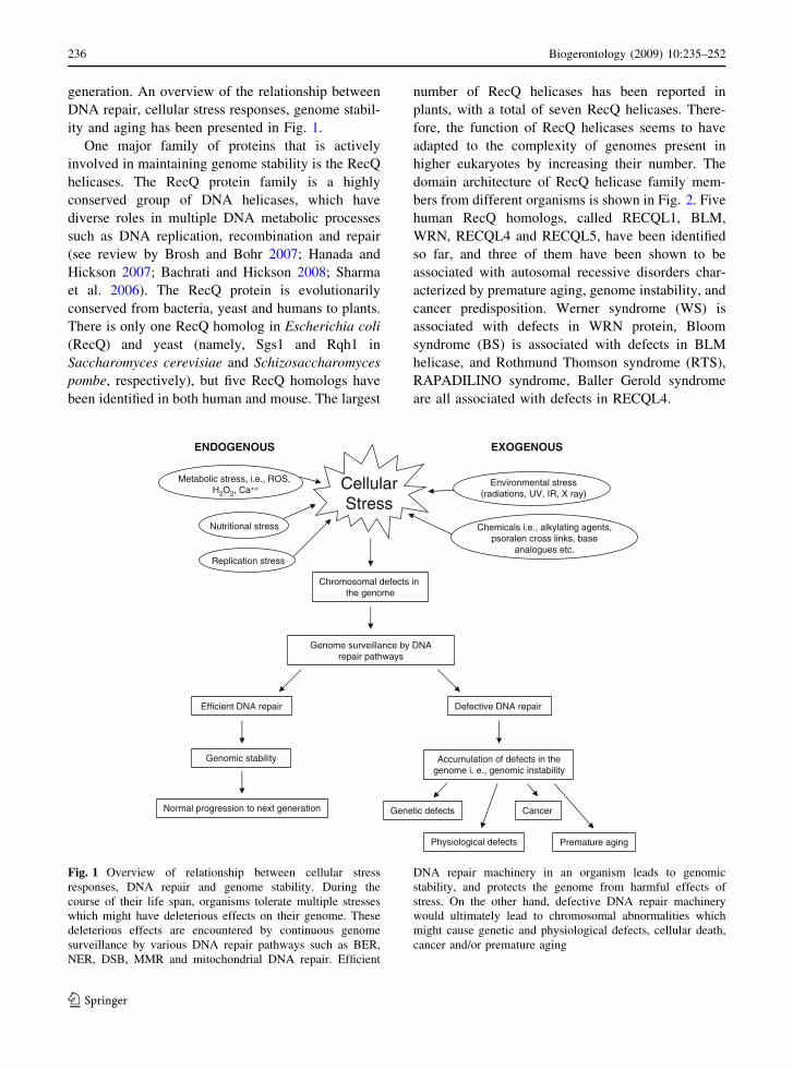
generation. An overview of the relationship between
DNA repair, cellular stress responses, genome stabil-
ity and aging has been presented in Fig. 1.
One major family of proteins that is actively
involved in maintaining genome stability is the RecQ
helicases. The RecQ protein family is a highly
conserved group of DNA helicases, which have
diverse roles in multiple DNA metabolic processes
such as DNA replication, recombination and repair
(see review by Brosh and Bohr 2007; Hanada and
Hickson 2007; Bachrati and Hickson 2008; Sharma
et al. 2006). The RecQ protein is evolutionarily
conserved from bacteria, yeast and humans to plants.
There is only one RecQ homolog in Escherichia coli
(RecQ) and yeast (namely, Sgs1 and Rqh1 in
Saccharomyces cerevisiae and Schizosaccharomyces
pombe, respectively), but five RecQ homologs have
been identified in both human and mouse. The largest
number of RecQ helicases has been reported in
plants, with a total of seven RecQ helicases. There-
fore, the function of RecQ helicases seems to have
adapted to the complexity of genomes present in
higher eukaryotes by increasing their number. The
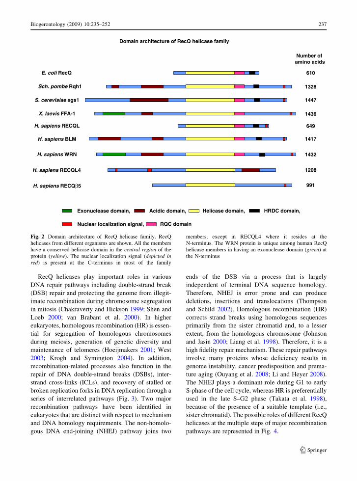
domain architecture of RecQ helicase family mem-
bers from different organisms is shown in Fig. 2. Five
human RecQ homologs, called RECQL1, BLM,
WRN, RECQL4 and RECQL5, have been identified
so far, and three of them have been shown to be
associated with autosomal recessive disorders char-
acterized by premature aging, genome instability, and
cancer predisposition. Werner syndrome (WS) is
associated with defects in WRN protein, Bloom
syndrome (BS) is associated with defects in BLM
helicase, and Rothmund Thomson syndrome (RTS),
RAPADILINO syndrome, Baller Gerold syndrome
are all associated with defects in RECQL4.
CellularStress
Chromosomal defects in the genome
Genome surveillance by DNA repair pathways
Efficient DNA repair Defective DNA repair
Chemicals i.e., alkylating agents,psoralen cross links, base
analogues etc.
Metabolic stress, i.e., ROS, H2O2, Ca++
Environmental stress(radiations, UV, IR, X ray)
Nutritional stress
Replication stress
Genomic stability Accumulation of defects in the genome i. e., genomic instability
Normal progression to next generation
SUONEGOXESUONEGODNE
Genetic defects
Physiological defects
Cancer
Premature aging
Fig. 1 Overview of relationship between cellular stress
responses, DNA repair and genome stability. During the
course of their life span, organisms tolerate multiple stresses
which might have deleterious effects on their genome. These
deleterious effects are encountered by continuous genome
surveillance by various DNA repair pathways such as BER,
NER, DSB, MMR and mitochondrial DNA repair. Efficient
DNA repair machinery in an organism leads to genomic
stability, and protects the genome from harmful effects of
stress. On the other hand, defective DNA repair machinery
would ultimately lead to chromosomal abnormalities which
might cause genetic and physiological defects, cellular death,
cancer and/or premature aging
236 Biogerontology (2009) 10:235–252
123
RecQ helicases play important roles in various
DNA repair pathways including double-strand break
(DSB) repair and protecting the genome from illegit-
imate recombination during chromosome segregation
in mitosis (Chakraverty and Hickson 1999; Shen and
Loeb 2000; van Brabant et al. 2000). In higher
eukaryotes, homologous recombination (HR) is essen-
tial for segregation of homologous chromosomes
during meiosis, generation of genetic diversity and
maintenance of telomeres (Hoeijmakers 2001; West
2003; Krogh and Symington 2004). In addition,
recombination-related processes also function in the
repair of DNA double-strand breaks (DSBs), inter-
strand cross-links (ICLs), and recovery of stalled or
broken replication forks in DNA replication through a
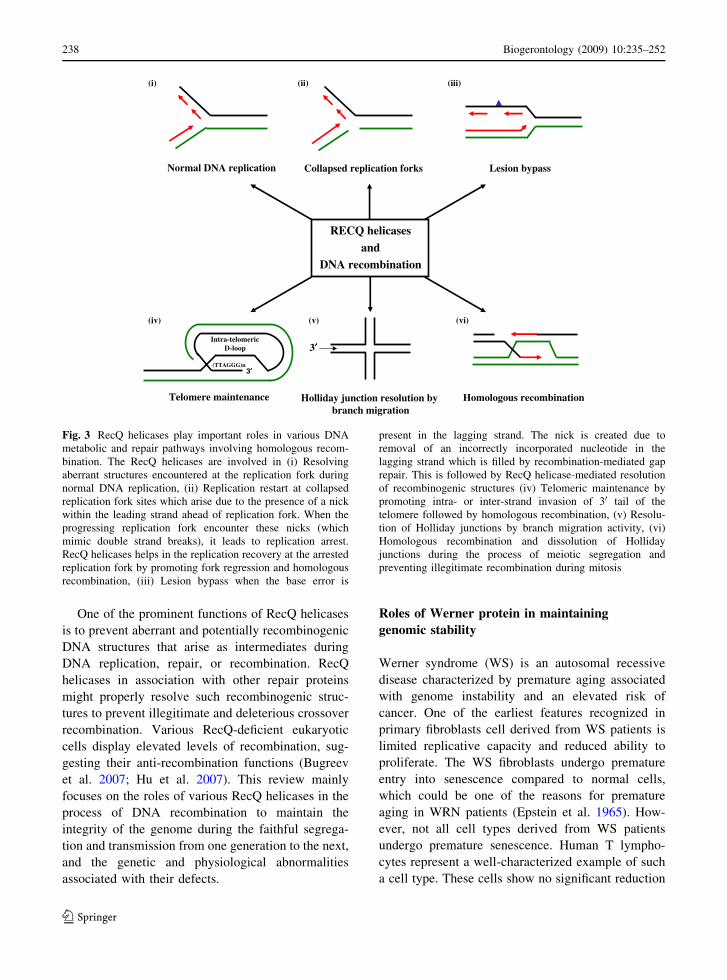
series of interrelated pathways (Fig. 3). Two major
recombination pathways have been identified in
eukaryotes that are distinct with respect to mechanism
and DNA homology requirements. The non-homolo-
gous DNA end-joining (NHEJ) pathway joins two
ends of the DSB via a process that is largely
independent of terminal DNA sequence homology.
Therefore, NHEJ is error prone and can produce
deletions, insertions and translocations (Thompson
and Schild 2002). Homologous recombination (HR)
corrects strand breaks using homologous sequences
primarily from the sister chromatid and, to a lesser
extent, from the homologous chromosome (Johnson
and Jasin 2000; Liang et al. 1998). Therefore, it is a
high fidelity repair mechanism. These repair pathways
involve many proteins whose deficiency results in
genome instability, cancer predisposition and prema-
ture aging (Ouyang et al. 2008; Li and Heyer 2008).
The NHEJ plays a dominant role during G1 to early
S-phase of the cell cycle, whereas HR is preferentially
used in the late S–G2 phase (Takata et al. 1998),
because of the presence of a suitable template (i.e.,
sister chromatid). The possible roles of different RecQ
helicases at the multiple steps of major recombination
pathways are represented in Fig. 4.
E. coli RecQ 610
Sch. pombe Rqh1 1328
S. cerevisiae sgs1 1447
X. laevis FFA-1 1436
H. sapiens RECQL 649
H. sapiens BLM 1417
H. sapiens WRN 1432
H. sapiens RECQL4 1208
H. sapiens RECQβ5 991
Number of amino acids
Exonuclease domain, Acidic domain, Helicase domain, HRDC domain,
Nuclear localization signal,
Domain architecture of RecQ helicase family
RQC domain
Fig. 2 Domain architecture of RecQ helicase family. RecQ
helicases from different organisms are shown. All the members
have a conserved helicase domain in the central region of the
protein (yellow). The nuclear localization signal (depicted inred) is present at the C-terminus in most of the family
members, except in RECQL4 where it resides at the
N-terminus. The WRN protein is unique among human RecQ
helicase members in having an exonuclease domain (green) at
the N-terminus
Biogerontology (2009) 10:235–252 237
123
One of the prominent functions of RecQ helicases
is to prevent aberrant and potentially recombinogenic
DNA structures that arise as intermediates during
DNA replication, repair, or recombination. RecQ
helicases in association with other repair proteins
might properly resolve such recombinogenic struc-
tures to prevent illegitimate and deleterious crossover
recombination. Various RecQ-deficient eukaryotic
cells display elevated levels of recombination, sug-
gesting their anti-recombination functions (Bugreev
et al. 2007; Hu et al. 2007). This review mainly
focuses on the roles of various RecQ helicases in the
process of DNA recombination to maintain the
integrity of the genome during the faithful segrega-
tion and transmission from one generation to the next,
and the genetic and physiological abnormalities
associated with their defects.
Roles of Werner protein in maintaining
genomic stability
Werner syndrome (WS) is an autosomal recessive
disease characterized by premature aging associated
with genome instability and an elevated risk of
cancer. One of the earliest features recognized in
primary fibroblasts cell derived from WS patients is
limited replicative capacity and reduced ability to
proliferate. The WS fibroblasts undergo premature
entry into senescence compared to normal cells,
which could be one of the reasons for premature
aging in WRN patients (Epstein et al. 1965). How-
ever, not all cell types derived from WS patients
undergo premature senescence. Human T lympho-
cytes represent a well-characterized example of such
a cell type. These cells show no significant reduction
Collapsed replication forksNormal DNA replication
Homologous recombination
RECQ helicases
andDNA recombination
3′′
Holliday junction resolution by branch migration
Telomere maintenance
3′′(TTAGGG)n
Intra-telomericD-loop
Lesion bypass
)iii()ii()i(
)iv()v()vi(
Fig. 3 RecQ helicases play important roles in various DNA
metabolic and repair pathways involving homologous recom-
bination. The RecQ helicases are involved in (i) Resolving
aberrant structures encountered at the replication fork during
normal DNA replication, (ii) Replication restart at collapsed
replication fork sites which arise due to the presence of a nick
within the leading strand ahead of replication fork. When the
progressing replication fork encounter these nicks (which
mimic double strand breaks), it leads to replication arrest.
RecQ helicases helps in the replication recovery at the arrested
replication fork by promoting fork regression and homologous
recombination, (iii) Lesion bypass when the base error is
present in the lagging strand. The nick is created due to
removal of an incorrectly incorporated nucleotide in the
lagging strand which is filled by recombination-mediated gap
repair. This is followed by RecQ helicase-mediated resolution
of recombinogenic structures (iv) Telomeric maintenance by
promoting intra- or inter-strand invasion of 30 tail of the
telomere followed by homologous recombination, (v) Resolu-
tion of Holliday junctions by branch migration activity, (vi)
Homologous recombination and dissolution of Holliday
junctions during the process of meiotic segregation and
preventing illegitimate recombination during mitosis
238 Biogerontology (2009) 10:235–252
123

in growth capacity compared to normal controls
(James et al. 2000), but they are hypersensitive to
DNA damaging agents, with aberrations consistent
with WRN deficiency (Fukuchi et al. 1990). Cytoge-
netic observations of WS cells show various types of
chromosomal aberrations including deletions, trans-
locations, and rearrangements as well as increased
spontaneous mutation (Hoehn et al. 1975; Salk et al.
1985; Fukuchi et al. 1989). Various studies have
observed that WS cells show selective sensitivity
towards many different types of DNA damaging
agents such as 4-nitroquinoline-N-oxide (4-NQO,
causing replication fork stalling), camptothecin
(CPT) (causing replication fork collapse), ICLs, and
ionizing radiation (IR) (Okada et al. 1998; Poot et al.
1999, 2002; Ogburn et al. 1997; Pichierri et al. 2000).
Thus, the WS cellular phenotype is dependent on the
sensitivities of WS cells to different DNA damaging
agents and efficiency of DNA repair mechanisms
including HR to counteract these damages.
Roles of Werner in genetic recombination
Cellular studies have shown marked reduction in cell
proliferation following mitotic recombination in WS
fibroblasts (Prince et al. 2001). These findings
suggested a role of WRN in mitotic recombination.
Prince et al. (2001), showed spontaneous genome
instability of WS cells by measuring the capacity for
mitotic recombination of the WS fibroblasts cell
lines. This group observed that WS fibroblast cell
lines failed to resolve recombinant products. Thus, in
the absence of WRN, unresolved or disrupted gene
conversion products may lead to gene rearrangement
or loss mediated by other processes and result in
recombination-initiated mitotic arrest, and cell death
(Prince et al. 2001).
Several lines of evidence suggest that WRN is
actively involved in the homologous recombination
pathway. Saintigny et al. (2002), have postulated that
the physiological role of WRN protein in the cell is to
Synapsis
DNA-PKcs/Ku/WRN
Helicase
Processing/Gap filling
Ligase IVXRCC4
Error-prone
RAD51/BRCA2RPA
Homology search and strand invasion,
5’3’
Error-free
DNA polymerasesLigaseResolvases
RAD52RAD54M/R/N
BRCA1/Bard1
H2AX
Exonuclease
ATM
M/R/N
5′′ strand resection
5’3’
Rad51 mediated nucleoprotein
filament formation
D-loop extension and promotion of DNA
synthesis
RecQ helicases interactome
and major recombination
pathways
)enorp rorre()eerf rorre(
Homologous recombination Non-homologous end-joining
Fig. 4 RecQ helicases are involved in multiple steps of major
recombination pathways. The members of the RecQ helicase
family interacts with various proteins involved in different
steps of the major recombination pathways i.e., error free
homologous recombination (HR) pathway and error prone non-
homologous end-joining (NHEJ) pathway. (See text for details)
Biogerontology (2009) 10:235–252 239
123
resolve RAD51-mediated homologous recombination
(HR) products, and its failure to do so in WS leads to
WS cellular phenotypes such as defective recombina-
tion resolution, mitotic arrest, cell death, or genomic
instability (Saintigny et al. 2002). In accordance with
this hypothesis, it has also been shown that WRN
interacts physically and functionally with the homol-
ogous recombination mediator/single strand annealing
protein RAD52 which has been found at arrested
replication forks. Biochemical studies have shown that
RAD52 both inhibits and enhances WRN helicase
activity in a DNA structure dependent manner. WRN,
in turn, stimulates RAD52-mediated homologous
strand annealing between complementary sequences.
Thus, the coordinated activities of WRN and RAD52
may be involved in replication fork rescue after DNA
damage (Baynton et al. 2003).
Another major recombination pathway is the
NHEJ pathway that is error-prone rejoining process
of DSBs. A defect in this pathway consequently
results in a loss or gain of genetic information.
Oshima et al. (2002) found extensive deletions at non
homologous joining ends of the linear plasmids with
incompatible ends when introduced into WS cells.
Thus, WRN might suppress extensive nucleotide loss
during NHEJ and prevent aberrant DNA repair
potentially by stabilizing the broken DNA ends or
by direct competition with other helicases or exon-
ucleases (Oshima et al. 2002). Thus, in the absence of
WRN, regulatory processes controlling NHEJ may be
disrupted and relatively large and potentially onco-
genic deletions would be generated, leading to
accelerated decline in the fidelity of DSB repair. In
a very recent study, it has been shown that WRN
physically and functionally interacts with the major
NHEJ factor XRCC4-DNA ligase IV complex
(X4L4) which stimulates WRN exonuclease activity
but not WRN helicase activity. Further, X4L4 is able
to ligate a substrate processed by WRN exonuclease,
suggesting the functional importance of this interac-
tion (Kusumoto et al. 2008).
Roles of Werner in repair of interstrand
cross-links
DNA interstrand cross-links (ICLs) covalently bind
the complementary strands of the double helix, thus
blocking DNA replication and transcription. ICLs are
some of the most cytotoxic and genotoxic DNA lesions
known (Akkari et al. 2000). ICLs cause replication
forks to stall, eventually leading to generation of one-
sided DSBs near the ICL site. In eukaryotes, ICL repair
is poorly understood, but is thought to involve a
combination of nucleotide excision repair (NER),
translesion synthesis (TLS) and/or recombination (De
Silva et al. 2000; Zheng et al. 2003). Since cells
lacking WRN are hypersensitive to DNA interstrand
cross-links (ICLs), WRN is likely involved in the
repair of ICLs to restore normal replication forks in the
cell (Pichierri and Rosselli 2004).
It has been shown that WRN relocates to the sites
of arrested replication induced by ICLs, where it
physically and functionally interacts with RAD52
(Baynton et al. 2003). Molecular studies by Cheng
et al. showed that WRN cooperates physically and
functionally with BRCA1 in cellular response to
ICLs. WRN helicase activity, but not its exonuclease
activity is required to process the DNA ICLs. The
BRCA1/BARD1 complex also associates with WRN
and stimulates WRN helicase activity on forked and
Holliday junction substrates (Cheng et al. 2006).
WRN also cooperates with MRN complex both in
vivo and in vitro via its association with Nbs1 (Cheng
et al. 2004). Further, Otterlei et al. showed that WRN
participates in a multiprotein complex containing
RAD51, RAD54, RAD54B and ATR in cells where
replication has been arrested by ICLs. These findings
suggest that WRN plays a role in the recombination
step of ICL repair (Otterlei et al. 2006).
One of the steps in HR is the formation of Holliday
junctions (HJs) as a recombination intermediate.
Although WRN is able to unwind Holliday junctions,
it is unclear whether HJs accumulate in WS cells.
Recently, Rodriguez-Lopez et al. (2007) created
isogenic WS cell lines expressing a nuclear targeted
bacterial HJ endonuclease, RusA and showed that
Holliday junction resolution by RusA restores DNA
replication capacity in primary WS fibroblasts and
enhances their proliferation. Furthermore, RusA
expression rescued the hypersensitivity of the WS
fibroblasts cell to camptothecin and 4-NQO inducing
the formation of a double-strand break and fork
collapse (Rodriguez-Lopez et al. 2007). This finding
suggests that HJs may persist in the absence of WRN
(i.e., in WS cells), leading to illegitimate recombina-
tion. Thus, WRN is important in vivo in preventing
accumulation of HJs. WRN also promotes the ATP-
dependent translocation of Holliday junctions which
240 Biogerontology (2009) 10:235–252
123

are consistent with the model in which WRN prevents
aberrant recombination events at sites of stalled
replication forks by dissociating recombination inter-
mediates (Constantinou et al. 2000). The role of
WRN in resolving steps of recombination is sup-
ported by the observation that WRN contains an
enzymatic property which unwinds Holliday junction
structures. In vitro studies have demonstrated that the
WRN helicase activity is able to unwind HJs through
a branch migration-like activity (Constantinou et al.
2000; Shen and Loeb 2001; Bachrati and Hickson
2003; Khakhar et al. 2003 Lee et al. 2005).
Roles of Werner in stalled replication forks
WRN also plays an important role in the response to
replication fork arrest and its recovery during the
S-phase of the cell cycle. Cultured WS cells show
poor S-phase progression with much lower levels of
DNA synthesis activity and an apparent G1 DNA
content (Rodriguez-Lopez et al. 2002). These pheno-
types account for the loss of proliferative capacity of
WS cells and appear to be responsible for early onset
of cellular senescence. It has been further observed
that there is significant asymmetry in bidirectional
replication fork in the absence of functional WRN
protein which suggests that WRN might acts to
prevent collapse of replication forks or to resolve
DNA junctions at stalled replication fork in normal
cells (Rodriguez-Lopez et al. 2002; Sidorova et al.
2008). Thus, the WRN protein is important in the
elongation stage of DNA replication. Further, as
mentioned earlier WRN deficient cells are hypersen-
sitive to clastogens (induces replication fork
blockage), DNA interstrand cross-linking agents,
camptothecin, and hydroxyurea (HU) (Okada et al.
1998; Poot et al. 2001; Pichierri et al. 2001; Bohr
et al. 2001). These findings lead us to propose that the
WRN helicase/exonuclease normally acts in the sites
of stalled or collapsed replication forks.
Although the trigger(s) recruiting WRN to stalled
replication sites are largely unknown, available
evidence suggest that WRN involvement in response
to replication stress is an ATM/ATR-dependent
(Pichierri et al. 2003). In addition, several lines of
evidence support the view that WRN might play an
upstream role in response to DSBs at replication
forks. WRN is required for activation of ATM as well
as phosphorylation of downstream ATM substrates in
cells with collapsed replication forks (Cheng et al.
2008). WRN also associates and colocalizes with the
MRN complex following DNA replication arrest
(Franchitto and Pichierri 2002, 2004).
Replication fork stalling or collapse is dependent on
whether the fork-blocking lesion is on the leading
strand or lagging strand. If the polymerase is kept in
the coupled replication complex and skips ahead to the
next primer to synthesis a new Okazaki fragment,
leaving behind a single strand gap containing the
lesion, HR is the preferred gap repair pathway.
Another pathway to repair the gap is the generation
of Displacement loop (D-loop) structures (recombi-
nation intermediates) by invasion of the blocked
nascent lagging strand into its sister chromatid and
extension of the invading strand by DNA polymerase.
The extended D-loop structures are excellent sub-
strates for WRN (and BLM helicase) and the
dissociated D-loops would be used as a substrate for
replication restart. A recent biochemical study sug-
gests that WRN protein catalyses fork regression and
HJ formation on a model replication fork in an ATP
dependent manner (Machwe et al. 2007). Further,
WRN exonuclease activity enhances regression of
forks with smaller gaps on the leading arm. This
finding suggests that WRN might regress replication
forks in vivo, proposing a role for WRN in the
recovery of replication arrest (Machwe et al. 2007).
Roles of Werner and other RecQ helicases
in telomeric maintenance and preservation
Increasing evidence suggests that telomeric dysfunc-
tion is likely to be an important factor for premature
senescence and decreased replicative capacity
observed in WS cells. Consistent with roles of other
RecQ helicases at the telomeres, human BS cells also
show telomere defects (Lillard-Wetherell et al. 2004).
Du et al. (2004) found that Wrn and Blm mutations,
introduced in a telomerase null mice, accelerates the
onset of the pathological phenotype, which includes
increased telomeric loss and chromosomal end
fusion, which is normally observed very late in
telomerase null mice (Du et al. 2004). These findings
suggest important roles played by RecQ helicases in
telomere maintenance.
It has been shown that most human somatic cells do
not possess sufficient telomerase to maintain telomere
length intact through successive generations and in the
Biogerontology (2009) 10:235–252 241
123

absence of telomerase, telomeres progressively
become shortened in each generation and eventually
become dysfunctional, leading to genomic instability,
growth arrest, and apoptosis (Harley et al. 1990;
Blasco 2005; Opresko 2008). The primary role of
telomeres in the cell is to protect the ends of linear
chromosomes and prevent them from being recog-
nized as double strand breaks (DSBs) by the cellular
damage response machinery. The telomere protects
the ends of the linear chromosome by forming a
‘‘telosome complex’’ consisting of six member core
proteins and telomeric DNA (de Lange 2005). In
humans, telomeres contain 2–10 kb of TTAGGG
tandem repeats and end in a 30 single strand G-rich
tail that serves as the substrate for telomerase. The
telomeric proteins remodel the telomere ends into a
structure that sequesters the 30 tail and protects them
from degradation by nucleases, or prevents them from
forming aberrant structures. Existing evidence
supports a model whereby telomeric proteins modu-
late t-loop formation (lasso structure) in which the
30 tail invades the telomeric duplex and forms a
recombination-like D-loop (Griffith et al. 1999, see
also Fig. 3iv). Loss of telomere structure and function
induces a DNA damage response that involves several
proteins that normally respond to the process of DSBs
(de Lange 2005). Moreover, WRN and BLM proteins
interact physically and functionally with at least three
critical members of telosome core complex, namely
TRF2 and TRF1 which bind duplex telomeric DNA,
and POT1 which binds single stranded TTAGGG
repeats and protects the 30 end of the telomere
(Opresko et al. 2002; Stavropoulos et al. 2002;
Machwe et al. 2004; Lillard-Wetherell et al. 2004).
RecQ helicases have been implicated in the telo-
mere-based DNA damage response that is induced by
mimics of telomeric ssDNA tails (Eller et al. 2006).
Studies in yeast have shown the involvement of RecQ
helicase protein in homologous recombination path-
ways at the telomeres. In budding yeast, RecQ helicase
(Sgs1) functions in an alternative pathway for length-
ening of telomeres (ALT) that occur via
recombination in type II survivors of telomerase-
negative mutants (Johnson et al. 2001; Huang et al.
2001). Evidence for an ALT-like pathway has also
been detected in telomerase-negative mammalian
cells from tumors or upon SV40 transformation
(Yeager et al. 1999). WRN protein colocalize with
telomeric DNA in human cell lines that maintain
telomeres by ALT (Johnson et al. 2001; Opresko et al.
2004. The precise mechanism of the ALT pathway in
human cells is poorly understood, but several models
have proposed the involvement of intra- and inter-
telomeric D-loop formation. For example, the 30
telomeric tail invades telomeric duplex DNA in the
same telomere (t-loop/D-loop), the sister chromatid,
or in a separate chromosome (Neumann and Reddel
2002). A DNA polymerase initiates synthesis at the 30-OH of the invading strand and increases the length of
the telomere, followed by dissociation of the recom-
bination intermediate. It has been postulated that the
initiation of telomeric recombination may signal
WRN recruitment to either suppress recombination
or to dissociate intermediates to ensure proper sepa-
ration of the recombining telomeric strand. In yeast,
RecQ helicase Sgs1 acts in the resolution of recom-
bination intermediates in telomerase deficient yeast
strains near senescence, when critically shortened
telomeres undergo recombination in an effort to
restore telomere length (Lee et al. 2007). Failure to
resolve these recombinant structures results in rapid
senescence. Further data support the suggestion that
Sgs1 is involved in the resolution of telomeric
recombination intermediates rather than preventing
its formation at the initiation stage (Lee et al. 2007).
An elevated level of sister chromatid exchange at
the telomere (T-SCEs) has been observed in WS,
indicating hyperrecombination at the telomere in the
absence of WRN. Cells from late generation (G5)
mTerc-/-Wrn-/- mice with shortened telomeres
show elevated T-SCEs compared to cells from G5
mTerc-/-Wrn± heterozygotes, which could be
suppressed with WRN helicase activity (Laud et al.
2005). This increased propensity towards telo-
meric recombination correlates with an increase in
emergence of immortalized clones from G5
mTerc-/-Wrn-/- cells which necessarily maintains
telomeres by ALT, since telomerase is absent (Laud
et al. 2005). These findings suggest that WRN
normally suppresses sister chromatid exchanges
between telomeres in the shortened or dysfunctional
telomeres. Indeed, late passage embryonic stem cells
from mTerc-/- mice display an increased propensity
towards T-SCEs as the telomeres become critically
short (Wang et al. 2005). Thus, a requirement for
WRN and regulation of recombination at the telo-
meres to suppress ALT may become more crucial as
the telomeres shorten.
242 Biogerontology (2009) 10:235–252
123
The proteins of the NHEJ pathway of recombina-
tion, i.e., the Ku70/80 heterodimer, also localize to
telomeres and function in telomere maintenance. Ku
suppresses recombination at telomeres that are made
dysfunctional through the loss of telomeric protein
TRF2. Deletion of TRF2 in Ku70-/- mouse cells
results in an elevated levels of T-SECs (Celli et al.
2006). WRN physically interacts with both POT1 and
the Ku heterodimer, which stimulate WRN helicase
and exonuclease activities, respectively (Cooper et al.
2000; Opresko et al. 2005). Very recently, it has been
shown that POT1 promotes the apparent processivity
of WRN helicase by maintaining partially unwound
DNA strands in a melted state, rather than preventing
WRN dissociation from the substrate (Sowd et al.
2008). Thus, WRN may function in pathways with Ku
or POT1 to suppress recombination at telomeric ends.
Physiological consequences of WRN loss
and its relation to aging
Taken together, the above findings support a multi-
dimensional role of WRN in protecting the genome
from aberrations and instability. The loss of WRN in
cells leads to proliferative defects, limited replicative
capacity, and premature cellular senescence. These
phenotypes could be due to global genomic damage
which results in the rapid exit from the cell cycle of
WRN defective cells compared to normal control
cells (Faragher et al. 1993).
Another important aspect of the aging phenotype
is telomeric erosion or dysfunction. Telomeres play
an important role in the genome stability and various
theories have been put forward suggesting that
telomeric dysfunction is directly linked to cellular
senescence and the aging process. As discussed
above, WRN protein, in co-ordination with other
telomeric proteins like TRF1 and TRF2, is directly
involved in preventing telomeric erosion and
genome instability. The loss of WRN in the cell
leads to elevated telomeric deletions and dysfunction.
Therefore, another reason for premature senes-
cence behaviors of WRN fibroblasts could be the
telomeric driven senescence due to WRN deficiency
(Cox and Faragher 2007). In accordance with
this hypothesis, ectopic expression of telomerase
(hTERT) prevents WRN fibroblasts from undergoing
premature senescence and the WS cells become
immortalized (Wyllie et al. 2000; Choi et al. 2001).
However, there are some significant differences in the
gene expression patterns between WS and normal
hTERT-immortalized cells, indicating that telome-
rase expression does not prevent the phenotypic drift,
or destabilized genotype, resulting from the WRN
defect (Choi et al. 2001). However, immortalization
of WRN cells by hTERT suggests that telomere
effects are the predominant trigger of premature
senescence in WRN cells. Contrary to this hypoth-
esis, work of Baird et al. (2004) using single telomere
length analysis (STELA) showed that WS dermal
fibroblasts display normal rates of telomere erosion,
suggesting that accelerated replicative decline seen in
WS fibroblasts does not result primarily from accel-
erated telomere erosion (Baird et al. 2004).
Therefore, it is likely that the senescence observed
in WS cells is a consequence of the combined effects
of irreversible cell cycle exit due to genome insta-
bility and accelerated telomere-driven senescence,
and that there is complex interplay between the two
phenomena.
Roles of BLM protein in genomic stability
Bloom syndrome (BS) is an autosomal recessive
disorder characterized by growth retardation, sunlight
sensitivity and predisposition to the development of
cancer (German 1995; Luo et al. 2000). Cellular
investigations show that BS is associated with
inherent genomic instability (Bachrati and Hickson
2003; Hickson 2003). BS cells show an elevated level
of several types of chromosomal aberrations, includ-
ing breaks, quadriradials and translocations. The
hallmark feature of BS cells is highly elevated levels
of the frequency of sister chromatid exchange
(SCEs), exchanges between homologous chromo-
somes, and loss of heterozygosity, which can be used
as a molecular diagnostic for this disease (Chaganti
et al. 1974; German 1995). These reciprocal DNA
exchanges arise primarily as part of HR events that
occur during repair of DNA damage in the S or G2
phases of the cell cycle.
Roles of BLM in homologous recombination
BLM forms a part of multienzyme complex that
appears to play roles both in the disruption of
Biogerontology (2009) 10:235–252 243
123
alternative DNA structures such as quadruplexes and
in the resolution of DNA intermediates that arises
during homologous recombination. Consistent with
its roles in HR, BLM physically interacts with HR
proteins RAD51 and Rad51D, as well as with several
other proteins involved in DNA repair and DNA
damage signaling such as Mus81, MLH1, MSH6,
RPA and ATM (Wu et al. 2001; Braybrooke et al.
2003; Beamish et al. 2002; Sharma et al. 2006;
Pedrazzi et al. 2003).
The repair of DSBs by HR is a multistep process.
One of the key steps in the HR pathway is the
formation of nucleoprotein filaments by RAD51
binding to ssDNA resected ends of DSBs (Sung
et al. 2003; West 2003). These RAD51 nucleoprotein
filaments possess the interesting ability to ‘‘search’’
the entire genome for a homologous duplex sequence
and then catalyses the ssDNA strand exchange
reaction with the identical strand in the homologous
duplex through complementary base pairing, result-
ing in the formation of a displacement loop (D-loop).
This structure facilitates repair synthesis using the
intact homologous sequence as the template strand
and invading ssDNA as a primer for DNA polymer-
ase during DNA repair synthesis.
Recent studies showed two novel pro- and anti-
recombination activities of the human BLM helicase
at different stages (Bugreev et al. 2007). In the early
phase of HR, BLM disrupts the Rad51-ssDNA
filament by dislodging human Rad51 protein from
ssDNA in an ATPase dependent manner, thus
preventing the formation of D-loop. These data are
consistent with the established role of BLM in
suppression of HR at an early stage (Bugreev et al.
2007). Evidence also suggests that BLM may act
downstream of D-loop formation (Wu and Hickson
2006). HR can proceed down several pathways. Two
pathways, known as synthesis-dependent strand-
annealing (SDSA) and double Holliday junction
dissolution (DJD), result exclusively in the formation
of non-crossover products. BLM has been implicated
in effecting both SDSA and DJD (Adams et al. 2003;
Wu and Hickson 2003). In one case, a D-loop may
eventually convert to a double Holliday junction, and
is then processed by dissolution of the Holliday
junction. However, SDSA requires the dissociation of
a D-loop allowing complementary 30 ssDNA tails of
the broken chromosome to anneal and be ligated
following DNA repair gap filling. Bugreev et al.
(2007) showed that BLM may promote SDSA by
facilitating D-loop mediated DNA repair synthesis.
Studies have also shown that BLM interacts
physically and functionally with the type IA topoiso-
merase Topo IIIa, and catalyses a novel reaction in the
resolution of recombination intermediates involving
Double Holliday junctions (DHJs), termed as ‘‘Holli-
day junction dissolution’’ (Hu et al. 2001; Wu and
Hickson 2003). This reaction gives rise exclusively to
non-cross-over products, which fits very well with the
role of BLM as a suppressor of SCEs. The BLM-Topo
IIIa pair is tightly associated with a third protein called
BLAP75. Attenuation of BLAP75 levels by RNA
interference destabilizes both BLM and Topo IIIa(Yin et al. 2005). Biochemical analyses have revealed
specific and direct interactions of BLAP75 with BLM
and Topo IIIa and a strong enhancement of the BLM-
Topo IIIa-mediated DHJ dissolution reaction by this
novel protein (Wu et al. 2006; Raynard et al. 2006).
Recently, Bussen et al. (2007) demonstrated that
BLAP75 in conjunction with Topo IIIa greatly
enhances the HJ unwinding activity of BLM. This
functional interaction is highly specific, as the
BLAP75-Topo IIIa pair has no effect on either WRN
or Escherichia coli RecQ helicase activity, nor can
E. coli Top3 substitute for Topo IIIa in the enhance-
ment of the BLM helicase activity (Bussen et al. 2007).
Role of BLM in rescuing stalled replication fork
BLM also plays an important role in the repair of
stalled or collapsed replication fork during the S-phase
of the cell cycle. BS cells exhibit abnormal replication
intermediate formation, delayed Okazaki fragment
maturation and hypersensitivity to various inhibitors
of replication (Davies et al. 2004; Lonn et al. 1990). In
response to hydroxyurea-induced replicative stress,
BLM localizes to repair centers at collapsed replica-
tion forks, which are dependent on stress-activated
kinases ATM and ATR (Davalos et al. 2004).
When the replication fork encounters lesions on
the leading strand, it causes the replication fork to
stall. One potential role of BLM at the stalled
replication fork is to promote the fork regression after
which the nascent leading and lagging strands anneal
to create a structure know as a ‘‘chicken foot’’ (Ralf
et al. 2006). This structure will facilitate a process
known as ‘‘template switching’’ in which the nascent
lagging strand is used as a template and the leading
244 Biogerontology (2009) 10:235–252
123
strand extends further to bypass the lesion which is
later processed by HR. However, the mechanism by
which BLM catalyses regression of the replication
fork requires further investigation.
As a result of HR-mediated restart/repair of a
damaged replication fork, sister chromatids become
covalently linked by Holliday junctions, which need
to be resolved prior to mitosis. BLM is able to both
bind and branch migrate synthetic Holliday junctions.
It has been shown in S. cerevisiae that loss of Sgs1
results in the accumulation of HR-dependent repli-
cation intermediates that resemble Holliday junctions
(Liberi et al. 2005) suggesting that BLM might
function in resolving Holliday junctions in a TopIIIaand BLAP75 dependent manner (Karow et al. 2000;
Johnson et al. 2000).
Roles of RECQL4 in maintaining genome stability
Defects in the RECQL4 gene are the cause of three
rare autosomal recessive diseases, namely Rothmund
Thomson syndrome (RTS), RAPADILINO syndrome
and Baller–Gerold (BGS) syndrome (Kitao et al.
1999; Siitonen et al. 2003; Van Maldergem et al.
2006). Rothmund Thomson syndrome is an unusual
disorder characterized by poikiloderma, growth defi-
ciency, juvenile cataracts, premature aging and
predisposition to malignant tumors especially osteo-
sarcomas (Vennos et al. 1992; Stinco et al. 2008).
Most of the RTS patients show mutations in the
RECQL4 helicase domain, resulting in truncated
protein due to premature termination of protein
synthesis (Lindor et al. 2000). Cytological investi-
gations of various cell types derived from RTS
patients show genomic instability and chromosomal
abnormalities such as trisomy, aneuploidy and chro-
mosomal rearrangements (Vennos et al. 1992; Der
Kaloustian et al. 1990; Orstavik et al. 1994; Durand
et al. 2002; Anbari et al. 2000). The different cell
types derived from RECQL4-knockout mice display
an overall aneuploidy phenotype and a significant
increase in the frequency of premature centromere
separation (Mann et al. 2005). These results suggest a
role of RECQL4 gene in preventing tumorigenesis
and maintenance of genome integrity in humans.
There have been contradictory reports of sensi-
tivity to different genotoxic agents of patient-derived
RECQL4-deficient fibroblasts. In two independent
studies, RTS cells showed sensitivity to H2O2, which
creates oxidative damage, and ionizing radiation
(Werner et al. 2006; Vennos and James 1995),
resulting in irreversible growth arrest, decreased
DNA synthesis and concomitant reduction of cells
in S-phase compared to normal fibroblasts (Werner
et al. 2006). However, in another recent study,
primary RTS fibroblasts showed no sensitivity to
wide variety of genotoxic agents including ionizing
or UV radiation, nitrogen mustard, 4-NQO, 8-MOP,
Cis-Pt, MMC, H2O2, HU, or UV plus caffeine,
suggesting the complexity of various RTS cells
towards genotoxic responses (Cabral et al. 2008). In
another interesting report, it has been shown that,
compared to wild type (wt) fibroblasts, primary
fibroblasts carrying two deleterious RECQL4 muta-
tions have increased sensitivity to HU, CPT, and
doxorubicin (DOX), which exert their effects primar-
ily during S-phase, suggesting a major role of
RECQL4 protein in DNA replication (Jin et al.
2008). Further, RTS cells showed modest sensitivity
to other DNA damaging agents including ultraviolet
(UV) irradiation, ionizing radiation (IR), and cisplatin
(CDDP) (Jin et al. 2008). The RTS cells also showed
relative resistance to 4-NQO, unlike WS and BS cells
which are hypersensitive to this drug (Jin et al. 2008).
Mutant human cells lacking RECQL4 escaped from
the S-phase arrest following UV or HU treatment,
whereas BLM-defective cells exhibited a normal
S-phase arrest following UV irradiation (Park et al.
2006). RECQL4 also formed discrete nuclear foci
coincident with the nucleotide excision repair factor
XPA, in response to UV irradiation and 4-NQO,
suggesting that it could be involved in efficient
removal of UV lesions (Fan and Luo 2008). How-
ever, the discrepancies among different reports might
be due to different experimental approaches that have
been employed for these studies. These results
indicate functional differences among RecQ helicase
family members in their possible involvement in
various DNA repair and replication pathways.
The cellular functions of RECQL4 are largely
unknown. However, data arising from its sensitivity
towards different genotoxic agents are indicative of its
involvement in distinct DNA metabolic and repair
pathways. RECQL4 has been shown to interact with
UBR1 and UBR2, members of a family of E3 ubiquitin
ligase of the N-end rule pathway, which is a part of the
ubiquitin-proteosome system (Yin et al. 2004).
Biogerontology (2009) 10:235–252 245
123
RECQL4 has been proposed to function in the
initiation of DNA replication with its N terminus
required for the recruitment of DNA polymerase a(Sangrithi et al. 2005; Matsuno et al. 2006). RECQL4
is also known to interact with Cut5 (a homologue of
Dpb11 that is required for loading DNA polymerases
onto chromatin (Hashimoto and Takisawa 2003).
Further, RECQL4 interacts with poly (ADP-ribose)
polymerase1 (PARP-1) which is involved in different
pathways of DNA metabolism such as DNA recom-
bination, repair, and transcriptional regulation (Woo
et al. 2006). In response to the induction of DSBs by
treatment with etoposide, a portion of RecQL4 and
Rad51 nuclear foci colocalized, suggesting that REC-
QL4 plays a role in the repair of DSBs by homologous
recombination (Petkovic et al. 2005). However, the
mechanistic details of this interaction are unknown.
Roles of RECQL1 in genome stability
RECQL1 is found to be the most abundant of all five
human RecQ helicases in resting B cells (Kawabe
et al. 2000). Studies in chicken DT40 cells have
shown that RECQL1 and RECQ5 have roles in cell
viability under BLM-impaired conditions, indicating
the redundant function of these helicases (Wang et al.
2003). Recent studies have shown that depletion of
RECQL1 makes human cells sensitive to IR or
camptothecin, and such cells show a high level of
spontaneous c-H2AX foci and elevated SCE, indi-
cating an accumulation of double strand breaks.
Further, its physical interaction with Rad51 suggests
that RECQL1 may be involved in the repair of DSB
by HR (Sharma and Brosh 2007). Consistent with a
role of RECQL1 in HR, very recently, it has been
shown that RECQL1 possesses ATPase-dependent
DNA branch migration activity (Bugreev et al. 2008).
A specific feature of RECQ1-catalysed branch migra-
tion is a strong preference towards the 30 ? 50
polarity in both the three and four stranded reactions,
which is very unique property of RECQ1. This
specific 30 ? 50 branch migration activity allows
RECQL1 to disrupt recombination intermediates
(D-loop) formed by invasion of tailed DNA with
the 50-protruding ends. These D-loops, in contrast to
the D-loops formed by invasion of tailed DNA
with the 30-protruding ends, cannot be readily
extended by DNA polymerase and therefore may
represent unproductive recombination intermediates
during DSB repair. Therefore, RECQL1 branch
migration may prevent accumulation of these uncon-
ventional and potentially toxic intermediates in vivo.
Role of RecQ5 in maintaining genome stability
RecQ5 is one of the members of RecQ helicase
family that has not been yet linked to any genetic
disease. In both Drosophila and humans, RecQ5
exists in different isoforms generated by alternative
splicing (Sekelsky et al. 1999). In humans there are
three RecQ helicase isomers, RecQ5a, RecQ5b and
RecQ5c. Two of these isomers RecQ5a and RecQ5c,
are small and localized in the cytoplasm, while
RecQ5b migrates into the nucleus and exists in the
nucleoplasm, like other RecQ helicases (Shimamoto
et al. 2000).
Recent studies in mouse models have shown that
deletion of RecQ5 results in increased susceptibility
to cancer. RecQ5-deleted cells exhibit elevated
frequencies of spontaneous double stranded breaks
(DSBs) and HR (Hu et al. 2007). Mechanistically,
human RecQ5 binds to the Rad51 recombinase
and inhibits Rad51 mediated D-loop formation by
displacing Rad51 from ssDNA. These results sug-
gest that RecQ5 may minimize gross chromosomal
rearrangements (GCRs) and tumorigenesis by sup-
pressing the accumulation of DSBs, and attenuate HR
by disrupting the Rad51 pre-synaptic filament (Hu
et al. 2007).
The results discussed above suggest that higher
organisms have multiple pathways to regulate HR and
that members of the RecQ helicase family might have
overlapping functions in modulating HR. Studies in
chicken B-lymphocyte line DT40 cells showed that
both RecQ1-/-/BLM-/- and RecQ5-/-/BLM-/-
cells grew much more slowly than BLM-/- cells,
indicating that RecQ1 and RecQ5 are involved in cell
viability when BLM function is impaired. Moreover,
RecQ5-/-/BLM-/- cells also showed a higher fre-
quency of SCE than BLM-/- cells, indicating that
RecQ5 suppresses SCE under the BLM function-
impaired conditions. These results suggest that RecQ1
and RecQ5 in combination with TOP3a partially
substitute the function of BLM under BLM function-
impaired conditions, indicating the existence of func-
tional redundancy between different RecQ helicase
246 Biogerontology (2009) 10:235–252
123

members (Otsuki et al. 2008; Wang et al. 2003).
However, a contradicting result has been observed in
mouse embryonic stem (ES) cells, in which disruption
of either Blm or the Recq5 gene resulted in a
significant increase in the frequency of sister chro-
matid exchange (SCE) compared to wild type (wt),
whereas deleting both Blm and Recq5 leads to an even
higher frequency of SCE (Hu et al. 2005). Further,
these authors also show that embryonic fibroblasts
derived from Recq5 knockout mice also exhibit a
significantly increased frequency of SCE compared to
corresponding wild type controls. These results indi-
cate that BLM and Recq5 have non-redundant
functions in suppressing crossovers in mouse ES cells
(Hu et al. 2005).
Conclusions and future perspectives
The RecQ helicases are essential parts of cellular
machinery engaged in DNA metabolism and genome
stability. The presence of five different types of RecQ
helicases in human and mouse is an adaptive feature
towards complexity in the genome among higher
eukaryotes. RecQ helicases are involved in various
DNA metabolic pathways and ensure error-free DNA
transactions in each generation. Therefore, defective
RecQ helicases lead to diverse chromosomal abnor-
malities, genomic instability and premature aging.
Existing literature suggests that different RecQ
helicases have overlapping functions in the DNA
metabolic pathways. There is likely to be interplay
among RecQ helicases in cellular pathways, with one
helicase complementing the function of other in a
particular type of stress response; some might have
non-redundant functions. In the future it would be
very interesting to gain insight into how different
RecQ helicases function together to ensure genome
stability. Among the RecQ helicases only WRN and
BLM have been studied in detail, however, the
evidence discussed here suggests that other members
are equally important in maintaining genome stabil-
ity. A major focus in the future would be to
characterize the cellular and biological functions of
other RecQ helicases in response to various stresses.
Acknowledgments We would like to thank Drs. Jian Lu and
Avik K. Ghosh for critical reading of the manuscript. This
work was in part supported by funds from the Intramural
Program of the National Institute on Aging, NIH. This work
was also in part supported by funds from the BK 21 Project in
2008 and KRF-2008-521-C00211 from KRF.
References
Adams MD, McVey M, Sekelsky JJ (2003) Drosophila BLM in
double-strand break repair by synthesis-dependent strand
annealing. Science 299:265–267
Akkari YM, Bateman RL, Reifsteck CA, Olson SB, Grompe M
(2000) DNA replication is required to elicit cellular
responses to psoralen-induced DNA interstrand cross-
links. Mol Cell Biol 20:8283–8289. doi:10.1128/MCB.
20.21.8283-8289.2000
Anbari KK, Ierardi-Curto LA, Silber JS, Asada N, Spinner N,
Zackai EH, Belasco J, Morrissette JD, Dormans JP (2000)
Two primary osteosarcomas in a patient with Rothmund–
Thomson syndrome. Clin Orthop Relat Res 378:213–223.
doi:10.1097/00003086-200009000-00032
Bachrati CZ, Hickson ID (2003) RecQ helicases: suppressors
of tumorigenesis and premature aging. Biochem J
374:577–606. doi:10.1042/BJ20030491
Bachrati CZ, Hickson ID (2008) RecQ helicases: guardian
angels of the DNA replication fork. Chromosoma 117:
219–233. doi:10.1007/s00412-007-0142-4
Baird DM, Davis T, Rowson J, Jones CJ, Kipling D (2004)
Normal telomere erosion rates at the single cell level in
Werner syndrome fibroblast cells. Hum Mol Genet
13:1515–1524. doi:10.1093/hmg/ddh159
Baynton K, Otterlei M, Bjoras M, von Kobbe C, Bohr VA,
Seeberg E (2003) WRN interacts physically and function-
ally with the recombination mediator protein RAD52. J Biol
Chem 278:36476–36486. doi:10.1074/jbc.M303885200
Beamish H, Kedar P, Kaneko H, Chen P, Fukao T, Peng C,
Beresten S, Gueven N, Purdie D, Lees-Miller S, Ellis N,
Kondo N, Lavin MF (2002) Functional link between BLM
defective in Bloom’s syndrome and the ataxia-telangiec-
tasia-mutated protein, ATM. J Biol Chem 277:30515–
30523. doi:10.1074/jbc.M203801200
Blasco MA (2005) Telomeres and human disease: ageing,
cancer and beyond. Nat Rev Genet 6:611–622
Bohr VA, Souza Pinto N, Nyaga SG, Dianov G, Kraemer K,
Seidman MM, Brosh RM Jr (2001) DNA repair and
mutagenesis in Werner syndrome. Environ Mol Mutagen
38:227–234. doi:10.1002/em.1076
Braybrooke JP, Li JL, Wu L, Caple F, Benson FE, Hickson ID
(2003) Functional interaction between the Bloom’s syn-
drome helicase and the RAD51 paralog, RAD51L3
(RAD51D). J Biol Chem 278:48357–48366. doi:10.1074/
jbc.M308838200
Brosh RM Jr, Bohr VA (2007) Human premature aging, DNA
repair and RecQ helicases. Nucleic Acids Res 35:7527–
7544. doi:10.1093/nar/gkm1008
Bugreev DV, Yu X, Egelman EH, Mazin AV (2007) Novel pro-
and anti-recombination activities of the Bloom’s syndrome
helicase. Genes Dev 21:3085–3094. doi:10.1101/gad.1609007
Bugreev DV, Brosh RM Jr, Mazin AV (2008) RECQ1 pos-
sesses DNA branch migration activity. J Biol Chem
283:20231–20242. doi:10.1074/jbc.M801582200
Biogerontology (2009) 10:235–252 247
123
Bussen W, Raynard S, Busygina V, Singh AK, Sung P (2007)
Holliday junction processing activity of the BLM-topo
IIIalpha-BLAP75 complex. J Biol Chem 282:31484–
31492. doi:10.1074/jbc.M706116200
Cabral RE, Queille S, Bodemer C, de Prost Y, Neto JB, Sarasin
A, Daya-Grosjean L (2008) Identification of new REC-
QL4 mutations in Caucasian Rothmund–Thomson
patients and analysis of sensitivity to a wide range of
genotoxic agents. Mutat Res 643:41–47. doi:10.1016/
j.mrfmmm.2008.06.002
Celli GB, Denchi EL, de Lange T (2006) Ku70 stimulates
fusion of dysfunctional telomeres yet protects chromo-
some ends from homologous recombination. Nat Cell Biol
8:885–890. doi:10.1038/ncb1444
Chaganti RS, Schonberg S, German J (1974) A manyfold
increase in sister chromatid exchanges in Bloom’s syn-
drome lymphocytes. Proc Natl Acad Sci USA 71:4508–
4512
Chakraverty RK, Hickson ID (1999) Defending genome
integrity during DNA replication: a proposed role for
RecQ family helicases. Bioessays 21:286–294. doi:
10.1002/(SICI)1521-1878(199904)21:4\286::AID-BIES4
[3.0.CO;2-Z
Cheng WH, von Kobbe C, Opresko PL, Arthur LM, Komatsu
K, Seidman MM, Carney JP, Bohr VA (2004) Linkage
between Werner syndrome protein and the Mre11 com-
plex via Nbs1. J Biol Chem 279:21169–21176. doi:
10.1074/jbc.M312770200
Cheng WH, Kusumoto R, Opresko PL, Sui X, Huang S,
Nicolette ML, Paull TT, Campisi J, Seidman M, Bohr VA
(2006) Collaboration of Werner syndrome protein and
BRCA1 in cellular responses to DNA interstrand cross-
links. Nucleic Acids Res 34:2751–2760. doi:10.1093/
nar/gkl362
Cheng WH, Muftic D, Muftuoglu M, Dawut L, Morris C,
Helleday T, Shiloh Y, Bohr VA (2008) WRN is required
for ATM Activation and the S-phase checkpoint in
response to interstrand crosslink-induced DNA double
strand breaks. Molecular biology of the cell. Mol Biol
Cell 19:3923–3933
Choi D, Whittier PS, Oshima J, Funk WD (2001) Telomerase
expression prevents replicative senescence but does not
fully reset mRNA expression patterns in Werner syn-
drome cell strains. FASEB J 15:1014–1020. doi:10.1096/
fj.00-0104com
Constantinou A, Tarsounas M, Karow JK, Brosh RM, Bohr
VA, Hickson ID, West SC (2000) Werner’s syndrome
protein (WRN) migrates Holliday junctions and co-
localizes with RPA upon replication arrest. EMBO Rep
1:80–84. doi:10.1093/embo-reports/kvd004
Cooper MP, Machwe A, Orren DK, Brosh RM, Ramsden D,
Bohr VA (2000) Ku complex interacts with and stimulates
the Werner protein. Genes Dev 14:907–912
Cox LS, Faragher RG (2007) From old organisms to new
molecules: integrative biology and therapeutic targets in
accelerated human ageing. Cell Mol Life Sci 64:2620–
2641. doi:10.1007/s00018-007-7123-x
Davalos AR, Kaminker P, Hansen RK, Campisi J (2004) ATR
and ATM-dependent movement of BLM helicase during
replication stress ensures optimal ATM activation and
53BP1 focus formation. Cell Cycle 3:1579–1586
Davies SL, North PS, Dart A, Lakin ND, Hickson ID (2004)
Phosphorylation of the Bloom’s syndrome helicase and its
role in recovery from S-phase arrest. Mol Cell Biol
24:1279–1291. doi:10.1128/MCB.24.3.1279-1291.2004
de Lange T (2005) Shelterin: the protein complex that shapes
and safeguards human telomeres. Genes Dev 19:2100–
2110. doi:10.1101/gad.1346005
De Silva IU, McHugh PJ, Clingen PH, Hartley JA (2000)
Defining the roles of nucleotide excision repair and
recombination in the repair of DNA interstrand cross-links
in mammalian cells. Mol Cell Biol 20:7980–7990. doi:
10.1128/MCB.20.21.7980-7990.2000
Der Kaloustian VM, McGill JJ, Vekemans M, Kopelman HR
(1990) Clonal lines of aneuploid cells in Rothmund–
Thomson syndrome. Am J Med Genet 37:336–339. doi:
10.1002/ajmg.1320370308
Du X, Shen J, Kugan N, Furth EE, Lombard DB, Cheung C,
Pak S, Luo G, Pignolo RJ, DePinho RA, Guarente L,
Johnson FB (2004) Telomere shortening exposes func-
tions for the mouse Werner and Bloom syndrome genes.
Mol Cell Biol 24:8437–8446. doi:10.1128/MCB.24.19.
8437-8446.2004
Durand F, Castorina P, Morant C, Delobel B, Barouk E,
Modiano P (2002) Rothmund–Thomson syndrome, tri-
somy 8 mosaicism and RECQ4 gene mutation. Ann
Dermatol Venereol 129:892–895
Eller MS, Liao X, Liu S, Hanna K, Backvall H, Opresko PL,
Bohr VA, Gilchrest BA (2006) A role for WRN in telo-
mere-based DNA damage responses. Proc Natl Acad Sci
USA 103:15073–15078. doi:10.1073/pnas.0607332103
Epstein CJ, Martin GM, Motulsky AG (1965) Werner’s syn-
drome; caricature of aging. A genetic model for the study
of degenerative diseases. Trans Assoc Am Physicians
78:73–81
Fan W, Luo J (2008) RecQ4 facilitates UV-induced DNA
damage repair through interaction with nucleotide exci-
sion repair factor XPA. J Biol Chem 283:29037–29044
Faragher RG, Kill IR, Hunter JA, Pope FM, Tannock C, Shall
S (1993) The gene responsible for Werner syndrome may
be a cell division ‘‘counting’’ gene. Proc Natl Acad Sci
USA 90:12030–12034. doi:10.1073/pnas.90.24.12030
Franchitto A, Pichierri P (2002) Protecting genomic integrity
during DNA replication: correlation between Werner’s
and Bloom’s syndrome gene products and the MRE11
complex. Hum Mol Genet 11:2447–2453. doi:10.1093/
hmg/11.20.2447
Franchitto A, Pichierri P (2004) Werner syndrome protein and
the MRE11 complex are involved in a common pathway
of replication fork recovery. Cell Cycle 3:1331–1339
Fukuchi K, Martin GM, Monnat RJ Jr (1989) Mutator pheno-
type of Werner syndrome is characterized by extensive
deletions. Proc Natl Acad Sci USA 86:5893–5897. doi:
10.1073/pnas.86.15.5893
Fukuchi K, Tanaka K, Kumahara Y, Marumo K, Pride MB,
Martin GM, Monnat RJ Jr (1990) Increased frequency of
6-thioguanine-resistant peripheral blood lymphocytes in
Werner syndrome patients. Hum Genet 84:249–252. doi:
10.1007/BF00200569
German J (1995) Bloom’s syndrome. Dermatol Clin 13:7–18
Griffith JD, Comeau L, Rosenfield S, Stansel RM, Bianchi A,
Moss H, de Lange T (1999) Mammalian telomeres end in
248 Biogerontology (2009) 10:235–252
123

a large duplex loop. Cell 97:503–514. doi:10.1016/
S0092-8674(00)80760-6
Hanada K, Hickson ID (2007) Molecular genetics of RecQ
helicase disorders. Cell Mol Life Sci 64:2306–2322. doi:
10.1007/s00018-007-7121-z
Harley CB, Futcher AB, Greider CW (1990) Telomeres shorten
during ageing of human fibroblasts. Nature 345:458–460.
doi:10.1038/345458a0
Hashimoto Y, Takisawa H (2003) Xenopus Cut5 is essential
for a CDK-dependent process in the initiation of DNA
replication. EMBO J 22:2526–2535. doi:10.1093/emboj/
cdg238
Hickson ID (2003) RecQ helicases: caretakers of the genome.
Nat Rev Cancer 3:169–178. doi:10.1038/nrc1012
Hoehn H, Bryant EM, Au K, Norwood TH, Boman H, Martin
GM (1975) Variegated translocation mosaicism in human
skin fibroblast cultures. Cytogenet Cell Genet 15:282–
298. doi:10.1159/000130526
Hoeijmakers JH (2001) Genome maintenance mechanisms for
preventing cancer. Nature 411:366–374. doi:10.1038/350
77232
Hu P, Beresten SF, van Brabant AJ, Ye TZ, Pandolfi PP, Johnson
FB, Guarente L, Ellis NA (2001) Evidence for BLM and
topoisomerase IIIalpha interaction in genomic stability. Hum
Mol Genet 10:1287–1298. doi:10.1093/hmg/10.12.1287
Hu Y, Lu X, Barnes E, Yan M, Lou H, Luo G (2005) Recql5
and Blm RecQ DNA helicases have nonredundant roles in
suppressing crossovers. Mol Cell Biol 25:3431–3442. doi:
10.1128/MCB.25.9.3431-3442.2005
Hu Y, Raynard S, Sehorn MG, Lu X, Bussen W, Zheng L,
Stark JM, Barnes EL, Chi P, Janscak P, Jasin M, Vogel H,
Sung P, Luo G (2007) RECQL5/Recql5 helicase regulates
homologous recombination and suppresses tumor forma-
tion via disruption of Rad51 presynaptic filaments. Genes
Dev 21:3073–3084. doi:10.1101/gad.1609107
Huang P, Pryde FE, Lester D, Maddison RL, Borts RH,
Hickson ID, Louis EJ (2001) SGS1 is required for telo-
mere elongation in the absence of telomerase. Curr Biol
11:125–129. doi:10.1016/S0960-9822(01)00021-5
James SE, Faragher RG, Burke JF, Shall S, Mayne LV (2000)
Werner’s syndrome T lymphocytes display a normal in
vitro life-span. Mech Ageing Dev 121:139–149. doi:
10.1016/S0047-6374(00)00205-0
Jin W, Liu H, Zhang Y, Otta SK, Plon SE, Wang LL (2008)
Sensitivity of RECQL4-deficient fibroblasts from Roth-
mund–Thomson syndrome patients to genotoxic agents.
Hum Genet 123:643–653. doi:10.1007/s00439-008-0518-4
Johnson RD, Jasin M (2000) Sister chromatid gene conversion
is a prominent double-strand break repair pathway in
mammalian cells. EMBO J 19:3398–3407. doi:10.1093/
emboj/19.13.3398
Johnson FB, Lombard DB, Neff NF, Mastrangelo MA, Dewolf
W, Ellis NA, Marciniak RA, Yin Y, Jaenisch R, Guarente
L (2000) Association of the Bloom syndrome protein with
topoisomerase IIIalpha in somatic and meiotic cells.
Cancer Res 60:1162–1167
Johnson FB, Marciniak RA, McVey M, Stewart SA, Hahn WC,
Guarente L (2001) The Saccharomyces cerevisiae WRN
homolog Sgs1p participates in telomere maintenance in
cells lacking telomerase. EMBO J 20:905–913. doi:
10.1093/emboj/20.4.905
Karow JK, Constantinou A, Li JL, West SC, Hickson ID
(2000) The Bloom’s syndrome gene product promotes
branch migration of holliday junctions. Proc Natl Acad
Sci USA 97:6504–6508. doi:10.1073/pnas.100448097
Kawabe T, Tsuyama N, Kitao S, Nishikawa K, Shimamoto A,
Shiratori M, Matsumoto T, Anno K, Sato T, Mitsui Y, Seki
M, Enomoto T, Goto M, Ellis NA, Ide T, Furuichi Y, Su-
gimoto M (2000) Differential regulation of human RecQ
family helicases in cell transformation and cell cycle.
Oncogene 19:4764–4772. doi:10.1038/sj.onc.1203841
Khakhar RR, Cobb JA, Bjergbaek L, Hickson ID, Gasser SM
(2003) RecQ helicases: multiple roles in genome mainte-
nance. Trends Cell Biol 13:493–501. doi:10.1016/S0962-
8924(03)00171-5
Kitao S, Shimamoto A, Goto M, Miller RW, Smithson WA,
Lindor NM, Furuichi Y (1999) Mutations in RECQL4
cause a subset of cases of Rothmund–Thomson syndrome.
Nat Genet 22:82–84. doi:10.1038/8788
Krogh BO, Symington LS (2004) Recombination proteins in
yeast. Annu Rev Genet 38:233–271. doi:10.1146/annurev.
genet.38.072902.091500
Kusumoto R, Dawut L, Marchetti C, Wan Lee J, Vindigni A,
Ramsden D, Bohr VA (2008) Werner protein cooperates
with the XRCC4-DNA ligase IV complex in end-pro-
cessing. Biochemistry 47:7548–7556. doi:10.1021/bi702
325t
Laud PR, Multani AS, Bailey SM, Wu L, Ma J, Kingsley C,
Lebel M, Pathak S, DePinho RA, Chang S (2005)
Elevated telomere-telomere recombination in WRN-
deficient, telomere dysfunctional cells promotes escape
from senescence and engagement of the ALT pathway.
Genes Dev 19:2560–2570. doi:10.1101/gad.1321305
Lee JW, Harrigan J, Opresko PL, Bohr VA (2005) Pathways
and functions of the Werner syndrome protein. Mech
Ageing Dev 126:79–86. doi:10.1016/j.mad.2004.09.011
Lee JY, Kozak M, Martin JD, Pennock E, Johnson FB (2007)
Evidence that a RecQ helicase slows senescence by
resolving recombining telomeres. PLoS Biol 5:e160. doi:
10.1371/journal.pbio.0050160
Li X, Heyer WD (2008) Homologous recombination in DNA
repair and DNA damage tolerance. Cell Res 18:99–113.
doi:10.1038/cr.2008.1
Liang F, Han M, Romanienko PJ, Jasin M (1998) Homology-
directed repair is a major double-strand break repair
pathway in mammalian cells. Proc Natl Acad Sci USA
95:5172–5177. doi:10.1073/pnas.95.9.5172
Liberi G, Maffioletti G, Lucca C, Chiolo I, Baryshnikova A,
Cotta-Ramusino C, Lopes M, Pellicioli A, Haber JE,
Foiani M (2005) Rad51-dependent DNA structures accu-
mulate at damaged replication forks in sgs1 mutants
defective in the yeast ortholog of BLM RecQ helicase.
Genes Dev 19:339–350. doi:10.1101/gad.322605
Lillard-Wetherell K, Machwe A, Langland GT, Combs KA,
Behbehani GK, Schonberg SA, German J, Turchi JJ, Orren
DK, Groden J (2004) Association and regulation of the
BLM helicase by the telomere proteins TRF1 and TRF2.
Hum Mol Genet 13:1919–1932. doi:10.1093/hmg/ddh193
Lindor NM, Furuichi Y, Kitao S, Shimamoto A, Arndt C, Jalal
S (2000) Rothmund–Thomson syndrome due to RECQ4
helicase mutations: report and clinical and molecular
comparisons with Bloom syndrome and Werner
Biogerontology (2009) 10:235–252 249
123
syndrome. Am J Med Genet 90:223–228. doi:10.1002/
(SICI)1096-8628(20000131)90:3\223::AID-AJMG7[3.0.
CO;2-Z
Lonn U, Lonn S, Nylen U, Winblad G, German J (1990) An
abnormal profile of DNA replication intermediates in
Bloom’s syndrome. Cancer Res 50:3141–3145
Luo G, Santoro IM, McDaniel LD, Nishijima I, Mills M,
Youssoufian H, Vogel H, Schultz RA, Bradley A (2000)
Cancer predisposition caused by elevated mitotic recom-
bination in Bloom mice. Nat Genet 26:424–429. doi:
10.1038/82548
Machwe A, Xiao L, Orren DK (2004) TRF2 recruits the
Werner syndrome (WRN) exonuclease for processing of
telomeric DNA. Oncogene 23:149–156. doi:10.1038/
sj.onc.1206906
Machwe A, Xiao L, Lloyd RG, Bolt E, Orren DK (2007)
Replication fork regression in vitro by the Werner syn-
drome protein (WRN): Holliday junction formation, the
effect of leading arm structure and a potential role for
WRN exonuclease activity. Nucleic Acids Res 35:5729–
5747. doi:10.1093/nar/gkm561
Mann MB, Hodges CA, Barnes E, Vogel H, Hassold TJ, Luo G
(2005) Defective sister-chromatid cohesion, aneuploidy
and cancer predisposition in a mouse model of type II
Rothmund–Thomson syndrome. Hum Mol Genet 14:813–
825. doi:10.1093/hmg/ddi075
Matsuno K, Kumano M, Kubota Y, Hashimoto Y, Takisawa H
(2006) The N-terminal noncatalytic region of Xenopus
RecQ4 is required for chromatin binding of DNA poly-
merase alpha in the initiation of DNA replication. Mol
Cell Biol 26:4843–4852. doi:10.1128/MCB.02267-05
Neumann AA, Reddel RR (2002) Telomere maintenance and
cancer–look, no telomerase. Nat Rev Cancer 2:879–884.
doi:10.1038/nrc929
Ogburn CE, Oshima J, Poot M, Chen R, Hunt KE, Gollahon
KA, Rabinovitch PS, Martin GM (1997) An apoptosis-
inducing genotoxin differentiates heterozygotic carriers
for Werner helicase mutations from wild-type and
homozygous mutants. Hum Genet 101:121–125. doi:
10.1007/s004390050599
Okada M, Goto M, Furuichi Y, Sugimoto M (1998) Differen-
tial effects of cytotoxic drugs on mortal and immortalized
B-lymphoblastoid cell lines from normal and Werner’s
syndrome patients. Biol Pharm Bull 21:235–239
Opresko PL (2008) Telomere ResQue and preservation–roles
for the Werner syndrome protein and other RecQ heli-
cases. Mech Ageing Dev 129:79–90. doi:10.1016/j.mad.
2007.10.007
Opresko PL, von Kobbe C, Laine JP, Harrigan J, Hickson ID,
Bohr VA (2002) Telomere-binding protein TRF2 binds to
and stimulates the Werner and Bloom syndrome helicases.
J Biol Chem 277:41110–41119. doi:10.1074/jbc.M2
05396200
Opresko PL, Otterlei M, Graakjaer J, Bruheim P, Dawut L,
Kolvraa S, May A, Seidman MM, Bohr VA (2004) The
Werner syndrome helicase and exonuclease cooperate to
resolve telomeric D loops in a manner regulated by TRF1
and TRF2. Mol Cell 14:763–774. doi:10.1016/j.mol
cel.2004.05.023
Opresko PL, Mason PA, Podell ER, Lei M, Hickson ID, Cech
TR, Bohr VA (2005) POT1 stimulates RecQ helicases
WRN and BLM to unwind telomeric DNA substrates. J
Biol Chem 280:32069–32080. doi:10.1074/jbc.M5052
11200
Orstavik KH, McFadden N, Hagelsteen J, Ormerod E, van der
Hagen CB (1994) Instability of lymphocyte chromosomes
in a girl with Rothmund–Thomson syndrome. J Med
Genet 31:570–572
Oshima J, Huang S, Pae C, Campisi J, Schiestl RH (2002) Lack
of WRN results in extensive deletion at nonhomologous
joining ends. Cancer Res 62:547–551
Otsuki M, Seki M, Inoue E, Abe T, Narita Y, Yoshimura A,
Tada S, Ishii Y, Enomoto T (2008) Analyses of functional
interaction between RECQL1, RECQL5, and BLM which
physically interact with DNA topoisomerase IIIalpha.
Biochim Biophys Acta 1782:75–81
Otterlei M, Bruheim P, Ahn B, Bussen W, Karmakar P,
Baynton K, Bohr VA (2006) Werner syndrome protein
participates in a complex with RAD51, RAD54, RAD54B
and ATR in response to ICL-induced replication arrest. J
Cell Sci 119:5137–5146. doi:10.1242/jcs.03291
Ouyang KJ, Woo LL, Ellis NA (2008) Homologous recombi-
nation and maintenance of genome integrity: cancer and
aging through the prism of human RecQ helicases. Mech
Ageing Dev 129:425–440. doi:10.1016/j.mad.2008.03.003
Park SJ, Lee YJ, Beck BD, Lee SH (2006) A positive
involvement of RecQL4 in UV-induced S-phase arrest.
DNA Cell Biol 25:696–703. doi:10.1089/dna.2006.25.696
Pedrazzi G, Bachrati CZ, Selak N, Studer I, Petkovic M,
Hickson ID, Jiricny J, Stagljar I (2003) The Bloom’s
syndrome helicase interacts directly with the human DNA
mismatch repair protein hMSH6. Biol Chem 384:1155–
1164. doi:10.1515/BC.2003.128
Petkovic M, Dietschy T, Freire R, Jiao R, Stagljar I (2005) The
human Rothmund-Thomson syndrome gene product,
RECQL4, localizes to distinct nuclear foci that coincide
with proteins involved in the maintenance of genome sta-
bility. J Cell Sci 118:4261–4269. doi:10.1242/jcs.02556
Pichierri P, Rosselli F (2004) The DNA crosslink-inducedS-phase checkpoint depends on ATR-CHK1 and ATR-
NBS1-FANCD2 pathways. EMBO J 23:1178–1187. doi:
10.1038/sj.emboj.7600113
Pichierri P, Franchitto A, Mosesso P, Palitti F (2000) Werner’s
syndrome cell lines are hypersensitive to camptothecin-
induced chromosomal damage. Mutat Res 456:45–57. doi:
10.1016/S0027-5107(00)00109-3
Pichierri P, Franchitto A, Mosesso P, Palitti F (2001) Werner’s
syndrome protein is required for correct recovery after
replication arrest and DNA damage induced in S-phase of
cell cycle. Mol Biol Cell 12:2412–2421
Pichierri P, Rosselli F, Franchitto A (2003) Werner’s syndrome
protein is phosphorylated in an ATR/ATM-dependent
manner following replication arrest and DNA damage
induced during the S phase of the cell cycle. Oncogene
22:1491–1500. doi:10.1038/sj.onc.1206169
Poot M, Gollahon KA, Rabinovitch PS (1999) Werner syn-
drome lymphoblastoid cells are sensitive to camptothecin-
induced apoptosis in S-phase. Hum Genet 104:10–14. doi:
10.1007/s004390050903
Poot M, Yom JS, Whang SH, Kato JT, Gollahon KA, Rabi-
novitch PS (2001) Werner syndrome cells are sensitive to
DNA cross-linking drugs. FASEB J 15:1224–1226
250 Biogerontology (2009) 10:235–252
123

Poot M, Gollahon KA, Emond MJ, Silber JR, Rabinovitch
PS (2002) Werner syndrome diploid fibroblasts are
sensitive to 4-nitroquinoline-N-oxide and 8-methoxyp-
soralen: implications for the disease phenotype. FASEB J
16:757–758
Prince PR, Emond MJ, Monnat RJ Jr (2001) Loss of Werner
syndrome protein function promotes aberrant mitotic
recombination. Genes Dev 15:933–938. doi:10.1101/
gad.877001
Ralf C, Hickson ID, Wu L (2006) The Bloom’s syndrome
helicase can promote the regression of a model replication
fork. J Biol Chem 281:22839–22846. doi:10.1074/jbc.
M604268200
Raynard S, Bussen W, Sung P (2006) A double Holliday
junction dissolvasome comprising BLM, topoisomerase
IIIalpha, and BLAP75. J Biol Chem 281:13861–13864.
doi:10.1074/jbc.C600051200
Rodriguez-Lopez AM, Jackson DA, Iborra F, Cox LS
(2002) Asymmetry of DNA replication fork progression
in Werner’s syndrome. Aging Cell 1:30–39. doi:10.1046/
j.1474-9728.2002.00002.x
Rodriguez-Lopez AM, Whitby MC, Borer CM, Bachler MA,
Cox LS (2007) Correction of proliferation and drug sen-
sitivity defects in the progeroid Werner’s syndrome by
Holliday junction resolution. Rejuvenation Res 10:27–40.
doi:10.1089/rej.2006.0503
Saintigny Y, Makienko K, Swanson C, Emond MJ, Monnat RJ
Jr (2002) Homologous recombination resolution defect in
werner syndrome. Mol Cell Biol 22:6971–6978. doi:
10.1128/MCB.22.20.6971-6978.2002
Salk D, Au K, Hoehn H, Martin GM (1985) Cytogenetic aspects
of Werner syndrome. Adv Exp Med Biol 190:541–546
Sangrithi MN, Bernal JA, Madine M, Philpott A, Lee J,
Dunphy WG, Venkitaraman AR (2005) Initiation of DNA
replication requires the RECQL4 protein mutated in
Rothmund–Thomson syndrome. Cell 121:887–898. doi:
10.1016/j.cell.2005.05.015
Sekelsky JJ, Brodsky MH, Rubin GM, Hawley RS (1999)
Drosophila and human RecQ5 exist in different isoforms
generated by alternative splicing. Nucleic Acids Res
27:3762–3769. doi:10.1093/nar/27.18.3762
Sharma S, Brosh RM Jr (2007) Human RECQ1 is a DNA
damage responsive protein required for genotoxic stress
resistance and suppression of sister chromatid exchanges.
PLOS One 2:e1297. doi:10.1371/journal.pone.0001297
Sharma S, Doherty KM, Brosh RM Jr (2006) Mechanisms of
RecQ helicases in pathways of DNA metabolism and
maintenance of genomic stability. Biochem J 398:319–
337. doi:10.1042/BJ20060450
Shen JC, Loeb LA (2000) The Werner syndrome gene: the
molecular basis of RecQ helicase-deficiency diseases.
Trends Genet 16:213–220. doi:10.1016/S0168-9525(99)
01970-8
Shen J, Loeb LA (2001) Unwinding the molecular basis of the
Werner syndrome. Mech Ageing Dev 122:921–944. doi:
10.1016/S0047-6374(01)00248-2
Shimamoto A, Nishikawa K, Kitao S, Furuichi Y (2000)
Human RecQ5beta, a large isomer of RecQ5 DNA heli-
case, localizes in the nucleoplasm and interacts with
topoisomerases 3alpha and 3beta. Nucleic Acids Res
28:1647–1655. doi:10.1093/nar/28.7.1647
Sidorova JM, Nianzhen L, Folch A, Monnat RJ (2008) The
RecQ helicase WRN is required for normal replication
fork progression after DNA damage or replication fork
arrest. Cell Cycle 7:796–807
Siitonen HA, Kopra O, Kaariainen H, Haravuori H, Winter
RM, Saamanen AM, Peltonen L, Kestila M (2003)
Molecular defect of RAPADILINO syndrome expands the
phenotype spectrum of RECQL diseases. Hum Mol Genet
12:2837–2844. doi:10.1093/hmg/ddg306
Sowd G, Lei M, Opresko PL (2008) Mechanism and substrate
specificity of telomeric protein POT1 stimulation of the
Werner syndrome helicase. Nucleic Acids Res 36:4242–
4256. doi:10.1093/nar/gkn385
Stavropoulos DJ, Bradshaw PS, Li X, Pasic I, Truong K, Ikura
M, Ungrin M, Meyn MS (2002) The Bloom syndrome
helicase BLM interacts with TRF2 in ALT cells and
promotes telomeric DNA synthesis. Hum Mol Genet
11:3135–3144. doi:10.1093/hmg/11.25.3135
Stinco G, Governatori G, Mattighello P, Patrone P (2008)
Multiple cutaneous neoplasms in a patient with Roth-
mund–Thomson syndrome: case report and published
work review. J Dermatol 35:154–161. doi:10.1111/j.1346-
8138.2008.00436.x
Sung P, Krejci L, Van Komen S, Sehorn MG (2003) Rad51
recombinase and recombination mediators. J Biol Chem
278:42729–42732. doi:10.1074/jbc.R300027200
Takata M, Sasaki MS, Sonoda E, Morrison C, Hashimoto M,
Utsumi H, Yamaguchi-Iwai Y, Shinohara A, Takeda S
(1998) Homologous recombination and non-homologous
end-joining pathways of DNA double-strand break repair
have overlapping roles in the maintenance of chromo-
somal integrity in vertebrate cells. EMBO J 17:5497–
5508. doi:10.1093/emboj/17.18.5497
Thompson LH, Schild D (2002) Recombinational DNA repair
and human disease. Mutat Res 509:49–78. doi:10.1016/
S0027-5107(02)00224-5
van Brabant AJ, Stan R, Ellis NA (2000) DNA helicases,
genomic instability, and human genetic disease. Annu Rev
Genomics Hum Genet 1:409–459. doi:10.1146/annurev.
genom.1.1.409
Van Maldergem L, Siitonen HA, Jalkh N, Chouery E, De Roy
M, Delague V, Muenke M, Jabs EW, Cai J, Wang LL,
Plon SE, Fourneau C, Kestila M, Gillerot Y, Megarbane
A, Verloes A (2006) Revisiting the craniosynostosis-
radial ray hypoplasia association: Baller–Gerold syn-
drome caused by mutations in the RECQL4 gene. J Med
Genet 43:148–152. doi:10.1136/jmg.2005.031781
Vennos EM, James WD (1995) Rothmund–Thomson syn-
drome. Dermatol Clin 13:143–150
Vennos EM, Collins M, James WD (1992) Rothmund–Thom-
son syndrome: review of the world literature. J Am
Acad Dermatol 27:750–762. doi:10.1016/0190-9622(92)
70249-F
Wang W, Seki M, Narita Y, Nakagawa T, Yoshimura A, Ot-
suki M, Kawabe Y, Tada S, Yagi H, Ishii Y, Enomoto T
(2003) Functional relation among RecQ family helicases
RecQL1, RecQL5, and BLM in cell growth and sister
chromatid exchange formation. Mol Cell Biol 23:3527–
3535. doi:10.1128/MCB.23.10.3527-3535.2003
Wang Y, Erdmann N, Giannone RJ, Wu J, Gomez M, Liu Y
(2005) An increase in telomere sister chromatid exchange
Biogerontology (2009) 10:235–252 251
123
in murine embryonic stem cells possessing critically
shortened telomeres. Proc Natl Acad Sci USA 102:10256–
10260. doi:10.1073/pnas.0504635102
Werner SR, Prahalad AK, Yang J, Hock JM (2006) RECQL4-
deficient cells are hypersensitive to oxidative stress/
damage: insights for osteosarcoma prevalence and hetero-
geneity in Rothmund–Thomson syndrome. Biochem
Biophys Res Commun 345:403–409. doi:10.1016/j.bbrc.
2006.04.093
West SC (2003) Molecular views of recombination proteins
and their control. Nat Rev Mol Cell Biol 4:435–445
Woo LL, Futami K, Shimamoto A, Furuichi Y, Frank KM (2006)
The Rothmund–Thomson gene product RECQL4 localizes
to the nucleolus in response to oxidative stress. Exp Cell Res
312:3443–3457. doi:10.1016/j.yexcr.2006.07.023
Wu L, Hickson ID (2003) The Bloom’s syndrome helicase
suppresses crossing over during homologous recombina-
tion. Nature 426:870–874. doi:10.1038/nature02253
Wu L, Hickson ID (2006) DNA helicases required for
homologous recombination and repair of damaged repli-
cation forks. Annu Rev Genet 40:279–306. doi:10.1146/
annurev.genet.40.110405.090636
Wu L, Davies SL, Levitt NC, Hickson ID (2001) Potential role
for the BLM helicase in recombinational repair via a
conserved interaction with RAD51. J Biol Chem
276:19375–19381. doi:10.1074/jbc.M009471200
Wu L, Bachrati CZ, Ou J, Xu C, Yin J, Chang M, Wang W, Li
L, Brown GW, Hickson ID (2006) BLAP75/RMI1
promotes the BLM-dependent dissolution of homologous
recombination intermediates. Proc Natl Acad Sci USA
103:4068–4073. doi:10.1073/pnas.0508295103
Wyllie FS, Jones CJ, Skinner JW, Haughton MF, Wallis C,
Wynford-Thomas D, Faragher RG, Kipling D (2000)
Telomerase prevents the accelerated cell ageing of Wer-
ner syndrome fibroblasts. Nat Genet 24:16–17. doi:
10.1038/71630
Yeager TR, Neumann AA, Englezou A, Huschtscha LI, Noble
JR, Reddel RR (1999) Telomerase-negative immortalized
human cells contain a novel type of promyelocytic leu-
kemia (PML) body. Cancer Res 59:4175–4179
Yin J, Kwon YT, Varshavsky A, Wang W (2004) RECQL4,
mutated in the Rothmund–Thomson and RAPADILINO
syndromes, interacts with ubiquitin ligases UBR1 and
UBR2 of the N-end rule pathway. Hum Mol Genet
13:2421–2430. doi:10.1093/hmg/ddh269
Yin J, Sobeck A, Xu C, Meetei AR, Hoatlin M, Li L, Wang W
(2005) BLAP75, an essential component of Bloom’s
syndrome protein complexes that maintain genome
integrity. EMBO J 24:1465–1476. doi:10.1038/sj.emboj.
7600622
Zheng H, Wang X, Warren AJ, Legerski RJ, Nairn RS,
Hamilton JW, Li L (2003) Nucleotide excision repair- and
polymerase eta-mediated error-prone removal of mito-
mycin C interstrand cross-links. Mol Cell Biol 23:754–
761. doi:10.1128/MCB.23.2.754-761.2003
252 Biogerontology (2009) 10:235–252
123