Embed Size (px)
Citation preview
Chen et al. Journal of Translational Medicine 2012, 10:228http://www.translational-medicine.com/content/10/1/228
REVIEW Open Access
Roles of microRNA on cancer cell metabolismBing Chen1†, Hongbin Li1†, Xiao Zeng1, Pengbo Yang1, Xinyu Liu1, Xia Zhao1,2 and Shufang Liang1*
Abstract
Advanced studies of microRNAs (miRNAs) have revealed their manifold biological functions, including control of cellproliferation, cell cycle and cell death. However, it seems that their roles as key regulators of metabolism havedrawn more and more attention in the recent years. Cancer cells display increased metabolic autonomy incomparison to non-transformed cells, taking up nutrients and metabolizing them in pathways that support growthand proliferation. MiRNAs regulate cell metabolic processes through complicated mechanisms, including directlytargeting key enzymes or transporters of metabolic processes and regulating transcription factors, oncogenes /tumor suppressors as well as multiple oncogenic signaling pathways. MiRNAs like miR-375, miR-143, miR-14 andmiR-29b participate in controlling cancer cell metabolism by regulating the expression of genes whose proteinproducts either directly regulate metabolic machinery or indirectly modulate the expression of metabolic enzymes,serving as master regulators, which will hopefully lead to a new therapeutic strategy for malignant cancer. Thisreview focuses on miRNA regulations of cancer cell metabolism,including glucose uptake, glycolysis, tricarboxylicacid cycle and insulin production, lipid metabolism and amino acid biogenesis, as well as several oncogenicsignaling pathways. Furthermore, the challenges of miRNA-based strategies for cancer diagnosis, prognosis andtherapeutics have been discussed.
Keywords: MicroRNA, Cell metabolism, MiRNA biomarker
BackgroundMicroRNAs (miRNAs) are endogenous ~22 nt RNAs thatcan play important regulatory roles in a variety of biologicalprocesses. They are genome-encoded, endogenous negativeregulators of translation and mRNA stability originatingfrom long primary transcripts with local hairpin structures[1]. Conserved seed pairing indicates that over one third ofhuman genes appear to be conserved miRNA targets [2].MiRNAs are involved in cell proliferation, intercellular sig-naling, cell growth, cell death [3,4], cellular differentiation,apoptosis [5] and cellular metabolism [6,7]. Meanwhile theyhave emerged as key post-transcriptional regulators of geneexpression, and their dysregulation may lead to abnormalgene expression, which is associated to human diseasessuch as cancer. For example, miR-378* (expressed from the3'-arm), which mediates metabolic shift in breast cancercells, leading to a reduction in tricarboxylic acid cycle geneexpression and oxygen consumption as well as an increase
* Correspondence: [email protected]†Equal contributors1State Key Laboratory of Biotherapy, West China Hospital, Sichuan University,No.17, third section of Renmin South Road, Chengdu 610041, People’sRepublic ChinaFull list of author information is available at the end of the article
© 2012 Chen et al.; licensee BioMed Central LCommons Attribution License (http://creativecreproduction in any medium, provided the or
in lactate production, via the PGC-1β/ERRγ transcriptionalpathway [8].Recent studies have shown that miRNAs play impor-
tant roles in energy metabolism, including glucose andlipid metabolism and amino acid biogenesis [9] (Table 1).Besides, miRNAs are also able to recognize and modu-late metabolic factors in transcriptional levels, relevantboth in non-neoplastic and in cancer cells [10]. Thealtered metabolism of tumor cells may be a potentialmeans to evade programmed cell death in order to favorsurvival and growth. The best characterized metabolicphenotype observed in tumor cells is the Warburg effect,in which the deregulation of miRNAs contributes tohigh glycolysis [11,12].MiRNAs participate in controlling cancer cell metabo-
lism by regulating the expression of genes whose proteinproducts either directly regulate metabolic machinery orindirectly modulate the expression of metabolic enzymes,serving as master regulators. Generally, miRNA signaturesmay distinguish physiological, pathologic from cancerousstates, which could be useful biomarkers in targetedtherapeutic-diagnostics for cancer. Therefore, this reviewwill focus on discussing the important roles of miRNA
td. This is an Open Access article distributed under the terms of the Creativeommons.org/licenses/by/2.0), which permits unrestricted use, distribution, andiginal work is properly cited.
Table 1 Summary of miRNA regulation in energy metabolism
miRNA Tissue / cell lines miRNA functions Target gene/Pathway Reference
miR-103/107
obese mice: ob/ob miceand diet-induced-obese (DIO) C57BL/6J mice
regulate insulin sensitivity caveolin-1 41
miR-122 primary mouse hepatocytesand AML12
regulator of cholesterol andfatty-acid metabolism
23,26,38,49,5054,60
miR-133 293FT cells decreased GLUT4 expressionand reduced insulin-mediatedglucose uptake in cardiomyocytes
31,34
miR-14 Drosophila regulate fat metabolism 36
miR-143 liver of obese mouse models impairs insulin-stimulatedAKT activation and glucosehomeostasis
Orp8 / Akt pathway 12,26,35,36, 37,91
miR-146 diabetic db/db mice islets / MIN6B1 cells cell death Irak1 & Traf6 / AP-1 pathway 52
miR-15a/16-1
leukemic cell line model (MEG-01)and in primary CLL samples
directly or indirectly affectapoptosis and cell cycle
MCL1, BCL2, ETS1,or JUN 27
miR-195-5p bladder cancer T24 cells inhibited cell growth andpromoted cell apoptosisthrough suppression ofGLUT3 expression
25
miR-210 human pulmonary arterial endothelial cells(HPAECs)
cellular metabolism andadaptation to cellular stress
ISCU1/2 42
miR-23a/b human P-493 B cells regulate expression ofglutaminase and glutaminemetabolism
c-Myc 6
miR-277 D. melanogaster a metabolic switch controllingamino acid catabolism
61
miR-27a 3T3-L1 suppress adipocytedifferentiation
PPARγ 51
Male C57BL/6J mice and 3T3-L1 cells a negative regulator ofadipocyte differentiation
miR-29b human kidney cells (HEK293) control metabolic pathwayof amino acid catabolism
mRNA for DBT 62
miR-335 liver of obese mouse affects adipocytedifferentiationand lipid accumulation
PPARγ & aP2 53
miR-33a/b mouse peritoneal macrophages regulate both HDL biogenesisin the liver and cellularcholesterol efflux
ABCA1 58
miR-34a diabetic db/db mice islets / MIN6B1 cells sensitization to apoptosisand impaired nutrient-inducedsecretion
BclII / p53 pathway 52,68
miR-370 liver of mouse affects lipid metabolism Cpt1α 54
miR-375 pancreatic endocrine cells (MIN6 cells) suppressed glucose-inducedinsulin secretion
Mtpn 45,76
miR-378 NMuMG cells and NT2196 reduce tricarboxylic acid cyclegene expression and oxygenconsumption as well asincreaselactate production
ERRγ and GABPA 8
INS-1E cells/primary rat islets decreased glucose-stimulatoryaction on insulin geneexpressionand DNA synthesis
Cell growth Eef1e1 76
cell growth Cadm1 76
Chen et al. Journal of Translational Medicine 2012, 10:228 Page 2 of 12http://www.translational-medicine.com/content/10/1/228
Table 1 Summary of miRNA regulation in energy metabolism (Continued)
negatively regulate cellulargrowthand proliferation
C1qbp 76
regulate cell cycle andcellular proliferation
Cav1 / p53 pathway or MAPKpathway
76,79
angiogenesis and cellproliferation
Id3 / VEGF pathway or MAPKpathway
76,79
regulate cell growth/survival Rasd1 / Ras pathway 76,81
cell-cell signaling Rgs16 76
mitochondrial morphologyand cristae structure,cell survival and death
Aifm1 76
negative regulation ofproliferative activity
HuD 76,83
Chen et al. Journal of Translational Medicine 2012, 10:228 Page 3 of 12http://www.translational-medicine.com/content/10/1/228
expression and deregulation in the altered metabolism incancer cells.
MiRNAs involved in cancer cell metabolismThe biogenesis of miRNAs is tightly associated withtheir action mechanism (Figure 1). Most miRNAsderived from independent transcription units [13,14]and are encoded by a bewildering array of genes. Theirtranscription is typically performed by RNA polymeraseII, with transcripts capped and polyadenylated. Theresulting primary or pri-miRNA transcript extends both5’ and 3’ from the miRNA sequence. The sequential pro-cessing reaction excises the stem-loop from the remain-der of the transcript to create a pre-miRNA product,which occurs in the nucleus and is mostly carried out bya nuclear member of the RNase III family (Drosha). Thefollowing step excises the terminal loop from the pre-miRNA stem to create a mature miRNA duplex ofapproximately 22 bp length, which is carried out by thecanonical Dicer enzyme in the cytoplasm. Either of thestrands becomes stably associated with RNA-inducedsilenced complex (RISC), which can be called miRISCcomplex [15,16]. The miRISC complex acts as a regula-tor of target gene by specially recognizing and regulatingparticular mRNAs to inhibit target genes [17].A shift in glucose metabolism from oxidative phosphor-
ylation to aerobic glycolysis was a key biochemical hall-mark of tumor cells [18,19]. The altered metabolism wascalled “Warburg phenomenon”, which consists of an in-crease in glycolysis maintained in conditions of high oxy-gen tension and gives rise to enhanced lactate production[20,21]. Metabolic shift in cancer cells seems to be influ-enced by oncogene and tumor suppressor networks [22].What’s more, most of these tumor suppressors are miRNAtargets. For example, phosphatidylinositol 3-kinase, a lipidkinase that regulates the levels of phosphorylated phos-phatidylinositol at the plasma membrane, plays a key role
in cancer cell metabolism, which is targeted by miR-320,miR-123a, miR-422, miR-506 and miR-136.There are several lines of evidence that many key
molecules in cell metabolism are miRNA targets, thusgiving a clue that miRNA regulates cell metabolism.Since miRNAs regulate a substantial fraction of genes inanimal genomes, Tibiche and Wang systematically ana-lyzed the human metabolic network by integratingmiRNA target genes into the network [23]. They per-formed randomization tests to determine whether amultiple-gene-node is significantly regulated by miRNAsand defined 79 multiple-gene-nodes as miRNA targets.They merged the miRNA targets of single-gene-nodeswith the multiple-gene-nodes, and found that 238 (22%)nodes are miRNA targets. The functional associationanalysis of miRNAs and metabolic pathways uncoveredthat miRNAs predominantly regulate central metabolicpathways such as amino acid biosynthesis, certain sugarand lipid metabolism (Figure 2).
Regulation of metabolic activity by miRNAsMiRNAs regulate cell metabolic processes through com-plicated mechanisms, including directly targeting keymolecules (transporters or enzymes / kinases) of meta-bolic processes and regulating multiple oncogenic sig-naling pathways (Figure 3). MiRNAs could directlymodulate the expression of metabolic transporters or en-zyme activities. In addition, MiRNAs also play pivotalroles in the expression level of transcription factors andoncogenes or tumor suppressors, including p53, c-Myc,AMPK and AKT signaling pathway.The molecular mechanisms driving the Warburg effect
in cancer cells were taken as an example to explainmiRNA regulation in energy metabolism. As shown inFigure 3, several miRNAs affect gene transcription and ex-pression of glucose transporters (GLUTs) which are re-sponsible for transporting glucose into cytoplasm. In theinitial step of glucose metabolism, glucose could be
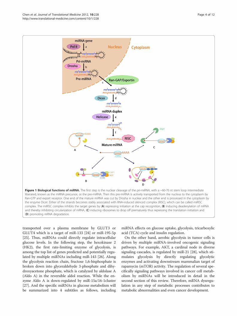
Figure 1 Biological functions of miRNA. The first step is the nuclear cleavage of the pri-miRNA, with a ~60-70 nt stem loop intermediateliberated, known as the miRNA precursor, or the pre-miRNA. Then this pre-miRNA is actively transported from the nucleus to the cytoplasm byRan-GTP and export receptor. One end of the mature miRNA was cut by Drosha in nuclear and the other end is processed in the cytoplasm bythe enzyme Dicer. Either of the strands becomes stably associated with RNA-induced silenced complex (RISC), which can be called miRISCcomplex. The miRISC complex inhibits the target genes by (A) repressing initiation at the cap recognition, (B) inducing deadenylation of mRNAand thereby inhibiting circularization of mRNA, (C) inducing ribosomes to drop off prematurely thus repressing the translation initiation and(D) promoting mRNA degradation.
Chen et al. Journal of Translational Medicine 2012, 10:228 Page 4 of 12http://www.translational-medicine.com/content/10/1/228
transported over a plasma membrane by GLUT3 orGLUT4 which is a target of miR-133 [24] or miR-195-5p[25]. Thus, miRNAs could directly regulate intracellularglucose levels. In the following step, the hexokinase 2(HK2), the first rate-limiting enzyme of glycolysis, isamong the top list of genes predicted and potentially regu-lated by multiple miRNAs including miR-143 [26]. Alongthe glycolysis reaction chain, fructose 1,6-bisphosphate isbroken down into glyceraldehyde 3-phosphate and dihy-droxyacetone phosphate, which is catalyzed by aldolase A(Aldo A) in the reversible aldol reaction. While the en-zyme Aldo A is down-regulated by miR-15a/16-1cluster[27]. And the specific miRNAs in glucose metabolism willbe summarized into 4 subtitles as follows, including
miRNA effects on glucose uptake, glycolysis, tricarboxylicacid (TCA) cycle and insulin regulation.On the other hand, aerobic glycolysis in tumor cells is
driven by multiple miRNA-involved oncogenic signalingpathways. For example, AKT, a cardinal node in diversesignaling cascades, is regulated by miR-21 [28], which sti-mulates glycolysis by directly regulating glycolyticenzymes and activating downstream mammalian target ofrapamycin (mTOR) activity. The regulation of several spe-cifically signaling pathways involved in cancer cell metab-olism by miRNAs will be introduced in detail in thesecond section of this review. Therefore, mRNA dysregu-lation in any step of metabolic processes contributes tometabolic abnormalities and even cancer development.
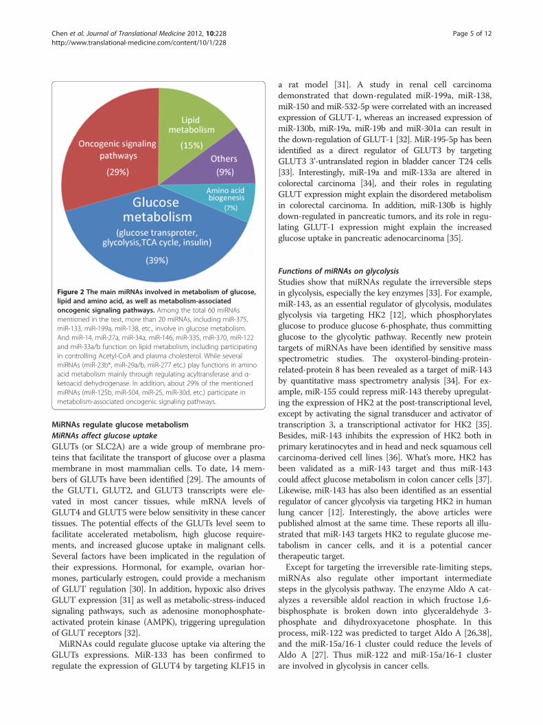
Figure 2 The main miRNAs involved in metabolism of glucose,lipid and amino acid, as well as metabolism-associatedoncogenic signaling pathways. Among the total 60 miRNAsmentioned in the text, more than 20 miRNAs, including miR-375,miR-133, miR-199a, miR-138, etc., involve in glucose metabolism.And miR-14, miR-27a, miR-34a, miR-146, miR-335, miR-370, miR-122and miR-33a/b function on lipid metabolism, including participatingin controlling Acetyl-CoA and plasma cholesterol. While severalmiRNAs (miR-23b*, miR-29a/b, miR-277 etc.) play functions in aminoacid metabolism mainly through regulating acyltransferase and α-ketoacid dehydrogenase. In addition, about 29% of the mentionedmiRNAs (miR-125b, miR-504, miR-25, miR-30d, etc.) participate inmetabolism-associated oncogenic signaling pathways.
Chen et al. Journal of Translational Medicine 2012, 10:228 Page 5 of 12http://www.translational-medicine.com/content/10/1/228
MiRNAs regulate glucose metabolismMiRNAs affect glucose uptakeGLUTs (or SLC2A) are a wide group of membrane pro-teins that facilitate the transport of glucose over a plasmamembrane in most mammalian cells. To date, 14 mem-bers of GLUTs have been identified [29]. The amounts ofthe GLUT1, GLUT2, and GLUT3 transcripts were ele-vated in most cancer tissues, while mRNA levels ofGLUT4 and GLUT5 were below sensitivity in these cancertissues. The potential effects of the GLUTs level seem tofacilitate accelerated metabolism, high glucose require-ments, and increased glucose uptake in malignant cells.Several factors have been implicated in the regulation oftheir expressions. Hormonal, for example, ovarian hor-mones, particularly estrogen, could provide a mechanismof GLUT regulation [30]. In addition, hypoxic also drivesGLUT expression [31] as well as metabolic-stress-inducedsignaling pathways, such as adenosine monophosphate-activated protein kinase (AMPK), triggering upregulationof GLUT receptors [32].MiRNAs could regulate glucose uptake via altering the
GLUTs expressions. MiR-133 has been confirmed toregulate the expression of GLUT4 by targeting KLF15 in
a rat model [31]. A study in renal cell carcinomademonstrated that down-regulated miR-199a, miR-138,miR-150 and miR-532-5p were correlated with an increasedexpression of GLUT-1, whereas an increased expression ofmiR-130b, miR-19a, miR-19b and miR-301a can result inthe down-regulation of GLUT-1 [32]. MiR-195-5p has beenidentified as a direct regulator of GLUT3 by targetingGLUT3 3’-untranslated region in bladder cancer T24 cells[33]. Interestingly, miR-19a and miR-133a are altered incolorectal carcinoma [34], and their roles in regulatingGLUT expression might explain the disordered metabolismin colorectal carcinoma. In addition, miR-130b is highlydown-regulated in pancreatic tumors, and its role in regu-lating GLUT-1 expression might explain the increasedglucose uptake in pancreatic adenocarcinoma [35].
Functions of miRNAs on glycolysisStudies show that miRNAs regulate the irreversible stepsin glycolysis, especially the key enzymes [33]. For example,miR-143, as an essential regulator of glycolysis, modulatesglycolysis via targeting HK2 [12], which phosphorylatesglucose to produce glucose 6-phosphate, thus committingglucose to the glycolytic pathway. Recently new proteintargets of miRNAs have been identified by sensitive massspectrometric studies. The oxysterol-binding-protein-related-protein 8 has been revealed as a target of miR-143by quantitative mass spectrometry analysis [34]. For ex-ample, miR-155 could repress miR-143 thereby upregulat-ing the expression of HK2 at the post-transcriptional level,except by activating the signal transducer and activator oftranscription 3, a transcriptional activator for HK2 [35].Besides, miR-143 inhibits the expression of HK2 both inprimary keratinocytes and in head and neck squamous cellcarcinoma-derived cell lines [36]. What’s more, HK2 hasbeen validated as a miR-143 target and thus miR-143could affect glucose metabolism in colon cancer cells [37].Likewise, miR-143 has also been identified as an essentialregulator of cancer glycolysis via targeting HK2 in humanlung cancer [12]. Interestingly, the above articles werepublished almost at the same time. These reports all illu-strated that miR-143 targets HK2 to regulate glucose me-tabolism in cancer cells, and it is a potential cancertherapeutic target.Except for targeting the irreversible rate-limiting steps,
miRNAs also regulate other important intermediatesteps in the glycolysis pathway. The enzyme Aldo A cat-alyzes a reversible aldol reaction in which fructose 1,6-bisphosphate is broken down into glyceraldehyde 3-phosphate and dihydroxyacetone phosphate. In thisprocess, miR-122 was predicted to target Aldo A [26,38],and the miR-15a/16-1 cluster could reduce the levels ofAldo A [27]. Thus miR-122 and miR-15a/16-1 clusterare involved in glycolysis in cancer cells.
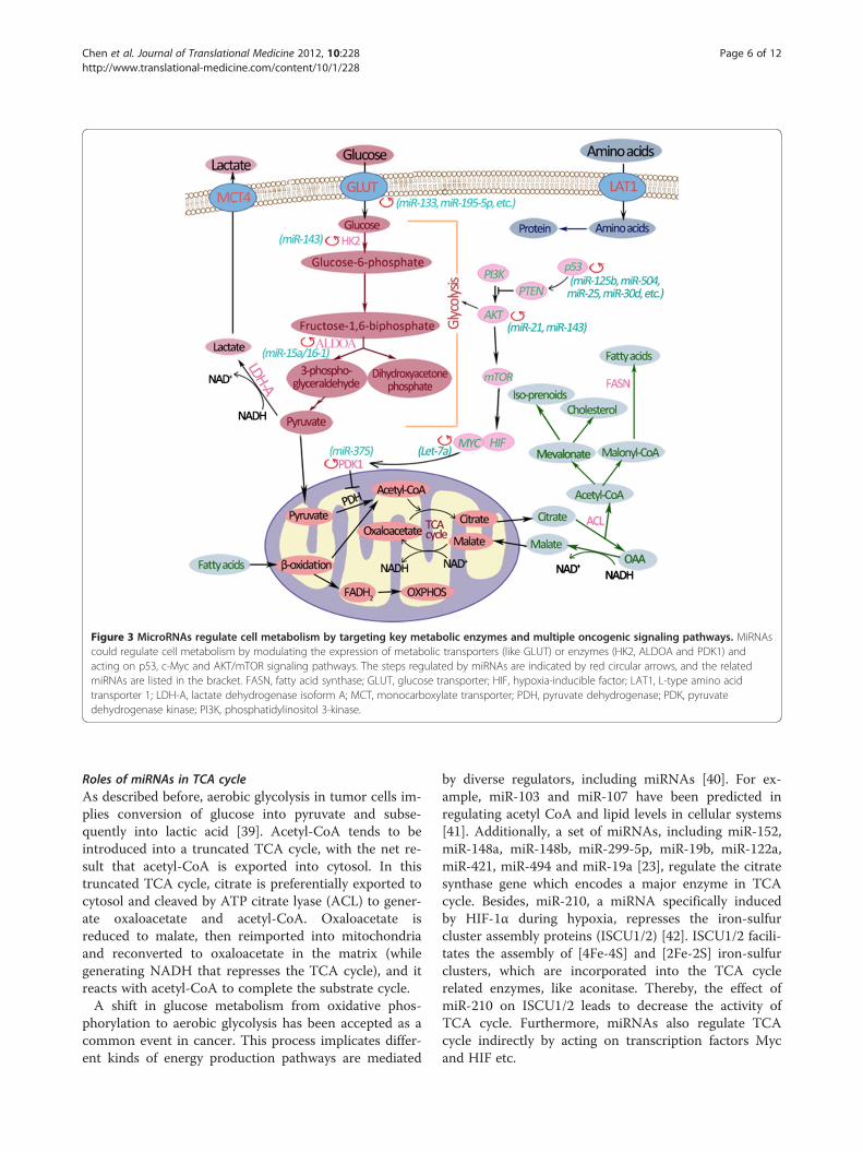
Figure 3 MicroRNAs regulate cell metabolism by targeting key metabolic enzymes and multiple oncogenic signaling pathways. MiRNAscould regulate cell metabolism by modulating the expression of metabolic transporters (like GLUT) or enzymes (HK2, ALDOA and PDK1) andacting on p53, c-Myc and AKT/mTOR signaling pathways. The steps regulated by miRNAs are indicated by red circular arrows, and the relatedmiRNAs are listed in the bracket. FASN, fatty acid synthase; GLUT, glucose transporter; HIF, hypoxia-inducible factor; LAT1, L-type amino acidtransporter 1; LDH-A, lactate dehydrogenase isoform A; MCT, monocarboxylate transporter; PDH, pyruvate dehydrogenase; PDK, pyruvatedehydrogenase kinase; PI3K, phosphatidylinositol 3-kinase.
Chen et al. Journal of Translational Medicine 2012, 10:228 Page 6 of 12http://www.translational-medicine.com/content/10/1/228
Roles of miRNAs in TCA cycleAs described before, aerobic glycolysis in tumor cells im-plies conversion of glucose into pyruvate and subse-quently into lactic acid [39]. Acetyl-CoA tends to beintroduced into a truncated TCA cycle, with the net re-sult that acetyl-CoA is exported into cytosol. In thistruncated TCA cycle, citrate is preferentially exported tocytosol and cleaved by ATP citrate lyase (ACL) to gener-ate oxaloacetate and acetyl-CoA. Oxaloacetate isreduced to malate, then reimported into mitochondriaand reconverted to oxaloacetate in the matrix (whilegenerating NADH that represses the TCA cycle), and itreacts with acetyl-CoA to complete the substrate cycle.A shift in glucose metabolism from oxidative phos-
phorylation to aerobic glycolysis has been accepted as acommon event in cancer. This process implicates differ-ent kinds of energy production pathways are mediated
by diverse regulators, including miRNAs [40]. For ex-ample, miR-103 and miR-107 have been predicted inregulating acetyl CoA and lipid levels in cellular systems[41]. Additionally, a set of miRNAs, including miR-152,miR-148a, miR-148b, miR-299-5p, miR-19b, miR-122a,miR-421, miR-494 and miR-19a [23], regulate the citratesynthase gene which encodes a major enzyme in TCAcycle. Besides, miR-210, a miRNA specifically inducedby HIF-1α during hypoxia, represses the iron-sulfurcluster assembly proteins (ISCU1/2) [42]. ISCU1/2 facili-tates the assembly of [4Fe-4S] and [2Fe-2S] iron-sulfurclusters, which are incorporated into the TCA cyclerelated enzymes, like aconitase. Thereby, the effect ofmiR-210 on ISCU1/2 leads to decrease the activity ofTCA cycle. Furthermore, miRNAs also regulate TCAcycle indirectly by acting on transcription factors Mycand HIF etc.
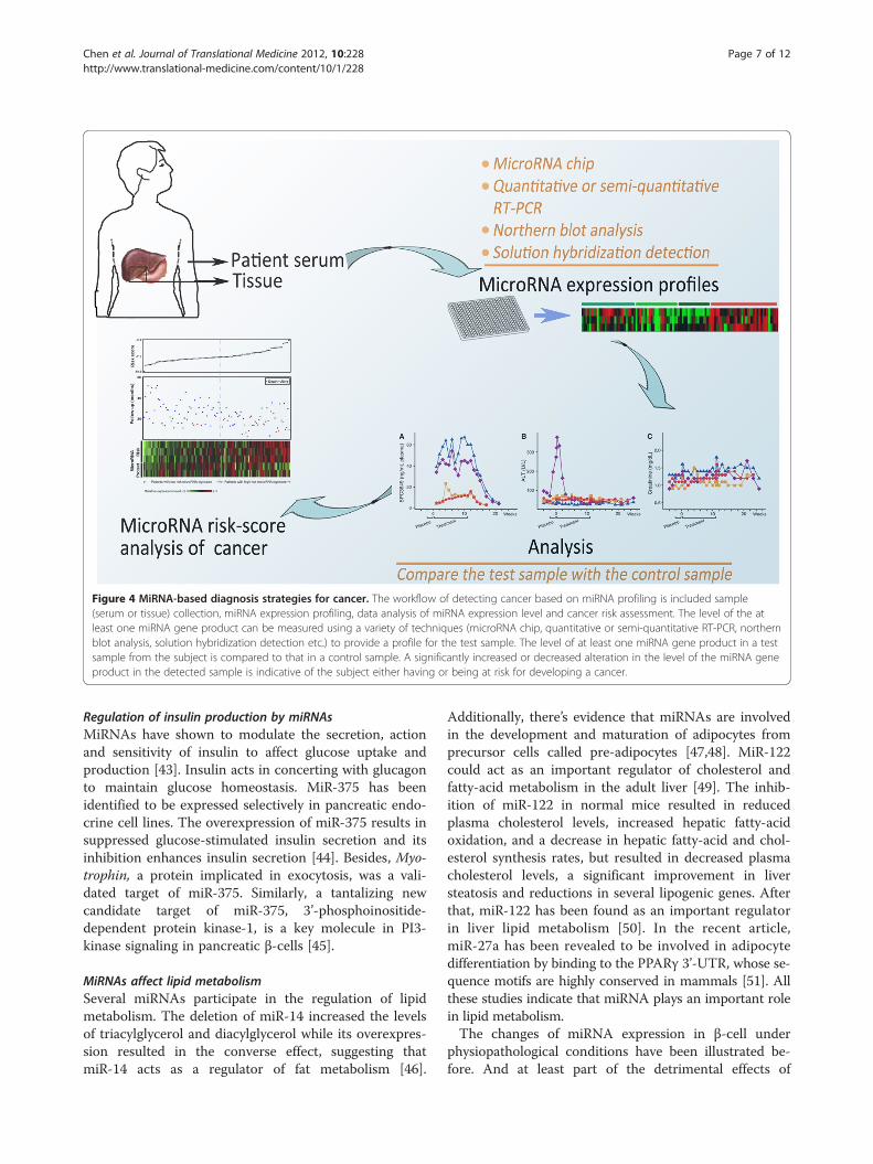
Figure 4 MiRNA-based diagnosis strategies for cancer. The workflow of detecting cancer based on miRNA profiling is included sample(serum or tissue) collection, miRNA expression profiling, data analysis of miRNA expression level and cancer risk assessment. The level of the atleast one miRNA gene product can be measured using a variety of techniques (microRNA chip, quantitative or semi-quantitative RT-PCR, northernblot analysis, solution hybridization detection etc.) to provide a profile for the test sample. The level of at least one miRNA gene product in a testsample from the subject is compared to that in a control sample. A significantly increased or decreased alteration in the level of the miRNA geneproduct in the detected sample is indicative of the subject either having or being at risk for developing a cancer.
Chen et al. Journal of Translational Medicine 2012, 10:228 Page 7 of 12http://www.translational-medicine.com/content/10/1/228
Regulation of insulin production by miRNAsMiRNAs have shown to modulate the secretion, actionand sensitivity of insulin to affect glucose uptake andproduction [43]. Insulin acts in concerting with glucagonto maintain glucose homeostasis. MiR-375 has beenidentified to be expressed selectively in pancreatic endo-crine cell lines. The overexpression of miR-375 results insuppressed glucose-stimulated insulin secretion and itsinhibition enhances insulin secretion [44]. Besides, Myo-trophin, a protein implicated in exocytosis, was a vali-dated target of miR-375. Similarly, a tantalizing newcandidate target of miR-375, 3’-phosphoinositide-dependent protein kinase-1, is a key molecule in PI3-kinase signaling in pancreatic β-cells [45].
MiRNAs affect lipid metabolismSeveral miRNAs participate in the regulation of lipidmetabolism. The deletion of miR-14 increased the levelsof triacylglycerol and diacylglycerol while its overexpres-sion resulted in the converse effect, suggesting thatmiR-14 acts as a regulator of fat metabolism [46].
Additionally, there’s evidence that miRNAs are involvedin the development and maturation of adipocytes fromprecursor cells called pre-adipocytes [47,48]. MiR-122could act as an important regulator of cholesterol andfatty-acid metabolism in the adult liver [49]. The inhib-ition of miR-122 in normal mice resulted in reducedplasma cholesterol levels, increased hepatic fatty-acidoxidation, and a decrease in hepatic fatty-acid and chol-esterol synthesis rates, but resulted in decreased plasmacholesterol levels, a significant improvement in liversteatosis and reductions in several lipogenic genes. Afterthat, miR-122 has been found as an important regulatorin liver lipid metabolism [50]. In the recent article,miR-27a has been revealed to be involved in adipocytedifferentiation by binding to the PPARγ 3’-UTR, whose se-quence motifs are highly conserved in mammals [51]. Allthese studies indicate that miRNA plays an important rolein lipid metabolism.The changes of miRNA expression in β-cell under
physiopathological conditions have been illustrated be-fore. And at least part of the detrimental effects of
Chen et al. Journal of Translational Medicine 2012, 10:228 Page 8 of 12http://www.translational-medicine.com/content/10/1/228
palmitate on pancreatic β-cells has been caused by alter-ation in the levels of specific miRNAs, like miR-34a andmiR-146 [52]. Apart from that, the up-regulation ofmiR-335 has also been found in obesity by microarrayanalysis [53]. Besides, the expression of miR-335 was up-regulated in liver and white adipose tissue in obese mice,which was associated with an elevated body, liver andWAT weight, and hepatic triglyceride and cholesterol.Additionally, miR-370 acting via miR-122 may accumu-late hepatic triglycerides by modulating initially the ex-pression of SREBP-1c, DGAT2, and Cpt1α and,subsequently, the expression of other genes that affectlipid metabolism [54].Recently, miR-33a/b has been discovered to govern
cholesterol / lipid metabolism and energy homeostasis[55]. MiR-33a/b embeds within intron sequences of thehuman SREBF genes and controls the levels of ATP-binding cassette transporter ABCA1, a cholesterol effluxpump critical for high-density lipoprotein synthesis andreversing cholesterol transport from peripheral tissues[56,57]. MiR-33a/b also acts in the lipid homeostasispathway by controlling the expression of fatty acidβ-oxidation genes including carnitine O-octanoyltransfer-ase, hydroxyacyl-CoA-dehydrogenase, and carnitine pal-mitoyltransferase 1A, as well as energy homeostasisregulators AMPK a1, SIRT6, and insulin receptor sub-strate 2 [58]. These reports bring us a further view ofmiRNA function on lipid metabolism.
Effects of miRNA in amino acid metabolismAmino acid metabolism is linked to biosynthesis of pro-tein, nucleotide and lipids, redox homeostasis, and en-ergy metabolism. MiR-23b* (expressed from the 3'-arm)mediates proline oxidase, the first enzyme in proline ca-tabolism, down-regulation in human kidney tumors [59].Furthermore, the metabolic link between proline andglutamine afforded by Myc emphasizes the complexityof tumor metabolism. While miR-122 was reported todownregulate the high affinity cationic amino acid trans-porter CAT-1 [60], thereby regulating amino acid metab-olism. Involving evidences have been found inDrosophila, where miR-277 plays a role as a metabolicswitch controlling amino acid catabolism by bioinfor-matics approaches [61]. Additionally, miR29b has beenidentified to control the component of the branchedchain a-ketoacid dehydrogenase complex, which cata-lyzes the irreversible step in branched chain amino acids(including leucine, isoleucine and valine) catabolism[62], suggesting that miR-29b exerts effects of control-ling on amino acid catabolism.
miRNA regulation of signaling pathways in cell metabolismThe intertwined connections between aberrant expres-sion of microRNAs and unbalanced signaling pathways
contribute to abnormal cell metabolism and carcinogen-esis. The specific p53, c-Myc, AMPK and AKT signalingpathways are included to clarify their roles in miRNA-mediated metabolism.
p53 pathwayp53, one of the most common tumour suppressor genes,functions to prevent tumour development by inhibitingthe outgrowth of stressed or damaged cells. In additionto well established functions to block cell proliferation,recent studies have revealed regulation roles for p53involved in metabolism [63]. The p53 can inhibit the ex-pression of GLUT-1, GLUT-4, phosphoglyceromutaseand TIGAR to affect glycolysis. TIGAR is TP53-inducedglycolysis and apoptosis regulator protein, and it inhibitsthe glycolytic enzyme PFKFB2 [64]. Additonally, p53could also activate the expression of synthesis of cyto-chrome c oxidase 2 at transcriptional level [65] and in-duce the expression of the ribonucleotide reductasesubunit p53R2 [66], leading to the restraint on glycolyticrate.Several miRNAs are able to control p53 activity. The
miR- 125b has been identified as a negative regulator ofp53 in both zebrafish and human [67]. To date, miRNAsincluding miR-125b, miR-504, miR-25, miR-30d, miR-34a,miR-122, miR-29, miR-192, miR-194 and miR-215 havebeen shown to regulate p53 abundance and/or activity.Among these, miR-125b, miR-504, miR-25 and miR-30dnegatively regulate p53 by binding to its 3'UTR whereasthe others indirectly influence p53 abundance and/oractivity by regulating the regulators of p53 [68]. The func-tions of these miRNAs on p53 give a clue of their effectsin cancer cell metabolism.
c-Myc pathwayThe c-Myc is a transcription factor that regulates the ex-pression of genes involved in nucleotide metabolism,DNA replication, and ribosomal and mitochondrial bio-genesis. Studies in the past few years have led to theidentification of miRNAs as novel regulators of c-Mycactivity. A mutated version of Myc leads to the unregu-lated expression of many genes, some of which areinvolved in cell proliferation and results in the formationof cancer [69]. For example, c-Myc has crucial roles inglutamine metabolism mediated by miR-23b [70]. More-over, in concerts with HIF1 to regulate glucose uptakeand glycolytic enzyme expression, thus favouring tumourgrowth in hostile environments [71].The regulation of Myc mRNA by let-7a has been con-
firmed [72]. Similiarly, the overexpression of let-7a caninhibit the growth of lung cancer transplanted subcuta-neously in nude mice by suppression of k-Ras and c-Myc [73]. Inspiringly, c-Myc transcriptionally repressesmiR-23a and miR-23b, resulting in increased expression

Chen et al. Journal of Translational Medicine 2012, 10:228 Page 9 of 12http://www.translational-medicine.com/content/10/1/228
of mitochondrial glutaminase, enhancing glutamine ca-tabolism through increased mitochondrial glutaminaseexpression [6].
AMPK pathwayAMPK acts as a metabolic master switch regulating sev-eral intracellular systems including the cellular uptake ofglucose, the β-oxidation of fatty acids and the biogenesisof GLUT4 and mitochondria [74]. AMPK controls glu-cose homeostasis by regulating metabolism in multipleperipheral tissues, such as skeletal muscle, liver, adiposetissues, and pancreatic β cells [75].The functions of miR-375 on glucose homeostasis
have been studied [76]. Total 381 putative direct targetsof miR-375 were selected, which contained a miR-375recognition motif, and confirmed 10 of these genes, in-volving caveolin1 [77,78], inhibitor of DNA binding 3[79,80], Smarca2, Ras-dexamethasone-induced-1 [81],regulator of G protein signaling 16 [82], eukaryoticelongation factor 1 epsilon 1, apoptosis-inducing factor,mitochondrion-associated 1, cell adhesion molecule 1,HuD antigen [83], and complement component 1 q sub-component binding protein. Published data have shownthat some of these genes play a role in AMPK signaling,inducing apoptosis and inhibiting normal developmentalgrowth processes or the proliferation of tumors.
AKT pathwayThe PI3K/AKT/mTOR pathway is an intracellular sig-nalling pathway, which is important in apoptosis. It hasrisen to prominence as a key regulator of cell cycle pro-liferation, growth, survival, protein synthesis, and glu-cose metabolism [84]. Activation of PI3K leads to theactivation of downstream effectors including Akt andmTOR that support cellular biosynthesis [85-87].Enhanced PI3K/Akt signal increases the expressionof nutrient transporters, enabling increased uptake ofglucose, amino acids, and other nutrients. Additionally,Akt-dependent stimulation of hexokinase and phospho-fructokinase drives glycolysis. Furthermore, AKT-involvedsignaling enhances transcription of genes to involve inglycolysis and lipid genesis [88-90].Regulation of this pathway by miRNAs mainly results
in altered glucose and lipid metabolism. For example,miR-21, which inhibits a negative regulator PTEN of thePIK/AKT pathway, is induced in gemcitabine-resistantpancreatic cancer cells [28]. And AKT pathway can in-volve in glycolysis by directly regulating glycolyticenzymes and activating downstream mTOR activity.Similiarly, ORP8 has been identied as an miR-143 targetand the reduction of ORP8 expression in cultured livercells impairs the ability of insulin to induce AKT activa-tion, revealing an ORP8-dependent mechanism of AKTregulation [91].
MiRNAs affect multiple targets in regulatory networksCertain miRNAs have also been shown to affect multipletargets in linear pathways or interconnected nodes inregulatory networks [25], thereby exerting a largercumulative effect [9,55]. For example, miR-33a and miR-33b, as described before, interact with the SREBPtranscription factors to regulate cholesterol and lipidhomeostasis. Furthermore, they may also influence insu-lin signaling and glucose regulation by targeting IRS2,SIRT6 and AMPK α1 [58]. MiR-34a, a miRNA that mayhave important function in a network with SIRT1 andp53, has additionally been implicated in cholesterol, lipidand energy homeostasis [52,68]. MiRNAs typically haverather modest effects on target protein levels, and com-binatorial actions on multiple functionally related targetsare probably required for single miRNAs to significantlyinfluence a complex biological process such as metabolichomeostasis.
MiRNAs as biomarkers for human cancerBy targeting and controlling the expression of mRNA,miRNAs can control highly complex signal transductionpathways and multiple metabolic processes, which areusually involved in different oncogenic pathways [92].The knowledge that miRNA expression is frequentlydysregulated in cancer has uncovered an entirely newrepertoire of molecular factors upstream of gene expres-sion, with exciting potential as novel biomarkers andtherapeutic targets in cancer [93]. Exploiting the uniquecharacteristics of these molecules including their stabil-ity, tissue specificity, ease of detection and manipulation,will bring clinicians ever closer to achieving the goal ofindividualized cancer treatment [94].On the one hand, miRNAs are produced in a tissue-
specific manner, and changes in miRNA within a tissuetype can be correlated with disease status. The tissueconcentrations of specific miRNAs have been associatedwith tumor invasiveness, metastatic potential, and otherclinical characteristics for several types of cancers, in-cluding chronic lymphocytic leukemia, and breast, colo-rectal, hepatic, lung, pancreatic, and prostate cancers[95]. On the other hand, there has been an accumulatingbody of evidence to support circulating miRNAs as non-invasive, sensitive biomarkers of disease states, partic-ularly cancers (breast, lung, pancreas, ovarian, andprostate) [96]. For example, miR-9 and miR-9* (expressedfrom the 3' mature sequence), mostly neuronal and thusexpressed in central nervous system tumors but absentin other tumors, present their potential as tumor mar-kers [97]. In addition, the reduced levels of miR-126,members of the miR-17-92 cluster, inflammation-relatedmiR-155, and smooth muscle-enriched miR-145 inpatients with coronary artery disease compared withhealthy controls [98]. What’s more, published data
Chen et al. Journal of Translational Medicine 2012, 10:228 Page 10 of 12http://www.translational-medicine.com/content/10/1/228
showed that plasma miR-29a and miR-92a have strongpotential as novel noninvasive biomarkers for early de-tection of colorectal carcinoma [99].Furthermore, since they are abundant in blood, easy to
measure, highly stable and disease associated, serummicroRNAs are attractive disease biomarkers [100].There have been over 200 publications on circulatingmiRNA in cancers including prostate, breast, colon,lung, ovarian and leukemia since 2008. Considering thesources of variation, state of microRNA in plasma andorigin and implications for disease specificity, miRNAexpression profiles of potential patients could beassessed by measuring circulating miRNAs in patientserum. This profile could be hopefully used for early de-tection of cancer.
Conclusion and perspectiveMiRNAs are important regulators of numerous aspectsof metabolic homeostasis, physiology and disease. Ingeneral, miRNAs could mainly have two ways to regulatecellular metabolism. MiRNAs could regulate transcrip-tion factors or signaling proteins, which in turn regulatemetabolic enzymes. Alternatively, miRNAs could regu-late the production of certain metabolites by directlyregulating the genes that encode metabolic enzymes[101]. In addition, miRNAs could regulate mRNAsthrough chromatin remodeling [102]. The emergence ofmiRNAs as important regulators of metabolism has gar-nered much interest not only from a scientific point ofview but also from a clinical perspective. The function ofmiRNAs on cellular metabolism reveals molecular strat-egies for controlling metabolic flux by miRNAs in livingorganisms, thus lighting up one aspect of miRNA thera-peutics. MiRNAs are promising in the diagnosis of can-cer, drug target identification and clinical treatment inthe future (Figure 4). The use of miRNAs, such as oligo-nucleotide complementary [103] or antisense oligonu-cleotides [104] in miRNA inhibition, to suppress cellmetabolism altering will hopefully lead to a new thera-peutic strategy for malignant cancer [105,106]. For ex-ample, endothelial miR-126 is deregulated in patientswith type 2 diabetes, which may ultimately lead to novelbiomarkers for risk estimation and classification andcould be exploited for miRNA-based therapeutic inter-ventions of vascular complications associated with thisdisease [107].So far, a variety of new strategies to identify and
characterize the targets of individual miRNAs have beendeveloped. Because miRNAs can also regulate othernon-coding RNAs, these interactions will increase thecomplexity of gene regulation. Moreover, cost-effectivemiRNA profiling strategies and larger studies are neededto determine its advantage for cancer diagnosis. Add-itionally, a new class of miRNA-based drugs that are
capable of targeting molecules outside the range of trad-itional medicinal chemistry, their clinical implementa-tion will require improvements in drug composition anddelivery. Since these challenges lie on the way, molecularstrategies for cancer therapy by miRNAs are still in theirinfancy. Nevertheless, the successful development ofmiRNA biology technologies could ultimately translateour understanding of miRNA functions in cancer intostrategies for the control of cancer.
AbbreviationsACL: ATP citrate lyase; AldoA: Aldolase A; AMPK: Adenosine monophosphate-activated protein kinase; GLUT: Glucose transporter; ISCU1/2: Iron-sulfurcluster assembly proteins; HIF1: Hypoxia-inducible factor 1; HK2: Hexokinase2; MiRNA: MicroRNA; MTOR: Mammalian target of rapamycin; ORP: Oxysterol-binding-protein-related-protein; RISC: RNA-induced silenced complex;TCA: Tricarboxylic acid.
Competing interestsThe authors declare that they have no competing interests.
Authors’ contributionsAll authors participated in the preparation of the manuscript, read andapproved the final manuscript.
AcknowledgementsThis work was financially supported by the grants from National Key BasicResearch Program of China (2011CB910703, 2013CB911303), National NaturalSciences Foundation of China (30970654, 31071235), and grants for NewCentury Excellent Talents in University (NCET-10-0595), specialized researchfund for the Doctoral Program of Higher Education (20120181110025). Thisresearch was also funded by Sichuan Province Program (2010JQ0016,2012SZ0002) and Chengdu Local Scientific Project (11DXYB356JH-027).
Author details1State Key Laboratory of Biotherapy, West China Hospital, Sichuan University,No.17, third section of Renmin South Road, Chengdu 610041, People’sRepublic China. 2Department of Gynecology and Obstetrics, West ChinaSecond Hospital, Sichuan University, Chengdu 610041, People’s RepublicChina.
Received: 27 September 2012 Accepted: 16 November 2012Published: 20 November 2012
Reference1. Kutter C, Svoboda P: miRNA, siRNA, piRNA. RNA Biol 2008, 5:181–188.2. Lewis BP, Burge CB, Bartel DP: Conserved seed pairing, often flanked by
adenosines, indicates that thousands of human genes are microRNAtargets. Cell 2005, 120:15–20.
3. Bartel DP: MicroRNAs: genomics, biogenesis, mechanism, and function.Cell 2004, 116:281–297.
4. Krutzfeldt J, Stoffel M: MicroRNAs: a new class of regulatory genesaffecting metabolism. Cell Metab 2006, 4:9–12.
5. Redova M, Svoboda M, Slaby O: MicroRNAs and their target genenetworks in renal cell carcinoma. Biochem Biophys Res Commun 2011,405:153–156.
6. Gao P, Tchernyshyov I, Chang TC, Lee YS, Kita K, Ochi T, Zeller KI, De MarzoAM, Van Eyk JE, Mendell JT, Dang CV: c-Myc suppression of miR-23a/benhances mitochondrial glutaminase expression and glutaminemetabolism. Nature 2009, 458:762–765.
7. Rayner KJ, Suarez Y, Davalos A, Parathath S, Fitzgerald ML, Tamehiro N,Fisher EA, Moore KJ, Fernandez-Hernando C: MiR-33 contributes to theregulation of cholesterol homeostasis. Science 2010, 328:1570–1573.
8. Eichner LJ, Perry M-C, Dufour CR, Bertos N, Park M, St-Pierre J, Giguère V:miR-378∗ mediates metabolic shift in breast cancer cells via the PGC-1β/ERRγ transcriptional pathway. Cell Metab 2010, 12:352–361.
9. Rottiers V, Naar AM: MicroRNAs in metabolism and metabolic disorders.Nat Rev Mol Cell Biol 2012, 13:239–250.
Chen et al. Journal of Translational Medicine 2012, 10:228 Page 11 of 12http://www.translational-medicine.com/content/10/1/228
10. Pucci S, Mazzarelli P: MicroRNA dysregulation in colon cancermicroenvironment interactions: the importance of small things inmetastases. Cancer Microenviron 2011, 4:155–162.
11. Cairns RA, Harris IS, Mak TW: Regulation of cancer cell metabolism. Nat RevCancer 2011, 11:85–95.
12. Fang R, Xiao T, Fang Z, Sun Y, Li F, Gao Y, Feng Y, Li L, Wang Y, Liu X, et al:miR-143 regulates cancer glycolysis via targeting hexokinase 2. J BiolChem 2012, 287:23227–23235.
13. Lagos-Quintana M, Rauhut R, Lendeckel W, Tuschl T: Identification of novelgenes coding for small expressed RNAs. Sci STKE 2001, 294:853.
14. Lee RC, Ambros V: An extensive class of small RNAs in Caenorhabditiselegans. Science 2001, 294:862–864.
15. He L, Hannon GJ: MicroRNAs: small RNAs with a big role in generegulation. Nat Rev Genet 2004, 5:522–531.
16. Winter J, Jung S, Keller S, Gregory RI, Diederichs S: Many roads to maturity:microRNA biogenesis pathways and their regulation. Nat Cell Biol 2009,11:228–234.
17. Carthew RW, Sontheimer EJ: Origins and mechanisms of miRNAs andsiRNAs. Cell 2009, 136:642–655.
18. Hanahan D, Weinberg RA: Hallmarks of cancer: the next generation. Cell2011, 144:646–674.
19. Hsu PP, Sabatini DM: Cancer cell metabolism: Warburg and beyond. Cell2008, 134:703–707.
20. Kroemer G, Pouyssegur J: Tumor cell metabolism: cancer's Achilles' heel.Cancer Cell 2008, 13:472–482.
21. Vander Heiden MG, Locasale JW, Swanson KD, Sharfi H, Heffron GJ, Amador-Noguez D, Christofk HR, Wagner G, Rabinowitz JD, Asara JM, Cantley LC:Evidence for an alternative glycolytic pathway in rapidly proliferatingcells. Science 2010, 329:1492–1499.
22. Jones RG, Thompson CB: Tumor suppressors and cell metabolism: arecipe for cancer growth. Gene Dev 2009, 23:537–548.
23. Tibiche C, Wang E: MicroRNA regulatory patterns on the humanmetabolic network. Open Syst Biol J 2008, 1:1–8.
24. Horie T, Ono K, Nishi H, Iwanaga Y, Nagao K, Kinoshita M, Kuwabara Y,Takanabe R, Hasegawa K, Kita T: MicroRNA-133 regulates the expression ofGLUT4 by targeting KLF15 and is involved in metabolic control incardiac myocytes. Biochem Biophys Res Commun 2009, 389:315–320.
25. Fei X, Qi M, Wu B, Song Y, Wang Y, Li T: MicroRNA-195-5p suppressesglucose uptake and proliferation of human bladder cancer T24 cells byregulating GLUT3 expression. FEBS Lett 2012, 586:392–397.
26. Coulouarn C, Factor VM, Andersen JB, Durkin ME, Thorgeirsson SS: Loss of miR-122 expression in liver cancer correlates with suppression of the hepaticphenotype and gain of metastatic properties. Oncogene 2009, 28:3526–3536.
27. Calin GA, Cimmino A, Fabbri M, Ferracin M, Wojcik SE, Shimizu M, Taccioli C,Zanesi N, Garzon R, Aqeilan RI, et al: MiR-15a and miR-16-1 clusterfunctions in human leukemia. Proc Natl Acad Sci USA 2008, 105:5166–5171.
28. Ali S, Ahmad A, Banerjee S, Padhye S, Dominiak K, Schaffert JM, Wang Z,Philip PA, Sarkar FH: Gemcitabine sensitivity can be induced in pancreaticcancer cells through modulation of miR-200 and miR-21 expression bycurcumin or its analogue CDF. Cancer Res 2010, 70:3606–3617.
29. Thorens B, Mueckler M: Glucose transporters in the 21st century. Am JPhysiol Endocrinol Metab 2010, 298:E141–E145.
30. Rivenzon-Segal D, Boldin-Adamsky S, Seger D, Seger R, Degani H: Glycolysisand glucose transporter 1 as markers of response to hormonal therapyin breast cancer. Int J Cancer 2003, 107:177–182.
31. Macheda ML, Rogers S, Best JD: Molecular and cellular regulation of glucosetransporter (GLUT) proteins in cancer. J Cell Physiol 2005, 202:654–662.
32. Koh HJ, Toyoda T, Fujii N, Jung MM, Rathod A, Middelbeek RJ, Lessard SJ,Treebak JT, Tsuchihara K, Esumi H, et al: Sucrose nonfermenting AMPK-related kinase (SNARK) mediates contraction-stimulated glucosetransport in mouse skeletal muscle. Proc Natl Acad Sci USA 2010,107:15541–15546.
33. Singh PK, Mehla K, Hollingsworth MA, Johnson KR: Regulation of aerobicglycolysis by microRNAs in cancer. Mol Cell Pharmacol 2011, 3:125–134.
34. Kruger M, Moser M, Ussar S, Thievessen I, Luber CA, Forner F, Schmidt S,Zanivan S, Fassler R, Mann M: SILAC mouse for quantitative proteomicsuncovers kindlin-3 as an essential factor for red blood cell function. Cell2008, 134:353–364.
35. Jiang S, Zhang LF, Zhang HW, Hu S, Lu MH, Liang S, Li B, Li Y, Li D, Wang ED,Liu MF: A novel miR-155/miR-143 cascade controls glycolysis by regulatinghexokinase 2 in breast cancer cells. EMBO J 2012, 31:1985–1998.
36. Peschiaroli A, Giacobbe A, Formosa A, Markert EK, Bongiorno-Borbone L,Levine AJ, Candi E, D'Alessandro A, Zolla L, Finazzi Agro A, Melino G: miR-143 regulates hexokinase 2 expression in cancer cells. Oncogene 2012,doi:10.1038/onc.2012.100.
37. Gregersen LH, Jacobsen A, Frankel LB, Wen J, Krogh A, Lund AH:microRNA-143down-regulates Hexokinase 2 in colon cancer cells. BMC Cancer 2012, 12:232.
38. Tsai WC, Hsu PW, Lai TC, Chau GY, Lin CW, Chen CM, Lin CD, Liao YL, WangJL, Chau YP, et al: MicroRNA-122, a tumor suppressor microRNA thatregulates intrahepatic metastasis of hepatocellular carcinoma. Hepatology2009, 49:1571–1582.
39. Ward PS, Thompson CB: Metabolic reprogramming: a cancer hallmarkeven warburg did not anticipate. Cancer Cell 2012, 21:297–308.
40. Singh PK, Brand RE, Mehla K: MicroRNAs in pancreatic cancermetabolism. Nat Rev Gastro Hepat 2012, 9:334–344.
41. Wilfred BR, Wang WX, Nelson PT: Energizing miRNA research: a reviewof the role of miRNAs in lipid metabolism, with a prediction that miR-103/107 regulates human metabolic pathways. Mol Genet Metab 2007,91:209–217.
42. Chan SY, Zhang YY, Hemann C, Mahoney CE, Zweier JL, Loscalzo J:MicroRNA-210 controls mitochondrial metabolism during hypoxia byrepressing the iron-sulfur cluster assembly proteins ISCU1/2. Cell Metab2009, 10:273–284.
43. Trajkovski M, Hausser J, Soutschek J, Bhat B, Akin A, Zavolan M, Heim MH,Stoffel M: MicroRNAs 103 and 107 regulate insulin sensitivity. Nature2011, 474:649–653.
44. Poy MN, Eliasson L, Krutzfeldt J, Kuwajima S, Ma X, MacDonald PE, Pfeffer S,Tuschl T, Rajewsky N, Rorsman P: A pancreatic islet-specific microRNAregulates insulin secretion. Nature 2004, 432:226–230.
45. El Ouaamari A, Baroukh N, Martens GA, Lebrun P, Pipeleers D, vanObberghen E: miR-375 targets 3'-phosphoinositide-dependent proteinkinase-1 and regulates glucose-induced biological responses inpancreatic beta-cells. Diabetes 2008, 57:2708–2717.
46. Xu P, Vernooy SY, Guo M, Hay BA: The Drosophila microRNA Mir-14suppresses cell death and is required for normal fat metabolism. CurrBiol 2003, 13:790–795.
47. Esau C, Kang X, Peralta E, Hanson E, Marcusson EG, Ravichandran LV, Sun Y,Koo S, Perera RJ, Jain R, et al: MicroRNA-143 regulates adipocytedifferentiation. J Biol Chem 2004, 279:52361–52365.
48. Kajimoto K, Naraba H, Iwai N: MicroRNA and 3T3-L1 pre-adipocytedifferentiation. RNA 2006, 12:1626–1632.
49. Esau C, Davis S, Murray SF, Yu XX, Pandey SK, Pear M, Watts L, Booten SL,Graham M, McKay R, et al: miR-122 regulation of lipid metabolismrevealed by in vivo antisense targeting. Cell Metab 2006, 3:87–98.
50. Lynn FC: Meta-regulation: microRNA regulation of glucose and lipidmetabolism. Trends Endocrinol Metab 2009, 20:452–459.
51. Kim SY, Kim AY, Lee HW, Son YH, Lee GY, Lee JW, Lee YS, Kim JB:miR-27a is a negative regulator of adipocyte differentiation viasuppressing PPARgamma expression. Biochem Biophys Res Commun2010, 392:323–328.
52. Lovis P, Roggli E, Laybutt DR, Gattesco S, Yang JY, Widmann C,Abderrahmani A, Regazzi R: Alterations in microRNA expressioncontribute to fatty acid-induced pancreatic beta-cell dysfunction.Diabetes 2008, 57:2728–2736.
53. Nakanishi N, Nakagawa Y, Tokushige N, Aoki N, Matsuzaka T, Ishii K, Yahagi N,Kobayashi K, Yatoh S, Takahashi A, et al: The up-regulation of microRNA-335is associated with lipid metabolism in liver and white adipose tissue ofgenetically obese mice. Biochem Biophys Res Commun 2009, 385:492–496.
54. Iliopoulos D, Drosatos K, Hiyama Y, Goldberg IJ, Zannis VI: MicroRNA-370controls the expression of MicroRNA-122 and Cpt1 and affects lipidmetabolism. J Lipid Res 2010, 51:1513–1523.
55. Rottiers V, Najafi-Shoushtari SH, Kristo F, Gurumurthy S, Zhong L, Li Y, CohenDE, Gerszten RE, Bardeesy N, Mostoslavsky R, Naar AM: MicroRNAs inmetabolism and metabolic diseases. Cold Spring Harb Symp Quant Biol2011, 76:225–233.
56. Najafi-Shoushtari SH, Kristo F, Li Y, Shioda T, Cohen DE, Gerszten RE, NaarAM: MicroRNA-33 and the SREBP host genes cooperate to controlcholesterol homeostasis. Sci Signalling 2010, 328:1566.
57. Gerin I, Clerbaux LA, Haumont O, Lanthier N, Das AK, Burant CF, Leclercq IA,MacDougald OA, Bommer GT: Expression of miR-33 from an SREBP2intron inhibits cholesterol export and fatty acid oxidation. J Biol Chem2010, 285:33652–33661.

Chen et al. Journal of Translational Medicine 2012, 10:228 Page 12 of 12http://www.translational-medicine.com/content/10/1/228
58. Dávalos A, Goedeke L, Smibert P, Ramírez CM, Warrier NP, Andreo U, Cirera-Salinas D, Rayner K, Suresh U, Pastor-Pareja JC: miR-33a/b contribute to theregulation of fatty acid metabolism and insulin signaling. Proc Natl AcadSci USA 2011, 108:9232–9237.
59. Liu W, Le A, Hancock C, Lane AN, Dang CV, Fan TW, Phang JM:Reprogramming of proline and glutamine metabolism contributes to theproliferative and metabolic responses regulated by oncogenictranscription factor c-MYC. Proc Natl Acad Sci USA 2012, 109:8983–8988.
60. Chang J, Nicolas E, Marks D, Sander C, Lerro A, Buendia MA, Xu C, Mason WS,Moloshok T, Bort R: Research Paper miR-122, a Mammalian Liver-SpecificmicroRNA, is Processed from mRNA and May Downregulate the HighAffinity Cationic Amino Acid Transporter CAT-1. RNA Biol 2004, 1:106–113.
61. Stark A, Brennecke J, Russell RB, Cohen SM: Identification of DrosophilaMicroRNA targets. PLoS Biol 2003, 1:E60.
62. Mersey BD, Jin P, Danner DJ: Human microRNA (miR29b) expressioncontrols the amount of branched chain alpha-ketoacid dehydrogenasecomplex in a cell. Hum Mol Genet 2005, 14:3371–3377.
63. Cheung EC, Vousden KH: The role of p53 in glucose metabolism. CurrOpin Cell Biol 2010, 22:186–191.
64. Vousden KH, Ryan KM: p53 and metabolism. Nat Rev Cancer 2009, 9:691–700.65. Gottlieb E, Vousden KH: p53 regulation of metabolic pathways. Cold
Spring Harb Perspect Biol 2010, 2:a001040.66. Bourdon A, Minai L, Serre V, Jais JP, Sarzi E, Aubert S, Chretien D, de Lonlay
P, Paquis-Flucklinger V, Arakawa H, et al: Mutation of RRM2B, encodingp53-controlled ribonucleotide reductase (p53R2), causes severemitochondrial DNA depletion. Nat gene 2007, 39:776–780.
67. Le MT, Teh C, Shyh-Chang N, Xie H, Zhou B, Korzh V, Lodish HF, Lim B:MicroRNA-125b is a novel negative regulator of p53. Genes Dev 2009,23:862–876.
68. Jones M, Lal A: MicroRNAs, wild-type and mutant p53: more questionsthan answers. RNA Biol 2012, 9:781–791.
69. Soucek L, Whitfield J, Martins CP, Finch AJ, Murphy DJ, Sodir NM, KarnezisAN, Swigart LB, Nasi S, Evan GI: Modelling Myc inhibition as a cancertherapy. Nature 2008, 455:679–683.
70. Dang CV, Le A, Gao P: MYC-induced cancer cell energy metabolism andtherapeutic opportunities. Clin Cancer Res 2009, 15:6479–6483.
71. Gordan JD, Thompson CB, Simon MC: HIF and c-Myc: sibling rivals for controlof cancer cell metabolism and proliferation. Cancer Cell 2007, 12:108–113.
72. Sampson VB, Rong NH, Han J, Yang Q, Aris V, Soteropoulos P, Petrelli NJ,Dunn SP, Krueger LJ: MicroRNA let-7a down-regulates MYC and revertsMYC-induced growth in Burkitt lymphoma cells. Cancer Res 2007,67:9762–9770.
73. He XY, Chen JX, Zhang Z, Li CL, Peng QL, Peng HM: The let-7a microRNAprotects from growth of lung carcinoma by suppression of k-Ras and c-Myc in nude mice. J Cancer Res Clin Oncol 2010, 136:1023–1028.
74. Ojuka EO: Role of calcium and AMP kinase in the regulation of mitochondrialbiogenesis and GLUT4 levels in muscle. Proc Nutr Soc 2004, 63:275–278.
75. Long YC, Zierath JR: AMP-activated protein kinase signaling in metabolicregulation. J Clin Invest 2006, 116:1776–1783.
76. Poy MN, Hausser J, Trajkovski M, Braun M, Collins S, Rorsman P, Zavolan M,Stoffel M: miR-375 maintains normal pancreatic alpha- and beta-cellmass. Proc Natl Acad Sci USA 2009, 106:5813–5818.
77. Galbiati F, Liu J, Capozza F, Frank PG, Zhu L, Pestell RG, Lisanti MP: Caveolin-1 expression negatively regulates cell cycle progression by inducing G0/G1 arrest via a p53/p21WAF1/Cip1-dependent mechanism. Mol Biol Cell2001, 12:2229–2244.
78. Cohen AW, Park DS, Woodman SE, Williams TM, Chandra M, Shirani J,Pereira de Souza A, Kitsis RN, Russell RG, Weiss LM, et al: Caveolin-1 nullmice develop cardiac hypertrophy with hyperactivation of p42/44 MAPkinase in cardiac fibroblasts. Am J Physiol Cell Physiol 2003, 284:C457–474.
79. Bain G, Cravatt CB, Loomans C, Alberola-Ila J, Hedrick SM, Murre C:Regulation of the helix-loop-helix proteins, E2A and Id3, by the Ras-ERKMAPK cascade. Nat Immunol 2001, 2:165–171.
80. Kim DS, Franklyn JA, Boelaert K, Eggo MC, Watkinson JC, McCabe CJ:Pituitary tumor transforming gene (PTTG) stimulates thyroid cellproliferation via a vascular endothelial growth factor/kinase insertdomain receptor/inhibitor of DNA binding-3 autocrine pathway. J ClinEndocrinol Metab 2006, 91:4603–4611.
81. Vaidyanathan G, Cismowski MJ, Wang G, Vincent TS, Brown KD, Lanier SM:The Ras-related protein AGS1/RASD1 suppresses cell growth. Oncogene2004, 23:5858–5863.
82. Neubig RR, Siderovski DP: Regulators of G-protein signalling as newcentral nervous system drug targets. Nat Rev Drug Discov 2002, 1:187–197.
83. Akamatsu W, Fujihara H, Mitsuhashi T, Yano M, Shibata S, Hayakawa Y,Okano HJ, Sakakibara S, Takano H, Takano T, et al: The RNA-binding proteinHuD regulates neuronal cell identity and maturation. Proc Natl Acad SciUSA 2005, 102:4625–4630.
84. Yap TA, Garrett MD, Walton MI, Raynaud F, de Bono JS, Workman P:Targeting the PI3K-AKT-mTOR pathway: progress, pitfalls, and promises.Curr Opin Pharmacol 2008, 8:393–412.
85. Samuels Y, Wang Z, Bardelli A, Silliman N, Ptak J, Szabo S, Yan H, Gazdar A,Powell SM, Riggins GJ: High frequency of mutations of the PIK3CA genein human cancers. Science 2004, 304:554–554.
86. Samuels Y, Diaz LA Jr, Schmidt-Kittler O, Cummins JM, Delong L, CheongI, Rago C, Huso DL, Lengauer C, Kinzler KW, et al: Mutant PIK3CApromotes cell growth and invasion of human cancer cells. Cancer Cell2005, 7:561–573.
87. Jia S, Liu Z, Zhang S, Liu P, Zhang L, Lee SH, Zhang J, Signoretti S, Loda M,Roberts TM, Zhao JJ: Essential roles of PI(3)K-p110beta in cell growth,metabolism and tumorigenesis. Nature 2008, 454:776–779.
88. Edinger AL, Thompson CB: Akt maintains cell size and survival by increasingmTOR-dependent nutrient uptake. Mol Biol Cell 2002, 13:2276–2288.
89. Bauer DE, Harris MH, Plas DR, Lum JJ, Hammerman PS, Rathmell JC, Riley JL,Thompson CB: Cytokine stimulation of aerobic glycolysis in hematopoieticcells exceeds proliferative demand. FASEB J 2004, 18:1303–1305.
90. Wullschleger S, Loewith R, Hall MN: TOR signaling in growth andmetabolism. Cell 2006, 124:471–484.
91. Jordan SD, Krüger M, Willmes DM, Redemann N, Wunderlich FT, Brönneke HS,Merkwirth C, Kashkar H, Olkkonen VM, Böttger T, et al: Obesity-inducedoverexpression of miRNA-143 inhibits insulin-stimulated AKT activation andimpairs glucose metabolism. Nat Cell Biol 2011, 13:434–446.
92. Garzon R, Calin GA, Croce CM: MicroRNAs in Cancer. Annu Rev Med 2009,60:167–179.
93. Cho WC: MicroRNAs: potential biomarkers for cancer diagnosis, prognosisand targets for therapy. Int J Biochem Cell Biol 2010, 42:1273–1281.
94. Osaki M, Takeshita F, Ochiya T: MicroRNAs as biomarkers and therapeuticdrugs in human cancer. Biomarkers 2008, 13:658–670.
95. Bartels CL, Tsongalis GJ: MicroRNAs: novel biomarkers for human cancer.Clin Chem 2009, 55:623–631.
96. Heneghan HM, Miller N, Kerin MJ: MiRNAs as biomarkers and therapeutictargets in cancer. Curr Opin Pharmacol 2010, 10:543–550.
97. De Smaele E, Ferretti E, Gulino A: MicroRNAs as biomarkers for CNS cancerand other disorders. Brain Res 2010, 1338:100–111.
98. Fichtlscherer S, De Rosa S, Fox H, Schwietz T, Fischer A, Liebetrau C, WeberM, Hamm CW, Roxe T, Muller-Ardogan M, et al: Circulating microRNAs inpatients with coronary artery disease. Circ Res 2010, 107:677–684.
99. Huang Z, Huang D, Ni S, Peng Z, Sheng W, Du X: Plasma microRNAs arepromising novel biomarkers for early detection of colorectal cancer. Int JCancer 2010, 127:118–126.
100. Brase JC, Wuttig D, Kuner R, Sultmann H: Serum microRNAs as non-invasive biomarkers for cancer. Mol Cancer 2010, 9:306.
101. Krutzfeldt J, Rajewsky N, Braich R, Rajeev KG, Tuschl T, Manoharan M, Stoffel M:Silencing of microRNAs in vivo with 'antagomirs'. Nature 2005, 438:685–689.
102. Gonzalez S, Pisano DG, Serrano M: Mechanistic principles of chromatinremodeling guided by siRNAs and miRNAs. Cell Cycle 2008, 7:2601–2608.
103. Lanford RE, Hildebrandt-Eriksen ES, Petri A, Persson R, Lindow M, Munk ME,Kauppinen S, Orum H: Therapeutic silencing of microRNA-122 in primateswith chronic hepatitis C virus infection. Science 2010, 327:198–201.
104. Stenvang J, Petri A, Lindow M, Obad S, Kauppinen S: Inhibition ofmicroRNA function by antimiR oligonucleotides. Silence 2012, 3:1.
105. Rossi JJ: New hope for a microRNA therapy for liver cancer. Cell 2009,137:990–992.
106. Bader AG, Brown D, Winkler M: The promise of microRNA replacementtherapy. Cancer Res 2010, 70:7027–7030.
107. Zampetaki A, Kiechl S, Drozdov I, Willeit P, Mayr U, Prokopi M, Mayr A, Weger S,Oberhollenzer F, Bonora E, et al: Plasma microRNA profiling reveals loss ofendothelial miR-126 and other microRNAs in type 2 diabetes. Circ Res 2010,107:810–817.
doi:10.1186/1479-5876-10-228Cite this article as: Chen et al.: Roles of microRNA on cancer cellmetabolism. Journal of Translational Medicine 2012 10:228.


![Novel Roles for the Polyphenol Oxidase Enzyme in ......Novel Roles for the Polyphenol Oxidase Enzyme in Secondary Metabolism and the Regulation of CellDeathinWalnut1[W][OPEN] Soha](https://img.pdfslide.us/doc/110x75/5f1a449c92fe23609900d20e/novel-roles-for-the-polyphenol-oxidase-enzyme-in-novel-roles-for-the-polyphenol.jpg)

![Bioinformatic identification of novel … pecial R epoRt Petrossian & Clarke the HEN1 microRNA methyltransferase [8,10], all known enzymes that play roles in epigenesis. Remarkably,](https://img.pdfslide.us/doc/110x75/5afa55837f8b9aff288e598d/bioinformatic-identification-of-novel-pecial-r-eport-petrossian-clarke-the-hen1.jpg)