Embed Size (px)
Citation preview
Retina
Roles of Cannabinoid Receptors Type 1 and 2 on theRetinal Function of Adult Mice
Bruno Cecyre,1,2 Nawal Zabouri,1,2 Frederic Huppe-Gourgues,3 Jean-Francois Bouchard,2
and Christian Casanova1
1Laboratoire des neurosciences de la vision, Ecole d’optometrie, Universite de Montreal, Quebec, Canada2Laboratoire de neuropharmacologie, Ecole d’optometrie, Universite de Montreal, Quebec, Canada3Laboratoire de neurobiologie de la cognition visuelle, Ecole d’optometrie, Universite de Montreal, Montreal, Quebec, Canada
Correspondence: Jean-Francois Bou-chard, Laboratoire de neuropharma-cologie, Ecole d’optometrie,Universite de Montreal, C.P. 6128Succursale Centre-Ville, Montreal,QC, Canada H3C 3J7;[email protected].
BC and NZ contributed equally to thework presented here and shouldtherefore be regarded as equivalentauthors.
Submitted: May 31, 2013Accepted: November 1, 2013
Citation: Cecyre B, Zabouri N, Huppe-Gourgues F, Bouchard J-F, Casanova C.Roles of cannabinoid receptors type 1and 2 on the retinal function of adultmice. Invest Ophthalmol Vis Sci.2013;54:8079–8090. DOI:10.1167/iovs.13-12514
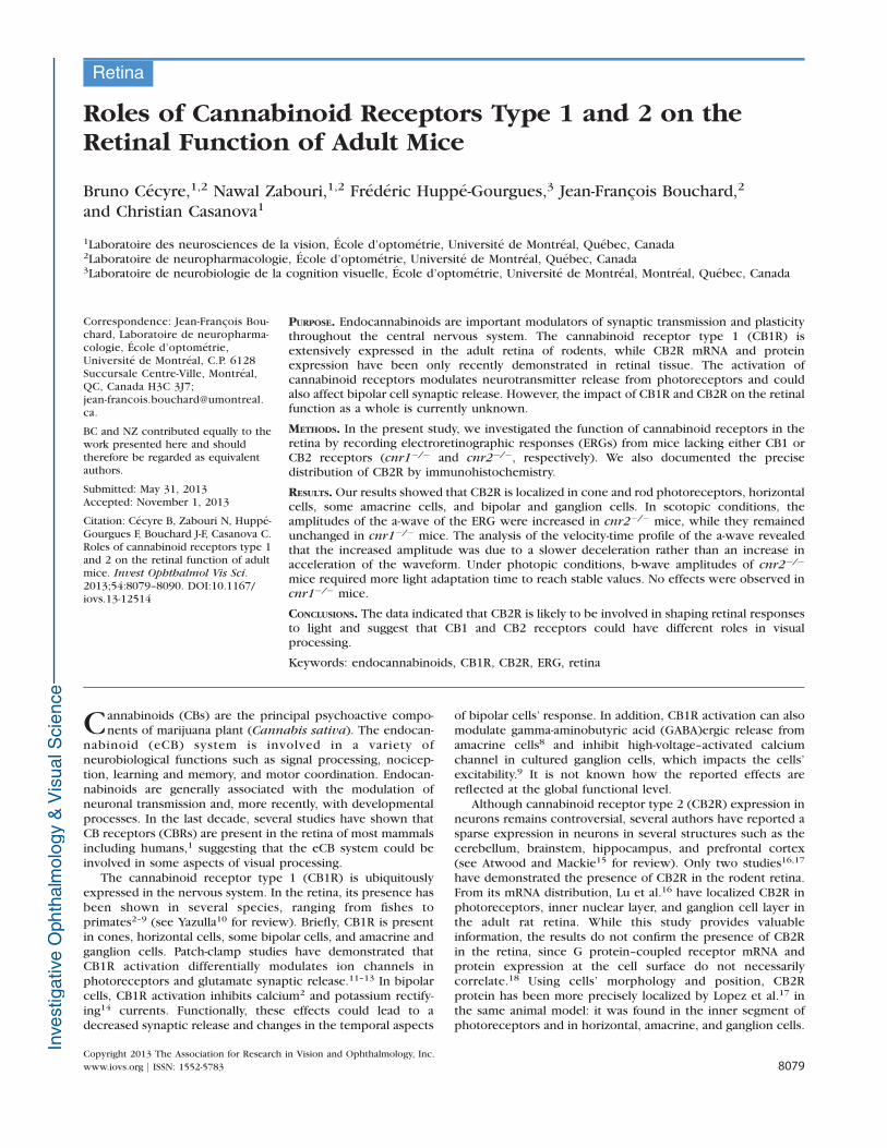
PURPOSE. Endocannabinoids are important modulators of synaptic transmission and plasticitythroughout the central nervous system. The cannabinoid receptor type 1 (CB1R) isextensively expressed in the adult retina of rodents, while CB2R mRNA and proteinexpression have been only recently demonstrated in retinal tissue. The activation ofcannabinoid receptors modulates neurotransmitter release from photoreceptors and couldalso affect bipolar cell synaptic release. However, the impact of CB1R and CB2R on the retinalfunction as a whole is currently unknown.
METHODS. In the present study, we investigated the function of cannabinoid receptors in theretina by recording electroretinographic responses (ERGs) from mice lacking either CB1 orCB2 receptors (cnr1�/� and cnr2�/�, respectively). We also documented the precisedistribution of CB2R by immunohistochemistry.
RESULTS. Our results showed that CB2R is localized in cone and rod photoreceptors, horizontalcells, some amacrine cells, and bipolar and ganglion cells. In scotopic conditions, theamplitudes of the a-wave of the ERG were increased in cnr2�/� mice, while they remainedunchanged in cnr1�/� mice. The analysis of the velocity-time profile of the a-wave revealedthat the increased amplitude was due to a slower deceleration rather than an increase inacceleration of the waveform. Under photopic conditions, b-wave amplitudes of cnr2�/�
mice required more light adaptation time to reach stable values. No effects were observed incnr1�/� mice.
CONCLUSIONS. The data indicated that CB2R is likely to be involved in shaping retinal responsesto light and suggest that CB1 and CB2 receptors could have different roles in visualprocessing.
Keywords: endocannabinoids, CB1R, CB2R, ERG, retina
Cannabinoids (CBs) are the principal psychoactive compo-nents of marijuana plant (Cannabis sativa). The endocan-
nabinoid (eCB) system is involved in a variety ofneurobiological functions such as signal processing, nocicep-tion, learning and memory, and motor coordination. Endocan-nabinoids are generally associated with the modulation ofneuronal transmission and, more recently, with developmentalprocesses. In the last decade, several studies have shown thatCB receptors (CBRs) are present in the retina of most mammalsincluding humans,1 suggesting that the eCB system could beinvolved in some aspects of visual processing.
The cannabinoid receptor type 1 (CB1R) is ubiquitouslyexpressed in the nervous system. In the retina, its presence hasbeen shown in several species, ranging from fishes toprimates2–9 (see Yazulla10 for review). Briefly, CB1R is presentin cones, horizontal cells, some bipolar cells, and amacrine andganglion cells. Patch-clamp studies have demonstrated thatCB1R activation differentially modulates ion channels inphotoreceptors and glutamate synaptic release.11–13 In bipolarcells, CB1R activation inhibits calcium2 and potassium rectify-ing14 currents. Functionally, these effects could lead to adecreased synaptic release and changes in the temporal aspects
of bipolar cells’ response. In addition, CB1R activation can alsomodulate gamma-aminobutyric acid (GABA)ergic release fromamacrine cells8 and inhibit high-voltage–activated calciumchannel in cultured ganglion cells, which impacts the cells’excitability.9 It is not known how the reported effects arereflected at the global functional level.
Although cannabinoid receptor type 2 (CB2R) expression inneurons remains controversial, several authors have reported asparse expression in neurons in several structures such as thecerebellum, brainstem, hippocampus, and prefrontal cortex(see Atwood and Mackie15 for review). Only two studies16,17
have demonstrated the presence of CB2R in the rodent retina.From its mRNA distribution, Lu et al.16 have localized CB2R inphotoreceptors, inner nuclear layer, and ganglion cell layer inthe adult rat retina. While this study provides valuableinformation, the results do not confirm the presence of CB2Rin the retina, since G protein–coupled receptor mRNA andprotein expression at the cell surface do not necessarilycorrelate.18 Using cells’ morphology and position, CB2Rprotein has been more precisely localized by Lopez et al.17 inthe same animal model: it was found in the inner segment ofphotoreceptors and in horizontal, amacrine, and ganglion cells.
Copyright 2013 The Association for Research in Vision and Ophthalmology, Inc.
www.iovs.org j ISSN: 1552-5783 8079
TA
BLE
1.
An
tib
od
ies
Use
din
Th
isSt
ud
y
An
tib
od
yT
arg
et
Imm
un
ogen
Dil
uti
on
Ho
stP
roven
an
ce
CB
2R
Can
nab
ino
idre
cep
tor
typ
e2
N-t
erm
inu
s1
4aa
of
hu
man
CB
2R
(20
–3
3
resi
du
es)
I,1
:20
0
W,
1:2
00
0
Rab
bit
10
15
50
;C
aym
anC
hem
ical
,A
nn
Arb
or,
MI
CB
1R
Can
nab
ino
idre
cep
tor
typ
e1
N-t
erm
inu
s7
7aa
of
rat
CB
1R
(1–7
7
resi
du
es)
W,
1:1
00
0R
abb
itC
12
33
;Si
gm
a-A
ldri
ch
,O
akville
,
ON
,C
anad
a
NA
PE
-PLD
N-a
cyl
ph
osp
hat
idye
than
ola
min
e
ph
osp
ho
lip
ase
D
N-t
erm
inu
s1
3aa
of
mo
use
NA
PE
-PLD
(9–2
1re
sid
ues)
W,
1:5
00
Rab
bit
NB
11
0-8
00
70
;N
ovu
sB
iolo
gic
als,
Oak
ville
,O
N,
Can
ada
DA
GLa
Dia
cyl
gly
cero
llip
ase
aC
-term
inu
s4
2aa
of
mo
use
DA
GLa
(10
03
–1
04
4re
sid
ues)
W,
1:2
00
Rab
bit
DG
La-
Rb
-Af3
80
;Fro
nti
er
Inst
itu
te,
Ish
ikar
i,H
okkai
do
,Ja
pan
FAA
HFat
tyac
idam
ide
hyd
rola
seC
-term
inu
s1
9aa
rat
FAA
H(5
61
–5
79
resi
du
es)
W,
1:3
00
0R
abb
it1
01
60
0;
Cay
man
Ch
em
ical
MG
LM
on
oac
ylgly
cero
llip
ase
N-t
erm
inu
s3
5aa
of
mo
use
MG
L(1
–3
5
resi
du
es)
W,
1:2
00
Rab
bit
MG
L-R
b-A
f20
0;
Fro
nti
er
Inst
itu
te
GA
PD
HLo
adin
gco
ntr
ol
Th
efu
ll-le
ngth
rab
bit
mu
scle
GA
PD
H
pro
tein
W,
1:2
0,0
00
Mo
use
G8
79
5;
Sigm
a-A
ldri
ch
Mo
use
co
ne-a
rrest
in(L
UM
Ij)
Co
ne
ph
oto
recep
tors
C-t
erm
inu
s1
3aa
of
the
mC
AR
pro
tein
(36
9–3
81
resi
du
es)
I,1
:10
00
Rab
bit
Ch
ery
lM
.C
raft
,M
ary
D.
Allen
Lab
ora
tory
for
Vis
ion
Rese
arch
,
Do
hen
yE
yeIn
stit
ute
,U
SC,
Lo
s
An
gele
s,C
A
Reco
veri
nR
od
ph
oto
recep
tors
,an
dO
Nan
dO
FF
co
ne
bip
ola
rcells
Th
efu
ll-le
ngth
reco
mb
inan
th
um
an
reco
veri
n
I,1
:20
00
Rab
bit
AB
55
85
;E
MD
Milip
ore
Co
rpo
rati
on
,B
ille
rica,
MA
Cal
bin
din
(D-2
8K
)H
ori
zon
tal
and
amac
rin
ecells
Reco
mb
inan
tra
tcal
bin
din
D-2
8K
full
len
gth
I,1
:10
00
Rab
bit
CB
-38
a;Sw
ant,
Mar
ly,
Swit
zerl
and
PK
C(c
lon
eH
7)
Ro
db
ipo
lar
cells
C-t
erm
inu
s2
8aa
of
hu
man
pro
tein
(64
5–
67
2re
sid
ues)
I,1
:50
0M
ou
seSc
-83
93
;Sa
nta
Cru
z
Bio
tech
no
logy,
San
taC
ruz,
CA
Syn
tax
in(c
lon
eH
PC
1)
Am
acri
ne
cells
Syn
apto
som
alp
lasm
afr
acti
on
of
rat
hip
po
cam
pu
s
I,1
:50
0M
ou
seS0
66
4;
Sigm
a-A
ldri
ch
Brn
-3a
(clo
ne
5A
3.2
)G
anglio
ncells
Am
ino
acid
s1
86
–2
24
of
Brn
-3a
fuse
dto
the
T7
gen
e1
0p
rote
in
I,1
:10
0M
ou
seM
AB
15
85
;E
MD
Millip
ore
Co
rpo
rati
on
Glu
tam
ine
syn
theta
se(G
S,clo
ne
GS-
6)
Mu
ller
cells
Th
efu
llp
rote
inp
uri
fied
fro
msh
eep
bra
in
I,1
:30
00
Mo
use
MA
B3
02
;E
MD
Millip
ore
Co
rpo
rati
on
aa,
amin
oac
ids;
I,im
mu
no
his
toch
em
istr
y;W
,W
est
ern
blo
t.
Roles of CB1R and CB2R in the Retina IOVS j December 2013 j Vol. 54 j No. 13 j 8080
However, the CB2R antibody used by these authors was notfully characterized. Thus, the exact localization of CB2R inretinal cells remains an open question. To our knowledge, nostudies have investigated the effect of CB2R activation at theretinal level.
The objective of this study was to evaluate the roles of CBRsin the retinal function in a mammalian in vivo model. Toachieve this goal, electroretinograms (ERGs, representing theglobal evoked response of the retina) of mice lacking CB1 orCB2 receptors (cnr1�/� and cnr2�/�, respectively) and theirwild-type (WT) controls were compared under different lightconditions. Given that the exact expression of pattern CB2R inthe retina remains debatable, the presence of CB2R in the miceretina was confirmed and precisely assessed with a specificCB2R antibody and specific retinal markers. Our resultsindicate that CB2R is largely expressed by retinal neurons ofmice and suggest that this receptor is likely to play a greaterrole in retinal processing than CB1R.
METHODS
Animals
All procedures were performed in accordance with theguidelines from the Canadian Council on Animal Care andthe ARVO Statement for the Use of Animals in Ophthalmic andVision Research, and were approved by the Ethics Committeeon Animal Research of the Universite de Montreal. The cnr1�/�
and cnr2�/� transgenic mice were obtained from Beat Lutz(Institute of Physiological Chemistry and Pathobiochemistry,University of Mainz, Germany) and Jackson Laboratory (BarHarbor, ME), respectively. The cnr1�/� and cnr2�/� mice wereon a C57BL/6N and C57BL/6J genetic background, respective-ly. Both transgenic mice were compared to background andage-matched WT controls from separate colonies. All animalswere maintained in-house, under a 12-hour dark/light cycle.Male and female adult mice (3–4 months old) were used for theexperiments.
Tissue Preparation
Mice were euthanized by an overdose of isoflurane. One eyewas immediately removed for Western blot analysis. The retinawas dissected on ice, promptly frozen, and kept at�808C untilfurther processing. Simultaneously, a transcardiac perfusionwas conducted with phosphate-buffered 0.9% saline (PBS; 0.1M, pH 7.4), followed by phosphate-buffered 4% paraformalde-hyde (PFA), until the head was lightly fixed. The nasal part ofthe second eye was marked with a suture and removed. Twosmall holes were made in the cornea before a first postfixationin 4% PFA for a period of 30 minutes. The cornea and lens wereremoved and the eyecups were postfixed for 30 minutes in 4%PFA. The eyecups were then washed in PBS, cryoprotected in30% sucrose overnight, embedded in Neg 50 tissue EmbeddingMedia (Fisher Scientific, Ottawa, ON, Canada), flash-frozen andkept at�808C. Sections (14 lm thick) were cut with a cryostat(Leica Microsystems, Concord, ON, Canada) and placed ongelatin/chromium-coated slides.
Western Blot
Retinas were homogenized in RIPA buffer (150 mM NaCl, 20mM Tris, pH 8.0, 1% NP-40, 0.1% SDS, 1 mM EDTA),supplemented with a protease inhibitor mixture (aprotinin,leupeptin, pepstatin [1 lg/mL] and phenylmethylsulfonylfluoride [0.2 mg/mL]; Roche Applied Science, Laval, QC,
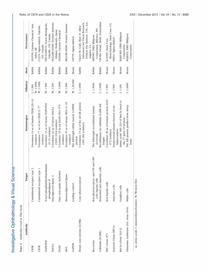
Canada). Thirty micrograms of protein per sample of thehomogenate were resolved by electrophoresis on a 10% SDS-polyacrylamide gel, transferred onto a nitrocellulose mem-brane, blocked with 5% skim milk, and incubated overnightwith antibodies directed against CB2R, CB1R, N-acyl phospha-tidyethanolamine phospholipase D (NAPE-PLD), diacylglycerollipase a (DAGLa), fatty acid amide hydrolase (FAAH), mono-acylglycerol lipase (MGL), or glyceraldehyde 3-phosphatedehydrogenase (GAPDH), the latter serving as a loadingcontrol. The blots were exposed to the appropriate horserad-ish peroxidase–coupled secondary antibodies (Jackson Immu-noresearch Laboratories, West Grove, PA). Detection wascarried out by using homemade ECL Western blot detectionreagents (final concentrations: 2.5 mM luminol, 0.4 mM p-coumaric acid, 0.1 M Tris-HCl pH 8.5, 0.018% H2O2).
Immunohistochemistry
Frozen sections were washed in PBS, postfixed for 10 minutesin cold acetone, washed, and then blocked in 1% bovine serumalbumin, bovine gelatin, and 0.5% Triton X-100 in PBS for 1hour. Some sections from WT animals were incubatedovernight in rabbit anti-CB2R solution with an antibody againstvarious retinal markers. In addition, sections from the threegenotypes were incubated with retinal markers (see Table 1) tocompare the distribution and morphology of retinal cells. Thesections were then washed in PBS, blocked for 30 minutes,incubated for 1 hour with Alexa Fluor 647 donkey anti-rabbitfor CB2R and Alexa Fluor 488 donkey anti-rabbit or Alexa Fluor488 donkey anti-mouse for cell type markers (MolecularProbes, Eugene, OR). After washes, the sections were mountedwith homemade PVA-Dabco mounting medium. The CB2R/recoverin, CB2R/cone-arrestin,19–21 and CB2R/calbindin com-binations required serial incubations, as the retinal marker hostwas rabbit, as previously performed on retinal tissues.6 Thedilution factors, the immunogens, and provenance of theantibodies are provided in Table 1.
Antibody Characterization
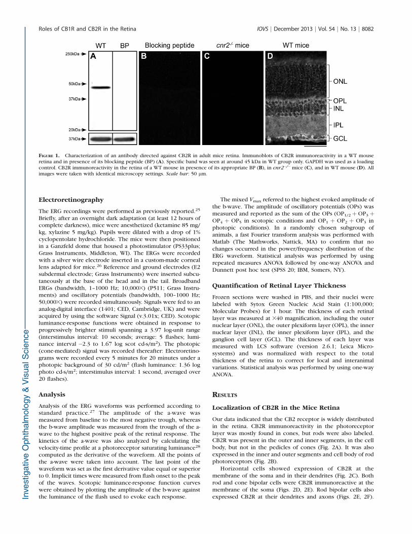
The CB2R antibody has been characterized in mice renaltissue22; consequently, we tested its specificity in the miceretina by Western blot and immunohistochemistry. The anti-CB2R reacted with a robust band at 45 kDa (Fig. 1A), whichwas absent with the addition of the immunizing peptide. Nounspecific signal could be observed on retinal sections whenCB2R antibody was preincubated with its immunizing peptide(Fig. 1B). Moreover, no unspecific signal was visible in thecnr2�/� mice (Fig. 1C), whereas the CB2R staining in the WTmouse yielded a strong signal in the retina (Fig. 1D). It shouldbe noted that this antibody showed inconsistent results fromone lot to another. These results display the specificity of lotNo. 0424681-1.
Confocal Microscopy
Images of the central retina (within 200 lm of the optic nervehead) were taken with a laser scanning confocal microscope(TCS SP2; Leica Microsystems), with an 340 oil immersionobjective. Image stacks (1024 3 1024 pixels 3 0.5 lm perstack) were captured by using the Leica confocal (LCS)software (version 2.6.1; Leica Microsystems). Offline process-ing was done with the Fiji software (version 1.47g; www.fiji.sc).23 The stack images were taken sequentially to ensure no‘‘bleed-through’’ between channels. Gaussian noise fromimages was partially removed by using the PureDenoise pluginfor Fiji24 and stacks were collapsed to projection images.
Roles of CB1R and CB2R in the Retina IOVS j December 2013 j Vol. 54 j No. 13 j 8081
Electroretinography
The ERG recordings were performed as previously reported.25
Briefly, after an overnight dark adaptation (at least 12 hours ofcomplete darkness), mice were anesthetized (ketamine 85 mg/kg, xylazine 5 mg/kg). Pupils were dilated with a drop of 1%cyclopentolate hydrochloride. The mice were then positionedin a Ganzfeld dome that housed a photostimulator (PS33plus;Grass Instruments, Middleton, WI). The ERGs were recordedwith a silver wire electrode inserted in a custom-made corneallens adapted for mice.26 Reference and ground electrodes (E2subdermal electrode; Grass Instruments) were inserted subcu-taneously at the base of the head and in the tail. BroadbandERGs (bandwidth, 1–1000 Hz; 10,0003) (P511; Grass Instru-ments) and oscillatory potentials (bandwidth, 100–1000 Hz;50,0003) were recorded simultaneously. Signals were fed to ananalog-digital interface (1401; CED, Cambridge, UK) and wereacquired by using the software Signal (v.3.01x; CED). Scotopicluminance-response functions were obtained in response toprogressively brighter stimuli spanning a 3.97 log-unit range(interstimulus interval: 10 seconds; average: 5 flashes; lumi-nance interval �2.3 to 1.67 log scot cd-s/m2). The photopic(cone-mediated) signal was recorded thereafter: Electroretino-grams were recorded every 5 minutes for 20 minutes under aphotopic background of 30 cd/m2 (flash luminance: 1.36 logphoto cd-s/m2; interstimulus interval: 1 second, averaged over20 flashes).
Analysis
Analysis of the ERG waveforms was performed according tostandard practice.27 The amplitude of the a-wave wasmeasured from baseline to the most negative trough, whereasthe b-wave amplitude was measured from the trough of the a-wave to the highest positive peak of the retinal response. Thekinetics of the a-wave was also analyzed by calculating thevelocity-time profile at a photoreceptor saturating luminance28
computed as the derivative of the waveform. All the points ofthe a-wave were taken into account. The last point of thewaveform was set as the first derivative value equal or superiorto 0. Implicit times were measured from flash onset to the peakof the waves. Scotopic luminance-response function curveswere obtained by plotting the amplitude of the b-wave againstthe luminance of the flash used to evoke each response.
The mixed Vmax referred to the highest evoked amplitude ofthe b-wave. The amplitude of oscillatory potentials (OPs) wasmeasured and reported as the sum of the OPs (OP1/2þ OP3þOP4 þ OP5 in scotopic conditions and OP1 þ OP2 þ OP3 inphotopic conditions). In a randomly chosen subgroup ofanimals, a fast Fourier transform analysis was performed withMatlab (The Mathworks, Nattick, MA) to confirm that nochanges occurred in the power/frequency distribution of theERG waveform. Statistical analysis was performed by usingrepeated measures ANOVA followed by one-way ANOVA andDunnett post hoc test (SPSS 20; IBM, Somers, NY).
Quantification of Retinal Layer Thickness
Frozen sections were washed in PBS, and their nuclei werelabeled with Sytox Green Nucleic Acid Stain (1:100,000;Molecular Probes) for 1 hour. The thickness of each retinallayer was measured at 340 magnification, including the outernuclear layer (ONL), the outer plexiform layer (OPL), the innernuclear layer (INL), the inner plexiform layer (IPL), and theganglion cell layer (GCL). The thickness of each layer wasmeasured with LCS software (version 2.6.1; Leica Micro-systems) and was normalized with respect to the totalthickness of the retina to correct for local and interanimalvariations. Statistical analysis was performed by using one-wayANOVA.
RESULTS
Localization of CB2R in the Mice Retina
Our data indicated that the CB2 receptor is widely distributedin the retina. CB2R immunoreactivity in the photoreceptorlayer was mostly found in cones, but rods were also labeled.CB2R was present in the outer and inner segments, in the cellbody, but not in the pedicles of cones (Fig. 2A). It was alsoexpressed in the inner and outer segments and cell body of rodphotoreceptors (Fig. 2B).
Horizontal cells showed expression of CB2R at themembrane of the soma and in their dendrites (Fig. 2C). Bothrod and cone bipolar cells were CB2R immunoreactive at themembrane of the soma (Figs. 2D, 2E). Rod bipolar cells alsoexpressed CB2R at their dendrites and axons (Figs. 2E, 2F).
FIGURE 1. Characterization of an antibody directed against CB2R in adult mice retina. Immunoblots of CB2R immunoreactivity in a WT mouseretina and in presence of its blocking peptide (BP) (A). Specific band was seen at around 45 kDa in WT group only. GAPDH was used as a loadingcontrol. CB2R immunoreactivity in the retina of a WT mouse in presence of its appropriate BP (B), in cnr2�/�mice (C), and in WT mouse (D). Allimages were taken with identical microscopy settings. Scale bar: 50 lm.
Roles of CB1R and CB2R in the Retina IOVS j December 2013 j Vol. 54 j No. 13 j 8082
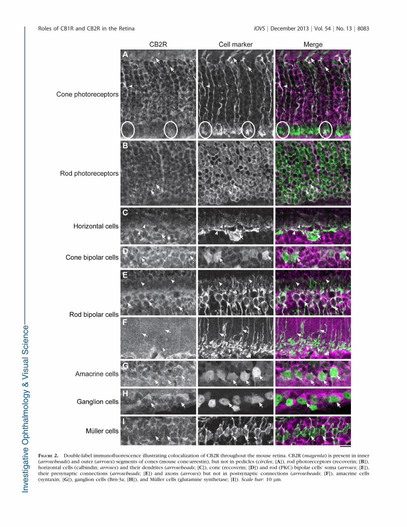
FIGURE 2. Double-label immunofluorescence illustrating colocalization of CB2R throughout the mouse retina. CB2R (magenta) is present in inner(arrowheads) and outer (arrows) segments of cones (mouse cone-arrestin), but not in pedicles (circles; [A]), rod photoreceptors (recoverin; [B]),horizontal cells (calbindin; arrows) and their dendrites (arrowheads; [C]), cone (recoverin; [D]) and rod (PKC) bipolar cells’ soma (arrows; [E]),their presynaptic connections (arrowheads; [E]) and axons (arrows) but not in postsynaptic connections (arrowheads; [F]), amacrine cells(syntaxin; [G]), ganglion cells (Brn-3a; [H]), and Muller cells (glutamine synthetase; [I]). Scale bar: 10 lm.
Roles of CB1R and CB2R in the Retina IOVS j December 2013 j Vol. 54 j No. 13 j 8083
Some amacrine cells showed also CB2R immunoreactivity atthe membrane of the soma (Fig. 2G).
CB2R staining was detected in the GCL and was present inthe ganglion cells’ soma (Fig. 2H). CB2R was not expressed atthe membrane of the Muller cells’ soma or in their inner andouter processes (Fig. 2I).
The Dark-Adapted Retinal Function
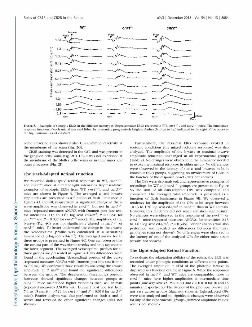
We recorded dark-adapted retinal responses in WT, cnr1�/�,and cnr2�/� mice at different light intensities. Representativeexamples of scotopic ERGs from WT, cnr1�/�, and cnr2�/�
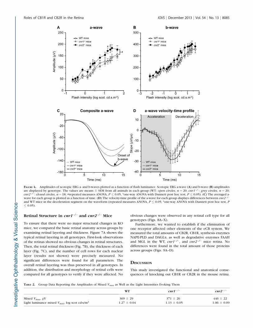
mice are shown in Figure 3. The averaged a- and b-waveamplitudes are presented as a function of flash luminance inFigures 4A and 4B, respectively. A significant change in the a-wave amplitude was observed in cnr2�/� but not in cnr1�/�
mice (repeated measures ANOVA with Dunnett post hoc test,for intensities 0.13 to 1.67 log scot cd-s/m2, P ¼ 0.798 forcnr1�/� and P¼ 0.037 for cnr2�/�mice). The amplitude of theb-wave (Fig. 4C) was not significantly altered in cnr1�/� orcnr2�/� mice. To better understand the change in the a-wave,the velocity-time profile was calculated at a saturatingluminance (1.3 log scot cd-s/m2). The averaged a-wave for allthree groups is presented in Figure 4C. One can observe thatthe earliest part of the waveforms overlay and only separate inthe latest segment. The averaged velocity-time profiles for allthree groups are presented in Figure 4D. No differences werefound in the accelerating (descending) portion of the curve(repeated measures ANOVA with Dunnett post hoc test from 0to 7.4 ms). We confirmed those results by analyzing the a-waveamplitude at 7 ms29 and found no significant differencesbetween the groups. The deceleration (ascending) portion,however, showed significant changes between groups ascnr2�/� mice maintained higher velocities than WT animals(repeated measures ANOVA with Dunnett post hoc test from7.4 to 15 ms, P¼ 0.729 for cnr1�/� and P¼ 0.018 for cnr2�/�
mice). Fourier analysis was also performed on both a- and b-waves and revealed no other significant changes (data notshown).
Furthermore, the maximal ERG response evoked inscotopic conditions (the mixed rod-cone response) was alsoanalyzed. The amplitude of the b-wave at maximal b-waveamplitude remained unchanged in all experimental groups(Table 2). No changes were observed in the luminance neededto evoke the maximal response in either group. No differenceswere observed in the latency of the a- and b-waves in bothknockout (KO) groups, suggesting no involvement of CBRs inthe kinetics of the response onset (data not shown).
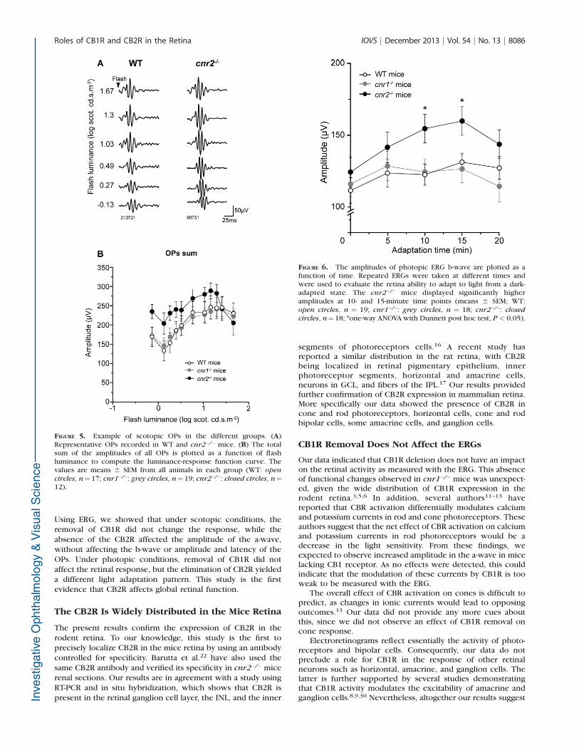
The OPs were also analyzed, and representative examples ofrecordings for WT and cnr2�/� groups are presented in Figure5A.The sum of all dark-adapted OPs was computed andcompared. The averaged total amplitude is presented as afunction of flash luminance in Figure 5B. We observed atendency for the amplitude of the OPs to be larger between�0.13 to 1.30 log scot cd-s/m2 in cnr2�/� than in WT animals.However, this tendency did not reach statistical significance.No changes were observed in the response of the cnr1�/� orcnr2�/� mice (repeated measures ANOVA, for intensities 0.13to 1.67 log scot cd-s/m2, P ¼ 0.345). Fourier analysis was alsoperformed and revealed no differences between the threegenotypes (data not shown). No differences were observed inthe latency of any of the analyzed OPs for either mice strain(results not shown).
The Light-Adapted Retinal Function
To evaluate the adaptation abilities of the retina, the ERG wasrecorded under photopic conditions at different time points.The averaged amplitude 6 SEM of the photopic b-wave isdisplayed as a function of time in Figure 6. While the responsesobserved in cnr1�/� and WT mice are comparable, those incnr2�/� mice have higher amplitudes at intermediate timepoints (one-way ANOVA, P¼0.021 and P¼0.018 for 10 and 15minutes, respectively). The latency of the photopic b-wave didnot vary across groups (data not shown). Light-adapted OPswere also analyzed and no significant changes were observedfor any of the experimental groups (summed amplitude values;results not shown).
FIGURE 3. Example of scotopic ERGs in the different genotypes. Representative ERGs recorded in WT, cnr1�/�, and cnr2�/�mice. The luminance-response function of each animal was established by presenting progressively brighter flashes (bottom to top) indicated to the right of the traces asthe log luminance (scot cd-s/m2).
Roles of CB1R and CB2R in the Retina IOVS j December 2013 j Vol. 54 j No. 13 j 8084
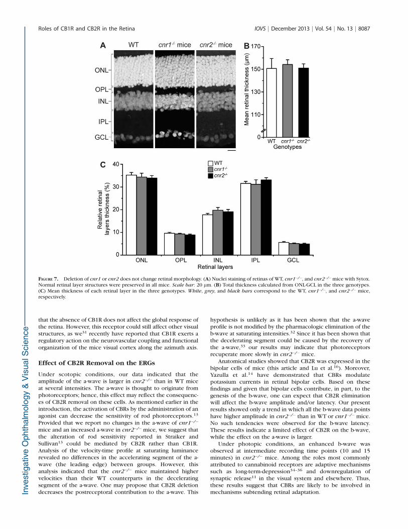
Retinal Structure in cnr1�/� and cnr2�/� Mice
To ensure that there were no major structural changes in KO
mice, we compared the basic retinal anatomy across groups by
examining retinal layering and thickness. Figure 7A shows the
typical retinal layering in all genotypes. First-look observations
of the retinas showed no obvious changes in retinal structures.
Then, the total retinal thickness (Fig. 7B), the thickness of each
layer (Fig. 7C), and the number of cell rows for each nuclear
layer (results not shown) were precisely measured. No
significant differences were found for all parameters. The
overall retinal layering was thus preserved in all genotypes. In
addition, the distribution and morphology of retinal cells were
compared for all genotypes to verify if they were affected. No
obvious changes were observed in any retinal cell type for allgenotypes (Figs. 8A–X).
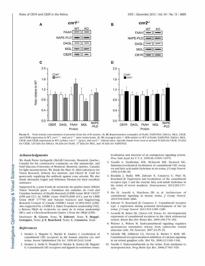
Furthermore, we wanted to establish if the elimination ofone receptor affected other elements of the eCB system. Wemeasured the total amounts of CB2R, CB1R, synthesis enzymesNAPE-PLD and DAGLa, as well as degradative enzymes FAAHand MGL in the WT, cnr1�/�, and cnr2�/� mice retina. Nodifferences were found in the total amount of those proteinsacross groups (Figs. 9A–D).
DISCUSSION
This study investigated the functional and anatomical conse-quences of knocking out CB1R or CB2R in the mouse retina.
TABLE 2. Group Data Reporting the Amplitudes of Mixed Vmax as Well as the Light Intensities Evoking Them
WT cnr1�/� cnr2�/�
Mixed Vmax, lV 369 6 29 371 6 26 446 6 22
Light luminance mixed Vmax, log scot cd-s/m2 1.27 6 0.04 1.13 6 0.05 1.06 6 0.09
FIGURE 4. Amplitudes of scotopic ERG a- and b-waves plotted as a function of flash luminance. Scotopic ERG a-wave (A) and b-wave (B) amplitudesare displayed by genotype. The values are means 6 SEM from all animals in each group (WT: open circles, n ¼ 20; cnr1�/�: grey circles, n ¼ 20;cnr2�/�: closed circles, n¼ 18; #repeated measures ANOVA, P � 0.05, *one-way ANOVA with Dunnett post hoc test, P � 0.05). (C) The averaged a-wave for each group is plotted as a function of time. (D) The velocity-time profile of the a-wave for each group displays differences between cnr2�/�
and WT mice in the deceleration segment on the waveform (repeated measures ANOVA, P � 0.05; *one-way ANOVA with Dunnett post hoc test, P
� 0.05).
Roles of CB1R and CB2R in the Retina IOVS j December 2013 j Vol. 54 j No. 13 j 8085
Using ERG, we showed that under scotopic conditions, theremoval of CB1R did not change the response, while theabsence of the CB2R affected the amplitude of the a-wave,without affecting the b-wave or amplitude and latency of theOPs. Under photopic conditions, removal of CB1R did notaffect the retinal response, but the elimination of CB2R yieldeda different light adaptation pattern. This study is the firstevidence that CB2R affects global retinal function.
The CB2R Is Widely Distributed in the Mice Retina
The present results confirm the expression of CB2R in therodent retina. To our knowledge, this study is the first toprecisely localize CB2R in the mice retina by using an antibodycontrolled for specificity. Barutta et al.22 have also used thesame CB2R antibody and verified its specificity in cnr2�/�micerenal sections. Our results are in agreement with a study usingRT-PCR and in situ hybridization, which shows that CB2R ispresent in the retinal ganglion cell layer, the INL, and the inner
segments of photoreceptors cells.16 A recent study hasreported a similar distribution in the rat retina, with CB2Rbeing localized in retinal pigmentary epithelium, innerphotoreceptor segments, horizontal and amacrine cells,neurons in GCL, and fibers of the IPL.17 Our results providedfurther confirmation of CB2R expression in mammalian retina.More specifically our data showed the presence of CB2R incone and rod photoreceptors, horizontal cells, cone and rodbipolar cells, some amacrine cells, and ganglion cells.
CB1R Removal Does Not Affect the ERGs
Our data indicated that CB1R deletion does not have an impacton the retinal activity as measured with the ERG. This absenceof functional changes observed in cnr1�/�mice was unexpect-ed, given the wide distribution of CB1R expression in therodent retina.3,5,6 In addition, several authors11–13 havereported that CBR activation differentially modulates calciumand potassium currents in rod and cone photoreceptors. Theseauthors suggest that the net effect of CBR activation on calciumand potassium currents in rod photoreceptors would be adecrease in the light sensitivity. From these findings, weexpected to observe increased amplitude in the a-wave in micelacking CB1 receptor. As no effects were detected, this couldindicate that the modulation of these currents by CB1R is tooweak to be measured with the ERG.
The overall effect of CBR activation on cones is difficult topredict, as changes in ionic currents would lead to opposingoutcomes.13 Our data did not provide any more cues aboutthis, since we did not observe an effect of CB1R removal oncone response.
Electroretinograms reflect essentially the activity of photo-receptors and bipolar cells. Consequently, our data do notpreclude a role for CB1R in the response of other retinalneurons such as horizontal, amacrine, and ganglion cells. Thelatter is further supported by several studies demonstratingthat CB1R activity modulates the excitability of amacrine andganglion cells.8,9,30 Nevertheless, altogether our results suggest
FIGURE 5. Example of scotopic OPs in the different groups. (A)Representative OPs recorded in WT and cnr2�/� mice. (B) The totalsum of the amplitudes of all OPs is plotted as a function of flashluminance to compute the luminance-response function curve. Thevalues are means 6 SEM from all animals in each group (WT: open
circles, n¼17; cnr1�/�: grey circles, n¼19; cnr2�/�: closed circles, n¼12).
FIGURE 6. The amplitudes of photopic ERG b-wave are plotted as afunction of time. Repeated ERGs were taken at different times andwere used to evaluate the retina ability to adapt to light from a dark-adapted state. The cnr2�/� mice displayed significantly higheramplitudes at 10- and 15-minute time points (means 6 SEM; WT:open circles, n ¼ 19; cnr1�/�: grey circles, n ¼ 18; cnr2�/�: closed
circles, n¼18; *one-way ANOVA with Dunnett post hoc test, P < 0.05).
Roles of CB1R and CB2R in the Retina IOVS j December 2013 j Vol. 54 j No. 13 j 8086
that the absence of CB1R does not affect the global response ofthe retina. However, this receptor could still affect other visualstructures, as we31 recently have reported that CB1R exerts aregulatory action on the neurovascular coupling and functionalorganization of the mice visual cortex along the azimuth axis.
Effect of CB2R Removal on the ERGs
Under scotopic conditions, our data indicated that theamplitude of the a-wave is larger in cnr2�/� than in WT miceat several intensities. The a-wave is thought to originate fromphotoreceptors; hence, this effect may reflect the consequenc-es of CB2R removal on these cells. As mentioned earlier in theintroduction, the activation of CBRs by the administration of anagonist can decrease the sensitivity of rod photoreceptors.13
Provided that we report no changes in the a-wave of cnr1�/�
mice and an increased a-wave in cnr2�/�mice, we suggest thatthe alteration of rod sensitivity reported in Straiker andSullivan13 could be mediated by CB2R rather than CB1R.Analysis of the velocity-time profile at saturating luminancerevealed no differences in the accelerating segment of the a-wave (the leading edge) between groups. However, thisanalysis indicated that the cnr2�/� mice maintained highervelocities than their WT counterparts in the deceleratingsegment of the a-wave. One may propose that CB2R deletiondecreases the postreceptoral contribution to the a-wave. This
hypothesis is unlikely as it has been shown that the a-waveprofile is not modified by the pharmacologic elimination of theb-wave at saturating intensities.32 Since it has been shown thatthe decelerating segment could be caused by the recovery ofthe a-wave,33 our results may indicate that photoreceptorsrecuperate more slowly in cnr2�/� mice.
Anatomical studies showed that CB2R was expressed in thebipolar cells of mice (this article and Lu et al.16). Moreover,Yazulla et al.14 have demonstrated that CBRs modulatepotassium currents in retinal bipolar cells. Based on thesefindings and given that bipolar cells contribute, in part, to thegenesis of the b-wave, one can expect that CB2R eliminationwill affect the b-wave amplitude and/or latency. Our presentresults showed only a trend in which all the b-wave data pointshave higher amplitude in cnr2�/� than in WT or cnr1�/�mice.No such tendencies were observed for the b-wave latency.These results indicate a limited effect of CB2R on the b-wave,while the effect on the a-wave is larger.
Under photopic conditions, an enhanced b-wave wasobserved at intermediate recording time points (10 and 15minutes) in cnr2�/� mice. Among the roles most commonlyattributed to cannabinoid receptors are adaptive mechanismssuch as long-term-depression34–36 and downregulation ofsynaptic release11 in the visual system and elsewhere. Thus,these results suggest that CBRs are likely to be involved inmechanisms subtending retinal adaptation.
FIGURE 7. Deletion of cnr1 or cnr2 does not change retinal morphology. (A) Nuclei staining of retinas of WT, cnr1�/�, and cnr2�/�mice with Sytox.Normal retinal layer structures were preserved in all mice. Scale bar: 20 lm. (B) Total thickness calculated from ONL-GCL in the three genotypes.(C) Mean thickness of each retinal layer in the three genotypes. White, grey, and black bars correspond to the WT, cnr1�/�, and cnr2�/� mice,respectively.
Roles of CB1R and CB2R in the Retina IOVS j December 2013 j Vol. 54 j No. 13 j 8087

We showed that functional changes observed in cnr1�/� andcnr2�/� mice are not due to abnormal retinal cell distributionor compensation of the eCB system activity. Previous studieshave shown that the eCB system interacts with GABA,glutamate, and dopamine systems.37–40 For example, CB1Ragonists stimulate dopamine release from the guinea pigretina.37 Hence, the changes in retinal response in cnr2�/�
mice, and/or lack thereof in cnr1�/�mice, could be partly due
to compensation mechanisms in GABA, glutamate, or dopa-mine systems.
In conclusion, this study demonstrated that CB2R isexpressed in several cell types in the mice retina. Our resultsalso suggest that CB1R and CB2R contribute differently tovisual functions: CB1R does not seem to be involved in globalretinal responses, while CB2R appears to be implicated in rodand cone sensitivity and light adaptation.
FIGURE 8. Absence of CB1R or CB2R does not produce any obvious changes in the distribution and morphology of cone photoreceptors (mousecone-arrestin; [A–C]), rod photoreceptors (recoverin; [D–F]), horizontal cells (calbindin; [G–I]), cone bipolar cells (recoverin; [J–L]), rod bipolarcells (PKC; [M–O]), amacrine cells (HPC; [P–R]), ganglion cells (Brn-3a; [S–U]), and Muller cells (glutamine synthetase; [V–X]) in WT, cnr1�/�, andcnr2�/� mice. Normal morphology and distribution of every retinal cell types were preserved in all mice. Scale bars: 10 lm.
Roles of CB1R and CB2R in the Retina IOVS j December 2013 j Vol. 54 j No. 13 j 8088
Acknowledgments
We thank Pierre Lachapelle (McGill University, Montreal, Quebec,Canada) for his constructive comments on this manuscript, andVasil Diaconu (University of Montreal, Montreal, Quebec, Canada)for light measurements. We thank the Mary D. Allen Laboratory forVision Research, Doheny Eye Institute, and Cheryl M. Craft forgenerously supplying the antibody against cone arrestin. We alsothank Alexandra Gagne and Sebastien Thomas for their excellentassistance.
Supported by a joint Fonds de recherche du Quebec-Sante (FRQS)Vision Network grant – Fondation des maladies de l’oeil andCanadian Institutes of Health Research (CIHR) Grant MOP 130337(J-FB and CC), by NSERC Grant 194670-2009 (CC), and by CIHRGrant MOP 177796 and Natural Sciences and EngineeringResearch Council of Canada (NSERC) Grant 311892-2010 (J-FB).Also supported by a CIHR-E.A. Baker Foundation studentship (NZ),a Reseau FRQS de recherche en sante de la vision studentship(BC), and a Chercheur-Boursier Junior 2 from the FRQS (J-FB).
Disclosure: B. Cecyre, None; N. Zabouri, None; F. Huppe-Gourgues, None; J.-F. Bouchard, None; C. Casanova, None
References
1. Straiker A, Maguire G, Mackie K, Lindsey J. Localization ofcannabinoid CB1 receptors in the human anterior eye andretina. Invest Ophthalmol Vis Sci. 1999;40:2442–2448.
2. Straiker A, Stella N, Piomelli D, Mackie K, Karten HJ, MaguireG. Cannabinoid CB1 receptors and ligands in vertebrate retina:
localization and function of an endogenous signaling system.Proc Natl Acad Sci U S A. 1999;96:14565–14570.
3. Yazulla S, Studholme KM, McIntosh HH, Deutsch DG.Immunocytochemical localization of cannabinoid CB1 recep-tor and fatty acid amide hydrolase in rat retina. J Comp Neurol.1999;415:80–90.
4. Bouskila J, Burke MW, Zabouri N, Casanova C, Ptito M,Bouchard JF. Expression and localization of the cannabinoidreceptor type 1 and the enzyme fatty acid amide hydrolase inthe retina of vervet monkeys. Neuroscience. 2012;202:117–130.
5. Hu SS, Arnold A, Hutchens JM, et al. Architecture ofcannabinoid signaling in mouse retina. J Comp Neurol.2010;518:3848–3866.
6. Zabouri N, Bouchard JF, Casanova C. Cannabinoid receptortype 1 expression during postnatal development of the ratretina. J Comp Neurol. 2011;519:1258–1280.
7. Leonelli M, Britto LR, Chaves GP, Torrao AS. Developmentalexpression of cannabinoid receptors in the chick retinotectalsystem. Brain Res Dev Brain Res. 2005;156:176–182.
8. Warrier A, Wilson M. Endocannabinoid signaling regulatesspontaneous transmitter release from embryonic retinalamacrine cells. Vis Neurosci. 2007;24:25–35.
9. Lalonde MR, Jollimore CA, Stevens K, Barnes S, Kelly ME.Cannabinoid receptor-mediated inhibition of calcium signalingin rat retinal ganglion cells. Mol Vis. 2006;12:1160–1166.
10. Yazulla S. Endocannabinoids in the retina: from marijuana toneuroprotection. Prog Retin Eye Res. 2008;27:501–526.
FIGURE 9. Total retinal concentration of proteins from the eCB system. (A, B) Representative examples of FAAH, NAPE-PLD, DAGLa, MGL, CB1R,and CB2R expression in WT, cnr1�/�, and cnr2�/�mice retina lysate. (C, D) Averaged ratio 6 SEM relative to WT of FAAH, NAPE-PLD, DAGLa, MGL,CB1R, and CB2R expression in WT (white), cnr1�/� (grey), and cnr2�/� (black) mice. Specific bands were seen at around 53 kDa for CB1R, 45 kDafor CB2R, 120 kDa for DAGLa, 66 kDa for FAAH, 37 kDa for MGL, and 46 kDa for NAPE-PLD.
Roles of CB1R and CB2R in the Retina IOVS j December 2013 j Vol. 54 j No. 13 j 8089
11. Fan SF, Yazulla S. Retrograde endocannabinoid inhibition ofgoldfish retinal cones is mediated by 2-arachidonoyl glycerol.Vis Neurosci. 2007;24:257–267.
12. Fan SF, Yazulla S. Biphasic modulation of voltage-dependentcurrents of retinal cones by cannabinoid CB1 receptor agonistWIN 55212-2. Vis Neurosci. 2003;20:177–188.
13. Straiker A, Sullivan JM. Cannabinoid receptor activationdifferentially modulates ion channels in photoreceptors ofthe tiger salamander. J Neurophysiol. 2003;89:2647–2654.
14. Yazulla S, Studholme KM, McIntosh HH, Fan SF. Cannabinoidreceptors on goldfish retinal bipolar cells: electron-micro-scope immunocytochemistry and whole-cell recordings. Vis
Neurosci. 2000;17:391–401.
15. Atwood BK, Mackie K. CB2: a cannabinoid receptor with anidentity crisis. Br J Pharmacol. 2010;160:467–479.
16. Lu Q, Straiker A, Lu Q, Maguire G. Expression of CB2cannabinoid receptor mRNA in adult rat retina. Vis Neurosci.2000;17:91–95.
17. Lopez EM, Tagliaferro P, Onaivi ES, Lopez-Costa JJ. Distributionof CB2 cannabinoid receptor in adult rat retina. Synapse.2011;65:388–392.
18. de Sousa Abreu R, Penalva LO, Marcotte EM, Vogel C. Globalsignatures of protein and mRNA expression levels. Mol
Biosyst. 2009;5:1512–1526.
19. Nikonov SS, Brown BM, Davis JA, et al. Mouse cones requirean arrestin for normal inactivation of phototransduction.Neuron. 2008;59:462–474.
20. Zhu X, Brown B, Li A, Mears AJ, Swaroop A, Craft CM. GRK1-dependent phosphorylation of S and M opsins and theirbinding to cone arrestin during cone phototransduction in themouse retina. J Neurosci. 2003;23:6152–6160.
21. Zhu X, Li A, Brown B, Weiss ER, Osawa S, Craft CM. Mousecone arrestin expression pattern: light induced translocationin cone photoreceptors. Mol Vis. 2002;8:462–471.
22. Barutta F, Piscitelli F, Pinach S, et al. Protective role ofcannabinoid receptor type 2 in a mouse model of diabeticnephropathy. Diabetes. 2011;60:2386–2396.
23. Schindelin J, Arganda-Carreras I, Frise E, et al. Fiji: an open-source platform for biological-image analysis. Nat Methods.2012;9:676–682.
24. Luisier F, Blu T. SURE-LET multichannel image denoising:interscale orthonormal wavelet thresholding. IEEE Trans
Image Processing. 2008;17:482–492.
25. Plaziac C, Lachapelle P, Casanova C. Effects of methanol on theretinal function of juvenile rats. Neurotoxicology. 2003;24:255–260.
26. Sagdullaev BT, DeMarco PJ, McCall MA. Improved contact lens
electrode for corneal ERG recordings in mice. Doc Ophthal-
mol. 2004;108:181–184.
27. Marmor MF, Fulton AB, Holder GE, et al. ISCEV Standard forfull-field clinical electroretinography (2008 update). Doc
Ophthalmol. 2009;118:69–77.
28. Weymouth AE, Vingrys AJ. Rodent electroretinography:methods for extraction and interpretation of rod and coneresponses. Prog Retin Eye Res. 2008;27:1–44.
29. Heckenlively JR, Arden GB. Principles and Practice of Clinical
Electrophysiology of Vision. 2nd ed. Cambridge, MA: MITPress; 2006.
30. Middleton T, Protti D. Cannabinoids modulate spontaneoussynaptic activity in retinal ganglion cells. Vis Neurosci. 2011;28:393–402.
31. Abbas Farishta R, Robert C, Vanni MP, Minville K, Bouchard JF,Casanova C. Effects of Cannabinoid CB1 Receptor on
Hemodynamic Responses and Functional Organization of
the Primary Visual Cortex: 2010 Neuroscience Meeting
Planner. San Diego, CA: Society for Neuroscience; 2010.
32. Lei B. Rod-driven OFF pathway responses in the distal retina:dark-adapted flicker electroretinogram in mouse. PLoS One.2012;7:e43856.
33. Kang Derwent JJ, Linsenmeier RA. Intraretinal analysis of the a-wave of the electroretinogram (ERG) in dark-adapted intact catretina. Vis Neurosci. 2001;18:353–363.
34. Liu CH, Heynen AJ, Shuler MG, Bear MF. Cannabinoid receptorblockade reveals parallel plasticity mechanisms in differentlayers of mouse visual cortex. Neuron. 2008;58:340–345.
35. Huang Y, Yasuda H, Sarihi A, Tsumoto T. Roles of endocanna-binoids in heterosynaptic long-term depression of excitatorysynaptic transmission in visual cortex of young mice. J
Neurosci. 2008;28:7074–7083.
36. Sjostrom PJ, Turrigiano GG, Nelson SB. Neocortical LTD viacoincident activation of presynaptic NMDA and cannabinoidreceptors. Neuron. 2003;39:641–654.
37. Schlicker E, Timm J, Gothert M. Cannabinoid receptor-mediated inhibition of dopamine release in the retina.Naunyn Schmiedebergs Arch Pharmacol. 1996;354:791–795.
38. Romero J, de Miguel R, Ramos JA, Fernandez-Ruiz JJ. Theactivation of cannabinoid receptors in striatonigral GABAergicneurons inhibited GABA uptake. Life Sci. 1998;62:351–363.
39. Chan PK, Chan SC, Yung WH. Presynaptic inhibition ofGABAergic inputs to rat substantia nigra pars reticulataneurones by a cannabinoid agonist. Neuroreport. 1998;9:671–675.
40. Shen M, Piser TM, Seybold VS, Thayer SA. Cannabinoidreceptor agonists inhibit glutamatergic synaptic transmissionin rat hippocampal cultures. J Neurosci. 1996;16:4322–4334.
Roles of CB1R and CB2R in the Retina IOVS j December 2013 j Vol. 54 j No. 13 j 8090