Embed Size (px)
Citation preview
ROLE OF TICKS IN TRANSMISSION OF BRUCELLOSIS AND
SKIN/HIDE DAMAGE IN SMALL AND LARGE RUMINANTS
MUHAMMAD ZAIN SALEEM
(2008-VA-158)
A THESIS SUBMITTED IN THE PARTIAL FULFILLMENT OF THE
REQUIREMENTS FOR THE DEGREE
OF
DOCTOR OF PHILOSOPHY
IN
PATHOLOGY
UNIVERSITY OF VETERINARY & ANIMAL SCIENCES,
LAHORE
2019

To
The Controller of Examinations,
University of Veterinary and Animal Sciences,
Lahore.
We, the supervisory committee, certify that the contents and form of the thesis, submitted
by MUHAMMAD ZAIN SALEEM, Regd. No. 2008-VA-158 have been found satisfactory and
recommend that it to be processed for the evaluation by the External Examiner(s) for the award of
the degree.
DR. RAHEELA AKHTAR _______________________
SUPERVISOR
PROF. DR. ASIM ASLAM _______________________
MEMBER
PROF. DR. MUHAMMAD IMRAN RASHID ______________________
MEMBER

i
DEDICATION
I feel privileged to dedicate this humble effort
To
My learned and inspiring parents

ii
ACKNOWLEDGEMENTS
This dissertation would not have been possible without the guidance and the help of several
individuals who in one way or another contributed and extended their valuable assistance in
the preparation and completion of this study. I was financially supported by HEC indigenous
scholarship “Indigenous PhD fellowship for MS/MPhil leading to PhD studies” Phase II,
Batch II, 2013.
Above all, I am deeply grateful to my supervisor Dr. Raheela Akhtar, Associate Professor,
Department of Pathology, University of Veterinary and Sciences, Lahore for providing me an
opportunity to work under her supervision. I find it hard to imagine that anyone could be a
more sincere, kind and better research advisor than what she has been. Her thorough analysis
of my work and rigorous critique not only improved the quality of this dissertation but also my
overall understanding of the subject. Her enthusiasm towards science and your pursuit to do
“good science” helped to shape my own scientific value.
I’ve been most fortunate to have the guidance of Prof. Dr. Asim Aslam, Chairman,
Department of Pathology, University of Veterinary Sciences, Lahore. I would like to record
my gratitude for his kind supervision as a member in my supervisory committee, sympathetic
attitude, constructive advices, and scientific discussions.
I am also thankful to Prof. Dr. Zafar Iqbal Chaudhary and Dr. Muhammad Imran,
Assistant Professor, IBBT, UVAS Lahore. They guided me at each step of my research and
were always there for kind supervision, regarding my research.
I wish to pay my gratitude to Prof. Dr. Muhammad Imran Rashid, Department of
Parasitology, University of Veterinary and Animal Sciences, Lahore, for his support,
encouragement, ever helping behavior and valuable guidance.
Muhammad Zain Saleem

iii
CONTENTS
DEDICATION (i)
ACKNOWLEDGEMENTS (ii)
LIST OF TABLES (iv)
LIST OF FIGURES (v)
ABSTRACT (vi)
SR. NO. CHAPTERS PAGE NO.
1 INTRODUCTION 1
2 REVIEW OF LITERATURE 4
3 MATERIALS AND METHODS 8
4 RESULTS 12
5 DISCUSSION 26
6 SUMMARY 29
7 LITERATURE CITED 30

iv
LISTOF TABLES
TABLE NO. TITLE PAGE NO.
3.1
Species specific primers for conventional PCR (Bricker and
Halling 1994)
9
3.2 Species specific primers and probe for Real-time PCR
(Probert et al. 2004) 10
4.1 Conventional PCR assay results of serum samples 12
4.2 Real-time PCR assay results of serum samples 13
4.3 Real-time PCR assay results of Blood samples 13
4.4 Real-time PCR assay results of seronegative serum
samples 15
4.5 Chi-square analysis of PCR techniques on serum samples
for brucellosis 16
4.6 Chi-square analysis of serum and blood samples for the
diagnosis of brucellosis 16
4.7 Hyalomma spp. of ticks from cattle 17
4.8 Boophilus spp. of ticks from cattle 17
4.9 Hyalomma spp. of ticks from buffalo 18
4.10 Boophilus spp. of ticks from buffalo 18
4.11 Rhipicephalus spp. of ticks from goat 19
4.12 Hyalomma spp. of ticks from goat 19
4.13 Rhipicephalus spp. of ticks from sheep
20
4.14 Hyalomma spp. of ticks from sheep
20
4.15 Real-time results of Brucella in ticks 21

v
LISTOF FIGURES
FIGURE NO. TITLE PAGE NO.
3.1 Sampling areas depicted in GIS-Map based on their
geo-coordinates using QGIS® software version 2.18.9 8
4.1 Electrophoretic pattern of PCR products 498 bp on
1.5% agarose gel stained with Ethidium bromide. 12
4.2
Electrophoretic pattern of PCR products 731 bp of
positive samples on 1.5% agarose gel stained with
Ethidium bromide.
13
4.3 Real-time amplification of Positive control of B.
abortus (30ng/µL) 14
4.4 Negative control real-time result /splenic pulp of
Spleen (goat aborted fetus) 14
4.5 Overall real-time amplification results 15
4.6 Real-time curve of ticks positive for Brucella spp 21
4.7 Female Hyalomma anatolicum 22
4.8 Male Hyalomma anatolicum 22
4.9 Female Boophilus microplus 22
4.10 Male Boophilus microplus 22
4.11 Female Rhipicephalus sanguineus 22
4.12 Male Rhipicephalus sanguineus 22
4.13
Hide of bovine showing marked ticks bite lesions
with small pin point pores after pickling and bluing
process of leather tanning process.
23
4.14
Tissue slide of bovine hide. Epidermal hyperkeratosis
and degeneration from basal lamina is seen. Collagen
degeneration, necrosis and presence of some
mononuclear cells in the dermis are also seen.
24
4.15
Tissue slide of goat skin. Focal areas of adjacent sub
epidermal edema is seen. In this area there was total
sloughing of the epidermis up to stratum basale layer
24
4.16
Hyperkeratosis and sloughing of epidermal keratin
layer is also seen. Infiltration of mononuclear cells at
sub epidermal layer in sheep skin and widening
intracellular spaces in epidermal
25

vi
ABSTRACT
Brucellosis is a highly infectious disease which induces significant economic losses to the
livestock industry by causing abortion and production losses in ruminants. Additionally, Brucella
has zoonotic potential to cause Malta fever in humans. Brucella has many species which infect
their particular hosts. However mix livestock farming and sharing of same pasture may enhance
cross-species transmission to non-preferred hosts. Cross-transmission of Brucella species to
peripheral hosts greatly complicates the diagnosis of brucellosis in both animals and humans. There
are many risk factors involved in brucellosis. Among these factors ectoparasites, particularly ticks,
are important vectors that haven’t received much scrutiny from epidemiologists investigating this
disease. Ticks belong to class Arachnida and subclass Acarina. Ticks harbor uncountable microbes
in their gut and as a clade transmit bacterial, viral, and protozoal pathogens to animals and humans;
these pathogens are referred to as tick-borne diseases (TBD). Brucella is one of the TBD which has
been reported previously in many countries, in Pakistan, however, no investigations have been
conducted regarding brucellosis in ticks. In view of the economic importance of brucellosis, the
present study was designed to investigate the inter-species transmission of brucellosis in non-
preferred hosts using molecular-based tests, comparative evaluation of molecular techniques and
preferred clinical specimens for diagnosis of brucellosis, to investigate the role of ticks as vectors
of brucellosis and histopathological investigations of skin and hides of tick infested ruminants. In
present study blood, serum and ticks were collected from 692 tick infested cattle, 798 buffalo, 471
sheep and 960 goats having a history of abortions in a farm, sharing of same pasture, close contact
and mixed farming of small and large ruminants. All serum samples were subjected to screening
with Rose-Bengal Plate test. After screening with RBPT the seropositive serum samples were
subjected to duplex conventional and real-time PCR for diagnosis of brucellosis, cross-
transmission of Brucella species, and comparative evaluation of real-time PCR with conventional
PCR. The blood samples of respective seropositive samples were subjected to real-time PCR for
comparative evaluation of serum with the blood for a preferred specimen. Seronegative samples
were also diagnosed by real-time PCR assay to investigate the role of non-reactive ruminants in
brucellosis. Ticks harvested from real-time PCR positive ruminants were identified; female ticks
were subjected to real-time PCR assay. The tissue samples of naturally tick infested skins and hides
were studied at the microscopic level. We uncovered cross-species transmission of B. abortus in
caprine and ovine serum samples while B. melitensis DNA traces were detected in bovine serum
samples. Brucellosis was detected in seronegative small and large ruminants. We also developed
and tested a real-time PCR assay more sensitive than conventional PCR and established that
brucellosis detection was more accurate when serum samples were used rather than whole blood.
The presence of DNA from several Brucella species were detected in ticks using real-time PCR
assay. Histopathological examination showed ticks cause significant damage to skin and hides by
inducing degenration of the epidermal layer from basal layer, collagen degeneration with a focal
area of necrosis, adjacent subdermal abscess and infiltration of neutrophils. Control of ticks should
be given consideration to reduce the severity of hide damage and concomitant losses in the domestic
leather industry. Ticks are known as a vector of numerous pathogens; efforts are underway to
educate farmers about financial loss of skin and hide due to tick infestation and preventive control
measures.

1
CHAPTER 1
INTRODUCTION Brucellosis is an important zoonotic disease which is prevalent in ruminants. Control and
eradication of the disease are imperative from a public health point of view. In recent years, the rate of brucellosis infection has been increasing in Pakistan (Ali et al. 2015). In developed countries, it has been eradicated but it is still present in developing and tropical countries (Pappas et al. 2006). Most of the control strategies rely on serological screening methods, which are not able to definitively identify Brucella at the species level. In a country like Pakistan where no strict biosafety procedures are followed in farm and domestic animals, there are issues of Brucella transmission from other reservoir hosts. Detection of these hosts is very important to adopt effective control measures. Reservoirs provide the niche and link between pathogen and animals for infection transmission (Zheludkov and Tsirelson 2010). There are hosts which could harbor the pathogen. Reservoir hosts for zoonoses like Brucella, often serve as hosts for arthropod parasites such as ticks. Ticks harbor uncountable microbes in their gut. Literature confirms that Brucella is capable of colonizing the tick gut and is likely transmissible to healthy animals (Wang et al., 2018; Hosseini-Chegeni et al. 2017). To date, data proving this is scarce; currently no recent report is available in Pakistan regarding brucellosis due to ticks. Brucellosis causes significant economic losses to livestock and is a public health issue in Pakistan. The aim of the present study was to identify and to investigate the ticks collected from ruminants as a reservoir of Brucella using real-time PCR. Ticks belong to Arachnida class, divided into two families, Ixodidae and Argasidae. This subclass is an important vector of viral, bacterial and protozoal diseases labelled Tick-Born diseases (TBD) (Shemshad et al. 2012). Ticks attach firmly to the hosts and bite at the site of attachment for blood feeding. During feeding, tick transmit TBDs to their host. TBDs include Babesiosis, theileriosis, Anaplasmosis, Q fever, Ehrlichiosis, Lyme disease, and Louping illness (Jubb and Kennedy 2016). Ticks are seen as an important vector of disease transmission but in literature, little attention has been given to their role in skin and hide damage and their impact on the leather industry. Leather is an important material for clothing worldwide. In Pakistan, more than 800 tanneries employ more than 0.5 million people and contribute 6.56% of the total export and 6.15% of the manufacturing economy (Chaudhary et al. 2011). In domestic ruminants, most of the skin and hide damage is due to hard ticks (Ixodidae). Hard ticks pierce the hide and skin of its host by inserting a chelicerate (mouthpart) which is equipped with a hypostome (Gashaw and Mersha 2013). In Pakistan, 33.88% of the skin and hide were damaged; tick infestation was responsible for 3.08% of hide damage (Chaudhary et al. 2011). Skin damage starts after the initial bite which leads to inflammatory responses, hypersensitivity, necrosis, collagen degeneration, sloughing of the epidermal layer, papules, wheals, irritation, wounds, subsequent ulceration, and scar formation (Gbolagunte et al. 2009). These complications may lead to secondary bacterial infection and fly myiasis. The TBDs and tick bite results in poor quality of skin and hide (Minjauw and Mcleod 2003). Heavy infestation of ticks on the flank and back region of the animal make small pinpoint pores (Fentahun et al. 2012). Skin and hides are used in the leather industry which is an important source of income. Tick infestation induced hide damage negatively impacts the leather industry, which is a fairly significant contributor of Pakistan’s economy (Sertse and Wossene 2007). Tick bite scars after bluing process of skin and hides in the tanning industry appear as pinpoint pores that reduce the market price of leather (Chaudhary et al. 2011). Due to the economic importance of the leather industry, the objective of the present study was to investigate the damage at the microscopic level to confirm the damaging effects of ticks on skin and hides.
Mixed farming and same housing of small and large ruminants may lead to cross-transmission of Brucella species to non-preferred hosts, complicating diagnosis and control measures to contain brucellosis. There are eleven reported Brucella species. Each species has a specific primary (or preferred) host. In past, host specificity of Brucella pathogen has been recognized as a phenotype (Sneath et al. 1986). However, due to sharing of same pasture, mixed farming of small and large ruminants and uncontrolled animals movements often leads to cross-infection of Brucella species with non-specific hosts. Polypathogenicity and Eco-plasticity enable the Brucella to cross the species barrier to infect host outside the normal pathogen host range. This transmission called an

Introduction
2
inter-species transmission, which is a primary barrier for accurate diagnosis and complicates efforts to control or eradicate brucellosis (Ali et al. 2015). The projects on detection of prevalent species of Brucella in small and large ruminants, reservoirs host, fomites and wildlife species are essential for effective implementation of control and eradication strategies (Muendo et al. 2012). Recently, Brucella infections in non-preferred hosts has become the focus of epidemiological studies, as little is currently known about interspecies transmission of Brucella and the disease in non-preferred hosts. To decrease the economic losses due to brucellosis, accurate, safe and more sensitive molecular-based diagnostic techniques are needed. There are several diagnostic tests for brucellosis. These diagnostic assays include traditional ones such as serological methods as well as more recent molecular-based assays. Culture methods entail maintenance of a living host and are both time consuming and hazardous for lab workers (Navarro et al. 2004). Diagnosis of brucellosis in small and large ruminants by serological techniques is not recommended due to cross-reactivity with other pathogens (Nielsen et al. 2004). The traditional method for the diagnosis of Brucella spp. is based on phenotypic characters, but protocols accompanying this approach have a high risk of laboratory-acquired contamination and are time consuming (Navarro et al. 2004). There are numerous molecular-based diagnostic assays that are more convenient, faster, safer, easier, and more accurate which have been developed (Scott et al. 2007; Foster et al. 2008). Molecular-based assays have been used for the diagnosis of Brucella in a wide variety of clinical specimens, have been proven as accurate and sensitive for the diagnosis of Brucella species. Molecular-based techniques offer an alternative way of diagnosing brucellosis. Genomic amplification techniques, such as PCR, a highly sensitive and specific assay, can eliminate the limitations of conventional techniques. Only a handful of studies, however, have addressed the direct detection of Brucella species in preferred clinical specimens of ruminants for diagnostic purposes. In the present study, our aim was to compare a more sensitive diagnostic PCR assay and define the optimal clinical specimen for the diagnosis of brucellosis. Peripheral blood samples, i.e., whole blood and serum, from seropositive ruminates were examined and real-time PCR was compared with conventional PCR for rapid and sensitive diagnosis of brucellosis in ruminants.
There are numerous control measures for brucellosis including serological screening, vaccination, and selective culling. Regular serological screening is essential for adopting control measures against brucellosis in ruminants. Recently, many countries like the USA and Australia have eradicated brucellosis from their herds, and many other countries are significantly reducing the prevalence of the Brucella infection in livestock populations. However, in developing countries where the brucellosis is still endemic, serological screening is performed by Rose-Bengal Plate test (RBPT). RBPT is widely performed on serum samples of ruminants to detect the antibody of Brucella spp. in a herd. It is inexpensive, easy to perform and appropriate for screening the individually each animal in a herd. But serological screening by RBPT may also lead to false positive serological reactions with lipopolysaccharide (LPS) of Escherichia coli 0157: H7, Yersinia enterocolitica 0: 9 and cross-reactive antigens from other bacteria like Pasteurella and Salmonella (Nielsen et al. 2004). Therefore, to confirm an individual free from brucellosis, a molecular-based test like PCR technique is advisable. Brucella is both intracellular and extracellular in a host. The humoral immune response is the basis for the indirect diagnosis of brucellosis by the serological assays used: complement fixation, Rose Bengal Plate Test and ELISA. In milk samples diagnosis of brucellosis mainly depends on the milk ring test (MRT), which detects antibodies of Brucella spp. in the host (Godfroid et al. 2002). Similarly, Brucella pathogen in the host can be diagnosed by a number of serological assays, but the serological assays have a limitation when the organism is harbored intracellularly in macrophages and the disease goes into the chronic stage. In this condition, the antibody titers may decrease below the diagnostic threshold; these animals may shed pathogen in milk constituting a risk to humans and animals in a herd. The keeping of seronegative nonreactive animals in the herd are potentially harmful to healthy livestock and the environment. These nonreactive animals also impede surveillance and control programs of brucellosis. Therefore diagnosis of Brucella infection should be always preferred by molecular-based diagnosis in herds at risk (Sarker et al. 2016). In Pakistan, in spite of the number of research works on seroprevalences of brucellosis in ruminants and humans, there is little work on detection and isolation of Brucella spp. from serologically negative nonreactive small and large ruminants. In the present study,

Introduction
3
Brucella spp. were detected in seronegative cattle, buffalo, sheep and goats with a history of stillbirth, abortion, repeat breeding and retention of the placenta using real-time PCR.

4
CHAPTER 2
REVIEW OF LITERATURE 2.1. Brucellosis caused ticks
Zotova in 1943 proved experimentally the susceptibility of ticks for brucellosis in-vitro. In this study, it was experimentally confirmed that both Hyalomma marginatum and Hyalomma savignyi ticks can harbor the Brucella pathogen and can transmit the pathogen to healthy animals. Zotova collected the engorged ticks from guinea pigs suffering from brucellosis in vitro. After collection, some ticks were used to inoculate culture medium with their gut contents, while the remaining ticks were used to inject the healthy guinea pigs in the form of an emulsion after maceration. A culture of Brucella was obtained from tick gut contents and from the organs of sacrificed guinea pigs. The sera of guinea pigs gave a positive Wright agglutination reaction, which showed that ticks can acquire pathogen from experimentally infected guinea pigs (Zotova and Balditsyna 1943). Galuzo in 1944 described the role of ticks as a vector of brucellosis. Ticks of genera Rhipicephalus, Ornithodoros and Borfhilus were involved in the transmission of brucellosis. In this study, a guinea pig, experimentally infected with Dermancentor; which was obtained from sheep pasture, gave a positive Wright reaction and a culture of Brucella was obtained from its organs. The larvae obtained from females that had engorged on this guinea pig were placed on healthy guinea pigs, which on later showed a positive Wright reaction. These pig hosts showed no sign of infection, however. In second experiment larvae and nymphs of engorged Haemaphysalis were obtained from infected sheep, and placed on healthy guinea pigs, which on later reacted positively. In third experiment adult Hyalomma and Dermacentor were placed on guinea pigs which were artificially infected with Brucella melitensis. These adult ticks produced infection in healthy guinea pigs and yielded Brucella colonies on culture medium. From these experiments, it can be inferred that larvae and nymph can successfully transmit infection to healthy animals, confirming both horizontal and vertical transmission of brucellosis. In this study a conclusion was made that Brucella abortus in cows is unreachable to the ticks owing to its position in the body of the animals, and cannot, therefore be picked up by them, but that B. melitensis can be ingested by ticks that feed on diseased sheep and transmitted to other animals through the bite of the subsequent stages (Galuzo and Kaitmazova 1944). Tovar in 1947 experimentally confirmed the role of ectoparasites in a transmission of brucellosis in Mexico. Healthy guinea pigs and mice were injected with suspensions of B. abortus, B. melitensis and B. suis subcutaneously and intraperitoneally; ticks, fleas, and bed-bugs were placed on them after 24 hours. Results indicated ectoparasites can be infected with brucellosis which was demonstrated isolation of Brucella from the parasite gut. These ectoparasites can be infected by many species of Brucella. Brucella has been detected in feces of six Amblyomma, eight of Boophilus, five of the bed-bugs and five of the fleas out of ten examples of each isolated after feeding on hosts. Infected ectoparasites were also allowed to feed on healthy guinea pigs and mice, which later on confirmed that only two species of ticks transmitted the Brucella. Infection was produced in guinea pigs due to subcutaneous injection of suspensions of eggs from infected ticks. Tovar also collected Boohilus annulatus from the beds of Brucella infected human patients. Emulsion made from these was injected in healthy guinea pigs, which later were shown to carry Brucella abortus (Tovar 1947). Boldicina confirmed experimental infection of ticks with brucellosis under laboratory conditions. Ticks were placed on guinea pigs suffering from brucellosis. Ticks were used for culture and emulsion for injection into healthy guinea pig hosts. These hosts subsequently became infected, proving tranmissability (Zotova and Boldicina 1951). Pritulin in 1954 detected brucellosis in Hyalomma marginatum and Dermacentor nutalli ticks infesting cattle. The outbreak of brucellosis at cattle herd in the presence of abundant Hyalomma marginatum and Dermacentor nutalli lead to the assumption that these ticks were responsible for the spread and transmission of the brucellosis in cattle. The role of ticks in the transmission of brucellosis was experimentally confirmed. In this study, 120 samples of D. nutalli and 30 of H. marginatum were taken from infected cows. These ticks were applied to guinea pig hosts experimentally. These guinea pigs gave serological positive results after 30 days and bacteriological examination also confirmed brucellosis. An emulsion made from the organs of these infested guinea pigs; this was injected into another batch of healthy guinea pigs which were later
Review of Literature
5
confirmed as Brucella seropositive. Additionally, injection of tick eggs (from known Brucella vector lines) made guinea pig hosts serologically positive for Brucella. It was concluded in this study that ticks may transmit brucellosis and carry viable pathogenic Brucella species in their gut for extended periods of time, making them important Brucella vectors (Pritulin 1954). Gudoshnik in 1955 collected Dermacentor marginatus and Dermacentor pictus from farms, pastures and domestic animals of Siberia previously reported as endemic for Brucella. These ticks were fed on guinea pigs and successfully transmitted Brucella melitensis. In another experiment, Dermacentor marginatus were allowed to feed on guinea pigs infected with Brucella melitensis for eight hours then placed on healthy sheep for eight days. Both the ram and ticks were positive for Brucella melitensis. Gudoshnik concluded that ticks can harbor Brucella melitensis and pathogen could be retained and transmitted throughout its cycle of development (Gudoshnik 1955). Gudoshnik in 1958 uncovered the role of rodents and pasture ticks in the transmission of Brucella. In this study, he collected the ticks from pasture and rodents where the Brucella outbreak occurred. After collection emulsion was made from these ticks and organs of rodents. The emulsion experimentally introduced in healthy guinea pigs. After infection guinea pigs proved serologically positive for Brucella (Gudoshnik 1958). Sidrov in 1960 infected the ticks with Brucella abortus. These ticks remained infected with Brucella for thirty days. He found out that Brucella multiplied in ticks, lived in the gut and were excreted by the ticks (Sidrov 1960). Rementsova (1962) showed that Brucella may remain viable and virulent residing in the tick gut for up to two years. Ticks act as a possible reservoir of Brucella infection in nature. Rementsova discussed the role of blood-sucking ectoparasites (particularly ticks) as possible reservoirs and vectors of Brucella infection. In his study, he discussed in detail about the experimental and natural transmission of brucellosis in ticks (Rementsova 1962). Hutcheson (1963) reported a case of Brucella transmission in a slaughterhouse worker from a tick in a meat processing plant. According to his report, a butcher had removed an engorged red tick from his shoulder which had probably been transferred from a live calf at work (Hutcheson RH 1963). Khrustcheva in 1969 found out the carrier role of Ixodid ticks, Dermacentor daghestanicus for the transmission of Brucellosis. Reservoir potential of ticks was proven in vitro for brucellosis. It was also demonstrated that both Ixodidae and Argasidae could harbor the Brucella and could be infected with pathogen at any developmental stage, Brucella could be transmitted to healthy animals after bloodsucking (Khrustcheva and Alma-Ata 1969). Aslanyan and Vershilova in 1979 divided all domestic and wild animals and ticks infected by brucellosis into two groups. Ticks were placed in group one which comprised of species that acquired brucellosis from farm animals. The other group included the animals that carried the Brucella organism regardless of their prevalence among host (Aslanyan and Vershilova 1979). Peres in 1986 confirmed that ticks could harbor Brucella canis. Peres isolated the Brucella canis from Rhipicephalus sanguineus (Peres 1986). In a study by Kahn, the role of ticks in the transmission of brucellosis was confirmed through culture of tick gut contents. Khan found out the vector role of Ixodid ticks in the transmission of bacterial diseases in cattle and buffalo. In this study B. abortus, Staphylococcus pyogenes and Pasteurella multocida were isolated from different tick species of cattle and buffalo for the first time in Pakistan. B. abortus was isolated from H. anatolicum and B. microplus infesting buffalos while B. microplus infesting cattle. Bacteria were isolated on basic culture media and characterized on the basis of biochemical tests. Quite interestingly, 70% of Boophilus microplus found on cattle and buffalo hosts were carrying B. abortus. 45% Hyalomma anatolicum infesting buffaloes were found with Staphylococcus pyogenes and B. abortus (Khan et al. 1997). Corn (2001) defined the role of ectoparasites in the transmission of brucellosis in southeastern USA (Corn and Nettles 2001). Jongejan in 2004 mentioned the ticks as a potential vector and reservoir for Brucella abortus, Pasteurella multocida and Salmonella typhimurium in his review (Jongejan and Uilenberg 2004). Godfroid in 2005 elaborated that Brucella abortus was isolated from ticks in Brazil which defined the role of ticks as a vector for brucellosis. No direct evedince of brucellosis transmission by tick (or any other arthropod vector) between infected and healthy animal sub-populations was found. (Godfroid et al. 2005). Zheludkov in 2010 identified ticks as vector and reservoir for pathogenic Brucella which could transmit Brucella through all developmental stages and can transmit Brucella to healthy animals (Zheludkov and Tsirelson 2010). Simsek in 2011 reported the possible transmission of brucellosis through ingestion of raw milk and milk products and tick bite. In this study, five cases were reported due to ingestion of raw

Review of Literature
6
milk while only one case was reported caused by tick bite as a possible transmission route (Simsek et al. 2011). Jasik in 2015 identified ticks transmission possible vectors of congenital diseases, including brucellosis. Jasik cited Brucella as one of many TBDs (Jasik et al. 2015). Kioko in 2015 conducted ethno therapy for TBDs, brucellosis was included in this study (Kioko et al. 2015). Miler in 2016 found out quite interestingly that hygiene, farm biosecurity and the control of ticks in cattle, goats may reduce brucellosis infection rates. There were protective effects from tick control measures on both B. abortus and B. melitensis. There was a direct association between tick control and brucellosis infection rates. Ticks act as a vector for brucellosis and may play a role in the epidemiology of Brucella infection in farms (Miler et al. 2016). Hosseini in 2017 used molecular techniques for the detection of Brucella in Boophilus ticks. Ticks can harbor many organisms which could be pathogenic, non-pathogenic or endosymbiotic. Ticks are the host of many unidentified microbes that continued to be described. Brucella was detected by PCR using Boophilus ticks as a DNA source, confirming the vector role of Boophilus for brucellosis. Hosseini recommended that further studies should be conducted to confirm the vector role of ticks and specific Brucella species should be identified (Hosseini-Chegeni et al. 2017). Wang in 2018 experimentally proved the transovarial transmission of the Brucella. Both Brucella abortus and Brucella melitensis could be detected in eggs, larva, nymph and adult stages of the Dermacentor marginatus through PCR. This study confirms the transmission of Brucella in the different developmental stages of D. marginatus (Wang et al. 2018). 2.2. Role of ectoparasites in skin and hide damage
Guglielmone et al. (1999) studied the role Haematobia irritans in causing bovine hide tissue damage. In his study, four different groups of ten Holstein cows were kept under different levels of infestation for 58 weeks with H. irritans. Significant differences were found between mean hide damage from steers maintained continuously under low H. irritans infestation levels compared with all other groups. Hyperemia was lower in the hides of cows with low infestation levels than in the hides of non-treated cows. Mononuclear and neutrophilic infiltration was significantly lower when the population of H. irritans was less than six per cow than when the infestation was more than 100 flies per cow. Nafstad et al. (2001) performed histological examination of hides from infected hosts. In his study scanning electron microscope uncovered post tanning spotting and light flecks in hides harvested from eleven cows infested with biting lice (Damalinia bovis). Nine cows from the herds which were free from lice infestation were included as control group. Histopathological investigation of skin biopsies from six of the cows having lice infestation showed hyperkeratosis with perivascular and diffused dermatitis infiltrated by mononuclear cells and eosinophilic granulocytes. Spots and light flecks were found after tanning on all skin/hides of the infested group. None of the hides collected from control group showed tissue damage. Histopathological examination of tissue sections from tanned hides having spots and light flecks exhibited loss of epidermal layer (grain loss). The link between louse infestation and damage of hides, suggested that louse infestations may lead to a weakening in the epidermal layer. This weakening of dermis may cause superficial loss of grain surface during tanning process. Coles et al. (2003) highlighted the association of lice infestation and hide damage in an experiment consisting of sixty-one cattle, half of them were treated with for lice infestation control. Hides were collected from both infested and healthy calves at an abattoir, then tanned and examined for damage due to lice, commercially referred as light spot or flecks. Hadley et al. (2005) showed that lice infestation in calves may cause tissue damage, which shows up when host hides are processed into leather products. All the calves
were treated and were kept at slaughter weight. The hides were removed at the abattoir, tanned, inspected for lice-related damage, and graded according to their suitability for the production of high quality leather. Kuhn et al. (2008) described mange infestation as an important disease of skin and hides which can infect humans, small and large ruminants, horses, pigs, rabbits, and dogs. Almost 300 million are affected worldwide and in livestock industry the infestation may lead to substantial losses leading to depression in growth and production. Diagnosis of Sarcoptes infestation is difficult and only a few serological tests have been developed using whole mite antigen for diagnosis of mange in animals.
Review of Literature
7
2.3. STATEMENT OF PROBLEM
Brucellosis represents one of the main zoonoses leading to serious economic loss due to abortion
in livestock and human infection worldwide. It has been controlled in developed countries but in
developing countries like Pakistan, eradication has proven more difficult. This may be due to cross-
species transmission of Brucella to their non-preferred hosts, and a large population of arthropod
ectoparasites serving as a major reservoir for pathogenic Brucella. Ticks not only act as a vector of
a number of pathogens but it also causes damage to skin and hides in ruminants. Leather and leather
products represent the most important and dynamic industrial sectors in Pakistan after Cotton and
Textiles. Tick infestation causes pinhole spots at the point of their attachment, and secondary
bacterial infections may worsen tissue damage initiated by feeding parasites. The current study has
been designed with following objectives.
To determine cross-transmission of Brucella spp. to non-preferred small and large
ruminant hosts.
To determine brucellosis infection rates among seronegative non-reactive small and
large using molecular assays.
Comparative evaluation of real-time PCR assay with conventional PCR assay and
serum with blood test screening results for diagnosis of brucellosis in ruminants.
Molecular-based investigation of Brucella spp. in ticks of Brucella positive small and
large ruminants by real-time PCR assay.
Histopathological investigation of skin and hides damage due to natural ticks
infestation.

8
CHAPTER 3
MATERIAL AND METHODS
3.1. Experimental station
The naturally infested ticks, blood and serum samples of 692 cattle, 798 buffalo, 471 sheep, and
960 goats were collected from Kasur (Latitude: 31.0896 ° N, Longitude: 74.1240° E) and
Sheikhupura (Latitude: 31.6243° N, Longitude: 74.1240° E). These samples were collected from
six different herds in each district having a history of abortion, retained placenta, infertility, and
reproductive problems. Blood samples were collected aseptically in both BD-Vacutainer® EDTA
blood collection tubes and BD-Vacutainer® Plus plastic serum tubes with spray-coated silica.
Serum was separated in a lab and stored at -20°C. Blood samples were stored at 4°C. Ticks were
collected from dewlap, perennial and axillary regions of animals by forceps (Farooqi et al. 2017).
The collected ticks were preserved in 15mL falcon tubes containing 70% ethanol to preserve their
morphological features (Ali et al. 2013). These areas were selected because not only the ruminants
and humans but the soil was also reported as a niche for brucellosis (Ahmed et al. 2017). Sampling
areas are given in GIS map in figure 3.1. GIS (Geographic information system) mapping was
developed by using QGIS® software 2.18.9 version (Quantum geographic information system)
licensed under GNU general public license (http://www.gnu.org/license). All samples transferred
to department of pathology for experimental research.
3.2. Experimental design
All serum samples were screened by Rose-Bengal Plate test (RBPT). The Rose Bengal antigen
was obtained from the Veterinary Research Institute, Livestock and Dairy Development
Department, Government of Punjab Pakistan. On a clean glass slide, 30µL of serum was mixed
with an equal quantity antigen stained with Rose Bengal by a wooden stick. Then slide was agitated
both clockwise and anti-clockwise. After four minutes, agglutination was observed (Aldomy et al.
2009). After screening with RBPT all seropositive and seronegative serum samples were labeled
and stored for molecular investigation.
Figure: 3.1. Sampling areas depicted in GIS-Map based on their geo-coordinates using
QGIS® software version 2.18.9.

Materials and Methods
9
3.3. Experiment 1
After screening with Rose-Bengal antigen all seropositive, seronegative serum samples and
blood samples of respective seropositive samples were proceeded to genomic extraction. In the first
part of an experiment, a duplex real-time PCR assay was performed to detect the B. abortus and B.
melitensis in seropositive serum samples of cattle, buffalo, sheep and goats for confirmation of
cross-species transmission. In the second part of the experiment, seronegative serum samples were
proceeded to real-time PCR assay to detect B. abortus in large ruminants (cattle and buffalo) and
B. melitensis in small ruminants. In the third part of the experiment, blood samples of respective
seropositive samples were used in real-time PCR assay for the detection of B. abortus in large
ruminants and B. melitensis in small ruminants. Blood samples were used only for comparative
evaluation with serum samples to find out the preferred clinical specimen for diagnosis of
brucellosis. In the fourth part of the experiment, duplex conventional PCR assay was used to detect
the B. abortus and B. melitensis in seropositive serum samples. Conventional PCR assay was
employed for the comparison with real-time PCR assay.
3.3.1. DNA extraction
DNA was extracted from blood and serum samples of animals by using ExgeneTM Blood SV-
mini Kit (GeneALL® Biotechnology Co. Ltd, Songpa-gu, Korea) according to manufacturer’s
instruction. 200µL of blood/serum samples were mixed with 200 µL buffer BL (Blood lysis buffer)
and 20µL of proteinase K (20mg/mL). These were incubated in a water bath for 10 minutes at
56°C. After incubation lysis mixture was transferred to spin columns provided in the kit. Columns
were spun with wash buffer BW and TW according to the protocol of kit in a centrifuge machine.
At the end, the extracted genome was collected in Eppendorf tubes with the help of elution buffer
of kit. The extracted genome concentration was determined by using Nano-Drop. Genomic samples
were stored at -20 °C. DNA samples were analyzed by species-specific primers of B. abortus and
B. melitensis for conventional PCR as given in Table 3.1. Real-time PCR primers with the probe
are given in Table 3.2.
3.3.2. Real-time PCR amplification
Amplification of reaction mixture was performed by using prepared Real-AmpTM TaqMan
qPCR master mix (Cat# 801-020, GeneALL® Biotechnology Co. Ltd, Songpa-gu, Korea). A
reaction mixture of 20µL containing 10µL of master mix, 0.5µL (200nmol) of each forward and
reverse primers, 1µL (100nmol) of probe, 1µL of DNA and nuclease-free water were used for
amplification. The cycle threshold (Ct-value) below 40 was considered as positive. The PCR
reaction was optimized for standard concentrations DNA, primers and probe. The known
concentration of DNA standard was provided by VRI, Lahore. PCR conditions were followed and
reaction mixture composition was prepared (Probert et al. 2004). Amplification of desired DNA
was done in 72-well Rotor-Gene®Q real-time PCR cycler (Qiagen Q-Rex Software 2.3.11.4.9-
Windows platforms). Initial denaturation at 94°C for 10 minutes followed by 40 cycles of each
consisting denaturation at 94°C for 30 sec, annealing at 60°C for 30 sec and extension at 72°C for
30 sec. Final extension was done at 72°C for 7 minutes. Double-stranded PCR product was detected
by fluorescent dye associated with Probe at each extension step. An amplification curve of PCR
product was analyzed and recorded through computerized software. Holding temperature was 4°C
till further testing.
Table 3.1: Species specific primers for conventional PCR (Bricker and Halling 1994)
Specie Primer
s
Sequence (5’ to 3’) Targ
et
Prod
uct
B. abortus
B. melitensis
Forward
Reverse
Forward
Reverse
GACGAACGGAATTTTTCCAATCCC
TGCCGATCACTTAAGGGCCTTCAT
AAATCGCGTCCTTGCTGGTCTGA
TGCCGATCACTTAAGGGCCTTCAT
IS711/
alkB
498 bp
731bp

Materials and Methods
10
Table. 3.2: Species specific primers and probe for Real-time PCR (Probert et al. 2004)
Specie Primers/
Probe
Sequence (5’ to 3’) Targe
t
5Fluorophore/
3quencher
B. abortus Forward GCGGCTTTTCTATCACGGTATTC alkB HEX/BHQ1
Reverse CATGCGCTATGATCTGGTTACG
Probe CGCTCATGCTCGCCAGACTTCAATG
B.
melitensis
Forward AACAAGCGGCACCCCTAAAA alkB Texas
Red/BHQ2 Reverse CATGCGCTATGATCTGGTTACG
Probe CAGGAGTGTTTCGGCTCAGAATAATCCACA
3.3.3. Conventional PCR amplification
GeneAmpTM PCR master mix (GeneALL® Biotechnology Co. Ltd, Songpa-gu, Korea) was
used as for amplification reaction. A reaction mixture of 20 µL containing 10µL of master mix,
1µL each forward and reverse primers having 10 pmol/µL, 2 µL genome of interest and nuclease-
free water was subjected to PCR. DNA of Brucella reference strains (BA-544 and BM-16M)
obtained from the Veterinary Research Institute (VRI), Lahore, Pakistan was used as positive
control. Nuclease-free water was used as negative control (NC). Amplification of reaction was done
in 96 well microplate thermocycler (Thermo Fisher Scientific Inc., Agilent Technologies, Santa
Clara USA) having cycling conditions as follow; initial decontamination at 50°C for 5 minutes then
initial denaturation at 95°C for 10 minutes followed by 40 cycles each consisting denaturation at
95°C for 30 seconds and annealing at 60°C for 30 seconds followed by extension at 72°C for 1
minute. A final extension of amplification was done at 72°C for 5 minutes.
3.4. Experiment 2
In the first part of experiment 2, ticks were identified. Ticks from B. abortus positive large
ruminants and B. melitensis in small ruminant’s serum samples by real-time PCR assay were
proceeded for identification in the lab. In the second part of the experiment, only female identified
ticks were subjected to genomic extraction. The extracted genome was subjected to real-time PCR
assay for the detection of B. abortus in ticks from large ruminants and B. melitensis in ticks from
small ruminants.
3.4.1. Ticks identification
Ticks from small and large ruminants were identified on the basis of morphological features
by using ticks identification computer software (Multikey Version 2.1, GUI built using
WxWindows 2.4.2® University of Edinburgh 2002) with ticks identification key (Walker 2014).
3.4.2. Genomic extraction of ticks
Genomic extraction of ticks was performed by using the Qiamp® DNA extraction kit for tissue
protocol (Qiagen® Hilden, Germany). Ticks were macerated in ATL buffer (provided in the kit) in
pestle and mortar with the help of liquid nitrogen. These were treated with Proteinase-K (100
mg/mL) and placed in water-bath for 16h at 56°C followed by DNA extraction (Halos et al. 2004).
Species-specific primer and probe used for PCR are given in table 3.2.
3.4.3. Real-time PCR amplification
Amplification was performed by using prepared Real-AmpTM TaqMan qPCR master mix (Cat#
801-020, GeneALL® Biotechnology Co. Ltd, Songpa-gu, Korea). A reaction mixture of 25µL
containing 12.5µL of master mix, 0.5µL (500nM) of each forward and reverse primers targeting
the gene alkB, 1µL (200nM) of probe, 1µL of DNA and nuclease free water was used for
amplification. The cycle threshold (Ct-value) below 40 was considered as positive. The PCR
reaction was optimized for standard concentrations DNA, primers and probe. The known
concentration of DNA standards were provided Veterinary Research Institute, Lahore. The
concentration of known DNA was 100pg/µL. About three different concentrations of forward
primer, reverse primer and probe like 10, 20 and 30 pmol per reaction were used, respectively.
Along with primers and probe, about four different concentrations (10, 15, 20 and 30 ng/reaction)

Materials and Methods
11
of DNA were used for optimization. PCR conditions was followed and reaction mixture
composition was prepared as described by Probert et al. (2004). Amplification of desired DNA was
done in 72-well Rotor-Gene®Q real-time PCR cycler (Qiagen Q-Rex Software 2.3.11.4.9-Windows
platforms). Initial denaturation at 94°C for 10 minutes followed by 40 cycles of each consisting
denaturation at 94°C for 30 sec, annealing at 60°C for 30 sec and extension at 72°C for 30 sec.
Final extension was done at 72°C for 7 minutes. Double stranded PCR product was detected by
fluorescent dye associated with probe at each extension step. An amplification curve of PCR
product was analyzed and recorded through computerized software. Holding temperature was 4°C
till further testing.
3.5. Experiment 3
3.5.1. Sampling
Tissue samples of skin and hides infested with ticks were collected from slaughterhouses of
Lahore (31.5204° N, 74.3587° E). Naturally acquired tick infected 25 cattle, 25 buffalo, 25 sheep,
and 25 goats were identified, tagged with a number and marked the tick infected area before
slaughtering. Tissue samples of skin and hides were cut post mortem at previously tagged infected
areas of dewlap, axillary and perineal regions. Tissue samples were preserved in 10% buffered
formalin. The labeled tissue samples were transported to histopathology lab of University of
Veterinary and Animal Sciences, Lahore.
3.5.2. Paraffin embedding of skin and hide tissue samples
Tissue samples including skin and hide infected with ticks were fixed for 24 hours in 10%
buffered formalin solution, dehydrated, embedded in paraffin wax, and sectioned. After fixation
with formalin, all samples were placed under running tap water for hydration purpose for 26h. Then
samples were dehydrated with ascending concentrations of alcohol. Alcohol from tissue samples
was removed by xylene. Then samples were infiltrated with paraffin wax. After embedding blocks
of tissue samples were made, sectioned at the 4µm size and mounted on a glass slide. These glass
slides were placed in an oven at 56°C for 2h (Kingston et al. 2007).
3.5.3. Staining of paraffin embedded tissues
Glass slides mounted with tissue sections were subjected to staining protocol. Before staining
the paraffin wax was removed by xylene and then xylene was removed by descending concentration
of alcohol. Alcohol from tissues was removed by water. After washing all the slides were subjected
to basic stain (hematoxylin) for 15 minutes. Then stained slides were washed with water, acid
alcohol, and ammonia alcohol. After washing slides were counterstained with eosin for 3 minutes.
These slides were washed with alcohol and xylene. In the last step, all the slide tissues were covered
with a coverslip. These were observed under microscope preinstalled with camera and display
monitor screen.
3.6. Sequencing of PCR products
The genomic samples of ticks which were positive for brucellosis by real-time PCR assay were
run by conventional PCR and confirmed on a Gel. Confirmed bands of required PCR products from
ruminants and ticks genome were then extracted by ExgeneTM Gel SV-mini Kit (GeneALL®
Biotechnology Co. Ltd, Songpa-gu, Korea) according to manufacturer’s instructions. The extracted
genome of PCR products was sequenced by automated sequencer of ABI-3130XL. Sequences
obtained were analyzed for similarity on NCBI Blast sequence, and were submitted to GenBank.
3.7. Statistical analysis
Standard error sample proportion was calculated by using the SEP formula as given. SEP =
Square root [P (1 – P)/ n] and statistical analysis using chi-square by using SPSS for Windows
version 20, SPSS Inc., Chicago, IL, USA (Statistical Package for Social Science). P< 0.05 was
considered significant.

12
CHAPTER 4
RESULTS
4.1. Conventional PCR results of seropositive serum samples
All the serum samples after collection were screened using Rose Bengal antigen. Agglutination
reaction confirmed the samples to be positive for brucellosis. Screening confirmed antibodies for
brucellosis at the genus level. Through RBPT we find out the 73 (10.54% ± 0.0116) seropositive
samples of cattle out of 692. We find out that 61 (7.644% ± 0.0093) serum samples of buffalo were
positive through RBPT out 798 collected samples. 118 serum samples (12.29% ± 0.0105) were
positive out of 960 goats. 91 serum samples (19.32% ± 0.289) were seropositive out of 471 serum
samples of sheep. PCR assay confirmed the 13 (17.80% ± 0.044) samples positive for B. melitensis
in cattle out of 73 seropositive samples. 9 (14.75% ± 0.047) serum samples from buffalo were
confirmed B. melitensis positive via PCR out of 61 seropositive samples. All seropositive samples
of caprine and ovine tested for B. abortus using species-specific PCR. PCR confirmed B. abortus
in 23 samples (19.49% ± 0.036) out of 118 seropositive samples in goats while 16 samples (17.58%
± 0.039) out of 91 seropositive samples of sheep. Results are summarized in table 4.1. The positive
samples of PCR assay are shown in figure 4.1 and figure 4.2.
Table: 4.1. Conventional PCR assay results of serum samples
Species RBPT Conventional PCR Assay
Seropositive B. abortus B. melitensis
Cattle (692) 73 (10.54% ± 0.0116) 45(61.64% ± 0.056 13 (17.80% ± 0.044)
Buffalo (798) 61 (7.644% ± 0.0093) 37(60.65%±0.062) 09 (14.75% ± 0.047)
Sheep (471) 91 (19.32% ± 0.289) 16 (17.58% ± 0.039) 34(37.36%± 0.050)
Goats (960) 118 (12.29% ± 0.0105) 23 (19.49% ± 0.036) 47 (39.83%± 0.044)
Figure: 4.1. Electrophoretic pattern of PCR products 498 bp on 1.5% agarose gel stained
with Ethidium bromide. Lane M is standard DNA marker, Lane P is positive control, Lane
N is negative control, Lane-2,3,4,6,7,8,9,10,11,12 is positive PCR products of B. abortus 498
bp and Lane 1 and 5 is negative PCR products for B. abortus.

Results
13
Figure: 4.2. Electrophoretic pattern of PCR products 731 bp of positive samples on 1.5%
agarose gel stained with Ethidium bromide. Lane M is standard DNA marker, Lane P is
positive control, Lane N is negative control and Lane-1-9 is positive PCR products of B.
melitensis 731 bp
4.2. Real-time PCR results of seropositive serum samples
Real-time PCR confirmed B. abortus in 74 samples (62.71% ± 0.044) out of 118 seropositive
samples in goats while 63 samples (69.23% ± 0.048) were PCR positive out of 91 seropositive
samples of sheep. Real-time PCR assay confirmed the 47 (64.38% ± 0.055) samples positive for B.
melitensis in cattle out 73 seropositive samples. 42 (68.85% ± 0.059) serum samples were
confirmed as B. melitensis positive out of 61 seropositive samples. The results are given in table
4.2. Real-time assay was also used for detection of brucellosis in blood samples for comparison.
The results of B. abortus and B. melitensis detected in blood samples are given in table 4.3. Real-
time PCR positive control is shown in figure 4.3; negative control is shown in figure 4.4. Overall
real-time amplification curve is shown in figure 4.5.
Table: 4.2. Real-time PCR assay results of serum samples
Species RBPT Real-time PCR Assay
Seropositive B. abortus B. melitensis
Cattle (692) 73 (10.54% ± 0.0116) 66(90.41%± 0.034) 47 (64.38% ± 0.055)
Buffalo (798) 61 (7.644% ± 0.0093) 53(86.88%± 0.042) 42 (68.85% ± 0.059)
Sheep (471) 91 (19.32% ± 0.289) 63 (69.23% ± 0.048) 59(64.83%± 0.050)
Goats (960) 118 (12.29% ± 0.0105) 74 (62.71% ± 0.044) 81(68.64%± 0.042)
Table: 4.3. Real-time PCR assay results of Blood samples
Species RBPT
Seropositive
Brucella spp.
detected
Real-time PCR
assay
Cattle (692) 73 (10.54% ± 0.0116) B. abortus 38(52.05%± 0.058)
Buffalo (798) 61 (7.644% ± 0.0093) 29(47.54%± 0.063)
Sheep (471) 91 (19.32% ± 0.289) B. melitensis 44(48.35%± 0.051)
Goats (960) 118 (12.29% ± 0.0105) 53(44.91%± 0.045)

Results
14
Figure: 4.3. Real-time amplification of Positive control of B. abortus (30ng/µL)
Figure: 4.4. Negative control real-time result

Results
15
Figure: 4.5. Overall real-time amplification results
4.3. Results of seronegative serum samples
Serum samples were collected from cattle, buffalo, sheep, and goats. All serum samples were
subjected to serological screening for brucellosis by RBPT. The precipitation reaction confirmed
the seropositive animals. Through serological screening, we found out the seroprevalences of
10.54% ± 0.0116, 7.644% ± 0.0093, 19.32% ± 0.289 and 12.29% ± 0.0105 in cattle, buffalo, sheep,
and goats respectively. Serum samples without precipitation reaction were considered
seronegative. 89.4% ± 0.0116 cattle, 92.3% ± 0.0093 buffalo, 80.65% ± 0.289 sheep and 87.7% ±
0.0105 goats tested as seronegative. Only seronegative serum samples were subjected to real-time
PCR for the detection of Brucella. Species-specific reported primers and probe attached with dye
and quencher were used. Real-time PCR detected B. abortus in 53 (8.5% ± 0.014) and 79 (10.7%
± 0.011) cattle and buffalo, respectively while B. melitensis in 28 (7.3% ± 0.013) sheep and 46
(5.4% ± 0.007) goats out of 619, 737, 380 and 842 seronegative serum samples of cattle, buffalo,
sheep and goats respectively. The results were used to generate a computer display system. The
results are given in table 4.4.
Table: 4.4. Real-time PCR assay results of seronegative serum samples
Species RBPT
Seronegative
Brucella spp.
detected
Real-time PCR
assay
Cattle (692) 619 (89.4% ± 0.011) B. abortus 53 (8.5% ± 0.014)
Buffalo (798) 737 (92.3% ± 0.0093) 79 (10.7% ± 0.011)
Sheep (471) 380 (80.65% ± 0.289) B. melitensis 28 (7.3% ± 0.013)
Goats (960) 842 (87.7% ± 0.0105) 46 (5.4% ± 0.007)
4.4. Results of comparative evaluation of molecular assay
Blood samples and serum samples were collected from cattle, buffalo, sheep and goats. All
serum samples were proceeded to serological screening for brucellosis using Rose Bengal antigen.
The precipitation reaction confirmed the seropositive animals. Through serological screening we
found out the seroprevalences of 10.54% ± 0.0116, 7.644% ± 0.0093, 19.32% ± 0.289 and 12.29%
± 0.0105 in cattle, buffalo, sheep and goats respectively. In the first part of the experiment, only
seropositive samples were subjected to real-time PCR and conventional PCR for checking the

Results
16
comparative efficacy of these molecular techniques. Real-time PCR detected significantly (P<
0.05) more B. abortus in cattle and buffalo. Similarly, significantly (P< 0.05) more B. melitensis
in sheep and goats. Real-time PCR detected 90%±0.039 and 86%±0.044 B. abortus than
conventional PCR detected 61%±0.057 and 60%±0.062 in seropositive cattle and buffalo
respectively. Similarly, real-time PCR detected 64%±0.050 and 68%±0.042 B. melitensis than
conventional PCR detected 37%±0.050 and 39%±0.044 in seropositive sheep and goats,
respectively. These results confirmed that real-time PCR is more sensitive than conventional PCR.
These results are shown in table 4.5.
Table 4.5: Chi-square analysis of PCR techniques on serum samples for brucellosis
Brucella
species
Animals Results Chi-square
analysis
RBPT Real-time
PCR
Conventional
PCR
Chi-square
(χ2)
P< 0.05
B. abortus Cattle 73 (10.54% ±
0.0116)
66
(90%±0.039)
45
(61%±0.057)
16.573 0.000
Buffalo 61 (7.644% ±
0.0093)
53
(86%±0.044)
37
(60%±0.062)
10.844 0.001
B. melitensis Sheep 91 (19.32% ±
0.289)
59
(64%±0.050)
34
(37%±0.050)
17.339 0.000
Goat 118 (12.29%
± 0.0105)
81
(68%±0.042)
47
(39%±0.044)
19.735 0.000
4.5. Results of comparative evaluation of serum with blood
Serum and blood samples were compared using real-time PCR for the diagnosis of brucellosis
in small and large ruminants. After serological screening, the serum and respective blood samples
of seropositive cattle, buffalo, sheep, and goats were subjected to real-time PCR. Real-time PCR
detected B. abortus in 90%±0.035 and 86%±0.044 in serum samples from cattle and buffalo,
respectively. Blood samples from cattle and buffalo showed B. abortus RT-PCR positive rates of
65% +/- 0.055 and 63%+/- 0.061, respectively. Similarly, real-time PCR detected B. melitensis in
64%±0.050, 68%±0.042 of serum samples and 48%±0.052, 53%±0.045 of blood samples from
seropositive cattle and buffalo, respectively. Comparison through chi-square has confirmed
significantly (P< 0.05) more B. abortus and B. melitensis detection in serum samples of large and
small ruminants, respectively. The results of the second part of the experiment are shown in table
4.6.
Table 4.6: Chi-square analysis of serum and blood samples for the diagnosis of brucellosis
Brucella
species
Animals Real-time PCR Chi-square analysis
RBPT Serum Blood Chi-square
(χ2)
P< 0.05
B. abortus Cattle 73 (10.54% ±
0.0116)
66
(90%±0.035)
48
(65%±0.055)
12.967 0.000
Buffalo 61 (7.644% ±
0.0093)
53
(86%±0.044)
39
(63%±0061)
8.664 0.003
B. melitensis Sheep 91 (19.32% ±
0.289)
59
(64%±0.050)
44
(48%±0.052)
5.033 0.025
Goat 118 (12.29%
± 0.0105)
81
(68%±0.042)
63
(53%±0.045)
5.772 0.016

Results
17
4.6. Results of cattle ticks
All the serum samples after collection were screened by using Rose Bengal antigen.
Agglutination reaction confirmed the positive samples for brucellosis. Through RBPT we found
out the 73 (10.54% ± 0.0116) seropositive samples of cattle out of 692. Real-time PCR assay
confirmed the B. abortus in 66 serum samples out of 73 seropositive serum samples of cattle. The
ticks from 66 naturally infected hosts with B. abortus (confirmed by real-time PCR) were identified
by using tick identification key of Walker, (2014), (ISBN 0-9545173-0-X). From these 66 cattle,
223 ticks were collected. 78 (34.97% ± 0.031) were Hyalomma and 145 (65.02% ± 0.031) were
Boophilus. Among Hyalomma the distribution pattern of different species was identified as: 18
(23.07% ± 0.046) H. anatolicum excavatum, 15 (19.23% ± 0.044) H. dromedrii, 15 (19.23% ±
0.044) H. anatolicum anatolicum, 07 (8.97% ± 0.031) H. impeltatum, 05 (06.41% ± 0.027) H.
detritum scupense, 05 (6.41% ± 0.027) H. impressum, 05 (06.41% ± 0.027) H. marginatum
marginatum, 04 (05.12% ± 0.024) H. marginatum rufipes, and 04 (05.12% ± 0.024) H. detritum
detritum. In Hyalomma males were 25 (32% ± 0.051) and females were 53 (68% ±0.051).
Hyalomma tick prevalence is shown in table 9. A total of 145 (65.02% ± 0.031) ticks were
identified as Boophilus among which the distribution pattern of different species was identified as:
52 (35.86% ± 0.038), B. annualtus, 33 (22.75% ± 0.034) B. decoloratus, 35 (24.13% ± 0.035) B.
microplus, and 25 (17.24% ± 0.031) B. geigyi. For Boophilus ticks, 05 (3.44% ± 0.014) were male
and 140 (96.55% ± 0.014) were females. Only females Hyalomma and Boophilus ticks were
proceeded to real-time PCR assay. RT-PCR detected B. abortus in 03 (5.6% ± 0.031) Hyalomma
anatolicum anatolicum ticks and 07 (5% ± 0.031) ticks of B. microplus. None of the other species
of ticks were identified as carriers of Brucella. Data is presented in table 4.7 and 4.8.
Table: 4.7. Hyalomma spp. of ticks from cattle
Hyalomma spp. Male Female Total
H. anatolicum anatolicum 04 11 15 (19.23% ± 0.044)
H. anatolicum excavatum 03 15 18 (23.07% ± 0.046)
H. marginatum marginatum 04 01 05 (06.41% ± 0.027)
H. marginatum rufipes 00 04 04 (05.12% ± 0.024)
H. detritum detritum 04 00 04 (05.12% ± 0.024)
H. detritum scupense 02 03 05 (06.41% ± 0.027)
H. dromedrii 02 13 15 (19.23% ± 0.044)
H. impressum 01 04 05 (6.41% ± 0.027)
H. impeltatum 05 02 07 (8.97% ± 0.031)
Total 25 (32% ± 0.051) 53 (68% ±0.051) 78 (34.97% ± 0.031)
Table: 4.8. Boophilus spp. of ticks from cattle
Boophilus spp. Male Female Total
B. annulatus 01 51 52 (35.86% ± 0.038)
B. microplus 04 31 35 (24.13% ± 0.035)
B. decoloratus 00 33 33 (22.75% ± 0.034)
B. geigyi 00 25 25 (17.24% ± 0.031)
Total 05 (3.44% ±
0.014)
140 (96.55% ±
0.014)
145 (65.02% ± 0.031)
4.7. Results of buffalo ticks
From 53 buffalo, 149 ticks were collected. 82 (55.03% ± 0.054) were Hyalomma and 67
(44.96% ± 0.040) were Boophilus. Distribution pattern of different Hyalomma species and
Boophilus species is given in tables 11 and 12. Among Hyalomma ticks, distribution pattern was
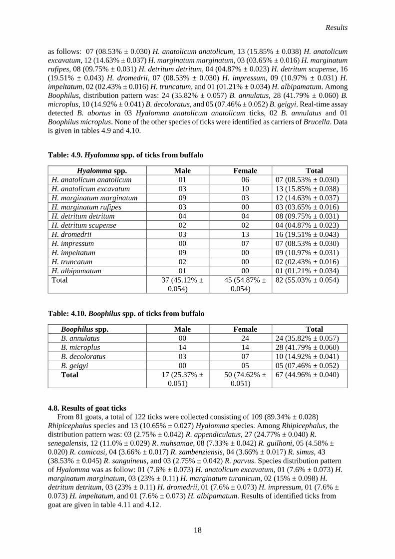
Results
18
as follows: 07 (08.53% ± 0.030) H. anatolicum anatolicum, 13 (15.85% ± 0.038) H. anatolicum
excavatum, 12 (14.63% ± 0.037) H. marginatum marginatum, 03 (03.65% ± 0.016) H. marginatum
rufipes, 08 (09.75% ± 0.031) H. detritum detritum, 04 (04.87% ± 0.023) H. detritum scupense, 16
(19.51% ± 0.043) H. dromedrii, 07 (08.53% ± 0.030) H. impressum, 09 (10.97% ± 0.031) H.
impeltatum, 02 (02.43% ± 0.016) H. truncatum, and 01 (01.21% ± 0.034) H. albipamatum. Among
Boophilus, distribution pattern was: 24 (35.82% ± 0.057) B. annulatus, 28 (41.79% ± 0.060) B.
microplus, 10 (14.92% ± 0.041) B. decoloratus, and 05 (07.46% ± 0.052) B. geigyi. Real-time assay
detected B. abortus in 03 Hyalomma anatolicum anatolicum ticks, 02 B. annulatus and 01
Boophilus microplus. None of the other species of ticks were identified as carriers of Brucella. Data
is given in tables 4.9 and 4.10.
Table: 4.9. Hyalomma spp. of ticks from buffalo
Hyalomma spp. Male Female Total
H. anatolicum anatolicum 01 06 07 (08.53% ± 0.030)
H. anatolicum excavatum 03 10 13 (15.85% ± 0.038)
H. marginatum marginatum 09 03 12 (14.63% ± 0.037)
H. marginatum rufipes 03 00 03 (03.65% ± 0.016)
H. detritum detritum 04 04 08 (09.75% ± 0.031)
H. detritum scupense 02 02 04 (04.87% ± 0.023)
H. dromedrii 03 13 16 (19.51% ± 0.043)
H. impressum 00 07 07 (08.53% ± 0.030)
H. impeltatum 09 00 09 (10.97% ± 0.031)
H. truncatum 02 00 02 (02.43% ± 0.016)
H. albipamatum 01 00 01 (01.21% ± 0.034)
Total 37 (45.12% ±
0.054)
45 (54.87% ±
0.054)
82 (55.03% ± 0.054)
Table: 4.10. Boophilus spp. of ticks from buffalo
Boophilus spp. Male Female Total
B. annulatus 00 24 24 (35.82% ± 0.057)
B. microplus 14 14 28 (41.79% ± 0.060)
B. decoloratus 03 07 10 (14.92% ± 0.041)
B. geigyi 00 05 05 (07.46% ± 0.052)
Total 17 (25.37% ±
0.051)
50 (74.62% ±
0.051)
67 (44.96% ± 0.040)
4.8. Results of goat ticks
From 81 goats, a total of 122 ticks were collected consisting of 109 (89.34% ± 0.028)
Rhipicephalus species and 13 (10.65% ± 0.027) Hyalomma species. Among Rhipicephalus, the
distribution pattern was: 03 (2.75% ± 0.042) R. appendiculatus, 27 (24.77% ± 0.040) R.
senegalensis, 12 (11.0% ± 0.029) R. muhsamae, 08 (7.33% ± 0.042) R. guilhoni, 05 (4.58% ±
0.020) R. camicasi, 04 (3.66% ± 0.017) R. zambenziensis, 04 (3.66% ± 0.017) R. simus, 43
(38.53% ± 0.045) R. sanguineus, and 03 (2.75% ± 0.042) R. parvus. Species distribution pattern
of Hyalomma was as follow: 01 (7.6% ± 0.073) H. anatolicum excavatum, 01 (7.6% ± 0.073) H.
marginatum marginatum, 03 (23% ± 0.11) H. marginatum turanicum, 02 (15% ± 0.098) H.
detritum detritum, 03 (23% ± 0.11) H. dromedrii, 01 (7.6% ± 0.073) H. impressum, 01 (7.6% ±
0.073) H. impeltatum, and 01 (7.6% ± 0.073) H. albipamatum. Results of identified ticks from
goat are given in table 4.11 and 4.12.

Results
19
Table: 4.11. Rhipicephalus spp. of ticks from goat
Rhipicephalus
spp.
Male Female Total
R. appendiculatus 00 03 03 (2.75% ± 0.042)
R. senegalensis 02 25 27 (24.77% ± 0.040)
R. muhsamae 12 00 12 (11.0% ± 0.029)
R. guilhoni 03 05 08 (7.33% ± 0.042)
R. camicasi 00 05 05 (4.58% ± 0.020)
R. zambenziensis 00 04 04 (3.66% ± 0.017)
R. simus 04 00 04 (3.66% ± 0.017)
R. sanguineus 16 27 43 (38.53% ± 0.045)
R. parvus 00 03 03 (2.75% ± 0.042)
Total 37 (33.94% ±
0.045)
72 (66.05% ±
0.045)
109 (89.34% ± 0.028)
Table: 4.12. Hyalomma spp. of ticks from goat
Hyalomma spp. Male Female Total
H. anatolicum excavatum 01 00 01 (7.6% ± 0.073)
H. marginatum marginatum 00 01 01 (7.6% ± 0.073)
H. marginatum turanicum 03 00 03 (23% ± 0.11)
H. detritum detritum 00 02 02 (15% ± 0.098)
H. dromedrii 00 03 03 (23% ± 0.11)
H. impressum 01 00 01 (7.6% ± 0.073)
H. impeltatum 01 00 01 (7.6% ± 0.073)
H. albipamatum 01 00 01 (7.6% ± 0.073)
Total 07 (53.84% ±
0.13)
06 (46.15% ±
0.13)
13 (10.65% ±
0.027)
4.9. Result of sheep ticks
From 59 sheep, a total of 138 ticks were collected. 90 (65.21% ± 0.040) ticks were
Rhipicephalus species while 48 (34.78% ± 0.040) were Hyalomma species. Among Rhipicephalus
the distribution pattern was as follow: 11 (12.22% ± 0.034) R. appendiculatus, 07 (7.77% ± 0.26)
R. senegalensis, 08 (8.88% ± 0.028) R. muhsamae, 06 (6.66% ± 0.024) R. guilhoni, 08 (8.88% ±
0.028) R. zambenziensis, 42 (46.66% ± 0.051) R. sanguineus, and 08 (8.88% ± 0.028) R. turanicus.
Among Hyalomma species the distribution pattern was; 01 (2.08% ± 0.023) H. anatolicum
anatolicum, 01 (2.08% ± 0.023) H. anatolicum excavatum, 23 (47.91% ± 0.071) H. marginatum
marginatum, 01 (2.08% ± 0.023) H. detritum detritum, 02 (4.16% ± 0.29) H. detritum scupense,
19 (39.58% ± 0.070) H. dromedrii, and 01 (2.08% ± 0.023) H. impressum. The results are given in
table 4.13 and 4.14.

Results
20
Table: 4.13. Rhipicephalus spp. of ticks from sheep
Rhipicephalus spp. Male Female Total
R. appendiculatus 04 07 11 (12.22% ± 0.034)
R. senegalensis 01 06 07 (7.77% ± 0.26)
R. muhsamae 07 01 08 (8.88% ± 0.028)
R. guilhoni 03 03 06 (6.66% ± 0.024)
R. zambenziensis 03 05 08 (8.88% ± 0.028)
R. sanguineus 16 26 42 (46.66% ± 0.051)
R. turanicus 06 02 08 (8.88% ± 0.028)
Total 40 (44.44% ±
0.052)
50 (55.55% ±
0.052)
90 (65.21% ± 0.040)
Table: 4.14. Hyalomma spp. of ticks from sheep
Hyalomma spp. Male Female Total
H. anatolicum anatolicum 00 01 01 (2.08% ± 0.023)
H. anatolicum excavatum 01 00 01 (2.08% ± 0.023)
H. marginatum marginatum 19 04 23 (47.91% ± 0.071)
H. detritum detritum 01 00 01 (2.08% ± 0.023)
H. detritum scupense 02 00 02 (4.16% ± 0.29)
H. dromedrii 06 13 19 (39.58% ± 0.070)
H. impressum 01 00 01 (2.08% ± 0.023)
Total 30 (62.5% ±
0.07)
18 (37.5% ±
0.07)
48 (34.78% ± 0.040)
4.10. Results of real-time assay for Brucella detection in ticks
After screening by RBPT, animals were confirmed for brucellosis by real-time PCR assay. Only
the female ticks of Brucella positive ruminants were subjected to real-time PCR. Real-time PCR
assay detected B. abortus in 03 (5.5% ± 0.031) Hyalomma anatolicum anatolicum and 07 (5% ±
0.018) Boophilus microplus ticks collected from Brucella positive cattle. In ticks collected from
buffalo, B. abortus was detected in 03 (6.6% ± 0.036) Hyalomma anatolicum anatolicum, 01 (2%
± 0.062) Boophilus microplus, and 02 (4% ± 0.027) Boophilus annulatus. In sheep and goats, the
Rhipicephalus was identified responsible for Brucella. B. melitensis was detected in 01 (2% ±
0.062) Rhipicephalus appendiculatus collected from sheep and 02 (2.7% ± 0.018) Rhipicephalus
sanguineus from goats. Results are given in table 4.15. Real-time amplification results can be seen
in figure 4.6.

Results
21
Table: 4.15. Real-time results of Brucella in ticks
Animal Ticks genus Ticks species Brucella
spp.
Cattle Hyalomma spp.
(53)
Hyalomma anatolicum anatolicum
03 (5.5% ± 0.031)
B. abortus
Boophilus spp
(140)
Boophilus microplus
07 (5% ± 0.018)
Buffalo Hyalomma spp
(45)
Hyalomma anatolicum anatolicum
03 (6.6% ± 0.036)
Boophilus spp
(50)
Boophilus microplus
01 (2% ± 0.062)
Boophilus annulatus
02 (4% ± 0.027)
Sheep Rhipicephalus spp.
(50)
Rhipicephalus appendiculatus
01 (2% ± 0.062)
B. melitensis
Hyalomma spp.
(18)
-
Goat Rhipicephalus spp.
(72)
Rhipicephalus sanguineus
02 (2.7% ± 0.018)
Hyalomma spp.
(06)
-
Figure: 4.6. Real-time curve of ticks positive for Brucella spp
4.11. Accession number of PCR products
The sequences were submitted to Gen-Bank for accession number.
Accession# MH615814: B. abortus, isolated from large ruminants
Accession# MH615815: B. abortus, isolated from ticks of large ruminants
Accession# MH615816: B. melitensis, isolated from small ruminants
Accession# MH615817: B. melitensis, isolated from large ruminants
Accession# MH615818: B. melitensis, isolate from ticks of small ruminants
Accession# MH615819: B. melitensis, isolated from ticks of small ruminants
Results
22
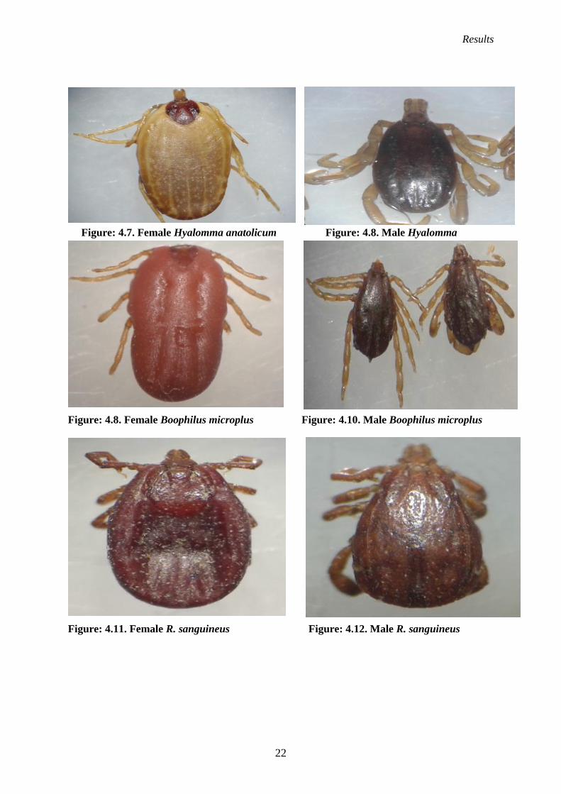
Figure: 4.7. Female Hyalomma anatolicum Figure: 4.8. Male Hyalomma
Figure: 4.8. Female Boophilus microplus Figure: 4.10. Male Boophilus microplus
Figure: 4.11. Female R. sanguineus Figure: 4.12. Male R. sanguineus

Results
23
4.11. Results of histopathological findings of tick bite lesions
Gross skin and hide examinations were performed. Hides of tick infested cattle and buffalo
showed mild swelling, red papule and erythema at the site tick bite lesion. In some tissue samples,
vesicles and ulceration were seen. Tick bites on processed bovine hides showed up as pinpoint
lesions, or holes (figure 1). Hyperkeratosis and widening of intracellular spaces with degeneration
of epidermal layer from basal lamina was seen in hides of infested bovine tissues samples. Tick
infested hides displayed focal areas of necrosis, collagen degeneration, spatial disorientation in the
grain and dermal layer infiltrated with neutrophils, basophils, eosinophils and mononuclear cells.
Hyperplasia of keratinocytes was also seen at sites of tick bite disrupted epidermis. These
histopathological changes can be seen in figure 2. The primary lesions found in skin of both sheep
and goats featured focal epidermal edema with adjacent dermal edema/ sub epidermal edema.
Sloughing of epidermal keratin layer is also seen. These lesion can be seen in figure 3. The
microscopic analysis revealed predominance of neutrophils over basophils, eosinophils and
mononuclear cells. In the stratum spinosum, marked spongiosis was observed with edematous fluid
between keratinocytes with stretched desmosome accompanied by basophilic micro-abscesses.
Figure: 4.13. Hide of bovine showing marked ticks bite lesions with small pin point pores
after pickling and bluing process of leather tanning process

Results
24
Figure: 4.14. Histopathology of bovine hide. Epidermal hyperkeratosis and degeneration
from basal lamina is seen. Collagen degeneration, necrosis and presence of some mononuclear
cells in the dermis are also seen
Figure: 4.15. Histopathology of goat skin. Focal areas of adjacent sub epidermal edema is
seen. In this area there was total sloughing of the epidermis up to stratum basale

Results
25
Figure: 4.16. Hyperkeratosis and sloughing of epidermal keratin layer is also seen.
Infiltration of mononuclear cells at sub epidermal layer in sheep skin and widening
intracellular spaces in epidermal layer

26
CHAPTER 5
DISCUSSION
5.1. Vector role of ticks in brucellosis
Ticks have been shown to harbor many organisms which may be pathogenic, non-pathogenic or
endosymbiotic. Ticks are the host of many unidentified microbes that continued to be described
including Brucella. Brucellosis is an important disease which is prevalent in ruminants and humans.
The prevalence of disease depends upon different factors. In the present study different species of
Hyalomma, Boophilus, and Rhipicephalus of Brucella positive ruminants were subjected to real-
time PCR to determine their role in Brucella transmission. Real-time PCR detected B. abortus in
Hyalomma anatolicum anatolicum and Boophilus microplus of cattle ticks. While in buffalo ticks,
B. abortus was detected in Hyalomma anatolicum anatolicum, Boophilus annulatus and Boophilus
microplus. B. melitensis was detected in Rhipicephalus sanguineus ticks of sheep and
Rhipicephalus appendiculatus ticks of goat. All other species of Hyalomma and Boophilus were
negative for B. abortus and B. melitensis. Previously Brucella was detected in Boophilus which is
in agreement with present study (Hosseini et al. 2017; Khan et al. 1997). Hosseini et al. (2017)
suggested a vector role Boophilus in nature for brucellosis. In present work specific species of
Brucella were investigated in identified species of Boophilus to confirm the role of ticks as vector.
Present study is also in accordance with Khan et al. (1997) who cultured the B. abortus from
Boophilus microplus and Hyalomma anatolicum anatolicum ticks in Pakistan. In current study
molecular based investigation Brucella from ticks infesting small and large ruminants has been
carried out first time in Pakistan. In present study only adult ticks were collected for investigation
of brucellosis. While previously, transovarial transmission of Brucella was investigated in
developmental stages of Dermacentor marginatus. Both B. abortus and B. melitensis were detected
in eggs, larva, nymph and adult stages of the Dermacentor marginatus through PCR (Wang et al.
2018). Hyalomma, Boophilus and Rhipicephalus were identified in present work which might have
a role in horizontal and vertical transmission of brucellosis. Their role can be highlighted through
investigation of Brucella in developmental stages of ticks. Control of ticks in cattle and goats may
reduce brucellosis infection rates, which provides further evidence implicating ticks as vector for
brucellosis among domestic animal populations (Miler et al. 2016). Currently ticks have been
detected for brucellosis but to gain an understanding of Brucella transmission by tick bites, in-vitro
based studies are required.
5.2. Histopathological changes in skin/hide due to ticks
Gross lesions of papules and erythema develop due to the mouth parts of tick which penetrate
the epidermal layer and lodge within the dermal papillae. Mouthpart piercing of the dermal blood
vessels may lead to hemorrhages and collagen degeneration at the site of bite (Wall and Shearer
2008). In the present study, hyperkeratosis and widening of intracellular spaces with degeneration
of stratum cornium layer from stratum granulosum was seen in hide of bovine tissues sample. The
hide was also damaged by focal area of necrosis, collagen degeneration, spatial disorientation in
the grain and dermal layer infiltrated with neutrophils, basophils and mononuclear cells.
Hyperplasia of keratinocytes was also seen at side of ruptured epidermis. Tick bite lesions induce
rupture and ulceration of skin which become vulnerable to secondary botfly infestation, or myiasis.
Tick bite lesions resulting in wounds provide a place for flies larvae which causes itching and
irritation on skin inducing exudation and may lead to secondary bacterial infections (Gashaw and
Mersha 2013). Tick infestation causes damage to both skin and hides of small and large ruminants
at both gross and microscopic levels, inducing economic losses to leather industry in Pakistan. In
Pakistan 3.08% skin and hide damage have been reported due to ticks (Chaudhry et al. 2011). Tick
bite causes a local injury during feeding. Bite lesions are prone to secondary bacterial infection and
fly myiasis, both of which may significantly increase the level of tissue damage at tick bite sites
(Taylor et al. 2007). Heavy infestation of ticks causes a loss in live weight gain, poor production
and decrease in the quality of skin and hide, as a result of undermining host physiological health

Discussions
27
(Kettle 1995). Ticks not only transmit pathogens to animals but also causes skin and hide damage
which ultimately leads to a financial loss in the livestock sector. The quality of skin and hides to
large extent are related to grain surface. The damage to the skin caused by ectoparasites, scratches
on animals during handling at farm and transportation to the abattoir, during flaying process and
inappropriate handling and preservation technique of skin and hides after slaughtering may reduce
the value of the hide and its products. Among ectoparasites, ticks are most important (Chaudhary
et al. 2011). Several ectoparasites induce skin damage, in Pakistan, ticks are thought to be the
primary culprits. Control of ticks and treatment of animals is imperative to compensate for the loss
of damages to hides.
5.3. Inter-species transmission of Brucella
In the present study, after screening with Rose Bengal antigen, seropositive samples were
subjected to real-time PCR assay. PCR assay determined the presence of 64.38% and 68.85% of B.
melitensis in both cattle and buffalo respectively in seropositive serum samples. Previously the
cause of abortion in cows due B. melitensis was investigated and it was observed that mix farming
with sheep and goats was the main reason for cross-infection (Wang et al. 2018). There are a
number of factors that might play a key role in cross-infection of species. In a country like Pakistan,
the lack of strict biosafety measures could be the main pillar of control failure. Mixed farming,
where both small and large animals share same housing, could be main cause of transmission of B.
melitensis to large ruminants (Ali et al. 2015). Ectoparasites have recently been investigated as
responsible for Brucella transmission, including ticks, mites, and lice (Wang et al. 2018). These
ectoparasites with fomites, water, and soil should be included in the investigation of cross-
transmission of Brucella species. In developing countries, poor farmers usually do not cull
Brucella-positive animals; rather, they sell them to other poor farmers, which leads to the spread
of infection to other animals in a mixed herd (Ali et al. 2015). Brucellosis is a rapidly emerging
disease; spread has been complicated by infections of nonpreferred hosts. Consideration of cross-
transmission of B. melitensis into nonspecific hosts should be included for effective control
measures. Abortion due to B. melitensis in large ruminants cannot be identified on serological tests,
and serological-based screening may lead to misleading planning measures for a control program.
Species-specific identification and isolation of Brucella are key for control measures. Early and
accurate detection of Brucella species and biovar/biotypes is fundamental for the control and
eradication of brucellosis. The presence of B. melitensis in cattle and buffalo is not only an
impediment in control and eradication but also poses a serious potential risk for public health. Little
attention has been given to B. melitensis in large ruminants, because B. abortus is considered the
main agent. Bovine should be vaccinated against B. melitensis because this specie does not lose its
pathogenicity and causes a significant abortion storm in cattle (Al-Mariri et al. 2015). More
attention should be paid to interspecies transmission of B. melitensis in cattle during control and
eradication of brucellosis (El-Hady et al. 2016).
5.4. Comparative evaluation of real-time PCR with conventional PCR In present study real-time assay detected more B. abortus in 90% cattle and 86% buffalo than
conventional assay which detected B. abortus in 61% cattle and 60% buffalo seropositive samples.
B. melitensis was detected in 64% sheep and 68% goat by real time assay as compare to
conventional assay which detected B. melitensis in 37% sheep and 39% goat. The diagnosis of
brucellosis is very important in adopting the control measures. Isolation and bacterial culture is still
a gold standard test for the diagnosis of brucellosis. But this method is hazardous (Wareth et al.
2014). The rapid, more sensitive and safe method of diagnosis is based on molecular tests (Akhtar
et al. 2010). Molecular assays can detect both live, dead even highly contaminated samples as
compared to culture method which cannot detect dead and contaminated samples. In the present
study, we compared two molecular techniques. We compared the real-time PCR assay with
conventional PCR assay. We found out that real-time assay is more sensitive than the conventional
type of molecular assay. The present research work is in agreement with Sidor et al. (2013) who

Discussions
28
proved multiplex real time PCR more sensitivity as compared to conventional. Greater sensitivity
is due of elimination of post PCR amplification steps in real-time PCR assay, while conventional
assay requires the run of amplified products on a Gel (Redkar et al. 2001). In real-time PCR reaction
amplification can be visualized through computerized generated software consisting of log and lag
phase. Due to visualization of amplification with the passage of time this assay called as a real-
time assay. In conventional PCR, we can confirm the reaction only after completion of cycles of
amplification. Even after successful amplification, there could be an error during post PCR steps.
Post PCR steps include Gel preparation, Ethidium bromide inclusion, Gel Electrophoresis, and
visualization through UV illuminator which are time consuming and labor intensive (Yang et al.
2007).
5.5. Brucellosis in seronegative small and large ruminants Seronegative animals could be serious problem because such animals are overlooked and
ignored during a routine screening and these seronegative animals could be a source of Brucella
infection in the farm (El-Diasty et al. 2018). In present study real-time PCR detected B. abortus in
53 and 79 cattle and buffalo while B. melitensis in 28 sheep and 46 goats out of 619, 737, 380 and
842 seronegative serum samples of cattle, buffalo, sheep and goats respectively. Previously B.
abortus was recovered in Egypt from a vaccinated dairy cattle herd which was declared as free
from brucellosis (Wareth et al. 2016). The presence of a small number of Brucella pathogens in
the blood circulation is not able to initiate a humoral immune response in the host, thus is unable
to produce antibodies (or cause abortion) and result in the negative immune response to serological
diagnostic tests. As a result, infected animals may register as brucellosis negative regarding
serological tests, yet remain significant carriers of pathogenic Brucella. Serological examination of
animals post abortion is of little value, as the antibodies against Brucella are detectable at the
earliest 14 days after the onset of Brucella infections. A number of factors may complicate the
diagnosis of Brucella, as bovine, caprine and ovine brucellosis can be diagnosed by various
serological assays. But these assays have limitations; when the Brucella pathogen remains
intracellular in macrophages, the disease goes into a chronic stage and the antibody titers may
decline (Islam et al. 2018). Seronegative non-reactive small and large ruminants in the current
research project that tested positive by IS711-based qPCR might have been exposed previously to
Brucella infection and then become serologically non-reactive after a certain time period. Sampling
at an early stage of Brucella infection, i.e. within the first fourteen days of infection during which
humoral immune response is not at detectable levels of antibodies in the host will often show
seronegative results (Gardner et al. 2000). Ruminants infected in the early postnatal period or in
utero can become latently infected with brucellosis and thus never become serologically reactive.
Almost 3.5% of infected cows are assessed to deliver latent infected offspring (Lefever et al. 2010).
The risks of false negatives obtained from serological testing of infectious animals clearly indicate
that brucellosis screening regimens should include molecular techniques. Seronegative animals
should therefore be included in future control programs for effective eradication measures of
brucellosis, and PCR testing may prove to be an effective assay for uncovering infectious Brucella
carriers that test as seronegative.
5.6. Conclusion
Detection of Brucella in tick species may play their role for brucellosis. The histopathological
investigation revealed that ticks have significant damage to skin and hides, ultimately decreasing
the quality of leather. Comparative evaluation of techniques shows that real-time PCR assay is
more suitable and serum is preferred clinical specimen for the diagnosis of brucellosis. The current
study is helpful for control programs and vaccine development strategies in livestock sector.
5.7. Suggestion
In future, ticks can be used in-vitro study for the confirmation of horizontal and vertical
transmission of brucellosis.

29
CHAPTER 6
SUMMARY Brucellosis is a bacterial disease which causes abortion storm in bovines, and also is a
significant zoonoses. It is highly prevalent in Pakistan. Transmission of the pathogen may be due to many risk factors. The eradication of disease mainly depends upon the control of risk factors. Many of factors have been cited as responsible for brucellosis. Among these risk factors, ectoparasites, particularly ticks, have emerged as highly significant as reservoirs and vectors for pathogenic Brucella species. Ticks typically harbor a great variety of microbes in their gut. Previously, little attention has been given to brucellosis as a tick borne disease. Ticks not only act as a vector for a number of the pathogens but may inflict significant levels of injury to host skin and hides, ultimately leading to economic losses to the leather industry by decreasing the quality of leather. In the present study samples of blood, serum, and ticks were collected from cattle, buffalo, sheep, and goats in Sheikhupura and Kasur districts. After collection serum samples were screened through Rose Bengal antigen. After screening only the seropositive serum samples were subjected to real-time PCR assay for the detection of B. abortus in cattle and buffalo, and B. melitensis in sheep and goats. Brucella positive cattle, buffalo, sheep, and goat ticks were identified under the microscope by AR Walker Key (2014). Female ticks were subjected to real-time PCR assay for the detection of B. abortus in ticks collected from cattle and buffalo; ticks collected from sheep and goats were tested for the presence of B. melitensis. The positive samples of ticks and ruminants were confirmed by conventional PCR. The PCR products were extracted from Gel and sequenced. The product sequences were submitted to Gen-Bank NCBI. Both the seropositive, seronegative serum samples and blood samples of respective seropositive hosts were subjected conventional PCR and real-time PCR for detection of brucellosis. In this experiment cross-transmission of B. abortus in caprine and ovine and B. melitensis in bovine were confirmed. Comparative evaluation of real-time PCR with conventional PCR and serum with blood for preferred clinical specimen were also performed. The study proved the real-time PCR assay to be more sensitive and serum as a preferred specimen for the diagnosis of brucellosis. The role of seronegative ruminants was also proven to be a factor in the spread of brucellosis. In the third part of the experiment, tick infected skin and hides of cattle, buffalo, sheep, and goats were collected from slaughterhouses. After collection, all the tissue samples were subjected to histopathological examination. Results revealed significant levels of damage to both skin and hides. Therefore, control of ticks would not only alleviate the spread of tick-borne diseases such as brucellosis but also serve to reduce incidences of skin and hide damage.

30
CHAPTER 7
LITERATURE CITED
Abd-El Halim MH, Mohamed AAE, Shalaby NA. 2017. Prevalence of brucellosis in bufaloes and
its control measures. J Vet Med Res. 24 (1): 161-170.
Ahmed R, Muhammad K, Rabbani M, Khan MS. 2017. Spatial Distribution of Soil Borne Brucella
Species Specific DNA in Punjab, Pakistan. Pak J Zool. 49 (5): 1739-1748.
Ahmed Y, Sokkar SM, Desouky H, Madbouly A. 2012. Pathological studies on buffalo-cows
naturally infected with Brucella melitensis. Global Veterinaria. 9 (2): 663-668.
Akhtar R, Anwar M, Khan I, El-Adawy H, Aslam A, Mustafa G, Rehmani S, Saleem M, Naz S.
2017. Pathological Investigations of Organ Affinity of Brucella Species and their Cross
Species Transmission. Pak Vet J. 37 (2): 372-374.
Akhtar R, Chaudhry ZI, Shakoori A, Ahmad M, Aslam A. 2010. Comparative efficacy of
conventional diagnostic methods and evaluation of polymerase chain reaction for the
diagnosis of bovine brucellosis. Vet World. 3 (2): 53-56.
Aldomy F, Alkhawaldeh M, Younis I. 2009. Immune responses of goats (Shami breed) to
vaccination with a full, reduced and conjunctival dose of brucevac (Brucella melitensis
Rev. 1) vaccine. Pak Vet J. 29 (2): 149-153.
Al-Mariri A. 2015. Isolation of Brucella melitensis strains from Syrian bovine milk samples. Bulg
J Vet Med. 18 (1): 40-48.
Ali Z, Maqbool A, Muhammad K, Khan M, Younis M. 2013. Prevalence of Theileria annulata
infected hard ticks of cattle and buffalo in Punjab, Pakistan. J Anim Plant Sci. 23 (1): 20-
26.
Ali S, Akhter S, Neubauer H, Melzer F, Khan I, Ali Q, Irfan M. 2015. Serological, cultural, and
molecular evidence of Brucella infection in small ruminants in Pakistan. J Infect Dev
Ctries. 9 (5): 470-475.
Alvarez J, Saez JL, Garcia N, Serrat C, Perez-Sancho M, Gonzalez S, Ortega MJ, Gou J, Carbajo
L, Garrido F. 2011. Management of an outbreak of brucellosis due to B. melitensis in dairy
cattle in Spain. Res Vet Sci. 90 (2): 208-211.
Arellano-Reynoso B, Suarez-Guemes F, Estrada FM, Michel-GomezFlores F, Hernandez-Castro
R, Acosta RB, Diaz-Aparicio E. 2013. Isolation of a field strain of Brucella abortus from
RB51-vaccinated-and brucellosis-seronegative bovine yearlings that calved normally. Trop
Anim Health Prod. 45 (2): 695-697.
Aslanyan RG and Vershilova. 1979. Recent aspects of natural focality of brucellosis, in Sbornik
Nauch. Trudov NIIEM im. N.F. Gamalei AMN SSSR Collected scientific Papers of the
Gamaleya Research Institute of Epidemiology and Microbiology. USSR Acad Med Sci.
59-63.
Bricker BJ, Halling SM. 1994. Differentiation of Brucella abortus bv. 1, 2, and 4, Brucella
melitensis, Brucella ovis, and Brucella suis bv. 1 by PCR. J Clin Microbiol. 32 (11): 2660-
2666.
Coles GC, Milnes AS, Green LE, Hadley PJ, Stosic PJ, Garnsworthy PC. 2003. Relationship
between lice infestation and leather damage in cattle. Vet Rec. 153 (9): 255-259.
Dehkordi FS, Khamesipour F, Momeni M. 2014. Brucella abortus and Brucella melitensis in
Iranian bovine and buffalo semen samples: The first clinical trial on seasonal, Senile and
geographical distribution using culture, Conventional and real-time polymerase chain
reaction assays. Kafkas Uni Vet Fak Derg. 20 (6): 821-828.
Dehkordi FS, Saberian S, Momtaz H. 2012. Detection and Segregation of Brucella abortus and
Brucella melitensis in Aborted Bovine, Ovine, Caprine, Buffaloes and Camelid Fetuses by
Application of Conventional and Real-time Polymerase Chain Reaction. Thai J Vet Med.
42 (1): 13-20.

Literature Cited
31
El-Diasty M, Wareth G, Melzer F, Mustafa S, Sprague LD, Neubauer H. 2018. Isolation of Brucella
abortus and Brucella melitensis from Seronegative Cows is a Serious Impediment in
Brucellosis Control. Vet Sci. 5 (1): 28-32.
El-Hady AM, Sayed-Ahmed M, Saleh ME, Younis EE. 2016. Seroprevalence and molecular
epidemiology of brucellosis in cattle in Egypt. Adv Dairy Res. 1-4.
Farooqi SH, Ijaz M, Saleem MH, Rashid MI, Oneeb M, Khan A, Aqib AI, Mahmood S. 2017.
Distribution of ixodid tick species and associated risk factors in temporal zones of Khyber
Pakhtunkhwa Province, Pakistan. Pak J Zool. 49 (6): 1-4 .
Corn JL, Nettles VF. 2001. Health protocol for translocation of free-ranging elk. J Wildl Dis. 37
(3): 413-426.
Chaudhry ZI, Saiddain A, Sabir N, Malik NA, Azeem S, Sajid A. 2011. Prevalence of pathological
conditions causing skin damage and consequently reducing its market value in domestic
ruminants of Punjab, Pakistan. Vet Sci Develop. 1 (1): 1-4.
Fentahun T, Woldemariam F, Chanie M, Berhan M. 2012. Prevalence of ectoparasites on small
ruminants in and around Gondar Town. Eur J Sci Res. 7 (2): 106-111.
Foster JT, Okinaka RT, Svensson R, Shaw KDBK, Robison RA, Probert WS, Kenefic LJ, Brown
WD, Keim P. 2008. Real-time PCR assays of single-nucleotide polymorphisms defining
the major Brucella clades. J Cin Microbiol. 46 (1): 296-301.
Gardner IA, Stryhn H, Lind P, Collins MT. 2000. Conditional dependence between tests affects the
diagnosis and surveillance of animal diseases. Prev Vet Med. 45 (2): 107-122.
Galuzo I and kaitmazova E. 1944. Ixodid Ticks-the possible Vectors of Brucellosis. Ixodid Ticks-
the possible Vectors of Brucellosis. 1 (3): 1-2.
Gashaw B and Mersha C. 2013. Pathology of Tick Bite Lesions in Naturally Infested Skin and
Hides of Ruminants: A Review. Acta Parasitologica Globalis. 4 (1): 59-63.
Gwida M, El-Ashker M, Melzer F, El-Diasty M, El-Beskawy M, Neubauer H. 2015. Use of
serology and real time PCR to control an outbreak of bovine brucellosis at a dairy cattle
farm in the Nile Delta region, Egypt. Irish Vet J. 69 (1): 3-6.
Gbolagunte GD, Hambolu JO, Akpavie SO. 2009. Pathology and leather surface appearance of
disease afflicted Nigerian small ruminant skins. Au J Tech. 12 (2): 271-283.
Godfroid J, Cloeckaert A, Liautard J-P, Kohler S, Fretin D, Walravens K, Garin-Bastuji B,
Letesson J-J. 2005. From the discovery of the Malta fever’s agent to the discovery of a
marine mammal reservoir, brucellosis has continuously been a re-emerging zoonosis. Vet
Res. 36 (3): 313-326.
Godfroid J, Nielsen K, Saegerman C. 2010. Diagnosis of brucellosis in livestock and wildlife. Croat
Med J. 51 (4): 296-305.
Godfroid J, Saegerman C, Wellemans V, Walravens K, Letesson J-J, Tibor A, Mc Millan A,
Spencer S, Sanna M, Bakker D. 2002. How to substantiate eradication of bovine brucellosis
when aspecific serological reactions occur in the course of brucellosis testing. Vet
Microbiol. 90 (4): 461-477.
Gudoshnik A. 1955. Study of the Role of Ixodid Ticks in the Circulation of the causal Agent of
Brucellosis. Microbiol Epidemiol Immunobiol. 29 (8): 113-117.
Gudoshnik A. 1958. Role of pasture ticks and rodents in dissemination of Brucella. Microbiol
Epidemiol Immunobiol. 29 (8): 113-117.
Guglielmone A, Gimeno E, Idiart J, Fisher WF, Volpogni M, Quaino O, Anziani S, Flores G,
Warnke O. 1999. Skin lesions and cattle hide damage from Haematobia irritans
infestations. Med Vet Entomol. 13 (1): 324-329.
Hadley PJ, Forbes AB, Rice BJ, Garnsworthy PC. 2005. Impact of the duration of control of cattle
lice with eprinomectin on leather quality. Vet Rec. 157 (1):841-845.
Halos L, Jamal T, Vial L, Maillard R, Suau A, Le Menach A, Boulouis H-J, Vayssier-Taussat M.
2004. Determination of an efficient and reliable method for DNA extraction from ticks. Vet
Res. 35 (6): 709-713.

Literature Cited
32
Herrera E, Rivera A, Palomares EG, Hernández-Castro R, Díaz-Aparicio E. 2011. Isolation of
Brucella melitensis from a RB51-vaccinated seronegative goat. Trop Anim Health Prod.
43 (6): 1069-1070.
Hosseini-Chegeni A, Tavakoli M, Telmadarraiy Z, Sedaghat MM, Faghihi F. 2017. Detection of a
Brucella-like (Alphaproteobacteria) Bacterium in Boophilus spp.(Acari: Ixodidae) from
Iran. J Med Microbiol Infect Dis. 5 (2): 66-68.
Hutchen RH. 1963. Tick-Borne Brucellosis. N Engl J Med. 1(2): 430.
Ica T, Aydin F, Gumussoy KS, Percin D, Sumerkan AB, Ocak F, Abay S, Dogan HO, Findik A,
Ciftci A. 2012. Conventional and molecular biotyping of Brucella strains isolated from
cattle, sheep and human. Ankara Üniv. Vet. Fak. Derg. 59 (3): 259-264.
Islam M, Khatun M, Saha S, Basir M, Hasan M-M. 2018. Molecular detection of Brucella spp.
from milk of seronegative cows from some selected area in Bangladesh. J Patho. 13 (2): 1-
7.
Junqueira Junior D, Rosinha G, Carvalho C, Oliveira C, Sanches C, Lima‐Ribeiro A. 2013.
Detection of Brucella spp. DNA in the semen of seronegative bulls by polymerase chain
reaction. Trans Emerg Dis. 60 (4): 376-377.
Jasik KP, Okła H, Słodki J, Rozwadowska B, Słodki A, Rupik W. 2015. Congenital tick borne
diseases: is this an alternative route of transmission of tick-borne pathogens in mammals.
Vector-Borne Zoon Dis. 15 (11): 637-644.
Jongejan F and Uilenberg G. 2004. The global importance of ticks. Parasitology. 129 (1): 3-14.
Jubb KVF. 2016. Pathology of domestic animals. 6th Edition. Academic press Elsevier, pp. 684.
Kettle DS. 1995. Medical and veterinary entomology. 2nd Edition, CAB international, PP. 469.
Kingston EF, Goulding H, Bateman AC. 2007. Vascular invasion is underrecognized in colorectal
cancer using conventional hematoxylin and eosin staining. Dis Col Rec. 50 (2): 1867-1872.
Kioko J, Baker J, Shannon A, Kiffner C. 2015. Ethnoecological knowledge of ticks and treatment
of tick-borne diseases among Maasai people in Northern Tanzania. Vet World. 8 (6): 755-
780.
Khan MN, Hayat CS, Iqbal Z. 1997. Role of ixodid ticks in transmission of bacterial diseases in
cattle and buffaloes in Pakistan. Pak J Agri Sci. 34 (1): 0-4.
Khan MZ, Usman T, Sadique U, Qureshi MS, Farooque M, Hassan MS, Khan A. 2017. Molecular
Characterization of Brucella abortus and Brucella melitensis in Cattle and Humans at the
North West of Pakistan. Pak Vet J. 37 (4): 360-363.
Khrustcheva N. 1969. Experimental Observations of Brucellosis Carriage in Bloodsucking Ticks.
Alma-Ata: Kazakhstan. Cand. Sci. Dissertation, Alma-Ata. Vet World. 8 (6): 755-780.
Kuhn C, Lucius R, Matthes HF, Meusel G, Reich B, Kalinna BH. 2008. Characterization of
recombinant immunoreactive antigens of the scab mite Sarcoptes scabies. Vet Parasitol.
153 (3): 329-337.
Lefevere C, Blancous J, Chermette R, UIlEngerg G. 2010. Infectious and parasitic disease of
livestock. Laveisies. Paris. 302-315.
Lindahl-Rajala E, Hoffman T, Fretin D, Godfroid J, Sattorov N, Boqvist S, Lundkvist A,
Magnusson U. 2017. Detection and characterization of Brucella spp. in bovine milk in
small-scale urban and peri-urban farming in Tajikistan. PLoS Negl Trop Dis. 11 (3): 53-
67.
Miller R, Nakavuma J, Ssajjakambwe P, Vudriko P, Musisi N, Kaneene J. 2016. The Prevalence
of Brucellosis in Cattle, Goats and Humans in Rural Uganda: A Comparative Study. Trans
Emerg Dis. 63 (6): 197-210.
Minjauw B, McLeod A. 2003. Tick-borne diseases and poverty: the impact of ticks and tick-borne
diseases on the livelihoods of small-scale and marginal livestock owners in India and
eastern and southern Africa. Tick-borne diseases and poverty: the impact of ticks and tick-
borne diseases on the livelihoods of small-scale and marginal livestock owners in India and
eastern and southern Africa. 2nd Edition, CAB international, pp. 124

Literature Cited
33
Muendo EN, Mbatha PM, Macharia J, Abdoel TH, Janszen PV, Pastoor R, Smits HL. 2012.
Infection of cattle in Kenya with Brucella abortus biovar 3 and Brucella melitensis biovar
1 genotypes. Trop Anim Health Prod. 44 (2): 17-20.
Mullen GR, Durden L.A. 2002. Medical and veterinary entomology. Academic press, Elsevier
Science China, pp. 518-554.
Nafstad O, Wisloff H, and Gronstol H. 2001. Nafstad O, Wisloff H, and Gronstol H. 2001. Acta
Vet Scand. 42 (1): 107-112.
Navarro E, Casao MA, Solera J. 2004. Diagnosis of human brucellosis using PCR. Expert review
of molecular diagnostics. 4 (1): 115-123.
Nielsen K, Smith P, Widdison J, Gall D, Kelly L, Kelly W, Nicoletti P. 2004. Serological
relationship between cattle exposed to Brucella abortus, Yersinia enterocolitica O: 9 and
Escherichia coli O157: H7. Vet Microbiol. 100 (2): 25-30.
O’Grady D, Byrne W, Kelleher P, O’Callaghan H, Kenny K, Heneghan T, Power S, Egan J, Ryan
F. 2014. A comparative assessment of culture and serology in the diagnosis of brucellosis
in dairy cattle. Vet J. 199 (3): 370-375.
Osman AE, Hassan AN, Ali AE, Abdoel TH, Smits HL. 2015. Brucella melitensis Biovar 1 and
Brucella abortus S19 vaccine strain infections in milkers working at cattle farms in the
Khartoum Area, Sudan. PloS one. https://doi.org/10.1371/journal.pone.0123374
Pappas G, Papadimitriou P, Akritidis N, Christou L, Tsianos EV. 2006. The new global map of
human brucellosis. Lancet Infect Dis. 6 (3): 91-99.
Peres J. 1986. Isolation of Brucella canis from ticks (Rhipicephalus sanguineus).
Pritulin P. 1954. On the Transmission of Brucellosis by the Pasture Ticks Dermacentor nuttallia
and Hyalomma marginatum. Veterinariya. 31 (7): 12-16.
Probert WS, Schrader KN, Khuong NY, Bystrom SL, Graves MH. 2004. Real-time multiplex PCR
assay for detection of Brucella spp., B. abortus, and B. melitensis. J. Clin. Microbiol. 42
(3): 1290-1293.
Rajput ZI, Hu Sh, Chen Wj, Arijo AG, Xiao Cw. 2006. Importance of ticks and their chemical and
immunological control in livestock. J Zhejiang Univ-Sc B. 7 (4): 912-921.
Redkar R, Rose S, Bricker B, DelVecchio V. 2001. Real-time detection of Brucella abortus,
Brucella melitensis and Brucella suis. Mol Cell Probes. 15 (1): 43-52.
Rementsova MM. 1962. Brucelsosis of wild animals.Alma-Ata: Nauka.
Samaha H, Al-Rowaily M, Khoudair RM, Ashour HM. 2008. Multicenter study of brucellosis in
Egypt. Emerg Infect Dis. 14 (12): 1916-1920.
Sarker M, Sarker R, Begum M, Shafy N, Islam M, Ehsan M, Bhattacharjee P, Rahman M, Melzer
F, Neubauer H. 2016. Seroprevalence and Molecular Diagnosis of Brucella abortus and
Brucella Melitensis in Bangladesh. Bangl J Vet Med. 14 (2): 221-226.
Scott JC, Koylass MS, Stubberfield MR, Whatmore AM. 2007. Multiplex assay based on single-
nucleotide polymorphisms for rapid identification of Brucella isolates at the species level.
Appl. Environ Microbiol. 73 (22): 7331-7337.
Sertse T, Wossene A. 2007. Effect of ectoparasites on quality of pickled skins and their impact on
the tanning industries in Amhara regional state, Ethiopia. Small rumin Res. 69 (2): 55-61.
Shemshad M, Shemshad K, Sedaghat MM, Shokri M, Barmaki A, Baniardalani M, Rafinejad J.
2012. First survey of hard ticks (Acari: Ixodidae) on cattle, sheep and goats in Boeen Zahra
and Takistan counties, Iran. Asian Pacific J Trop Biomed. 2M(1): 489-492.
Sidorov V. E. 1960. Body cavity of argasid ticks as a habitat of spirochetes and Brucella. J
Microbiol. 3 (1): 1101-1108.
Simsek F, Yildirmak M, Gedik H, Kantürk A, Iris E. 2011. Pulmonary involvement of Brucellosis:
a report of six cases. Afr Health Sci. 11 (3): 112-116.
Sneath PH, Mair NS, Sharpe ME, Holt JG. 1986. Bergey's manual of systematic bacteriology,
Volume 2. Williams & Wilkins. Vet World. 8 (6): 755-780.

Literature Cited
34
Sola MC, da Veiga Jardim EA, de Freitas MR, de Mesquita AJ. 2014. Real-time PCR detection of
Brucella spp. DNA in lesions and viscera of bovine carcasses. J Microbiol Methods. 104
(1): 87-91.
Taylor M, Coop R, Wall R. 2007. Veterinary Parasitology. 3rd edn. Blackwell Publishing. In: UK,
pp. 693-708
Tovar RM. 1947. Infection and transmission of Brucella by ectoparasites. Amerc J Vet Res. 8 (26).
Wall RL, Shearer D. 2008. Veterinary ectoparasites: biology, Pathology and Control. 2nd Edition,
John Wiley & Sons, pp. 55-199.
Walker AR. 2014. Ticks of domestic animals in Africa: a guide to identification of species.
Bioscience Reports Edinburgh.
Wang Q, Zhao S, Wureli H, Xie S, Chen C, Wei Q, Cui B, Tu C, Wang Y. 2018. Brucella melitensis
and B. abortus in eggs, larvae and engorged females of Dermacentor marginatus. Ticks
Tick Borne Dis. 9 (4): 1045-1048.
Wareth G, Melzer F, Elschner MC, Neubauer H, Roesler U. 2014. Detection of Brucella melitensis
in bovine milk and milk products from apparently healthy animals in Egypt by real-time
PCR. J Infect Dev Ctries 8 (6): 1339-1343.
Wareth G, Melzer F, Bottcher D, El-Diasty M, El-Beskawy M, Rasheed N, Schmoock G, Roesler
U, Sprague LD, Neubauer H. 2016. Molecular typing of isolates obtained from aborted
foetuses in Brucella-free Holstein dairy cattle herd after immunisation with Brucella
abortus RB51 vaccine in Egypt. Acta tropica. 164 (2): 267-271.
Yang SJ, Shim HS, Woo JT, Kim HS, Lee SS. 2007. Bacteriological detection of Brucella abortus
and its characterization by PCR in the sporadic outbreak of bovine brucellosis in Gyeonggi
province. Korean Society Poul Sci. 30 (2): 251-258.
Zajac AM, Conboy GA. 2016. Veterinary clinical parasitology. 8th Edition John Wiley & Sons, pp.
55-56.
Zheludkov M, Tsirelson L. 2010. Reservoirs of Brucella infection in nature. Biology bulletin. 37
(2): 709-715.
Zotova A, Boldicina K. 1951. The experimental infection of ticks with brucellosis under laboratory
conditions. In: DTIC Document.
Zotova AA, Balditsina KS. 1943. Expereicne in infecting ticks with brucellosis under laboratory
conditions. Ser Zool Alma-Ata. 1(2): 73-79.
Zowghi E, Ebadi A, Mohseni B. 1990. Isolation of Brucella organisms from the milk of
seronegative cows. Rev Sci Tech. 9 (4): 1175-1178.