Embed Size (px)
Citation preview
Neuropsychologia 51 (2013) 601–612
Contents lists available at SciVerse ScienceDirect
Neuropsychologia
0028-39
http://d
n Corr
Pavillon
France.
E-m
journal homepage: www.elsevier.com/locate/neuropsychologia
Role of the superior parietal lobules in letter-identity processing withinstrings: FMRI evidence from skilled and dyslexic readers
Caroline Reilhac a,b,n, Carole Peyrin c, Jean-Franc-ois Demonet d, Sylviane Valdois c
a INSERM, Imagerie Cerebrale et Handicaps neurologique, UMRS 825, F-31059 Toulouse, Franceb Universite de Toulouse, UPS, Imagerie Cerebrale et Handicaps neurologique, UMRS 825, F-31059 Toulouse, Francec Laboratoire de Psychologie et Neuro-Cognition (UMR 5105 CNRS), Universite Pierre Mend�es France, BP 47, 38040, Grenoble Cedex 9, Franced Leenaards Memory Center, CHUV and University of Lausanne, Switzerland
a r t i c l e i n f o
Article history:
Received 27 May 2012
Received in revised form
10 December 2012
Accepted 17 December 2012Available online 25 December 2012
Keywords:
Parietal cortex
Word form system
Developmental dyslexia
Reading
Letter string processing
Visual word recognition
32/$ - see front matter & 2013 Elsevier Ltd. A
x.doi.org/10.1016/j.neuropsychologia.2012.12
espondence to: UMR 825 Inserm/Universite
BAUDOT, CHU Purpan, Place du Dr Joseph Bay
Tel.: þ33 5 62 74 62 11; fax: þ33 5 62 74 61 6
ail address: [email protected] (C. Re
a b s t r a c t
Traditionally, the ventral occipito-temporal (vOT) area, but not the superior parietal lobules (SPLs), is
thought as belonging to the neural system of visual word recognition. However, some dyslexic children
who exhibit a visual attention span disorder – i.e. poor multi-element parallel processing – further
show reduced SPLs activation when engaged in visual multi-element categorization tasks. We
investigated whether these parietal regions further contribute to letter-identity processing within
strings. Adult skilled readers and dyslexic participants with a visual attention span disorder were
administered a letter-string comparison task under fMRI. Dyslexic adults were less accurate than
skilled readers to detect letter identity substitutions within strings. In skilled readers, letter identity
differs related to enhanced activation of the left vOT. However, specific neural responses were further
found in the superior and inferior parietal regions, including the SPLs bilaterally. Two brain regions that
are specifically related to substituted letter detection, the left SPL and the left vOT, were less activated
in dyslexic participants. These findings suggest that the left SPL, like the left vOT, may contribute to
letter string processing.
& 2013 Elsevier Ltd. All rights reserved.
1. Introduction
A large body of research has supported difficulties withphonological processing as the core disorder in developmentaldyslexia (Bishop & Snowling, 2004; Ramus, 2003; Vellutino,Fletcher, Snowling, & Scanlon, 2004). Accordingly, a number ofleft hemisphere language-related regions have been described asshowing atypical function in individuals with dyslexia (Demonet,Taylor, & Chaix, 2004, for a review). Involvement of the leftinferior frontal gyrus (for phonological short-term memory andphonological processing; Dufor, Serniclaes, Sprenger-Charolles, &Demonet, 2007; Heim, Eulitz, & Elbert, 2003), the left superiortemporal gyrus (for speech sound analysis and letter-soundmapping; Blau et al., 2010; Brambati et al., 2006; Paulesu et al.,2001) and the left temporo-parietal areas (for letter-to-soundconversion; Aylward et al., 2003; Price & Mechelli, 2005; Templeet al., 2003) is well in line with the phonological theory ofdevelopmental dyslexia (Snowling, 2000). A further brain region,the left inferior temporal area that more specifically relates to
ll rights reserved.
.010
Toulouse III—Paul Sabatier,
lac, 31024 Toulouse Cedex 3,
3.
ilhac).
visual recognition of letter strings (Dehaene & Cohen, 2011) hasalso been reported as consistently impaired in developmentaldyslexia (Richlan, Kronbichler, & Wimmer, 2011). However, thereis now strong evidence that not all dyslexic children have aphonological disorder (Vidyasagar & Pammer, 2010). A visualattention span disorder – a parallel letter string processingimpairment due to reduced visual attention capacity – has beenfound to account for the poor reading outcome of a subgroup ofdyslexic individuals (Bosse, Tainturier, & Valdois, 2007). Somestudies have identified the superior parietal lobules (SPLs) asvisual attention (VA) span brain correlates (Peyrin, Demonet,N’Guyen-Morel, Le Bas, & Valdois, 2011; Peyrin et al., 2012).These regions are specifically activated when the task requiresmulti-element processing, regardless of the alphanumeric or non-alphanumeric nature of the stimuli to be processed (Lobier,Zoubrinetzky, & Valdois, 2012c). However, evidence for SPLinvolvement in developmental dyslexia derives from tasks thatdid not directly relate to reading but required multi-elementvisual categorization. Thus, no direct evidence was provided forthis region to be involved in letter identification within strings.Moreover, typically this brain region is not recognized as part ofthe reading network, although being involved in reading undersome specific conditions (Cohen, Dehaene, Vinckier, Jobert, &Montavont, 2008; Valdois et al. 2006). As letter string
C. Reilhac et al. / Neuropsychologia 51 (2013) 601–612602
identification is a necessary component process of word recogni-tion, the current study aimed at exploring whether the SPLs areinvolved in letter identity encoding within strings thus contribut-ing to the early phase of the reading process.
1.1. Role of the left ventral occipito-temporal (vOT) cortex
in word recognition
The report of cases of pure alexia showed that some regions ofthe neural system are dedicated to visual word recognition. Purealexia is an acquired deficit of reading that results from lesions ofthe left vOT (Binder & Mohr, 1992; Cohen et al., 2003; Gaillardet al., 2006; Leff et al., 2001; Leff, Spitsyna, Plant, & Wise, 2006).Numerous neuroimaging studies of reading have identified part ofthis cortical region – namely the ‘Visual Word Form Area’ (VWFA)– as playing an important role in visual word recognition (Cohenet al., 2000; Cohen et al., 2002; Dehaene & Cohen, 2011). Itsactivity is strictly visual (Dehaene, Le Clec, Poline, Le Bihan, &Cohen, 2002), yet invariant for spatial location (right or left visualfield) (Cohen et al., 2000), or typographical characteristics(Dehaene et al., 2001). Left vOT activity increases with wordvisibility and this perceptual sensitivity to word visibility corre-lates with the ability to quickly read words by sight (Ben-Shachar,Dougherty, Deutsch, & Wandell, 2011). Further, this brain regionis sensitive to word frequency (Bruno, Zumberge, Manis, Lu, &Goldman, 2008). Thus, it is widely accepted that the left vOTencodes the abstract identity of strings of visual letters on thebasis of fast and parallel processing of letter identification (Cohenet al., 2000; Szwed et al., 2011; Tagamets, Novick, Chalmers, &Friedman, 2000; Vigneau, Jobard, Mazoyer, & Tzourio-Mazoyer,2005). However considerable literature argues for a domain-general role of the left vOT cortex that is not strictly specializedfor recognition of written words (Hellyer, Woodhead, Leech, &Wise, 2011; Price & Devlin, 2003, 2011; Starrfelt & Gerlach, 2007;Twomey, Kawabata Duncan, Price, & Devlin, 2011). Indeed in linewith the recycling hypothesis (Dehaene & Cohen, 2007), the leftvOT cortex is not exclusively activated by words but furtherresponds to pseudowords, consonant strings or even objects’ linedrawings or false font strings, thus suggesting a more generalinvolvement in the identification of multi-element ‘‘objects’’(Inhoff & Tousman, 1990). Thanks to its general property inmultipart object processing, the vOT area plays a crucial role inthe perceptual expertise required for reading.
The vOT area further belongs to the network of brain areasinvolved in developmental dyslexia (Wandell, Rauschecker, &Yeatman, 2012). Reading skills in children with dyslexia corre-lates with the magnitude of vOT cortex activation (Shaywitz et al.2002). In their meta-analysis of 18 studies, Richlan et al. (2011)found support for a dysfunction of the vOT cortex in both childrenand adults with developmental dyslexia. They reported agemodulated activations within this region and located the peakof age difference within the VWFA. In a longitudinal study ofchildren with familial risk of dyslexia, Maurer et al. (2007) foundreduced electrophysiological tuning in vOT area for those kinder-garten children who showed a specific reading disorder two yearslater. This result clarifies that in dyslexia visual tuning for print isreduced from the early phase of reading acquisition. A similarreduction was reported for adults with developmental dyslexia(Helenius, Tarkiainen, Cornelissen, Hansen, & Salmelin, 1999).Thus there is strong evidence that the vOT region develops into afast word recognition system over time and that a failure torecruit the vOT cortex characterizes developmental dyslexia.Specialization of this region in word identification may be dueto its dense interconnection with language regions, so thatreduced activation in developmental dyslexia may relate to thepoor phonological skills and language disorder typically reported
in dyslexic children (Price & Devlin, 2011; Wandell, et al., 2012).However, a growing body of evidence suggests that a subset ofdyslexic children exhibit a multi-element string processing dis-order in the absence of any phonological disorder.
1.2. A multi-element string processing disorder
in developmental dyslexia
There is strong evidence that dyslexic children exhibit a multi-element parallel processing disorder that reflects poor visualattention processes (Hawelka & Wimmer, 2005; Jones, Branigan,& Kelly, 2008; Pammer, Lavis, Hansen, & Cornelissen, 2004;Pammer & Vidyasagar, 2005; Valdois et al., 2003). Several studieshave explored multi-element visual processing in dyslexic chil-dren using 5-consonant report tasks (Bosse et al., 2007; Bosse &Valdois, 2009; Valdois, et al., 2003; Valdois, Bosse, & Tainturier,2004). These tasks require identifying as many letters as possibleor a single cued letter within 5-consonant letter strings that arebriefly displayed. Impaired performance on these tasks wasinterpreted as reflecting a simultaneous processing disorder,known as a visual attention (VA) span deficit (Valdois et al.,2004). This disorder prevents dyslexic children from processing asmany letters in parallel as non-dyslexic children do. There isevidence that the VA span disorder is strictly visual since it is notaffected by concurrent phonological processing (Valdois, Lassus-Sangosse, & Lobier, 2012) and further extends to non-verbal tasksand non-verbal material (Lobier et al., 2012c; Pammer et al.,2004; Pammer & Vidyasagar, 2005). Furthermore, both groupstudies and single case studies have shown that the VA spandisorder typically dissociates from phonological problems indevelopmental dyslexia (Bosse et al., 2007; Lallier, Donnadieu,Berger, & Valdois, 2010; Valdois et al., 2011; Valdois et al., 2003).Research on typical development further showed that VA spanabilities contribute to reading performance independently of thechild’s phonological skills (Bosse & Valdois, 2009). They suggest amore specific involvement of VA span abilities in irregular wordprocessing and reading speed, even if the VA span further con-tributes to pseudo-word reading. This is consistent with report ofVA span disorders in children with a selective irregular wordreading disorder (Dubois et al., 2010; Valdois et al., 2003) and incases of mixed dyslexia (Valdois et al., 2011). This disorder isinterpreted as a reduction in the amount of visual attention thatcan be distributed over the letter string, so that the attentional loadallocated to each letter within the string is not sufficient to allowaccurate identification of the whole letters simultaneously. Areduced visual attention span would prevent normal processingof the whole word sequence, thus affecting words’ fast recognitionin reading. In preventing identification of their whole constitutiveletters, such a reduction would further impact normal processingof relevant multi-letter sub-lexical units, thus leading to impairedpseudo-word reading. A VA span disorder was expected to relate tolower activity in those brain regions specifically dedicated toattentional processing.
1.3. Role of superior parietal lobules (SPLs) in letter string processing
The neural underpinnings of the VA span have been investi-gated in dyslexic individuals and skilled readers. Using categor-ization tasks under fMRI, Peyrin et al. (2011, 2008) identified thesuperior parietal lobules bilaterally (SPLs) as the neural correlatesof VA span. Peyrin et al. (2011) investigated the VA span cerebralsubstrates in normal reading children and in a group of dyslexicchildren chosen to have a VA span disorder at the behavioral level(i.e. poor performance on the letter report tasks). In this study,two flanked and isolated letter categorization tasks were designedwhich differently taxed visual attention. In typical readers, the
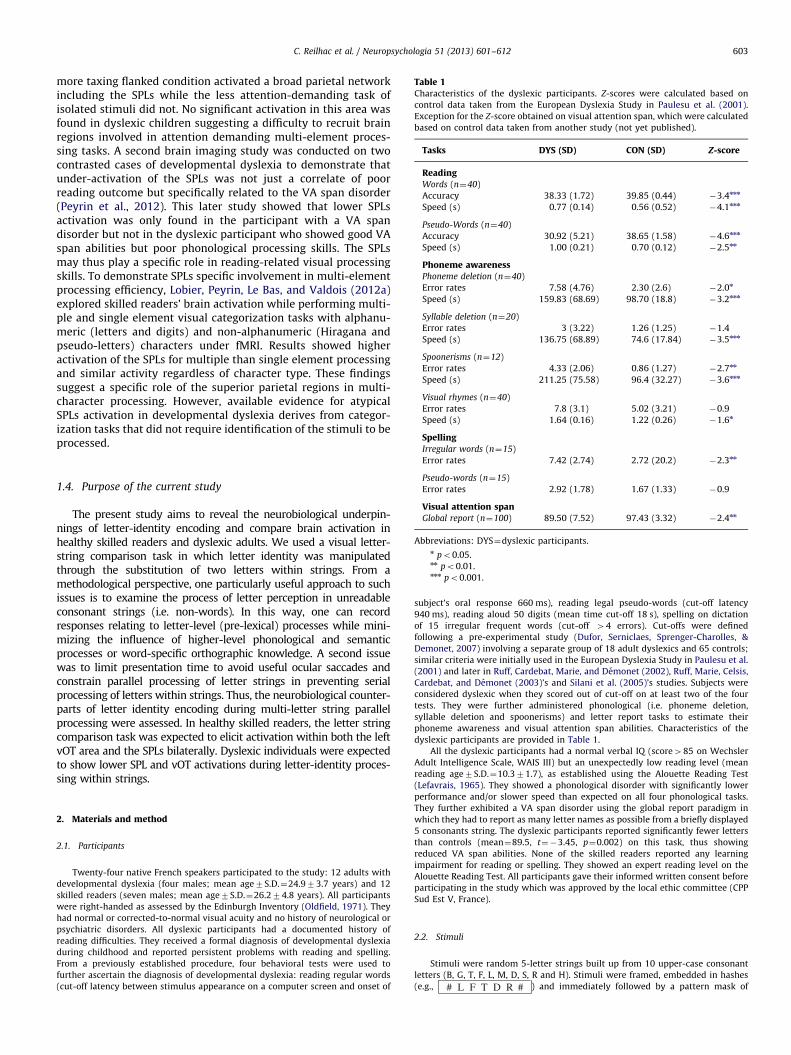
Table 1Characteristics of the dyslexic participants. Z-scores were calculated based on
control data taken from the European Dyslexia Study in Paulesu et al. (2001).
Exception for the Z-score obtained on visual attention span, which were calculated
based on control data taken from another study (not yet published).
Tasks DYS (SD) CON (SD) Z-score
ReadingWords (n¼40)
Accuracy 38.33 (1.72) 39.85 (0.44) �3.4nnn
Speed (s) 0.77 (0.14) 0.56 (0.52) �4.1nnn
Pseudo-Words (n¼40)
Accuracy 30.92 (5.21) 38.65 (1.58) �4.6nnn
Speed (s) 1.00 (0.21) 0.70 (0.12) �2.5nn
Phoneme awarenessPhoneme deletion (n¼40)
Error rates 7.58 (4.76) 2.30 (2.6) �2.0n
Speed (s) 159.83 (68.69) 98.70 (18.8) �3.2nnn
Syllable deletion (n¼20)
Error rates 3 (3.22) 1.26 (1.25) �1.4
Speed (s) 136.75 (68.89) 74.6 (17.84) �3.5nnn
Spoonerisms (n¼12)
Error rates 4.33 (2.06) 0.86 (1.27) �2.7nn
Speed (s) 211.25 (75.58) 96.4 (32.27) �3.6nnn
Visual rhymes (n¼40)
Error rates 7.8 (3.1) 5.02 (3.21) �0.9
Speed (s) 1.64 (0.16) 1.22 (0.26) �1.6n
SpellingIrregular words (n¼15)
C. Reilhac et al. / Neuropsychologia 51 (2013) 601–612 603
more taxing flanked condition activated a broad parietal networkincluding the SPLs while the less attention-demanding task ofisolated stimuli did not. No significant activation in this area wasfound in dyslexic children suggesting a difficulty to recruit brainregions involved in attention demanding multi-element proces-sing tasks. A second brain imaging study was conducted on twocontrasted cases of developmental dyslexia to demonstrate thatunder-activation of the SPLs was not just a correlate of poorreading outcome but specifically related to the VA span disorder(Peyrin et al., 2012). This later study showed that lower SPLsactivation was only found in the participant with a VA spandisorder but not in the dyslexic participant who showed good VAspan abilities but poor phonological processing skills. The SPLsmay thus play a specific role in reading-related visual processingskills. To demonstrate SPLs specific involvement in multi-elementprocessing efficiency, Lobier, Peyrin, Le Bas, and Valdois (2012a)explored skilled readers’ brain activation while performing multi-ple and single element visual categorization tasks with alphanu-meric (letters and digits) and non-alphanumeric (Hiragana andpseudo-letters) characters under fMRI. Results showed higheractivation of the SPLs for multiple than single element processingand similar activity regardless of character type. These findingssuggest a specific role of the superior parietal regions in multi-character processing. However, available evidence for atypicalSPLs activation in developmental dyslexia derives from categor-ization tasks that did not require identification of the stimuli to beprocessed.
Error rates 7.42 (2.74) 2.72 (20.2) �2.3nn
Pseudo-words (n¼15)
Error rates 2.92 (1.78) 1.67 (1.33) �0.9
Visual attention spanGlobal report (n¼100) 89.50 (7.52) 97.43 (3.32) �2.4nn
Abbreviations: DYS¼dyslexic participants.n po0.05.nn po0.01.nnn po0.001.
1.4. Purpose of the current study
The present study aims to reveal the neurobiological underpin-nings of letter-identity encoding and compare brain activation inhealthy skilled readers and dyslexic adults. We used a visual letter-string comparison task in which letter identity was manipulatedthrough the substitution of two letters within strings. From amethodological perspective, one particularly useful approach to suchissues is to examine the process of letter perception in unreadableconsonant strings (i.e. non-words). In this way, one can recordresponses relating to letter-level (pre-lexical) processes while mini-mizing the influence of higher-level phonological and semanticprocesses or word-specific orthographic knowledge. A second issuewas to limit presentation time to avoid useful ocular saccades andconstrain parallel processing of letter strings in preventing serialprocessing of letters within strings. Thus, the neurobiological counter-parts of letter identity encoding during multi-letter string parallelprocessing were assessed. In healthy skilled readers, the letter stringcomparison task was expected to elicit activation within both the leftvOT area and the SPLs bilaterally. Dyslexic individuals were expectedto show lower SPL and vOT activations during letter-identity proces-sing within strings.
2. Materials and method
2.1. Participants
Twenty-four native French speakers participated to the study: 12 adults with
developmental dyslexia (four males; mean age7S.D.¼24.973.7 years) and 12
skilled readers (seven males; mean age7S.D.¼26.274.8 years). All participants
were right-handed as assessed by the Edinburgh Inventory (Oldfield, 1971). They
had normal or corrected-to-normal visual acuity and no history of neurological or
psychiatric disorders. All dyslexic participants had a documented history of
reading difficulties. They received a formal diagnosis of developmental dyslexia
during childhood and reported persistent problems with reading and spelling.
From a previously established procedure, four behavioral tests were used to
further ascertain the diagnosis of developmental dyslexia: reading regular words
(cut-off latency between stimulus appearance on a computer screen and onset of
subject’s oral response 660 ms), reading legal pseudo-words (cut-off latency
940 ms), reading aloud 50 digits (mean time cut-off 18 s), spelling on dictation
of 15 irregular frequent words (cut-off 44 errors). Cut-offs were defined
following a pre-experimental study (Dufor, Serniclaes, Sprenger-Charolles, &
Demonet, 2007) involving a separate group of 18 adult dyslexics and 65 controls;
similar criteria were initially used in the European Dyslexia Study in Paulesu et al.
(2001) and later in Ruff, Cardebat, Marie, and Demonet (2002), Ruff, Marie, Celsis,
Cardebat, and Demonet (2003)’s and Silani et al. (2005)’s studies. Subjects were
considered dyslexic when they scored out of cut-off on at least two of the four
tests. They were further administered phonological (i.e. phoneme deletion,
syllable deletion and spoonerisms) and letter report tasks to estimate their
phoneme awareness and visual attention span abilities. Characteristics of the
dyslexic participants are provided in Table 1.
All the dyslexic participants had a normal verbal IQ (score485 on Wechsler
Adult Intelligence Scale, WAIS III) but an unexpectedly low reading level (mean
reading age7S.D.¼10.371.7), as established using the Alouette Reading Test
(Lefavrais, 1965). They showed a phonological disorder with significantly lower
performance and/or slower speed than expected on all four phonological tasks.
They further exhibited a VA span disorder using the global report paradigm in
which they had to report as many letter names as possible from a briefly displayed
5 consonants string. The dyslexic participants reported significantly fewer letters
than controls (mean¼89.5, t¼�3.45, p¼0.002) on this task, thus showing
reduced VA span abilities. None of the skilled readers reported any learning
impairment for reading or spelling. They showed an expert reading level on the
Alouette Reading Test. All participants gave their informed written consent before
participating in the study which was approved by the local ethic committee (CPP
Sud Est V, France).
2.2. Stimuli
Stimuli were random 5-letter strings built up from 10 upper-case consonant
letters (B, G, T, F, L, M, D, S, R and H). Stimuli were framed, embedded in hashes
(e.g., # L F T D R # ) and immediately followed by a pattern mask of
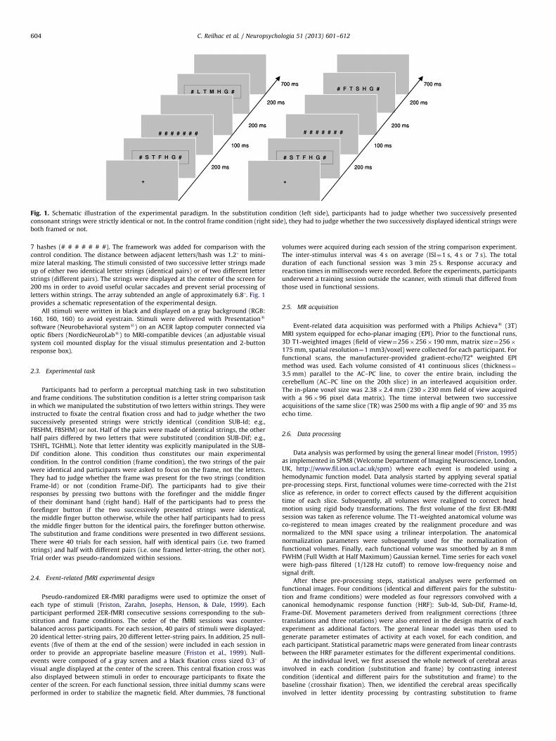
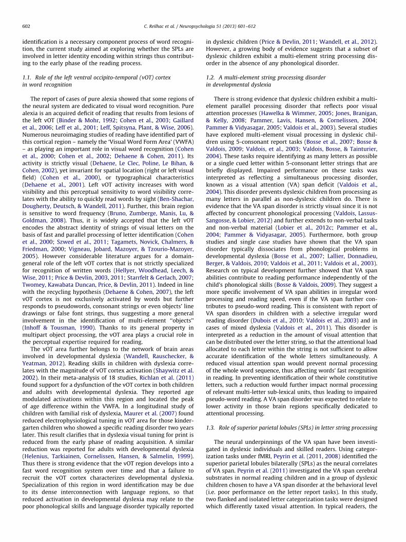
Fig. 1. Schematic illustration of the experimental paradigm. In the substitution condition (left side), participants had to judge whether two successively presented
consonant strings were strictly identical or not. In the control frame condition (right side), they had to judge whether the two successively displayed identical strings were
both framed or not.
C. Reilhac et al. / Neuropsychologia 51 (2013) 601–612604
7 hashes (# # # # # # #). The framework was added for comparison with the
control condition. The distance between adjacent letters/hash was 1.21 to mini-
mize lateral masking. The stimuli consisted of two successive letter strings made
up of either two identical letter strings (identical pairs) or of two different letter
strings (different pairs). The strings were displayed at the center of the screen for
200 ms in order to avoid useful ocular saccades and prevent serial processing of
letters within strings. The array subtended an angle of approximately 6.81. Fig. 1
provides a schematic representation of the experimental design.
All stimuli were written in black and displayed on a gray background (RGB:
160, 160, 160) to avoid eyestrain. Stimuli were delivered with Presentations
software (Neurobehavioral systems) on an ACER laptop computer connected via
optic fibers (NordicNeuroLabs) to MRI-compatible devices (an adjustable visual
system coil mounted display for the visual stimulus presentation and 2-button
response box).
2.3. Experimental task
Participants had to perform a perceptual matching task in two substitution
and frame conditions. The substitution condition is a letter string comparison task
in which we manipulated the substitution of two letters within strings. They were
instructed to fixate the central fixation cross and had to judge whether the two
successively presented strings were strictly identical (condition SUB-Id; e.g.,
FBSHM, FBSHM) or not. Half of the pairs were made of identical strings, the other
half pairs differed by two letters that were substituted (condition SUB-Dif; e.g.,
TSHFL, TGHML). Note that letter identity was explicitly manipulated in the SUB-
Dif condition alone. This condition thus constitutes our main experimental
condition. In the control condition (frame condition), the two strings of the pair
were identical and participants were asked to focus on the frame, not the letters.
They had to judge whether the frame was present for the two strings (condition
Frame-Id) or not (condition Frame-Dif). The participants had to give their
responses by pressing two buttons with the forefinger and the middle finger
of their dominant hand (right hand). Half of the participants had to press the
forefinger button if the two successively presented strings were identical,
the middle finger button otherwise, while the other half participants had to press
the middle finger button for the identical pairs, the forefinger button otherwise.
The substitution and frame conditions were presented in two different sessions.
There were 40 trials for each session, half with identical pairs (i.e. two framed
strings) and half with different pairs (i.e. one framed letter-string, the other not).
Trial order was pseudo-randomized within sessions.
2.4. Event-related fMRI experimental design
Pseudo-randomized ER-fMRI paradigms were used to optimize the onset of
each type of stimuli (Friston, Zarahn, Josephs, Henson, & Dale, 1999). Each
participant performed 2ER-fMRI consecutive sessions corresponding to the sub-
stitution and frame conditions. The order of the fMRI sessions was counter-
balanced across participants. For each session, 40 pairs of stimuli were displayed:
20 identical letter-string pairs, 20 different letter-string pairs. In addition, 25 null-
events (five of them at the end of the session) were included in each session in
order to provide an appropriate baseline measure (Friston et al., 1999). Null-
events were composed of a gray screen and a black fixation cross sized 0.31 of
visual angle displayed at the center of the screen. This central fixation cross was
also displayed between stimuli in order to encourage participants to fixate the
center of the screen. For each functional session, three initial dummy scans were
performed in order to stabilize the magnetic field. After dummies, 78 functional
volumes were acquired during each session of the string comparison experiment.
The inter-stimulus interval was 4 s on average (ISI¼1 s, 4 s or 7 s). The total
duration of each functional session was 3 min 25 s. Response accuracy and
reaction times in milliseconds were recorded. Before the experiments, participants
underwent a training session outside the scanner, with stimuli that differed from
those used in functional sessions.
2.5. MR acquisition
Event-related data acquisition was performed with a Philips Achievas (3T)
MRI system equipped for echo-planar imaging (EPI). Prior to the functional runs,
3D T1-weighted images (field of view¼256�256�190 mm, matrix size¼256�
175 mm, spatial resolution¼1 mm3/voxel) were collected for each participant. For
functional scans, the manufacturer-provided gradient-echo/T2n weighted EPI
method was used. Each volume consisted of 41 continuous slices (thickness¼
3.5 mm) parallel to the AC–PC line, to cover the entire brain, including the
cerebellum (AC–PC line on the 20th slice) in an interleaved acquisition order.
The in-plane voxel size was 2.38�2.4 mm (230�230 mm field of view acquired
with a 96�96 pixel data matrix). The time interval between two successive
acquisitions of the same slice (TR) was 2500 ms with a flip angle of 901 and 35 ms
echo time.
2.6. Data processing
Data analysis was performed by using the general linear model (Friston, 1995)
as implemented in SPM8 (Welcome Department of Imaging Neuroscience, London,
UK, http://www.fil.ion.ucl.ac.uk/spm) where each event is modeled using a
hemodynamic function model. Data analysis started by applying several spatial
pre-processing steps. First, functional volumes were time-corrected with the 21st
slice as reference, in order to correct effects caused by the different acquisition
time of each slice. Subsequently, all volumes were realigned to correct head
motion using rigid body transformations. The first volume of the first ER-fMRI
session was taken as reference volume. The T1-weighted anatomical volume was
co-registered to mean images created by the realignment procedure and was
normalized to the MNI space using a trilinear interpolation. The anatomical
normalization parameters were subsequently used for the normalization of
functional volumes. Finally, each functional volume was smoothed by an 8 mm
FWHM (Full Width at Half Maximum) Gaussian kernel. Time series for each voxel
were high-pass filtered (1/128 Hz cutoff) to remove low-frequency noise and
signal drift.
After these pre-processing steps, statistical analyses were performed on
functional images. Four conditions (identical and different pairs for the substitu-
tion and frame conditions) were modeled as four regressors convolved with a
canonical hemodynamic response function (HRF): Sub-Id, Sub-Dif, Frame-Id,
Frame-Dif. Movement parameters derived from realignment corrections (three
translations and three rotations) were also entered in the design matrix of each
experiment as additional factors. The general linear model was then used to
generate parameter estimates of activity at each voxel, for each condition, and
each participant. Statistical parametric maps were generated from linear contrasts
between the HRF parameter estimates for the different experimental conditions.
At the individual level, we first assessed the whole network of cerebral areas
involved in each condition (substitution and frame) by contrasting interest
condition (identical and different pairs for the substitution and frame) to the
baseline (crosshair fixation). Then, we identified the cerebral areas specifically
involved in letter identity processing by contrasting substitution to frame
C. Reilhac et al. / Neuropsychologia 51 (2013) 601–612 605
conditions ([Sub-Id4Frame-Id] and ([Sub-Dif4Frame-Dif] contrasts). We then
performed a separate random-effect group analysis for controls and dyslexics on
the contrast images from the individual analyses (Friston et al. 1998), using one-
sample t-tests. Finally, two-sample t-tests were performed to statistically compare
brain activity between controls and dyslexics on the relevant contrasts. Clusters of
activated voxels were then identified, based on the intensity of the individual
responses (po0.001 uncorrected for multiple comparisons, T44.02, extended
threshold of 15 voxels).
While this uncorrected threshold may seem liberal, it is in line with those of
previous papers reporting significant activations to identify pre-orthographic
character string neural correlates (Lobier et al., 2012a) as well as activation
differences between skilled and dyslexic readers within the parietal (Peyrin,
Demonet, N’Guyen-Morel, Le Bas, & Valdois, 2011; Peyrin et al., 2008) or
occipito-temporal areas (Van Der Mark et al., 2009). To facilitate comparisons
with other studies, a transformation of MNI into Talairach and Tournoux (1988)
coordinates was performed using the MNI2TAL function (created by Matthew
Brett, available at http://www.mrc-cbu.cam.ac.uk/Imaging).
Analysis was finally completed by statistically comparing activity for skilled
and dyslexic readers within regions of interest (ROIs). A set of a priori ROIs were
defined using predefined masks from the Wake Forest University (WFU) PickAtlas
(Maldjian, Laurienti, Kraft, & Burdette, 2003). ROI masks were created with the
automated anatomical labeling atlas, which uses an anatomical parcellation of the
MNI MRI single-subject brain and sulcal boundaries to define each anatomical
volume. All ROIs were constructed using the SPM Marsbar toolbox (http://
marsbar.sourceforge.net). Parameter estimates (% signal change relative to the
global mean intensity of signal) of event-related responses were then extracted
from these ROIs for each participant. The average parameter of activity was
calculated for each skilled and dyslexic reader and each ROI and ROIs’ activity was
compared between groups. Previous research has linked behavioral deficits in
simultaneous visual processing in dyslexia to lower activation in parietal brain
areas, and more specifically in the superior parietal lobule bilaterally and the left
inferior parietal lobule lobule (Peyrin et al., 2011; Peyrin et al., 2012). Atypical
activation patterns in dyslexia have been also observed in the left inferior frontal
areas – including Broca’s area – implicated in output phonology and articulatory
processing (Paulesu et al., 1996; Shaywitz et al., 1998; Wimmer et al., 2010). Other
research showed a deficit in activation in ventral pathway of reading centered in
the left middle and inferior temporal gyrus (Demonet et al., 2004; Paulesu et al.,
2001). Thus, six a priori defined cortical ROIs were investigated: left SPL, BA 7;
right SPL, BA7; left IPL; BA 40; right IPL, BA 40; left IFG, BA 44; left ITG, BA 37.
3. Results
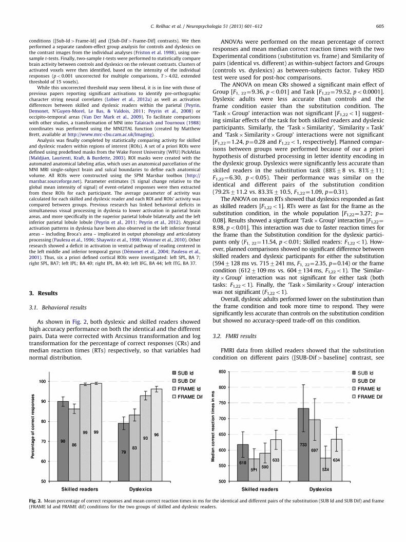
3.1. Behavioral results
As shown in Fig. 2, both dyslexic and skilled readers showedhigh accuracy performance on both the identical and the differentpairs. Data were corrected with Arcsinus transformation and logtransformation for the percentage of correct responses (CRs) andmedian reaction times (RTs) respectively, so that variables hadnormal distribution.
Fig. 2. Mean percentage of correct responses and mean correct reaction times in ms for
(FRAME Id and FRAME dif) conditions for the two groups of skilled and dyslexic reade
ANOVAs were performed on the mean percentage of correctresponses and mean median correct reaction times with the twoExperimental conditions (substitution vs. frame) and Similarity ofpairs (identical vs. different) as within-subject factors and Groups(controls vs. dyslexics) as between-subjects factor. Tukey HSDtest were used for post-hoc comparisons.
The ANOVA on mean CRs showed a significant main effect ofGroup [F1, 22¼9.36, po0.01] and Task [F1,22¼79.52, po0.0001].Dyslexic adults were less accurate than controls and theframe condition easier than the substitution condition. The‘Task�Group’ interaction was not significant [F1,22o1] suggest-ing similar effects of the task for both skilled readers and dyslexicparticipants. Similarly, the ‘Task� Similarity’, ‘Similarity�Task’and ‘Task� Similarity�Group’ interactions were not significant[F1,22¼1.24, p¼0.28 and F1,22o1, respectively]. Planned compar-isons between groups were performed because of our a priorihypothesis of disturbed processing in letter identity encoding inthe dyslexic group. Dyslexics were significantly less accurate thanskilled readers in the substitution task (88%78 vs. 81%711;F1,22¼6.30, po0.05). Their performance was similar on theidentical and different pairs of the substitution condition(79.2%711.2 vs. 83.3%710.5, F1,22¼1.09, p¼0.31).
The ANOVA on mean RTs showed that dyslexics responded as fastas skilled readers [F1,22o1]. RTs were as fast for the frame as thesubstitution condition, in the whole population [F1,22¼3.27; p¼
0.08]. Results showed a significant ‘Task�Group’ interaction [F1,22¼
8.98, po0.01]. This interaction was due to faster reaction times forthe frame than the Substitution condition for the dyslexic partici-pants only (F1, 22¼11.54, po0.01; Skilled readers: F1,22o1). How-ever, planned comparisons showed no significant difference betweenskilled readers and dyslexic participants for either the substitution(5947128 ms vs. 7157241 ms, F1, 22¼2.35, p¼0.14) or the framecondition (6127109 ms vs. 6047134 ms, F1,22o1). The ‘Similar-ity�Group’ interaction was not significant for either task (bothtasks: F1,22o1). Finally, the ‘Task� Similarity�Group’ interactionwas not significant (F1,22o1).
Overall, dyslexic adults performed lower on the substitution thanthe frame condition and took more time to respond. They weresignificantly less accurate than controls on the substitution conditionbut showed no accuracy-speed trade-off on this condition.
3.2. FMRI results
FMRI data from skilled readers showed that the substitutioncondition on different pairs ([SUB-Dif4baseline] contrast, see
the identical and different pairs of the substitution (SUB Id and SUB Dif) and frame
rs.
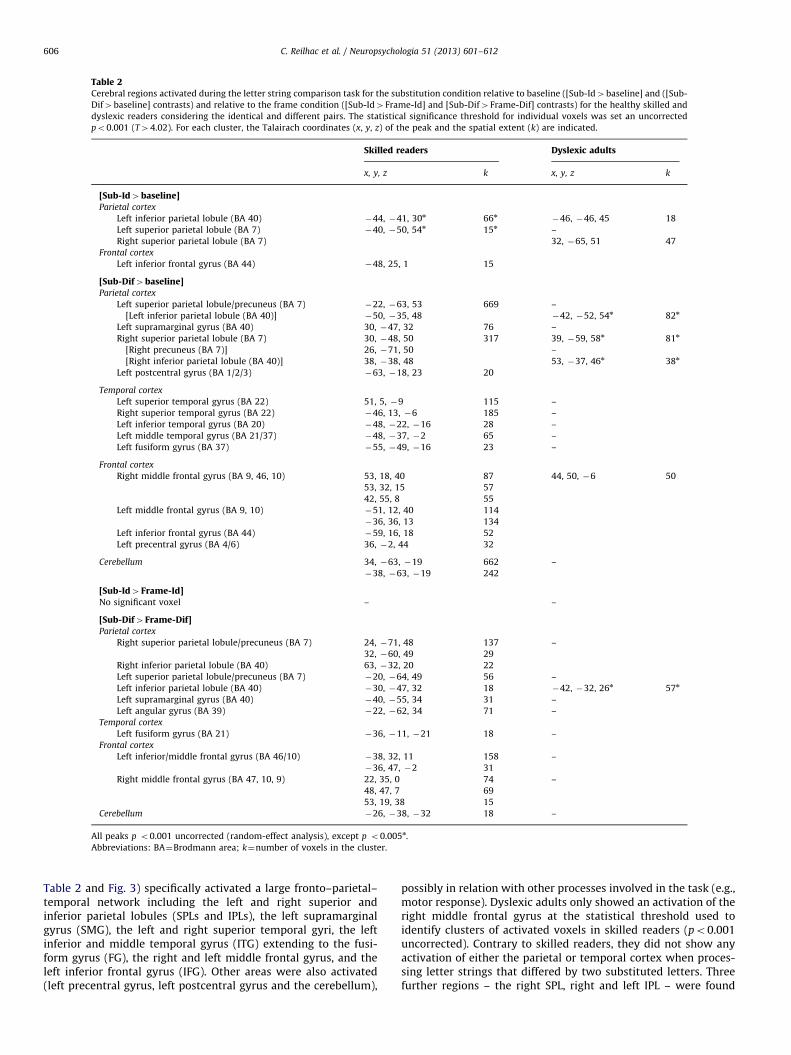
Table 2Cerebral regions activated during the letter string comparison task for the substitution condition relative to baseline ([Sub-Id4baseline] and ([Sub-
Dif4baseline] contrasts) and relative to the frame condition ([Sub-Id4Frame-Id] and [Sub-Dif4Frame-Dif] contrasts) for the healthy skilled and
dyslexic readers considering the identical and different pairs. The statistical significance threshold for individual voxels was set an uncorrected
po0.001 (T44.02). For each cluster, the Talairach coordinates (x, y, z) of the peak and the spatial extent (k) are indicated.
Skilled readers Dyslexic adults
x, y, z k x, y, z k
[Sub-Id4baseline]Parietal cortex
Left inferior parietal lobule (BA 40) �44, �41, 30n 66n�46, �46, 45 18
Left superior parietal lobule (BA 7) �40, �50, 54n 15n –
Right superior parietal lobule (BA 7) 32, �65, 51 47
Frontal cortex
Left inferior frontal gyrus (BA 44) �48, 25, 1 15
[Sub-Dif4baseline]Parietal cortex
Left superior parietal lobule/precuneus (BA 7) �22, �63, 53 669 –
[Left inferior parietal lobule (BA 40)] �50, �35, 48 �42, �52, 54n 82n
Left supramarginal gyrus (BA 40) 30, �47, 32 76 –
Right superior parietal lobule (BA 7) 30, �48, 50 317 39, �59, 58n 81n
[Right precuneus (BA 7)] 26, �71, 50 –
[Right inferior parietal lobule (BA 40)] 38, �38, 48 53, �37, 46n 38n
Left postcentral gyrus (BA 1/2/3) �63, �18, 23 20
Temporal cortex
Left superior temporal gyrus (BA 22) 51, 5, �9 115 –
Right superior temporal gyrus (BA 22) �46, 13, �6 185 –
Left inferior temporal gyrus (BA 20) �48, �22, �16 28 –
Left middle temporal gyrus (BA 21/37) �48, �37, �2 65 –
Left fusiform gyrus (BA 37) �55, �49, �16 23 –
Frontal cortex
Right middle frontal gyrus (BA 9, 46, 10) 53, 18, 40 87 44, 50, �6 50
53, 32, 15 57
42, 55, 8 55
Left middle frontal gyrus (BA 9, 10) �51, 12, 40 114
�36, 36, 13 134
Left inferior frontal gyrus (BA 44) �59, 16, 18 52
Left precentral gyrus (BA 4/6) 36, �2, 44 32
Cerebellum 34, �63, �19 662 –
�38, �63, �19 242
[Sub-Id4Frame-Id]No significant voxel – –
[Sub-Dif4Frame-Dif]Parietal cortex
Right superior parietal lobule/precuneus (BA 7) 24, �71, 48 137 –
32, �60, 49 29
Right inferior parietal lobule (BA 40) 63, �32, 20 22
Left superior parietal lobule/precuneus (BA 7) �20, �64, 49 56 –
Left inferior parietal lobule (BA 40) �30, �47, 32 18 �42, �32, 26n 57n
Left supramarginal gyrus (BA 40) �40, �55, 34 31 –
Left angular gyrus (BA 39) �22, �62, 34 71 –
Temporal cortex
Left fusiform gyrus (BA 21) �36, �11, �21 18 –
Frontal cortex
Left inferior/middle frontal gyrus (BA 46/10) �38, 32, 11 158 –
�36, 47, �2 31
Right middle frontal gyrus (BA 47, 10, 9) 22, 35, 0 74 –
48, 47, 7 69
53, 19, 38 15
Cerebellum �26, �38, �32 18 –
All peaks p o0.001 uncorrected (random-effect analysis), except p o0.005n.
Abbreviations: BA¼Brodmann area; k¼number of voxels in the cluster.
C. Reilhac et al. / Neuropsychologia 51 (2013) 601–612606
Table 2 and Fig. 3) specifically activated a large fronto–parietal–temporal network including the left and right superior andinferior parietal lobules (SPLs and IPLs), the left supramarginalgyrus (SMG), the left and right superior temporal gyri, the leftinferior and middle temporal gyrus (ITG) extending to the fusi-form gyrus (FG), the right and left middle frontal gyrus, and theleft inferior frontal gyrus (IFG). Other areas were also activated(left precentral gyrus, left postcentral gyrus and the cerebellum),
possibly in relation with other processes involved in the task (e.g.,motor response). Dyslexic adults only showed an activation of theright middle frontal gyrus at the statistical threshold used toidentify clusters of activated voxels in skilled readers (po0.001uncorrected). Contrary to skilled readers, they did not show anyactivation of either the parietal or temporal cortex when proces-sing letter strings that differed by two substituted letters. Threefurther regions – the right SPL, right and left IPL – were found
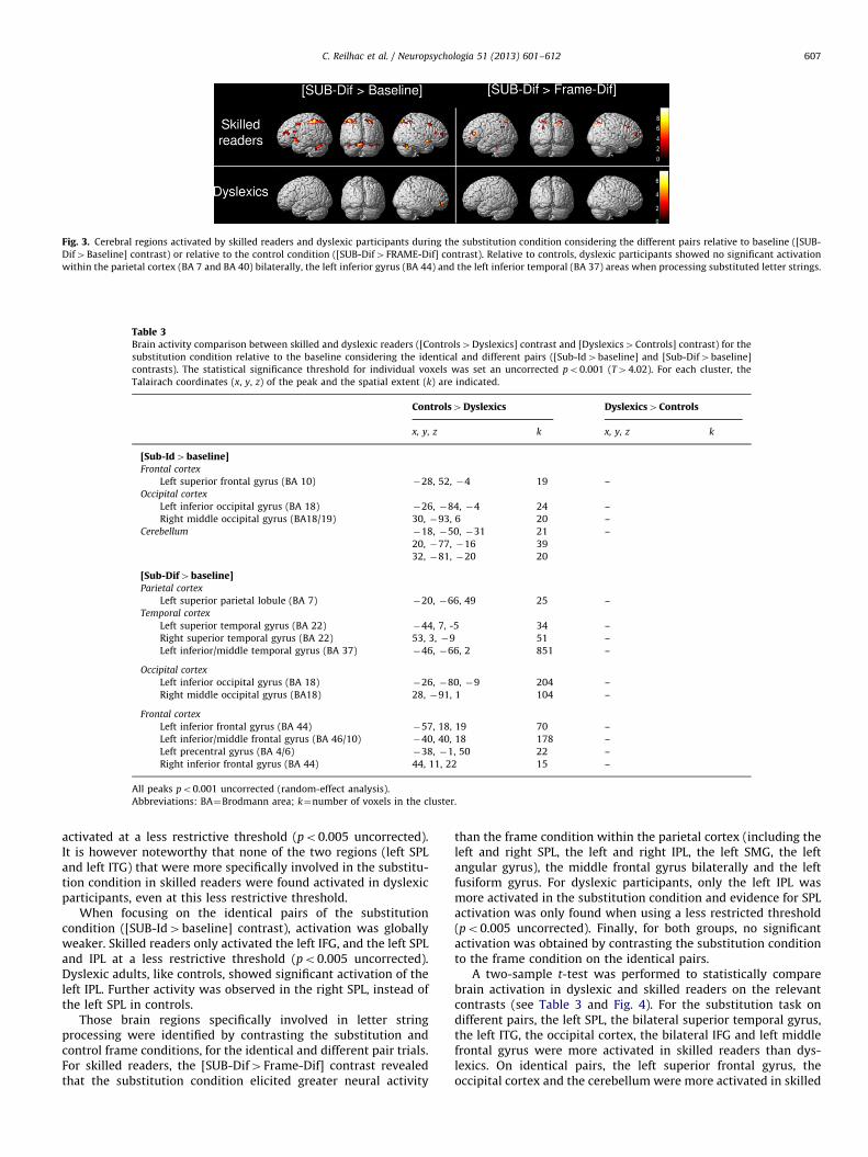
Fig. 3. Cerebral regions activated by skilled readers and dyslexic participants during the substitution condition considering the different pairs relative to baseline ([SUB-
Dif4Baseline] contrast) or relative to the control condition ([SUB-Dif4FRAME-Dif] contrast). Relative to controls, dyslexic participants showed no significant activation
within the parietal cortex (BA 7 and BA 40) bilaterally, the left inferior gyrus (BA 44) and the left inferior temporal (BA 37) areas when processing substituted letter strings.
Table 3Brain activity comparison between skilled and dyslexic readers ([Controls4Dyslexics] contrast and [Dyslexics4Controls] contrast) for the
substitution condition relative to the baseline considering the identical and different pairs ([Sub-Id4baseline] and [Sub-Dif4baseline]
contrasts). The statistical significance threshold for individual voxels was set an uncorrected po0.001 (T44.02). For each cluster, the
Talairach coordinates (x, y, z) of the peak and the spatial extent (k) are indicated.
Controls4Dyslexics Dyslexics4Controls
x, y, z k x, y, z k
[Sub-Id4baseline]Frontal cortex
Left superior frontal gyrus (BA 10) �28, 52, �4 19 –
Occipital cortex
Left inferior occipital gyrus (BA 18) �26, �84, �4 24 –
Right middle occipital gyrus (BA18/19) 30, �93, 6 20 –
Cerebellum �18, �50, �31 21 –
20, �77, �16 39
32, �81, �20 20
[Sub-Dif4baseline]Parietal cortex
Left superior parietal lobule (BA 7) �20, �66, 49 25 –
Temporal cortex
Left superior temporal gyrus (BA 22) �44, 7, -5 34 –
Right superior temporal gyrus (BA 22) 53, 3, �9 51 –
Left inferior/middle temporal gyrus (BA 37) �46, �66, 2 851 –
Occipital cortex
Left inferior occipital gyrus (BA 18) �26, �80, �9 204 –
Right middle occipital gyrus (BA18) 28, �91, 1 104 –
Frontal cortex
Left inferior frontal gyrus (BA 44) �57, 18, 19 70 –
Left inferior/middle frontal gyrus (BA 46/10) �40, 40, 18 178 –
Left precentral gyrus (BA 4/6) �38, �1, 50 22 –
Right inferior frontal gyrus (BA 44) 44, 11, 22 15 –
All peaks po0.001 uncorrected (random-effect analysis).
Abbreviations: BA¼Brodmann area; k¼number of voxels in the cluster.
C. Reilhac et al. / Neuropsychologia 51 (2013) 601–612 607
activated at a less restrictive threshold (po0.005 uncorrected).It is however noteworthy that none of the two regions (left SPLand left ITG) that were more specifically involved in the substitu-tion condition in skilled readers were found activated in dyslexicparticipants, even at this less restrictive threshold.
When focusing on the identical pairs of the substitutioncondition ([SUB-Id4baseline] contrast), activation was globallyweaker. Skilled readers only activated the left IFG, and the left SPLand IPL at a less restrictive threshold (po0.005 uncorrected).Dyslexic adults, like controls, showed significant activation of theleft IPL. Further activity was observed in the right SPL, instead ofthe left SPL in controls.
Those brain regions specifically involved in letter stringprocessing were identified by contrasting the substitution andcontrol frame conditions, for the identical and different pair trials.For skilled readers, the [SUB-Dif4Frame-Dif] contrast revealedthat the substitution condition elicited greater neural activity
than the frame condition within the parietal cortex (including theleft and right SPL, the left and right IPL, the left SMG, the leftangular gyrus), the middle frontal gyrus bilaterally and the leftfusiform gyrus. For dyslexic participants, only the left IPL wasmore activated in the substitution condition and evidence for SPLactivation was only found when using a less restricted threshold(po0.005 uncorrected). Finally, for both groups, no significantactivation was obtained by contrasting the substitution conditionto the frame condition on the identical pairs.
A two-sample t-test was performed to statistically comparebrain activation in dyslexic and skilled readers on the relevantcontrasts (see Table 3 and Fig. 4). For the substitution task ondifferent pairs, the left SPL, the bilateral superior temporal gyrus,the left ITG, the occipital cortex, the bilateral IFG and left middlefrontal gyrus were more activated in skilled readers than dys-lexics. On identical pairs, the left superior frontal gyrus, theoccipital cortex and the cerebellum were more activated in skilled
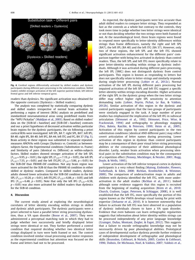
Fig. 4. Cerebral regions differentially activated by skilled readers and dyslexic
participants during different pairs processing in the substitution condition. Skilled
readers exhibit stronger activation of the left superior parietal lobule, left inferior
frontal gyrus and left inferior temporal gyrus.
C. Reilhac et al. / Neuropsychologia 51 (2013) 601–612608
readers than dyslexics. No significant activation was obtained forthe opposite contrasts (Dyslexics4Skilled readers).
The analysis was completed by statistically comparing dyslexicand skilled readers irrespective of normal brain activation byperforming a region of interest (ROIs) analysis on predefined andstandardized neuroanatomical areas using predefined masks fromthe ‘‘WFU Pickatlas’’ (Maldjian et al., 2003). Based on skilled readers’data on the [SUB-Id4baseline] and [SUB-Dif4baseline] contrastsand our a priori hypotheses of decreased activation within particularbrain regions for the dyslexic participants, the six following a prioricortical ROIs were investigated: left SPL, BA 7; right SPL, BA7; left IPL;BA 40; right IPL, BA 40; left IFG, BA 44; left ITG and FG, BA 37 (Fig. 5).Brain activity in these regions was submitted to separate repeated-measures ANOVAs with Groups (Dyslexics vs. Controls) as between-subjects factor, the Experimental conditions (Substitution vs. Frame)and Similarity of pairs (Identical vs. Different) as within-subjectsfactors. Skilled readers showed stronger activation of the left SPL(F1,22¼9.95, po0.01), the right SPL (F1,22¼7.19, po0.05), the left IPL(F1,22¼7.55, po0.05) and the left ITG/FG (F1,22¼5.86, po0.05) forthe SUB-Dif than FRAME-Dif condition. Not any brain region wasmore activated for the SUB-Id than the FRAME-Id condition in eitherskilled or dyslexic readers. Compared to skilled readers, dyslexicadults showed lower activation for the SUB-Dif condition in the leftSPL (F1,22¼10.20, po0.01), left ITG/FG (F1,22¼6.88, po0.05) and leftIFG (F1,22¼6.48, po0.05). Note that only the left IFG (F1,22¼6.58,po0.05) was also more activated for skilled readers than dyslexicsfor the SUB-Id condition.
4. Discussion
The current study aimed at exploring the neurobiologicalcorrelates of letter identity encoding within strings in skilledand dyslexic adult participants. The dyslexic participants were apriori selected to have a parallel letter string processing dysfunc-tion, thus a VA span disorder (Bosse et al., 2007). They wereadministered a perceptual matching task in which they had tojudge whether two successively and briefly displayed letterstrings were identical or not. They further performed a controlcondition that required deciding whether two identical letterstrings displayed in turn were both framed or not. The controlcondition involved similar visual processing and motor responsesas the experimental condition but attention was focused on theframe and letters had not to be processed.
As expected, the dyslexic participants were less accurate thanadult skilled readers to compare letter strings. They responded asfast as the controls as a whole but contrary to the controls, theytook more time to judge whether two letter strings were identicalor not than deciding whether the two strings were both framed ornot. At the neurobiological level, three brain regions were foundto respond more specifically to letter-identity differences withinstrings than frame differences, namely the right and left SPL(BA7), the left IPL (BA 40) and the left ITG (BA 37). However, onlytwo of these regions, the left SPL and the left ITG showedsignificant activation enhancement for the different pairs’ com-parison together with being less activated in dyslexic than controlreaders. Thus, the left SPL and left ITG more specifically relate topoor letter-identity encoding within strings in dyslexic indivi-duals. Although it was activated during different pairs processing,the left IPL (SMG) does not distinguish dyslexic from controlparticipants. This region is known as responding to letters butdoes not specifically relate to letter-strings and similarly respondsduring single-letter processing (Lobier et al., 2012c). Normalactivation of the left IPL during different pairs processing butimpaired activation of the left SPL and left ITG suggest a specificletter-identity within strings encoding disorder. Higher activationof the right SPL in the dyslexic group when the two letter stringsdiffer may reflect higher right SPL sensitivity to attentiondemanding tasks (Lobier, Peyrin, Pichat, Le Bas, & Valdois,2012b). Similar activation of this region in the dyslexic andcontrol participants suggests that the two groups of participantswere similarly engaged in the task. A large number of priorstudies has emphasized the implication of the left IFG in subvocalarticulation (Demonet et al., 1992; Demonet, Price, Wise, &Frackowiak, 1994) and orthography to phonology mapping(Fiez, Balota, Raichle, & Petersen, 1999; Heim et al., 2005).Activation of this region by control participants in the twosubstitution conditions (identical AND different pairs) may reflectcovert articulation of letter names during processing. Loweractivation of this region in dyslexic than control participantsmay be a consequence of their poor visual letter-string processingabilities or the consequence of their additional phonologicaldisorder. Lastly, weaker brain activation when processing iden-tical pairs in the substitution condition may be the consequenceof a repetition effect (Penney, Mecklinger, & Nessler, 2001; Rugg,Doyle, & Wells, 1995).
Lower activation of the left inferior temporal cortex in dyslexicparticipants is a very robust finding (Maisog, Einbinder, Flowers,Turkeltaub, & Eden, 2008; Richlan, Kronbichler, & Wimmer,2009). The comparison of underactivation maps in adults andchildren with dyslexia identified the left ITG, with more under-activation in the adult studies (Richlan et al., 2011). Indeedalthough some evidence suggests that this region is recruitedfrom the beginning of reading acquisition (Brem et al., 2010;Church, Coalson, Lugar, Petersen, & Schlaggar, 2008), it is wellestablished that the left ITG, more specifically the portion of thisregion known as the VWFA develops visual tuning for print withexpertise (Dehaene et al., 2010). It is however noteworthy thatfailure to activate the left ITG was here observed in a populationof dyslexic individuals chosen to have a VA span disorder.Evidence that even baboons can learn orthographic regularitiessuggests that information about letter identity within strings canbe processed independently of any prior language knowledge(Grainger, Dufau, Montant, Ziegler, & Fagot, 2012). This suggeststhat a failure to encode orthographic knowledge may not benecessarily driven by poor phonological abilities. Prototypicalcases of developmental surface dyslexia provide further evidenceof independent development of orthographic and phonologicalskills (Brunsdon, Coltheart, & Nickels, 2005; Castles & Coltheart,1996; Dubois, De Micheaux, Noel, & Valdois, 2007; Valdois et al.,
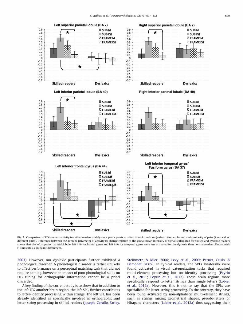
Fig. 5. Comparison of ROIs neural activity in skilled readers and dyslexic participants as a function of condition (substitution vs. frame) and similarity of pairs (identical vs.
different pairs). Difference between the average parameter of activity (% change relative to the global mean intensity of signal) calculated for skilled and dyslexic readers
shows that the left superior parietal lobule, left inferior frontal gyrus and left inferior temporal gyrus were less activated for the dyslexic than normal readers. The asterisk
(*) indicates significant differences.
C. Reilhac et al. / Neuropsychologia 51 (2013) 601–612 609
2003). However, our dyslexic participants further exhibited aphonological disorder. A phonological disorder is rather unlikelyto affect performance on a perceptual matching task that did notrequire naming, however an impact of poor phonological skills onITG tuning for orthographic information cannot be a prioridiscarded.
A key finding of the current study is to show that in addition tothe left ITG another brain region, the left SPL, further contributesto letter-identity processing within strings. The left SPL has beenalready identified as specifically involved in orthographic andletter string processing in skilled readers (Joseph, Cerullo, Farley,
Steinmetz, & Mier, 2006; Levy et al., 2009; Pernet, Celsis, &Demonet, 2005). In typical readers, the SPLs bilaterally werefound activated in visual categorization tasks that requiredmulti-element processing but no identity processing (Peyrinet al., 2011; Peyrin et al., 2012). These brain regions morespecifically respond to letter strings than single letters (Lobieret al., 2012a). However, this is not to say that the SPLs arespecialized for letter-string processing. To the contrary, they havebeen found activated by non-alphabetic multi-element strings,such as strings mixing geometrical shapes, pseudo-letters orHiragana characters (Lobier et al., 2012a) thus suggesting their
C. Reilhac et al. / Neuropsychologia 51 (2013) 601–612610
more general involvement in multi-character visual parallelprocessing. The current findings however show that these regionsfurther contribute to multiple letter identity processing. Previousinvolvement of the SPLs in string processing was mainly observedin categorization tasks that did not require stimuli identification.Evidence for SPLs involvement in a perceptual matching task thatrequires letter identification is more direct evidence that thisregion may be involved in the earlier stages of visual wordprocessing.
Traditionally, it is believed that the posterior parietal cortex thatbelongs to the dorsal visual processing stream codes for spatiallocation (the ‘‘where’’ system) whereas the ventral system codes forletter (or object) identity (the ‘‘what’’ system). The current findingssuggest that letter identity processing within strings is not confinedto the ventral pathway but further involves the dorsal visual path-way, more specifically the left SPL. Such findings are compatible withcurrent knowledge on object visual processing, showing that the twodorsal and ventral visual neural systems process very similar visualinformation (Konen & Kastner, 2008; Xu & Chun, 2007, 2009).
In the current study, VA span impaired dyslexic childrenunder-activated both the left inferior temporal cortex and theleft superior parietal lobule during letter-identity processingwithin strings. Cooperation of the dorsal and ventral visual path-ways during visual word recognition has already been empha-sized (Rosazza, Cai, Minati, Paulignan, & Nazir, 2009) and reading-related connectivity between the posterior parietal cortex and theinferior temporal cortex has been identified. Through resting-state connectivity investigation, Vogel, Miezin, Petersen, andSchlaggar (2012) identified SPLs as belonging to the networkassociated with the VWFA. Significant connectivity betweenVWFA and SPLs was reported in typical readers but not found indyslexic individuals (Van Der Mark et al., 2009). Available datathus suggests involvement of the dorsal attention network inreading. While our data does not directly bear on the question ofhow the VWFA and SPLs contribute to reading acquisition, wespeculate that these two regions may play a complementary rolein perceptual learning and visual specialization. Although mostreading models ignore the role of visual attention (Coltheart,Rastle, Perry, Langdon, & Ziegler, 2001; Harm & Seidenberg,1999), the computational Multi-Trace Memory (MTM) model ofreading (Ans, Carbonnel, & Valdois, 1998) to the contrary postu-lates that visual attention plays a key role in both skilled readingand reading acquisition (Valdois et al., 2004). Indeed, the imple-mented network includes a visual attention component thatdelineates the amount of orthographic information that is pro-cessed at each step of reading. After individuation of the ortho-graphic information to be processed, this attentional componentfurther identifies and encodes visual information on the inputletter string under focus to match previously learned informationabout orthographic regularities in long-term memory. We spec-ulate that the superior parietal lobules hold the attentionalnetwork that subserves individuation and early visual processingof letter-strings in reading. In support of this hypothesis, dis-rupted activation of this dorsal attentional region was specificallyreported in dyslexic individuals who showed a VA span disorder,thus a reduction of the number of letters they could simulta-neously process in reading (Peyrin et al., 2011; Peyrin et al.,2012). Further studies are required to determine how the twoventral (ITG and VWFA) and dorsal (SPLs) brain regions interact toensure perceptual learning.
Acknowledgments
This study was supported by a Grant from the ANR (ResearchNational Agency, Programme Blanc ‘‘VASRA’’ no. 07-BLAN-0019-01)
to SV and JFD. CR was funded by the ANR grant. We thank CedricPichat, Emilie Longeras and Patrice Peran for their help withdata analysis. Finally, we address special thanks to the dyslexicand non-dyslexic adults who participated to this study for theirtime and motivation.
References
Ans, B., Carbonnel, S., & Valdois, S. (1998). A connectionist multiple-trace memorymodel for polysyllabic word reading. Psychological Review, 105(4), 678–723.
Aylward, E. H., Richards, T. L., Berninger, V. W., Nagy, W. E., Field, K. M., Grimme, A. C.,et al. (2003). Instructional treatment associated with changes in brain activationin children with dyslexia. Neurology, 61(2), 212–219.
Ben-Shachar, M., Dougherty, R. F., Deutsch, G. K., & Wandell, B. A. (2011). Thedevelopment of cortical sensitivity to visual word forms. Journal of CognitiveNeuroscience, 23(9), 2387–2399.
Binder, J. R., & Mohr, J. P. (1992). The topography of callosal reading pathways.A case-control analysis. Brain, 115(Pt 6), 1807–1826.
Bishop, D. V., & Snowling, M. J. (2004). Developmental dyslexia and specificlanguage impairment: Same or different? Psychological Bulletin, 130(6),858–886.
Blau, V., Reithler, J., Van Atteveldt, N., Seitz, J., Gerretsen, P., Goebel, R., et al.(2010). Deviant processing of letters and speech sounds as proximate cause ofreading failure: A functional magnetic resonance imaging study of dyslexicchildren. Brain, 133(3), 868–879.
Bosse, M. L., Tainturier, M. J., & Valdois, S. (2007). Developmental dyslexia: Thevisual attention span deficit hypothesis. Cognition, 104(2), 198–230.
Bosse, M. L., & Valdois, S. (2009). Influence of the visual attention span on childreading performance: A crosssectional study. Journal of Research in Reading,32(2), 230–253.
Brambati, S. M., Termine, C., Ruffino, M., Danna, M., Lanzi, G., Stella, G, et al. (2006).Neuropsychological deficits and neural dysfunction in familial dyslexia. BrainResearch, 1113(1), 174–185.
Brem, S., Bach, S., Kucian, K., Guttorm, T. K., Martin, E., Lyytinen, H., et al. (2010).Brain sensitivity to print emerges when children learn letter–speech soundcorrespondences. Proceedings of the National Academy of Sciences, 107(17),7939.
Bruno, J. L., Zumberge, A., Manis, F. R., Lu, Z. L., & Goldman, J. G. (2008). Sensitivityto orthographic familiarity in the occipito-temporal region. Neuroimage, 39(4),1988–2001.
Brunsdon, R., Coltheart, M., & Nickels, L. (2005). Treatment of irregular wordspelling in developmental surface dysgraphia. Cognitive Neuropsychology,22(2), 213–251.
Castles, A., & Coltheart, M. (1996). Cognitive correlates of developmental surfacedyslexia: A single case study. Cognitive Neuropsychology, 13(1), 25–50.
Church, J. A., Coalson, R. S., Lugar, H. M., Petersen, S. E., & Schlaggar, B. L. (2008).A developmental fMRI study of reading and repetition reveals changes inphonological and visual mechanisms over age. Cerebral Cortex, 18(9),2054–2065.
Cohen, L., Dehaene, S., Naccache, L., Lehericy, S., Dehaene-Lambertz, G., Henaff, M. A.,et al. (2000). The visual word form area. Brain, 123(2), 291.
Cohen, L., Dehaene, S., Vinckier, F., Jobert, A., & Montavont, A. (2008). Readingnormal and degraded words: contribution of the dorsal and ventral visualpathways. Neuroimage, 40(1), 353–366.
Cohen, L., Lehericy, S., Chochon, F., Lemer, C., Rivaud, S., & Dehaene, S. (2002).Language-specific tuning of visual cortex? Functional properties of the visualword form area. Brain, 125(Pt 5), 1054–1069.
Cohen, L., Martinaud, O., Lemer, C., Lehericy, S., Samson, Y., Obadia, M., et al.(2003). Visual word recognition in the left and right hemispheres: Anatomicaland functional correlates of peripheral alexias. Cerebral Cortex, 13(12),1313–1333.
Coltheart, M., Rastle, K., Perry, C., Langdon, R., & Ziegler, J. (2001). DRC: A dualroute cascaded model of visual word recognition and reading aloud. Psycho-logical Review, 108(1), 204–256.
Dehaene, S., & Cohen, L. (2007). Cultural recycling of cortical maps. Neuron, 56(2),384–398.
Dehaene, S., & Cohen, L. (2011). The unique role of the visual word form area inreading. Trends in Cognitive Sciences, 15(6), 254–262.
Dehaene, S., Le Clec, H. G., Poline, J. B., Le Bihan, D., & Cohen, L. (2002). The visualword form area: A prelexical representation of visual words in the fusiformgyrus. Neuroreport, 13(3), 321–325.
Dehaene, S., Naccache, L., Cohen, L., Bihan, D., Mangin, J. F., Poline, J. B., et al.(2001). Cerebral mechanisms of word masking and unconscious repetitionpriming. Nature neuroscience, 4(7), 752–758.
Dehaene, S., Pegado, F., Braga, L. W., Ventura, P., Nunes Filho, G., Jobert, A., et al.(2010). How learning to read changes the cortical networks for vision andlanguage. Science, 330(6009), 1359–1364.
Demonet, J. F., Chollet, F., Ramsay, S., Cardebat, D., Nespoulous, J. L., Wise, R., et al.(1992). The anatomy of phonological and semantic processing in normalsubjects. Brain, 115(6), 1753–1768.
Demonet, J. F., Price, C., Wise, R., & Frackowiak, R. S. (1994). A PET study ofcognitive strategies in normal subjects during language tasks. Influence of
C. Reilhac et al. / Neuropsychologia 51 (2013) 601–612 611
phonetic ambiguity and sequence processing on phoneme monitoring. Brain,117(Pt 4), 671–682.
Demonet, J. F., Taylor, M. J., & Chaix, Y. (2004). Developmental dyslexia. Lancet,363(9419), 1451–1460.
Dubois, M., De Micheaux, P. L., Noel, M. P., & Valdois, S. (2007). Preorthographicalconstraints on visual word recognition: evidence from a case study ofdevelopmental surface dyslexia. Cognitive Neuropsychology, 24(6), 623–660.
Dubois, M., Kyllingsbaek, S., Prado, C., Musca, S. C., Peiffer, E., Lassus-Sangosse, D.,et al. (2010). Fractionating the multi-character processing deficit in develop-mental dyslexia: Evidence from two case studies. Cortex, 46(6), 717–738.
Dufor, O., Serniclaes, W., Sprenger-Charolles, L., & Demonet, J. F. (2007). Top-downprocesses during auditory phoneme categorization in dyslexia: A PET study.Neuroimage, 34(4), 1692–1707.
Dufor, O., Serniclaes, W., Sprenger-Charolles, L., & Demonet, J. F. (2007). Top-downprocesses during auditory phoneme categorization in dyslexia: A PET study.Neuroimage, 34(4), 1692–1707.
Fiez, J. A., Balota, D. A., Raichle, M. E., & Petersen, S. E. (1999). Effects of lexicality,frequency, and spelling-to-sound consistency on the functional anatomy ofreading. Neuron, 24(1), 205–218.
Friston, K. J. (1995). Commentary and opinion: II. Statistical parametric mapping:ontology and current issues. Journal of Cerebral Blood Flow and Metabolism,15(3), 361–370.
Friston, K. J., Fletcher, P., Josephs, O., Holmes, A., Rugg, M. D., & Turner, R. (1998).Event-related fMRI: Characterizing differential responses. Neuroimage, 7(1),30–40.
Friston, K. J., Zarahn, E., Josephs, O., Henson, R. N., & Dale, A. M. (1999). Stochasticdesigns in event-related fMRI. Neuroimage, 10(5), 607–619.
Gaillard, R., Naccache, L., Pinel, P., Clemenceau, S., Volle, E., Hasboun, D, et al.(2006). Direct intracranial, FMRI, and lesion evidence for the causal role of leftinferotemporal cortex in reading. Neuron, 50(2), 191–204.
Grainger, J., Dufau, S., Montant, M., Ziegler, J. C., & Fagot, J. (2012). Orthographicprocessing in baboons (Papio papio). Science, 336(6078), 245–248.
Harm, M. W., & Seidenberg, M. S. (1999). Phonology, reading acquisition, and dyslexia:Insights from connectionist models. Psychological Review, 106(3), 491.
Hawelka, S., & Wimmer, H. (2005). Impaired visual processing of multi-elementarrays is associated with increased number of eye movements in dyslexicreading. Vision Research, 45(7), 855–863.
Heim, S., Alter, K., Ischebeck, A. K., Amunts, K., Eickhoff, S. B., Mohlberg, H., et al.(2005). The role of the left Brodmann’s areas 44 and 45 in reading words andpseudowords. Cognitive Brain Research, 25(3), 982–993.
Heim, S., Eulitz, C., & Elbert, T. (2003). Altered hemispheric asymmetry of auditoryP100m in dyslexia. European Journal of Neuroscience, 17(8), 1715–1722.
Helenius, P., Tarkiainen, A., Cornelissen, P., Hansen, P. C., & Salmelin, R. (1999).Dissociation of normal feature analysis and deficient processing of letter-strings in dyslexic adults. Cerebral Cortex, 9(5), 476–483.
Hellyer, P. J., Woodhead, Z. V., Leech, R., & Wise, R. J. (2011). An investigation oftwenty/20 vision in reading. Journal of Neuroscience, 31(41), 14631–14638.
Inhoff, A. W., & Tousman, S. (1990). Lexical priming from partial-word previews.Journal of Experimental Psychology, Learning, Memory and Cognition, 16(5),825–836.
Jones, M. W., Branigan, H. P., & Kelly, M. L. (2008). Visual deficits in developmentaldyslexia: Relationships between nonlinguistic visual tasks and their contribu-tion to components of reading. Dyslexia, 14(2), 95–115.
Joseph, J. E., Cerullo, M. A., Farley, A. B., Steinmetz, N. A., & Mier, C. R. (2006). fMRIcorrelates of cortical specialization and generalization for letter processing.Neuroimage, 32(2), 806–820.
Konen, C. S., & Kastner, S. (2008). Two hierarchically organized neural systems forobject information in human visual cortex. Nature neuroscience, 11(2),224–231.
Lallier, M., Donnadieu, S., Berger, C., & Valdois, S. (2010). A case study ofdevelopmental phonological dyslexia: Is the attentional deficit in the percep-tion of rapid stimuli sequences amodal? Cortex, 46(2), 231–241.
Lefavrais, P. (1965). Test d’Analyse de la Lecture et de la Dyslexie (Test del’Alouette). Paris: Masson.
Leff, A. P., Crewes, H., Plant, G. T., Scott, S. K., Kennard, C, & Wise, R. J. (2001). Thefunctional anatomy of single-word reading in patients with hemianopic andpure alexia. Brain, 124(Pt 3), 510–521.
Leff, A. P., Spitsyna, G., Plant, G., & Wise, R. (2006). Structural anatomy of pureand hemianopic alexia. Journal of Neurology, Neurosurgery & Psychiatry, 77(9),1004.
Levy, J., Pernet, C., Treserras, S., Boulanouar, K., Aubry, F., Demonet, J. F., et al.(2009). Testing for the dual-route cascade reading model in the brain: AnfMRI effective connectivity account of an efficient reading style. PLoS One,4(8), e6675.
Lobier, M., Peyrin, C., Le Bas, J. F., & Valdois, S. (2012a). Pre-orthographic characterstring processing and parietal cortex: A role for visual attention in reading?Neuropsychologia, 50(9), 2195–2204.
Lobier, M., Peyrin, C., Pichat, C., Le Bas, J. F., & Valdois, S. (2012b). Visual processingof multiple element in the dyslexic brain: Evidence for a parietal dysfunction.Paying visual attention to pre-orthographic processing in reading and devel-opmental dyslexia, Universite Pierre Mend�es France, 126–155.
Lobier, M., Zoubrinetzky, R., & Valdois, S. (2012c). The visual attention span deficitin dyslexia is visual and not verbal. Cortex, 48(6), 768–773.
Maisog, J. M., Einbinder, E. R., Flowers, D. L., Turkeltaub, P. E., & Eden, G. F. (2008).A meta-analysis of functional neuroimaging studies of dyslexia. Annals of NewYork Academy of Sciences, 1145, 237–259.
Maldjian, J. A., Laurienti, P. J., Kraft, R. A., & Burdette, J. H. (2003). An automatedmethod for neuroanatomic and cytoarchitectonic atlas-based interrogation offMRI data sets. Neuroimage, 19(3), 1233–1239.
Maurer, U., Brem, S., Bucher, K., Kranz, F., Benz, R., Steinhausen, H. C., et al. (2007).Impaired tuning of a fast occipito-temporal response for print in dyslexicchildren learning to read. Brain, 130(12), 3200–3210.
Oldfield, R. C. (1971). The assessment and analysis of handedness: the Edinburghinventory. Neuropsychologia, 9(1), 97–113.
Pammer, K., Lavis, R., Hansen, P., & Cornelissen, P. L. (2004). Symbol-stringsensitivity and children’s reading. Brain and Language, 89(3), 601–610.
Pammer, K., & Vidyasagar, T. R. (2005). Integration of the visual and auditorynetworks in dyslexia: A theoretical perspective. Journal of Research in Reading,28(3), 320–331.
Paulesu, E., Demonet, J. F., Fazio, F., McCrory, E., Chanoine, V., Brunswick, N., et al.(2001). Dyslexia: Cultural diversity and biological unity. Science, 291(5511),2165–2167.
Paulesu, E., Frith, U., Snowling, M., Gallagher, A., Morton, J., Frackowiak, R. S., et al.(1996). Is developmental dyslexia a disconnection syndrome? Evidence fromPET scanning. Brain, 119(Pt 1), 143–157.
Penney, T. B., Mecklinger, A., & Nessler, D. (2001). Repetition related ERP effects ina visual object target detection task. Brain Research. Cognitive Brain Research,10(3), 239–250.
Pernet, C., Celsis, P., & Demonet, J. F. (2005). Selective response to lettercategorization within the left fusiform gyrus. Neuroimage, 28(3), 738–744.
Peyrin, C., Demonet, J. F., N’Guyen-Morel, M. A., Le Bas, J. F., & Valdois, S. (2011).Superior parietal lobule dysfunction in a homogeneous group of dyslexicchildren with a visual attention span disorder. Brain and Language, 118(3),128–138.
Peyrin, C., Demonet, J. F., N’Guyen-Morel, M. A., Le Bas, J. F., & Valdois, S. (2011).Superior parietal lobule dysfunction in a homogeneous group of dyslexicchildren with a visual attention span disorder. Brain and Language, 118(3),128–138.
Peyrin, C., Lallier, M., Baciu, M., & Valdois, S. (2008). Brain mechanisms of thevisual attention span in normal and dyslexic readers. In M. Baciu (Ed.),Behavioral, neuropsychological and neuroimaging studies of spoken and writtenlanguage. Signpost Edition (pp. 22–43).
Peyrin, C., Lallier, M., Demonet, J. F., Pernet, C., Baciu, M., Le Bas, J. F., et al. (2012).Neural dissociation of phonological and visual attention span disorders indevelopmental dyslexia: fMRI evidence from two case studies. Brain andLanguage, 120(3), 381–394.
Price, C. J., & Devlin, J. T. (2003). The myth of the visual word form area.Neuroimage, 19(3), 473–481.
Price, C. J., & Devlin, J. T. (2011). The interactive account of ventral occipitotem-poral contributions to reading. Trends in Cognitive Sciences, 15(6), 246–253.
Price, C. J., & Mechelli, A. (2005). Reading and reading disturbance. Current Opinionin Neurobiology, 15(2), 231–238.
Ramus, F. (2003). Developmental dyslexia: Specific phonological deficit or generalsensorimotor dysfunction? Current Opinion in Neurobiolology, 13(2), 212–218.
Richlan, F., Kronbichler, M., & Wimmer, H. (2009). Functional abnormalities in thedyslexic brain: A quantitative meta-analysis of neuroimaging studies. HumanBrain Mapping, 30(10), 3299–3308.
Richlan, F., Kronbichler, M., & Wimmer, H. (2011). Meta-analyzing brain dysfunc-tions in dyslexic children and adults. Neuroimage, 56(3), 1735–1742.
Rosazza, C., Cai, Q., Minati, L., Paulignan, Y., & Nazir, T. A. (2009). Early involvementof dorsal and ventral pathways in visual word recognition: An ERP study. BrainResearch, 1272, 32–44.
Ruff, S., Cardebat, D., Marie, N., & Demonet, J. F. (2002). Enhanced response of theleft frontal cortex to slowed down speech in dyslexia: An fMRI study.Neuroreport, 13(10), 1285–1289.
Ruff, S., Marie, N., Celsis, P., Cardebat, D., & Demonet, J. F. (2003). Neural substratesof impaired categorical perception of phonemes in adult dyslexics: An fMRIstudy. Brain and Cognition, 53(2), 331–334.
Rugg, M. D., Doyle, M. C., & Wells, T. (1995). Word and nonword repetition within-and across-modality: An event-related potential study. Journal of CognitiveNeuroscience, 7(2), 209–227.
Shaywitz, B. A., Shaywitz, S. E., Pugh, K. R., Mencl, W. E., Fulbright, R. K., Skudlarski, P.,et al. (2002). Disruption of posterior brain systems for reading in children withdevelopmental dyslexia. Biological Psychiatry, 52(2), 101–110.
Shaywitz, S. E., Shaywitz, B. A., Pugh, K. R., Fulbright, R. K., Constable, R. T., Mencl, W. E.,et al. (1998). Functional disruption in the organization of the brain for reading indyslexia. Proceedings of the National Academy of Sciences of the United States ofAmerica, 95(5), 2636–2641.
Silani, G., Frith, U., Demonet, J. F., Fazio, F., Perani, D., Price, C., et al. (2005). Brainabnormalities underlying altered activation in dyslexia: A voxel based mor-phometry study. Brain, 128(10), 2453–2461.
Snowling, M. J. (2000). Dyslexia. Wiley-Blackwell.Starrfelt, R., & Gerlach, C. (2007). The visual what for area: Words and pictures in
the left fusiform gyrus. Neuroimage, 35(1), 334–342.Szwed, M., Dehaene, S., Kleinschmidt, A., Eger, E., Valabregue, R., Amadon, A., et al.
(2011). Specialization for written words over objects in the visual cortex.Neuroimage, 56(1), 330–344.
Tagamets, M. A., Novick, J. M., Chalmers, M. L., & Friedman, R. B. (2000).A parametric approach to orthographic processing in the brain: An fMRIstudy. Journal of Cognitive Neuroscience, 12(2), 281–297.
Talairach, J., & Tournoux, P. (1988). Co-planar stereotaxic atlas of the human brain:3-dimensional proportional system: an approach to cerebral imaging: Thieme.
C. Reilhac et al. / Neuropsychologia 51 (2013) 601–612612
Temple, E., Deutsch, G. K., Poldrack, R. A., Miller, S. L., Tallal, P., Merzenich, M. M.,et al. (2003). Neural deficits in children with dyslexia ameliorated bybehavioral remediation: Evidence from functional MRI. Proceedings of the
National Academy of Sciences, 100(5), 2860.Twomey, T., Kawabata Duncan, K. J., Price, C. J., & Devlin, J. T. (2011). Top-down
modulation of ventral occipito-temporal responses during visual word recog-nition. Neuroimage, 55(3), 1242–1251.
Valdois, S., Bidet-Ildei, C., Lassus-Sangosse, D., Reilhac, C., N’Guyen-Morel, M. A.,Guinet, E., et al. (2011). A visual processing but no phonological disorder in achild with mixed dyslexia. Cortex, 47(10), 1197–1218.
Valdois, S., Bosse, M. L., Ans, B., Carbonnel, S., Zorman, M., David, D., et al. (2003).Phonological and visual processing deficits can dissociate in developmentaldyslexia: Evidence from two case studies. Reading and Writing, 16(6),541–572.
Valdois, S., Bosse, M. L., & Tainturier, M. J. (2004). The cognitive deficits responsiblefor developmental dyslexia: Review of evidence for a selective visual atten-tional disorder. Dyslexia, 10(4), 339–363.
Valdois, S., Carbonnel, S., Juphard, A., Baciu, M., Ans, B., Peyrin, C., et al. (2006).Polysyllabic pseudo-word processing in reading and lexical decision: Conver-ging evidence from behavioral data, connectionist simulations and functionalMRI. Brain Research, 1085(1), 149–162.
Valdois, S., Lassus-Sangosse, D., & Lobier, M. (2012b). Impaired letter stringprocessing in developmental dyslexia: What visual-to-phonological codemapping disorder? Dyslexia, 18(2), 77–93.
Van Der Mark, S., Bucher, K., Maurer, U., Schulz, E., Brem, S., Buckelmuller, J., et al.(2009). Children with dyslexia lack multiple specializations along the visualword-form (VWF) system. Neuroimage, 47(4), 1940–1949.
Vellutino, F. R., Fletcher, J. M., Snowling, M. J., & Scanlon, D. M. (2004). Specificreading disability (dyslexia): What have we learned in the past four decades?Journal of Child Psychology and Psychiatry, 45(1), 2–40.
Vidyasagar, T. R., & Pammer, K. (2010). Dyslexia: A deficit in visuo-spatialattention, not in phonological processing. Trends in Cognitive Sciences, 14(2),57–63.
Vigneau, M., Jobard, G., Mazoyer, B., & Tzourio-Mazoyer, N. (2005). Word and non-word reading: What role for the visual word form area? Neuroimage, 27(3),694–705.
Vogel, A. C., Miezin, F. M., Petersen, S. E., & Schlaggar, B. L. (2012). The putativevisual word form area is functionally connected to the dorsal attentionnetwork. Cerebral Cortex, 22(3), 537–549.
Wandell, B. A., Rauschecker, A. M., & Yeatman, J. D. (2012). Learning to see words.Annual Review of Psychology, 63, 31–53.
Wimmer, H., Schurz, M., Sturm, D., Richlan, F., Klackl, J., Kronbichler, M., et al.(2010). A dual-route perspective on poor reading in a regular orthography: AnfMRI study. Cortex: A Journal Devoted to The Study of The Nervous System andBehavior, 46(10), 1284.
Xu, Y., & Chun, M. M. (2007). Visual grouping in human parietal cortex. Proceedingsof the National Academy of Sciences, 104(47), 18766.
Xu, Y., & Chun, M. M. (2009). Selecting and perceiving multiple visual objects.Trends in Cognitive Sciences, 13(4), 167–174.