Embed Size (px)
Citation preview
Leading Edge
Review
Role of the Microbiotain Immunity and Inflammation
Yasmine Belkaid1,* and Timothy W. Hand1,21Immunity at Barrier Sites Initiative, National Institute of Allergy and Infectious Diseases, National Institutes of Health, Bethesda,
MD 20892, USA2Mucosal Immunology Section, Laboratory of Parasitic Diseases, National Institute of Allergy and Infectious Disease, National Institutes of
Health, Bethesda, MD 20892, USA
*Correspondence: [email protected]
http://dx.doi.org/10.1016/j.cell.2014.03.011
Themicrobiota plays a fundamental role on the induction, training, and function of the host immunesystem. In return, the immune systemhas largely evolved as ameans tomaintain the symbiotic rela-tionship of the host with these highly diverse and evolvingmicrobes.When operating optimally, thisimmune system-microbiota alliance allows the induction of protective responses to pathogens andthe maintenance of regulatory pathways involved in the maintenance of tolerance to innocuous an-tigens. However, in high-income countries, overuse of antibiotics, changes in diet, and eliminationof constitutive partners, such as nematodes, may have selected for a microbiota that lack the resil-ience and diversity required to establish balanced immune responses. This phenomenon is pro-posed to account for some of the dramatic rise in autoimmune and inflammatory disorders in partsof the world where our symbiotic relationship with the microbiota has been the most affected.
‘‘The states of health or disease are the expressions
of the success or failure experienced by the organism
in its efforts to respond adaptively to environmental
challenges.’’—Rene Dubos, 1965
IntroductionMulticellular organisms exist as meta-organisms comprised of
both the macroscopic host and its symbiotic commensal mi-
crobiota. With an estimated composition of 100 trillion cells,
human symbionts outnumber host cells by at least a factor of
10 and express at least 10-fold more unique genes than their
host’s genome (Ley et al., 2006a). These complex communities
of microbes that include bacteria, fungi, viruses, and other mi-
crobial and eukaryotic species provide a tremendous enzy-
matic capability and play a fundamental role in controlling
many aspects of host physiology. Over the past few years,
the field of immunology has been revolutionized by the growing
understanding of the fundamental role of the microbiota in the
induction, education, and function of the mammalian immune
system.
The immune system is composed of a complex network of
innate and adaptive components endowed with an extraordinary
capacity to adapt and respond to highly diverse challenges.
Collectively, this cellular network acts as a formidable regulator
of host homeostasis that operates to sustain and restore tissue
function in the context of microbial and environmental encoun-
ters. The development of defined arms of the immune sys-
tem—and, more particularly, those associated with adaptive
immunity—has coincided with the acquisition of a complex
microbiota, supporting the concept that a large fraction of this
machinery has evolved as a means to maintain a symbiotic rela-
tionships with these highly diverse microbial communities. In
turn, the microbiota promote and calibrate multiple aspects of
the immune system.
When operating optimally, the immune system-microbiota
alliance interweaves the innate and adaptive arms of immunity
in a dialog that selects, calibrates, and terminates responses
in the most appropriate manner. However, both the acquisition
of a complex immune system and its reliance on the microbiota
came at a price. Pathologies that increasingly affect humans,
such as allergies, autoimmune, and inflammatory disorders, all
arise from a failure to control misdirected immune responses
against self, microbiota-derived, or environmental antigens.
Further, alteration of the composition and function of the micro-
biota as a result of antibiotic use, diet evolution, and recent
elimination of constitutive partners such as helminth worms
has transformed our microbial allies into potential liabilities.
Although members of the microbiota are often referred to
as commensals, symbiosis between the microbiota and its
mammalian host encompasses various forms of relationship,
including mutualistic, parasitic, or commensal. However, how
defined members of the microbiota interact with their host can
be highly contextual, with the same microbe developing as
mutualist or parasite according to the nutritional, coinfection,
or genetic landscape of its host. Over the past decade, explora-
tion of optimal and dysregulated partnerships between the
microbiota and its mammalian host has taken center stage in
the field of immunology and has led to the rediscovery of a
more holistic view of host physiology. Indeed, the notion that
microbial partners can promote human health is not a recent
concept and was originally proposed by the seminal work of
Doderlein (1892) and his understanding of the role of lactobacilli
Cell 157, March 27, 2014 ª2014 Elsevier Inc. 121
as gatekeepers of the vaginal ecosystem as well as the observa-
tion of Metchnikoff associating prolonged life with fermented
milk products. Recent sequencing efforts of the human meta-
genome have changed our understanding of the microbiome
and how variations in these populations can contribute to dis-
ease states. In this Review, we will discuss some of the major
concepts that have emerged from the recent dialog between
immunologists, geneticists, microbiologists, and clinicians that
highlight the complex role of the microbiota on the immune
system in health and diseases.
Microbiota-Immune System Interaction duringDevelopmentUnder normal conditions, the fetal gastrointestinal tract is
believed to be sterile, with the first exposure of the immune
system to commensals occurring during the passage through
the birth canal. These early interactions are considered to set
the tone of the mucosal and systemic immune system for the
long term. The mechanism by which neonate tissues adapt
to the formidable challenge of microbial colonization remains
incompletely understood, but factors contained in maternal
milk are believed to define some of these early responses to
commensals. Indeed, colostrum and breast milk contain live
microbes, metabolites, IgA, and immune cells as well as cyto-
kines. These factors synergize to shape the breast-fed infant
microbiota and the response of the host to these microbes.
For instance, maternal IgA restricts immune activation and mi-
crobial attachment by binding nutritional and microbial antigens,
and the presence of metabolites, including oligosaccharides in
mother’s milk, promotes the expansion of defined constituents
of the microbiota such as Bifidobacterium (Marcobal et al.,
2010; Marcobal and Sonnenburg, 2012). Bacterial translocation
from the mouse gut is increased during pregnancy and lacta-
tion, and bacterially loaded dendritic cells in the milk have
been proposed to contribute to neonatal immune imprinting
(Perez et al., 2007).
The capacity to accept the microbiota can also be explained
by the relative immaturity of the neonate immune system at birth
and the tolerogenic environment that defines early mammalian
life. Indeed, the developing immune system is characterized by
blunted inflammatory cytokine production and skewed T and B
cell development in favor of regulatory responses (PrabhuDas
et al., 2011; Siegrist, 2001). Although a consequence of this
blunted immune response is high susceptibility to infections,
this regulatory environment ensures that the establishment of
the microbiota occurs without overt inflammation. Recent re-
ports reveal that a defined population of erythroid cells enriched
in neonates contributes to the maintenance of this immunoregu-
latory environment and limits mucosal inflammation following
colonization with the microbiota (Elahi et al., 2013). Early expo-
sure of the host to commensals can also repress cells involved
in the induction of inflammatory responses such as invariant
natural killer T (iNKT) cells, an effect that has long-term conse-
quences for the host capacity to develop inflammatory diseases
(Olszak et al., 2012). A recent report proposed that this control
can be mediated by the direct interaction, early in life, of
unique inhibitory commensal-derived sphingolipids with iNKT
cells (An et al., 2014).
122 Cell 157, March 27, 2014 ª2014 Elsevier Inc.
One of the primary modes of dialog between the host and
the microbiota is mediated by the recognition of conserved
microbial-associated molecular patterns (MAMPs). The neonate
innate immune system integrates these signals in a unique
way to promote healthy microbial colonization. For instance,
although neonate innate cells express Toll-like receptor (TLR)
ligands, their response to microbial ligands is distinct from that
of adult cells, with notable impairment in the production of in-
flammatory mediators such as oxygen radicals and heightened
production of regulatory cytokines such as IL-10 (Kollmann
et al., 2012). Part of this phenomenon results from the action
of the microbiota itself. Indeed, early responses to microbial
ligands such as LPS, the endotoxin found in the outer membrane
of Gram-negative bacterial walls, condition gut epithelial cells to
become hypo-responsive to subsequent TLR stimulation (Chas-
sin et al., 2010; Lotz et al., 2006). How the innate immune system
integrates microbial-derived signals remains unclear, but recent
findings support the idea that expression of epigenome-
modifying enzymes by epithelial cells may be required for the
coordination of commensal dependent intestinal homeostasis
(Alenghat et al., 2013).
Commensals also contribute to the postnatal development of
the immune system that, in turn, contributes to their contain-
ment. Studies performed in animals raised in the absence of
live microbes referred to as germ-free (GF), revealed that the
microbiota plays a critical role in secondary lymphoid structure
development. This is particularly evident in the gastrointestinal
tract, where germ-freemice are characterized by smaller Peyer’s
patch size and a reduced number of CD4+T cells and IgA-pro-
ducing plasma cells (Bauer et al., 1963; Hamada et al., 2002;
Macpherson et al., 2001; Mazmanian et al., 2005; Smith et al.,
2007; Talham et al., 1999). In the intestine, tertiary lymphoid
structures such as isolated lymphoid follicle or crytopatches
are induced after birth as a result of commensal exposure (Bou-
skra et al., 2008; Ohnmacht et al., 2011). As further discussed
below, commensals can also contribute to the fortification of
the intestinal barrier by various mechanisms, including the pro-
motion of epithelial cell maturation and angiogenesis (Hooper
et al., 2001; Stappenbeck et al., 2002).
When operating properly, the highly regulatory tone of the
neonate immune system and the action of commensals in the
development and training of this system lead to the establish-
ment of a durable and homeostatic host/commensal relation-
ship. These primary encounters between the host immune
system and the microbiota have profound and long-term impli-
cations for human health. Indeed, epidemiological observations
revealed that alteration of the microbiota in mothers or in neo-
nates may predispose to diseases associated with dysregulated
barrier responses, such as asthma (Ege et al., 2011).
Containing the MicrobiotaAn important point to consider when exploring the role of the
microbiota on the immune system is that pathogenicity is, in
most cases, a contextual state. Indeed, the capacity of a given
microbe, including those composing the microbiota, to trigger
or promote disease is highly dependent on the state of activation
of the host, the host’s genetic predisposition, and the localization
of the particular microbe. As such, the mechanisms utilized by

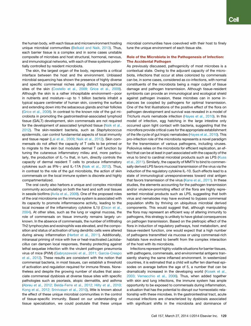
Figure 1. The Mucosal Firewall(1) The mucus represents the primary barrier limiting contact between themicrobiota and host tissue preventing microbial translocation. (2) Epithelialcells produce antimicrobial peptides that also play a significant role in limitingexposure to the commensal microbiota. (3) Translocating commensals arerapidly eliminated by tissue-resident macrophages. (4) Commensals orcommensal antigens can also be captured by CD103+ CD11b+ DCs that trafficto the mLN from the lamina propria but do not penetrate further. Presentationof commensal antigens by these DCs leads to the differentiation ofcommensal-specific regulatory cells (Treg), Th17 cells, and IgA-producing Bcells. Commensal-specific lymphocytes traffic to the lamina propria andPeyer’s patches. In the Peyer’s patches, Treg can further promote classswitching and IgA generation against commensals. The combination of theepithelial barrier, mucus layer, IgA, and DCs and T cells comprises the‘‘mucosal firewall,’’ which limits the passage and exposure of commensals tothe gut-associated lymphoid tissue, preventing untoward activation andpathology.
the immune system to maintain its relationship with the micro-
biota are highly analogous to the ones that are used to constrain
organisms with pathogenic potential.
An enormous fraction of the immune system’s constitutive
function is aimed at controlling our relationship with the micro-
biota. As such, the highest number of immune cells in the body
are resident at sites colonized by commensals such as the skin
or the GI tract. In turn, to protect their ecological niche, a domi-
nant action of the healthy microbiota on the immune system is
aimed at reinforcing barrier immunity and therefore their own
containment. A central strategy utilized by the host to maintain
its homeostatic relationship with the microbiota is to minimize
contact between microorganisms and the epithelial cell surface,
thereby limiting tissue inflammation and microbial translocation.
In the gastrointestinal tract, home to the largest density of com-
mensals, this segregation is accomplished by the combined
action of epithelial cells, mucus, IgA, antimicrobial peptides,
and immune cells. Collectively, these structural and immunolog-
ical components have been referred to as the ‘‘mucosal firewall’’
(Figure 1) (Macpherson et al., 2009).
The mucus represents the primary shield limiting contact be-
tween the microbiota and host tissue and preventing microbial
translocation (McGuckin et al., 2011). In addition to the produc-
tion of mucus by goblet cells, all intestinal epithelial cell lineages
can produce antimicrobial peptides that play a significant role in
limiting exposure to the commensal microbiota (Hooper and
Macpherson, 2010). These proteins can exert antimicrobial func-
tions resulting from enzymatic attack of the bacterial cell wall or
by disrupting the bacterial inner membrane (Hooper and Mac-
pherson, 2010). Some of these molecules, such as a-defensins,
are constitutively expressed by epithelial cells, whereas in other
cases, engagement of pattern recognition receptors (PRRs) by
commensally derived products is required (Hooper and Mac-
pherson, 2010). One of the best-characterized mucosal antimi-
crobial peptides is RegIIIg, which is expressed soon after birth
or following colonization of germ-free mice (Cash et al., 2006).
Production of this lectin is tightly controlled by the flora in a
MyD88-dependent manner and has a direct microbicidal effect
on Gram-positive bacteria (Brandl et al., 2007; Cash et al.,
2006; Ismail et al., 2011; Mukherjee et al., 2009). Accumulation
of antimicrobial peptides such as RegIIIg in the mucus contrib-
utes to the maintenance of the segregation between the micro-
biota and the host intestine, creating a physical separation
referred to as the ‘‘demilitarized zone’’ (Vaishnava et al., 2011).
Compartmentalization of intestinal bacteria also depends on
secreted immunoglobulin A (IgA). IgA specific for commensals
is produced with the help of intestinal dendritic cells that sample
commensals associated with the epithelium and interact with
B and T cells in the Peyer’s patches to produce IgA specific for
commensal-derived antigens (Macpherson and Uhr, 2004).
Further, commensals that translocate across the intestinal
epithelial cell barrier can be rapidly engulfed and eliminated by
macrophages that reside in the lamina propria or carried alive
by dendritic cells (DC) (Kelsall, 2008; Macpherson and Uhr,
2004). The bacteria loaded DC traffic to the mesenteric lymph
node via the intestinal lymphatics but do not penetrate further,
allowing the induction of a mucosal-compartmentalized IgA
response (Macpherson and Uhr, 2004). IgA+B cells migrate to
the intestinal lamina propria and secrete IgA that are subse-
quently transcytosed across epithelial cells. These transcytosed
IgAs control host commensal interaction by both impacting
commensal gene expression (Peterson et al., 2007) and prevent-
ing adhesion of commensal bacteria to the epithelial surfaces
(Figure 1) (Boullier et al., 2009; Fernandez et al., 2003; Hornquist
and Lycke, 1995; Martinoli et al., 2007; Wade and Wade, 2008;
Wei et al., 2011). Mucosal IgA responses lack classical memory
characteristics and are able to respond to flux in commensal mi-
crobiota composition. Indeed, established IgA-producing clones
are outcompeted by novel antibacterial responses, allowing the
mucosal immune system to respond to a constantly changing
microbiota (Hapfelmeier et al., 2010).
Cell 157, March 27, 2014 ª2014 Elsevier Inc. 123

Figure 2. Promotion of Immune Regulation
by the Microbiota during Steady State and
Inflammation(Left) Commensals promote the induction ofregulatory T cells via direct sensing of microbialproducts or metabolites by T cells or dendriticcells. Further commensals promote the inductionof Th17 cells that can regulate the function andhomeostasis of epithelial cells. In the context ofinflammation, similar mechanisms may accountfor the regulatory role of the microbiota. (Right)Commensal-derived metabolites can also have alocal and systemic effect on inflammatory cells.For example, SCFA can inhibit neutrophil activa-tion. Upon entrance in the tissue, inflammatorymonocytes can also respond to microbial-derivedligands by producingmediators such as PGE2 thatlimit neutrophil activation and tissue damage.
Most activated or memory T cells reside in tissues that are
constitutively colonized by commensals such as the skin
and the GI tract. Notably, at steady state, most IL-17 (Th17)
and IFNg-producing (Th1) T cells are found in the GI tract and
develop from signals that are derived from the microbiota
(Gaboriau-Routhiau et al., 2009; Ivanov et al., 2008; Macpherson
and Harris, 2004). The current view is that constitutive sensing of
commensals plays an important homeostatic role, whereas
active responses against the flora are believed to be associated
with pathogenesis. However, this distinction is clearly not abso-
lute and needs to be revisited in light of the observation that
healthy human serum normally contains antibodies and T cells
specific to commensals (Ergin et al., 2011; Macpherson et al.,
1996), suggesting that a certain degree of commensal recogni-
tion is a common occurrence and, in most circumstances, is not
associated with pathogenic responses. Although tissue-derived
cues can dictate the induction and maintenance of Th17 cells
irrespective of antigen specificity, we could speculate that, in
a similar manner to that proposed for Treg cells, antigenic spec-
ificities of tissue-resident effector T cells are highly enriched for
commensal antigens. Notably, Th17 cells produce cytokines
such as IL-17 and, more particularly, IL-22, which contribute
to the homeostatic dialog with commensals via the capacity of
these cytokines to act on epithelial cell function. Failure to main-
tain the Th17 lineage in the gut, as observed during HIV or SIV
infection, is associated with microbial translocation that contrib-
utes to dissemination of the virus (Klatt et al., 2013). The action
of IL-22 on the mucosal immune system is highly pleiotropic and
promotes the production of antimicrobial peptides, enhances of
epithelial regeneration, increases mucus production, and regu-
lates wound repair (Wolk and Sabat, 2006; Zenewicz and Flavell,
2011). This cytokine can also be produced by other cell lineages
and, more particularly, by a population of gut-resident innate
124 Cell 157, March 27, 2014 ª2014 Elsevier Inc.
cells referred to as group 3 innate
lymphoid cells (ILCs). Although some re-
ports propose that the development of
these cells is independent of signals
derived from the microbiota, their pheno-
type and functional capacity can evolve
to accommodate physiologic alterations
in the intestinal environment following microbial colonization at
birth (Satoh-Takayama et al., 2008). Production of IL-22 by ILC
promotes the containment of specific members of the micro-
biota community and, more particularly, microbes that reside
in mucosal lymphoid structures, such as bacteria of the
Alcaligenes genus (Qiu et al., 2013; Sonnenberg et al., 2012).
Thus, in addition to broad and nonspecific modes of commensal
containment, discrete pathways may have evolved to promote
the selective containment of communities of microbes residing
in unique ecological niches.
Induction of Regulatory Responses by the MicrobiotaMaintenance of tissue homeostasis is imperative for host
survival. This fundamental process relies on a complex and
coordinated set of innate and adaptive responses that selects
and calibrates responses against self, food, commensals, and
pathogens in the most appropriate manner. To this end, special-
ized populations of cells have to integrate local cues such as
definedmetabolites, cytokines, or hormones, allowing the induc-
tion of responses in a way that preserves the physiological and
functional requirements of each tissue. As such, the regulatory
pathways that are involved in the maintenance of a homeostatic
relationship with the microbiota are likely to be tissue specific.
However, most of our current understanding of commensal-
dependent regulatory pathways relates to the gastrointestinal
environment. In the gut, the formidable challenge represented
by the exposure to the microbiota, food-derived antigens,
metabolites, and pathogens requires a highly complex network
of regulatory pathways that is only beginning to be understood
(Figure 2). Failure to regulate these responses can lead to severe
pathological outcomes ranging from inflammatory bowel dis-
eases (IBD) to allergies or, as further discussed, metabolic
syndromes.

Commensals are a critical and active inducer of regulatory
responses. Notably, the establishment of tolerance—the active
suppression of inflammatory responses to food and other orally
ingested antigens—could not be induced in the absence of gut
flora-derived signals (Kiyono et al., 1982; Sudo et al., 1997;
Wannemuehler et al., 1982; Weiner et al., 2011). Although immu-
nological tolerance is likely to be achieved via multiple and
redundant mechanisms (Weiner et al., 2011), over the past few
years, Foxp3+ regulatory T (Treg) cells have taken central stage
in our understanding of this process. These cells maintain both
peripheral and mucosal homeostasis throughout the lifespan of
the host, and disruption of the homeostasis of these cells results
in loss of oral tolerance and development of aberrant effector re-
sponses in the gut (Josefowicz et al., 2012a; Mucida et al., 2005;
Worbs et al., 2006). Although Foxp3+ Treg can arise as differenti-
ated cells in the thymus, the gastrointestinal tract environment is
a privileged site for the induction of Treg cells in response to oral
antigens (Coombes et al., 2007; Mucida et al., 2005; Sun et al.,
2007). A current consensus is that optimal maintenance of toler-
ance to commensal and environmental antigens requires the
combined effect of both thymically and GI-induced Treg (Cebula
et al., 2013; Josefowicz et al., 2012a; Lathrop et al., 2011). This
specialized property of the gut to induce Treg can be, at least in
part, explained by the presence of specialized populations of an-
tigen-presenting cells, such as the CD103+CD11b+ DC. Notably,
these gut-resident dendritic cells are endowed with the capacity
to produce factors involved in the induction of Treg, such as the
cytokine TGF-b and the vitamin A metabolite retinoic acid (RA)
(Coombes et al., 2007; Mucida et al., 2007; Sun et al., 2007).
Tissue-specific factors such as vitamin A and MUC2, a mucus
glycoprotein produced by intestinal goblet cells, contribute to
the regulatory specialization of mucosal dendritic cells (Kleban-
off et al., 2013; Shan et al., 2013). The importance of this pathway
for the control of mucosal homeostasis is highlighted by the
finding that a proportion of induced Treg in the colonic tissue is
specific for antigens derived from the commensal microbiota
(Lathrop et al., 2011). Induction of Treg cells is proposed as one
of the mechanisms of action of probiotics—defined bacteria
known to confer a health benefit to the host. Indeed, some of
the regulatory effects of probiotics in the context of inflammatory
diseases and atopic eczema in neonates and infants is believed
to be associated with the induction or expansion of Treg (Di
Giacinto et al., 2005; Feleszko et al., 2007; Karimi et al., 2009;
Zoumpopoulou et al., 2008) and the manipulation of mucosal
DCs toward a proregulatory function (Foligne et al., 2007; Smits
et al., 2005). Commensals can also control oral antigen sampling
by mucosal DCs and promote the induction of lamina propria-
resident macrophages associated with local expansion of Tregcells (Chieppa et al., 2006; Niess and Adler, 2010). Aside from
the direct influence of the microbiota on the immune machinery
associated with the induction of oral tolerance, commensal-
specific Treg can promote class switching to IgA in an antigen-
specific manner (Cong et al., 2009; Tsuji et al., 2009), thereby
controlling the host relationship with the microbiota via multi-
ple mechanisms (Peterson et al., 2007; Suzuki et al., 2004)
(Figure 2).
The first demonstration that a unique symbiont molecule could
promote regulatory responses was provided by the identification
of the polysaccharide A (PSA), which is produced by a prominent
human symbiont Bacteroides fragilis (Mazmanian et al., 2005).
B. fragilis, via PSA expression, can protect mice from experi-
mental colitis induced by Helicobacter hepaticus, a commensal
bacterium with pathogenic potential (Mazmanian et al., 2008).
This protective activity was associated with the capacity of
PSA to induce and expand IL-10-producing Treg cells (Mazma-
nian et al., 2008; O’Mahony et al., 2008; Ochoa-Reparaz et al.,
2010a). B. fragilis was able to promote Treg cell function and
induction via engagement of the microbial-derived PSA with
TLR2 expressed by T cells, a phenomenon associated with the
capacity of this bacterium to also limit Th17 responses (Round
et al., 2011). The discovery of a link between defined members
of themicrobiota and the induction of regulatory cells able to limit
mucosal inflammation and promote tolerance led to a rational
approach for the identification of the next generation of probiot-
ics with superior capacity to induce Treg cells. Induction of Tregcells is not restricted to B. fragilis, as the presence of an indige-
nous Clostridium species also promotes Treg cell accumulation
via, at least in part, its capacity to create a TGF-b rich environ-
ment (Atarashi et al., 2011). Of note, optimal induction of Treg in
the colonic environment relies on the synergistic effect of a con-
sortium of Clostridia strains, whereas individual species have
a modest effect on the immune system (Atarashi et al., 2013).
Based on the fundamental role of Treg in maintaining mucosal
homeostasis, it is likely that, rather than being restricted to
defined commensals, a large fraction of any given microbiota
or microbiota metabolism may have evolved to favor this aspect
of the regulatory network. Indeed, recent findings have shown
that many organisms can increase the frequency of Tregs in the
colon (Faith et al., 2014; Geuking et al., 2011).
Mammals rely on bacteria to break down undigestible dietary
components such as fiber (Ley et al., 2006a). One dominant
metabolite resulting from this process is short-chain fatty acids
(SCFA), ubiquitous bacterial fermentation products that are
highly enriched in the colonic environment (Cummings et al.,
1987). Although a role for SCFA in controlling various aspects
of immune responses has been long recognized (Meijer et al.,
2010), their link to lymphocyte function has only recently been
appreciated. Notably, SCFA and, in particular, butyrate regulate
the size and function of the regulatory T cell network by promot-
ing the induction and fitness of regulatory T cells in the colonic
environment (Arpaia et al., 2013; Furusawa et al., 2013; Smith
et al., 2013). Butyrate is well known to regulate gene expres-
sion epigenetically by inhibiting histone deacetylases (HDACs)
(Davie, 2003), and this property is currently proposed as an
underlying mechanism for enhanced Treg generation in the gut.
The action of SCFA likely results from themanipulation of various
cells involved in the induction of regulatory responses, and
indeed, the effect of SCFA on both T cell and dendritic cells
has been linked to this process (Arpaia et al., 2013; Furusawa
et al., 2013; Smith et al., 2013).
Altogether, these results reveal a major role for the microbiota
in shaping the repertoire, number, and activation of tissue-resi-
dent Treg cells and in the maintenance of host-microbe mutu-
alism at barrier sites. Based on the abundance and complexity
of the flora, one could also speculate that opportunity for cross
reactivity between commensals, pathogenic, and environmental
Cell 157, March 27, 2014 ª2014 Elsevier Inc. 125

Figure 3. Promotion of Protective Immunity by the MicrobiotaThe symbiosis between the microbiota and its mammalian host encompassesmultiple relationships, including mutualistic, parasitic, and commensal. Thecapacity of a given microbe, including those composing the microbiota, totrigger or promote disease is highly contextual, and some microbes can shiftfrom mutualist to commensal to parasite according to the state of activation ofthe host, co-infection, or localization. Commensals can control microbes withpathogenic potential (as normal constituent of the microbiota or acquired) viadistinct mechanisms. Commensals can compete for nutrients and produceantimicrobial molecules and metabolites that affect the survival and virulenceof pathogens. Commensals can promote the production of antimicrobialpeptides by epithelial cells and reinforce tight junctions. Finally, commensalscan modulate the function of dendritic cells and other innate cells both locallyand systemically in a manner that promotes the induction of effector T and Bcells responses against pathogens. When uncontrolled, this adjuvant propertyof the microbiota can promote inflammatory and autoimmune disorders.
antigens is high. Thus, microbial pressure in the gut could lead to
the induction and maintenance of a pool of activated Treg cells
that may not only contribute to the maintenance of a mutualistic
relationship with the microbiota, but also to the systemic control
of immune responses.
One of the first demonstrations of the protective role of com-
mensals during acute injury was provided by the observation
that, in the gut, TLR activation by commensals was required to
promote tissue repair and host survival (Rakoff-Nahoum et al.,
2004). Part of the protective effect of the microbiota in the
context of inflammation relates to its capacity to sustain the
aforementioned regulatory network (Arpaia et al., 2013; Boula-
doux et al., 2012; Furusawa et al., 2013; Smith et al., 2013).
Commensal-derived products can also act by controlling directly
or indirectly the function of inflammatory cells. For instance,
recognition of the commensal-derived metabolites SCFA by
innate immune cells is critical for the regulation of inflammation
in response not only to intestinal injury, but also in models of
arthritis and allergy (Maslowski et al., 2009). Furthermore, com-
mensals can also tune the function of inflammatory monocytes,
a population of cells involved in the control of pathogens
126 Cell 157, March 27, 2014 ª2014 Elsevier Inc.
(Figure 2). During acute mucosal infection, encounter of inflam-
matory monocytes with the microbiota in the gastrointestinal
tract promotes their production of the lipid mediator PGE2 that,
in turn, limits the level of activation of tissue-damaging neutro-
phils (Grainger et al., 2013). Although most of what is known
today about the regulatory properties of the microbiota arises
from the exploration of the bacterial component of the flora,
other microbes such as fungi and virus are likely to promote
similar or complementary aspects of the regularly network. In
the gastrointestinal tract, interaction of commensal fungi with
the C-type lectin receptor Dectin 1 was able to prevent inflam-
mation in the context of acute mucosal injury (Iliev et al., 2012).
Induction of Protective Responses by CommensalsTissues that are natural habitats of the microbiota such as the
skin, the GI tract, or the lung are also the portals by which path-
ogens access the host and, often, the primary site of infections.
This implies that the initial encounter of pathogens with the
immune system occurs in an environment conditioned and
regulated by its endogenous microbiota. As such, the fate of
commensals and pathogens (as well as their classification) is
highly interdependent (Figure 3). Notably, commensals can
directly and dynamically interact with pathogens and immune
cells, and the results of this interaction can define the pathogen-
esis and outcome of a given infection.
Protection of the host from exogenous pathogens by
commensal bacteria, a phenomenon referred to as colonization
resistance, was described more than five decades ago (Buffie
and Pamer, 2013; van der Waaij et al., 1971). One of the major
forms of interaction between the microbiota and invading
microbes relates to the need for both forms of organisms to
compete for the same ecological niche. Consequently, commen-
sals have been shown to limit pathogen colonization through
competition for defined metabolites in a process referred to as
colonization resistance (Kamada et al., 2013). Alteration of
nutrient availability by thehostmicrobiota canalsohaveprofound
consequences on the expression of virulence genes and growth
rateof pathogenssuchasenterohemorragicE. coliorClostridium
difficile (Kamada et al., 2013; Ng et al., 2013; Pacheco et al.,
2012). Manipulation of microbial virulence can also occur as a
consequence of commensal metabolism. In some instances,
the very samemetabolites involved in themanipulation of the im-
mune system, such as SCFA, can also directly act on pathogens
by downregulating the expression of virulence genes such as
those encoding the type 3 secretion system in Salmonella
enterica and typhimurium (Gantois et al., 2006). Commensals
can also promote the establishment of an environment hostile
for pathogen establishment, with the best illustration in the
vaginal environment in which lactobacilli can protect from patho-
genic colonization via reduction of the local pH (Turovskiy et al.,
2011). Finally, commensals can also produce antimicrobial pep-
tides that directly affect pathogen growth or survival (Hammami
et al., 2013). For instance, E. coli produces bacteriocins—pro-
teinaceous toxins that specifically inhibit the growth of the
same or similar bacterial strains—thus impairing the growth of
the related pathogen, enterohemorrhagic E. coli. Additionally,
the dominant skin commensal Staphylococcus epidermidis pro-
duces several antimicrobial proteins and proteases that can limit

the biofilm formation of Staphylococcus aureus (Iwase et al.,
2010; Schamberger and Diez-Gonzalez, 2002) (Figure 3).
In tandem with its direct action on invasive microbes, the mi-
crobiota’s capacity to control infection is also associated with
its remarkable ability to promote and calibrate both innate and
adaptive immunity (Molloy et al., 2012). Indeed, commensals
play a fundamental role in both the training of the immune system
and its functional tuning, thereby acting as adjuvants to the im-
mune system as a whole. As previously discussed, commensals
can reinforce defined components of barrier immunity, an effect
that has the dual function of promoting their own containment
and limiting pathogen invasion. For example, the commensal-
dependent antimicrobial peptide RegIIIg not only contributes
to maintenance of segregation between the microbiota and
host epithelial cells, but also promotes protection against
defined pathogens, such as vancomycin-resistant Enterococcus
faecalis (Brandl et al., 2008; Vaishnava et al., 2008, 2011). An
important role of the microbiota is associated with its capacity
to condition cells to respond to infectious challenge both sys-
temically and locally. For instance, commensals can tune innate
cells in a way that allows them to rapidly respond to pathogen
encounters. An example of this control is provided by the capac-
ity of the gutmicrobiota to control the production of IL-1b, a cyto-
kine that is involved in host defense. The microbiota contributes
to the homeostatic production of pro-IL-1b by intestinal resident
macrophages in a MyD88-dependent manner, thereby priming
these cells to respond rapidly to enteric infections by conversion
of pro-IL-1b to mature active IL1b (Franchi et al., 2012).
Some of the stimulatory effects of the microbiota can be
attributed to dominant microbial-derived signals. Indeed, com-
mensals and pathogenic microbes interact with the host immune
system through conserved ligands that are immutable features of
microorganisms (Sansonetti and Di Santo, 2007). Many of these
ligands signal through the Toll-like andNod-like families of recep-
tors (Sansonetti and Di Santo, 2007; Takeda et al., 2003). One
example of this is flagellin, a structural protein that forms the
main portion of flagella and promotes bacterial chemotaxis,
adhesion, and invasion of host tissue by pathogenic bacteria.
Flagellin is also expressed by a large number of commensal bac-
teria, and several lines of evidence indicate that interaction of
commensal flagellin with Toll-like receptor 5, in particular in the
context of barrier breach, plays an important role in thepromotion
of mucosal immunity. Notably, dendritic cells that reside in the
lamina propria of the intestine are poised to respond to flagellin
by rapidly expressing chemokines and antimicrobial peptides,
as well as cytokines involved in the initiation of immune re-
sponses (Kinnebrew et al., 2012; Uematsu et al., 2008; Uematsu
et al., 2006). In response to flagellin, CD103+DCproduce IL-23, in
part, to induce IL-22 by innate lymphoid cells (ILCs), thereby pro-
moting epithelial-mediated host defense (Kinnebrewet al., 2012).
Unmethylated cytosine phosphate guanosine (CpG) dinucleo-
tides, which are abundantwithin the prokaryotic DNAof intestinal
flora, can contribute to intestinal homeostasis under steady-state
conditions (Hall et al., 2008), and constitutive interaction between
CpG-expressing commensal DNA and TLR9 can act as a local
adjuvant of immune responses (Hall et al., 2008). Nonetheless,
under homeostatic conditions, both inflammatory and regulatory
signals are constantly integrated—the sum of these signals
leading to the establishment of an immune tone compatible
with tissue immunity.
The capacity of the microbiota to stimulate innate responses
translates into its important role in the induction of adaptive im-
munity (Figure 3). Indeed, early studies have identified signifi-
cantly impaired host immune responses to pathogens in mice
treatedwith antibiotics or raised under germ-free conditions (Ce-
bra, 1999; Hall et al., 2008; Ivanov et al., 2009; Mazmanian et al.,
2005). One of the first demonstrations of this ‘‘adjuvant’’ effect
was revealed in a parasitic model of small intestine infection
with Encephalitozoon cuniculi in which protective Th1 and
Th17 responses were severely compromised in the absence
of commensals (Hall et al., 2008). Similarly, protective Th17 re-
sponses failed to develop in response to Citrobacter rodentium,
a model of attaching and effacing infection that primarily affects
the large intestine (Ivanov et al., 2009). The adjuvant property of
themicrobiota has also been revealed in amodel of oral vaccina-
tion (Hall et al., 2008), an observation that may help to explain
some of the failures of oral vaccination in developing countries
in which malnutrition and infection have profoundly affected
the microbiota (Korpe and Petri, 2012).
In addition to the pleiotropic effects of conserved microbial
ligands or metabolites in the education and function of the im-
mune system, it is now becoming clear that unique microbes
or groups of bacteria can dominantly influence immune system
development and function under steady-state and inflammatory
conditions. In the language of ecological systems, these organ-
isms of paramount importance are termed ‘‘keystone species.’’
The prototype of a keystone species in theGI tract is represented
by the interaction between segmented filamentous bacteria
(SFB) and the immune system. Under steady-state conditions,
this spore-forming Gram-positive anaerobic bacteria colonizes
the terminal ileum of mice. SFB has a dominant effect on the
mucosal immune system by promoting the accumulation of
both Th17 and Th1 cells in the small intestine and driving the pro-
duction of IgA (Gaboriau-Routhiau et al., 2009; Ivanov et al.,
2009; Umesaki et al., 1999). In contrast to most commensals
that reside outside of the ‘‘demilitarized zone,’’ SFB interacts
closely with the mucosal tissue via tight adhesion to Peyer’s
patches and epithelial cells, inducing cytoskeletal reorganization
in these cells at the site of contact (Ivanov et al., 2008; Sczesnak
et al., 2011). This intimate contact with epithelial cells, a property
shared by a select minority of commensal organisms, is believed
to account for the capacity of SFB to heighten tissue immunity
and promote protective responses to pathogens (Ivanov et al.,
2009). The presence of bacteria with a keystone role on the im-
mune system raises an intriguing possibility. Although optimal
control of host metabolism and physiology may rely on complex
and redundant populations of microbes, we could speculate that
a more limited number of microbes may act as adjuvants of im-
mune responses. Indeed, overpopulation of the GI tract with
bacteria that have enhanced inflammatory potential can have
local and systemic pathological consequences. Under steady-
state conditions, these ambivalent members of the microbiota,
such as E. coli or SFB, are maintained in check by the immune
system and coopted by the host to control invasive microbes.
This protective effect of the microbiota has been revealed
in clinical and experimental settings in which broad antibiotic
Cell 157, March 27, 2014 ª2014 Elsevier Inc. 127

treatment can allow the domination of intestinal microbiota with
drug-resistant microbes such as vancomycin-resistant entero-
coccus (VRE), a pathogen that causes bloodstream infections
in immunocompromised patients (Buffie and Pamer, 2013; Mur-
ray, 2000). Infections caused by multidrug-resistant organisms
are on the rise and have developed into endemic and epidemic
situations worldwide (Gupta et al., 2011). Harnessing the micro-
biota to combat these infections represents an important thera-
peutic avenue, with the most spectacular results obtained thus
far in the context of Clostridium difficile colitis (van Nood et al.,
2013). During this recurrent infection, transfer of a microbiota
from a healthy donor eradicated the infection with a remarkable
efficiency (van Nood et al., 2013). Again highlighting the concept
of defined microbes endowed with superior adjuvant capacity,
the protective effect of the microbiota in VRE-infected patients
was highly dependent on the presence of the commensal
Barnesiella (Ubeda et al., 2013).
Thus, the microbiota is a required component of the effector
response of the host. Although currents studies are attempting
to link defined microbes to unique immunological states in hu-
man, the microbiota is a highly dynamic and complex composite
of microbes all expressing a large number of potential ligand and
metabolites. Under homeostatic conditions, both inflammatory
and regulatory signals are constantly integrated—the sum of
these signals leading to the establishment of a controlled inflam-
mation compatible with tissue immunity. Therefore, under most
settings, changes in inflammatory state associated with the
microbiota are unlikely due to a single microbial product or
metabolite but may result from a shift in the balance of signals.
Systemic Control of Protective ImmunityThough it is readily accepted that shifts in the gut microbiota
composition and density can affect local immune responses, it
is becoming apparent that these changes can also alter immu-
nity and inflammation in organs distal from the intestine (Belkaid
and Naik, 2013). For instance, reduction of gut commensals via
broad-spectrum antibiotic treatment results in blunted T and B
cell response against intranasal infection with influenza (Ichinohe
et al., 2011). This effect of the microbiota can be linked to its
capacity to promote the inflammasome-mediated induction of
IL-1b and IL-18 secretion (Ichinohe et al., 2011). In this setting,
rectal administration of TLR agonists restored protective im-
mune responses, indicating that either the microbial products
are capable of diffusing systemically or that inflammasome acti-
vation does not need to occur at the site of infection (Ichinohe
et al., 2011). Antibiotic treatment also impaired adaptive and
innate antiviral responses following exposure to systemic lym-
phocytic choriomeningitis virus (LCMV) and musosal influenza
virus (Abt et al., 2012). Genome-wide transcriptional profiling
of macrophages from antibiotic-treated mice revealed a broad
decrease of genes associated with antiviral immunity (Abt
et al., 2012). This bystander control of peripheral responses
can be, at least in part, explained by the unique requirement of
the GI tract for absorption resulting in the constant diffusion of
low-level microbial products such as TLR or NOD ligands and
metabolites into the bloodstream. For instance, commensal-
derived peptidoglycan can be found in the serum and has
been shown to improve the killing of Streptococcus pneumonia
128 Cell 157, March 27, 2014 ª2014 Elsevier Inc.
and Staphylococcus aureus by bone-marrow-derived neutro-
phils in a NOD1-dependent manner (Clarke et al., 2010). Exper-
imental evidence suggests that the tonic sensing of commensal
products or metabolites present in the blood stream also con-
tributes to steady-state hematopoiesis and monocyte egress
from the bone marrow (Maslowski et al., 2009; Shi et al., 2011).
Recent evidence implies that the capacity of commensals
to calibrate systemic immunity has profound consequences in
the context of immunotherapy. Total body irradiation, used in
defined settings of immunotherapy and bone marrow transplan-
tation, is associated with gut damage and microbial transloca-
tion, providing an adjuvant effect to the transferred anti-tumoral
T cells (Paulos et al., 2007). A similar mechanism is proposed to
explain the protective role of the microbiota in the context of
chemotherapy. Cyclophosphamide, a clinically important cancer
drug, also leads to intestinal damage, bacterial dysbiosis, and
translocation and induction of anti-commensal Th17 responses
that collectively contribute to the anti-tumoral response (Viaud
et al., 2013). Some of the effect of the microbiota can also occur
independently of gut damage. Indeed, disruption of the gut flora
via antibiotic treatment or in germ-free mice impairs the capacity
of the host to control subcutaneous tumors during immuno-
therapy (Iida et al., 2013). In these experimental settings, the
protective effect results from the capacity of the commensal-
derived ligands to control the status of activation of tumor
myeloid cells and, more particularly, their level of TNF-a and
reactive oxygen species, both associated with optimal tumor
control (Iida et al., 2013). Remarkably, tumor control was associ-
ated with the presence of defined commensal species such
as Alistipes shahii (Iida et al., 2013). Thus, commensals and
more particularly defined member of the microbiota can
control various aspects of immunity associated with anti-tumoral
responses, an effect that has profound clinical implications.
Altogether, these data reveal that exposure to microbial
ligands shapes systemic immunity at both the steady state and
in the context of inflammation. The basal tuning of the immune
system associated with constant sensing of microbial products
or ligands implies that subtle changes in this conditioning
may have long-term consequences on the capacity of the
host to mount systemic immune responses and develop inflam-
matory diseases. The mechanism underlying this phenomenon
remains incompletely understood, but we could speculate that
commensal bacteria-derived signals can influence gene expres-
sion profiles of immune cells via epigenetic modifications of
genes involved in innate responses, thus creating a transcrip-
tional state that enables basal expression of host-defense fac-
tors and rapid responses upon encounter with a pathogen.
How permanent the effects of microbial sensing are and to
what extent tissue and hematopoetic stem cells are permanently
influenced by this tonic sensing remain to be explored.
Compartmentalized Control of Tissue ImmunityAlthough the above mentioned observations propose that gut
commensals can control the systemic threshold of activation
of innate and adaptive cells, these studies do not exclude a
direct role for commensals residing in the lung, skin, or other bar-
rier sites in the control of local immunity. Indeed, microbial sur-
veys unveiled the remarkable partitioning of commensals within
the human body, with each tissue andmicroenvironment hosting
unique microbial communities (Belkaid and Naik, 2013). Thus,
each barrier tissue is a complex and in some cases unstable
composite of microbes and host structural, hormonal, nervous,
and immunological networks, with each of these systems poten-
tially controlled by resident microbiota.
The skin, the largest organ of the body, represents a critical
interface between the host and the environment. Unbiased
microbial sequencing has shown the presence of highly diverse
and specific commensal niches along distinct topographical
sites of the skin (Costello et al., 2009; Grice et al., 2009).
Although the skin is a rather inhospitable environment—poor
in nutrients and moisture—up to 1 billion bacteria inhabit a
typical square centimeter of human skin, covering the surface
and extending down into the sebaceous glands and hair follicles
(Grice et al., 2008). In contrast to the known role of the gut mi-
crobiota in promoting the gastrointestinal-associated lymphoid
tissue (GALT) development, skin commensals are not required
for the development of associated lymphoid tissue (Naik et al.,
2012). The skin-resident bacteria, such as Staphylococcus
epidermidis, can control fundamental aspects of local immunity
and tissue repair (Lai et al., 2009; Naik et al., 2012). Skin com-
mensals do not affect the capacity of T cells to be primed or
to migrate to the skin but modulate dermal T cell function by
tuning the cutaneous inflammatory milieu and, more particu-
larly, the production of IL-1a that, in turn, directly controls the
capacity of dermal resident T cells to produce inflammatory
cytokines such as IFN-g and IL-17A (Naik et al., 2012). Thus,
in contrast to the role of the gut microbiota, the action of skin
commensals on the local immune system is discrete and highly
compartmentalized.
The oral cavity also harbors a unique and complex microbial
community accumulating on both the hard and soft oral tissues
in sessile biofilms (Avila et al., 2009). One of the proposed roles
of the oral microbiome on the immune system is associated with
its capacity to promote inflammasome activity, leading to the
local increase of the inflammatory cytokine IL1b (Dixon et al.,
2004). At other sites, such as the lung or vaginal mucosa, the
role of commensals on tissue immunity remains largely un-
known. In the absence of commensals, the number of infiltrating
Th2 lymphocytes and eosinophils was elevated, and the compo-
sition and status of activation of lung dendritic cells were altered
during airway inflammation (Herbst et al., 2011). Additionally,
intranasal priming of mice with live or heat-inactivated Lactoba-
cillus can dampen local responses, thereby protecting against
lethal sequelae infection with the virulent pathogen pneumonia
virus of mice (PVM) (Gabryszewski et al., 2011; Garcia-Crespo
et al., 2012). These results are consistent with the notion that
commensal bacteria, in most tissues, can establish a threshold
of activation and regulation required for immune fitness. None-
theless and despite the growing number of studies that asso-
ciate commensal dysbiosis at diverse tissue sites with specific
pathologies such as psoriasis, atopic dermatitis, and asthma
(Abreu et al., 2012; Belda-Ferre et al., 2012; Hilty et al., 2010;
Kong et al., 2012; Srinivasan et al., 2010), little is known about
the effect of these unique microbial communities in the control
of tissue-specific immunity. Based on our understanding of
tissue specialization, we could postulate that these unique
microbial communities have coevolved with their host to finely
tune the unique environment of each tissue site.
Role of the Microbiota in the Pathogenesis of Infection:The Accidental PathogenAs previously discussed, pathogenicity of most microbes is a
contextual state. Owing to the adjuvant capacity of the micro-
biota, infections that occur at sites colonized by commensals
can be, in some cases, considered as co-infections, with normal
constituents of the microbiota being a major culprit of tissue
damage and pathogen transmission. Although tissue-resident
symbionts can provide an immunological and ecological shield
against pathogen invasion, these microbes can in some in-
stances be coopted by pathogens for optimal transmission.
One of the first illustrations of the positive effect of the flora on
pathogen development and survival was revealed in a model of
Trichuris muris nematode infection (Hayes et al., 2010). In this
model of infection, egg hatching in the large intestine only
occurred upon tight contact with bacteria, suggesting that the
microflora provide critical cues for the appropriate establishment
of the life cycle of gut tropic nematodes (Hayes et al., 2010). This
pro-infection role of the microbiota represents a novel paradigm
for the transmission of various pathogens, including viruses.
Poliovirus relies on the microbiota for efficient replication, an ef-
fect that can be at least in part associatedwith the capacity of the
virus to bind to cardinal microbial products such as LPS (Kuss
et al., 2011). Similarly, the capacity of MMTV to bind to commen-
sally derived LPS favorsmucosal transmission of the virus via the
induction of the regulatory cytokine IL-10. Such effects lead to a
state of immunological unresponsiveness toward viral antigen
that favors transmission of the virus (Kane et al., 2011). In these
studies, the elements accounting for the pathogen transmission
and/or virulence-promoting effect of the flora are highly repre-
sented microbial products such as LPS, suggesting that both
virus and nematodes may have evolved to bypass commensal
population shifts by thriving on ubiquitous microbial derived
components. This would suggest that, although manipulating
the flora may represent an efficient way of altering immunity to
pathogens, this strategy is unlikely to have global consequences
on pathogen transmission. Based on the pleitropic effect of the
flora in induction of regulatory pathways, host metabolism, and
tissue-resident function, one would expect that a high number
of pathogens transmitted via mucosa or using commensal-rich
habitats have evolved to benefit from the complex interaction
of the host with its microbiota.
Infections represent highly volatile situations for barrier tissues,
with pathogens, commensals, and environmental antigens tran-
siently sharing the same inflamed environment. In westernized
countries, it is estimated that a child will suffer ten diarrheal epi-
sodes on average before the age of 5, a number that can be
dramatically increased in the developing world (Kosek et al.,
2003; Vernacchio et al., 2006). Thus, when added together
with skin and lung infections, the immune system has ample
opportunity to be exposed to commensals during inflammation,
a situation that has the potential to disrupt our homeostatic rela-
tionship with these microbes. In the gastrointestinal tract, acute
mucosal infections are characterized by dysbiosis associated
with significant shifts in the microbiota and dominance of
Cell 157, March 27, 2014 ª2014 Elsevier Inc. 129

bacteria, with enhanced invasive and inflammatory properties
that can directly exacerbate inflammation and tissue damage
(Egan et al., 2012; Heimesaat et al., 2006; Lupp et al., 2007;
Stecher et al., 2007). In particular, g-proteobacteria dominance
has emerged as a hallmark of acute mucosal infections and
enhanced pathology (Benson et al., 2009; Craven et al., 2012;
Egan et al., 2012;Heimesaat et al., 2006; Luppet al., 2007;Molloy
et al., 2013; Raetz et al., 2013; Stecher et al., 2007). Various
mechanisms could contribute to proteobacterial ‘‘blooms,’’
including the capacity of these commensals to thrive onmetabo-
lites derived from inflammatory setting such as nitrates and
benefit from the selective death of Paneth cells (Raetz et al.,
2013;Winter et al., 2013). Becauseof thepathogenic role of these
bacteria, the immune system may have evolved specific modes
of control. Indeed, in the context of acute mucosal infection,
both neutrophils and monocytes can exit the lamina propria
andenter thegut lumen, therebycreatingacontainment structure
referred toas intraluminal cast,which limits epithelial contactwith
these invasive microbes and translocation (Molloy et al., 2013).
In a number of models of gastrointestinal infections, such as
Toxoplasma gondii and Yersinia pseudotuberculosis, immuno-
pathology can also induce the translocation of commensal bac-
teria (Brenchley and Douek, 2012; Brenchley et al., 2006; Estes
et al., 2010; Hand et al., 2012; Heimesaat et al., 2006; Jung
et al., 2012; Meinzer et al., 2012). A consequence of infection
that results from the induction of inflammation and enhanced
microbial translocation is that, in contrast to steady-state re-
sponses, commensal-specific T cells, much like pathogen-spe-
cific cells, become activated and differentiate to an inflammatory
phenotype (Hand et al., 2012). In the GI tract, these commensal-
specific T cells form memory cells that are phenotypically
and functionally indistinguishable from pathogen-specific
T cells (Hand et al., 2012). Because of the extraordinary number
of potential antigens expressed by the host microbiota, this im-
plies that a significant fraction of memory cells are expected to
be commensal specific and will develop over time in response
to successive infections and/or various barrier breaches. Thus,
primary exposure to a pathogen in the skin, lung, and GI tract
is likely to occur in the context of a much broader recall response
against commensal bacteria. Much like pathogen-specific CD4
T cells, commensal-specific memory T cells declined steadily
over time (Hand et al., 2012; Homann et al., 2001; Pepper
et al., 2010). As CD4 T cells carry out the complex task of remem-
bering and responding to pathogenic and commensal organisms
in a constantly changing environment, perhaps development
of CD4 memory reflects this necessity for flexibility. An evolving
pool of specificities within the regulatory and effector CD4 T cell
compartment may allow for the maintenance of tolerance and
barrier function in the face of variable commensal populations
and intermittent infection.
The physiological consequence of long-term T cell memory
against commensals remains to be addressed. One possible
consequence may be the induction of heterologous memory,
wherein antigen-specific responses against commensal bacteria
could drive the rapid production of inflammatory cytokines upon
secondary infection, leading to increased protection against
both pathogens and commensal bacteria. In support of this
hypothesis, healthy human serum contains antibodies that are
130 Cell 157, March 27, 2014 ª2014 Elsevier Inc.
specific to the skin and intestinal microbiota (Haas et al.,
2011). In addition, recent studies suggest that CD4 T cell clones
that are cross-reactive to commensals and viruses are common
in healthy individuals (Su et al., 2013). Conversely, aberrant
accumulation of commensal-specific T cells may lead to the
development of inflammation and IBD (Sartor, 2006). A further
exploration of antigen-specific memory T cells residing at all
barrier sites and their regulation would inform us of the potential
of impact of effector responses against commensals at steady
state or in the context of inflammation.
Role of the Microbiota in Chronic DiseasesOptimal microbiota host interaction implies that balance
between stimulatory and regulatory signals would allow the
development of immunity without compromising the capacity
of the host to maintain tolerance to innocuous antigens. Yet, in
westernized countries, the overuse of antibiotics, changes in
the diet, and elimination of chronic parasitic infections, such
as those caused by helminths, including roundworms, hook-
worms, and whipworms, may have selected for a microbiota
that lacks the resilience required for the establishment of
balanced immune responses. For instance, as recently as
1940, the prevalence of helminthic worm infection in children in
some rural areas of the United States was as high as 70% (John-
ston et al., 2014; Weinstock et al., 2004). Further, the use of
antibiotics and changes in diet have caused the chronic disap-
pearance of potentially critical components of the human micro-
biota (Blaser and Falkow, 2009). These profound changes in the
microbiota and, as a direct result, the immune system are now
believed to contribute to the dramatic and rapid increase in
chronic inflammatory and autoimmune disorders seen in high-in-
come countries. Indeed, though each inflammatory disease is
associated with unique genetic and biological mechanisms, a
unifying trend seems to be that many inflammatory diseases
are associated with significant shifts in the resident microbiota
from a ‘‘healthy’’ to ‘‘diseased’’ state. One tantalizing hypothesis
is that the partially penetrant nature of genetic polymorphisms
associated with many complex diseases is due to the necessity
of changes in the microbiota for the onset of pathology. An
alarming consequence of this hypothesis is that susceptibility
to some diseases is partly due to the stochastic acquisition of
specific commensal organisms. Interestingly, similar ‘‘opportun-
ists’’ are associated with multiple disease states. Therefore,
some bacteria may be particularly adept at surviving in and
contributing to inflammation, and the recent acquisition of a
particular set of inflammatory clades of bacteria may be an
important contributor to the etiology of inflammatory and autoim-
mune diseases. Below, wewill discuss a number of diseases and
their associations with the microbiota.
Role of the Microbiota in IBD and Autoimmune DiseaseThe gastrointestinal tract is home to the largest population of
commensal organisms in the human body and, as such, is
home to a unique set of immunoregulatory mechanisms that pre-
vent the unnecessary activation of the immune system against
innocuous antigens, including those expressed by the micro-
biota. A breakdown in these overlapping regulatory mecha-
nisms results in a set of chronic inflammatory conditions that

are collectively known as inflammatory bowel disease (IBD). The
relationship between mucosal immune dysfunction and IBD is
illustrated by the fact that both Crohn’s disease (CD) and ulcer-
ative colitis (UC) are associated with genes that are critical in
maintenance of the epithelial barrier and the regulation of innate
and adaptive immune responses (Rivas et al., 2011). The etiology
of IBD is complex and is believed to be the consequence of
genetic factors, the host immune system, and environmental
factors, such as the microbiota (Maloy and Powrie, 2011).
Stressors such as defined infections have also been proposed
to contribute to the induction of these disorders (Cadwell
et al., 2010).
IBD is associated with profound changes in the composition
of the intestinal microbiota, highlighting the importance of the
microbiota in disease etiology. Notably, numerous studies
have shown that both CD and UC are associated with a reduced
complexity of the commensal microbiota and consistent shifts in
the consortia to a dysbiotic state. For example, in a similar
manner to what is observed during acute mucosal infections,
both CD and UC are characterized by the outgrowth of the phyla
proteobacteria and, in particular, the Enterobacteriaceae family
(Frank et al., 2007). Notably Crohn’s disease has been associ-
ated with commensals that are intrinsically inflammatory and
blur the line between commensal and pathogen. For example,
adherent-invasive E. coli, Yersinia, and Clostridium difficile are
much more common in patients suffering from Crohn’s disease
than in healthy individuals and in some mouse models have
been shown to be key contributors to IBD (Issa et al., 2008;
Lamps et al., 2003; Rolhion and Darfeuille-Michaud, 2007). In
mice deficient in IL-10 or in IL-10 and TGF-b and therefore prone
to develop colitis, defined commensals such as Helicobacter
hepaticus or commensalBacteroides are sufficient to induce dis-
ease (Bloom et al., 2011; Cahill et al., 1997; Kullberg et al., 2002).
It is hypothesized that these commensal bacteria with enhanced
inflammatory potential contribute to disease via their capacity to
invade and promote innate and adaptive immune responses to
otherwise benign food antigens and commensal organisms
(Elson and Cong, 2012; Packey and Sartor, 2008). Indeed,
increased serum antibody responses against antigens derived
from the microbiota are characteristic of Crohn’s disease
patients (Dubinsky et al., 2006; Lodes et al., 2004). One simple
explanation as to why particular commensal organisms are
able to ‘‘bloom’’ during inflammation is that these organisms
are closely related to obligate pathogens and therefore possess
modules that allow for survival under the harsh conditions of
immune activation. Indeed, commensal E. coli, which is benign
under homeostatic conditions, is capable of using inflammatory
nitric oxides generated by the immune response as an energy
source, conferring a significant growth advantage (Winter
et al., 2013). Other organisms such as Bilophila wadsworthia
can take advantage of dietary-induced bile acids to dominate
the intestine, contributing to disease in mouse strains prone to
colitis, providing a glimpse at how diet may trigger IBD via its
profound impact on the microbiota (Devkota et al., 2012). An
intriguing hypothesis that can be derived from these studies is
that IBD is caused or exacerbated by a positive feedback loop,
where host mutation leads to dysregulated immune responses
in the GI tract, which drive the outgrowth of inflammatory bacte-
ria that in turn promote more inflammation. One striking experi-
mental example of the influence of the flora on IBD is that of
mice deficient in both T-bet and adaptive immunity that develop
a spontaneous and transferrable form of ulcerative colitis (TRUC
mice) (Garrett et al., 2007). Deficiency of T-bet in the innate
immune system leads to exaggerated tumor necrosis factor a
(TNF-a) production by dendritic cells, which together with the
absence of Treg, creates a chronic inflammatory state that mod-
ulates the composition of the microflora and eventually leads to
the development of colorectal cancer (Garrett et al., 2007; Gar-
rett et al., 2009). Interestingly, transfer of the microbiota from
TRUC mice into wild-type recipients also transfers the colitis.
Defined species of commensals such as Proteus mirabilis,
Klebsiella pneumoniae, or Helicobacter typhlonius are found at
increased frequency in TRUC mice and can transfer disease in
wild-type mice (Garrett et al., 2010; Powell et al., 2012). Further
demonstration that colitis can, at least in experimental settings,
become ‘‘contagious’’ was revealed in a number of mouse
models in which innate immune defects in components of the in-
flammasome or IL-22 led to aberrant responses to commensals
(Elinav et al., 2011; Zenewicz et al., 2013). Deficiency of the
NLRP6 component of the inflammasome from colonic cells re-
sults in reduced IL-18 levels and shifts in microbiota composition
characterized by expansion of the bacterial phyla Bacteroidetes
(in particular, the family Prevotellaceae) and TM7. Consequent to
the microbial shift, these mice spontaneously develop intestinal
hyperplasia and are more susceptible to chemically induced
colitis and colonic cancer (Elinav et al., 2011; Hu et al., 2013).
As for the TRUC mice, the colitic phenotype can be transferred
by the microbiota to neonatal or adult wild-type mice that have
no innate immune defect, though it should be noted that colitis
is less severe in these animals (Garrett et al., 2007). How these
infectious and dysbiotic microbiota contribute to human disor-
ders remains to be addressed but is of critical importance if we
are to understand the recent rise of IBD incidence.
Perhaps equally important to the outgrowth of inflammatory
components of the microbiota in the induction of IBD is the
loss of symbiotic commensal organisms that rely upon fermenta-
tion as their energy source. Sequencing studies have shown that
the bacteria of the phylum Firmicutes and, in particular, the class
Clostridia are reduced in patients suffering IBD (Frank et al.,
2007). This is potentially important because of the effect that
bacteria within this class can have on GI-resident Treg cells.
Clostridia has been shown to directly induce colonic Tregs that
can oppose colitis induction (Atarashi et al., 2011, 2013). Addi-
tionally and as previously discussed, single-chain fatty acids
(SCFA) have been shown to limit GI inflammation both by the
induction of Treg and by the direct inhibition of macrophage
and neutrophil activation (Arpaia et al., 2013; Furusawa et al.,
2013; Maslowski et al., 2009; Smith et al., 2013). The enzymatic
processes necessary for the fermentation of fiber to SCFA
are largely dependent upon bacteria within the Clostridia class
(Pryde et al., 2002). Thus, the mucosa-associated immune sys-
tem is shaped by commensal dysbiosis in at least two distinct
ways. First, the outgrowth of opportunistic clades of bacteria
drives increased inflammation. And second, the loss of benign
fermenting bacteria that produce ‘‘keystone’’ metabolites results
in reduced immunoregulation.
Cell 157, March 27, 2014 ª2014 Elsevier Inc. 131

The microbiome may also contribute to autoimmune disease
at other barrier and systemic sites. Though it was initially thought
to be largely sterile, the resident microbiota of the respiratory
tract has recently been described (Beck et al., 2012) and is a
likely contributor to the etiology of asthma and other airway in-
flammatory disorders, such as cystic fibrosis (CF). Cystic fibrosis
is caused by inactivating mutations in the cystic fibrosis trans-
membrane conductance regulator (CFTR) gene that is required
for the homeostatic control of chloride ions in the lung. The
expression of mutated CFTR proteins leads to the build-up of
thick mucus on the surface of the airway that cannot be easily
removed by the action of the cilia. The build-up of mucus on
the surface of the lung allows inflammatory constituents of the
lung microbiota to gain a foothold and grow out, leading to an
influx of neutrophils and macrophages and immune-mediated
damage to the airway that severely limits pulmonary function.
The bacteria that are most commonly associated with the dis-
ease, Pseudomonas and Staphylococcus, are often constituents
of the healthy lungmicrobiota (Blainey et al., 2012), and therefore
CF is perhaps the simplest example of a genetic disease whose
mechanism is the induction of dysbiosis within the microbiota
and subsequent activation of the immune system, leading to
uncontrolled inflammation.
Given the microbiota’s role in setting the systemic immune
tone, a number of groups have investigated the role of the
commensal microbiota in animal models of autoimmune dis-
ease. Rheumatoid arthritis is an autoimmune disease that is
characterized by the invasion of joints by immune cells and, in
particular, immune complexes formed from autoreactive anti-
bodies. In a murine model of rheumatoid arthritis, disease is
significantly reduced in the absence of commensal bacteria,
with diminished disease associated with a reduction in the proin-
flammatory Th17 response (Wu et al., 2010). Arthritic pheno-
types were completely restored when germ-free animals were
monocolonized with SFB, which drives the differentiation and/
or dissemination of both intestinal and splenic Th17 cells (Wu
et al., 2010). Analysis of the microbiome of patients suffering
RA revealed a surprising relationship between the disease and
a potentially inflammatory clade of Prevotellaceae, but how the
microbiota contribute to immune activation and dissemination
of T cells and antibodies to the joints in RA is unknown (Scher
et al., 2013).
Multiple sclerosis is an autoimmune disease characterized by
the invasion of the central nervous system by immune cells, re-
sulting in the demyelination of neurons and subsequent pathol-
ogy. The incidence of disease in the mouse model of multiple
sclerosis, experimental autoimmune encephalitis (EAE), is also
dependent upon the commensal microbiota (Berer et al., 2011;
Lee et al., 2011; Ochoa-Reparaz et al., 2009, 2010a, 2010b).
These data are in agreement with what had been known anec-
dotally for many years—that disease varied among genetically
identical animals housed in different settings. Multiple studies
have now shown that the activation of myelin-specific B cells
and the invasion of the neurons by effector T cells require the
commensal microbiota (Berer et al., 2011; Ochoa-Reparaz
et al., 2009). It should be noted that the role of the microbiota
in inducing EAE is contextual. For example, the commensal mi-
crobiota may be protective in this disease, as PSA of B. fragilis
132 Cell 157, March 27, 2014 ª2014 Elsevier Inc.
can ameliorate symptoms via the induction of IL-10-producing
regulatory T cells (Lee et al., 2011).
How the microbiota is acting to drive disease in these models
is not completely understood. It does seem likely that, in individ-
uals prone to autoimmunity, the intrinsic inflammatory nature of
defined commensal bacteria could lead to an increase in the sys-
temic threshold of activation. Further, the average microbiota
possesses millions of potential antigens, and as recently high-
lighted, the possibility of dangerous cross-reactivity between
commensals, pathogens, and self is higher than previously
understood (Su et al., 2013).
As alluded to previously, the commensal microbiota can
provide protection against some inflammatory disorders. For
instance, incidence of disease in the nonobese diabetic (NOD)
mouse model of type 1 diabetes (T1D) is maximal in germ-free
animals, and the presence of SFB within the microbiota segre-
gates with protection from the disease (Kriegel et al., 2011; Poz-
zilli et al., 1993). Moreover, MyD88 knockout mice bred to the
pro-diabetic background are also protected from disease due
to the possession of a ‘‘protective’’ microbiota that can be trans-
mitted frommother to child (Wen et al., 2008). The mouse model
of diabetes, but not the human disease, has a significant prefer-
ence for females, which is in concert with most other autoim-
mune disorders. Interestingly, this sexual dimorphism is also
dependent upon the microbiota, as it is not seen in germ-free
mice, and the transfer of a male microbiota to females equalizes
the incidence of the disease as a result of changes in the expres-
sion of sex hormones (Markle et al., 2013; Yurkovetskiy et al.,
2013). How the microbiota shapes the immune system and
which bacterial clades are important in T1D are still not known,
but this research may lead to an understanding of factors
contributing to the initiation of T1D, a disease that has been
increasing in prevalence worldwide without a known stressor.
Additionally, how the microbiota may shape host physiology,
such as sex hormones, resulting in modification of the immune
response adds an additional layer of complexity that will be an
important component of future studies.
Role of the Microbiota in CancerPerhaps the first indication that the commensal microbiota could
drive cancer was provided by the observation that stomach
ulcers and subsequent stomach cancer were caused by the
presence of a single type of bacteria, Helicobacter pylori
(Marshall andWarren, 1984). The vastmajority of people carrying
H. pylori are asymptomatic, and thus, as for most host-microbe
interactions, the host response to the colonization determines
whether chronic inflammation and carcinogenesis occurs.
Similar phenomena seem to be important to the etiology of can-
cers at other barrier surfaces—most notably, the intestine. On
the other hand, H. pylori is associated with protection from other
types of cancer (Atherton and Blaser, 2009), further highlighting
the functional contextuality of commensal.
Intestinal cancer is inextricably linked to inflammation in the GI
tract, and commensal-driven immune activation is an important
contributing factor to the etiology of these diseases (Grivennikov
and Karin, 2010). Notably, IBD and, in particular, CD are associ-
ated with the development of colon cancer (Elinav et al., 2013).
Liver cancer has also been linked to the microbiota via induction

of specific bile acids that damage DNA within the liver (Yoshi-
moto et al., 2013). A number of studies have attempted to link
signaling via innate immune receptors in the GI tract and disease
in mouse models of colorectal cancer; however, the results have
been disparate, perhaps due to the confounding factor of themi-
crobiota (Elinav et al., 2013; Fukata et al., 2007; Lowe et al., 2010;
Rakoff-Nahoum and Medzhitov, 2007; Salcedo et al., 2010).
Indeed, dysbiosis of the microbiota seems to be an important
factor in determining whether the immune system contributes
or protects against the initiation of cancer. For example, in the
NLRP6-deficient mice that lack a key component of the inflam-
masome and TRUC mice, susceptibility to colorectal cancer is
associated with dysbiosis, which is directly related to immune
dysfunction (Garrett et al., 2009; Hu et al., 2013). Moreover,
as a direct illustration of the importance of the microbiota in co-
lon cancer, disease susceptibility can be transferred to healthy
mice via the commensal microbiota (Garrett et al., 2009; Hu
et al., 2013). The microbiome of human patients suffering from
colorectal cancer is also modestly disrupted, with an increased
representation of Prevotella (Sobhani et al., 2011). Additionally,
mouse models of colorectal cancer have been shown to be
exacerbated by specific strains of bacteria. For example,
E. coli that express the polyketide synthase genotoxic island
can promote tumorigenesis in defined models of colon cancer,
and E. coli that express the same gene module are found in pa-
tients afflicted with colon cancer (Arthur et al., 2012). Bacteria of
the phylum Fusobacteria are also found at a high frequency
residing within the tumor tissue and are important in inducing
the traffic of tumor-promoting myeloid cells (Kostic et al., 2012,
2013; Rubinstein et al., 2013). In general, commensal-driven infil-
tration of intestinal tumors by immune cells is believed to be an
important driving factor in tumorigenesis. Studies have shown
that colonic tumors have a reducedmucus layer and are suscep-
tible to bacterial translocation and the influx of Th17 cells (Gri-
vennikov et al., 2012). Related to this, in the multiple intestinal
neoplasia mouse model, chronic colonization with the Th17-
inducing bacteria enterotoxigenic Bacteroides fragilis leads
to increased tumorigenesis (Wu et al., 2009). Therefore, colon
cancer, much like IBD and stomach cancer, may be caused or
exacerbated by an over-exuberant immune response against a
dysbiotic commensal microbiota. However, it should be noted
that commensal-induced inflammation is a ‘‘double-edged
sword’’ for the host. As previously discussed, the microbiota is
also an integral part of the capacity of the host to control tumor
in the context of immunotherapy and chemotherapy. Thus, the
role of the microbiota in tumor development and control is highly
contextual and very much dependent upon the exact constitu-
ents of the microbiota and the state of the host immune system.
Role of the Microbiota in Type II Diabetes and MetabolicSyndromeMetabolic syndrome is a set of risk factors for heart disease,
stroke, and diabetes that are related to increased adiposity
and sedentary lifestyles. Type II diabetes (T2D) is an inflamma-
tory condition in which increased adiposity and metabolic de-
fects drive chronic production of inflammatory cytokines such
as TNF-a and IL-1 (Gregor and Hotamisligil, 2011). These inflam-
matory cytokines inhibit insulin signaling, leading to insulin resis-
tance and increased blood sugar (Osborn and Olefsky, 2012).
The microbiota has been proposed to play a prominent role in
this process. Indeed, the commensal microbiota of obese indi-
viduals is significantly altered such that the ratio of firmicutes is
increased in relation to bacteroidetes, a shift that is associated
with increased nutrient production for the host (Turnbaugh
et al., 2006). Importantly, these changes are transferrable, as
the microbiota of genetically obese mice leads to increased
adiposity among healthy cage mates (Turnbaugh et al., 2006).
Significant changes to the commensal microbiota have been
observed in T2D as well and, interestingly, are associated with
a reduction in SCFA-producing strains of Clostridia and in-
creases in E. coli (Karlsson et al., 2013; Qin et al., 2012). Serum
LPS levels are also increased as a result of T2D, indicating that
increased translocation of bacterial products may be taking
place; in mouse models, high-fat diets have been associated
with increased translocation of commensal E. coli (Amar et al.,
2011; Jayashree et al., 2013). Finally, there is some evidence
that susceptibility to obesity itself is regulated by the immune
system. Mice that are deficient in TLR5 develop exacerbated
metabolic syndrome and grow obese faster than wild-type con-
trols (Vijay-Kumar et al., 2010). The transfer of the microbiota
alone is sufficient to transfer the susceptibility to metabolic
syndrome, and tellingly, these mice are particularly susceptible
to the outgrowth of E. coli (Carvalho et al., 2012; Vijay-Kumar
et al., 2010). Therefore, in T2D as in IBD, the disease can be
exacerbated by complex interactions between the immune
system and the microbiota, leading to chronic inflammation.
Interdependence of Diet, Commensals, and the ImmuneSystemThe immune system is not only controlled by its symbiotic rela-
tionship with the microbiota but is also exquisitely sensitive to
the nutritional status of the host. Evidence now exists for a multi-
directional interaction between the diet, immune system, and
commensal microflora. The intestine serves as the primary site
of nutrient absorption in the body. As such, the immune system
and commensal microbiota are sensitive to changes in diet.
Dietary control of immune cells is mediated both by metabolic
requirements and direct sensing of food-derived metabolites.
For instance, metabolite of vitamin A, retinoic acid, which is high-
ly enriched in the gut, can directly control the capacity of lympho-
cytes to respond to antigen and to migrate to the gastrointestinal
tract (Hall et al., 2011a, 2011b). In addition, dietary-derived AHR
ligands from cruciferous vegetables are important signals for
intestinal immune development and immune responses (Kiss
et al., 2011; Li et al., 2011). The concept that definedmetabolites
could control unique aspects of the immune system is believed
to be a highly generalizable observation extending to all micronu-
trients explored thus far, including iron, folate, zinc, selenium,
and vitamins C, D, and E (Mora et al., 2008; Savy et al., 2009;
von Essen et al., 2010). However, the diet has a profound and
highly dynamic impact on the composition and function of the
microbiota. Therefore, an understanding of the role of diet in im-
mune responses requires integration of the commensal micro-
biota as well (Figure 4). As an example, mice fed a high-fat diet
have altered commensal communities that not only effect energy
harvest but that can also alter immunity and increase the severity
Cell 157, March 27, 2014 ª2014 Elsevier Inc. 133
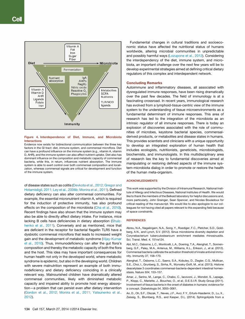
Figure 4. Interdependence of Diet, Immune, and Microbiota
InteractionsEvidence now exists for bidirectional communication between the three keyfactors in the GI tract: diet, immune system, and commensal microflora. Dietcan have a profound influence on the immune system (e.g., vitamin A, vitaminD, AHR), and the immune system can also affect nutrient uptake. Diet also hasdominant influence on the composition and metabolic capacity of commensalbacteria, while this, in return, influences nutrient absorption. The immunesystem is able to exert control over both commensal composition and locali-zation, whereas commensal signals are critical for development and functionof the immune system.
of disease states such as colitis (Devkota et al., 2012; Gregor and
Hotamisligil, 2011; Ley et al., 2006b; Monira et al., 2011). Defined
dietary deficiency can also alter commensal communities. For
example, the essential micronutrient vitamin A, which is required
for the induction of protective immunity, has also profound
effects on the composition of the microbiota (Cha et al., 2010).
Recent findings have also shown that the immune system may
also be able to directly affect dietary intake. For instance, mice
lacking B cells have deficiencies in dietary absorption (Shulz-
henko et al., 2011). Conversely and as mentioned, mice that
are deficient in the receptor for bacterial flagellin TLR5 have a
dysbiotic commensal microflora that leads to increased weight
gain and the development of metabolic syndrome (Vijay-Kumar
et al., 2010). Thus, immunodeficiency can alter the gut flora’s
composition and thereby themetabolic capacity of both the flora
and the host. This idea may have important consequences for
human health not only in the developed world, where metabolic
syndrome is epidemic, but also in the developing world. Children
with severe malnutrition represent an example of both immu-
nodeficiency and dietary deficiency coinciding in a clinically
relevant way. Malnourished children have dramatically altered
commensal communities, likely with diminished metabolic
capacity and impaired ability to promote host energy absorp-
tion—a problem that can persist even after dietary intervention
(Gordon et al., 2012; Monira et al., 2011; Yatsunenko et al.,
2012).
134 Cell 157, March 27, 2014 ª2014 Elsevier Inc.
Fundamental changes in cultural traditions and socioeco-
nomic status have affected the nutritional status of humans
worldwide, altering microbial communities in unpredictable
and possibly harmful ways (Lozupone et al., 2012). Considering
the interdependency of the diet, immune system, and micro-
biota, an important challenge over the next few years will be to
develop experimental strategies aimed at defining critical dietary
regulators of this complex and interdependent network.
Concluding RemarksAutoimmune and inflammatory diseases, all associated with
dysregulated immune responses, have been rising dramatically
over the past few decades. The field of immunology is at a
fascinating crossroad. In recent years, immunological research
has evolved from a lymphoid-tissue-centric view of the immune
system to the understanding of tissue microenvironments as a
fundamental determinant of immune responses. This area of
research has led to the integration of the microbiota as an
intrinsic regulator of all immune responses. There is today an
explosion of discoveries associated with the role of commu-
nities of microbes, keystone bacterial species, commensal-
derived products, or metabolites and disease states in humans.
This provides scientists and clinicians with a unique opportunity
to develop an integrated exploration of human health that
includes ecologists, nutritionists, geneticists, microbiologists,
biochemists, and immunologists. In this multidisciplinary area
of research lies the key to fundamental discoveries aimed at
manipulating or restoring defined aspects of the immune sys-
tem-microbiota dialog in order to promote or restore the health
of the human meta-organism.
ACKNOWLEDGMENTS
This work was supported by theDivision of Intramural Research, National Insti-
tute of Allergy and Infectious Diseases, National Institutes of Health. We would
like to thank themembers of the Belkaid laboratory for helpful discussions and,
more particularly, John Grainger, Sean Spencer, and Nicolas Bouladoux for
critical reading of the manuscript. We would like to also apologize to our col-
leagues for not having cited all papers relevant to this expanding field because
of space constraints.
REFERENCES
Abreu, N.A., Nagalingam, N.A., Song, Y., Roediger, F.C., Pletcher, S.D., Gold-
berg, A.N., and Lynch, S.V. (2012). Sinus microbiome diversity depletion and
Corynebacterium tuberculostearicum enrichment mediates rhinosinusitis.
Sci. Transl. Med. 4, 151ra124.
Abt, M.C., Osborne, L.C., Monticelli, L.A., Doering, T.A., Alenghat, T., Sonnen-
berg, G.F., Paley, M.A., Antenus, M., Williams, K.L., Erikson, J., et al. (2012).
Commensal bacteria calibrate the activation threshold of innate antiviral immu-
nity. Immunity 37, 158–170.
Alenghat, T., Osborne, L.C., Saenz, S.A., Kobuley, D., Ziegler, C.G., Mullican,
S.E., Choi, I., Grunberg, S., Sinha, R., Wynosky-Dolfi, M., et al. (2013). Histone
deacetylase 3 coordinates commensal-bacteria-dependent intestinal homeo-
stasis. Nature 504, 153–157.
Amar, J., Serino, M., Lange, C., Chabo, C., Iacovoni, J., Mondot, S., Lepage,
P., Klopp, C., Mariette, J., Bouchez, O., et al.; D.E.S.I.R. Study Group (2011).
Involvement of tissue bacteria in the onset of diabetes in humans: evidence for
a concept. Diabetologia 54, 3055–3061.
An, D., Oh, S.F., Olszak, T., Neves, J.F., Avci, F.Y., Erturk-Hasdemir, D., Lu, X.,
Zeissig, S., Blumberg, R.S., and Kasper, D.L. (2014). Sphingolipids from a

symbiotic microbe regulate homeostasis of host intestinal natural killer T cells.
Cell 156, 123–133.
Arpaia, N., Campbell, C., Fan, X., Dikiy, S., van der Veeken, J., deRoos, P., Liu,
H., Cross, J.R., Pfeffer, K., Coffer, P.J., andRudensky, A.Y. (2013). Metabolites
produced by commensal bacteria promote peripheral regulatory T-cell gener-
ation. Nature 504, 451–455.
Arthur, J.C., Perez-Chanona, E., Muhlbauer, M., Tomkovich, S., Uronis, J.M.,
Fan, T.J., Campbell, B.J., Abujamel, T., Dogan, B., Rogers, A.B., et al. (2012).
Intestinal inflammation targets cancer-inducing activity of the microbiota.
Science 338, 120–123.
Atarashi, K., Tanoue, T., Shima, T., Imaoka, A., Kuwahara, T., Momose, Y.,
Cheng, G., Yamasaki, S., Saito, T., Ohba, Y., et al. (2011). Induction of colonic
regulatory T cells by indigenous Clostridium species. Science 331, 337–341.
Atarashi, K., Tanoue, T., Oshima, K., Suda, W., Nagano, Y., Nishikawa, H.,
Fukuda, S., Saito, T., Narushima, S., Hase, K., et al. (2013). Treg induction
by a rationally selected mixture of Clostridia strains from the human micro-
biota. Nature 500, 232–236.
Atherton, J.C., and Blaser, M.J. (2009). Coadaptation of Helicobacter
pylori and humans: ancient history, modern implications. J. Clin. Invest. 119,
2475–2487.
Avila, M., Ojcius, D.M., and Yilmaz, O. (2009). The oral microbiota: living with
a permanent guest. DNA Cell Biol. 28, 405–411.
Bauer, H., Horowitz, R.E., Levenson, S.M., and Popper, H. (1963). The
response of the lymphatic tissue to the microbial flora. Studies on germfree
mice. Am. J. Pathol. 42, 471–483.
Beck, J.M., Young, V.B., and Huffnagle, G.B. (2012). The microbiome of the
lung. Transl. Res. 160, 258–266.
Belda-Ferre, P., Alcaraz, L.D., Cabrera-Rubio, R., Romero, H., Simon-Soro, A.,
Pignatelli, M., and Mira, A. (2012). The oral metagenome in health and disease.
ISME J. 6, 46–56.
Belkaid, Y., and Naik, S. (2013). Compartmentalized and systemic control of
tissue immunity by commensals. Nat. Immunol. 14, 646–653.
Benson, A., Pifer, R., Behrendt, C.L., Hooper, L.V., and Yarovinsky, F. (2009).
Gut commensal bacteria direct a protective immune response against Toxo-
plasma gondii. Cell Host Microbe 6, 187–196.
Berer, K., Mues,M., Koutrolos, M., Rasbi, Z.A., Boziki, M., Johner, C.,Wekerle,
H., and Krishnamoorthy, G. (2011). Commensal microbiota andmyelin autoan-
tigen cooperate to trigger autoimmune demyelination. Nature 479, 538–541.
Blainey, P.C., Milla, C.E., Cornfield, D.N., and Quake, S.R. (2012). Quantitative
analysis of the human airway microbial ecology reveals a pervasive signature
for cystic fibrosis. Sci. Transl. Med. 4, 153ra130.
Blaser, M.J., and Falkow, S. (2009). What are the consequences of the disap-
pearing human microbiota? Nat. Rev. Microbiol. 7, 887–894.
Bloom, S.M., Bijanki, V.N., Nava, G.M., Sun, L., Malvin, N.P., Donermeyer,
D.L., Dunne, W.M., Jr., Allen, P.M., and Stappenbeck, T.S. (2011). Commensal
Bacteroides species induce colitis in host-genotype-specific fashion in a
mouse model of inflammatory bowel disease. Cell Host Microbe 9, 390–403.
Bouladoux, N., Hall, J.A., Grainger, J.R., dos Santos, L.M., Kann, M.G., Nagar-
ajan, V., Verthelyi, D., and Belkaid, Y. (2012). Regulatory role of suppressive
motifs from commensal DNA. Mucosal Immunol. 5, 623–634.
Boullier, S., Tanguy, M., Kadaoui, K.A., Caubet, C., Sansonetti, P., Corthesy,
B., and Phalipon, A. (2009). Secretory IgA-mediated neutralization of Shigella
flexneri prevents intestinal tissue destruction by down-regulating inflammatory
circuits. J. Immunol. 183, 5879–5885.
Bouskra, D., Brezillon, C., Berard, M., Werts, C., Varona, R., Boneca, I.G., and
Eberl, G. (2008). Lymphoid tissue genesis induced by commensals through
NOD1 regulates intestinal homeostasis. Nature 456, 507–510.
Brandl, K., Plitas, G., Schnabl, B., DeMatteo, R.P., and Pamer, E.G. (2007).
MyD88-mediated signals induce the bactericidal lectin RegIII gamma and pro-
tect mice against intestinal Listeria monocytogenes infection. J. Exp. Med.
204, 1891–1900.
Brandl, K., Plitas, G., Mihu, C.N., Ubeda, C., Jia, T., Fleisher, M., Schnabl, B.,
DeMatteo, R.P., and Pamer, E.G. (2008). Vancomycin-resistant enterococci
exploit antibiotic-induced innate immune deficits. Nature 455, 804–807.
Brenchley, J.M., and Douek, D.C. (2012). Microbial translocation across the GI
tract. Annu. Rev. Immunol. 30, 149–173.
Brenchley, J.M., Price, D.A., Schacker, T.W., Asher, T.E., Silvestri, G., Rao, S.,
Kazzaz, Z., Bornstein, E., Lambotte, O., Altmann, D., et al. (2006). Microbial
translocation is a cause of systemic immune activation in chronic HIV infection.
Nat. Med. 12, 1365–1371.
Buffie, C.G., and Pamer, E.G. (2013). Microbiota-mediated colonization
resistance against intestinal pathogens. Nat. Rev. Immunol. 13, 790–801.
Cadwell, K., Patel, K.K., Maloney, N.S., Liu, T.C., Ng, A.C., Storer, C.E., Head,
R.D., Xavier, R., Stappenbeck, T.S., and Virgin, H.W. (2010). Virus-plus-
susceptibility gene interaction determines Crohn’s disease gene Atg16L1
phenotypes in intestine. Cell 141, 1135–1145.
Cahill, R.J., Foltz, C.J., Fox, J.G., Dangler, C.A., Powrie, F., and Schauer, D.B.
(1997). Inflammatory bowel disease: an immunity-mediated condition trig-
gered by bacterial infection with Helicobacter hepaticus. Infect. Immun. 65,
3126–3131.
Carvalho, F.A., Koren, O., Goodrich, J.K., Johansson, M.E., Nalbantoglu, I.,
Aitken, J.D., Su, Y., Chassaing, B., Walters, W.A., Gonzalez, A., et al. (2012).
Transient inability to manage proteobacteria promotes chronic gut inflamma-
tion in TLR5-deficient mice. Cell Host Microbe 12, 139–152.
Cash, H.L., Whitham, C.V., Behrendt, C.L., and Hooper, L.V. (2006). Symbiotic
bacteria direct expression of an intestinal bactericidal lectin. Science 313,
1126–1130.
Cebra, J.J. (1999). Influences of microbiota on intestinal immune system
development. Am. J. Clin. Nutr. 69, 1046S–1051S.
Cebula, A., Seweryn, M., Rempala, G.A., Pabla, S.S., McIndoe, R.A., Denning,
T.L., Bry, L., Kraj, P., Kisielow, P., and Ignatowicz, L. (2013). Thymus-derived
regulatory T cells contribute to tolerance to commensal microbiota. Nature
497, 258–262.
Cha, H.R., Chang, S.Y., Chang, J.H., Kim, J.O., Yang, J.Y., Kim, C.H., and
Kweon, M.N. (2010). Downregulation of Th17 cells in the small intestine
by disruption of gut flora in the absence of retinoic acid. J. Immunol. 184,
6799–6806.
Chassin, C., Kocur, M., Pott, J., Duerr, C.U., Gutle, D., Lotz, M., and Hornef,
M.W. (2010). miR-146a mediates protective innate immune tolerance in the
neonate intestine. Cell Host Microbe 8, 358–368.
Chieppa, M., Rescigno, M., Huang, A.Y., and Germain, R.N. (2006). Dynamic
imaging of dendritic cell extension into the small bowel lumen in response to
epithelial cell TLR engagement. J. Exp. Med. 203, 2841–2852.
Clarke, T.B., Davis, K.M., Lysenko, E.S., Zhou, A.Y., Yu, Y., and Weiser, J.N.
(2010). Recognition of peptidoglycan from the microbiota by Nod1 enhances
systemic innate immunity. Nat. Med. 16, 228–231.
Cong, Y., Feng, T., Fujihashi, K., Schoeb, T.R., and Elson, C.O. (2009). A domi-
nant, coordinated T regulatory cell-IgA response to the intestinal microbiota.
Proc. Natl. Acad. Sci. USA 106, 19256–19261.
Coombes, J.L., Siddiqui, K.R., Arancibia-Carcamo, C.V., Hall, J., Sun, C.M.,
Belkaid, Y., and Powrie, F. (2007). A functionally specialized population of
mucosal CD103+ DCs induces Foxp3+ regulatory T cells via a TGF-beta and
retinoic acid-dependent mechanism. J. Exp. Med. 204, 1757–1764.
Costello, E.K., Lauber, C.L., Hamady, M., Fierer, N., Gordon, J.I., and Knight,
R. (2009). Bacterial community variation in human body habitats across space
and time. Science 326, 1694–1697.
Craven, M., Egan, C.E., Dowd, S.E., McDonough, S.P., Dogan, B., Denkers,
E.Y., Bowman, D., Scherl, E.J., and Simpson, K.W. (2012). Inflammation drives
dysbiosis and bacterial invasion in murine models of ileal Crohn’s disease.
PLoS ONE 7, e41594.
Cummings, J.H., Pomare, E.W., Branch, W.J., Naylor, C.P., and Macfarlane,
G.T. (1987). Short chain fatty acids in human large intestine, portal, hepatic
and venous blood. Gut 28, 1221–1227.
Cell 157, March 27, 2014 ª2014 Elsevier Inc. 135
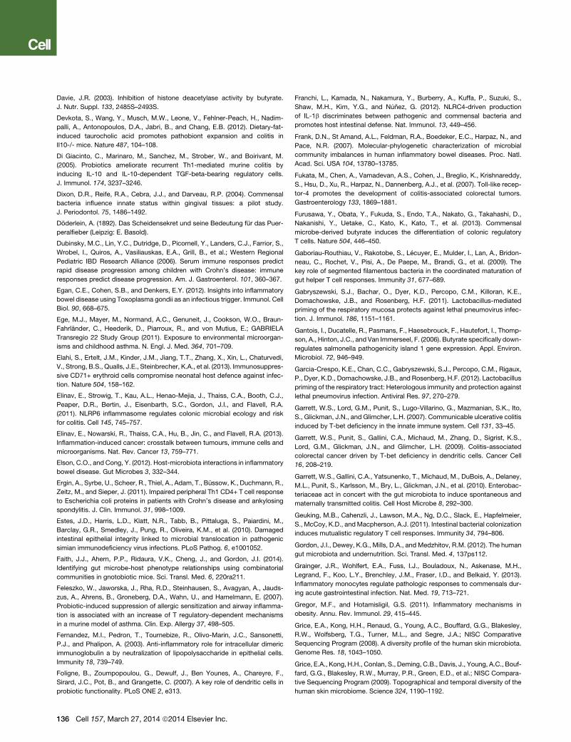
Davie, J.R. (2003). Inhibition of histone deacetylase activity by butyrate.
J. Nutr. Suppl. 133, 2485S–2493S.
Devkota, S., Wang, Y., Musch, M.W., Leone, V., Fehlner-Peach, H., Nadim-
palli, A., Antonopoulos, D.A., Jabri, B., and Chang, E.B. (2012). Dietary-fat-
induced taurocholic acid promotes pathobiont expansion and colitis in
Il10-/- mice. Nature 487, 104–108.
Di Giacinto, C., Marinaro, M., Sanchez, M., Strober, W., and Boirivant, M.
(2005). Probiotics ameliorate recurrent Th1-mediated murine colitis by
inducing IL-10 and IL-10-dependent TGF-beta-bearing regulatory cells.
J. Immunol. 174, 3237–3246.
Dixon, D.R., Reife, R.A., Cebra, J.J., and Darveau, R.P. (2004). Commensal
bacteria influence innate status within gingival tissues: a pilot study.
J. Periodontol. 75, 1486–1492.
Doderlein, A. (1892). Das Scheidensekret und seine Bedeutung fur das Puer-
peralfieber (Leipzig: E. Basold).
Dubinsky, M.C., Lin, Y.C., Dutridge, D., Picornell, Y., Landers, C.J., Farrior, S.,
Wrobel, I., Quiros, A., Vasiliauskas, E.A., Grill, B., et al.; Western Regional
Pediatric IBD Research Alliance (2006). Serum immune responses predict
rapid disease progression among children with Crohn’s disease: immune
responses predict disease progression. Am. J. Gastroenterol. 101, 360–367.
Egan, C.E., Cohen, S.B., and Denkers, E.Y. (2012). Insights into inflammatory
bowel disease using Toxoplasma gondii as an infectious trigger. Immunol. Cell
Biol. 90, 668–675.
Ege, M.J., Mayer, M., Normand, A.C., Genuneit, J., Cookson, W.O., Braun-
Fahrlander, C., Heederik, D., Piarroux, R., and von Mutius, E.; GABRIELA
Transregio 22 Study Group (2011). Exposure to environmental microorgan-
isms and childhood asthma. N. Engl. J. Med. 364, 701–709.
Elahi, S., Ertelt, J.M., Kinder, J.M., Jiang, T.T., Zhang, X., Xin, L., Chaturvedi,
V., Strong, B.S., Qualls, J.E., Steinbrecher, K.A., et al. (2013). Immunosuppres-
sive CD71+ erythroid cells compromise neonatal host defence against infec-
tion. Nature 504, 158–162.
Elinav, E., Strowig, T., Kau, A.L., Henao-Mejia, J., Thaiss, C.A., Booth, C.J.,
Peaper, D.R., Bertin, J., Eisenbarth, S.C., Gordon, J.I., and Flavell, R.A.
(2011). NLRP6 inflammasome regulates colonic microbial ecology and risk
for colitis. Cell 145, 745–757.
Elinav, E., Nowarski, R., Thaiss, C.A., Hu, B., Jin, C., and Flavell, R.A. (2013).
Inflammation-induced cancer: crosstalk between tumours, immune cells and
microorganisms. Nat. Rev. Cancer 13, 759–771.
Elson, C.O., and Cong, Y. (2012). Host-microbiota interactions in inflammatory
bowel disease. Gut Microbes 3, 332–344.
Ergin, A., Syrbe, U., Scheer, R., Thiel, A., Adam, T., Bussow, K., Duchmann, R.,
Zeitz, M., and Sieper, J. (2011). Impaired peripheral Th1 CD4+ T cell response
to Escherichia coli proteins in patients with Crohn’s disease and ankylosing
spondylitis. J. Clin. Immunol. 31, 998–1009.
Estes, J.D., Harris, L.D., Klatt, N.R., Tabb, B., Pittaluga, S., Paiardini, M.,
Barclay, G.R., Smedley, J., Pung, R., Oliveira, K.M., et al. (2010). Damaged
intestinal epithelial integrity linked to microbial translocation in pathogenic
simian immunodeficiency virus infections. PLoS Pathog. 6, e1001052.
Faith, J.J., Ahern, P.P., Ridaura, V.K., Cheng, J., and Gordon, J.I. (2014).
Identifying gut microbe-host phenotype relationships using combinatorial
communities in gnotobiotic mice. Sci. Transl. Med. 6, 220ra211.
Feleszko, W., Jaworska, J., Rha, R.D., Steinhausen, S., Avagyan, A., Jauds-
zus, A., Ahrens, B., Groneberg, D.A., Wahn, U., and Hamelmann, E. (2007).
Probiotic-induced suppression of allergic sensitization and airway inflamma-
tion is associated with an increase of T regulatory-dependent mechanisms
in a murine model of asthma. Clin. Exp. Allergy 37, 498–505.
Fernandez, M.I., Pedron, T., Tournebize, R., Olivo-Marin, J.C., Sansonetti,
P.J., and Phalipon, A. (2003). Anti-inflammatory role for intracellular dimeric
immunoglobulin a by neutralization of lipopolysaccharide in epithelial cells.
Immunity 18, 739–749.
Foligne, B., Zoumpopoulou, G., Dewulf, J., Ben Younes, A., Chareyre, F.,
Sirard, J.C., Pot, B., and Grangette, C. (2007). A key role of dendritic cells in
probiotic functionality. PLoS ONE 2, e313.
136 Cell 157, March 27, 2014 ª2014 Elsevier Inc.
Franchi, L., Kamada, N., Nakamura, Y., Burberry, A., Kuffa, P., Suzuki, S.,
Shaw, M.H., Kim, Y.G., and Nunez, G. (2012). NLRC4-driven production
of IL-1b discriminates between pathogenic and commensal bacteria and
promotes host intestinal defense. Nat. Immunol. 13, 449–456.
Frank, D.N., St Amand, A.L., Feldman, R.A., Boedeker, E.C., Harpaz, N., and
Pace, N.R. (2007). Molecular-phylogenetic characterization of microbial
community imbalances in human inflammatory bowel diseases. Proc. Natl.
Acad. Sci. USA 104, 13780–13785.
Fukata, M., Chen, A., Vamadevan, A.S., Cohen, J., Breglio, K., Krishnareddy,
S., Hsu, D., Xu, R., Harpaz, N., Dannenberg, A.J., et al. (2007). Toll-like recep-
tor-4 promotes the development of colitis-associated colorectal tumors.
Gastroenterology 133, 1869–1881.
Furusawa, Y., Obata, Y., Fukuda, S., Endo, T.A., Nakato, G., Takahashi, D.,
Nakanishi, Y., Uetake, C., Kato, K., Kato, T., et al. (2013). Commensal
microbe-derived butyrate induces the differentiation of colonic regulatory
T cells. Nature 504, 446–450.
Gaboriau-Routhiau, V., Rakotobe, S., Lecuyer, E., Mulder, I., Lan, A., Bridon-
neau, C., Rochet, V., Pisi, A., De Paepe, M., Brandi, G., et al. (2009). The
key role of segmented filamentous bacteria in the coordinated maturation of
gut helper T cell responses. Immunity 31, 677–689.
Gabryszewski, S.J., Bachar, O., Dyer, K.D., Percopo, C.M., Killoran, K.E.,
Domachowske, J.B., and Rosenberg, H.F. (2011). Lactobacillus-mediated
priming of the respiratory mucosa protects against lethal pneumovirus infec-
tion. J. Immunol. 186, 1151–1161.
Gantois, I., Ducatelle, R., Pasmans, F., Haesebrouck, F., Hautefort, I., Thomp-
son, A., Hinton, J.C., and Van Immerseel, F. (2006). Butyrate specifically down-
regulates salmonella pathogenicity island 1 gene expression. Appl. Environ.
Microbiol. 72, 946–949.
Garcia-Crespo, K.E., Chan, C.C., Gabryszewski, S.J., Percopo, C.M., Rigaux,
P., Dyer, K.D., Domachowske, J.B., and Rosenberg, H.F. (2012). Lactobacillus
priming of the respiratory tract: Heterologous immunity and protection against
lethal pneumovirus infection. Antiviral Res. 97, 270–279.
Garrett, W.S., Lord, G.M., Punit, S., Lugo-Villarino, G., Mazmanian, S.K., Ito,
S., Glickman, J.N., and Glimcher, L.H. (2007). Communicable ulcerative colitis
induced by T-bet deficiency in the innate immune system. Cell 131, 33–45.
Garrett, W.S., Punit, S., Gallini, C.A., Michaud, M., Zhang, D., Sigrist, K.S.,
Lord, G.M., Glickman, J.N., and Glimcher, L.H. (2009). Colitis-associated
colorectal cancer driven by T-bet deficiency in dendritic cells. Cancer Cell
16, 208–219.
Garrett, W.S., Gallini, C.A., Yatsunenko, T., Michaud, M., DuBois, A., Delaney,
M.L., Punit, S., Karlsson, M., Bry, L., Glickman, J.N., et al. (2010). Enterobac-
teriaceae act in concert with the gut microbiota to induce spontaneous and
maternally transmitted colitis. Cell Host Microbe 8, 292–300.
Geuking, M.B., Cahenzli, J., Lawson, M.A., Ng, D.C., Slack, E., Hapfelmeier,
S., McCoy, K.D., andMacpherson, A.J. (2011). Intestinal bacterial colonization
induces mutualistic regulatory T cell responses. Immunity 34, 794–806.
Gordon, J.I., Dewey, K.G., Mills, D.A., and Medzhitov, R.M. (2012). The human
gut microbiota and undernutrition. Sci. Transl. Med. 4, 137ps112.
Grainger, J.R., Wohlfert, E.A., Fuss, I.J., Bouladoux, N., Askenase, M.H.,
Legrand, F., Koo, L.Y., Brenchley, J.M., Fraser, I.D., and Belkaid, Y. (2013).
Inflammatory monocytes regulate pathologic responses to commensals dur-
ing acute gastrointestinal infection. Nat. Med. 19, 713–721.
Gregor, M.F., and Hotamisligil, G.S. (2011). Inflammatory mechanisms in
obesity. Annu. Rev. Immunol. 29, 415–445.
Grice, E.A., Kong, H.H., Renaud, G., Young, A.C., Bouffard, G.G., Blakesley,
R.W., Wolfsberg, T.G., Turner, M.L., and Segre, J.A.; NISC Comparative
Sequencing Program (2008). A diversity profile of the human skin microbiota.
Genome Res. 18, 1043–1050.
Grice, E.A., Kong, H.H., Conlan, S., Deming, C.B., Davis, J., Young, A.C., Bouf-
fard, G.G., Blakesley, R.W., Murray, P.R., Green, E.D., et al.; NISC Compara-
tive Sequencing Program (2009). Topographical and temporal diversity of the
human skin microbiome. Science 324, 1190–1192.

Grivennikov, S.I., and Karin, M. (2010). Dangerous liaisons: STAT3 and
NF-kappaB collaboration and crosstalk in cancer. Cytokine Growth Factor
Rev. 21, 11–19.
Grivennikov, S.I., Wang, K., Mucida, D., Stewart, C.A., Schnabl, B., Jauch, D.,
Taniguchi, K., Yu, G.Y., Osterreicher, C.H., Hung, K.E., et al. (2012). Adenoma-
linked barrier defects and microbial products drive IL-23/IL-17-mediated
tumour growth. Nature 491, 254–258.
Gupta, N., Limbago, B.M., Patel, J.B., and Kallen, A.J. (2011). Carbapenem-
resistant Enterobacteriaceae: Epidemiology and prevention. Clin. Infect. Dis.
53, 60–67.
Haas, A., Zimmermann, K., Graw, F., Slack, E., Rusert, P., Ledergerber, B.,
Bossart, W., Weber, R., Thurnheer, M.C., Battegay, M., et al.; Swiss HIV
Cohort Study (2011). Systemic antibody responses to gut commensal bacteria
during chronic HIV-1 infection. Gut 60, 1506–1519.
Hall, J.A., Bouladoux, N., Sun, C.M.,Wohlfert, E.A., Blank, R.B., Zhu, Q., Grigg,
M.E., Berzofsky, J.A., and Belkaid, Y. (2008). Commensal DNA limits regulato-
ry T cell conversion and is a natural adjuvant of intestinal immune responses.
Immunity 29, 637–649.
Hall, J.A., Cannons, J.L., Grainger, J.R., Dos Santos, L.M., Hand, T.W., Naik,
S., Wohlfert, E.A., Chou, D.B., Oldenhove, G., Robinson, M., et al. (2011a).
Essential role for retinoic acid in the promotion of CD4(+) T cell effector
responses via retinoic acid receptor alpha. Immunity 34, 435–447.
Hall, J.A., Grainger, J.R., Spencer, S.P., and Belkaid, Y. (2011b). The role of
retinoic acid in tolerance and immunity. Immunity 35, 13–22.
Hamada, H., Hiroi, T., Nishiyama, Y., Takahashi, H., Masunaga, Y., Hachimura,
S., Kaminogawa, S., Takahashi-Iwanaga, H., Iwanaga, T., Kiyono, H., et al.
(2002). Identification of multiple isolated lymphoid follicles on the antimesen-
teric wall of the mouse small intestine. J. Immunol. 168, 57–64.
Hammami, R., Fernandez, B., Lacroix, C., and Fliss, I. (2013). Anti-infective
properties of bacteriocins: an update. Cell. Mol. Life Sci. 70, 2947–2967.
Hand, T.W., Dos Santos, L.M., Bouladoux, N., Molloy, M.J., Pagan, A.J., Pep-
per, M., Maynard, C.L., Elson, C.O., 3rd, and Belkaid, Y. (2012). Acute gastro-
intestinal infection induces long-lived microbiota-specific T cell responses.
Science 337, 1553–1556.
Hapfelmeier, S., Lawson, M.A., Slack, E., Kirundi, J.K., Stoel, M., Heiken-
walder, M., Cahenzli, J., Velykoredko, Y., Balmer, M.L., Endt, K., et al.
(2010). Reversible microbial colonization of germ-free mice reveals the
dynamics of IgA immune responses. Science 328, 1705–1709.
Hayes, K.S., Bancroft, A.J., Goldrick, M., Portsmouth, C., Roberts, I.S., and
Grencis, R.K. (2010). Exploitation of the intestinal microflora by the parasitic
nematode Trichuris muris. Science 328, 1391–1394.
Heimesaat, M.M., Bereswill, S., Fischer, A., Fuchs, D., Struck, D., Niebergall,
J., Jahn, H.K., Dunay, I.R., Moter, A., Gescher, D.M., et al. (2006). Gram-nega-
tive bacteria aggravate murine small intestinal Th1-type immunopathology
following oral infection with Toxoplasma gondii. J. Immunol. 177, 8785–8795.
Herbst, T., Sichelstiel, A., Schar, C., Yadava, K., Burki, K., Cahenzli, J., McCoy,
K., Marsland, B.J., and Harris, N.L. (2011). Dysregulation of allergic airway
inflammation in the absence of microbial colonization. Am. J. Respir. Crit.
Care Med. 184, 198–205.
Hilty, M., Burke, C., Pedro, H., Cardenas, P., Bush, A., Bossley, C., Davies, J.,
Ervine, A., Poulter, L., Pachter, L., et al. (2010). Disordered microbial commu-
nities in asthmatic airways. PLoS ONE 5, e8578.
Homann, D., Teyton, L., and Oldstone, M.B. (2001). Differential regulation of
antiviral T-cell immunity results in stable CD8+ but declining CD4+ T-cell mem-
ory. Nat. Med. 7, 913–919.
Hooper, L.V., and Macpherson, A.J. (2010). Immune adaptations that maintain
homeostasis with the intestinal microbiota. Nat. Rev. Immunol. 10, 159–169.
Hooper, L.V., Wong, M.H., Thelin, A., Hansson, L., Falk, P.G., and Gordon, J.I.
(2001). Molecular analysis of commensal host-microbial relationships in the
intestine. Science 291, 881–884.
Hornquist, E., and Lycke, N. (1995). Cholera toxin increases T lymphocyte
responses to unrelated antigens. Adv. Exp. Med. Biol. 371B, 1507–1512.
Hu, B., Elinav, E., Huber, S., Strowig, T., Hao, L., Hafemann, A., Jin, C.,
Wunderlich, C., Wunderlich, T., Eisenbarth, S.C., and Flavell, R.A. (2013).
Microbiota-induced activation of epithelial IL-6 signaling links inflamma-
some-driven inflammation with transmissible cancer. Proc. Natl. Acad. Sci.
USA 110, 9862–9867.
Ichinohe, T., Pang, I.K., Kumamoto, Y., Peaper, D.R., Ho, J.H., Murray, T.S.,
and Iwasaki, A. (2011). Microbiota regulates immune defense against respira-
tory tract influenza A virus infection. Proc. Natl. Acad. Sci. USA 108, 5354–
5359.
Iida, N., Dzutsev, A., Stewart, C.A., Smith, L., Bouladoux, N.,Weingarten, R.A.,
Molina, D.A., Salcedo, R., Back, T., Cramer, S., et al. (2013). Commensal
bacteria control cancer response to therapy bymodulating the tumormicroen-
vironment. Science 342, 967–970.
Iliev, I.D., Funari, V.A., Taylor, K.D., Nguyen, Q., Reyes, C.N., Strom, S.P.,
Brown, J., Becker, C.A., Fleshner, P.R., Dubinsky, M., et al. (2012). Interactions
between commensal fungi and the C-type lectin receptor Dectin-1 influence
colitis. Science 336, 1314–1317.
Ismail, A.S., Severson, K.M., Vaishnava, S., Behrendt, C.L., Yu, X., Benjamin,
J.L., Ruhn, K.A., Hou, B., DeFranco, A.L., Yarovinsky, F., and Hooper, L.V.
(2011). Gammadelta intraepithelial lymphocytes are essential mediators
of host-microbial homeostasis at the intestinal mucosal surface. Proc. Natl.
Acad. Sci. USA 108, 8743–8748.
Issa, M., Ananthakrishnan, A.N., and Binion, D.G. (2008). Clostridium difficile
and inflammatory bowel disease. Inflamm. Bowel Dis. 14, 1432–1442.
Ivanov, I.I., Frutos, Rde.L., Manel, N., Yoshinaga, K., Rifkin, D.B., Sartor, R.B.,
Finlay, B.B., and Littman, D.R. (2008). Specific microbiota direct the differen-
tiation of IL-17-producing T-helper cells in the mucosa of the small intestine.
Cell Host Microbe 4, 337–349.
Ivanov, I.I., Atarashi, K., Manel, N., Brodie, E.L., Shima, T., Karaoz, U., Wei, D.,
Goldfarb, K.C., Santee, C.A., Lynch, S.V., et al. (2009). Induction of intestinal
Th17 cells by segmented filamentous bacteria. Cell 139, 485–498.
Iwase, T., Uehara, Y., Shinji, H., Tajima, A., Seo, H., Takada, K., Agata, T., and
Mizunoe, Y. (2010). Staphylococcus epidermidis Esp inhibits Staphylococcus
aureus biofilm formation and nasal colonization. Nature 465, 346–349.
Jayashree, B., Bibin, Y.S., Prabhu, D., Shanthirani, C.S., Gokulakrishnan, K.,
Lakshmi, B.S., Mohan, V., and Balasubramanyam, M. (2013). Increased circu-
latory levels of lipopolysaccharide (LPS) and zonulin signify novel biomarkers
of proinflammation in patients with type 2 diabetes. Mol. Cell. Biochem. 388,
2033–210.
Johnston, C.J., McSorley, H.J., Anderton, S.M., Wigmore, S.J., and Maizels,
R.M. (2014). Helminths and immunological tolerance. Transplantation 97,
127–132.
Josefowicz, S.Z., Niec, R.E., Kim, H.Y., Treuting, P., Chinen, T., Zheng, Y.,
Umetsu, D.T., and Rudensky, A.Y. (2012a). Extrathymically generated regula-
tory T cells control mucosal TH2 inflammation. Nature 482, 395–399.
Jung, C., Meinzer, U., Montcuquet, N., Thachil, E., Chateau, D., Thiebaut, R.,
Roy, M., Alnabhani, Z., Berrebi, D., Dussaillant, M., et al. (2012). Yersinia
pseudotuberculosis disrupts intestinal barrier integrity through hematopoietic
TLR-2 signaling. J. Clin. Invest. 122, 2239–2251.
Kamada, N., Chen, G.Y., Inohara, N., and Nunez, G. (2013). Control of patho-
gens and pathobionts by the gut microbiota. Nat. Immunol. 14, 685–690.
Kane, M., Case, L.K., Kopaskie, K., Kozlova, A., MacDearmid, C., Chervonsky,
A.V., and Golovkina, T.V. (2011). Successful transmission of a retrovirus
depends on the commensal microbiota. Science 334, 245–249.
Karimi, K., Inman,M.D., Bienenstock, J., and Forsythe, P. (2009). Lactobacillus
reuteri-induced regulatory T cells protect against an allergic airway response
in mice. Am. J. Respir. Crit. Care Med. 179, 186–193.
Karlsson, F.H., Tremaroli, V., Nookaew, I., Bergstrom, G., Behre, C.J., Fager-
berg, B., Nielsen, J., and Backhed, F. (2013). Gut metagenome in European
women with normal, impaired and diabetic glucose control. Nature 498,
99–103.
Cell 157, March 27, 2014 ª2014 Elsevier Inc. 137

Kelsall, B. (2008). Recent progress in understanding the phenotype and
function of intestinal dendritic cells and macrophages. Mucosal Immunol. 1,
460–469.
Kinnebrew, M.A., Buffie, C.G., Diehl, G.E., Zenewicz, L.A., Leiner, I., Hohl,
T.M., Flavell, R.A., Littman, D.R., and Pamer, E.G. (2012). Interleukin 23 pro-
duction by intestinal CD103(+)CD11b(+) dendritic cells in response to bacterial
flagellin enhances mucosal innate immune defense. Immunity 36, 276–287.
Kiss, E.A., Vonarbourg, C., Kopfmann, S., Hobeika, E., Finke, D., Esser, C., and
Diefenbach, A. (2011). Natural aryl hydrocarbon receptor ligands control
organogenesis of intestinal lymphoid follicles. Science 334, 1561–1565.
Kiyono, H., McGhee, J.R., Wannemuehler, M.J., and Michalek, S.M. (1982).
Lack of oral tolerance in C3H/HeJ mice. J. Exp. Med. 155, 605–610.
Klatt, N.R., Funderburg, N.T., and Brenchley, J.M. (2013). Microbial transloca-
tion, immune activation, and HIV disease. Trends Microbiol. 21, 6–13.
Klebanoff, C.A., Spencer, S.P., Torabi-Parizi, P., Grainger, J.R., Roychoudhuri,
R., Ji, Y., Sukumar, M., Muranski, P., Scott, C.D., Hall, J.A., et al. (2013). Ret-
inoic acid controls the homeostasis of pre-cDC-derived splenic and intestinal
dendritic cells. J. Exp. Med. 210, 1961–1976.
Kollmann, T.R., Levy, O., Montgomery, R.R., and Goriely, S. (2012). Innate
immune function by Toll-like receptors: distinct responses in newborns and
the elderly. Immunity 37, 771–783.
Kong, H.H., Oh, J., Deming, C., Conlan, S., Grice, E.A., Beatson, M.A., Nomi-
cos, E., Polley, E.C., Komarow, H.D., Murray, P.R., et al.; NISC Comparative
Sequence Program (2012). Temporal shifts in the skin microbiome associated
with disease flares and treatment in children with atopic dermatitis. Genome
Res. 22, 850–859.
Korpe, P.S., and Petri, W.A., Jr. (2012). Environmental enteropathy: critical im-
plications of a poorly understood condition. Trends Mol. Med. 18, 328–336.
Kosek, M., Bern, C., and Guerrant, R.L. (2003). The global burden of diarrhoeal
disease, as estimated from studies published between 1992 and 2000. Bull.
World Health Organ. 81, 197–204.
Kostic, A.D., Gevers, D., Pedamallu, C.S., Michaud, M., Duke, F., Earl, A.M.,
Ojesina, A.I., Jung, J., Bass, A.J., Tabernero, J., et al. (2012). Genomic analysis
identifies association of Fusobacterium with colorectal carcinoma. Genome
Res. 22, 292–298.
Kostic, A.D., Chun, E., Robertson, L., Glickman, J.N., Gallini, C.A., Michaud,
M., Clancy, T.E., Chung, D.C., Lochhead, P., Hold, G.L., et al. (2013). Fusobac-
terium nucleatum potentiates intestinal tumorigenesis and modulates the
tumor-immune microenvironment. Cell Host Microbe 14, 207–215.
Kriegel, M.A., Sefik, E., Hill, J.A., Wu, H.J., Benoist, C., and Mathis, D. (2011).
Naturally transmitted segmented filamentous bacteria segregatewith diabetes
protection in nonobese diabetic mice. Proc. Natl. Acad. Sci. USA 108, 11548–
11553.
Kullberg, M.C., Jankovic, D., Gorelick, P.L., Caspar, P., Letterio, J.J., Cheever,
A.W., and Sher, A. (2002). Bacteria-triggered CD4(+) T regulatory cells sup-
press Helicobacter hepaticus-induced colitis. J. Exp. Med. 196, 505–515.
Kuss, S.K., Best, G.T., Etheredge,C.A., Pruijssers, A.J., Frierson, J.M., Hooper,
L.V., Dermody, T.S., and Pfeiffer, J.K. (2011). Intestinal microbiota promote
enteric virus replication and systemic pathogenesis. Science 334, 249–252.
Lai, Y., Di Nardo, A., Nakatsuji, T., Leichtle, A., Yang, Y., Cogen, A.L., Wu, Z.R.,
Hooper, L.V., Schmidt, R.R., von Aulock, S., et al. (2009). Commensal bacteria
regulate Toll-like receptor 3-dependent inflammation after skin injury. Nat.
Med. 15, 1377–1382.
Lamps, L.W., Madhusudhan, K.T., Havens, J.M., Greenson, J.K., Bronner,
M.P., Chiles, M.C., Dean, P.J., and Scott, M.A. (2003). Pathogenic Yersinia
DNA is detected in bowel and mesenteric lymph nodes from patients with
Crohn’s disease. Am. J. Surg. Pathol. 27, 220–227.
Lathrop, S.K., Bloom, S.M., Rao, S.M., Nutsch, K., Lio, C.W., Santacruz, N.,
Peterson, D.A., Stappenbeck, T.S., and Hsieh, C.S. (2011). Peripheral educa-
tion of the immune system by colonic commensal microbiota. Nature 478,
250–254.
138 Cell 157, March 27, 2014 ª2014 Elsevier Inc.
Lee, Y.K., Menezes, J.S., Umesaki, Y., andMazmanian, S.K. (2011). Proinflam-
matory T-cell responses to gut microbiota promote experimental autoimmune
encephalomyelitis. Proc. Natl. Acad. Sci. USA 108 (Suppl 1), 4615–4622.
Ley, R.E., Peterson, D.A., and Gordon, J.I. (2006a). Ecological and evolu-
tionary forces shaping microbial diversity in the human intestine. Cell 124,
837–848.
Ley, R.E., Turnbaugh, P.J., Klein, S., and Gordon, J.I. (2006b). Microbial ecol-
ogy: human gut microbes associated with obesity. Nature 444, 1022–1023.
Li, Y., Innocentin, S., Withers, D.R., Roberts, N.A., Gallagher, A.R., Grigorieva,
E.F., Wilhelm, C., and Veldhoen, M. (2011). Exogenous stimuli maintain
intraepithelial lymphocytes via aryl hydrocarbon receptor activation. Cell
147, 629–640.
Lodes, M.J., Cong, Y., Elson, C.O., Mohamath, R., Landers, C.J., Targan, S.R.,
Fort, M., and Hershberg, R.M. (2004). Bacterial flagellin is a dominant antigen
in Crohn disease. J. Clin. Invest. 113, 1296–1306.
Lotz, M., Gutle, D., Walther, S., Menard, S., Bogdan, C., and Hornef, M.W.
(2006). Postnatal acquisition of endotoxin tolerance in intestinal epithelial cells.
J. Exp. Med. 203, 973–984.
Lowe, E.L., Crother, T.R., Rabizadeh, S., Hu, B., Wang, H., Chen, S., Shimada,
K., Wong, M.H., Michelsen, K.S., and Arditi, M. (2010). Toll-like receptor 2
signaling protects mice from tumor development in a mouse model of coli-
tis-induced cancer. PLoS ONE 5, e13027.
Lozupone, C.A., Stombaugh, J.I., Gordon, J.I., Jansson, J.K., and Knight, R.
(2012). Diversity, stability and resilience of the human gut microbiota. Nature
489, 220–230.
Lupp, C., Robertson, M.L., Wickham, M.E., Sekirov, I., Champion, O.L., Gay-
nor, E.C., and Finlay, B.B. (2007). Host-mediated inflammation disrupts the
intestinal microbiota and promotes the overgrowth of Enterobacteriaceae.
Cell Host Microbe 2, 204.
Macpherson, A.J., and Harris, N.L. (2004). Interactions between commensal
intestinal bacteria and the immune system. Nat. Rev. Immunol. 4, 478–485.
Macpherson, A.J., and Uhr, T. (2004). Induction of protective IgA by intestinal
dendritic cells carrying commensal bacteria. Science 303, 1662–1665.
Macpherson, A., Khoo, U.Y., Forgacs, I., Philpott-Howard, J., and Bjarnason, I.
(1996). Mucosal antibodies in inflammatory bowel disease are directed against
intestinal bacteria. Gut 38, 365–375.
Macpherson, A.J., Hunziker, L., McCoy, K., and Lamarre, A. (2001). IgA
responses in the intestinal mucosa against pathogenic and non-pathogenic
microorganisms. Microbes Infect. 3, 1021–1035.
Macpherson, A.J., Slack, E., Geuking, M.B., and McCoy, K.D. (2009). The
mucosal firewalls against commensal intestinal microbes. Semin. Immunopa-
thol. 31, 145–149.
Maloy, K.J., and Powrie, F. (2011). Intestinal homeostasis and its breakdown in
inflammatory bowel disease. Nature 474, 298–306.
Marcobal, A., and Sonnenburg, J.L. (2012). Human milk oligosaccharide con-
sumption by intestinal microbiota. Clin. Microbiol. Infect. 18 (Suppl 4), 12–15.
Marcobal, A., Barboza, M., Froehlich, J.W., Block, D.E., German, J.B., Lebrilla,
C.B., and Mills, D.A. (2010). Consumption of human milk oligosaccharides by
gut-related microbes. J. Agric. Food Chem. 58, 5334–5340.
Markle, J.G., Frank, D.N., Mortin-Toth, S., Robertson, C.E., Feazel, L.M.,
Rolle-Kampczyk, U., von Bergen, M., McCoy, K.D., Macpherson, A.J., and
Danska, J.S. (2013). Sex differences in the gut microbiome drive hormone-
dependent regulation of autoimmunity. Science 339, 1084–1088.
Marshall, B.J., andWarren, J.R. (1984). Unidentified curved bacilli in the stom-
ach of patients with gastritis and peptic ulceration. Lancet 1, 1311–1315.
Martinoli, C., Chiavelli, A., and Rescigno, M. (2007). Entry route of Salmonella
typhimurium directs the type of induced immune response. Immunity 27,
975–984.
Maslowski, K.M., Vieira, A.T., Ng, A., Kranich, J., Sierro, F., Yu, D., Schilter,
H.C., Rolph,M.S.,Mackay, F., Artis, D., et al. (2009).Regulationof inflammatory
responses by gut microbiota and chemoattractant receptor GPR43. Nature
461, 1282–1286.

Mazmanian, S.K., Liu, C.H., Tzianabos, A.O., and Kasper, D.L. (2005). An
immunomodulatory molecule of symbiotic bacteria directs maturation of the
host immune system. Cell 122, 107–118.
Mazmanian, S.K., Round, J.L., and Kasper, D.L. (2008). A microbial symbiosis
factor prevents intestinal inflammatory disease. Nature 453, 620–625.
McGuckin, M.A., Linden, S.K., Sutton, P., and Florin, T.H. (2011). Mucin
dynamics and enteric pathogens. Nat. Rev. Microbiol. 9, 265–278.
Meijer, K., de Vos, P., and Priebe, M.G. (2010). Butyrate and other short-chain
fatty acids as modulators of immunity: what relevance for health? Curr. Opin.
Clin. Nutr. Metab. Care 13, 715–721.
Meinzer, U., Barreau, F., Esmiol-Welterlin, S., Jung, C., Villard, C., Leger, T.,
Ben-Mkaddem, S., Berrebi, D., Dussaillant, M., Alnabhani, Z., et al. (2012).
Yersinia pseudotuberculosis effector YopJ subverts the Nod2/RICK/TAK1
pathway and activates caspase-1 to induce intestinal barrier dysfunction.
Cell Host Microbe 11, 337–351.
Molloy, M.J., Bouladoux, N., and Belkaid, Y. (2012). Intestinal microbiota:
shaping local and systemic immune responses. Semin. Immunol. 24, 58–66.
Molloy, M.J., Grainger, J.R., Bouladoux, N., Hand, T.W., Koo, L.Y., Naik, S.,
Quinones, M., Dzutsev, A.K., Gao, J.L., Trinchieri, G., et al. (2013). Intraluminal
containment of commensal outgrowth in the gut during infection-induced
dysbiosis. Cell Host Microbe 14, 318–328.
Monira, S., Nakamura, S., Gotoh, K., Izutsu, K., Watanabe, H., Alam, N.H.,
Endtz, H.P., Cravioto, A., Ali, S.I., Nakaya, T., et al. (2011). Gut microbiota of
healthy and malnourished children in bangladesh. Front. Microbiol. 2, 228.
Mora, J.R., Iwata, M., and von Andrian, U.H. (2008). Vitamin effects on the
immune system: vitamins A and D take centre stage. Nat. Rev. Immunol. 8,
685–698.
Mucida, D., Kutchukhidze, N., Erazo, A., Russo, M., Lafaille, J.J., and Curotto
de Lafaille, M.A. (2005). Oral tolerance in the absence of naturally occurring
Tregs. J. Clin. Invest. 115, 1923–1933.
Mucida, D., Park, Y., Kim, G., Turovskaya, O., Scott, I., Kronenberg, M., and
Cheroutre, H. (2007). Reciprocal TH17 and regulatory T cell differentiation
mediated by retinoic acid. Science 317, 256–260.
Mukherjee, S., Partch, C.L., Lehotzky, R.E., Whitham, C.V., Chu, H., Bevins,
C.L., Gardner, K.H., and Hooper, L.V. (2009). Regulation of C-type lectin
antimicrobial activity by a flexible N-terminal prosegment. J. Biol. Chem.
284, 4881–4888.
Murray, B.E. (2000). Vancomycin-resistant enterococcal infections. N. Engl. J.
Med. 342, 710–721.
Naik, S., Bouladoux, N., Wilhelm, C., Molloy, M.J., Salcedo, R., Kastenmuller,
W., Deming, C., Quinones, M., Koo, L., Conlan, S., et al. (2012). Compartmen-
talized control of skin immunity by resident commensals. Science 337, 1115–
1119.
Ng, K.M., Ferreyra, J.A., Higginbottom, S.K., Lynch, J.B., Kashyap, P.C.,
Gopinath, S., Naidu, N., Choudhury, B., Weimer, B.C., Monack, D.M., and
Sonnenburg, J.L. (2013). Microbiota-liberated host sugars facilitate post-anti-
biotic expansion of enteric pathogens. Nature 502, 96–99.
Niess, J.H., and Adler, G. (2010). Enteric flora expands gut lamina propria
CX3CR1+ dendritic cells supporting inflammatory immune responses under
normal and inflammatory conditions. J. Immunol. 184, 2026–2037.
O’Mahony, C., Scully, P., O’Mahony, D., Murphy, S., O’Brien, F., Lyons, A.,
Sherlock, G., MacSharry, J., Kiely, B., Shanahan, F., and O’Mahony, L.
(2008). Commensal-induced regulatory T cells mediate protection against
pathogen-stimulated NF-kappaB activation. PLoS Pathog. 4, e1000112.
Ochoa-Reparaz, J., Mielcarz, D.W., Ditrio, L.E., Burroughs, A.R., Foureau,
D.M., Haque-Begum, S., and Kasper, L.H. (2009). Role of gut commensal
microflora in the development of experimental autoimmune encephalomy-
elitis. J. Immunol. 183, 6041–6050.
Ochoa-Reparaz, J., Mielcarz, D.W., Ditrio, L.E., Burroughs, A.R., Begum-
Haque, S., Dasgupta, S., Kasper, D.L., and Kasper, L.H. (2010a). Central
nervous system demyelinating disease protection by the human commensal
Bacteroides fragilis depends on polysaccharide A expression. J. Immunol.
185, 4101–4108.
Ochoa-Reparaz, J., Mielcarz, D.W., Wang, Y., Begum-Haque, S., Dasgupta,
S., Kasper, D.L., and Kasper, L.H. (2010b). A polysaccharide from the human
commensal Bacteroides fragilis protects against CNS demyelinating disease.
Mucosal Immunol. 3, 487–495.
Ohnmacht, C., Marques, R., Presley, L., Sawa, S., Lochner, M., and Eberl, G.
(2011). Intestinal microbiota, evolution of the immune system and the bad
reputation of pro-inflammatory immunity. Cell. Microbiol. 13, 653–659.
Olszak, T., An, D., Zeissig, S., Vera, M.P., Richter, J., Franke, A., Glickman,
J.N., Siebert, R., Baron, R.M., Kasper, D.L., and Blumberg, R.S. (2012). Micro-
bial exposure during early life has persistent effects on natural killer T cell func-
tion. Science 336, 489–493.
Osborn, O., and Olefsky, J.M. (2012). The cellular and signaling networks link-
ing the immune system and metabolism in disease. Nat. Med. 18, 363–374.
Pacheco, A.R., Curtis, M.M., Ritchie, J.M., Munera, D., Waldor, M.K., Moreira,
C.G., and Sperandio, V. (2012). Fucose sensing regulates bacterial intestinal
colonization. Nature 492, 113–117.
Packey, C.D., and Sartor, R.B. (2008). Interplay of commensal and pathogenic
bacteria, genetic mutations, and immunoregulatory defects in the pathogen-
esis of inflammatory bowel diseases. J. Intern. Med. 263, 597–606.
Paulos, C.M., Wrzesinski, C., Kaiser, A., Hinrichs, C.S., Chieppa, M., Cassard,
L., Palmer, D.C., Boni, A., Muranski, P., Yu, Z., et al. (2007). Microbial translo-
cation augments the function of adoptively transferred self/tumor-specific
CD8+ T cells via TLR4 signaling. J. Clin. Invest. 117, 2197–2204.
Pepper, M., Linehan, J.L., Pagan, A.J., Zell, T., Dileepan, T., Cleary, P.P., and
Jenkins, M.K. (2010). Different routes of bacterial infection induce long-lived
TH1 memory cells and short-lived TH17 cells. Nat. Immunol. 11, 83–89.
Perez, P.F., Dore, J., Leclerc, M., Levenez, F., Benyacoub, J., Serrant, P.,
Segura-Roggero, I., Schiffrin, E.J., and Donnet-Hughes, A. (2007). Bacterial
imprinting of the neonatal immune system: lessons from maternal cells?
Pediatrics 119, e724–e732.
Peterson, D.A., McNulty, N.P., Guruge, J.L., and Gordon, J.I. (2007). IgA
response to symbiotic bacteria as a mediator of gut homeostasis. Cell Host
Microbe 2, 328–339.
Powell, N., Walker, A.W., Stolarczyk, E., Canavan, J.B., Gokmen, M.R., Marks,
E., Jackson, I., Hashim, A., Curtis, M.A., Jenner, R.G., et al. (2012). The
transcription factor T-bet regulates intestinal inflammation mediated by
interleukin-7 receptor+ innate lymphoid cells. Immunity 37, 674–684.
Pozzilli, P., Signore, A., Williams, A.J., and Beales, P.E. (1993). NOD mouse
colonies around the world—recent facts and figures. Immunol. Today 14,
193–196.
PrabhuDas, M., Adkins, B., Gans, H., King, C., Levy, O., Ramilo, O., and Sieg-
rist, C.A. (2011). Challenges in infant immunity: implications for responses to
infection and vaccines. Nat. Immunol. 12, 189–194.
Pryde, S.E., Duncan, S.H., Hold, G.L., Stewart, C.S., and Flint, H.J. (2002). The
microbiology of butyrate formation in the human colon. FEMS Microbiol. Lett.
217, 133–139.
Qin, J., Li, Y., Cai, Z., Li, S., Zhu, J., Zhang, F., Liang, S., Zhang, W., Guan, Y.,
Shen, D., et al. (2012). A metagenome-wide association study of gut micro-
biota in type 2 diabetes. Nature 490, 55–60.
Qiu, J., Guo, X., Chen, Z.M., He, L., Sonnenberg, G.F., Artis, D., Fu, Y.X., and
Zhou, L. (2013). Group 3 innate lymphoid cells inhibit T-cell-mediated intestinal
inflammation through aryl hydrocarbon receptor signaling and regulation of
microflora. Immunity 39, 386–399.
Raetz, M., Hwang, S.H., Wilhelm, C.L., Kirkland, D., Benson, A., Sturge, C.R.,
Mirpuri, J., Vaishnava, S., Hou, B., Defranco, A.L., et al. (2013). Parasite-
induced T(H)1 cells and intestinal dysbiosis cooperate in IFN-gamma-depen-
dent elimination of Paneth cells. Nat. Immunol. 14, 136–142.
Rakoff-Nahoum, S., and Medzhitov, R. (2007). Regulation of spontaneous
intestinal tumorigenesis through the adaptor protein MyD88. Science 317,
124–127.
Rakoff-Nahoum, S., Paglino, J., Eslami-Varzaneh, F., Edberg, S., and Medzhi-
tov, R. (2004). Recognition of commensal microflora by toll-like receptors is
required for intestinal homeostasis. Cell 118, 229–241.
Cell 157, March 27, 2014 ª2014 Elsevier Inc. 139

Rivas, M.A., Beaudoin, M., Gardet, A., Stevens, C., Sharma, Y., Zhang, C.K.,
Boucher, G., Ripke, S., Ellinghaus, D., Burtt, N., et al.; National Institute of Dia-
betes and Digestive Kidney Diseases Inflammatory Bowel Disease Genetics
Consortium (NIDDK IBDGC); United Kingdom Inflammatory Bowel Disease
Genetics Consortium; International Inflammatory Bowel Disease Genetics
Consortium (2011). Deep resequencing of GWAS loci identifies independent
rare variants associated with inflammatory bowel disease. Nat. Genet. 43,
1066–1073.
Rolhion, N., and Darfeuille-Michaud, A. (2007). Adherent-invasive Escherichia
coli in inflammatory bowel disease. Inflamm. Bowel Dis. 13, 1277–1283.
Round, J.L., Lee, S.M., Li, J., Tran, G., Jabri, B., Chatila, T.A., and Mazmanian,
S.K. (2011). The Toll-like receptor 2 pathway establishes colonization by a
commensal of the human microbiota. Science 332, 974–977.
Rubinstein, M.R., Wang, X., Liu, W., Hao, Y., Cai, G., and Han, Y.W. (2013).
Fusobacterium nucleatum promotes colorectal carcinogenesis by modulating
E-cadherin/b-catenin signaling via its FadA adhesin. Cell Host Microbe 14,
195–206.
Salcedo, R., Worschech, A., Cardone, M., Jones, Y., Gyulai, Z., Dai, R.M.,
Wang, E., Ma, W., Haines, D., O’hUigin, C., et al. (2010). MyD88-mediated
signaling prevents development of adenocarcinomas of the colon: role of inter-
leukin 18. J. Exp. Med. 207, 1625–1636.
Sansonetti, P.J., and Di Santo, J.P. (2007). Debugging how bacteria manipu-
late the immune response. Immunity 26, 149–161.
Sartor, R.B. (2006). Mechanisms of disease: pathogenesis of Crohn’s disease
and ulcerative colitis. Nat. Clin. Pract. Gastroenterol. Hepatol. 3, 390–407.
Satoh-Takayama, N., Vosshenrich, C.A., Lesjean-Pottier, S., Sawa, S., Loch-
ner, M., Rattis, F., Mention, J.J., Thiam, K., Cerf-Bensussan, N., Mandelboim,
O., et al. (2008). Microbial flora drives interleukin 22 production in intestinal
NKp46+ cells that provide innate mucosal immune defense. Immunity 29,
958–970.
Savy, M., Edmond, K., Fine, P.E., Hall, A., Hennig, B.J., Moore, S.E., Mulhol-
land, K., Schaible, U., and Prentice, A.M. (2009). Landscape analysis of
interactions between nutrition and vaccine responses in children. J. Nutr.
139, 2154S–2218S.
Schamberger, G.P., and Diez-Gonzalez, F. (2002). Selection of recently
isolated colicinogenic Escherichia coli strains inhibitory to Escherichia coli
O157:H7. J. Food Prot. 65, 1381–1387.
Scher, J.U., Sczesnak, A., Longman, R.S., Segata, N., Ubeda, C., Bielski, C.,
Rostron, T., Cerundolo, V., Pamer, E.G., Abramson, S.B., et al. (2013). Expan-
sion of intestinal Prevotella copri correlates with enhanced susceptibility to
arthritis. eLife 2, e01202.
Sczesnak, A., Segata, N., Qin, X., Gevers, D., Petrosino, J.F., Huttenhower, C.,
Littman, D.R., and Ivanov, I.I. (2011). The genome of th17 cell-inducing
segmented filamentous bacteria reveals extensive auxotrophy and adapta-
tions to the intestinal environment. Cell Host Microbe 10, 260–272.
Shan, M., Gentile, M., Yeiser, J.R., Walland, A.C., Bornstein, V.U., Chen, K.,
He, B., Cassis, L., Bigas, A., Cols, M., et al. (2013). Mucus enhances gut
homeostasis and oral tolerance by delivering immunoregulatory signals. Sci-
ence 342, 447–453.
Shi, C., Jia, T., Mendez-Ferrer, S., Hohl, T.M., Serbina, N.V., Lipuma, L., Leiner,
I., Li, M.O., Frenette, P.S., and Pamer, E.G. (2011). Bone marrow mesen-
chymal stem and progenitor cells induce monocyte emigration in response
to circulating toll-like receptor ligands. Immunity 34, 590–601.
Shulzhenko, N., Morgun, A., Hsiao, W., Battle, M., Yao, M., Gavrilova, O.,
Orandle,M., Mayer, L., Macpherson, A.J., McCoy, K.D., et al. (2011). Crosstalk
between B lymphocytes, microbiota and the intestinal epithelium governs
immunity versus metabolism in the gut. Nat. Med. 17, 1585–1593.
Siegrist, C.A. (2001). Neonatal and early life vaccinology. Vaccine 19, 3331–
3346.
Smith, K., McCoy, K.D., andMacpherson, A.J. (2007). Use of axenic animals in
studying the adaptation of mammals to their commensal intestinal microbiota.
Semin. Immunol. 19, 59–69.
140 Cell 157, March 27, 2014 ª2014 Elsevier Inc.
Smith, P.M., Howitt, M.R., Panikov, N., Michaud, M., Gallini, C.A., Bohlooly-Y,
M., Glickman, J.N., and Garrett, W.S. (2013). The microbial metabolites,
short-chain fatty acids, regulate colonic Treg cell homeostasis. Science 341,
569–573.
Smits, H.H., Engering, A., van der Kleij, D., de Jong, E.C., Schipper, K., van
Capel, T.M., Zaat, B.A., Yazdanbakhsh, M., Wierenga, E.A., van Kooyk, Y.,
and Kapsenberg, M.L. (2005). Selective probiotic bacteria induce IL-10-pro-
ducing regulatory T cells in vitro by modulating dendritic cell function through
dendritic cell-specific intercellular adhesion molecule 3-grabbing nonintegrin.
J. Allergy Clin. Immunol. 115, 1260–1267.
Sobhani, I., Tap, J., Roudot-Thoraval, F., Roperch, J.P., Letulle, S., Langella,
P., Corthier, G., Tran Van Nhieu, J., and Furet, J.P. (2011). Microbial dysbiosis
in colorectal cancer (CRC) patients. PLoS ONE 6, e16393.
Sonnenberg, G.F., Monticelli, L.A., Alenghat, T., Fung, T.C., Hutnick, N.A.,
Kunisawa, J., Shibata, N., Grunberg, S., Sinha, R., Zahm, A.M., et al. (2012).
Innate lymphoid cells promote anatomical containment of lymphoid-resident
commensal bacteria. Science 336, 1321–1325.
Srinivasan, S., Liu, C., Mitchell, C.M., Fiedler, T.L., Thomas, K.K., Agnew, K.J.,
Marrazzo, J.M., and Fredricks, D.N. (2010). Temporal variability of human
vaginal bacteria and relationship with bacterial vaginosis. PLoS ONE 5,
e10197.
Stappenbeck, T.S., Hooper, L.V., and Gordon, J.I. (2002). Developmental
regulation of intestinal angiogenesis by indigenous microbes via Paneth cells.
Proc. Natl. Acad. Sci. USA 99, 15451–15455.
Stecher, B., Robbiani, R., Walker, A.W., Westendorf, A.M., Barthel, M.,
Kremer, M., Chaffron, S., Macpherson, A.J., Buer, J., Parkhill, J., et al.
(2007). Salmonella enterica serovar typhimurium exploits inflammation to
compete with the intestinal microbiota. PLoS Biol. 5, 2177–2189.
Su, L.F., Kidd, B.A., Han, A., Kotzin, J.J., andDavis, M.M. (2013). Virus-specific
CD4(+) memory-phenotype T cells are abundant in unexposed adults. Immu-
nity 38, 373–383.
Sudo, N., Sawamura, S., Tanaka, K., Aiba, Y., Kubo, C., and Koga, Y. (1997).
The requirement of intestinal bacterial flora for the development of an IgE
production system fully susceptible to oral tolerance induction. J. Immunol.
159, 1739–1745.
Sun, C.M., Hall, J.A., Blank, R.B., Bouladoux, N., Oukka, M., Mora, J.R., and
Belkaid, Y. (2007). Small intestine lamina propria dendritic cells promote
de novo generation of Foxp3 T reg cells via retinoic acid. J. Exp. Med. 204,
1775–1785.
Suzuki, K., Meek, B., Doi, Y., Muramatsu, M., Chiba, T., Honjo, T., and Fagar-
asan, S. (2004). Aberrant expansion of segmented filamentous bacteria in
IgA-deficient gut. Proc. Natl. Acad. Sci. USA 101, 1981–1986.
Takeda, K., Kaisho, T., and Akira, S. (2003). Toll-like receptors. Annu. Rev.
Immunol. 21, 335–376.
Talham, G.L., Jiang, H.Q., Bos, N.A., and Cebra, J.J. (1999). Segmented
filamentous bacteria are potent stimuli of a physiologically normal state of
the murine gut mucosal immune system. Infect. Immun. 67, 1992–2000.
Tsuji, M., Komatsu, N., Kawamoto, S., Suzuki, K., Kanagawa, O., Honjo, T.,
Hori, S., and Fagarasan, S. (2009). Preferential generation of follicular B helper
T cells from Foxp3+ T cells in gut Peyer’s patches. Science 323, 1488–1492.
Turnbaugh, P.J., Ley, R.E., Mahowald, M.A., Magrini, V., Mardis, E.R., and
Gordon, J.I. (2006). An obesity-associated gut microbiome with increased
capacity for energy harvest. Nature 444, 1027–1031.
Turovskiy, Y., Sutyak Noll, K., and Chikindas, M.L. (2011). The aetiology of
bacterial vaginosis. J. Appl. Microbiol. 110, 1105–1128.
Ubeda, C., Bucci, V., Caballero, S., Djukovic, A., Toussaint, N.C., Equinda, M.,
Lipuma, L., Ling, L., Gobourne, A., No, D., et al. (2013). Intestinal microbiota
containing Barnesiella species cures vancomycin-resistant Enterococcus
faecium colonization. Infect. Immun. 81, 965–973.
Uematsu, S., Jang, M.H., Chevrier, N., Guo, Z., Kumagai, Y., Yamamoto, M.,
Kato, H., Sougawa, N., Matsui, H., Kuwata, H., et al. (2006). Detection of path-
ogenic intestinal bacteria by Toll-like receptor 5 on intestinal CD11c+ lamina
propria cells. Nat. Immunol. 7, 868–874.

Uematsu, S., Fujimoto, K., Jang, M.H., Yang, B.G., Jung, Y.J., Nishiyama, M.,
Sato, S., Tsujimura, T., Yamamoto, M., Yokota, Y., et al. (2008). Regulation of
humoral and cellular gut immunity by lamina propria dendritic cells expressing
Toll-like receptor 5. Nat. Immunol. 9, 769–776.
Umesaki, Y., Setoyama, H., Matsumoto, S., Imaoka, A., and Itoh, K. (1999).
Differential roles of segmented filamentous bacteria and clostridia in develop-
ment of the intestinal immune system. Infect. Immun. 67, 3504–3511.
Vaishnava, S., Behrendt, C.L., Ismail, A.S., Eckmann, L., and Hooper, L.V.
(2008). Paneth cells directly sense gut commensals and maintain homeostasis
at the intestinal host-microbial interface. Proc. Natl. Acad. Sci. USA 105,
20858–20863.
Vaishnava, S., Yamamoto, M., Severson, K.M., Ruhn, K.A., Yu, X., Koren, O.,
Ley, R., Wakeland, E.K., and Hooper, L.V. (2011). The antibacterial lectin
RegIIIgamma promotes the spatial segregation of microbiota and host in the
intestine. Science 334, 255–258.
van der Waaij, D., Berghuis-de Vries, J.M., and Lekkerkerk Lekkerkerk-v.
(1971). Colonization resistance of the digestive tract in conventional and anti-
biotic-treated mice. J. Hyg. (Lond.) 69, 405–411.
van Nood, E., Vrieze, A., Nieuwdorp, M., Fuentes, S., Zoetendal, E.G., de Vos,
W.M., Visser, C.E., Kuijper, E.J., Bartelsman, J.F., Tijssen, J.G., et al. (2013).
Duodenal infusion of donor feces for recurrent Clostridium difficile. N. Engl.
J. Med. 368, 407–415.
Vernacchio, L., Vezina, R.M., Mitchell, A.A., Lesko, S.M., Plaut, A.G., and
Acheson, D.W. (2006). Diarrhea in American infants and young children in
the community setting: incidence, clinical presentation and microbiology.
Pediatr. Infect. Dis. J. 25, 2–7.
Viaud, S., Saccheri, F., Mignot, G., Yamazaki, T., Daillere, R., Hannani, D.,
Enot, D.P., Pfirschke, C., Engblom, C., Pittet, M.J., et al. (2013). The intestinal
microbiota modulates the anticancer immune effects of cyclophosphamide.
Science 342, 971–976.
Vijay-Kumar, M., Aitken, J.D., Carvalho, F.A., Cullender, T.C., Mwangi, S., Sri-
nivasan, S., Sitaraman, S.V., Knight, R., Ley, R.E., and Gewirtz, A.T. (2010).
Metabolic syndrome and altered gut microbiota inmice lacking Toll-like recep-
tor 5. Science 328, 228–231.
von Essen, M.R., Kongsbak, M., Schjerling, P., Olgaard, K., Odum, N., and
Geisler, C. (2010). Vitamin D controls T cell antigen receptor signaling and acti-
vation of human T cells. Nat. Immunol. 11, 344–349.
Wade, T.K., and Wade, W.F. (2008). Variable gene family usage of protective
and non-protective anti-Vibrio cholerae O1 LPS antibody heavy chains. Micro-
biol. Immunol. 52, 611–620.
Wannemuehler, M.J., Kiyono, H., Babb, J.L., Michalek, S.M., and McGhee,
J.R. (1982). Lipopolysaccharide (LPS) regulation of the immune response:
LPS converts germfree mice to sensitivity to oral tolerance induction.
J. Immunol. 129, 959–965.
Wei, M., Shinkura, R., Doi, Y., Maruya, M., Fagarasan, S., and Honjo, T. (2011).
Mice carrying a knock-in mutation of Aicda resulting in a defect in somatic
hypermutation have impaired gut homeostasis and compromised mucosal
defense. Nat. Immunol. 12, 264–270.
Weiner, H.L., da Cunha, A.P., Quintana, F., and Wu, H. (2011). Oral tolerance.
Immunol. Rev. 241, 241–259.
Weinstock, J.V., Summers, R., and Elliott, D.E. (2004). Helminths and har-
mony. Gut 53, 7–9.
Wen, L., Ley, R.E., Volchkov, P.Y., Stranges, P.B., Avanesyan, L., Stone-
braker, A.C., Hu, C., Wong, F.S., Szot, G.L., Bluestone, J.A., et al. (2008).
Innate immunity and intestinal microbiota in the development of Type 1 dia-
betes. Nature 455, 1109–1113.
Winter, S.E., Winter, M.G., Xavier, M.N., Thiennimitr, P., Poon, V., Keestra,
A.M., Laughlin, R.C., Gomez, G., Wu, J., Lawhon, S.D., et al. (2013). Host-
derived nitrate boosts growth of E. coli in the inflamed gut. Science 339,
708–711.
Wolk, K., and Sabat, R. (2006). Interleukin-22: a novel T- and NK-cell derived
cytokine that regulates the biology of tissue cells. Cytokine Growth Factor Rev.
17, 367–380.
Worbs, T., Bode, U., Yan, S., Hoffmann, M.W., Hintzen, G., Bernhardt, G.,
Forster, R., and Pabst, O. (2006). Oral tolerance originates in the intestinal
immune system and relies on antigen carriage by dendritic cells. J. Exp.
Med. 203, 519–527.
Wu, S., Rhee, K.J., Albesiano, E., Rabizadeh, S., Wu, X., Yen, H.R., Huso, D.L.,
Brancati, F.L., Wick, E., McAllister, F., et al. (2009). A human colonic
commensal promotes colon tumorigenesis via activation of T helper type 17
T cell responses. Nat. Med. 15, 1016–1022.
Wu, H.J., Ivanov, I.I., Darce, J., Hattori, K., Shima, T., Umesaki, Y., Littman,
D.R., Benoist, C., and Mathis, D. (2010). Gut-residing segmented filamentous
bacteria drive autoimmune arthritis via T helper 17 cells. Immunity 32,
815–827.
Yatsunenko, T., Rey, F.E., Manary, M.J., Trehan, I., Dominguez-Bello, M.G.,
Contreras, M., Magris, M., Hidalgo, G., Baldassano, R.N., Anokhin, A.P.,
et al. (2012). Human gut microbiome viewed across age and geography.
Nature 486, 222–227.
Yoshimoto, S., Loo, T.M., Atarashi, K., Kanda, H., Sato, S., Oyadomari, S.,
Iwakura, Y., Oshima, K., Morita, H., Hattori, M., et al. (2013). Obesity-induced
gut microbial metabolite promotes liver cancer through senescence secre-
tome. Nature 499, 97–101.
Yurkovetskiy, L., Burrows, M., Khan, A.A., Graham, L., Volchkov, P., Becker,
L., Antonopoulos, D., Umesaki, Y., and Chervonsky, A.V. (2013). Gender
bias in autoimmunity is influenced by microbiota. Immunity 39, 400–412.
Zenewicz, L.A., and Flavell, R.A. (2011). Recent advances in IL-22 biology. Int.
Immunol. 23, 159–163.
Zenewicz, L.A., Yin, X., Wang, G., Elinav, E., Hao, L., Zhao, L., and Flavell, R.A.
(2013). IL-22 deficiency alters colonic microbiota to be transmissible and
colitogenic. J. Immunol 190, 5306–5312.
Zoumpopoulou, G., Foligne, B., Christodoulou, K., Grangette, C., Pot, B., and
Tsakalidou, E. (2008). Lactobacillus fermentum ACA-DC 179 displays probi-
otic potential in vitro and protects against trinitrobenzene sulfonic acid
(TNBS)-induced colitis and Salmonella infection in murine models. Int. J.
Food Microbiol. 121, 18–26.
Cell 157, March 27, 2014 ª2014 Elsevier Inc. 141

![Clinical Nutritionist and Functional Medicine Practitioner...mediation of feelings of attachment in long term rela-tionships, rather than the facilitation of sexual arousal [1]. Kissing](https://img.pdfslide.us/doc/110x75/6001eea1ab89642a3d115f74/clinical-nutritionist-and-functional-medicine-practitioner-mediation-of-feelings.jpg)