Embed Size (px)
Citation preview
Proc. Natl. Acad. Sci. USAVol. 92, pp. 4616-4620, May 1995Neurobiology
Role of sodium and potassium ions in regulation of glucosemetabolism in cultured astroglia
([14C]deoxyglucose/Na+,K+-ATPase/glutamate/astrocyte)
SHINICHI TAKAHASHI, BERNARD F. DRISCOLL, MONA J. LAW, AND LOUIS SOKOLOFF*
Laboratory of Cerebral Metabolism, National Institute of Mental Health, Bethesda, MD 20892-4030
Contributed by Louis Sokoloff, January 9, 1995
ABSTRACT Effects of increasing extracellular K+ orintracellular Na+ concentrations on glucose metabolism incultures of rat astroglia and neurons were examined. Cellswere incubated in bicarbonate buffer, pH 7.2, containing 2 mMglucose, tracer amounts of [14C]deoxyglucose ([14C]dGlc),and 5.4, 28, or 56 mM KCI for 10, 15, or 30 min, and then for5 min in [14C]dGlc-free buffer to allow efflux of unmetabo-lized [14C]dGlc. Cells were then digested and assayed forlabeled products, which were shown to consist of 96-98%['4C]deoxyglucose 6-phosphate. Increased K+ concentrationssignificantly raised ['4C]deoxyglucose 6-phosphate accumu-lation in both neuronal and mixed neuronal-astroglial cul-tures at 15 and 30 min but did not raise it in astroglialcultures. Veratridine (75 ,iM), which opens voltage-depen-dent Na+ channels, significantly raised rates of [14C]dGlcphosphorylation in astroglial cultures (+20%), and theseelevations were blocked by either 1 mM ouabain, a specificinhibitor of Na+,K+-ATPase (EC 3.6.1.37), or 10 ,iM tetro-dotoxin, which blocks Na+ channels. The carboxylic sodiumionophore, monensin (10 ,lM), more than doubled [14C]dGlcphosphorylation; this effect was only partially blocked byouabain and unaffected by tetrodotoxin. L-Glutamate (500JIM) also stimulated ['4C]dGlc phosphorylation in astro-glia-not through N-methyl-D-aspartate or non-N-methyl-D-aspartate receptor mechanisms but via a Na+-dependentglutamate-uptake system. These results indicate that in-creased uptake of Na+ can stimulate energy metabolism inastroglial cells.
Glucose is normally the main substrate for the brain's energymetabolism (1). Applications of the [14C]deoxyglucose(['4C]dGlc) method for determination of local rates of glucoseutilization (ICMRgic) in neural tissues to conditions withaltered local functional activities have established that energymetabolism and functional activity are closely linked and thatthe increases in ICMRgic are quantitatively related to themagnitude of functional activation (2-4). During retinal stim-ulation with random light flashes, ICMRgic increases propor-tionately to the logarithm of the light intensity in regionsreceiving direct projections from the retina (3, 4). Electricalstimulation of the cervical sympathetic trunk or sciatic nerveincreases ICMRgic linearly with the spike frequency in thesuperior cervical ganglion and dorsal lumbar spinal cord,respectively (5, 6).These function-related increases in metabolism depend on
Na+,K+-ATPase activity. In neurohypophysial preparationsstimulated electrically in vitro the increases in [14C]dGlc uptakewere blocked by ouabain, a specific inhibitor of Na+,K+-ATPase (7). Opening voltage-dependent Na+ channels withveratridine and allowing Na+ entry into cells also stimulatedICMRgic; this stimulation was also blocked by ouabain (7).
The publication costs of this article were defrayed in part by page chargepayment. This article must therefore be hereby marked "advertisement" inaccordance with 18 U.S.C. §1734 solely to indicate this fact.
The function-driven increases in ICMRglc in vivo occurmainly in neuropil or synapse-rich regions (4, 6). The spatialresolution of the autoradiographic [14C]dGlc method is limitedto 100-200 ,tm (8); this is insufficient to identify the specificcellular or subcellular elements in the neuropil that share in thelCMRgic increases. Whether the function-driven increases inenergy metabolism are limited to axonal and/or dendriticprocesses or include astrocytic processes that envelop thesynapses therefore remains uncertain.Action potentials reflect Na+ influx and K+ efflux in
neurons, and increases in energy metabolism are proportionalto their frequency. Presumably, with higher spike-frequencies,increases in intracellular Na+ ([Na+]i) and extracellular K+([K+]o) concentrations are greater, and more Na+,K+-ATPaseactivity and energy metabolism are needed to restore ionicgradients to resting levels. Such restoration must occur inneuronal elements from which the action potentials are de-rived, but astrocytic processes might also be involved. Astro-cytes are believed to regulate [K+]o either by passive diffusion(9, 10) or active transport (11) after increases in [K+]o resultingfrom neuronal excitation (12, 13), and there have been reportsbased on studies with tissue sections (14-17) or cultured cells(18-22) that energy is consumed in the process. In the presentstudy we have attempted to simulate in vitro changes in theextracellular environment that might result from increasedspike activity in vivo and to examine their effects on glucosemetabolism of neurons and astroglia in culture. Effects ofelevated [K+]o and [Na+]i and of glutamate, the most prevalentexcitatory neurotransmitter in brain, were examined.
MATERIALS AND METHODSAnimals. Pregnant Sprague-Dawley rats with known dates
of conception were purchased from Taconic Farms. All pro-cedures on animals were in accordance with the NationalInstitutes of Health Guide for the Care and Use of LaboratoryAnimals and approved by the National Institute of MentalHealth Animal Care and Use Committee.
Materials. 2-Deoxy-D-[1-'4C]glucose ([14C]dGlc; specificactivity, 53 mCi or 1.96 GBq per mmol; 1 Ci = 37 GBq) waspurchased from DuPont/NEN. Other chemicals and materialswere obtained as follows: high-glucose (25 mM) Dulbecco'smodified Eagle medium, penicillin, and streptomycin fromLife Technologies (Gaithersburg, MD); defined fetal bovineserum from HyClone; Dulbecco's phosphate-buffered saline,L-glutamate, choline bicarbonate, choline chloride, poly(L-lysine), cytosine arabinoside, DL-2-amino-5-phosphonovaleric
Abbreviations: [14C]dGlc, [14C]deoxyglucose; ICMRgic, local cerebralglucose utilization; GFAP, glial fibrillary acidic protein; CNQX,6-cyano-7-nitroquinoxaline-2,3-dione; dbcAMP, N-6,2'-O-dibutyrylcAMP; [K+]o, extracellular K+ concentration; [Na+]i, intracellularNa+ concentration; NMDA, N-methyl-D-aspartate.*To whom reprint requests should be addressed at: Laboratory ofCerebral Metabolism, National Institute of Mental Health, Building36, Room 1A-05, 36 Convent Drive, MSC 4030, Bethesda, MD20892-4030.
4616
Dow
nloa
ded
by g
uest
on
Nov
embe
r 15
, 202
1
Proc. NatL Acad Sci. USA 92 (1995) 4617
acid, DL-threo-3-hydroxyaspartic acid, ouabain, tetrodotoxin,veratridine, and monensin from Sigma; 6-cyano-7-nitroquin-oxaline-2,3-dione (CNQX) from Research Biochemicals In-ternational (Natick, MA); trypsin-EDTA from BoehringerMannheim; and N-6,2'-O-dibutyryl cAMP (dbcAMP) fromCalbiochem.Neuronal and Mixed Neuronal-Astroglial Cultures. Neu-
ronal and mixed neuronal-glial cultures were prepared fromstriatum of fetal rats on embryonic day 16. Striatal tissue wasexcised and mechanically disrupted by passage through a22-gauge needle. The dissociated cells were counted andcultured in high-glucose (25 mM) Dulbecco's modified Eaglemedium containing 10% (vol/vol) fetal bovine serum, peni-cillin (100 units/ml), and streptomycin (100 ag/ml) at 37°C inhumidified air/7% CO2. For neuronal cultures viable cells (1.5x 10⁶ cells per ml) that excluded trypan blue were placed inpoly(L-lysine) (5 ,pg/ml)-coated 6-well culture plates or theeight center wells of 24-well culture plates. Cytosine arabino-side (10 ,AM) was added 2-3 days later. For mixed neuronal-astroglial cultures, 1.0 x 10⁶ cells per ml were placed inpoly(L-lysine)-coated plates, and no cytosine arabinoside wasadded. Assays were done on 6- to 8-day-old cultures.
Astroglial Cultures. Astroglial cultures were prepared fromcerebral cortex of newborn rats or striatum removed onembryonic day 16. Results were the same with both; therefore,only results obtained with astroglia of cortical origin arereported here. Meninges and blood vessels were removed frombrains obtained from newborn rats, and the fronto-parietalcortices were dissected out and mechanically disrupted. Thedissociated cells (2.5 x 10⁵ cells per ml) were cultured inmedium like that used for mixed neuronal-astroglial culturesin uncoated 75-cm2 culture flasks at 37°C in humidified air/7%CO2. Culture medium was changed every 2 days until thecultures reached confluence; the flask was then shaken over-night at room temperature to eliminate loosely adherentprocess-bearing cells. The adherent cells were treated withtrypsin-EDTA solution diluted 1:5 with Dulbecco's phos-phate-buffered saline for 1-2 min at 37°C, suspended in 4 volof fresh culture medium, and placed in uncoated 6-well cultureplates or the eight center wells of 24-well culture plates(secondary culture). Culture medium was changed every 3days, and the cultures were used when confluent (day 19-22)or 1 week later (day 26-29). Some cultures were treated withdbcAMP (0.5 mM) for 24 or 72 hr before the experiment toinduce morphological differentiation of the astroglia.Assay of [14C]dGlc Phosphorylation. The reaction mixture
for assay of [14C]dGlc phosphorylation contained 110 mMNaCl, 5.4 mM KCl, 1.8 mM CaCl2, 0.8 mM MgSO4, 0.9 mMNaH2PO4, 44 mM NaHCO3, 2.0 mM D-glucose, and 50 ,uM[14C]dGlc (2.5 ,uCi or 92.5 kBq per ml) in a final volume of 0.2ml per well in 24-well plates or 1.0 ml per well in 6-well plates.pH was adjusted with CO2 to 7.2 just before use. When KCIcontent was raised, NaCI concentration was decreased equallyto maintain constant osmolality. Immediately before assay theculture medium was replaced by [14C]dGlc-free reaction mix-ture. After 15 min of preincubation at 37°C in humidifiedair/7% C02, the preincubation medium was replaced byreaction mixture containing [14C]dGlc, and one of four con-centrations of K+ (2.7, 5.4, 28, or 56 mM), and incubation wascontinued for 10, 15, or 30 min.
Effects of veratridine (75 ,uM), monensin (10 pM), andglutamate (500 ,M) were examined with [K+]o at 5.4 mM and15 min of incubation. When choline was substituted for Na+ toobtain a Na+-free medium, or inhibitors (e.g., 1 mM ouabain,10 ,uM tetrodotoxin, 1 mM DL-threo-3-hydroxyaspartatic acid,1 mM DL-2-amino-5-phosphonovaleric acid, or 100 ,uMCNQX) were added, cultures were first preincubated with theappropriate medium without ['4C]dGlc for 15 min before finalincubation with [14C]dGlc.
At end of the incubation, the reaction mixture was replacedby fresh reaction mixture lacking [14C]dGlc, and incubationwas continued for 5 min to allow efflux of residual [14C]dGlcfrom the cells. The cell carpets were washed quickly three timeswith ice-cold Dulbecco's phosphate-buffered saline and di-gested for 2 hr in 0.2 ml of 1 M NaOH at room temperature.Cell digests were assayed for protein content (23), and 14Cconcentration was measured by liquid scintillation countingwith external standardization. Results are expressed as meandpm/,ug of protein ± SEM of quadruplicate wells. Separateexperiments with anion-exchange (Dowex-1-formate) columnchromatography confirmed that 96-98% of the total radioac-tivity recovered by this procedure was contained in [14C]deoxy-glucose 6-phosphate and further acidic metabolites of[14C]dGlc.
RESULTSEffects of Increased [K+]o on ['4C]dGlc Phosphorylation.
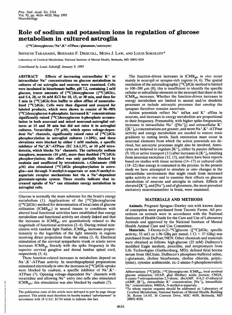
Increasing [K+]o from 5.4 to 28 and 56 mM stimulated[14C]dGlc phosphorylation in neuronal and mixed neuronal-astroglial cultures (day 6) incubated for 15 and 30 min (P <0.05); there was a lag in the stimulation because it was not seenafter 10 min of incubation (Fig. 1). In pure secondary astroglialcultures baseline rates of [14C]dGlc phosphorylation variedconsiderably from preparation to preparation, but no stimu-lation by elevated [K+]o was found in any of them (Fig. 1, Table1). Even when the range of [K+]o was expanded from 2.7 to 56mM, no K+-induced increases in rates of [14C]dGlc phosphor-ylation were observed in 20-day-old astroglial cultures (Table
.co
o
Q.
E0.
Q.CL
0
oC-L
0
o0O
C.
140 NEURONAL12010&80
10 min
5.4 28 56 5.4
15 min**
* r
II285628 56
30 min
5.4 28 56**
5.4 28 56
140a NEURONAL-ASTROGLIAL 30 min **12010 15 min
**
8 **
10 min
5.4 28 56 5.4 28 56 5.4 28 56
180- ASTROGLIAL 30min160- **
120 15 min
4180 10min ** I l 5
5.4 28 56 5.4 28 56 5.4 28 56Extracellular potassium ion concentration, mM
FIG. 1. Effects of [K+]o on rates of [14C]dGlc phosphorylation incultures of neurons (day 6), mixed neuronal-astroglia (day 6), andastroglia (day 19, no dbcAMP). Values are means ± SEM obtainedfrom quadruplicate wells. Numbers above bars indicate the durationof incubation. Data are representative of a minimum of three suchexperiments for each condition. *, P < 0.05; **, P < 0.01 comparedwith the 5.4 mM [K+]o (Dunnett's test for multiple comparison).
m
Neurobiology: Takahashi et at
Dow
nloa
ded
by g
uest
on
Nov
embe
r 15
, 202
1
4618 Neurobiology: Takahashi et at
Table 1. Effect of increased [K+]o on rates of phosphorylation of [14C]dGlc in cultured astrogliaIncubation Rate, dpm/tg of protein
Culture conditions time, min 2.7 mM [K+]o 5.4 mM [K+]o 28 mM [K+]o 56 mM [K+]oEffects of expanded range of [K+]o
20-day culture 10 39 + 1 40 ± 1 39 + 1 39 + 0No dbcAMP 15 65 ± 2 64 ± 0 73 ± 1* 69 ± 2
30 144 ±3 142 ± 2 133 ± 3 125 ± 4*Effects of age of culture
22-day culture 10 112 ± 2 108 ± 3 74 ± 3tNo dbcAMP 15 143 ± 4 131 ± 1t 127 ± 3t
30 273 ±8 251 ± 3t 215 ± 3t29-day culture 10 99 ± 2 87 ± 4t 60 ±+ tNo dbcAMP 15 147 ± 2 144 ± 3 124 ± 3t
30 328 + 3 283 + 6t 235 + 3tEffects of treatment of cultures with dbcAMP
22-day cultureNo dbcAMP 15 73 + 1 75 ± 3 81 ± 3 81 ± 8+ dbcAMP-1 day 15 96 ± 3§ 90 ± 3§ 100 + 2§ 95 ± 2+ dbcAMP-3 days 15 116 ±+ 1§ 106 + 1§ 125 ± 3§ 116 ± 7§All values are means ± SEM of quadruplicate wells. The results are representative of at least three
experiments for each condition; the culture ages in these experiments were 19-22 days for the firstexperimental series, 19-22 and 26-29 days for the effects of culture age, and 19-22 days for the last groupof experiments.*P < 0.01 compared with 2.7 mM [K+]o.tp < 0.01.tP < 0.05 compared with 5.4 mM [K+]o.§P < 0.01 compared with values in absence of dbcAMP (Dunnett's t test).
1). Age of the astroglial cultures was not a factor; neither22-day nor 29-day astroglial cultures showed elevation of[14C]dGlc phosphorylation in response to increased [K+]o(Table 1):'Addition of dbcAMP to the culture medium, whichtransforms astrocytes from protoplasmic to fibrous types,raised the baseline rate of [1 C]dGlc phosphorylation in 22-day-old astroglial cultures but did not lead to stimulation of[14C]dGlc phosphorylation by elevated [K+]o (Table 1).
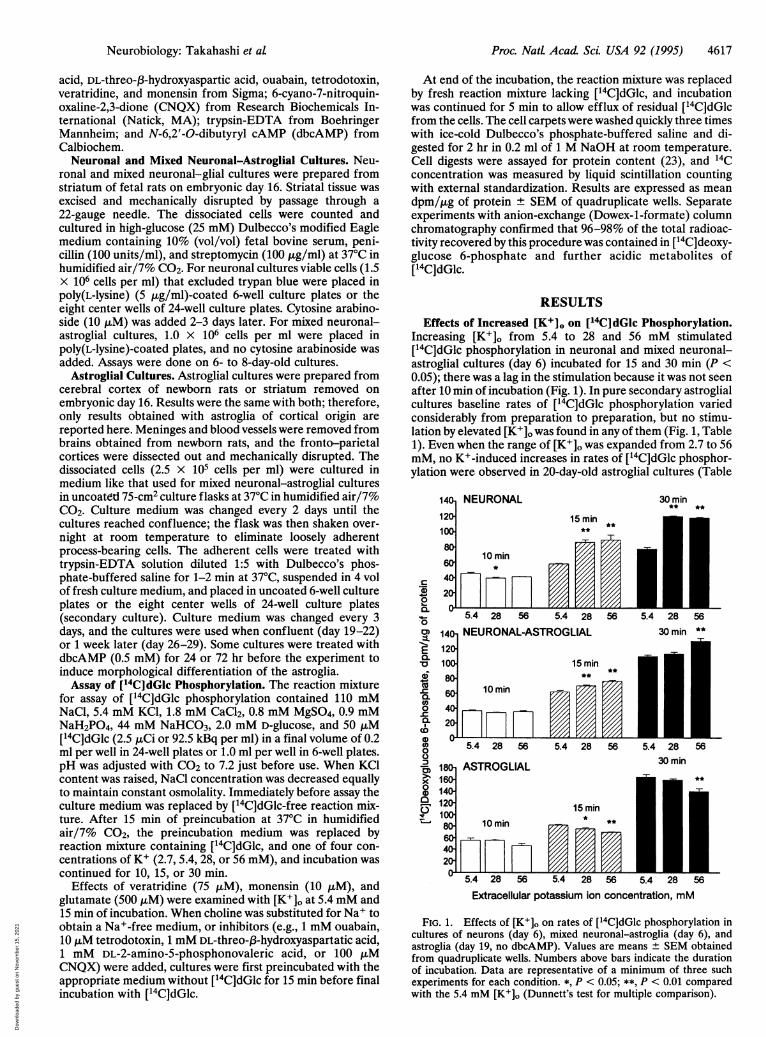
Effects of Veratridine and Monensin on [14C]dGlc Phos-phorylation. Facilitating Na+ entry into cells with veratridine(75 iaM), which opens membrane voltage-dependent Na+channels, or with the Na+ ionophore monensin (10 ,/M)stimulated ['4C]dGlc phosphorylation in astroglia by 20% and171%, respectively (Fig. 2). Ouabain (1 mM) suppressed thebasal rate of phosphorylation by -20%, completely eliminatedthe stimulation by veratridine, but only partially suppressed thestimulation by monensin (Fig. 2). Tetrodotoxin (10 /jM) didnot alter basal rates of ['4C]dGlc phosphorylation in astroglia;it did, however, eliminate the effects of veratridine but had noeffect on the stimulation by monensin (Fig. 2). Replacement ofNa+ with choline eliminated the stimulations by both veratri-dine and monensin (Fig. 2).
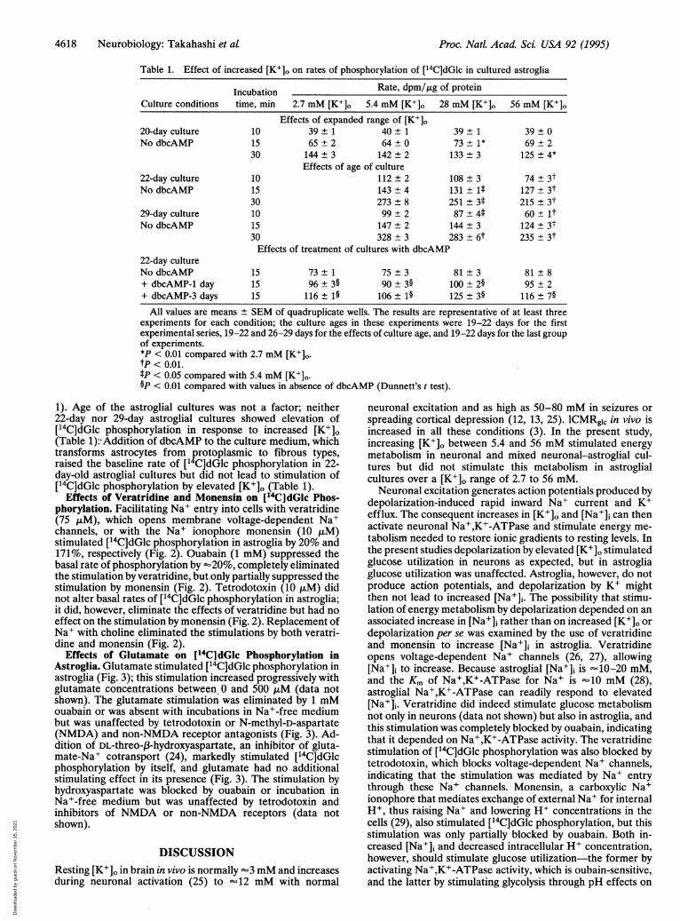
Effects of Glutamate on [l4C]dGIc Phosphorylation inAstroglia. Glutamate stimulated [14C]dGlc phosphorylation inastroglia (Fig. 3); this stimulation increased progressively withglutamate concentrations between 0 and 500 ,uM (data notshown). The glutamate stimulation was eliminated by 1 mMouabain or was absent with incubations in Na+-free mediumbut was unaffected by tetrodotoxin or N-methyl-D-aspartate(NMDA) and non-NMDA receptor antagonists (Fig. 3). Ad-dition of DL-threo-J3-hydroxyaspartate, an inhibitor of gluta-mate-Na+ cotransport (24), markedly stimulated [14C]dGlcphosphorylation by itself, and glutamate had no additionalstimulating effect in its presence (Fig. 3). The stimulation byhydroxyaspartate was blocked by ouabain or incubation inNa+-free medium but was unaffected by tetrodotoxin andinhibitors of NMDA or non-NMDA receptors (data notshown).
DISCUSSION
Resting [K+]o in brain in vivo is normally -3 mM and increasesduring neuronal activation (25) to "12 mM with normal
neuronal excitation and as high as 50-80 mM in seizures orspreading cortical depression (12, 13, 25). ICMRglc in vivo isincreased in all these conditions (3). In the present study,increasing [K+]o between 5.4 and 56 mM stimulated energymetabolism in neuronal and mixed neuronal-astroglial cul-tures but did not stimulate this metabolism in astroglialcultures over a [K+]o range of 2.7 to 56 mM.Neuronal excitation generates action potentials produced by
depolarization-induced rapid inward Na+ current and K+efflux. The consequent increases in [K+]o and [Na+]i can thenactivate neuronal Na+,K+-ATPase and stimulate energy me-tabolism needed to restore ionic gradients to resting levels. Inthe present studies depolarization by elevated [K+]o stimulatedglucose utilization in neurons as expected, but in astrogliaglucose utilization was unaffected. Astroglia, however, do notproduce action potentials, and depolarization by K+ mightthen not lead to increased [Na+]i. The possibility that stimu-lation of energy metabolism by depolarization depended on anassociated increase in [Na+]i rather than on increased [K+]o ordepolarization per se was examined by the use of veratridineand monensin to increase [Na+]i in astroglia. Veratridineopens voltage-dependent Na+ channels (26, 27), allowing[Na+]i to increase. Because astroglial [Na+]i is -10-20 mM,and the Km of Na+,K+-ATPase for Na+ is "10 mM (28),astroglial Na+,K+-ATPase can readily respond to elevated[Na+]i. Veratridine did indeed stimulate glucose metabolismnot only in neurons (data not shown) but also in astroglia, andthis stimulation was completely blocked by ouabain, indicatingthat it depended on Na+,K+-ATPase activity. The veratridinestimulation of [14C]dGlc phosphorylation was also blocked bytetrodotoxin, which blocks voltage-dependent Na+ channels,indicating that the stimulation was mediated by Na+ entrythrough these Na+ channels. Monensin, a carboxylic Na+ionophore that mediates exchange of external Na+ for internalH+, thus raising Na+ and lowering H+ concentrations in thecells (29), also stimulated [14C]dGlc phosphorylation, but thisstimulation was only partially blocked by ouabain. Both in-creased [Na+]i and decreased intracellular H+ concentration,however, should stimulate glucose utilization-the former byactivating Na+,K+-ATPase activity, which is oubain-sensitive,and the latter by stimulating glycolysis through pH effects on
Proc. Natl. Acad Sci. USA 92 (1995)
Dow
nloa
ded
by g
uest
on
Nov
embe
r 15
, 202
1
Neurobiology: Takahashi et at
VERATRIDINE
+20%**
MONENSIN
No Veratridine
e 75 ILM Veratridine
+0%n.s.
120C
0!0m 100o0.E0 80
-Co.( 600c-
.
CD
o040
0)xoD 20
rM.
No Monensin| 10 ,lM Monensin
+189%***
+Ouabain +TTX(1 mM) (10 P.M)
FIG. 2. Effects of veratridine (Upper) and monensin (Lower) on
rates of phosphorylation of [14C]dGlc in astroglia (day 21, no db-cAMP) and the effects of ouabain, tetrodotoxin (TTX); and Na+-freemedium on the veratridine and monensin effects. Values are means ±SEM obtained from quadruplicate wells. Numbers above bars indicatepercentage differences from each control. Data are representative ofthree such experiments in three separate astroglial preparations. **, P< 0.01; ***, P < 0.001; n.s., not statistically significant, compared witheach control (grouped t test).
phosphofructokinase (30), which is not oubain-sensitive.These results agree with those of Erecinska et at (31) in ratsynaptosomes but agree only partly with those of Yarowsky etat (16), who found the stimulation of [14C]dGlc uptake bymonensin in astroglia to be completely inhibited by ouabain.The fact that [14C]dGlc phosphorylation in astroglia is not
stimulated by increased [K+]o but is stimulated by veratridine-or monensin-induced increases in [Na+]i indicates that energymetabolism in astroglia can be stimulated, but the stimulationrequires an increase in intracellular Na+ concentration. As-troglial membrane can be depolarized by increased [K+]o, butthe depolarization is not associated with action potentials (32)and may, therefore, not lead to increased Na+ entry into thecells. This result may be due to either low density or differentproperties of voltage-dependent Na+ channels in astroglia(33). Barres et al (34) observed that type 1 astrocytes preparedfrom optic nerve do have voltage-dependent Na+ channels,but they open at more negative potentials and more slowly thanneuronal Na+ channels in response to depolarization. Also,Hisanaga et al (35) have reported that high [K+]o elicits c-fosexpression in neurons but does not elicit it in astroglia, furtherindicating that neurons and astroglia may respond differentlyto increased [K+]o.
Proc. Natl. Acad. Sci. USA 92 (1995) 4619
* No Glutamate
3 500 pM Glutamate
+440
Itt~
+2%n.s.
I'
+9%n.s.
I7
+50% +28%+38% ***
-19%
I+
Control +Ouabain +TTX +APV +CNQX +THA Na free(1 mM) (10 VM) (1 mM) (100 PIM) (1 mM)
FIG. 3. Effects of glutamate on [14C]dGlc phosphorylation inastroglia (day 22, no dbcAMP), and the effects of DL-2-amino-5-phosphonovaleric acid (APV), CNQX, ouabain, tetrodotoxin (TTX),DL-threo-p3-hydroxyaspartic acid (THA), and Na+-free medium on theglutamate effects. Values are means ± SEM obtained from quadru-plicate wells. Numbers above bars indicate percentage differencesfrom each control. Data are representative of three such experimentsin three separate astroglial preparations. ***, P < 0.001; n.s., notstatistically significant, from each control group (grouped t test).The failure of increased [K+]o to alter energy consumption
in astroglia also indicates that K+ uptake by astrocytes does notrequire energy. That astroglia do take up K+ is manifested bythe ability of an astroglial syncytium to move K+ from areas ofneuronal stimulation to areas of lower [K+]o-e.g., spatialbuffering of K+ (33). Also, astroglia in primary cultures havebeen reported to take up K+ avidly (32, 36), and the uptakerate is higher than that of neurons (37). Astroglial membranes,however, have a much higher K+ conductance than that ofneurons, and K+ uptake may, therefore, not be energy-dependent.cAMP and its analogue, dbcAMP, induce morphological
differentiation in cultured astroglia (38), and dbcAMP hasbeen reported to induce increased Na+,K+-ATPase activity(39). dbcAMP did increase the basal rate of [14C]dGlc phos-phorylation in our astroglial cultures, but increased [K+]o stilldid not stimulate this rate. Age of the culture could be a factor.Central nervous system cultures become more anaerobic andconsume glucose more rapidly with age of the culture (40), andNa+,K+-ATPase has been reported to require 28 days inculture to reach mature differentiation (39). In our studies thebasal rate of [14C]dGlc phosphorylation was indeed higher in26- to 29- than in 19- to 22-day-old cultures but was notstimulated by increased [K+]o in either.
Contrary to our results, there have been reports that in-creased [K+]o stimulates [14C]dGlc uptake in cultured astroglia(18-22). The reasons for this discrepancy are not obvious.Differences in the cell cultures could possibly be involved. Theastroglia derived from the cortex of newborns that we usedwere secondary cultures and were very pure; they contained noneurons and virtually no cells without glial fibrillary acidicprotein (GFAP). Some of our primary astroglial cultures,particularly those from embryonic tissue, appeared to be purewhen tested with anti-GFAP alone, but staining with anti-GFAP plus antivimentin revealed the presence of vimentin+/GFAP- cells. Such cells were never found in the secondarycultures. Inasmuch as mixed neuronal-astroglial cultures do
c
C0I.
EC"6-a0.o0i-0.
Doo
xo
0r.
-.ji A..,~ ID
ownl
oade
d by
gue
st o
n N
ovem
ber
15, 2
021
4620 Neurobiology: Takahashi et at
exhibit increased ['4C]dGlc phosphorylation in response toelevated [K+]o, contamination with other cell types must beconsidered in the reported appearance of metabolic responsesin primary astroglial cultures to increased [K+]o. Furthermore,neurons influence astrocytic differentiation in vitro (41). For,example, [3H]ouabain binding in cultured astroglia, whichreflects the level of the functional a2/a3 catalytic subunit ofNa+,K+-ATPase, has been reported to be low in pure astro-cytic cultures and much higher in neuron-enriched mixedcultures (42); this suggests an effect of neurons on the level orproperties of Na+,K+-ATPase in astroglia. Also, coculture ofastrocytes with neurons or neuron-conditioned medium hasbeen found to influence the expression of ion channels incultured astrocytes (43).The question remains whether astroglia contribute to the
increases in local energy metabolism that accompany in-creased function-related spike activity in neuropil. The presentresults indicate that neuronal elements can respond withincreased glucose utilization to changes in the extracellularionic environment associated with spike activity (i.e., increased[K+]o). Astroglial energy metabolism is, however stimulated byincreased Na+ entry and not increased [K+]o. Spike activity inaxonal terminals has, however, additional consequences tothose on [K+]o; it also leads to release of neurotransmittersthat could activate Na+ entry into astrocytes and thus stimu-late astroglial energy metabolism. Glutamate is the mostcommon and widely distributed excitatory neurotransmitter inthe central nervous system. Astroglia actively take up gluta-mate, and at least three different Na+-dependent glutamatetransporters have been cloned (44-46); one is expressedmainly in astrocytes (47). Glutamate uptake in astroglia isassociated with cotransport of Na+, leading to increased [Na+]iand depolarization (48), and, according to the present studies,stimulation of glucose utilization. This stimulation of metab-olism is eliminated when Na+ in the external medium isreplaced by choline. These results are in accord with a recentreport by Pellerin and Magistretti (49). It would appear thenthat astroglia can participate in the increased energy metab-olism associated with functional activity; in contrast to neu-rons, however, the stimulation is due not to increased [K+]o butrather to another mechanism, such as the release of glutamateand/or possibly other neurotransmitters that promote Na+entry into the cells.
1. Clarke, D. D. & Sokoloff, L. (1993) in Basic Neurochemistry, eds.Siegel, G. J., Agranoff, B. W., Albers, R. W. & Molinoff, P. B.(Raven, New York), 5th Ed., pp. 645-680.
2. Sokoloff, L., Reivich, M., Kennedy, C., Des Rosiers, M. H.,Patlak, C. S., Pettigrew, K. D., Sakurada, O. & Shinohara, M.(1977) J. Neurochem. 28, 897-916.
3. Sokoloff, L. (1981) J. Cereb. Blood Flow Metab. 1, 7-36.4. Sokoloff, L. (1994) Dev. Neurosci. 15, 194-206.5. Yarowsky, P., Kadekaro, M. & Sokoloff, L. (1983) Proc. Natl.
Acad. Sci. USA 80, 4179-4183.6. Kadekaro, M., Crane, A. M. & Sokoloff, L. (1985) Proc. Natl.
Acad. Sci. USA 82, 6010-6013.7. Mata, M., Fink, D. J., Gainer, H., Smith, C. B., Davidsen, L.,
Savaki, H., Schwartz, W. J. & Sokoloff, L. (1980) J. Neurochem.34, 213-215.
8. Smith, C. B. (1983) in Current Methods in Cellular Neurobiology:Anatomical Techniques, eds. Barker, J. L. & McKelvy, J. F.(Wiley, New York), Vol. 1, pp. 269-317.
9. Orkand, R. K., Nicholls, J. G. & Kuffler, S. W. (1966) J. Neuro-physiol. 29, 788-806.
10. Medzihradsky, F., Nandhasri, P. S., Idoyaga-Vargas, V. & Sell-inger, 0. Z. (1971) J. Neurochem. 18, 1599-1603.
11. Henn, F. A., Haljamae, H. & Hamberger, A. (1972) Brain Res. 43,437-443.
12. Hertz, L. (1977) Pharmacol. Rev. 29, 35-65.13. Erecinska, M. & Silver, I. A. (1994) Prog. Neurobiol. 43, 37-71.14. Kai-Kai, M. A. & Pentreath, V. W. (1981) J. Neurocytol. 10,
693-708.15. Pentreath, V. W. & Kai-Kai, M. A. (1982) Nature (London) 295,
59-61.16. Yarowsky, P., Boyne, A. F., Wierwille, R. & Brookes, N. (1986)
J. Neurosci. 6, 859-866.17. Badar-Goffer, R. S., Ben-Yoseph, O., Bachelard, H. S. & Morris,
P. G. (1992) Biochem. J. 282, 225-230.18. Cummins, C. J., Glover, R. A. & Sellinger, O. Z. (1979) Brain
Res. 170, 190-193.19. Cummins, C.J., Glover, R.A. & Sellinger, O. Z. (1979) J.
Neurochem. 33, 779-785.20. Brookes, N. & Yarowsky, P. J. (1985) J. Neurochem. 44, 473-479.21. Hertz, L. & Peng, L. (1992) Can. J. Physiol. Pharmacol. 70,
S145-S157.22. Peng, L., Zhang, X. & Hertz, L. (1994) Brain Res. 663, 168-172.23. Smith, P. R., Krohn, R. I., Hermanson, G. T., Mallia, A. K.,
Gartner, F. H., Provenzano, M. D., Fujimoto, E. E., Goeke,N. M., Olson, B. J. & Klenk, D. C. (1985) Anal. Biochem. 15,76-85.
24. Flott, B. & Seifert, W. (1991) Glia 4, 293-304.25. Somjen, G. G. (1975) Annu. Rev. Physiol. 37, 163-190.26. Li, P. P. & White, T. D. (1977) J. Neurochem. 28, 967-975.27. Catterall, W. A. (1992) Physiol. Rev. 72, S15-S48.28. Kimelberg, H. K., Biddlecome, S., Narumi, S. & Bourke, R. S.
(1978) Brain Res. 141, 305-323.29. Pressman, B. C. & Fahim, M. (1982) Annu. Rev. Pharmacol.
Toxicol. 22, 465-490.30. Trivedi, B. & Danforth, W. H. (1966) J. Biol. Chem. 241, 4110-
4112.31. Erecinska, M., Dagani, F., Nelson, D., Deas, J. & Silver, I. A.
(1991) J. Neurosci. 11, 2410-2421.32. Moonen, G., Frank, G. & Schoffeniels, E. (1980) Neurochem. Int.
2, 299-310.33. Barres, B. A. (1991) J. Neurosci. 11, 3685-3694.34. Barres, B. A., Chun, L. L. Y. & Corey, D. P. (1989) Neuron 2,
1375-1388.35. Hisanaga, K., Sagar, S. M., Hicks, K. J., Swanson, R. A. & Sharp,
F. R. (1990) Mol. Brain Res. 8, 69-75.36. Hertz, L. (1978) Brain Res. 145, 202-208.37. Hertz, L. (1981) J. Cereb. Blood Flow Metab. 1, 143-153.38. Shapiro, D. L. (1973) Nature (London) 241, 203-204.39. Moonen, G. & Franck, G. (1977) Neurosci. Lett. 4, 263-267.40. Danias, P., Nicklas, W. J., Ofori, S., Shen, J. & Mytilineou, C.
(1989) J. Neurochem. 53, 1149-1155.41. Hatten, M. E. (1985) J. Cell Biol. 100, 384-396.42. Brines, M. L. & Robbins, R. J. (1993) Brain Res. 631, 1-11.43. Thio, C. L., Waxman, S. G. & Sontheimer, H. (1994) J. Neuro-
physiol. 69, 819-831.44. Kanai, Y. & Hediger, M. A. (1992) Nature (London) 360, 467-
471.45. Pines, G., Danbolt, N. C., Bj0ras, M., Zhang, Y., Bendahan, A.,
Eide, L., Koepsell, H., Storm-Mathisen, J., Seeberg, E. & Kanner,B. I. (1992) Nature (London) 360, 464-467.
46. Storck, T., Schulte, S., Hofmann, K. & Stoffel, W. (1992) Proc.Natl. Acad. Sci. USA 89, 10955-10959.
47. Rothstein, J. D., Martin, L., Levey, A. I., Dykes-Hoberg, M., Jin,L., Wu, D., Nash, N. & Kuncl, R. W. (1994) Neuron 13, 713-725.
48. Bowman, C. L. & Kimelberg, H. K. (1984) Nature (London) 311,656-659.
49. Pellerin, L. & Magistretti, P. J. (1994) Proc. Natl. Acad. Sci. USA91, 10625-10629.
Proc. Natl. AcadL ScL USA 92 (1995)
Dow
nloa
ded
by g
uest
on
Nov
embe
r 15
, 202
1


![Molecular dynamics simulations of valinomycin …dreds of millions of potassium and sodium ions through a cell membrane per second [2-4]. The potassium concentration in the cell is](https://img.pdfslide.us/doc/110x75/5f2688e409644e38ee444d0a/molecular-dynamics-simulations-of-valinomycin-dreds-of-millions-of-potassium-and.jpg)