Embed Size (px)
Citation preview

Available online at www.sciencedirect.com
istry 20 (2009) 291–297
Journal of Nutritional BiochemRole of signaling pathways in the regulation of folate transport inethanol-fed rats
Abid Hamid, Jyotdeep Kaur⁎
Department of Biochemistry, Postgraduate Institute of Medical Education and Research, Chandigarh 160 012, India
Received 5 November 2007; received in revised form 6 February 2008; accepted 6 March 2008
Abstract
Folate is an essential cofactor for normal cellular proliferation and tissue regeneration. Alcohol-associated folate deficiency is common,primarily due to intestinal malabsorption, the mechanism of which needs attention. The aim of the present study was to evaluate theregulatory events of folate transport in experimental alcohol ingestion. For this, male Wistar rats were fed 1 g/kg body weight/day ethanol(20% solution) orally for 3 months and folate transport was studied in isolated intestinal epithelial cells across the crypt–villus axis. The roleof different signaling pathways in folate transport regulation was evaluated independently to that of reduced folate carrier (RFC) expression.The results showed that differentiated cells of villus possess high folate uptake activity as compared to mid villus and crypt base cells. Duringchronic ethanol ingestion, decrease in transport was observed all along the crypt–villus axis but was more pronounced at proliferating cryptbase stem cells. Studying the effect of modulators of signaling pathways revealed the folate transport system to be under the regulation ofcAMP-dependent protein kinase A (PKA), the activity of which was observed to decrease upon alcohol ingestion. In addition, protein kinaseC might have a role in folate transport regulation during alcoholic conditions. The deregulation in the folate transport system was associatedwith a decrease in RFC expression, which may result in lower transport efficiency observed at absorptive surface in alcohol-fed rats. Thestudy highlights the role that perturbed regulatory pathways and RFC expression play in the decreased folate transport at brush border surfaceduring alcohol ingestion.© 2009 Elsevier Inc. All rights reserved.
Keywords: Crypt–villus axis; Reduced folate carrier; Alcoholism; Signaling; Protein kinase
1. Introduction
The fast turnover rate of the epithelial cells is one of thecharacteristic features of the small intestinal tissue, and aconstant balance of different cofactors is required for themaintenance of a large number of mature cells [1]. Folate isone such cofactor that not only is required to sustain high rateof DNA synthesis in this highly proliferating tissue but alsoplays a critical role in maintaining normal metabolic, energy,differentiation and growth status of all mammalian cells [2].The exogenous folate required by mammals involves theinitial step of intestinal uptake across the brush bordermembrane via reduced folate carrier (RFC) as a major folatetransporter. Importantly, the consensus sequences for protein
⁎ Corresponding author. Tel.: +91 172 2747585 5181; fax: +91 1722744401x2745078.
E-mail address: [email protected] (J. Kaur).
0955-2863/$ – see front matter © 2009 Elsevier Inc. All rights reserved.doi:10.1016/j.jnutbio.2008.03.004
kinase C (PKC) and protein kinase A (PKA) have beenshown to exist in cloned RFC [3]. Some recent studies havebeen directed towards understanding the folate transportregulation by these kinases in different cell lines and suggestan important link between cellular transport system of folateand protein kinases [4]. However, the role of these pathwaysin folate transport in isolated intestinal epithelial cells underphysiological or pathophysiological conditions has not beenstudied yet.
Deficiency of folate is highly prevalent throughout theworld and affects 10% of the U.S. population [5]. Alcoholingestion has been the major contributor to folate deficiencyworldwide [6]. Folate deficiency during alcoholism candevelop because of dietary inadequacy, intestinal malabsorp-tion, altered hepatobiliary metabolism, enhanced colonicmetabolism and increased renal excretion [7–10]. However,it is evident from the literature that most of the ethanoliceffects on folate metabolism are reflected in its effect on
292 A. Hamid, J. Kaur / Journal of Nutritional Biochemistry 20 (2009) 291–297
intestinal absorption [11]. In addition, the recent over-whelming evidence suggests the existence of intricatemechanisms for controlling patterns of RFC expression andfunction in response to diverse tissue environments [12].Hence, we sought to evaluate the regulatory events mediatingfolate transport across crypt–villus axis epithelium underphysiological conditions and their roles in folate malabsorp-tion during chronic alcoholism. Such mechanistic insightscould lead to strategies for deducing the folate transportregulation in diverse cellular microenvironments and will beimportant for designing the therapeutic targets involvingregulatory derangements in folate transport systems inprimary absorptive epithelia.
2. Materials and methods
2.1. Animals
Young adult male albino rats (Wistar strain) weighing100–150 g were obtained from our institute's CentralAnimal House. The animals were housed in clean wiremesh cages with controlled temperature (23±1°C) andhumidity (45–55%) and were subjected to a 12-h dark/12-h light cycle throughout the study. The rats wererandomized into two groups of six animals each. The ratsin Group I were given a single dose of 1 g ethanol (20%solution)/kg body weight/day and those in Group II receivedan isocaloric amount of sucrose (36% solution) orally byintragastic intubation daily for 3 months. The dose wasadministrated daily between 9 and 10 a.m. to avoidperturbance in circadian rhythm. The rats were fedcommercially available pellet diet (Ashirwad Industries,Ropar, India) and water ad libitum. The body weights of ratswere recorded twice weekly. Animals from both groups weresacrificed under anesthesia using sodium pentothal, andblood was drawn for folate estimations.
The protocol of the study was approved by theInstitutional Animal Ethical Committee and the InstitutionalBiosafety Committee.
2.2. Chemicals
Radiolabeled [3′, 5′, 7, 9-3H]-folic acid potassium saltwith a specific activity of 24.0 Ci/mmol was purchased fromAmersham Pharmacia Biotech, Hong Kong. [γ-32P]-Ade-nosine-5′-triphosphate triethylammonium salt with a specificactivity of 5000 Ci/mmol was procured from Bhabha AtomicResearch Centre, Mumbai, India.
2.3. Measurement of folate levels
Serum and RBC folate levels were measured by employ-ing the microbiological assay using Lactobacillus casei asdescribed [13].
2.4. Isolation of intestinal epithelial cells
The intestinal epithelial cells were isolated as described[14]. Starting from the ligament of Treitz, the upper two
thirds of the small intestine was removed, flushedthoroughly with 0.9% saline. One end of the intestinewas tied with thread and filled with rinsing buffercontaining 1 mM DTT in normal saline, and the otherend was tied as well. The rinsing buffer that filled theintestine was then replaced with a solution consisting of1.5 mM KCl, 96 mM NaCl, 27 mM sodium citrate, 8 mMKH2PO4 and 8 mM Na2HPO4 and kept at 37°C for 15 minin a beaker containing PBS. We discarded the solution inthe intestine, and we filled the intestine with anothersolution containing 1.5 mM EDTA and 0.5 mM DTT inPBS, which was kept at 37°C for 30 min to isolate totalsmall intestinal epithelial cells or for different time intervalsto isolate cells of different origins along the crypt–villusaxis. Fraction numbers 1–3 were collected at 4-, 2- and2-min intervals whereas fraction numbers 4–6 werecollected at 3-, 4- and 5-min intervals, respectively.Similarly, fraction numbers 7–9 were collected at 7-, 10-and 15-min intervals, respectively. The three consecutivefractions were pooled and represented villus tip, mid villusand crypt base cells, respectively, from fraction numbers1–9. The cells were then centrifuged at 3000 rpm for 15min. Then, 5 ml of the cold PBS was added to the pelletcontents and centrifuged at 3000 rpm for 10 min. Twomore PBS washings were given to the pellet constitutingintestinal epithelial cells.
2.5. Preparation of brush border membrane vesicles(BBMVs)
BBMVs were prepared from the isolated total intestinalcells or cells from different origins at 4°C by the method ofKessler et al. [15] with some modifications. The final pelletcontaining isolated epithelial cells was homogenized in2 mM Tris–50 mM mannitol buffer. We added 10 mMMgCl2 to the major portion of the homogenate in 2 mMTris–50 mMmannitol buffer, followed by intermittent gentleshaking for 10 min. The contents were centrifuged at 3000×gfor 15 min, and the supernatant was run at 27,000×g for30 min. The pellet thus obtained was mixed with a smallamount of loading buffer containing 280 mM mannitol and20 mM HEPES–Tris, pH 7.4, and hand homogenizedfollowed by centrifugation at 27,000×g for 30 min. The finalpellet obtained, representing BBMV, was suspended inloading buffer to achieve a final protein concentration ofapproximately 5 mg/ml. Purity of the membrane prepara-tions was checked by measuring the specific activities ofalkaline phosphatase and Na+, K+-ATPase in brush bordermembranes and in the original homogenate [7]. The vesiclepreparations from both groups showed enrichment of 10- to12-fold with respect to alkaline phosphatase and showednegligible amount of Na+, K+-ATPase activity. The vesiclesused in the study were intact and stable as they showed theproperties of a typical brush border membrane as revealed bya transient overshoot of the intravesicular glucose concen-tration over its equilibrium uptake in the presence of sodiumgradient (data not shown).

293A. Hamid, J. Kaur / Journal of Nutritional Biochemistry 20 (2009) 291–297
2.6. Transport of [3H]-folic acid in BBMV across thecrypt–villus axis
Uptake studies were performed at 37°C using theincubation buffer consisting of 100 mM NaCl, 80 mMmannitol, 10 mM HEPES and 10 mM 3-[N-morpholino]ethanesulfonic acid (MES), pH 5.5. We added 10 μl ofvesicles (50 μg protein) prepared from different cell types toan incubation buffer containing 0.5μM[3H]-folic acid. Initialrate of transport was determined by stopping the reaction after30 s unless otherwise mentioned by adding ice-cold stopsolution containing 280 mM mannitol and 20 mM HEPES–Tris, pH 7.4, followed by rapid vacuum filtration [16].Nonspecific binding to the filters was determined by residualfilter counts after filtration of the incubation buffer andlabeled substrate without vesicles. Also, in the preliminaryexperiments, vesicular transport of [3H]-folic acid in thepresence of excess of unlabeled folic acid was measured andamounted to a negligible value, suggesting the specificity ofthe transport process. The radioactivity that remained on thefilters was determined by liquid scintillation counting(Beckman Coulter LS 6500).
2.7. Characterization of transport in cells
2.7.1. Time course and kinetics of folate transport inepithelial cells
The reaction was initiated by adding 10 μl of epithelialcells (100 μg protein) to 0.5 μM of [3H]-folic acid inincubation buffer (pH 5.5). The reaction was stopped atdifferent time intervals, namely, 10, 20, 30, 60 and 120 s. Fordetermining the Km and Vmax values, the [3H]-folic acidconcentrationwasvaried from0.125 to1.00μMin the reactionmedium and reaction was allowed for 30 s after cell addition.
2.7.2. Intracellular regulation of folic acid uptake inepithelial cells
During total cell isolation as described above, theconcentration of EDTA and DTT used was 0.75 and0.25 mM, respectively. A small aliquot from each isolatedcell preparation was checked for viability by trypan blueexclusion. The viable cells (50 μg protein) were thenincubated in the incubation buffer consisting of 100 mMNaCl, 80mMmannitol, 10mMHEPES and 10mMMES, pH5.5, containing activators or inhibitors of PKC-, PKA- andcAMP-mediated pathways, for 30 min as per an earlier study[1]. Transport reaction was initiated with 0.5 μM [3H]-folicacid and stopped with the stop solution of 280 mM mannitoland 20 mM HEPES–Tris, pH 7.4, after 30 s. The proteinkinase inhibitor (1.33 μg/50 μl reaction mixture), atropine(3 mM), dibutyryl cAMP (DbcAMP) (1 mM), chelerythrinechloride (7 μM) and phorbol-12-myristate-13-acetate (PMA)(10–30 μM) were the various modulators used in the study.
2.7.3. Estimation of cAMP-dependent PKA activityDuring assay, the phosphotransferase activity of the
viable total intestinal epithelial cells was monitored usinghistone IIA as a substrate by employing the method of
Roskoski [17] with modifications. We added 5 μl of thesubstrate histone (1 mg/ml) to a microcentrifuge tubecontaining 10 μl of the assay dilution buffer containing250 mM 3-[N-morpholino]propanesulfonic acid, 50 mMMgCl2 and 1.25 mg/ml BSA. Then, 5 μl of cAMP (50 μM)was added, followed by 5 μl of diluted [γ-32P]-ATP(100 μM), and volume was made to 30 μl with the distilledwater. The 10 μl of cell suspension (50 μg protein) wasadded to the whole reaction mixture and incubated at 37°Cfor 5 min with constant agitation. Two controls for eachsample, either without the external addition of cAMP orhistone substrate, were run simultaneously. Aliquots of 25 μlwere transferred and spotted on phosphocellulose strips(Whatman P81, 1×2 cm). The strips were washed with 10 mlof 75 mM phosphoric acid and swirled gently for 5 min.Phosphoric acid was decanted, and phosphocellulose stripswere washed twice more in phosphoric acid with gentleagitation. The strips were dried, and radioactivity wasmeasured as counts per minute in a scintillation cocktail.
2.7.4. Western blot analysis for RFC expression in BBMVFor expression studies, brush border membrane proteins
(100–150 μg) isolated from intestinal epithelial cells wereresolved on 10% SDS-PAGE and transferred to nitrocellulosemembrane for 4–5 h at 4°C, and the transfer was carried out at25 Vor 300 mA. Equal amounts of BBMV protein from thetwo groups of rats were loaded forWestern blot analysis usingthe procedure described by Towbin et al. [18] employingpolyclonal primary antibodies of rabbit anti-rat RFC (1:500dilutions), kindly provided by Dr. Hamid M. Said (Professor,Physiology and Biophysics, School of Medicine, Universityof California, Irvine, CA, USA), and were raised againstspecific region of rat RFC synthetic peptide corresponding toamino acids 495–512 of the rat RFC. The polyclonalantibodies against leucine aminopeptidase (LAP; an intest-inal brush border peptidase), which are rabbit anti-rat LAP(1:500), were used and served as an internal control.Secondary antibodies used were HRP-labeled goat anti-rabbit IgG (1:2000 dilution). The quantification of blots wascarried out by using Scion image software.
2.7.5. Statistical analysisThe data were computed as mean±S.D. Group means
were compared by using the Student's t test, and analysis ofvariance was used wherever necessary. The acceptable levelof significance was Pb.05 for each analysis.
3. Results
3.1. Growth and folate levels of rats
There was no significant change in the relative bodyweights of rats during the course of ethanol administration asreported earlier by us [16]. At the time of sacrifice, the meanbody weights of rats in control and ethanol-fed groups were201±8 and 196±9 g, respectively. Themean serum folate levelswere 49.64±5.29 and 33.71±4.95μg/L (Pb.001), and themean

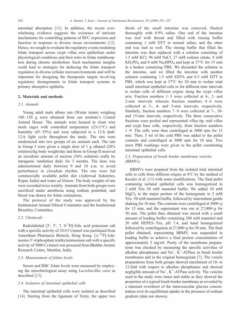
ig. 2. [3H]-Folic acid uptake in BBMVs isolated from villus tip, mid villusnd crypt base cells of the intestine. Each data point is the mean±S.D. ofree separate uptake determinations carried out in duplicate. **Pb.01,**Pb.001 versus Control and ##Pb.01, ###Pb.001 versus Villus tip.
294 A. Hamid, J. Kaur / Journal of Nutritional Biochemistry 20 (2009) 291–297
RBC folate values were 950±29.84 and 624±49.73 μg/L(Pb.001) in control and ethanol-fed rats, respectively.
3.2. Determination of kinetic constants
Time course of folate uptake at various intervals revealedthat the folate uptake in both groups showed maxima up to30–45 s and attained plateau level throughout thereafter. Inaddition, Km and Vmax values under physiological condi-tions in rats were determined in the presence of increasing[3H]-folic acid concentration from 0.125 to 1.0 μM at pH5.5. The observed saturation phenomena with increasingsubstrate concentration are indicative of Michaelis–Mentenkinetics with a Km value of 0.75±0.001 μM and a Vmax
value of 50.21±2.14 pmol/30 s/mg protein as calculated bymeans of a Lineweaver–Burk plot (Fig. 1).
3.3. Transport of [3H]-folic acid in BBMV across thecrypt–villus axis
The cell fractions (F1–F9) isolated from the smallintestine were characterized by an approximately eight-folddecrease in a villus cell marker enzyme, alkaline phospha-tase specific activity from F1 (villus tip) to F9 (crypt base)(data not shown). On the basis of the distribution patternsof the villus and the crypt cell markers, the nine cellfractions were grouped as the villus tip (F1–F3), mid villus(F4–F6) and crypt (F7–F9) cells, representing differen-tiated, differentiating and proliferating enterocytes, respec-tively. BBMVs were isolated from these pooled cellfractions, and the initial rate of [3H]-folic acid transportwas studied (Fig. 2). Folate transport was observed to besignificantly higher at the villus tip as compared to thecrypt base in both the control and the ethanol-fed group(Pb.01 and Pb.001, respectively). Ethanol feeding resultedin a significant decrease in folate transport all along thecrypt–villus axis, with the decrease being maximum (50%)at the crypt base.
Fig. 1. Lineweaver–Burk plot showing [3H]-folic acid uptake in intestinalepithelial cells as a function of substrate concentration. Uptake wasmeasured by varying [3H]-folic acid concentration from 0.125 to 1.0 μM inthe incubation medium (pH 5.5) after incubating cells (100 μg) for 30 s.Each data point is the mean±S.D. of three separate uptake determinationscarried out in duplicate.
Fath*
3.4. Intracellular regulation of folic acid transport inepithelial cells
Using trypan blue, we found that the isolated intestinalcells used for transport study were 70–80% viable in bothgroups of rats. Importantly, none of the modulators affectedthe viability of cells when cells were checked 1 h afterincubation. A 10-μl aliquot of cells containing an equalamount of protein (100 μg) was taken from both groups ofanimals for measurement of folate transport. Since there wasno alteration in histology of the two groups of animals withregard to villus height and architecture [19], it was assumedthat the number of cells per milligram of protein is equal inthe two groups (data not shown). The cells were pretreatedwith various activators and inhibitors for 30 min beforemeasurement of transport activity (Fig. 3). The resultsrevealed that when the cells were incubated in the presenceof protein kinase inhibitor specific to cAMP-dependentkinases, there was a 66% and 51% decrease (Pb.001) ofinitial rate of folic acid transport in control and ethanol-fedrats, respectively. Interestingly, when the cells wereincubated in a compound known to increase the intracellularcAMP level, for example, DbcAMP, the initial rate of [3H]-folic acid (0.5 μM) uptake was found to increase by 20% and
Fig. 3. Effect of positive and negative modulators of PKA and PKC on [3H]-folic acid uptake in viable isolated intestinal epithelial cells. Each data pointis the mean±S.D. of 12 independent observations. ***Pb.001 versusControl, #Pb.001, ###Pb.001 versus None.

ig. 5. (A) Western blot analysis of intestinal BBMV using anti-RFC5 kDa) and anti LAP (80 kDa) antibodies. (B) Densitometric analysispresenting relative change in RFC protein levels. Data shown are the meanf five separate sets of experiments. Lane 1, Control; lane 2, Ethanol.**Pb.001 versus Control.
295A. Hamid, J. Kaur / Journal of Nutritional Biochemistry 20 (2009) 291–297
43% (Pb.01), respectively, in the control and ethanol-fedrats. Atropine, which is an adenylyl cyclase inhibitor andlowers intracellular cAMP levels, resulted in reduction offolic acid uptake of 65% and 51% (Pb.001), respectively, inpretreated epithelial cells from control and ethanol-fed rats.The observed changes (increase or decrease) in folatetransport activity are attributed to the effects of thesemodulators on cAMP levels.
For the investigation of the role of PKC-mediatedpathway in the regulation of folic acid uptake, the epithelialcells were pretreated for 30 min with PKC activator (PMA)or the specific inhibitor chelerythrine chloride. None ofthese pretreatments significantly affected the folic aciduptake in the control group; however, under similarconditions, chelerythrine chloride could inhibit the transportsignificantly by 24% (Pb.05) in the ethanol-fed group.Interestingly, PMA could not impart any effect in theethanol-fed group even when its concentration wasincreased from 10 to 30 μM.
3.5. cAMP-dependent PKA activity in epithelial cells
To correlate the observed increase in folate transportactivity in response to exogenous cAMP levels with PKAactivity, we incubated cells in the presence or absence ofcAMP or histone (substrate) and we monitored PKA activityusing [γ-32P]-ATP in intestinal epithelial cells of control andethanol-fed rats (Fig. 4). Total PKA activity was decreasedby 21% (Pb.01) upon ethanol feeding to rats. In control rats,there was 35% and 49% (Pb.001) of total PKA activity in theabsence of cAMP or histone, respectively. In ethanol-fedrats, the levels of PKA activity under these conditions were43% and 55% (Pb.001), respectively.
3.6. Expression of the RFC at the brush border membranesurface
The effect of chronic alcoholism on the level ofexpression of the RFC protein at the brush border membranesurface was studied by Western blot analysis to identify theRFC using polyclonal antibodies raised against a specific
Fig. 4. PKA activities in isolated intestinal epithelial cells in the presenceof either exogenous cAMP or histone IIA or both. Each data point is themean±S.D. of 12 independent observations. *Pb.05, **Pb.01 versusControl and ###Pb.001 versus Total PKA activity.
F(6reo*
region of rat RFC, and reactivity was found at ∼65 kDa.Also, there was no cross-reaction of RFC antibodies againstany protein in the vesicular preparations used. Antiseraagainst the LAP showed reactivity at 80 kDa, which servedas an internal control. LAP is a membrane-bound amino-peptidase present in intestinal brush border membrane,whose activity has been found to be unaffected by chronicethanol feeding [20,21]. The expression of the RFC proteinwas observed to be three-fold higher in BBMV from thecontrol group when compared with the chronic, ethanol-fedgroup (Fig. 5).
4. Discussion
In the present study, the higher transport rate of folate atvillus tip cells was observed as compared to mid villus orcrypt base cells, which suggested that with maturation of theintestinal stem cells, the specialized folate transport system ispredominantly active in villus tip cells. Under suchconditions, ethanol treatment for 3 months reduced thefolate transport capability all across the crypt–villus axis,which resulted in an appreciable decrease in serum and RBCfolate levels. For the validation of the present cell modelunder physiological conditions, we observed kinetic con-stants in the intestinal epithelial cells comparable to that ofthe purified brush border membrane.
Using viable epithelial cells, the intracellular regulation offolic acid transport was focused on PKA- and PKC-mediatedpathways because consensus sequences for these kinaseshave been found in cloned folate transporters (i.e., RFC) indifferent mammalian species [4]. In the presence of inhibitorof cAMP-dependent protein kinase, a significant decrease infolic acid transport implied the involvement of proteinkinases for efficient folate transport process to be operative.Since the best defined target of cAMP is PKA, which, inturn, mediates most of the physiological effects of cAMP ineukaryotes, in order to deduce the role of cAMP in the folate

296 A. Hamid, J. Kaur / Journal of Nutritional Biochemistry 20 (2009) 291–297
transport, activator such as DbcAMP, which increases theintracellular cAMP levels, was employed. The observed 2.5-fold increase in folate transport in ethanol-fed rats under suchconditions suggested that cAMP may form an importantcomponent of signaling cascade, it being affected in chronicalcoholism. Moreover, atropine, which results in decreasedintracellular cAMP levels by inhibiting the activity ofadenylyl cyclase, inhibited the folic acid transport andagain emphasizes the role of cAMP in folate transport and itsperturbance during chronic alcoholism. Thus, these observa-tions suggest that the cAMP-mediated PKA pathway may beinvolved in the regulation of folic acid uptake in intestinalepithelial cells. Such a statement is plausible because PKArepresents a downstream target of cAMP action and cantransduce cAMP signal either by phosphorylating thepreexisting proteins in the cytoplasm or by activating thetranscription factors involved in the gene expression [22,23].Earlier studies have suggested that intracellular cAMP maynegatively affect the folic acid uptake process via PKA-independent mechanism in NCM460 [24], IEC-6 [1] andPaCa-2 cell lines [25]. However, the increased cAMP levelswere found to positively correlate with the folic acid uptakein the present study. Such a discrepancy may be related todifferences in the experimental setup employed in theprevious studies using cultured cell lines unlike viableepithelial cells in our study. In addition, the observedincrease in PKA activity in the presence of exogenous cAMPlevels in two groups of rats and the fact that there wassignificant decrease in PKA activity in ethanol-fed ratsfurther substantiated our results. Therefore, cAMP-mediatedPKA can be proposed to regulate the intestinal folatetransport system; the regulatory role of which gets alteredduring alcoholism-associated folate malabsorption via anunknown mechanism.
Furthermore, the lack of effect of PKC modulators onfolic acid uptake in the control group showed that thePKC pathway has no role in folic acid transport systemunder physiological conditions. Nevertheless, the signifi-cant effect of the PKC inhibitor chelerythrine chloride inethanol-fed group proposes that PKC may play a role infolic acid transport regulation during alcoholism. How-ever, the absence of any effect of the PKC activator PMAin the ethanol-fed group may suggest that either PMA assuch or its pharmacological range concentrations used inthe study were not sufficient to revert the observedreduction in folate transport after ethanol feeding. Overall,these findings suggest that protein kinases via cAMP areinvolved in physiological folic acid transport regulation,and after chronic ethanol ingestion in rats, PKC couldalso play a vital role via yet unidentified mechanism(s).This can be explained by the presumption that phosphorylationof PKC site has no subsequent effect on the transport activityunder physiological conditions or the site gets accessibility onlyin alcoholism due to change in membrane fluidity orconformation. Although no direct evidence exists regardingthe intracellular regulation of folate transporters (e.g., RFC) by
kinases, the effect of modulators on transport activity andthe presence of consensus sequences for these kinases incloned RFC makes it a fascinating field to study, especiallyin pathophysiological conditions of folate malabsorptionsuch as alcoholism. The disturbance in regulation of thefolate transport system via kinases reflected its effect asdecreased expression of the RFC at intestinal brush bordermembrane observed in the present study. Since the level ofRFC message is a reflection of functional uptake across theintestinal surface, such results suggest that a decrease in thenumber of transport carriers due to perturbed signaling isthe possible reason of the folate malabsorption duringalcoholism. These observations substantiated our earlierstudy in which chronic ethanol feeding resulted in alteredkinetic characteristics of folate transport process, reflectedby decreased Vmax in rat intestine [19,26]. The decreasedRFC protein molecules in the brush border membrane canoccur by greater turnover or degradation, which results inlesser number of transporter molecules available at thebrush border membrane surface for the uptake of folic acidduring alcoholism. Future studies should focus on whetherthe cAMP–PKA network is directly involved in phosphor-ylating the RFC and/or substrate or whether it influencesthe expression of the RFC at the transcriptional orposttranslational level. In addition, the role of signalingintermediates of these pathways on the expression of RFCwill help in determining the target of alcohol interference inthe folate transport system.
Taken together, the decreased transport all along thecrypt–villus axis associated with altered regulating pathwayswas reflected as low expression of the RFC in the intestineduring alcoholism.
Acknowledgments
Abid Hamid acknowledges the Council of Scientific andIndustrial Research (CSIR), New Delhi, India, for fellow-ship. The authors also thank ICMR, New Delhi, for grantsupport.
References
[1] Said HM, Ma TY, Ortiz A, Tapia A, Valerio CK. Intracellularregulation of intestinal folate uptake: studies with cultured IEC-6epithelial cells. Am J Physiol Cell Physiol 1997;272:C729–36.
[2] Matherly LH, Goldman ID. Membrane transport of folates. VitamHorm 2003;66:403–56.
[3] Said HM, Mohammed ZM. Intestinal absorption of water-solublevitamins: an update. Curr Opin Gastroenterol 2006;22:140–6.
[4] Said HM. Recent advances in carrier-mediated intestinal absorption ofwater-soluble vitamins. Annu Rev Physiol 2004;66:419–46.
[5] Novakovic P, Stempak JM, Sohn KJ, Kim YI. Effects of folatedeficiency on gene expression in the apoptosis and cancer pathways incolon cancer cells. Carcinogenesis 2006;27:916–24.
[6] Sakuta H, Suzuki T. Alcohol consumption and plasma homocysteine.Alcohol 2005;37:73–7.
[7] Hamid A, Kaur J. Chronic alcoholism alters the transport character-istics of folate in rat renal brush border membrane. Alcohol 2006;38:59–66.

297A. Hamid, J. Kaur / Journal of Nutritional Biochemistry 20 (2009) 291–297
[8] Homann N, Tillonen J, Salaspuro M. Microbially produced acetalde-hyde from ethanol may increase the risk of colon cancer via folatedeficiency. Int J Cancer 2000;86:169–73.
[9] Schalinske KL, Nieman KM. Disruption of methyl group metabolismby ethanol. Nutr Rev 2005;63:387–91.
[10] Villanueva J, Chandler CJ, Shimasaki N, Tang AB, Nakamura M,Phinney SD, et al. Effects of ethanol feeding on liver, kidney andjejunal membranes of micropigs. Hepatol 1994;19:1229–40.
[11] Villanueva JA, Devlin AM, Halsted CH. Reduced folate carrier: tissuedistribution and effects of chronic ethanol intake in the micropig.Alcohol Clin Exp Res 2001;25:415–20.
[12] Rajgopal A, Sierra EE, Zhao R, Goldman ID. Expression of thereduced folate carrier SLC19A1 in IEC-6 cells results in two distincttransport activities. Am J Physiol Cell Physiol 2001;281:C1579–86.
[13] Tamura T. In: Picciano MF, Stokstad ELR, Gregory JF, editors. Folicacid metabolism in health and disease. New York: Wiley-Liss; 1990.pp. 121–37.
[14] Weiser MM. Intestinal epithelial cell surface membrane glycoproteinsynthesis, I. An indicator of cellular differentiation. J Biol Chem 1973;248:2536–41.
[15] Kessler M, Acuto O, Storelli C, Murer H, Muller M, Samenza G.A modified procedure for the rapid preparation of efficientlytransporting vesicles from small intestinal brush border membranes.Their use in investigating some properties of D-glucose and cholinetransport. Biochem Biophys Acta 1978;506:136–54.
[16] Hamid A, Kaur J. Kinetic characteristics of folate binding to rat renalbrush border membrane in chronic alcoholism. Mol Cell Biochem2005;280:219–25.
[17] Roskoski Jr R. Assays of protein kinase. Methods Enzymol 1983;99:3–6.
[18] Towbin H, Staehelin T, Gordon J. Electrophoretic transfer of proteinsfrom polyacrylamide gels to nitrocellulose sheets: procedure and someapplications. Proc Natl Acad Sci U S A 1979;76:4350–4.
[19] Hamid A, Wani NA, Rana S, Vaiphei K, Mahmood A, Kaur J. Down-regulation of reduced folate carrier may result in folate malabsorptionacross intestinal brush border membrane during experimental alcohol-ism. FEBS J 2007;274:6317–28.
[20] Kaur J, Virender Jaswal MS, Nagpaul JP, Mahmood A. Chronicethanol feeding and microvillus membrane glycosylation in normaland protein malnourished rat intestine. Nutrition 1992;8:338–42.
[21] Kaur J, Nagpaul JP, Mahmood A. Expression of brush border enzymesin ethanol fed rat intestine. Indian J Med Res 1994;100:289–94.
[22] Muller U, Brandsch M, Prasad PD, Fei YJ, Ganapathy V, Leibach FH.Inhibition of the H+/peptide cotransporter in the human intestinal cellline Caco-2 by cyclic AMP. Biochem Biophys Res Commun 1996;218:461–5.
[23] Velasco G, Iglesias CF, Dominguez P, Barros F, Gascon S, Lazo PS.Protein kinase C from small intestine epithelial cells. Biochem BiophysRes Commun 1986;139:875–82.
[24] Kumar CK, Moyer MP, Dudeja PK, Said HM. A protein tyrosinekinase-regulated pH dependent, carrier mediated uptake system forfolate in human normal colonic epithelial cell line NCM460. J BiolChem 1997;272:6226–31.
[25] Nabokina SM, Ma TY, Said HM. Mechanism and regulation of folateuptake by human pancreatic epithelial MIA PaCa-2 cells. Am J PhysiolCell Physiol 2004;87:C142–8.
[26] Hamid A, Kaur J, Mahmood A. Evaluation of the kinetic propertiesof the folate transport system in intestinal absorptive epitheliumduring experimental ethanol ingestion. Mol Cell Biochem 2007;304:265–71.