Embed Size (px)
Citation preview
Original Contribution
ROLE OF REACTIVE OXYGEN SPECIES AND MAPKs IN VANADATE-INDUCED G2/M PHASE ARREST
ZHUO ZHANG,*† STEPHEN S. LEONARD,*† CHUANSHU HUANG,‡ VAL VALLYATHAN ,* V INCE CASTRANOVA,*and XIANGLIN SHI*†
*Pathology and Physiology Research Branch, National Institute for Occupational Safety and Health, Morgantown, WV, USA;†Department of Basic Pharmaceutical Sciences, West Virginia University, Morgantown, WV, USA; and‡Nelson Institute of
Environmental Medicine, New York University School of Medicine, New York, NY, USA
(Received 30 December 2002;Revised 21 February 2003;Accepted 21 February 2003)
Abstract—Cell growth arrest is an important mechanism in maintaining genomic stability and integrity in response toenvironmental stress. Using the human lung alveolar epithelial cancer cell line A549, we investigated the role of reactiveoxygen species (ROS), extracellular signal-regulated protein kinase (ERK), and p38 protein kinase in vanadate-inducedcell growth arrest. Exposure of cells to vanadate led to cell growth arrest at the G2/M phase and caused upregulationof p21 and phospho-cdc2 and degradation of cdc25C in a time- and dose-dependent manner. Vanadate stimulatedmitogen-activated protein kinases (MAPKs) family members, as determined by the phosphorylation of ERK and p38.PD98059, an inhibitor of ERK, and SB202190, an inhibitor of p38, inhibited vanadate-induced cell growth arrest,upregulation of p21 and cdc2, and degradation of cdc25C. In addition to hydroxyl radical (•OH) formation, cellularreduction of vanadate generated superoxide radical (O2
•�) and hydrogen peroxide (H2O2), as determined by confocalmicroscopy using specific dyes. Generation of O2
•� and H2O2 was inhibited by specific antioxidant enzymes,superoxide dismutase (SOD) and catalase, respectively. ROS activate ERK and p38, which in turn upregulate p21 andcdc2 and cause degradation of cdc25C, leading to cell growth arrest at the G2/M phase. Specific ROS affect differentMAPK family members and cell growth regulatory proteins with different potencies. © 2003 Elsevier Inc.
Keywords—MAPKs, Cell cycle regulatory proteins, Reactive oxygen species, Growth arrest, Vanadate
INTRODUCTION
Vanadium is an essential transition trace element foundin some plants and animals. It is widely distributed inrocks, soil, and to a lesser extent in water [1–3]. Vana-dium compounds (V(V)) exert potent toxic effects on awide variety of biological systems [1,4–8]. This metalregulates growth factor-mediated signal transductionpathways, promotes cell transformation, and decreasescell adhesion [9–11]. Occupational exposure to vana-dium occurs in mining, petrochemical industries, andcoal- and oil-fired plants. Epidemiological studies haveshown a correlation between vanadium exposure and theincidence of lung cancer in humans [6,8,12,13]. Vana-dium compounds were reported to modify DNA synthe-
sis and repair [14–16]. Vanadate induced forward muta-tions and DNA-protein cross-links in culturedmammalian cells [17]. While the biochemical mecha-nism of vanadium carcinogenicity still is not fully un-derstood, recent studies have indicated that vanadium-mediated generation of reactive oxygen species (ROS)may play an important role [18–26]. For example,through ROS, vanadium caused 2'-deoxyguanosine hy-droxylation and DNA damage [26], apoptosis [27,28],and activation of nuclear transcription factors [21] AP-1and NF-�B [29].
In mammalian cells, cell cycle transition is under thecontrol of a tightly regulated network of cell divisionkinases (cdks) and numerous surveillance mechanisms,the so-called checkpoints [30]. In most normal cells,DNA damage arrests proliferation in G1/S or G2/M phaseand then resumes proliferation after the damage is re-paired [30]. The cell cycle controls the onset of DNAreplication and mitosis to ensure the integrity of the
Address correspondence to: Dr. Xianglin Shi, National Institute forOccupational Safety and Health, Pathology and Physiology ResearchBranch, 1095 Willowdale Road, Morgantown, WV 26505, USA; Tel:(304) 285-6158; Fax: (304) 285-5938; E-Mail: [email protected].
Free Radical Biology & Medicine, Vol. 34, No. 10, pp. 1333–1342, 2003Copyright © 2003 Elsevier Inc.
Printed in the USA. All rights reserved0891-5849/03/$–see front matter
doi:10.1016/S0891-5849(03)00145-X
1333
genome [31–33]. Lack of fidelity in DNA replication andmaintenance can result in deleterious mutations, leadingto cell death or, in multicellular organisms, cancer [30].
Recent evidence indicates that ROS may function asintracellular messengers to modulate signaling pathways[34,35]. The changes of intracellular ROS have beendetected in a variety of cells stimulated with cytokines,growth factors, and agonists of receptors [34,36,37].Various experiments have shown that many protein ki-nases and transcription regulatory factors are activatedunder the conditions of oxidative stress [38–43]. Mito-gen-activated protein kinases (MAPKs) cascades areprotein kinase signal transduction pathways that havebeen remarkably conserved in evolution. They are dif-ferentially used to relay numerous extracellular signalswithin cells [44–46]. These MAPK cascades have beenfound to be involved in such diverse cellular functions asproliferation, differentiation, stress responses, and apo-ptosis.
Stress-activated protein kinases (SAPK)/Jun N-termi-nal kinase (JNK), p38, and extracellular signal-relatedkinase (ERK) are the most widely studied members ofthe MAPK family. Activation of MAPKs led to abnor-mal M phase transition in the cell cycle [47]. It has beenreported that p38 functions as a component of the spindleassembly checkpoint in somatic cell cycles [48]. Al-though ROS are frequently mentioned in the literature tobe inducers for MAPKs, many of the studies are indirect.For example, N-acetylcysteine (NAC) was used as aninhibitor [49] and the generation of ROS by the stimulantin cellular system was not well characterized. NAC canreadily react with “–SH” group of the protein and affectits function in a mechanism other than the scavenging ofROS.
The link between ROS and cell growth arrest has notyet been established. Many questions remain to be an-swered. For example, do ROS play a key role in theinduction of cell growth arrest? Which species amongROS are involved? Are MAPKs involved in ROS-medi-ated cell growth arrest? Do MAPKs and ROS affect cellgrowth regulatory proteins? The present study attemptsto answer these questions using vanadate as an inducer.
MATERIALS AND METHODS
Chemicals
Sodium metavanadate was purchased from AldrichChemical Co. (Milwaukee, WI, USA). RNase A andsuperoxide dismutase (SOD) were from Sigma ChemicalCo. (St. Louis, MO, USA). Catalase was from RocheMolecular Biochemicals (Indianapolis, IN, USA). Pro-pidium iodide (PI), 2', 7'-dichlorofluorescin diacetate(DCFH-DA), and dihydroethidium (HE) were from Mo-
lecular Probes (Eugene, OR, USA). Both F12K nutrientmixture medium and fetal bovine serum (FBS) werefrom Gibco BRL (Life Technologies, Gaithersburg, MD,USA). PD98058 and SB202190 were from Calbiochem(San Diego, CA, USA). Antibodies against p21 andcdc25C were from Santa Cruz Biotechnology (SantaCruz, CA, USA). Antibodies against p38, ERK, andphospho-Cdc2 and second AP-linked antirabbit IgGwere from Cell Signaling (Beverly, MA, USA).
Cell culture
The human alveolar epithelial cell line A549 wascultured in F12K nutrient mixture medium containing10% FBS, 2 mM L-glutamine, and 1000 U/ml penicillin-streptomycin in an incubator at 5% CO2 and 37°C.
Measurement of cell cycle/DNA content
DNA content was detected using flow cytometry[50,51]. A549 cells were fixed and permeabilized with70% ice-cold ethanol for more than 2 h and incubatedwith the freshly prepared staining buffer (0.1% TritonX-100 in PBS, 200 �g/ml RNAase A, and 20 �g/ml PI)for 15 min at 37°C. Cell cycle analysis was performed byflow cytometry with at least 10,000 cells for each sam-ple. The histogram was abstracted and the percentage ofcells in the G1/S and G2/M phases were then calculatedusing ModFit LT software (Verity Software House, Top-sham, ME, USA).
Western blot analysis
Whole cell extracts were mixed with Tris-GlycineSDS sample buffer and then subjected to Tris-Glycinegel electrophoresis. The resolved proteins were trans-ferred to a PVDF membrane. Western blotting was per-formed using antibodies against p21, cdc25C, phospho-cdc2, p38, ERK, and second antirabbit IgG. Afterreaction with ECF substrate, the signal was visualized byblue laser scanning using a Molecular DynamicsSTOM860 scanner (Molecular Dynamics, Sunnyvale,CA, USA).
Cellular hydrogen peroxide (H2O2) and superoxideanion (O2
•�) assay
Confocal microscopy was used to measure the gener-ation of H2O2 and O2
•�. DCFH-DA is a specific molec-ular probe for H2O2 and HE is a specific dye for O2
•�
detection. The principle of this assay is that DCFH-DAdiffuses through the cell membrane and is enzymaticallyhydrolyzed by intracellular esterases to nonfluorescentdichlorofluorescin (DCFH). In the presence of H2O2, thiscompound is rapidly oxidized to highly fluorescent di-chlorofluorescein (DCF) [52–54]. The blue fluorescentdye HE is oxidized by O2
•� to ethidium, which stains the
1334 Z. ZHANG et al.
nucleus a bright fluorescent red. A549 cells were cul-tured in 6-well plates containing 5 � 105 cells in eachwell. The cells were treated with 100 �M vanadate for1 h. DCFH-DA or HE (final concentration, 5 �M) wasadded to the cells and incubated for another 15–20 minprior to the measurement of fluorescence.
RESULTS
The effect of vanadate on the cell cycle
DNA content was measured by flow cytometry to in-vestigate vanadate-induced cell growth arrest. Human alve-olar epithelial cells (the A549 cell line) were first synchro-nized by serum starvation and then exposed to vanadate for24 h with various concentrations of vanadate. The resultsshow that exposure of the cells to 100 �M vanadate causedgrowth arrest at the G2/M phase in agreement with a pre-vious report [55]. At a vanadate concentration of 100 �M,vanadate-induced G2/M phase arrest peaked (32%) at 24 h.The control cells without exposure to vanadate exhibitedG2/M at 9%. The effects of vanadate concentration in theranges of 10–200 �M were examined after a 24 h exposure.An increase in vanadate concentration increased the per-centage of G2/M phase (Fig. 1B).
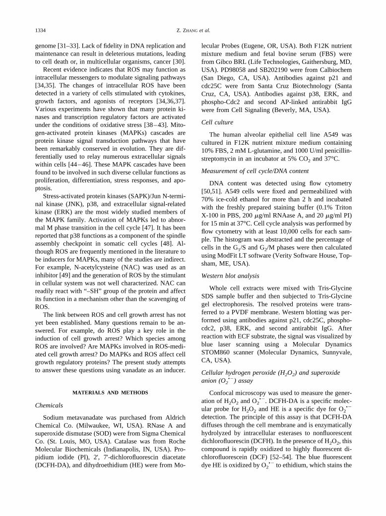
The effect of vanadate on cell growth regulatoryproteins
Several cell growth regulatory proteins, p21, phos-pho-cdc2, and cdc25C, were examined by Western blot-ting. These proteins were chosen due to their involve-ment in the regulation of G2/M phase arrest [56–58]. The
results are shown in Fig. 1. Treatment of the A549 cellswith 100 �M vanadate for different times increased thelevels of both p21 and phosphorylated cdc2 (left panel).Treatment of the cells with 100 �M vanadate for 6 hslightly increased the cdc25C level. An increase in incu-bation time caused degradation of cdc25C; further expo-sure of the cells to 100 �M vanadate for 48 h completelydegraded cdc25C.
The dose-dependent effects of vanadate on these threegrowth regulatory proteins also were examined. Asshown in Fig. 1 (right panel), vanadate caused a dose-dependent increase in the levels of both p21 and phos-phorylated cdc2. Vanadate at 25 �M caused degradationof cdc25C; at 100 �M, vanadate almost completelydegraded this regulatory protein.
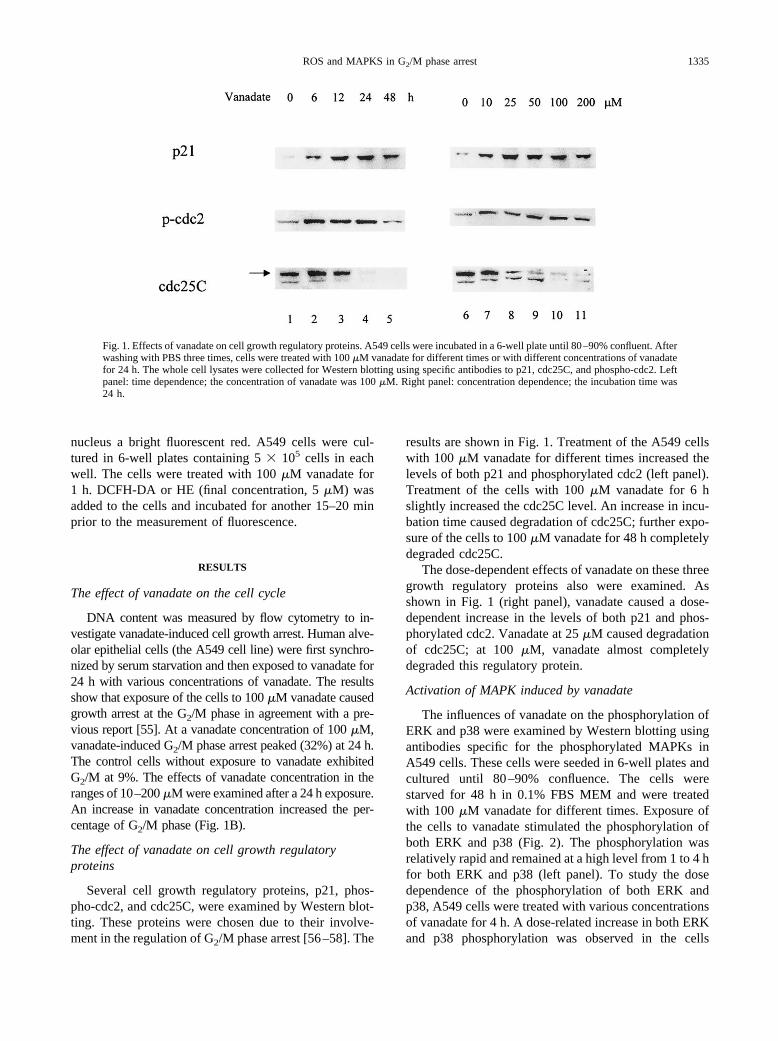
Activation of MAPK induced by vanadate
The influences of vanadate on the phosphorylation ofERK and p38 were examined by Western blotting usingantibodies specific for the phosphorylated MAPKs inA549 cells. These cells were seeded in 6-well plates andcultured until 80–90% confluence. The cells werestarved for 48 h in 0.1% FBS MEM and were treatedwith 100 �M vanadate for different times. Exposure ofthe cells to vanadate stimulated the phosphorylation ofboth ERK and p38 (Fig. 2). The phosphorylation wasrelatively rapid and remained at a high level from 1 to 4 hfor both ERK and p38 (left panel). To study the dosedependence of the phosphorylation of both ERK andp38, A549 cells were treated with various concentrationsof vanadate for 4 h. A dose-related increase in both ERKand p38 phosphorylation was observed in the cells
Fig. 1. Effects of vanadate on cell growth regulatory proteins. A549 cells were incubated in a 6-well plate until 80–90% confluent. Afterwashing with PBS three times, cells were treated with 100 �M vanadate for different times or with different concentrations of vanadatefor 24 h. The whole cell lysates were collected for Western blotting using specific antibodies to p21, cdc25C, and phospho-cdc2. Leftpanel: time dependence; the concentration of vanadate was 100 �M. Right panel: concentration dependence; the incubation time was24 h.
1335ROS and MAPKS in G2/M phase arrest
treated with increasing concentrations of vanadate from10 to 200 �M (right panel).
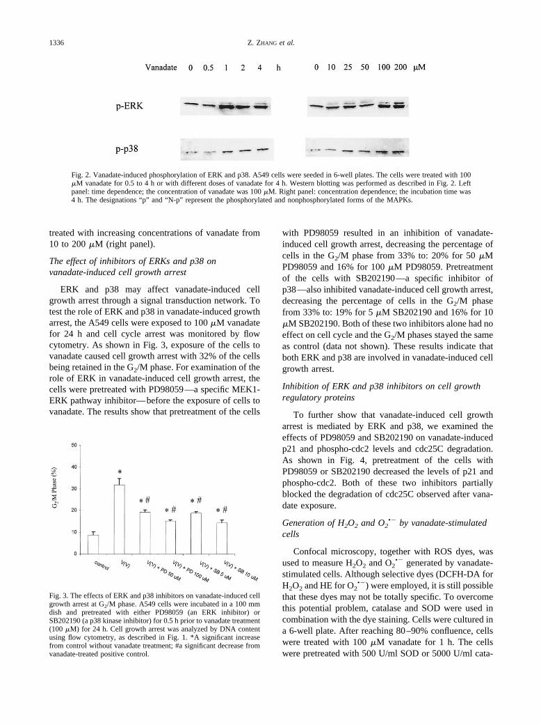
The effect of inhibitors of ERKs and p38 onvanadate-induced cell growth arrest
ERK and p38 may affect vanadate-induced cellgrowth arrest through a signal transduction network. Totest the role of ERK and p38 in vanadate-induced growtharrest, the A549 cells were exposed to 100 �M vanadatefor 24 h and cell cycle arrest was monitored by flowcytometry. As shown in Fig. 3, exposure of the cells tovanadate caused cell growth arrest with 32% of the cellsbeing retained in the G2/M phase. For examination of therole of ERK in vanadate-induced cell growth arrest, thecells were pretreated with PD98059—a specific MEK1-ERK pathway inhibitor—before the exposure of cells tovanadate. The results show that pretreatment of the cells
with PD98059 resulted in an inhibition of vanadate-induced cell growth arrest, decreasing the percentage ofcells in the G2/M phase from 33% to: 20% for 50 �MPD98059 and 16% for 100 �M PD98059. Pretreatmentof the cells with SB202190—a specific inhibitor ofp38—also inhibited vanadate-induced cell growth arrest,decreasing the percentage of cells in the G2/M phasefrom 33% to: 19% for 5 �M SB202190 and 16% for 10�M SB202190. Both of these two inhibitors alone had noeffect on cell cycle and the G2/M phases stayed the sameas control (data not shown). These results indicate thatboth ERK and p38 are involved in vanadate-induced cellgrowth arrest.
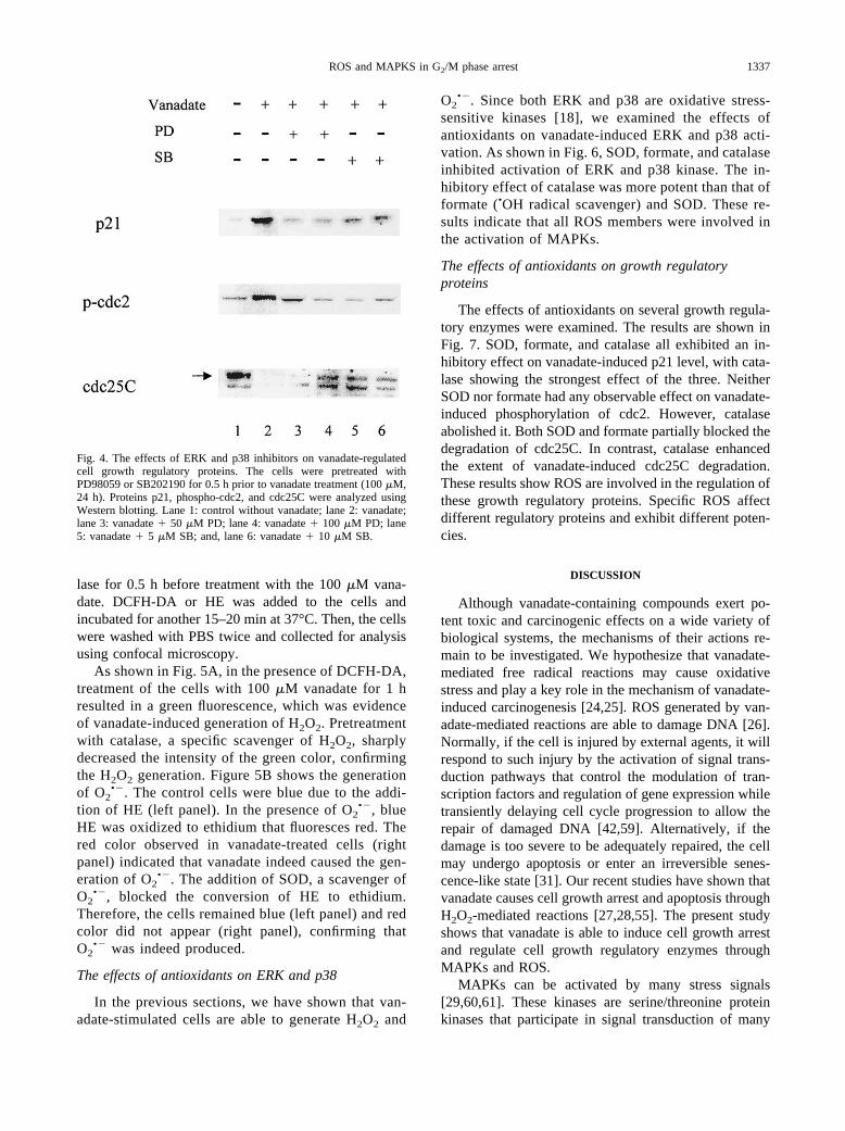
Inhibition of ERK and p38 inhibitors on cell growthregulatory proteins
To further show that vanadate-induced cell growtharrest is mediated by ERK and p38, we examined theeffects of PD98059 and SB202190 on vanadate-inducedp21 and phospho-cdc2 levels and cdc25C degradation.As shown in Fig. 4, pretreatment of the cells withPD98059 or SB202190 decreased the levels of p21 andphospho-cdc2. Both of these two inhibitors partiallyblocked the degradation of cdc25C observed after vana-date exposure.
Generation of H2O2 and O2•� by vanadate-stimulated
cells
Confocal microscopy, together with ROS dyes, wasused to measure H2O2 and O2
•� generated by vanadate-stimulated cells. Although selective dyes (DCFH-DA forH2O2 and HE for O2
•�) were employed, it is still possiblethat these dyes may not be totally specific. To overcomethis potential problem, catalase and SOD were used incombination with the dye staining. Cells were cultured ina 6-well plate. After reaching 80–90% confluence, cellswere treated with 100 �M vanadate for 1 h. The cellswere pretreated with 500 U/ml SOD or 5000 U/ml cata-
Fig. 2. Vanadate-induced phosphorylation of ERK and p38. A549 cells were seeded in 6-well plates. The cells were treated with 100�M vanadate for 0.5 to 4 h or with different doses of vanadate for 4 h. Western blotting was performed as described in Fig. 2. Leftpanel: time dependence; the concentration of vanadate was 100 �M. Right panel: concentration dependence; the incubation time was4 h. The designations “p” and “N-p” represent the phosphorylated and nonphosphorylated forms of the MAPKs.
Fig. 3. The effects of ERK and p38 inhibitors on vanadate-induced cellgrowth arrest at G2/M phase. A549 cells were incubated in a 100 mmdish and pretreated with either PD98059 (an ERK inhibitor) orSB202190 (a p38 kinase inhibitor) for 0.5 h prior to vanadate treatment(100 �M) for 24 h. Cell growth arrest was analyzed by DNA contentusing flow cytometry, as described in Fig. 1. *A significant increasefrom control without vanadate treatment; #a significant decrease fromvanadate-treated positive control.
1336 Z. ZHANG et al.
lase for 0.5 h before treatment with the 100 �M vana-date. DCFH-DA or HE was added to the cells andincubated for another 15–20 min at 37°C. Then, the cellswere washed with PBS twice and collected for analysisusing confocal microscopy.
As shown in Fig. 5A, in the presence of DCFH-DA,treatment of the cells with 100 �M vanadate for 1 hresulted in a green fluorescence, which was evidenceof vanadate-induced generation of H2O2. Pretreatmentwith catalase, a specific scavenger of H2O2, sharplydecreased the intensity of the green color, confirmingthe H2O2 generation. Figure 5B shows the generationof O2
•�. The control cells were blue due to the addi-tion of HE (left panel). In the presence of O2
•�, blueHE was oxidized to ethidium that fluoresces red. Thered color observed in vanadate-treated cells (rightpanel) indicated that vanadate indeed caused the gen-eration of O2
•�. The addition of SOD, a scavenger ofO2
•�, blocked the conversion of HE to ethidium.Therefore, the cells remained blue (left panel) and redcolor did not appear (right panel), confirming thatO2
•� was indeed produced.
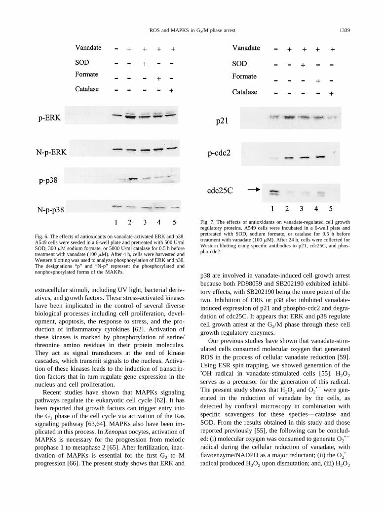
The effects of antioxidants on ERK and p38
In the previous sections, we have shown that van-adate-stimulated cells are able to generate H2O2 and
O2•�. Since both ERK and p38 are oxidative stress-
sensitive kinases [18], we examined the effects ofantioxidants on vanadate-induced ERK and p38 acti-vation. As shown in Fig. 6, SOD, formate, and catalaseinhibited activation of ERK and p38 kinase. The in-hibitory effect of catalase was more potent than that offormate (•OH radical scavenger) and SOD. These re-sults indicate that all ROS members were involved inthe activation of MAPKs.
The effects of antioxidants on growth regulatoryproteins
The effects of antioxidants on several growth regula-tory enzymes were examined. The results are shown inFig. 7. SOD, formate, and catalase all exhibited an in-hibitory effect on vanadate-induced p21 level, with cata-lase showing the strongest effect of the three. NeitherSOD nor formate had any observable effect on vanadate-induced phosphorylation of cdc2. However, catalaseabolished it. Both SOD and formate partially blocked thedegradation of cdc25C. In contrast, catalase enhancedthe extent of vanadate-induced cdc25C degradation.These results show ROS are involved in the regulation ofthese growth regulatory proteins. Specific ROS affectdifferent regulatory proteins and exhibit different poten-cies.
DISCUSSION
Although vanadate-containing compounds exert po-tent toxic and carcinogenic effects on a wide variety ofbiological systems, the mechanisms of their actions re-main to be investigated. We hypothesize that vanadate-mediated free radical reactions may cause oxidativestress and play a key role in the mechanism of vanadate-induced carcinogenesis [24,25]. ROS generated by van-adate-mediated reactions are able to damage DNA [26].Normally, if the cell is injured by external agents, it willrespond to such injury by the activation of signal trans-duction pathways that control the modulation of tran-scription factors and regulation of gene expression whiletransiently delaying cell cycle progression to allow therepair of damaged DNA [42,59]. Alternatively, if thedamage is too severe to be adequately repaired, the cellmay undergo apoptosis or enter an irreversible senes-cence-like state [31]. Our recent studies have shown thatvanadate causes cell growth arrest and apoptosis throughH2O2-mediated reactions [27,28,55]. The present studyshows that vanadate is able to induce cell growth arrestand regulate cell growth regulatory enzymes throughMAPKs and ROS.
MAPKs can be activated by many stress signals[29,60,61]. These kinases are serine/threonine proteinkinases that participate in signal transduction of many
Fig. 4. The effects of ERK and p38 inhibitors on vanadate-regulatedcell growth regulatory proteins. The cells were pretreated withPD98059 or SB202190 for 0.5 h prior to vanadate treatment (100 �M,24 h). Proteins p21, phospho-cdc2, and cdc25C were analyzed usingWestern blotting. Lane 1: control without vanadate; lane 2: vanadate;lane 3: vanadate � 50 �M PD; lane 4: vanadate � 100 �M PD; lane5: vanadate � 5 �M SB; and, lane 6: vanadate � 10 �M SB.
1337ROS and MAPKS in G2/M phase arrest
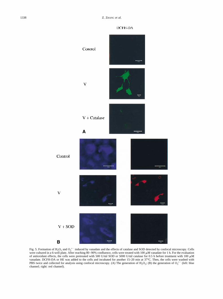
Fig. 5. Formation of H2O2 and O2•� induced by vanadate and the effects of catalase and SOD detected by confocal microscopy. Cells
were cultured in a 6-well plate. After reaching 80–90% confluence, cells were treated with 100 �M vanadate for 1 h. For the evaluationof antioxidant effects, the cells were pretreated with 500 U/ml SOD or 5000 U/ml catalase for 0.5 h before treatment with 100 �Mvanadate. DCFH-DA or HE was added to the cells and incubated for another 15–20 min at 37°C. Then, the cells were washed withPBS twice and collected for analysis using confocal microscopy. (A) The generation of H2O2; (B) the generation of O2
•� (left: bluechannel; right: red channel).
1338 Z. ZHANG et al.
extracellular stimuli, including UV light, bacterial deriv-atives, and growth factors. These stress-activated kinaseshave been implicated in the control of several diversebiological processes including cell proliferation, devel-opment, apoptosis, the response to stress, and the pro-duction of inflammatory cytokines [62]. Activation ofthese kinases is marked by phosphorylation of serine/threonine amino residues in their protein molecules.They act as signal transducers at the end of kinasecascades, which transmit signals to the nucleus. Activa-tion of these kinases leads to the induction of transcrip-tion factors that in turn regulate gene expression in thenucleus and cell proliferation.
Recent studies have shown that MAPKs signalingpathways regulate the eukaryotic cell cycle [62]. It hasbeen reported that growth factors can trigger entry intothe G1 phase of the cell cycle via activation of the Rassignaling pathway [63,64]. MAPKs also have been im-plicated in this process. In Xenopus oocytes, activation ofMAPKs is necessary for the progression from meioticprophase 1 to metaphase 2 [65]. After fertilization, inac-tivation of MAPKs is essential for the first G2 to Mprogression [66]. The present study shows that ERK and
p38 are involved in vanadate-induced cell growth arrestbecause both PD98059 and SB202190 exhibited inhibi-tory effects, with SB202190 being the more potent of thetwo. Inhibition of ERK or p38 also inhibited vanadate-induced expression of p21 and phospho-cdc2 and degra-dation of cdc25C. It appears that ERK and p38 regulatecell growth arrest at the G2/M phase through these cellgrowth regulatory enzymes.
Our previous studies have shown that vanadate-stim-ulated cells consumed molecular oxygen that generatedROS in the process of cellular vanadate reduction [59].Using ESR spin trapping, we showed generation of the•OH radical in vanadate-stimulated cells [55]. H2O2
serves as a precursor for the generation of this radical.The present study shows that H2O2 and O2
•� were gen-erated in the reduction of vanadate by the cells, asdetected by confocal microscopy in combination withspecific scavengers for these species—catalase andSOD. From the results obtained in this study and thosereported previously [55], the following can be conclud-ed: (i) molecular oxygen was consumed to generate O2
•�
radical during the cellular reduction of vanadate, withflavoenzyme/NADPH as a major reductant; (ii) the O2
•�
radical produced H2O2 upon dismutation; and, (iii) H2O2
Fig. 6. The effects of antioxidants on vanadate-activated ERK and p38.A549 cells were seeded in a 6-well plate and pretreated with 500 U/mlSOD, 300 �M sodium formate, or 5000 U/ml catalase for 0.5 h beforetreatment with vanadate (100 �M). After 4 h, cells were harvested andWestern blotting was used to analyze phosphorylation of ERK and p38.The designations “p” and “N-p” represent the phosphorylated andnonphosphorylated forms of the MAKPs.
Fig. 7. The effects of antioxidants on vanadate-regulated cell growthregulatory proteins. A549 cells were incubated in a 6-well plate andpretreated with SOD, sodium formate, or catalase for 0.5 h beforetreatment with vanadate (100 �M). After 24 h, cells were collected forWestern blotting using specific antibodies to p21, cdc25C, and phos-pho-cdc2.
1339ROS and MAPKS in G2/M phase arrest
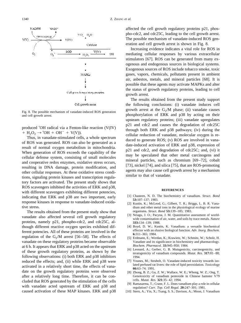
produced •OH radical via a Fenton-like reaction (V(IV)� H2O2 3
•OH � OH� � V(V)).Thus, in vanadate-stimulated cells, a whole spectrum
of ROS was generated. ROS can also be generated as aresult of normal oxygen metabolism in mitochondria.When generation of ROS exceeds the capability of thecellular defense system, consisting of small moleculesand cooperative redox enzymes, oxidative stress occurs,resulting in DNA damage, protein modification, andother cellular responses. At these oxidative stress condi-tions, signaling protein kinases and transcription regula-tory factors are activated. The present study shows thatROS scavengers inhibited the activities of ERK and p38,with different scavengers exhibiting different potencies,indicating that ERK and p38 are two important, earlyresponse kinases in response to vanadate-induced oxida-tive stress.
The results obtained from the present study show thatvanadate also affected several cell growth regulatoryproteins, namely p21, phospho-cdc2, and cdc25C, al-though different reactive oxygen species exhibited dif-ferent potencies. All of these proteins are involved in theregulation of the G2/M arrest [56–58]. The effects ofvanadate on these regulatory proteins became observableat 6 h. It appears that ERK and p38 acted on the upstreamof these growth regulatory proteins, as shown by thefollowing observations: (i) both ERK and p38 inhibitorsreduced the effects; and, (ii) while ERK and p38 wereactivated in a relatively short time, the effects of vana-date on the growth regulatory proteins were observedafter a relatively long time. Therefore, it can be con-cluded that ROS generated by the stimulation of the cellswith vanadate acted upstream of ERK and p38 andcaused activation of these MAP kinases. ERK and p38
affected the cell growth regulatory proteins p21, phos-pho-cdc2, and cdc25C, leading to the cell growth arrest.The possible mechanism of vanadate-induced ROS gen-eration and cell growth arrest is shown in Fig. 8.
Increasing evidence indicates a vital role for ROS inmediating cellular responses by various extracellularstimulators [67]. ROS can be generated from many ex-ogenous and endogenous sources in biological systems.Exogenous sources of ROS include tobacco smoke, toxicgases, vapors, chemicals, pollutants present in ambientair, asbestos, metals, and mineral particles [68]. It ispossible that these agents may activate MAPKs and alterthe status of growth regulatory proteins, leading to cellgrowth arrest.
The results obtained from the present study supportthe following conclusions: (i) vanadate induces cellgrowth arrest at the G2/M phase; (ii) vanadate causesphosphorylation of ERK and p38 by acting on theirupsteam regulatory proteins; (iii) vanadate upregulatesp21 and cdc2 and causes the degradation of cdc25Cthrough both ERK and p38 pathways; (iv) during thecellular reduction of vanadate, molecular oxygen is re-duced to generate ROS; (v) ROS are involved in vana-date-induced activation of ERK and p38, expression ofp21 and cdc2, and degradation of cdc25C; and, (vi) itmay be speculated that other metal carcinogens andmineral particles, such as chromium [69–72], cobalt[73], nickel [74], and silica [75], that are ROS-promotingagents may also cause cell growth arrest by a mechanismsimilar to that of vanadate.
REFERENCES
[1] Chasteen, N. D. The biochemistry of vanadium. Struct. Bond53:107–137; 1983.
[2] Kustin, K.; McLeod, G.; Gilbert, T. R.; Briggs, L. B. R. Vana-dium and other metal ions in the physiological ecology of marineorganisms. Struct. Bond 53:139–185; 1983.
[3] Nriagu, J. O.; Pacyna, J. M. Quanititative assessment of world-wide contamination of air, water, and soils by trace metals. Nature333:134–139; 1988.
[4] Boyd, D. W.; Kustin, K. Vanadium: a versatile biochemicaleffector with an elusive biological function. Adv. Inorg. Biochem.6:311–365; 1984.
[5] Erdmann, E.; Werdan, K.; Krawietz, W.; Schmitz, W.; Scholz, H.Vanadate and its significance in biochemistry and pharmacology.Biochem. Pharmacol. 33:945–950; 1984.
[6] Leonard, A.; Gerber, G. B. Mutagenicity, carcinogenicity, andteratogenicity of vanadium compounds. Mutat. Res. 317:81–88;1994.
[7] Younes, M.; Strubelt, O. Vanadate-induced toxicity towards iso-lated perfused rat livers: the role of lipid peroxidation. Toxicology66:63–74; 1991.
[8] Zhong, B. Z.; Gu, Z. W.; Wallace, W. E.; Whong, W. Z.; Ong, T.Genotoxicity of vanadium pentoxide in Chinese hamster V79cells. Mutat. Res. 321:35–42; 1994.
[9] Ramasarma, T.; Crane, F. L. Does vanadium play a role in cellularregulation? Curr. Top. Cell Regul. 20:247–301; 1981.
[10] Stern, A.; Yin, X.; Tsang, S. S.; Davison, A.; Moon, J. Vanadium
Fig. 8. The possible mechanism of vanadate-induced ROS generationand cell growth arrest.
1340 Z. ZHANG et al.
as a modulator of cellular regulatory cascades and oncogeneexpression. Biochem. Cell Biol. 71:103–112; 1993.
[11] Yin, X.; Davison, A. J.; Tsang, S. S. Vanadate-induced geneexpression in mouse C127 cells: roles of oxygen-derived activespecies. Mol. Cell. Biochem. 115:85–96; 1992.
[12] Hickey, R. J.; Schoff, E. P.; Clelland, R. C. Relationship betweenair pollution and certain chronic disease death rates. Multivariatestatistical studies. Arch. Environ. Health 15:728–738; 1967.
[13] Stock, P. On the relations between atmosphere pollution in urbanand rural location and mortality from cancer, bronchitis, pneumo-nia, with particular reference to 3,4-benzopyrene, beryllium, mo-lybdenum, vanadium, and arsenic. Br. J. Cancer 14:397–418;1965.
[14] Carpenter, G. Vanadate, epidermal growth factor, and the stimu-lation of DNA synthesis. Biochem. Biophys. Res. Commun. 102:1115–1121; 1981.
[15] Hori, C.; Oka, T. Vanadate enhances the stimulatory action ofinsulin on DNA synthesis in cultured mouse mammary gland.Biochim. Biophys. Acta 610:235–240; 1980.
[16] Sabbioni, E.; Pozzi, G.; Pintar, A.; Casella, L.; Garattini, S.Cellular retention, cytotoxicity, and morphological transformationby vanadium (IV) and vanadium (V) in BALB/3T3 cell lines.Carcinogenesis 12:47–52; 1991.
[17] Nechay, B. R.; Nanninga, L. B.; Nechay, P. S. Vanadyl (IV) andvanadate (V) binding to selected endogenous phosphate, car-boxyl, and amino ligands: calculations of cellular vanadium spe-cies distribution. Arch. Biochem. Biophys. 251:128–138; 1986.
[18] Ding, M.; Shi, X.; Dong, Z.; Chen, F.; Lu, Y.; Castranova, V.;Vallyathan, V. Freshly fractured crystalline silica induces activa-tor protein-1 activation through ERKs and p38 MAPK. J. Biol.Chem. 274:30611–30616; 1999.
[19] Carmichael, A. J. Vanadyl-induced Fenton-like reaction in RNA.An ESR and spin trapping study. FEBS Lett. 261:165–170; 1990.
[20] Carmichael, A. J. Reaction of vanadyl with hydrogen perxide. AnESR and spin trapping study. Free Radic. Res. Commun. 10:37–45; 1990.
[21] Huang, C.; Ding, M.; Li, J.; Leonard, S. S.; Rojanasakul, Y.;Castranova, V.; Vallyathan, V.; Ju, G.; Shi, X. Vanadium-inducednuclear factor of activated T-cells activation through hydrogenperoxide. J. Biol. Chem. 276:22397–22403; 2001.
[22] Keller, R. J.; Sharma, R. P.; Grover, T. A.; Piette, L. H. Vanadiumand lipid peroxidation: evidence for involvement of vanadyl andhydroxyl radical. Arch. Biochem. Biophys. 265:524–533; 1988.
[23] Ozawa, T.; Hanaki, A. ESR evidence for the formation of hy-droxyl radicals during the reaction of vanadyl ions with hydrogenperoxide. Chem. Pharm. Bull. 37:1407–1409; 1989.
[24] Shi, X.; Dalal, N. S. Glutathione reductase functions as vanadate(V) reductase. Arch. Biochem. Biophys. 278:288–290; 1990.
[25] Shi, X.; Dalal, N. S. Flavoenzymes reduce vanadium (V) andmolecular oxygen and generate hydroxyl radical. Arch. Biochem.Biophys. 289:355–361; 1991.
[26] Shi, X.; Wang, P.; Jiang, H.; Mao, Y.; Ahmed, N.; Dalal, N.Vanadium (IV) causes 2'-deoxyguanosine hydroxylation and de-oxyribonucleic acid damage via free radical reactions. Ann. Clin.Lab. Sci. 26:39–49; 1996.
[27] Ye, J.; Ding, M.; Leonard, S. S.; Robinson, V. A.; Millecchia, L.;Zhang, X.; Castranova, V.; Vallyathan, V.; Shi, X. Vanadateinduces apoptosis in epidermal JB6 P� cells via hydrogen perox-ide-mediated reactions. Mol. Cell. Biochem. 202:9–17; 1999.
[28] Huang, C.; Zhang, Z.; Ding, M.; Li, J.; Ye, J.; Leonard, S. S.;Shen, H. M.; Butterworth, L.; Lu, Y.; Costa, M.; Rojanasakul, Y.;Castranova, V.; Vallyathan, V.; Shi, X. Vanadate induces p53transactivation through hydrogen peroxide and causes apoptosis.J. Biol. Chem. 275:32516–32522; 2000.
[29] Ding, M.; Li, J. J.; Leonard, S. S.; Ye, J. P.; Shi, X.; Colburn,N. H.; Castranova, V.; Vallyathan, V. Vanadate-induced activa-tion of activator protein-1: role of reactive species. Carcinogen-esis 20:663–668; 1999.
[30] Shackelford, R. E.; Kaufmann, W. K.; Paules, R. S. Cell cyclecontrol, checkpoint mechanisms, and genotoxic stress. Environ.Health Perspect 107(Suppl. 1):5–24; 1999.
[31] Hartwell, L. H.; Weinert, T. A. Checkpoints: controls that ensurethe order of cell cycle events. Science 246:629–634; 1989.
[32] Hartwell, L. H.; Kastan, M. B. Cell cycle control and cancer.Science 266:1821–1828; 1994.
[33] Mercer, W. E. Checking on the cell cycle. J. Cell. Biochem.31:50–54; 1998.
[34] Ye, J.; Zhang, X.; Young, H. A.; Mao, Y.; Shi, X. Chromium(VI)-induced nuclear factor-�B activation in intact cells via free radicalreactions. Carcinogenesis 16:2401–2405; 1995.
[35] Cheng, T. H.; Shih, N. L.; Chen, S. Y.; Wang, D. L.; Chen, J. J.Reactive oxygen species modulate endothelin-1-induced c-fosgene expression in cardiomyocytes. Cardiovasc. Res. 41:654–662; 1999.
[36] Lander, H. M. An essential role for free radicals and derivedspecies in signal transduction. FASEB J. 11:118–124; 1977.
[37] Nakamura, K.; Kazuo, F.; Kouchi, H.; Mihara, K.; Miyazaki, M.;Ohe, T.; Namaba, M. Inhibitory effects of antioxidants on neo-natal rat cardiac myocyte hypertrophy induced by tumor necrosisfactor-� and angiotensin II. Circ. Res. 98:794–799; 1998.
[38] Adler, V.; Yin, Z.; Fuchs, S. Y.; Benezra, M.; Rosario, L.; Tew,K. D.; Pincus, M. R.; Sardana, M.; Henderson, C. J.; Wolf, C. R.;Davis, R. J.; Ronai, Z. Regulation of JNK signaling by GSTp.EMBO J. 18:1321–1334; 1999.
[39] Kurata, S. Sensitization of the HIV-1-LTR upon long term lowdose oxidative stress. J. Biol. Chem. 271:21798–21802; 1996.
[40] Kyriakis, J. M.; Banerjee, P.; Nikolakaki, E.; Dai, T.; Rubie,E. A.; Ahmad, M. F.; Avruch, J.; Woodgett, J. R. The stress-activated protein kinase subfamily of c-Jun kinases. Nature 369:156–160; 1994.
[41] Raingeaud, J.; Gupta, S.; Rogers, J. S.; Dickens, M.; Han, J.;Ulevitch, R. J.; Davis, R. J. Proinflammatory cytokines and en-vironmental stress cause p38 mitogen-activated protein kinaseactivation by dual phosphorylation on tyrosine and threonine.J. Biol. Chem. 270:7420–7426; 1995.
[42] Schreck, R.; Rieber, P.; Baeuerle, P. A. Reactive oxygen inter-mediates as apparently widely used messengers in the activationof the NF-�B transcription factor and HIV-1. EMBO J. 10:2247–2258; 1991.
[43] Stein, B.; Brady, H.; Yang, M. X.; Young, D. B.; Barbosa, M. S.Cloning and characterization of MEK6, a novel member of themitogen-activated protein kinase kinase cascade. J. Biol. Chem.271:11427–11433; 1996.
[44] Cohen, P. The search for physiological substances of MAP andSAP kinases in mammalian cells. Trends Cell Biol. 7:353–361;1997.
[45] Fanger, G. R.; Gerwins, P.; Widmann, C.; Jarpe, M. B.; Johnson,G. L. MEKKs, GCKs, MLKs, PALs, TAKs, and tpls: upstreamregulators of the c-jun amino-terminal kinases? Curr. Opin.Genet. Dev. 7:67–74; 1997.
[46] Herskowitz, I. MAP kinase pathways in yeast: for mating andmore. Cell 80:187–197; 1995.
[47] Kurata, S. Selective activation of p38 MAPK cascade and mitoticarrest caused by low level oxidative stress. J. Biol. Chem. 275:23413–23416; 2000.
[48] Takenaka, K.; Moriguchi, T.; Nishida, E. Activation of the proteinkinase p38 in the spindle assembly checkpoint and mitotic arrest.Science 280:599–602; 1998.
[49] Su, B.; Mitra, S.; Gregg, H.; Flavahan, S.; Chotani, M. A.; Clark,K. R.; Goldschmidt-Clermont, P. J.; Flavahan, N. A. Redoxregulation of vascular smooth muscle cell differentiation. Circ.Res. 89:39–46; 2001.
[50] Nicoletti, I.; Migliorati, G.; Pagliacci, M. C.; Grignani, F.; Ric-cardi, C. A rapid and simple method for measuring thymocyteapoptosis by propidium iodide staining and flow cytometry. J. Im-munol. Methods 139:271–279; 1991.
[51] Sgonic, R. Methods for the detection of apoptosis. Int. Arch.Allergy Immunol. 105:327–332; 1994.
[52] Bass, D. A.; Parce, J. W.; Dechatelet, L. R.; Szejda, P.; Seeds,M. C.; Thomas, M. Flow cytometric studies of oxidative productformation by neutrophils: a graded response to membrane stim-ulation. J. Immunol. 130:1910–1917; 1983.
1341ROS and MAPKS in G2/M phase arrest
[53] LeBel, C. P.; Ischiropoulos, H.; Bondy, S. C. Evaluation of theprobe 2',7'-dichlorofluorescin as an indicatior of reactive oxygenspecies formation and oxidative stress. Chem. Res. Toxicol.5:227–231; 1992.
[54] Kahn, E.; Frouin, F.; Souchier, C.; Bernengo, J. C.; Bruzzoni-Giovanelli, H.; Clement, O.; Frija, G.; Di Paola, R.; Calvo, F.;Linares-Cruz, G. Confocal multilaser focusing and single-lasercharacterization of ultraviolet excitable stains of cellular prepa-rations. Cytometry 40:42–49; 2000.
[55] Zhang, Z.; Huang, C.; Li, J.; Leonard, S. S.; Lanciotti, R.; But-terworth, L.; Shi, X. Vanadate-induced cell growth regulation andthe role of reactive oxygen species. Arch. Biochem. Biophys.392:311–320; 2001.
[56] Taylor, W. R.; Stark, G. R. Regulation of the G2/M transition byp53. Oncogene 20:1803–1815; 2001.
[57] Bunz, F.; Dutriaux, A.; Lengauer, C.; Waldman, T.; Zhou, S.;Brown, J. P.; Sedivy, J. M.; Kinzler, K. W.; Vogelstein, B.Requirement for p53 and p21 to sustain G2 arrest after DNAdamage. Science 282:1497–1501; 1998.
[58] Chan, T. A.; Hwang, P. M.; Hermeking, H.; Kinzler, K. W.;Vogelstein, B. Cooperative effects of genes controlling theG(2)/M checkpoint. Genet. Dev. 14:1584–1588; 2000.
[59] Kastan, M. B. Cell cycle. Checking two steps. Nature 410:766–777; 2001.
[60] Ono, K.; Han, J. The p38 signal transduction pathway: activationand function. Cell. Signal. 12:1–13; 2000.
[61] Tibbles, L. A.; Woodgett, J. R. The stress-activated protein kinasepathways. Cell. Mol. Life Sci. 55:1230–1254; 1999.
[62] Wilkinson, M. G.; Millar, J. B. Control of the eukaryotic cellcycle by MAP kinase signaling pathways. FASEB J. 14:2147–2157; 2000.
[63] Dobrowolski, S.; Harter, M.; Stacey, D. W. Cellular ras activity isrequired for passage through multiple points of the G0/G1 phasein BALB/c 3T3 cells. Mol. Cell. Biol. 14:5441–5449; 1994.
[64] Winston, J. T.; Coats, S. R.; Wang, Y. Z.; Pledger, W. J. Regu-lation of the cell cycle machinery by oncogenic ras. Oncogene12:127–134; 1996.
[65] Kosako, H.; Gotoh, Y.; Nishida, E. Requirement for the MAP
kinase kinase/MAP kinase cascade in Xenopus oocyte maturation.EMBO J. 13:2131–2138; 1994.
[66] Bitangcol, J. C.; Chau, A. S.; Stadnick, E.; Lohka, M. J.; Dicken,B.; Shibuya, E. K. Activation of the p42 mitogen-activated pro-tein kinase pathway inhibits Cdc2 activation and entry into M-phase in cycling Xenopus egg extracts. Mol. Biol. Cell 9:451–467; 1998.
[67] Lander, H. M. An essential role for free radicals and derivedspecies in signal transduction. FASEB J. 11:118–124; 1997.
[68] Vllyathan, V.; Shi, X. The role of oxygen free radicals in occu-pational and environmental lung diseases. Environ. Health Per-spect. 105(Suppl. 1):165–177; 1997.
[69] Shi, X.; Dalal, N. S.; Kasprzak, K. S. Generation of free radicalsfrom hydrogen peroxide and lipid hydroperoxides in the presenceof Cr(III). Arch. Biochem. Biophys. 302:294–299; 1993.
[70] Shi, X.; Dalal, N. S. On the hydroxyl radical formation in thereaction between hydrogen peroxide and biologically generatedchromium (V) species. Arch. Biochem. Biophys. 277:342–350;1990.
[71] Shi, X.; Dalal, N. S. Chromium (V) and hydroxyl radical forma-tion during the glutathione reductase-catalyzed reduction of chro-mium (VI). Biochem. Biophys. Res. Commun. 163:627–634;1989.
[72] Shi, X.; Dalal, N. S. One electron reduction of vanadium (V) byflavoenzymes/NADPH. Arch. Biochem. Biophys. 302:300–304;1993.
[73] Shi, X.; Dalal, N. S.; Kasprzak, K. S. Generation of free radicalsfrom model lipid hydroperoxides and H2O2 by Co(II) in thepresence of cysteinyl and histidyl chelators. Chem. Res. Toxicol.6:277–283; 1993.
[74] Shi, X.; Dalal, N. S.; Kasprzak, K. S. Generation of free radicalsfrom lipid hydroperoxides by Ni2� in the presence of oligopep-tides. Arch. Biochem. Biophys. 299:154–162; 1992.
[75] Ding, M.; Shi, X.; Lu, Y.; Huang, C.; Leonard, S. S.; Roberts, J.;Antonini, J.; Castranova, V.; Vallyathan, V. Induction of activatorprotein-1 through reactive oxygen species by crystalline silica inJB6 cells. J. Biol. Chem. 276:9108–9144; 2001.
1342 Z. ZHANG et al.