Embed Size (px)
Citation preview
Kidney International, Vol. 57 (2000), pp. 215–223
VASCULAR BIOLOGY – HEMODYNAMICS – HYPERTENSION
Role of protein kinase C in angiotensin II-induced constrictionof renal microvessels
TAKAHIKO NAGAHAMA, KOICHI HAYASHI, YURI OZAWA, TSUNEO TAKENAKA, and TAKAO SARUTA
Department of Internal Medicine, School of Medicine, Keio University, Tokyo, Japan
Role of protein kinase C in angiotensin II-induced constriction modynamics. Several lines of investigation suggest thatof renal microvessels. diverse mechanisms act in concert to control the vascular
Background. Although angiotensin II (Ang II) exerts its tone in AFF and EFF. These two microvessels respondaction through multiple vasomotor mechanisms, the contribu-differently to a variety of vasoactive substances, includ-tion of phosphoinositol hydrolysis products to Ang II-induceding atrial natriuretic peptide [1] and endothelin [2]. Fur-renal vasoconstriction remains undetermined.
Methods. The role of protein kinase C (PKC) in Ang II- thermore, the stimulus that causes membrane depolar-induced afferent (AFF) and efferent (EFF) arteriolar constric- ization preferentially constricts the AFF [3, 4], suggestingtion was examined using the isolated perfused hydronephrotic that a predominant distribution of voltage-dependentrat kidney.
calcium channels exists at the AFF. Such differencesResults. Ang II (0.3 nmol/L)-induced EFF constriction wasin vasoconstrictor mechanisms in AFF and EFF wouldrefractory to inhibition of voltage-dependent calcium channels
by pranidipine (1 mmol/L, 19 6 2% reversal) but was com- explain why glomerular filtration is altered in responsepletely reversed by a PKC inhibitor, chelerythrine (1 mmol/L, to a variety of vasoactive substances.96 6 2% reversal). Furthermore, direct PKC activation by It is well established that angiotensin II (Ang II)-medi-phorbol myristate acetate (PMA; 1 mmol/L) caused prominent
ated renal vascular tone constitutes an important deter-EFF constriction, and this constriction was inhibited by manga-minant of glomerular hemodynamics. Recent studiesnese and free calcium medium. In contrast, Ang II-induced AFF
constriction was completely abolished by pranidipine (98 6 4% demonstrate that Ang II-induced renal vasoconstrictionreversal) and was partially inhibited by chelerythrine (55 6 involves multiple intracellular activation steps [5, 6]. In3% reversal). Although PMA elicited marked AFF constric- many vascular beds, Ang II-induced vascular responsestion, this constriction was insensitive to the calcium antagonist,
involve the phosphoinositol pathway [7, 8]. Thus, activa-but was totally inhibited by manganese or free calcium medium.tion of phospholipase C (PLC) results in the generationConclusions. PKC plays an obligatory role in Ang II-
induced EFF constriction that requires extracellular calcium of inositol trisphosphate (IP3) and diacylglycerol (DAG).entry through nonselective cation channels. In contrast, in con- At the AFF, Ang II is reported to enhance intracellularcert with our recent findings demonstrating a complete dilation calcium release, presumably resulting from IP3-inducedby thapsigargin, Ang II-induced AFF constriction is mainly
calcium release from the sarcoplasmic reticulum [9]. Thismediated by inositol trisphosphate (IP3) and voltage-depen-elevated intracellular calcium would activate chloridedent calcium channel pathways, but could not be attributedchannels, resulting in membrane depolarization and sub-to the PKC-activated calcium entry pathway (for example,
nonselective cation channels). Rather, Ang II-stimulated PKC sequent activation of voltage-dependent calcium chan-may cross-talk to the IP3/voltage-dependent calcium channel nels [10]. The role of DAG, which is produced simultane-pathway and could modulate the vasoconstrictor mechanism ously with IP3 and stimulates protein kinase C (PKC),of the AFF. Thus, the role of PKC during Ang II stimulation
in the AFF remains fully undetermined. Furthermore,differs in AFF and EFF, which may constitute segmental heter-although Ang II-induced EFF constriction is relativelyogeneity in the renal microvasculature.refractory to the vasodilator action of the calcium antag-onist [6, 11], but requires influx of extracellular calcium
Renal afferent (AFF) and efferent arterioles (EFF) ion [12, 13], the mechanism of calcium entry in the EFFplay an important role in controlling the glomerular he- and the contribution of DAG-PKC pathways to the Ang
II-induced EFF constriction have not been evaluated.In the present study, we examined the role of PKC inKey words: afferent arterioles, efferent arterioles, cation channels, volt-
age-dependent Ca channels, vasoconstriction, renal microvasculature. mediating the Ang II-induced constriction of the renalmicrovasculature. Furthermore, segmental (that is, AFFReceived for publication May 26, 1999vs. EFF) differences in the contribution of PKC to Angand in revised form August 6, 1999
Accepted for publication August 23, 1999 II-induced vasoconstriction were also assessed. In orderto clarify these issues, we used the isolated perfused 2000 by the International Society of Nephrology
215
Nagahama et al: PKC in renal vasoconstriction216
rat hydronephrotic kidney model, which allowed direct Ang II-induced renal microvascular responses was as-visualization of the renal microvascular responsiveness sessed. After the determinations of Ang II (0.3 nmol/L)-to vasoactive stimuli [11, 12]. induced vasoconstrictor tone, a PLC inhibitor, 2-nitro-
4-carboxyphenyl-N,N-diphenyl-carbamate (NCDC; 200mmol/L; Sigma) [12, 16], was added into the perfusate.METHODSAfter 30 minutes, the ability of this agent to reverse AFF
All experimental procedures in this study were con- and EFF constriction was evaluated.ducted following the guidelines of the Animal Care Com- Role of voltage-dependent calcium channels in angio-mittee of Keio University. Chronic hydronephrosis was tensin II-induced renal vasoconstriction. Angiotensin IIestablished to facilitate subsequent visualization of the (0.3 nmol/L) was added to the perfusate to obtain basalrenal microcirculation [1, 14]. The right ureter of six- vascular tone of AFF and EFF. Then, increasing dosesweek-old male Wistar-Kyoto rats was ligated through a of a novel calcium antagonist, pranidipine (0.01, 0.1, andsmall midabdominal incision. After 8 to 10 weeks, at 1.0 mmol/L; Otsuka Pharmaceutical Co. Ltd., Tokyo,which time renal tubular atrophy had progressed to a Japan), were administered [17]. Finally, the effect of thestage that allowed direct microscopic visualization of PKC inhibitor chelerythrine (1 mmol/L; Sigma) [18] wasrenal microvessels, the kidneys were harvested for perfu- assessed.sion study. Role of protein kinase C in angiotensin II-induced renal
The renal artery of the hydronephrotic kidney was vasoconstriction. After administration of Ang II (0.3cannulated in situ through the superior mesenteric artery nmol/L), the direct vasodilator action of chelerythrineacross the aorta. The hydronephrotic kidney was placed (0.01, 0.1, and 1.0 mmol/L) on AFF and EFF was as-on the stage of an inverted microscope (IMT-2; Olympus, sessed. Subsequently, losartan (10 mmol/L; Banyu Phar-Tokyo, Japan) modified to accommodate a heated cham- maceutical Co. Ltd., Tokyo, Japan) was added to reverseber equipped with a thin-glass viewing port on the bot- the remaining vasoconstrictor tone.tom surface. Kidneys were perfused with a medium con-
To examine whether nitric oxide (NO) participated insisting of a Krebs-Ringer bicarbonate buffer containing
the chelerythrine-induced renal vascular responses [19],5 mmol/L d-glucose, 7.5% bovine serum albumin (Sigma
the vasodilator action of chelerythrine was evaluated inChemical Co., St. Louis, MO, USA), and a complementthe presence of nitro-l-arginine methyl ester (L-NAME;of amino acids [15]. The perfusion apparatus is illustrated100 mmol/L; Sigma).in our previous publication [11]. The perfusion medium
Direct stimulation of protein kinase C in renal micro-was saturated with a gas mixture of 95% oxygen/5%vasculature. The effects of PKC activators, phorbol 12-carbon dioxide, and the perfusion pressure, monitoredmyristate 13-acetate (PMA; 0.01, 0.1, and 1.0 mmol/L;at the level of the renal artery, was controlled by ad-Sigma) [20] and 1-oleoyl 2-acetyl sn-glycerol (OAG; 0.1,justing the back-pressure-type regulator (10 BP; Fair-1, and 3 mmol/L; Sigma) [21], on AFF and EFF tone werechild Industrial Products Co., Winston-Salem, NC,assessed. In each dose, at least a 30-minute equilibrationUSA).period was allowed to obtain stable PKC-induced arteri-Vessel diameters were measured as detailed pre-olar responses. Subsequently, pranidipine (1 mmol/L)viously [11]. In brief, video images from a video camerawas added to examine whether PKC-induced vascular(XC-77; Sony, Tokyo, Japan) were recorded with a videotone was mediated by voltage-dependent calcium chan-recorder and transmitted to an computer (PS55/Modelnels.5551; IBM Japan, Tokyo, Japan) equipped with a video
Role of extracellular calcium in phorbol myristate ace-acquisition and display board (Targa 161; Truevisiontate-induced vasoconstriction. The role of extracellularInc., Indianapolis, IN, USA). Vessel diameters were esti-calcium in PMA-induced renal vasoconstriction wasmated with an automated program custom designed toassessed. Initially, the kidney was perfused with zero cal-permit determination of the mean distance between par-cium medium containing ethylene glycol-bis-(b-amino-allel edges of the selected microvessels. Segments of AFFethyl ether)-N,N,N9,N9-tetraacetic acid (2 mmol/L; Sig-and EFF approximately 50 microns in length, near thema). Then, PMA (0.01, 0.1, and 1 mmol/L) was addedglomerulus, were scanned at 0.5- to 1.0-second intervals.into the perfusate, and whether PMA-induced vasocon-The mean vessel diameter was determined by averagingstriction required extracellular calcium was assessed.all measurements obtained during the plateau of the
In additional series of experiments, during the PMAresponse. To eliminate pressure-induced changes in ves-(1 mmol/L)-induced vasoconstrictor tone, manganesesel diameter, renal perfusion pressure was maintained(Mn; 10 mmol/L; Sigma) was administered to examineat 80 mm Hg throughout the experimental protocol.whether PMA-induced vasoconstriction was mediated by
Experimental protocols calcium entry through nonselective cation channels [22].Role of intracellular calcium in phorbol myristate ace-Role of phospholipase C in angiotensin II-induced re-
nal vasoconstriction. The role of PLC in mediating the tate-induced renal vasoconstriction. The role of calcium
Nagahama et al: PKC in renal vasoconstriction 217
both AFF (to 11.8 6 0.2 mm, P , 0.01) and EFF diameter(to 12.2 6 0.5 mm, P , 0.01).
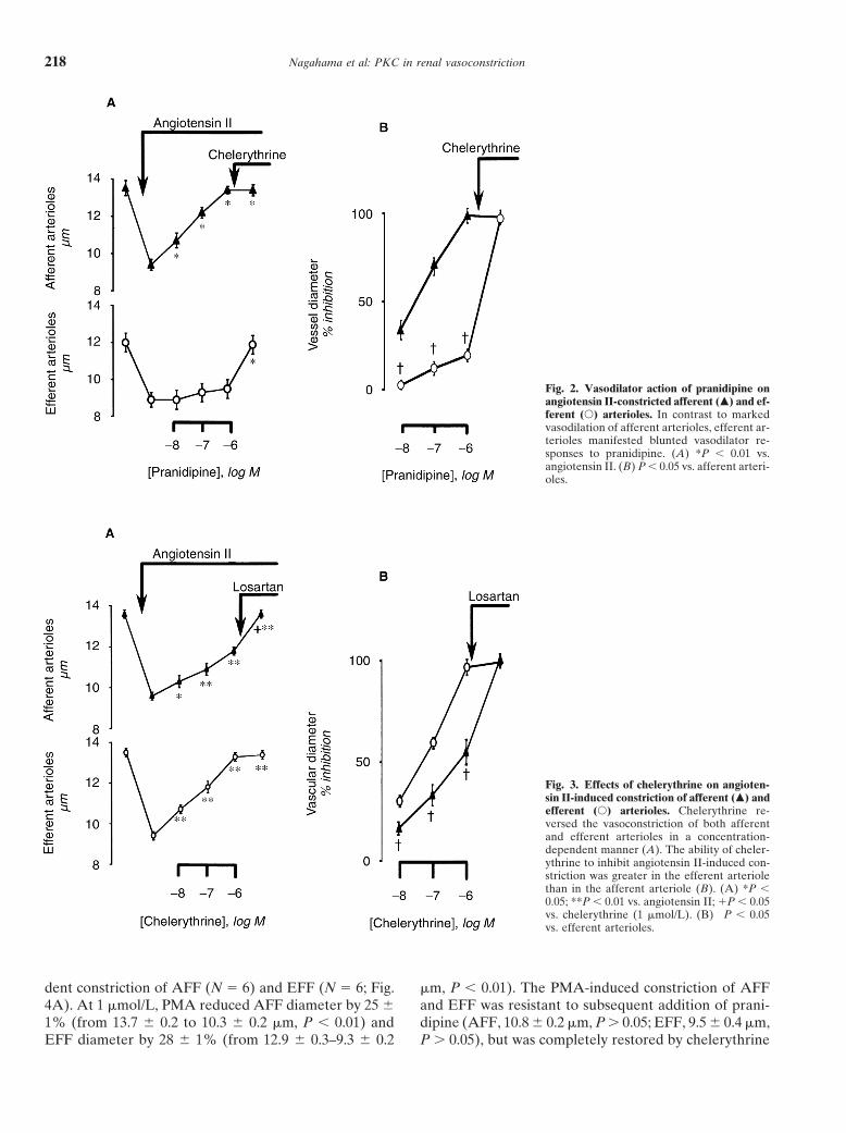
Role of voltage-dependent calcium channels inangiotensin II-induced renal vasoconstriction
The effects of pranidipine and chelerythrine on AngII-induced microvascular tone are summarized in Figure2. The Ang II-induced vasoconstriction of AFF (that is,from 13.5 6 0.4 to 9.4 6 0.3 mm, P , 0.01, N 5 6) wasreversed by pranidipine in a dose-dependent manner (10nmol/L, 10.7 6 0.4 mm, P , 0.01; 100 nmol/L, 12.2 60.3 mm, P , 0.01; 1 mmol/L, 13.4 6 0.2 mm, P , 0.01,vs. Ang II); at 1 mmol/L, pranidipine completely restoredthe AFF diameter (that is, 98 6 4% reversal). In contrast,the Ang II-induced EFF constriction (that is, from 12.0 60.5 to 8.9 6 0.4 mm, P , 0.01, N 5 6) was refractory topranidipine, with only 19 6 2% increment in diameterat 1 mmol/L (that is, 9.5 6 0.5 mm, P . 0.05, vs. Ang II).Thus, EFFs were much less responsive to the vasodilatoraction of pranidipine than AFF (Fig. 2B). The subse-quent addition of chelerythrine (1 mmol/L) completelyrestored EFF diameter (11.9 6 0.5 mm, P . 0.5, vs. pre-Ang II).
Role of protein kinase C in angiotensin II-inducedrenal vasoconstriction
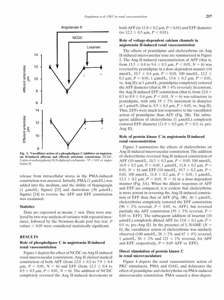
Figure 3 summarizes the effects of chelerythrine onAng II-induced microvascular constriction. The additionFig. 1. Vasodilator action of a phospholipase C inhibitor on angioten-of chelerythrine reversed Ang II-induced constriction ofsin II-induced afferent and efferent arteriolar constriction. NCDC,
2-nitro-4-carboxyphenyl-N,N-diphenyl-carbamate. *P , 0.01 vs. angio- AFF (10 nmol/L, 10.3 6 0.2 mm, P , 0.05; 100 nmol/L,tensin II. 10.9 6 0.2 mm, P , 0.05; 1 mmol/L, 11.8 6 0.2 mm, P ,
0.01, N 5 6) and EFF (10 nmol/L, 10.7 6 0.2 mm, P ,0.01; 100 nmol/L, 11.8 6 0.2 mm, P , 0.01; 1 mmol/L,release from intracellular stores in the PMA-induced13.3 6 0.2 mm, P , 0.01, N 5 6) in a dose-dependentconstriction was assessed. Initially, PMA (1 mmol/L) wasmanner (Fig. 3A). When the dilator responses of AFFadded into the medium, and the ability of thapsigarginand EFF are compared, it is evident that chelerythrine(1 mmol/L; Sigma) [23] and dantrolene (30 mmol/L;is more potent in reversing the Ang II-induced constric-Sigma) [24] to reverse the AFF and EFF constrictiontion of EFF than that of AFF (Fig. 3B). At 1 mmol/L,was examined.chelerythrine completely restored the EFF constriction(96 6 2% reversal, P , 0.05, vs. AFF), but reversedStatisticspartially the AFF constriction (55 6 3% reversal, P ,Data are expressed as means 6 sem. Data were ana-0.05 vs. EFF). The subsequent addition of losartan (10lyzed by two-way analysis of variance with repeated mea-mmol/L) completely dilated AFF (to 13.6 6 0.1 mm, P .sures, followed by the Newman–Keuls post hoc test. P0.5 vs. pre-Ang II). In the presence of L-NAME (N 5values , 0.05 were considered statistically significant.6), the vasodilator action of chelerythrine was similarlyobserved (100 nmol/L, 28 6 2% and 67 6 4% reversal;
RESULTS 1 mmol/L, 56 6 2% and 121 6 2% reversal, for AFFRole of phospholipase C in angiotensin II-induced and EFF, respectively, P , 0.05 AFF vs. EFF).renal vasoconstriction
Direct stimulation of protein kinase CFigure 1 depicts the effect of NCDC on Ang II-inducedin renal microvasculaturerenal microvascular constriction. Ang II elicited marked
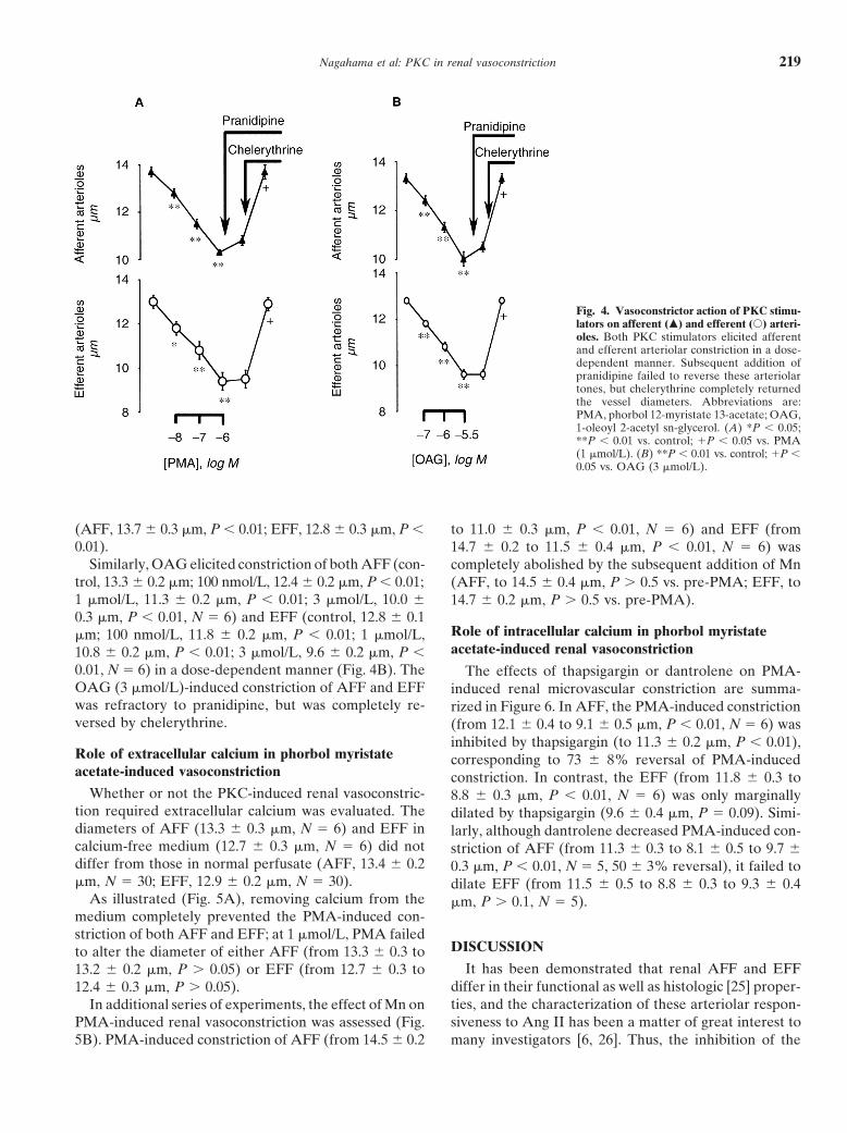
Figure 4 depicts the renal vasoconstrictor action ofconstriction of both AFF (from 12.0 6 0.2 to 7.9 6 0.4PKC stimulators, PMA and OAG, and delineates themm, P , 0.01, N 5 6) and EFF (from 12.3 6 0.4 toeffect of pranidipine and chelerythrine on PMA-induced8.5 6 0.5 mm, P , 0.01, N 5 6). The addition of NCDC
completely reversed the Ang II-induced decrements in microvascular constriction. PMA caused a dose-depen-
Nagahama et al: PKC in renal vasoconstriction218
Fig. 2. Vasodilator action of pranidipine onangiotensin II-constricted afferent (m) and ef-ferent (s) arterioles. In contrast to markedvasodilation of afferent arterioles, efferent ar-terioles manifested blunted vasodilator re-sponses to pranidipine. (A) *P , 0.01 vs.angiotensin II. (B) P , 0.05 vs. afferent arteri-oles.
Fig. 3. Effects of chelerythrine on angioten-sin II-induced constriction of afferent (m) andefferent (s) arterioles. Chelerythrine re-versed the vasoconstriction of both afferentand efferent arterioles in a concentration-dependent manner (A). The ability of cheler-ythrine to inhibit angiotensin II-induced con-striction was greater in the efferent arteriolethan in the afferent arteriole (B). (A) *P ,0.05; **P , 0.01 vs. angiotensin II; 1P , 0.05vs. chelerythrine (1 mmol/L). (B) †P , 0.05vs. efferent arterioles.
dent constriction of AFF (N 5 6) and EFF (N 5 6; Fig. mm, P , 0.01). The PMA-induced constriction of AFFand EFF was resistant to subsequent addition of prani-4A). At 1 mmol/L, PMA reduced AFF diameter by 25 6
1% (from 13.7 6 0.2 to 10.3 6 0.2 mm, P , 0.01) and dipine (AFF, 10.8 6 0.2 mm, P . 0.05; EFF, 9.5 6 0.4 mm,P . 0.05), but was completely restored by chelerythrineEFF diameter by 28 6 1% (from 12.9 6 0.3–9.3 6 0.2
Nagahama et al: PKC in renal vasoconstriction 219
Fig. 4. Vasoconstrictor action of PKC stimu-lators on afferent (m) and efferent (s) arteri-oles. Both PKC stimulators elicited afferentand efferent arteriolar constriction in a dose-dependent manner. Subsequent addition ofpranidipine failed to reverse these arteriolartones, but chelerythrine completely returnedthe vessel diameters. Abbreviations are:PMA, phorbol 12-myristate 13-acetate; OAG,1-oleoyl 2-acetyl sn-glycerol. (A) *P , 0.05;**P , 0.01 vs. control; 1P , 0.05 vs. PMA(1 mmol/L). (B) **P , 0.01 vs. control; 1P ,0.05 vs. OAG (3 mmol/L).
(AFF, 13.7 6 0.3 mm, P , 0.01; EFF, 12.8 6 0.3 mm, P , to 11.0 6 0.3 mm, P , 0.01, N 5 6) and EFF (from0.01). 14.7 6 0.2 to 11.5 6 0.4 mm, P , 0.01, N 5 6) was
Similarly, OAG elicited constriction of both AFF (con- completely abolished by the subsequent addition of Mntrol, 13.3 6 0.2 mm; 100 nmol/L, 12.4 6 0.2 mm, P , 0.01; (AFF, to 14.5 6 0.4 mm, P . 0.5 vs. pre-PMA; EFF, to1 mmol/L, 11.3 6 0.2 mm, P , 0.01; 3 mmol/L, 10.0 6 14.7 6 0.2 mm, P . 0.5 vs. pre-PMA).0.3 mm, P , 0.01, N 5 6) and EFF (control, 12.8 6 0.1
Role of intracellular calcium in phorbol myristatemm; 100 nmol/L, 11.8 6 0.2 mm, P , 0.01; 1 mmol/L,acetate-induced renal vasoconstriction10.8 6 0.2 mm, P , 0.01; 3 mmol/L, 9.6 6 0.2 mm, P ,
0.01, N 5 6) in a dose-dependent manner (Fig. 4B). The The effects of thapsigargin or dantrolene on PMA-OAG (3 mmol/L)-induced constriction of AFF and EFF induced renal microvascular constriction are summa-was refractory to pranidipine, but was completely re- rized in Figure 6. In AFF, the PMA-induced constrictionversed by chelerythrine. (from 12.1 6 0.4 to 9.1 6 0.5 mm, P , 0.01, N 5 6) was
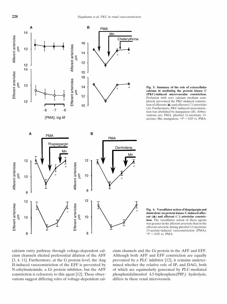
inhibited by thapsigargin (to 11.3 6 0.2 mm, P , 0.01),Role of extracellular calcium in phorbol myristate corresponding to 73 6 8% reversal of PMA-inducedacetate-induced vasoconstriction constriction. In contrast, the EFF (from 11.8 6 0.3 to
Whether or not the PKC-induced renal vasoconstric- 8.8 6 0.3 mm, P , 0.01, N 5 6) was only marginallytion required extracellular calcium was evaluated. The dilated by thapsigargin (9.6 6 0.4 mm, P 5 0.09). Simi-diameters of AFF (13.3 6 0.3 mm, N 5 6) and EFF in larly, although dantrolene decreased PMA-induced con-calcium-free medium (12.7 6 0.3 mm, N 5 6) did not striction of AFF (from 11.3 6 0.3 to 8.1 6 0.5 to 9.7 6differ from those in normal perfusate (AFF, 13.4 6 0.2 0.3 mm, P , 0.01, N 5 5, 50 6 3% reversal), it failed tomm, N 5 30; EFF, 12.9 6 0.2 mm, N 5 30). dilate EFF (from 11.5 6 0.5 to 8.8 6 0.3 to 9.3 6 0.4
As illustrated (Fig. 5A), removing calcium from the mm, P . 0.1, N 5 5).medium completely prevented the PMA-induced con-striction of both AFF and EFF; at 1 mmol/L, PMA failed
DISCUSSIONto alter the diameter of either AFF (from 13.3 6 0.3 toIt has been demonstrated that renal AFF and EFF13.2 6 0.2 mm, P . 0.05) or EFF (from 12.7 6 0.3 to
differ in their functional as well as histologic [25] proper-12.4 6 0.3 mm, P . 0.05).ties, and the characterization of these arteriolar respon-In additional series of experiments, the effect of Mn onsiveness to Ang II has been a matter of great interest toPMA-induced renal vasoconstriction was assessed (Fig.
5B). PMA-induced constriction of AFF (from 14.5 6 0.2 many investigators [6, 26]. Thus, the inhibition of the
Nagahama et al: PKC in renal vasoconstriction220
Fig. 5. Summary of the role of extracellularcalcium in mediating the protein kinase C(PKC)-induced microvascular constriction.Perfusion with zero calcium medium com-pletely prevented the PKC-induced constric-tion of afferent (m) and efferent (s) arterioles(A). Furthermore, PKC-induced vasoconstric-tion was abolished by manganese (B). Abbre-viations are: PMA, phorbol 12-myristate 13-acetate; Mn, manganese. *P , 0.05 vs. PMA.
Fig. 6. Vasodilator action of thapsigargin anddantrolene on protein kinase C-induced affer-ent (m) and efferent (s) arteriolar constric-tion. The vasodilator action of these agentswas greater in the afferent arteriole than in theefferent arteriole during phorbol 12-myristate13-acetate-induced vasoconstriction (PMA).*P , 0.05 vs. PMA.
calcium entry pathway through voltage-dependent cal- cium channels and the Gi protein in the AFF and EFF.Although both AFF and EFF constriction are equallycium channels elicited preferential dilation of the AFF
[3, 4, 11]. Furthermore, at the G protein level, the Ang prevented by a PLC inhibitor [12], it remains undeter-mined whether the relative role of IP3 and DAG, bothII-induced vasoconstriction of the EFF is prevented by
N-ethylmaleimide, a Gi protein inhibitor, but the AFF of which are equimolarly generated by PLC-mediatedphosphatidylinositol 4,5-biphosphate(PIP2) hydrolysis,constriction is refractory to this agent [12]. These obser-
vations suggest differing roles of voltage-dependent cal- differs in these renal microvessels.
Nagahama et al: PKC in renal vasoconstriction 221
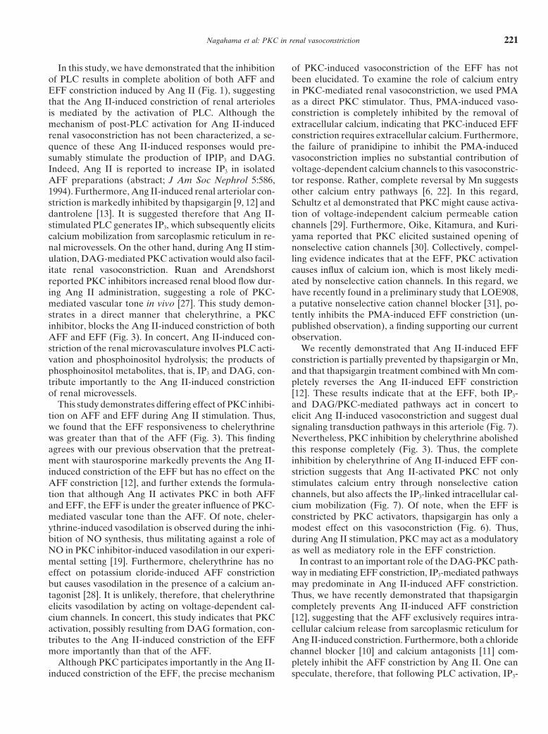
In this study, we have demonstrated that the inhibition of PKC-induced vasoconstriction of the EFF has notbeen elucidated. To examine the role of calcium entryof PLC results in complete abolition of both AFF and
EFF constriction induced by Ang II (Fig. 1), suggesting in PKC-mediated renal vasoconstriction, we used PMAas a direct PKC stimulator. Thus, PMA-induced vaso-that the Ang II-induced constriction of renal arterioles
is mediated by the activation of PLC. Although the constriction is completely inhibited by the removal ofextracellular calcium, indicating that PKC-induced EFFmechanism of post-PLC activation for Ang II-induced
renal vasoconstriction has not been characterized, a se- constriction requires extracellular calcium. Furthermore,the failure of pranidipine to inhibit the PMA-inducedquence of these Ang II-induced responses would pre-
sumably stimulate the production of IPIP3 and DAG. vasoconstriction implies no substantial contribution ofvoltage-dependent calcium channels to this vasoconstric-Indeed, Ang II is reported to increase IP3 in isolated
AFF preparations (abstract; J Am Soc Nephrol 5:586, tor response. Rather, complete reversal by Mn suggestsother calcium entry pathways [6, 22]. In this regard,1994). Furthermore, Ang II-induced renal arteriolar con-
striction is markedly inhibited by thapsigargin [9, 12] and Schultz et al demonstrated that PKC might cause activa-tion of voltage-independent calcium permeable cationdantrolene [13]. It is suggested therefore that Ang II-
stimulated PLC generates IP3, which subsequently elicits channels [29]. Furthermore, Oike, Kitamura, and Kuri-yama reported that PKC elicited sustained opening ofcalcium mobilization from sarcoplasmic reticulum in re-
nal microvessels. On the other hand, during Ang II stim- nonselective cation channels [30]. Collectively, compel-ling evidence indicates that at the EFF, PKC activationulation, DAG-mediated PKC activation would also facil-
itate renal vasoconstriction. Ruan and Arendshorst causes influx of calcium ion, which is most likely medi-ated by nonselective cation channels. In this regard, wereported PKC inhibitors increased renal blood flow dur-
ing Ang II administration, suggesting a role of PKC- have recently found in a preliminary study that LOE908,a putative nonselective cation channel blocker [31], po-mediated vascular tone in vivo [27]. This study demon-
strates in a direct manner that chelerythrine, a PKC tently inhibits the PMA-induced EFF constriction (un-published observation), a finding supporting our currentinhibitor, blocks the Ang II-induced constriction of both
AFF and EFF (Fig. 3). In concert, Ang II-induced con- observation.We recently demonstrated that Ang II-induced EFFstriction of the renal microvasculature involves PLC acti-
vation and phosphoinositol hydrolysis; the products of constriction is partially prevented by thapsigargin or Mn,and that thapsigargin treatment combined with Mn com-phosphoinositol metabolites, that is, IP3 and DAG, con-
tribute importantly to the Ang II-induced constriction pletely reverses the Ang II-induced EFF constriction[12]. These results indicate that at the EFF, both IP3-of renal microvessels.
This study demonstrates differing effect of PKC inhibi- and DAG/PKC-mediated pathways act in concert toelicit Ang II-induced vasoconstriction and suggest dualtion on AFF and EFF during Ang II stimulation. Thus,
we found that the EFF responsiveness to chelerythrine signaling transduction pathways in this arteriole (Fig. 7).Nevertheless, PKC inhibition by chelerythrine abolishedwas greater than that of the AFF (Fig. 3). This finding
agrees with our previous observation that the pretreat- this response completely (Fig. 3). Thus, the completeinhibition by chelerythrine of Ang II-induced EFF con-ment with staurosporine markedly prevents the Ang II-
induced constriction of the EFF but has no effect on the striction suggests that Ang II-activated PKC not onlystimulates calcium entry through nonselective cationAFF constriction [12], and further extends the formula-
tion that although Ang II activates PKC in both AFF channels, but also affects the IP3-linked intracellular cal-cium mobilization (Fig. 7). Of note, when the EFF isand EFF, the EFF is under the greater influence of PKC-
mediated vascular tone than the AFF. Of note, cheler- constricted by PKC activators, thapsigargin has only amodest effect on this vasoconstriction (Fig. 6). Thus,ythrine-induced vasodilation is observed during the inhi-
bition of NO synthesis, thus militating against a role of during Ang II stimulation, PKC may act as a modulatoryas well as mediatory role in the EFF constriction.NO in PKC inhibitor-induced vasodilation in our experi-
mental setting [19]. Furthermore, chelerythrine has no In contrast to an important role of the DAG-PKC path-way in mediating EFF constriction, IP3-mediated pathwayseffect on potassium cloride-induced AFF constriction
but causes vasodilation in the presence of a calcium an- may predominate in Ang II-induced AFF constriction.Thus, we have recently demonstrated that thapsigargintagonist [28]. It is unlikely, therefore, that chelerythrine
elicits vasodilation by acting on voltage-dependent cal- completely prevents Ang II-induced AFF constriction[12], suggesting that the AFF exclusively requires intra-cium channels. In concert, this study indicates that PKC
activation, possibly resulting from DAG formation, con- cellular calcium release from sarcoplasmic reticulum forAng II-induced constriction. Furthermore, both a chloridetributes to the Ang II-induced constriction of the EFF
more importantly than that of the AFF. channel blocker [10] and calcium antagonists [11] com-pletely inhibit the AFF constriction by Ang II. One canAlthough PKC participates importantly in the Ang II-
induced constriction of the EFF, the precise mechanism speculate, therefore, that following PLC activation, IP3-
Nagahama et al: PKC in renal vasoconstriction222
Fig. 7. Hypothetical diagram illustrating therole of protein kinase C in angiotensin II-induced constriction of renal microvessels. Inthe afferent arteriole, angiotensin II (Ang II)activates phospholipase C (PLC), and pro-duces inositol trisphosphate (IP3) and diacylglycerol (DAG; filled arrows). The increasedIP3 facilitates calcium release from intracellu-lar stores, activates chloride channels [10], andopens voltage-dependent calcium channels(VDCC) [3, 4, 11]. Although DAG stimulatesprotein kinase C (PKC), the Ang II-activatedPKC modulates the VDCC pathway by affect-ing IP3 pathways (hatched arrow). In contrast,when PKC is directly activated by phorbolester (PMA) without stimulation of IP3 path-ways (open arrows), PKC stimulates nonselec-tive cation channels (NSCC), but has no effecton VDCC. In the efferent arteriole, both IP3
and DAG elevate the intracellular calciumconcentration by different mechanisms [12].Although the calcium influx through NSCCconstitutes a major route for the PKC-medi-ated calcium entry, PKC also modulates theIP3-induced calcium release, resulting in en-hanced calcium releases from intracellularstores.
induced calcium release opens calcium-activated chloride the increase in intracellular calcium concentration in-duced by Ang II, but not by KCl [32]. Finally, Cameron etchannels and subsequent membrane depolarization, which
then activates voltage-dependent calcium channels. al demonstrated that PKC phosphorylated IP3 receptorsand up-regulated the calcium release from intracellularThe role of PKC in mediating Ang II-induced constric-
tion of the AFF merits comment. This study demon- stores [33]. It is possible therefore that during Ang IIstimulation, in which the IP3 pathway is also activated,strates that PKC plays a less important role in the AFF
than in the EFF during Ang II-induced constriction (Fig. PKC affects the AFF tone by simply modulating the IP3-mediated pathway, including membrane potential and3). We also revealed that staurosporine failed to prevent
Ang II-induced AFF constriction, but markedly inhib- voltage-dependent calcium channels, without substantialactivation of nonselective cation channels (Fig. 7). Ourited EFF constriction [12], a finding in accordance with
our results with regard to the diminished PKC activity observations that Ang II-induced AFF constriction iscompletely abolished by pranidipine and partially inhib-in the AFF. Whereas Ang II stimulates PLC and activates
the IP3-mediated pathway, it remains unclear why the ited by chelerythrine suggest cross-talk between thesemechanisms. In contrast, when directly activated byPKC-mediated signaling transduction pathway is osten-
sibly suppressed in the AFF. Actually, PMA-and OAG- PMA or OAG without stimulating the IP3 pathway, PKCwould preferentially gate nonvoltage-dependent calciuminduced PKC activation elicits marked AFF constriction
(Fig. 4), suggesting potent activity of a PKC-mediated entry pathways directly or indirectly by affecting intracel-lular calcium mobilization (Figs. 6 and 7) [32, 34]. Collec-signaling transduction pathway in the AFF. Neverthe-
less, this arteriolar response is refractory to the calcium tively, the role of PKC may vary depending on the typeof underlying stimuli. This postulate is consistent withantagonist despite requirement of extracellular calcium
entry. Because Ang II-induced AFF constriction is highly the observation that during the renal vasoconstrictionby endothelin, a well-known stimulus to enhance bothresponsive to the dilator action of the calcium antagonist,
the failure of this agent to reverse PKC-induced constric- IP3 and PKC activity, staurosporine, sensitizes the AFFresponsiveness to the calcium antagonist [35].tion militates against a pivotal role of PKC in Ang II-
induced AFF constriction. In this regard, Kirton and In summary, this study demonstrates that PKC con-tributes importantly to Ang II-induced EFF constriction.Loutzenhiser have recently demonstrated that moderate
activation of PKC, which per se does not cause vasocon- This PKC-mediated pathway exclusively requires extra-cellular calcium entry independent of voltage-dependentstriction, modulates delayed rectifier potassium channels
in the AFF, which would play a permissive role in de- calcium channels. In contrast, whereas in the AFF AngII-induced constriction is recognized to depend mainlytermining the membrane potential [28]. Salomonsson et
al also reported that in the AFF, chelerythrine blunted on the activation of the IP3/voltage-dependent calcium
Nagahama et al: PKC in renal vasoconstriction 223
of the renin-angiotensin system in the isolated perfused rat kidney.channel pathway, PKC affects the Ang II-induced con-Ren Physiol 2:244–256, 1980
striction as a factor modulating the IP3-mediated path- 16. Hishikawa K, Nakaki T, Marumo M, Hayashi M, Suzuki H, KatoR, Saruta T: Pressure promotes DNA synthesis in rat culturedway. This different PKC activation in AFF and EFF isvascular smooth muscle cells. J Clin Invest 93:1975–1980, 1994coupled to distinct calcium entry mechanisms and may
17. Nagahama T, Hayashi K, Ozawa Y, Fujiwara K, Saruta T:reflect a part of segmental heterogeneity in the renal Characterization of renal action of pranidipine in the rat. Drug
Res (in press)microvasculature.18. Herbert JM, Augereau JM, Gleye J, Maffrand JP: Chelerythrine
is a potent and specific inhibitor of protein kinase C. BiochemACKNOWLEDGMENTS Biophys Res Commun 172:993–999, 1990
19. Gopalakrishna R, Chen ZH, Gundimeda U: Nitric oxide andThis work was presented at the 30th and 31st annual meetings of thenitric oxide-generating agents induce a reversible inactivation ofAmerican Society of Nephrology, San Antonio, TX, USA, Novemberprotein kinase C activity and phorbol ester binding. J Biol Chem1997, and Philadelphia, PA, USA, November 1998, and was published268:27180–27185, 1993in abstract form (J Am Soc Nephrol 8:A1519, 1997, and J Am Soc
20. Kraft AS, Anderson WB, Cooper HL, Sando JJ: Decrease inNephrol 9:A1747, 1998).cytosolic calcium/phospholipid- dependent protein kinase activityfollowing phorbol ester treatment of EL4 thymoma cells. J BiolReprint requests to Takao Saruta, M.D., Department of InternalChem 257:13193–13196, 1982Medicine, School of Medicine, Keio University, 35 Shinanomachi, Shin-
21. Kaibuchi K, Takai Y, Sawamura M, Hoshijima M, Fujikura T,juku-ku, Tokyo 160-8582, Japan.Nishizuka Y: Synergistic functions of protein phosphorylation andE-mail: [email protected] mobilization in platelet activation. J Biol Chem 258:6701–6704, 1983
22. Van Renterghem C, Lazdunski M: Identification of the Ca21REFERENCEScurrent activated by vasoconstrictors in vascular smooth muscle
1. Loutzenhiser R, Hayashi K, Epstein M: Atrial natriuretic pep- cells. Pflugers Arch 429:1–6, 1994tide reverses afferent arteriolar vasoconstriction and potentiates 23. Thastrup O, Cullen PJ, Drobak BK, Hanley MR, Dawson AP:efferent arteriolar vasoconstriction in the isolated perfused rat Thapsigargin, a tumor promoter, discharges intracellular Ca21
kidney. J Pharmacol Exp Ther 246:522–528, 1988 stores by specific inhibition of the endoplasmic reticulum Ca21-2. Loutzenhiser R, Epstein M, Hayashi K, Horton C: Direct visual- ATPase. Proc Natl Acad Sci USA 87:2466–2470, 1990
ization of effects of endothelin on the renal microvasculature. Am 24. Ally AI, Horrobin DF, Manku MS, Morgan RO, KarmazynJ Physiol 266:F61–F68, 1990 M, Karmali RA, Cunnane SC: Dantrolene blocks intracellular
3. Loutzenhiser R, Chilton L, Trottier G: Membrane potential calcium release in smooth muscle: Competitive antagonism ofmeasurements in renal afferent and efferent arterioles: Actions of thromboxane A2. Can J Physiol Pharmacol 56:520–523, 1978angiotensin II. Am J Physiol 273:F307–F314, 1997 25. Kimura K, Nagai R, Sakai T, Aikawa M, Kuro-o M, Kobayashi
4. Carmines PK, Fowler BC, Bell PD: Segmentally distinct effects N, Shirato I, Inagami T, Oshi M, Suzuki N, Oba S, Mise N,of depolarization on intracellular [Ca21] in renal arterioles. Am J Tojo A, Hirata Y, Goto A, Yazaki Y, Omata M: Diversity andPhysiol 265:F677–F685, 1993 variability of smooth muscle phenotypes of renal arterioles as re-
5. Douglas JG, Hopfer U: Novel aspect of angiotensin receptors vealed by myosin isoform expression. Kidney Int 48:372–382, 1995and signal transduction in the kidney. Annu Rev Physiol 56:649– 26. Ito S, Arima S, Ren YL, Juncos LA, Carretero OA: Endothe-669, 1994 lium-derived relaxing factor/nitric oxide modulates angiotensin II
6. Navar LG: Integrating multiple paracrine regulators of renal mi- action in the isolated microperfused rabbit afferent but not efferentcrovascular dynamics. Am J Physiol 274:F433–F444, 1998 arteriole. J Clin Invest 91:2012–2019, 1993
7. Kondo T, Konishi F, Inui H, Inagami T: Diacylglycerol formation 27. Ruan X, Arendshorst WJ: Role of protein kinase C in angiotensinfrom phosphatidylcholine in angiotensin II-stimulated vascular II-induced renal vasoconstriction in genetically hypertensive rats.smooth muscle cells. Biochem Biophys Res Commun 187:1460– Am J Physiol 270:F945–F952, 19961465, 1992 28. Kirton CA, Loutzenhiser R: Alterations in basal protein kinase
8. Smith JB, Smith L, Brown ER, Barnes D, Sabir MA, Davis C activity modulate renal afferent arteriolar myogenic reactivity.JS, Farese RV: Angiotensin II rapidly increases phosphatidate- Am J Physiol 275:H467–H475, 1998phosphoinositide synthesis and phosphoinositide hydrolysis and 29. Schultz G, Rosenthal W, Hescheler J, Trautwein W: Rolemobilizes intracellular calcium in cultured arterial muscle cells. of G proteins in calcium channel modulation. Annu Rev PhysiolProc Natl Acad Sci USA 81:7812–7816, 1984 52:275–292, 1990
9. Inscho EW, Imig JD, Cook AK: Afferent and efferent arteriolar 30. Oike M, Kitamura K, Kuriyama H: Protein kinase C activatesvasoconstriction to angiotensin II and norepinephrine involves re- the non-selective cation channel in the rabbit portal vein. Pflugerslease of Ca21 from intracellular stores. Hypertension 29(Pt 2):222– Arch 424:159–164, 1993227, 1997 31. Krautwurst D, Hescheler J, Arndts D, Losel W, Hammer R,
10. Takenaka T, Kanno Y, Kitamura Y, Hayashi K, Suzuki H, Sar- Schultz G: Novel potent inhibitor of receptor-activated nonselec-uta T: Role of chloride channels in afferent arteriolar constriction. tive cation currents in HL-60 cells. Mol Pharmacol 43:655–659,Kidney Int 50:864–872, 1996 1994
11. Hayashi K, Nagahama T, Oka K, Epstein M, Saruta T: Disparate 32. Salomonsson M, Kornfeld M, Gutierrez AM, Magnusson M,effects of calcium antagonists on renal microcirculation. Hypertens Persson AEG: Effects of stimulation and inhibition of proteinRes 19:31–36, 1996 kinase C on the cytosolic calcium concentration in rabbit afferent
12. Takenaka T, Suzuki H, Fujiwara K, Kanno Y, Ohno Y, Hayashi arterioles. Acta Physiol Scand 161:271–279, 1997K, Nagahama T, Saruta T: Cellular mechanisms mediating rat 33. Cameron AM, Steiner JP, Roskams AJ, Ali SM, Ronnett GV,renal microvascular constriction by angiotensin II. J Clin Invest Snyder SH: Calcineurin associated with the inositol 1,4,5-trisphos-100:2107–2114, 1997 phate receptor-FKBP12 complex modulates Ca21 flux. Cell 83:463–
13. Conger JD, Falk SA, Robinette JB: Angiotensin II-induced 472, 1995changes in smooth muscle calcium in rat renal arterioles. J Am 34. Randriamampita C, Tsien RY: Emptying of intracellular Ca21
Soc Nephrol 3:1792–1803, 1993 stores releases a novel small messenger that stimulates Ca21 influx.14. Steinhausen M, Snoei H, Parekh N, Baker R, Johnson PC: Nature 364:809–814, 1993
Hydronephrosis: A new method to visualize vas afferens, efferens, 35. Takenaka T, Forster H, Epstein M: Protein kinase C and calciumand glomerular network. Kidney Int 23:794–806, 1983 channel activation as determinants of renal vasoconstriction by
15. Epstein M, Flamenbaum W, Loutzenhiser R: Characterization angiotensin II and endothelin. Circ Res 73:743–750, 1993