Embed Size (px)
Citation preview
This article is protected by copyright. All rights reserved
Article
Role of pncA gene mutations W68R and W68G in Pyrazinamide resistance†
Mansi Aggarwala #, Aditi Singhb,c #, Sonam Groverd, Bharati Pandeye, Anchala Kumarib,c
and Abhinav Groverb*
aAmity Institute of Biotechnology, Amity University, Sector-125, Noida (U.P.)- 201313
bSchool of Biotechnology, Jawaharlal Nehru University, New Delhi 110067, India
cDepartment of Biotechnology, TERI University, Vasant Kunj, New Delhi 110070, India
dKusuma School of Biological Sciences, Indian Institute of Technology Delhi 110016, India
eDepartment of Biotechnology, Panjab University, Chandigarh 160014, India
#Mansi Aggarwal and Aditi Singh contributed equally to this work.
*Corresponding Author
Mobile: +91-8130738032; +91-9891812561
Office Tel: +91-11-26738728
Fax: +91-11-26702040
Email: [email protected], [email protected]
†This article has been accepted for publication and undergone full peer review but has not been
through the copyediting, typesetting, pagination and proofreading process, which may lead to
differences between this version and the Version of Record. Please cite this article as doi:
[10.1002/jcb.26420]
Received 14 July 2017; Revised 14 September 2017; Accepted 3 October 2017
Journal of Cellular Biochemistry
This article is protected by copyright. All rights reserved
DOI 10.1002/jcb.26420
This article is protected by copyright. All rights reserved
Abstract
Mycobacterium tuberculosis (Mtb) resistance towards anti-tuberculosis drugs is a widespread
problem. Pyrazinamide (PZA) is a first line antitubercular drug that kills semi-dormant bacilli
when converted into its activated form i.e. pyrazinoic acid (POA) by Pyrazinamidase (PZase)
enzyme coded by pncA gene. In this study, we conducted several analyses on native and mutant
structures (W68R, W68G) of PZase before and after docking with the PZA drug to explore the
molecular mechanism behind PZA resistance caused due to pncA mutations. Structural changes
caused by mutations were studied with respect to their effects on functionality of protein.
Docking was performed to analyze the protein-drug binding and comparative analysis was done
to observe how the mutations affect drug binding affinity and binding site on protein. Native
PZase protein was observed to have the maximum binding affinity in terms of docking score as
well as shape complementarity in comparison to the mutant forms. Molecular dynamics
simulation analyses showed that mutation in the 68th residue of protein results in a structural
change at its active site which further affects the biological function of protein i.e. conversion of
PZA to POA. Mutations in the protein thereby led to PZA resistance in the bacterium due to the
inefficient binding. This article is protected by copyright. All rights reserved
Keywords: Mycobacterium tuberculosis, docking, molecular dynamics simulation,
pyrazinamidase, PZA resistance

This article is protected by copyright. All rights reserved
Introduction
Among humans, Tuberculosis (TB) which, spreads by aerosols is one of the leading cause of
infection related deaths. Tuberculosis (TB) manifests upon infection by its bacterial causative
agent, Mycobacterium tuberculosis (Mtb) [Organization, 2015]. As on 2015, 10.4 million
individuals were infected with TB which led to the death of 1.5 million individuals around the
world. One of the major reasons for this alarming rise in spread of TB is due to the emergence
of drug resistant strains [Stehr et al., 2015]. In 2015, > 4.7 million new cases of Multi drug-
resistant tuberculosis (MDR-TB) were detected which, resulted in about 250000 deaths
[Organization, 2016]. Moreover, 9.7% people with MDR-TB were reported to have Extensively
drug-resistant tuberculosis (XDR-TB) [Organization, 2015]. This burgeoning phenomenon of
antibiotic resistance is believed to be the result of several point mutations in specific key
resistance genes [Chan et al., 2007]. Thereby there is an urgent need to combat this disease. The
World Health Organization recommended protocol of treatment for new TB patients is DOTS
(directly observed treatment, short-course), which is a six months course involving four first-line
drugs; isoniazid, rifampin, pyrazinamide and ethambutol [Organization, 2015]. However, this
therapy is marred by the poor efficacy of first line anti-tubercular drugs and high toxic side
effects of second line drugs [Dooley et al., 2012]. It is due to the fact that Mtb is impervious to
numerous anti-toxins produced by other bacteria due to the presence of highly hydrophobic cell
envelope acting as a permeability barrier [Cole et al., 1998].
Pyrazinamide (PZA), a derivative of nicotinamide, has the unique ability to kill persistent bacilli
which resides in the low pH environment of the host macrophages, hence reducing the period of
chemotherapy [Gu et al., 2015; Mitchison, 1985]. Nicotinamide was reported to have activity
against Mycobacterium in animal models by Chorine in 1945 [Ryan, 1993]. PZA was identified
as an active analog of nicotinamide by direct testing on a mouse model of TB infection [Malone
et al., 1952; McKenzie et al., 1948; Solotorovsky et al., 1952]. Its role as an anti-tuberculosis
drug was discovered in 1952 [Yeager et al., 1952]. In 1996, it was confirmed that pncA gene was
strongly associated with intrinsic PZA resistance in Mtb [Scorpio and Zhang, 1996] and in 1997
it was found that mutations in pncA gene constituted the significant reason behind PZA
resistance [Scorpio and Zhang, 1996]. Pyrazinamidase (PZase) is a superfluous enzyme for the

This article is protected by copyright. All rights reserved
survival of Mtb and henceforth puts no specific pressure on what type of mutations which occur.
Hence a wide range of pncA mutations are endured leading to high diversity of mutations in
pncA gene [Cheng et al., 2000]. These mutations result in amino acid substitution, insertion or
deletion causing shift in the reading frame (Frameshift Mutations) that gives rise to abnormal,
non-functional or prematurely terminated proteins [Lee et al., 2001; Scorpio et al., 1997].
PZA is a pro-drug which is toxic to bacteria when converted into its active form, pyrazinoic acid
(POA). pncA gene encodes PZase protein which catalyses the conversion of PZA into POA.
PZase plays a crucial role in PZA drug action and is expressed in the cytoplasm [Chang et al.,
2011]. PZA targets only the semi-dormant bacilli. Accumulation of POA has been shown to
inhibit various functions in Mtb at a lower pH due to the deficient POA efflux mechanism. This
causes destabilization of potential of the membrane, blocking the membrane transport function
[Konno et al., 1967; Zhang et al., 2008; Zhang and Telenti, 2000]. Mycobacterium smegmatis is
naturally impervious to PZA because of the presence of a dynamic POA efflux mechanism
[Zhang et al., 1999]. PZA resistance is caused due to mutations in pncA, rpsA and panD genes.
Loss of PZase activity is associated with mutations in the pncA gene [Bamaga et al., 2002]. This
results in PZA resistance due to the non-synonymous mutations in the pncA gene [Zhang and
Mitchison, 2003]. RpsA is a target of PZA involved in the process of trans-translation. Mutations
in the rpsA gene prevents POA binding, thereby further inhibiting the trans-translation resulting
in accumulation of toxic polypeptides and cell death under stress conditions [Shi et al., 2011].
Moreover, panD encoding aspartate decarboxylase is involved in PZA resistance affecting the
pantothenate biosynthetic pathway [Pandey et al., 2016; Zhang et al., 2013]. It is thereby vital to
comprehend the phenomenon behind drug resistance by analyzing the 3D structure of the mutant
proteins.
In this report, we have studied the PZase enzyme, having a 185 amino acid long chain, that
consists of six parallel β-sheet having α-helices on both sides forming an α/β domain [Petrella et
al., 2011]. The substrate binding cavity has a catalytic triad made from the residues Asp8, Lys96,
and Cys138 before the termination of β-strand 3, β-strand 1 and at the N-terminal of α- helix 3,
respectively. Opposite to the substrate binding cavity is the metal ion binding site which holds
the Fe2+ ion. It is formed by the residues Asp49, His51, His57 and His71[Vats et al., 2015].
This article is protected by copyright. All rights reserved
Besides Fe2+ ion, PZA also binds in the cavity. Some degree of clustering observed at three
regions of PZase (3 to 17, 61 to 85, and 132 to 142). Mutations in these residues greatly affected
the function of PZase [Du et al., 2001; Lemaitre et al., 1999; Petrella et al., 2011; Sheen et al.,
2012].
The native PZase protein and mutant W68G and W68R PZase proteins were selected for this
study. Various analyses were performed which included binding pocket determination, shape
complementarity computation, receptor-ligand interaction, docking simulation and molecular
dynamics (MD) simulations to comprehend the impact of genetic mutations on structure and
function of the protein. MD simulations allowed the investigation of conformational changes that
happened to the 3D structure of the protein due to mutations in the gene encoding it. They
additionally helped to observe the dynamic conformational changes that occurred within the
interactions between the ligand and protein [Purohit et al., 2011; Singh et al., 2017]. MD
simulations for the native and mutant protein structures, solvated in water, were run for modeling
the protein behavior and to assess the physical reason for drug resistance. The principle goal of
this study was to comprehend the mechanism causing PZA resistance at molecular level in TB
infected patients which, in turn would enable the development of robust and improved molecular
diagnostic techniques. Moreover, the knowledge gained following this study, regarding the
mechanism of resistance of PZA towards Mtb could help to develop prophylactic measures
against the disease [Da Silva and Palomino, 2011].
Materials and methods
Data-set
We retrieved the structure of PncA protein (PDB ID 3PL1) [Petrella et al., 2011] from Protein
Data Bank (PDB) [Berman et al., 2000]. The PDB native structure was also composed of a Fe2+
ion situated in the substrate binding cavity coordinated by one Aspartate residue and three
Histidine residues. The crystallized water particles were excluded. The database did not had
mutant structures and hence two point mutations were induced at 68th position i.e. tryptophan to
glycine and tryptophan to arginine in the native protein structure to obtain the mutant structures
This article is protected by copyright. All rights reserved
using PyMOL [Schrödinger]. Also, the SMILES string of the drug PZA was retrieved from the
PubChem database [Kim et al., 2015] maintained in NCBI. This was submitted to CORINA for
construction of 3D structure of PZA. Energy minimization was done using GROMACS 5.0
package [Abraham et al., 2015] with all-atom OPLS force field for the protein structures.
Binding pocket determination
Binding cavity analysis was required to understand the protein-ligand interactions. We used
computed atlas of surface topography of proteins (CASTp) algorithm [Dundas et al., 2006] for
determining the binding pocket of native and mutant proteins. This algorithm used Delaunay
triangulation, alpha shape and discrete flow for taking measurements. It distinguished and
measured the pockets on surface and interior of the proteins. Following which, it calculated the
volume and area for each cavity and pocket, either present on the surface accessible to the
solvent (Richards’ surface) or surface of the molecule (Connolly’s surface). Probe radius of 1.4Å
was set as default and the pockets detected were ranked with their volumes and area. We selected
the pocket with maximum volume for analysis.
Shape complementarity computation
PatchDock [Duhovny et al., 2002; Schneidman-Duhovny et al., 2005] is a docking algorithm
based on geometry for protein-protein and protein-ligand complexes. This algorithm
comprehended the molecular shape representation of protein, surface patch matching, filtering
and scoring. It discovered the docking transformations which yielded the best complementarities
in molecular shape by initiating wide interface area and steric clashes which guaranteed that
different coordinated components of docked molecules having complementary qualities are
incorporated. The Connolly dot surface representation [Emekli et al., 2008] of molecules had
various shape-based patches: convex, concave and flat surfaces. The patches were coordinated
using Geometric Hashing and Pose-Clustering procedures to enhance the shape complementarity
function. Concave patches were coordinated with convex patches whereas the flat patches were
coordinated with either of the two to produce candidate transformations. These transformations
were analyzed to discard the ones with unsatisfactory penetrations and RMSD clustering with
default value of 4Å was used to discard the repetitive transformations as well. Every
transformation assessed was based on the geometric fit of the complex along with its atomic
This article is protected by copyright. All rights reserved
desolvation energy [Schneidman-Duhovny et al., 2005; Zhang et al., 1997]. A table was
generated which gave the geometric score, desolvation energy, interface area and rigid
transformation and the PDB file of the solution.
Receptor-ligand interaction
Receptor-ligand interaction was studied using the Glide module [Halgren et al., 2004] of
Schrodinger [Glide, 2009], similar to the previous studies of our group [Jamal et al., 2016;
Nagpal et al., 2015; Singh et al., 2016; Sinha et al., 2016]. In this study, we utilized the flexible
ligand docking procedures. Schrodinger’s protein preparation wizard was used to prepare the
protein structure [Sastry et al., 2013; Schrödinger, 2013] where the hydrogen atoms were
included, bond lengths were corrected, disulphide bonds were made, terminal residues capped
and selenomethionines were changed into methionine [Friesner et al., 2004]. Ligand was also
prepared using Schrodinger’s LigPrep module [Release, 2013] which produced possible chiral,
stereo chemical and ionization variations of PZA. Maximum 32 conformers were considered for
PZA. A grid was generated using Schrodinger’s Receptor grid generation application around the
active site residues Cys138, Thr135, Ala134, Ile133, Lys96, His71, Trp68, Phe58, His57, His51,
Asp49, Phe13 and Asp8 [Lakshmipathy et al., 2013; Unissa et al., 2011].
Extra precision docking protocol [Friesner et al., 2006] was implemented. Total ligand-receptor
interaction energy was calculated on the basis of van der Waals interaction forces, electrostatic
energy and H-bond interactions within the ligand and receptor. Electrostatic forces were made
appropriate to understand how a molecule interacts with other molecules in its vicinity.
Formation of H-bonds and van der Waals interactions depends on the integral surfaces which
pack together and also correctly position the charged atoms to form electrostatic bonds.
Consequently, these polar associations improve the overall stability of complex [Jones and
Thornton, 1996].
Molecular dynamics simulation
MD simulation was run on all the complexes using GROMACS 5.0 software package[Berendsen
et al., 1995; Hess et al., 2008; Van Der Spoel et al., 2005] and GROMOS-96 43A1 force field
[van Gunsteren et al., 1996]. The topology of ligand was generated using PRODRG web server
[SchuÈttelkopf and Van Aalten, 2004; Verma et al., 2016b]. Each complex was solvated in a

This article is protected by copyright. All rights reserved
cubic box with 1.5nm box space using simple point charge (spc) water model [Wu et al., 2006].
Wild protein and drug complex had 17673 SPC water molecules whereas the mutant W68G and
W68R complexes had 16951 and 17126 SPC water molecules, respectively in the cubic box.
Sodium and chloride ions were added into the cubic box by genion tool in GROMACS to
neutralize the systems. These ions replaced the water molecules which had maximum
electrostatic potential. Energy minimization was done for 50,000 cycles using the steepest
descent algorithm and terminated on reaching a maximum force of 1000 kJ mol-1 nm-1.
Equilibration was done in two steps, first using NVT ensemble at 300K and then NPT ensemble
for 300K and 1 bar pressure, each for 100ps. Temperature coupling was done using Berendsen
thermostat method [Berendsen et al., 1984] and pressure was maintained constant by Parrinello–
Rahman barostat [Parrinello and Rahman, 1981]. Linear constraint solver (LINCS) algorithm
[Hess et al., 1997] rectified all the bond lengths. Fast particle-mesh Ewald (PME) [Darden et al.,
1993] electrostatics method computed all the long-range electrostatic interactions. Finally, the
production MD simulations were carried out on each complex for 40,000ps. Various analyses
included in the scripts of GROMACS 5.0 package were performed [Van Der Spoel et al., 2005].
Trajectory files were analyzed by root-mean square deviation (RMSD), root-mean square
fluctuations (RMSF) and radius of gyration (Rg) values using GROMACS utilities. Number of
H-bonds, solvent accessible surface area (SASA) and energies like kinetic energy, potential
energy, total energy, coulomb energy and short range energies were also calculated. Graphs were
generated using the obtained values for each parameter.
Principal Component Analysis
Principal component analysis (PCA) is a mass-weighted covariance matrix based mathematical
method whichwas utilized for deliberately diminishing the dimensionality of complex system
[Verma et al., 2016c]. PCA extracted the dominant and concerted motion of proteins during
simulations which were relevant for its biological function [Amadei et al., 1993]. A list of
eigenvectors along with their respective eigenvalues was obtained. Eigenvectors are known as
the principal components and eigenvalues indicate the amount of dynamical fluctuations
contributed by them. Covariance matrix was plotted and diagonalized to obtain information
about the correlated motion of residues in native and mutant proteins. This was utilized to catch
the degree of co-linearity for every match of atoms [Verma et al., 2016a].
This article is protected by copyright. All rights reserved
Overall the movement of atoms along each eigenvector showed the direction of motion of the
protein. Trajectory files of MD simulations were utilized to portray the movement of native and
mutant protein structures concentrating on the Cα atoms. gmx covar tool was used to obtain a set
of eigenvectors and their respective eigenvalues as well as for diagonalization of matrix. gmx
anaeig tool was used for analysis and plotting of the eigenvectors.
Results and discussion
In this study, we have analyzed the effect of mutations W68R and W68G in PZase enzyme
coded by pncA gene causing PZA resistance. Various analyses were performed including
binding pocket determination, shape complementarity computation, receptor-ligand interactions
and molecular dynamics simulations using protein-ligand complex structures.
Binding pocket analysis
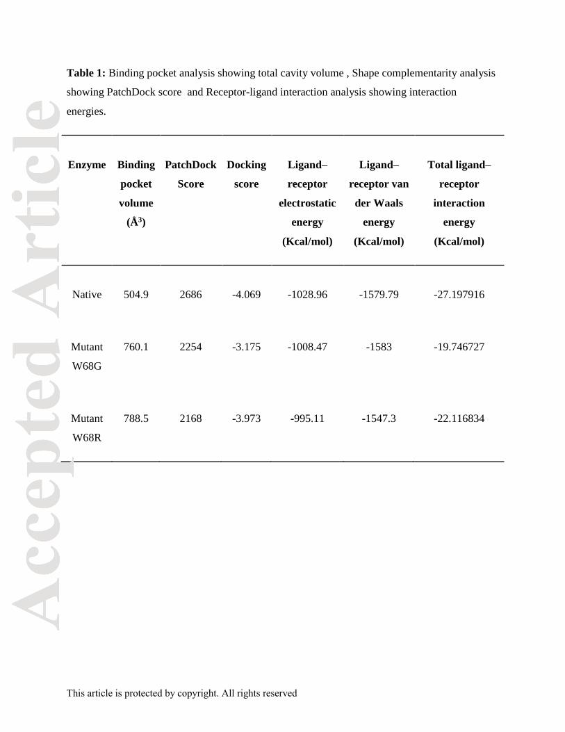
Binding pocket volume of PZase proteins was evaluated using CASTp server. It measured the
substrate binding cavity of protein which may differ between wild and mutant proteins and state
the outcome of mutation. Significant difference was observed in the volume of native and mutant
protein pockets. Binding pocket volume of native, mutant W68G and W68R was 504.9Ǻ3,
760.1Ǻ3 and 788.5 Ǻ3, respectively (Table1). The cavity volume for native protein was optimum
for ligand binding. Increase or decrease in the cavity volume affected ligand occupancy. Increase
in binding pocket volume of mutants indicated their role in altering significant protein side chain
interaction with the drug.
Complementarity analysis
PatchDock web server estimated shape complementarity by calculating the docking score
between receptor and ligand. The docking score obtained for PZA with native and mutants
W68G & W68R complexes was 2686, 2254, and 2168, respectively (Table1). Shape
complementarity between receptor and ligand indicated towards the strength of atomic
interactions. Higher docking score for native protein stated higher magnitude of geometric
complementarity for native protein and drug in comparison to the mutant forms.

This article is protected by copyright. All rights reserved
Estimation of interaction energy between receptor and ligand molecules
Interaction energy of ligand-receptor complex is critical to comprehend the biological function of
ligand molecule. Prior studies on PZase suggested that van der Waals forces were significant for
ligand binding. Van der Waals forces and electrostatic interaction forces of protein-drug
complexes were measured by Glide module of Schrodinger. Van der Waals forces and
electrostatic forces showed higher complementarity between the native PZase and PZA
molecules in comparison to the mutants (Table 1). Comparison of the native and mutant protein
total interaction energies with PZA and complementarity analysis signified the characteristic
difference of binding between the native and mutant protein-drug complexes. The docking score
for native PZase protein was -4.069 while for mutant W68R and W68G was -3.973 and -3.175,
respectively (Table 1). This showed that the PZA-PZase binding affinity for W68G mutant is
significantly low and hence W68G mutant protein has less stable binding with PZA than W68R
mutant or native PZase protein. . Thus, the overall deviation in the binding of ligand to receptor
was due to the deforming mutations present in the active site of PZase protein.
Molecular dynamics trajectory analysis of protein and drug complexes
To understand the effect of mutations on the PZase structure and function, MD simulations were
performed using the native and mutant proteins in complex with PZA drug. High performance
molecular dynamics simulations of the protein-ligand complexes were performed for 40 ns.
Various analyses were performed on the resultant trajectories. RMSD of backbone, RMSF of
protein residues, Rg of complex, SASA of protein and hbond between the protein and ligand
were investigated and compared for native and mutant proteins.
Root-mean-square deviations (RMSD) for backbone of the proteins was estimated which showed
least deviation for native protein in comparison to the mutant structures W68G and W68R
(Figure 1). Native structure showed deviations and achieved 0.23 nm of RMSD at ~6 ns. W68G
structure achieved 0.25 nm of RMSD at ~10 ns and showed more deviations whereas W68R
structure attained 0.35 nm at ~12 nm showed maximum deviation. From 22 ns to 40 ns, native
structure was stable and maintained 0.25nm of backbone of RMSD with least deviations. W68G
was stable from 22 ns to40 ns with a 0.26 nm of backbone of RMSD whereas W68R was stable
with 0.33 nm RMSD. RMSD values of native and mutant proteins were stable after ~ 22 ns and
showed similar fashion of deviation. MD trajectories reached convergence as plateau was

This article is protected by copyright. All rights reserved
obtained within the time period of 22-40 ns (Figure 1). The trajectories obtained were stable and
reliable and hence could be used for further analysis.
Potential energy of all protein complexes were calculated which revealed the potential energy of
mutant protein complexes was higher than the native protein complex (Figure 2). This showed
that the native protein complex was highly stable. Mutant W68R protein complex had a more
positive value than native. Mutant W68G protein complex had the most positive value and is not
stable. Like the RMSD values indicate, native protein is stably folded without any deviations
whereas the mutant proteins showed reduced stability during the course of molecular dynamics.
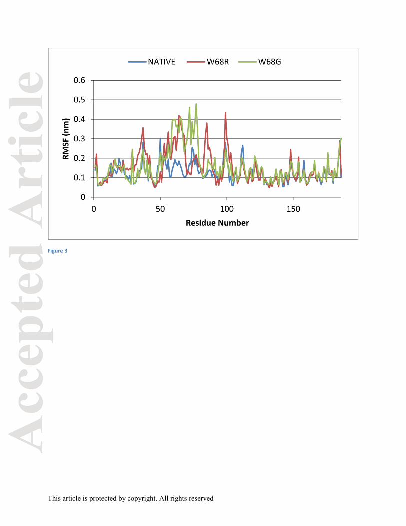
Root-mean-square fluctuation (RMSF) was calculated for every residue of the native and mutant
proteins. It can be observed that major fluctuations arose in the residues 51 to 104 (Figure 3).
Fluctuations in mutant proteins were higher than occurring in the native protein. Native protein
showed fluctuations ranging from 0.05 to 0.30 nm whereas W68G and W68R showed
fluctuations ranging from 0.06 to 0.48 nm and 0.05 to 0.43 nm respectively. A higher RMSF
value indicated high flexibility in the region whereas a low RMSF indicated constrained regions.
There was higher flexibility in the residues 51 to 104 for the mutant proteins which imparted
greater mobility to the amino acids forming a loosely packed protein throughout the MD
simulation period which might have contributed to the lower affinity with the drug, PZA. The
residues involved in the catalytic site and metal ion binding site showed major variations along
with the active site residues. The catalytic triad Asp 8, Lys 96 and Cys 138 showed variations in
the mutant W68G protein. On the other hand, the metal ion binding sites Asp 49, His 51, His57
and His71 showed major variations for both the mutant proteins W68G and W68R. The
conserved regions of amino acids 61 to 85 showed major fluctuations ranging from 0.1 to 0.47
nm. The loop region, 52 to 70, which constituted the flap closing the binding cavity for PZA,
showed higher fluctuations for the mutant proteins. Trp 68 is a part of the catalytic cleft which
formed direct hydrogen bond with His 57. Mutation at this site had affected the proper
positioning of PZA at the binding site in the protein. Fluctuations in the RMSF values proved the
same.
Rg, radius of gyration was outlined as root mean-square distance of a cluster of atoms based on
mass to weight ratio from the same center of mass. Rg was plotted against time which defines the
This article is protected by copyright. All rights reserved
degree of compactness for protein during MD simulations (Figure 4). A stable Rg value signifies
that there is no change in protein folding while varying values signify protein folding or
unfolding. The plot obtained in case of native protein maintained stability through the
simulation period, while mutant protein W68G showed highest variations. Mutant protein was
less compact due to the mutations resulting in lower stability. Solvent accessible surface area
(SASA) estimated the protein surface in contact with the solvent molecules. The solvation effect
is important for maintaining the stability of protein and its structure. SASA was estimated using
gmx sasa module for a simulation period of 40,000ps (Figure 5). SASA for the native protein
was in the range from ~94 nm2 to ~75 nm2. The mutant W68R had SASA of ~95 nm2 to ~77
nm2and for the mutant W68G it was ~ 95 nm2 to ~78 nm2. Thus, there was not much of a
difference in the solvent accessible surface of the native and mutant proteins.
NH bond analysis was done to comprehend the stability of the protein-ligand complex. Hydrogen
bonds play a significant role in imparting stability to the protein structure. H-bonds between the
protein and ligand molecule were analyzed during the 40 ns simulation period. Simulation of the
protein complexes showed significant difference in the H-bonds formed between the proteins and
ligand. H-bonds for the native as well as mutant protein-ligand structures were mostly 2 to 3
throughout the simulation (Figure 6).
Receptor-ligand interaction analysis
Hydrogen and hydrophobic interactions are important to maintain the proper functioning of
protein-ligand complex. Hydrogen bonds and hydrophobic interactions were estimated for
protein-ligand complexes after docking and MD simulation. Interaction plots were generated
using Maestro and CHIMERA [Huang et al., 1996]
Native protein formed two H-bonds with PZA for which nitrogen atom of His137 and Cys138
were involved with oxygen atom of PZA having bond length of 2.50Å and 2.76Å, respectively
(Fig 7). Mutant W68R also formed two H-bonds with PZA by Ala134 with bond length of
2.83Å and Cys 138 with bond length of 2.88Å (Fig 8). Mutant W68G formed two H-bonds with
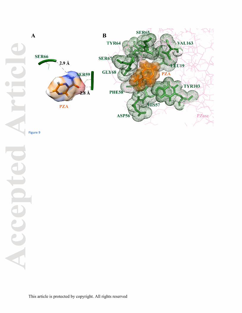
PZA. Oxygen atoms of Ser59 and Ser66 formed H-bond with nitrogen atom of PZA having bond
length of 3.21Å and 2.93Å, respectively (Fig 9). There was a significant increase in the bond
lengths of H-bond in the mutant proteins. This could be due to the increased binding pocket
volume and highly rigid and non-flexible structure.

This article is protected by copyright. All rights reserved
PZA showed hydrophobic interactions with Phe13, Asp49, His57 Phe58, Trp68, His71, Tyr103,
Ala134, Asp136, and Val163 of native protein. PZA formed hydrophobic bonds with Asp8,
Phe13, Leu19, Val21, Phe58, Ser66, Ser67, Ile133, His137, Val139, and Val163 of W68Rprotein
whereas the residues Leu19, Asp56, His57, Phe58, Ser65, Tyr64, Ser67, Gly68, Tyr103, and
Val163 were involved for mutant W68G protein. An overall increased number of hydrophobic
bonds were observed between the mutant proteins and ligand. The binding interactions between
PZA and PZase proteins have been listed in Table 2.
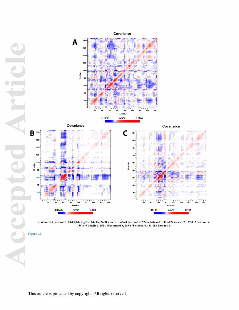
Essential dynamics analysis
Essential dynamics analysis was performed on the MD trajectory to study the changes in the
motion between the proteins. Large-scale collective movements of the protein structures were
estimated and dynamics of the proteins was obtained by characterization of their phase-space
behavior. Figure 10 shows the overall secondary structural features of pyrazinamidase protein.
Analysis of backbone atoms showed that the mutant proteins have less correlation in comparison
to the native form (Figure 11). The red regions are the positive regions associated with the
correlated residue movement in the same direction whereas the blue region is the negative region
indicating anti-correlated residue motions in the opposite direction.
Residues of native protein-ligand complex found in correlated motions were: β-strand 3 and α-
helix 1, α-helix 3 and α-helix 4. Anti-correlated motions were observed between β-strand 1 and
α-helix 4, α-helix 3 and 3/10-helix, α-helix 4 and 3/10-helix, β-strand 3 and 3/10-helix. All these
motions are important for optimal interaction between the protein and ligand, so that the protein
can effectively act on the ligand. The mutant W68Rprotein-ligand complex residues showed
correlated motions mostly similar to the native protein-ligand complex. The uncorrelated
motions were in α-helix 3 and 3/10-helix, α-helix 4 and 3/10-helix, β-strand 3 and 3/10-helix and
α-helix 1 and3/10-helix. The mutant W68G protein-ligand complex residues showed correlated
motions same as the native protein-ligand complex. The uncorrelated motions were in α-helix 3
and 3/10-helix, α-helix 4 and 3/10-helix, and β-strand 3 and 3/10-helix.
Projection of proteins’ motion was given by eigenvectors 1 and 2, eigenvectors 2 and 3, and
eigenvectors 7 and 8 from each of the MD trajectory obtained as seen in Figure 12. The

This article is protected by copyright. All rights reserved
corresponding eigenvalues indicated that variations in the protein system were restricted to first
three eigenvectors and the proteins attained their equilibrium fluctuations within 8 eigenvectors.
Analysis of these plots revealed that mutant W68R and W68G spread over a greater area of
phase space than native protein mainly along PC1, PC2 and PC3 planes. It can thus be implied
that the mutant proteins are more flexible than native protein at 310 K and tend to have more
conformational fluctuations. Projection of the trajectories along the seventh and eighth principal
components (PC7, PC8) was well defined for all the three native and mutant proteins. Hence, the
three well defined trajectory clusters as shown in figure indicated the changes of conformational
motion that occurred upon point mutation in the protein structure.
Conclusion
Due to increasing emergence of drug-resistant Mtb strains, present conventional therapies are
rendered ineffective. Understanding the effect of gene mutation on structure of protein and its
affinity for the ligand is required to aid the knowledge of drug development. This study is based
on comprehensive analysis of the PZA drug resistance in Mycobacterium tuberculosis due to
pncA gene mutations. Binding pocket determination, shape complementarity computation,
molecular docking, molecular dynamics simulation and essential dynamic studies have been
performed. Docking of the native protein showed best complementarity between the receptor and
ligand. The protein trajectories showed that there was increased flexibility in the mutant protein
atoms. The mutant proteins were loosely packed, less compact and show lower stability. All the
analyses showed that the protein had lost its compactness and stability because of the mutation.
The H-bond and hydrophobic interactions between the receptor and ligand were different for the
native and mutant proteins affecting the protein-drug interaction. The mutant W68G protein was
found to drastically affect the structure of binding site of protein. The protein-drug interaction
was inefficient due to the structural change resulting in PZA drug resistance in Mycobacterium
tuberculosis. Mutation in the 68th residue led to a structural change in the binding site of protein,
thus PZA was not converted to its active form that renders it ineffective for TB treatment. The
investigation of the dynamic behavior gave highly reliable information on the PZase mutation

This article is protected by copyright. All rights reserved
affecting its structure and insights into the change in protein–ligand interactions due to
conformational variations.
Acknowledgements
AG is thankful to Jawaharlal Nehru University for usage of all computational facilities. AG is
grateful to University Grants Commission, India for the Faculty Recharge position.
Competing financial interests
Authors declare no competing financial interests.
This article is protected by copyright. All rights reserved
References
Abraham MJ, Murtola T, Schulz R, Páll S, Smith JC, Hess B, Lindahl E. 2015. GROMACS:
High performance molecular simulations through multi-level parallelism from laptops to
supercomputers. SoftwareX 1:19-25.
Amadei A, Linssen A, Berendsen HJ. 1993. Essential dynamics of proteins. Proteins: Struct,
Funct, Bioinf 17:412-425.
Bamaga M, Wright D, Zhang H. 2002. Selection of in vitro mutants of pyrazinamide-resistant
Mycobacterium tuberculosis. Int J Antimicrob Agents 20:275-281.
Berendsen HJ, Postma Jv, van Gunsteren WF, DiNola A, Haak J. 1984. Molecular dynamics
with coupling to an external bath. J Chem Phys 81:3684-3690.
Berendsen HJ, van der Spoel D, van Drunen R. 1995. GROMACS: a message-passing parallel
molecular dynamics implementation. Comput Phys Commun 91:43-56.
Berman HM, Westbrook J, Feng Z, Gilliland G, Bhat T, Weissig H, Shindyalov IN, Bourne PE.
2000. The protein data bank. Nucleic Acids Res 28:235-242.
Chan RC, Hui M, Chan EW, Au T, Chin ML, Yip CK, AuYeang CK, Yeung CY, Kam KM, Yip
PC. 2007. Genetic and phenotypic characterization of drug-resistant Mycobacterium tuberculosis
isolates in Hong Kong. J Antimicrob Chemother 59:866-873.
Chang KC, Yew WW, Zhang Y. 2011. Pyrazinamide susceptibility testing in Mycobacterium
tuberculosis: a systematic review with meta-analyses. Antimicrob Agents Chemother 55:4499-
4505.
Cheng S-J, Thibert L, Sanchez T, Heifets L, Zhang Y. 2000. pncA mutations as a major
mechanism of pyrazinamide resistance in Mycobacterium tuberculosis: spread of a monoresistant
strain in Quebec, Canada. Antimicrob Agents Chemother 44:528-532.
Cole S, Brosch R, Parkhill J, Garnier T, Churcher C, Harris D, Gordon S, Eiglmeier K, Gas S,
Barry Cr. 1998. Deciphering the biology of Mycobacterium tuberculosis from the complete
genome sequence. Nature 393:537-544.
Da Silva PEA, Palomino JC. 2011. Molecular basis and mechanisms of drug resistance in
Mycobacterium tuberculosis: classical and new drugs. J Antimicrob Chemother 66:1417-1430.
Darden T, York D, Pedersen L. 1993. Particle mesh Ewald: An N⋅ log (N) method for Ewald
sums in large systems. J Chem Phys 98:10089-10092.

This article is protected by copyright. All rights reserved
Dooley KE, Mitnick CD, DeGroote MA, Obuku E, Belitsky V, Hamilton CD, Makhene M, Shah
S, Brust JC, Durakovic N. 2012. Old drugs, new purpose: retooling existing drugs for optimized
treatment of resistant tuberculosis. Clin. Infect. Dis. 55:572-581.
Du X, Wang W, Kim R, Yakota H, Nguyen H, Kim S-H. 2001. Crystal structure and mechanism
of catalysis of a pyrazinamidase from Pyrococcus horikoshii. Biochemistry 40:14166-14172.
Duhovny D, Nussinov R, Wolfson HJ. 2002. Efficient unbound docking of rigid molecules.
Algorithms in bioinformatics. Springer, p 185-200.
Dundas J, Ouyang Z, Tseng J, Binkowski A, Turpaz Y, Liang J. 2006. CASTp: computed atlas
of surface topography of proteins with structural and topographical mapping of functionally
annotated residues. Nucleic Acids Res 34:W116-W118.
Emekli U, Schneidman‐Duhovny D, Wolfson HJ, Nussinov R, Haliloglu T. 2008. HingeProt:
automated prediction of hinges in protein structures. Proteins: Struct, Funct, Bioinf 70:1219-
1227.
Friesner RA, Banks JL, Murphy RB, Halgren TA, Klicic JJ, Mainz DT, Repasky MP, Knoll EH,
Shelley M, Perry JK. 2004. Glide: a new approach for rapid, accurate docking and scoring. 1.
Method and assessment of docking accuracy. J Med Chem 47:1739-1749.
Friesner RA, Murphy RB, Repasky MP, Frye LL, Greenwood JR, Halgren TA, Sanschagrin PC,
Mainz DT. 2006. Extra precision glide: docking and scoring incorporating a model of
hydrophobic enclosure for protein-ligand complexes. J Med Chem 49:6177-6196.
Glide V. 2009. 5.5, Schrodinger. New York, NY.
Gu Y, Yu X, Jiang G, Wang X, Ma Y, Li Y, Huang H. 2015. Pyrazinamide resistance among
multidrug-resistant tuberculosis clinical isolates in a national referral center of China and its
correlations with pncA, rpsA, and panD gene mutations. Diagn Microbiol Infect Dis.
Halgren TA, Murphy RB, Friesner RA, Beard HS, Frye LL, Pollard WT, Banks JL. 2004. Glide:
a new approach for rapid, accurate docking and scoring. 2. Enrichment factors in database
screening. J Med Chem 47:1750-1759.
Hess B, Bekker H, Berendsen HJ, Fraaije JG. 1997. LINCS: a linear constraint solver for
molecular simulations. J Comput Chem 18:1463-1472.
Hess B, Kutzner C, Van Der Spoel D, Lindahl E. 2008. GROMACS 4: algorithms for highly
efficient, load-balanced, and scalable molecular simulation. J Chem Theory Comput 4:435-447.

This article is protected by copyright. All rights reserved
Huang CC, Couch GS, Pettersen EF, Ferrin TE. 1996. Chimera: an extensible molecular
modeling application constructed using standard components. Pacific symposium on
biocomputing. p 1519-1523.
Jamal S, Goyal S, Shanker A, Grover A. 2016. Integrating network, sequence and functional
features using machine learning approaches towards identification of novel Alzheimer genes.
BMC genomics 17:807.
Jones S, Thornton JM. 1996. Principles of protein-protein interactions. Proc Natl Acad Sci
93:13-20.
Kim S, Thiessen PA, Bolton EE, Chen J, Fu G, Gindulyte A, Han L, He J, He S, Shoemaker BA.
2015. PubChem substance and compound databases. Nucleic Acids Res:gkv951.
Konno K, Feldmann FM, McDermott W. 1967. Pyrazinamide Susceptibility and Amidase
Activity of Tubercle Bacilli 1, 2. Am Rev Respir Dis 95:461-469.
Lakshmipathy D, Ramasubban G, Therese L, Vetrivel U, Sivashanmugam M, Rajendiran S,
Sridhar R, Madhavan H, Meenakshi N. 2013. In silico Analysis of Novel Mutation ala102pro
Targeting pncA Gene of M. Tuberculosis. J Comp Sci & Sys Biol 2013.
Lee KW, Lee JM, Jung KS. 2001. Characterization of pncA mutations of pyrazinamide-resistant
Mycobacterium tuberculosis in Korea. J Korean Med Sci 16:537.
Lemaitre N, Sougakoff W, Truffot-Pernot C, Jarlier V. 1999. Characterization of New Mutations
in Pyrazinamide-Resistant Strains of Mycobacterium tuberculosisand Identification of Conserved
Regions Important for the Catalytic Activity of the Pyrazinamidase PncA. Antimicrob Agents
Chemother 43:1761-1763.
Malone L, Schurr A, Lindh H, McKenzie D, Kiser J, Williams J. 1952. The Effect of
Pyrazinamide (Aldinamide) on Experi-mental Tuberculosis in Mice. Am Rev Respir Dis 65:511-
18.
McKenzie D, Malone L, Kushner S, Oleson J, Subbarow Y. 1948. The Effect of Nieotinic Acid
Amide on Experimental Tuberculosis of White Mice. J Lab Clin Med 33:1249-53.
Mitchison D. 1985. The action of antituberculosis drugs in short-course chemotherapy. Tubercle
66:219-225.
Nagpal N, Goyal S, Wahi D, Jain R, Jamal S, Singh A, Rana P, Grover A. 2015. Molecular
principles behind Boceprevir resistance due to mutations in hepatitis C NS3/4A protease. Gene
570:115-121.
This article is protected by copyright. All rights reserved
Organization WH. 2015. Global tuberculosis report 2015.
Organization WH. 2016. Global tuberculosis report 2016.
Pandey B, Grover S, Tyagi C, Goyal S, Jamal S, Singh A, Kaur J, Grover A. 2016. Molecular
principles behind pyrazinamide resistance due to mutations in panD gene in Mycobacterium
tuberculosis. Gene 581:31-42.
Parrinello M, Rahman A. 1981. Polymorphic transitions in single crystals: A new molecular
dynamics method. J Appl Phys 52:7182-7190.
Petrella S, Gelus-Ziental N, Maudry A, Laurans C, Boudjelloul R, Sougakoff W. 2011. Crystal
structure of the pyrazinamidase of Mycobacterium tuberculosis: insights into natural and
acquired resistance to pyrazinamide. PLoS One 6:e15785.
Purohit R, Rajendran V, Sethumadhavan R. 2011. Relationship between mutation of serine
residue at 315th position in M. tuberculosis catalase-peroxidase enzyme and Isoniazid
susceptibility: an in silico analysis. J Mol Model 17:869-877.
Release S. 2013. 2: LigPrep, version 2.7. Schrödinger, LLC, New York, NY.
Ryan F. 1993. The Forgotten Plague: How the battle against tuberculosis was won--and lost.
Sastry GM, Adzhigirey M, Day T, Annabhimoju R, Sherman W. 2013. Protein and ligand
preparation: parameters, protocols, and influence on virtual screening enrichments. J Comput
Aided Mol Des 27:221-234.
Schneidman-Duhovny D, Inbar Y, Nussinov R, Wolfson HJ. 2005. PatchDock and SymmDock:
servers for rigid and symmetric docking. Nucleic Acids Res 33:W363-W367.
Schrödinger L. The PyMOL Molecular Graphics System. 1.4.: Schrödinger: LLC.
Schrödinger L. 2013. Schrödinger Suite 2011 Protein Preparation Wizard. Epik version 2.
SchuÈttelkopf AW, Van Aalten DM. 2004. PRODRG: a tool for high-throughput
crystallography of protein–ligand complexes. Acta Crystallogr D Biol Crystallogr 60:1355-1363.
Scorpio A, Lindholm-Levy P, Heifets L, Gilman R, Siddiqi S, Cynamon M, Zhang Y. 1997.
Characterization of pncA mutations in pyrazinamide-resistant Mycobacterium tuberculosis.
Antimicrob Agents Chemother 41:540-543.
Scorpio A, Zhang Y. 1996. Mutations in pncA, a gene encoding pyrazinamidase/nicotinamidase,
cause resistance to the antituberculous drug pyrazinamide in tubercle bacillus. Nat Med 2:662-
667.

This article is protected by copyright. All rights reserved
Sheen P, Ferrer P, Gilman RH, Christiansen G, Moreno-Román P, Gutiérrez AH, Sotelo J,
Evangelista W, Fuentes P, Rueda D. 2012. Role of metal ions on the activity of Mycobacterium
tuberculosis pyrazinamidase. Am J Trop Med Hyg 87:153-161.
Shi W, Zhang X, Jiang X, Yuan H, Lee JS, Barry CE, Wang H, Zhang W, Zhang Y. 2011.
Pyrazinamide inhibits trans-translation in Mycobacterium tuberculosis. Science 333:1630-1632.
Singh A, Goyal S, Jamal S, Subramani B, Das M, Admane N, Grover A. 2016. Computational
identification of novel piperidine derivatives as potential HDM2 inhibitors designed by
fragment-based QSAR, molecular docking and molecular dynamics simulations. Struct Chem
27:993-1003.
Singh A, Grover S, Sinha S, Das M, Somvanshi P, Grover A. 2017. Mechanistic principles
behind molecular mechanism of Rifampicin resistance in mutant RNA polymerase beta subunit
of Mycobacterium tuberculosis. J Cell Biochem.
Sinha S, Tyagi C, Goyal S, Jamal S, Somvanshi P, Grover A. 2016. Fragment based G-QSAR
and molecular dynamics based mechanistic simulations into hydroxamic-based HDAC inhibitors
against spinocerebellar ataxia. J Biomol Struct Dyn:1-15.
Solotorovsky M, Gregory F, Ironson E, Bugie E, O'Neill R, Pfister K. 1952. Pyrazinoic acid
amide-An agent active against experimental murine tuberculosis. Exp Biol Med 79:563-565.
Stehr M, Elamin AA, Singh M. 2015. Pyrazinamide: the importance of uncovering the
mechanisms of action in mycobacteria. Expert Rev Anti Infect Ther 13:593-603.
Unissa AN, Sudha S, Selvakumar N. 2011. Elucidating pyrazinamide resistance in
mycobacterium tuberculosis by molecular docking.
Van Der Spoel D, Lindahl E, Hess B, Groenhof G, Mark AE, Berendsen HJ. 2005. GROMACS:
fast, flexible, and free. J Comput Chem 26:1701-1718.
van Gunsteren WF, Billeter S, Eising A, Hünenberger PH, Krüger P, Mark AE, Scott W, Tironi
IG. 1996. Biomolecular simulation: The {GROMOS96} manual and user guide.
Vats C, Dhanjal JK, Goyal S, Gupta A, Bharadvaja N, Grover A. 2015. Mechanistic analysis
elucidating the relationship between Lys96 mutation in Mycobacterium tuberculosis
pyrazinamidase enzyme and pyrazinamide susceptibility. BMC genomics 16:1.
Verma S, Goyal S, Tyagi C, Jamal S, Singh A, Grover A. 2016a. BIM (BCL-2 interacting
mediator of cell death) SAHB (stabilized α helix of BCL2) not always convinces BAX (BCL-2-
associated X protein) for apoptosis. J Mol Graph Model 67:94-101.

This article is protected by copyright. All rights reserved
Verma S, Grover S, Tyagi C, Goyal S, Jamal S, Singh A, Grover A. 2016b. Hydrophobic
Interactions Are a Key to MDM2 Inhibition by Polyphenols as Revealed by Molecular Dynamics
Simulations and MM/PBSA Free Energy Calculations. PLoS One 11:e0149014.
Verma S, Tyagi C, Goyal S, Pandey B, Jamal S, Singh A, Grover A. 2016c. Mutations induce
conformational changes in folliculin C-terminal domain: possible cause of loss of guanine
exchange factor activity and Birt-Hogg-Dubé syndrome. J Biomol Struct Dyn:1-6.
Wu Y, Tepper HL, Voth GA. 2006. Flexible simple point-charge water model with improved
liquid-state properties. J Chem Phys 124:024503.
Yeager R, Munroe W, Dessau FI. 1952. Pyrazinamide (aldinamide) in the treatment of
pulmonary tuberculosis. American Review of Tuberculosis and Pulmonary Diseases 65:523-46.
Zhang C, Vasmatzis G, Cornette JL, DeLisi C. 1997. Determination of atomic desolvation
energies from the structures of crystallized proteins. J Mol Biol 267:707-726.
Zhang H, Deng JY, Bi LJ, Zhou YF, Zhang ZP, Zhang CG, Zhang Y, Zhang XE. 2008.
Characterization of Mycobacterium tuberculosis nicotinamidase/pyrazinamidase. FEBS J
275:753-762.
Zhang S, Chen J, Shi W, Liu W, Zhang W, Zhang Y. 2013. Mutations in panD encoding
aspartate decarboxylase are associated with pyrazinamide resistance in Mycobacterium
tuberculosis. Emerg Microbes Infect 2:e34.
Zhang Y, Mitchison D. 2003. The curious characteristics of pyrazinamide: a review. Int J Tuberc
Lung Dis 7:6-21.
Zhang Y, Scorpio A, Nikaido H, Sun Z. 1999. Role of acid pH and deficient efflux of pyrazinoic
acid in unique susceptibility of Mycobacterium tuberculosis to pyrazinamide. J Bacteriol
181:2044-2049.
Zhang Y, Telenti A. 2000. Genetics of drug resistance in Mycobacterium tuberculosis. Molecular
genetics of mycobacteria. ASM Press, Washington, DC.

This article is protected by copyright. All rights reserved
Figure Legends
Figure 1: Root mean square deviation (backbone) as a function of time for native and mutant
proteins bound to PZA.
Figure 2: Potential energy of the native and mutant proteins bound to PZA versus time.
Figure 3: Root-mean square fluctuation of all residues of native and mutant proteins bound to
PZA.
Figure 4: Radius of gyration of Cα atoms of native and mutant proteins bound to PZA.
Figure 5: Solvent accessible surface area of the complex of native and mutant proteins bound to
PZA.
Figure 6: Hydrogen bonds formed by the native and mutant protein with PZA.
Figure 7: PZA interaction with Native W68 PZase protein (A) Hydrogen bonds, (B)
hydrophobic interactions.
Figure 8: PZA interaction with Mutant W68R PZase protein (A) Hydrogen bonds, (B)
hydrophobic interactions.
Figure 9: PZA docking interaction with Mutant W68G PZase protein (A) Hydrogen bonds, (B)
hydrophobic interactions.
Figure 10: PZase protein illustration of secondary structure elements: α-helix and β-strand and
coordinated Fe atom.
Figure 11: Covariance matrix A. Native W68 protein, B. Mutant W68R protein, C. Mutant
W68G protein.
Figure 12: Projection of the motion of the protein in phase space at 300K A. Along the principal
eigenvectors 1 and 2, B. Along the principal eigenvectors 2 and 3, C. Along the principal
eigenvectors 7 and 8.
This article is protected by copyright. All rights reserved
Table 1: Binding pocket analysis showing total cavity volume , Shape complementarity analysis
showing PatchDock score and Receptor-ligand interaction analysis showing interaction
energies.
Enzyme Binding
volume
(Å3)
PatchDock
Score
Docking
score
Ligand–
receptor
electrostatic
energy
(Kcal/mol)
Ligand–
receptor van
der Waals
energy
(Kcal/mol)
Total ligand–
receptor
interaction
energy
(Kcal/mol)
Native 504.9 2686 -4.069 -1028.96 -1579.79 -27.197916
Mutant
W68G
760.1 2254 -3.175 -1008.47 -1583 -19.746727
Mutant
W68R
788.5 2168 -3.973 -995.11 -1547.3 -22.116834

This article is protected by copyright. All rights reserved
Table 2: Binding interactions between PZA and native and mutant PZase.
Protein H-bond Hydrophobic bond
NATIVE W68 protein-
ligand complex
His137, Cys138 Phe13, Asp49, His57 Phe58, Trp68,
His71, Tyr103, Ala134, Asp136,
Val163
Mutant W68R protein-
ligand complex
Ala134, Cys138 Asp8, Phe13, Leu19, Val21,Phe58,
Ser66, Ser67, Ile133, His137, Val139,
Val163
Mutant W68G protein-
ligand complex
Ser59, Ser66 Leu19, Asp56, His57, Phe58, Ser65,
Tyr64, Ser67, Gly68, Tyr103, Val163

This article is protected by copyright. All rights reserved
Figure 1

This article is protected by copyright. All rights reserved
Figure 2
This article is protected by copyright. All rights reserved
Figure 3

This article is protected by copyright. All rights reserved
Figure 4

This article is protected by copyright. All rights reserved
Figure 5

This article is protected by copyright. All rights reserved
Figure 6

This article is protected by copyright. All rights reserved
Figure 7

This article is protected by copyright. All rights reserved
Figure 8
This article is protected by copyright. All rights reserved
Figure 9

This article is protected by copyright. All rights reserved
Figure 10
This article is protected by copyright. All rights reserved
Figure 11

This article is protected by copyright. All rights reserved
Figure 12