Embed Size (px)
Citation preview

1
CIR-16-0382R[Martner]
Role of NOX2-derived reactive oxygen species in NK cell-mediated control of
murine melanoma metastasis
Ebru Aydin1, Junko Johansson
1, Faisal Hayat Nazir
1, 2, Kristoffer Hellstrand
1 and
Anna Martner1*
1 TIMM Laboratory, Sahlgrenska Cancer Center, University of Gothenburg, 405 30
Gothenburg, Sweden
2 Department of Psychiatry and Neurochemistry, University of Gothenburg, 405 30
Gothenburg, Sweden
Running title: NOX2 in regulation of melanoma metastasis
Key words: melanoma metastasis, NOX2, reactive oxygen species, NK cells, IFNγ,
immunotherapy
List of abbreviations: HDC, histamine dihydrochloride; NOX2, NADPH oxidase
isoform 2; ROS, reactive oxygen species
Financial support: This work was supported by the Swedish Research Council (2012-
2047 to AM, 2012-3205 to KH), the Swedish Society for Medical Research (SSMF to
AM), the Swedish Cancer Society (CAN 212/595 to AM, CAN 2015/411 to KH), the
Swedish state via the ALF agreement (ALFGBG-436961 to AM), the Erna and Victor
Hasselblad Foundation (to AM), the Torsten and Ragnar Söderberg Foundation (to
KH), the Assar Gabrielsson Foundation (to EA), BioCARE - a National Strategic
Research Program at University of Gothenburg and the Sahlgrenska Academy at
University of Gothenburg (to AM).
*Corresponding author: Anna Martner, TIMM Laboratory, Sahlgrenska Cancer
Center, University of Gothenburg, Medicinaregatan 1F, 41390 Gothenburg, Sweden;
Phone: +46 736 517644, E-mail: [email protected]
on August 21, 2018. © 2017 American Association for Cancer Research. cancerimmunolres.aacrjournals.org Downloaded from
Author manuscripts have been peer reviewed and accepted for publication but have not yet been edited. Author Manuscript Published OnlineFirst on July 31, 2017; DOI: 10.1158/2326-6066.CIR-16-0382

2
ABSTRACT
The NADPH oxidase of myeloid cells, NOX2, generates reactive oxygen species
(ROS) to eliminate pathogens and malignant cells. NOX2-derived ROS also have
been proposed to dampen functions of natural killer (NK) cells and other anti-
neoplastic lymphocytes in the microenvironment of established tumors. The
mechanisms by which NOX2 and ROS influence the process of distant metastasis
have only been partially explored. Here we utilized genetically NOX2-deficient mice
and pharmacological inhibition of NOX2 to elucidate the role of NOX2 for the
hematogenous metastasis of melanoma cells. After intravenous inoculation of B16F1
or B16F10 cells, lung metastasis formation was reduced in B6.129S6-Cybbtm1DinK
(Nox2-KO) vs. Nox2-sufficient wild-type (WT) mice. Systemic treatment with the
NOX2-inhibitor histamine dihydrochloride (HDC) reduced melanoma metastasis and
enhanced the infiltration of IFN-producing NK cells into lungs of WT but not of
Nox2-KO mice. IFN-deficient B6.129S7-Ifngtm1Ts
/J mice were prone to develop
melanoma metastases and did not respond to in vivo treatment with HDC. We propose
that NOX2-derived ROS facilitate metastasis of melanoma cells by down-modulating
NK cell function and that inhibition of NOX2 may restore IFNγ-dependent, NK cell-
mediated clearance of melanoma cells.
on August 21, 2018. © 2017 American Association for Cancer Research. cancerimmunolres.aacrjournals.org Downloaded from
Author manuscripts have been peer reviewed and accepted for publication but have not yet been edited. Author Manuscript Published OnlineFirst on July 31, 2017; DOI: 10.1158/2326-6066.CIR-16-0382

3
INTRODUCTION
Reactive oxygen species (ROS) are short-lived compounds that arise from electron
transfer across biological membranes where the electron acceptor is molecular oxygen
and the initial product is superoxide anion (O2-). ROS refer to oxygen radicals such as
O2- and the hydroxyl radical (OH
.) along with non-radicals, including hydrogen
peroxide, that share the oxidizing capacity of oxygen radicals and may be converted
into radicals (1). ROS are generated as by-products of mitochondrial ATP generation
in the electron transport chain but are also produced in a regulated fashion by the
nicotinamide adenine dinucleotide phosphate (NADPH) oxidases (NOX) and the dual
oxidases (DUOX). This family of transmembrane proteins comprises NOX1-5 and
DUOX1-2 whose only known function is to produce ROS (2).
The NOX proteins are structurally similar and utilize a similar principal mechanism
of ROS generation but vary in cellular and subcellular distribution. NOX2 is
expressed almost exclusively in cells of the myeloid lineage such as
monocyte/macrophages and neutrophilic granulocytes (3, 4). These cells utilize
NOX2-derived ROS to eliminate intra- and extracellular microorganisms (5). NOX2
also has been linked to immunosuppression in cancer: when released from myeloid
cells into the extracellular space, ROS generated by NOX2 may trigger dysfunction
and apoptosis of adjacent anti-neoplastic lymphocytes, including natural killer (NK)
cells (6, 7, 8, 9). The strategy to target ROS formation by myeloid cells has been
proposed to improve the efficiency of cancer immunotherapy (3, 10, 11, 12).
on August 21, 2018. © 2017 American Association for Cancer Research. cancerimmunolres.aacrjournals.org Downloaded from
Author manuscripts have been peer reviewed and accepted for publication but have not yet been edited. Author Manuscript Published OnlineFirst on July 31, 2017; DOI: 10.1158/2326-6066.CIR-16-0382

4
The role of ROS and NOX2 for the growth and metastatic spread of cancer cells is,
however, complex and controversial. Thus, although the genetic disruption of Nox2
reduces the subcutaneous growth of murine melanoma and lung carcinoma, it does
not affect sarcoma growth or prostate cancer growth in mice (13, 14). Also, the in
vivo administration of scavengers of ROS such as N-acetyl-cysteine reduces the
tumorigenicity of murine melanoma cells (15) but enhances lymph node metastasis in
other melanoma models, accelerates tumor progression in mouse models of B-RAF-
and K-RAS-induced lung cancer and accelerates the metastasis of xenografted human
melanoma cells in immunodeficient mice (16, 17, 18).
The detailed mechanisms of relevance to the discrepant impact of ROS for the growth
and spread of cancer cells remain to be eluciated. Further understanding of the role of
ROS for cancer progression requires experimental models that address a distinct
phase of tumor progression, define the source of ROS and take mechanisms of
immunosurveillance into account. For the present study, we aimed at determining the
impact of genetic and pharmacological inhibition of NOX2 in a murine NK cell-
dependent model of melanoma metastasis.
on August 21, 2018. © 2017 American Association for Cancer Research. cancerimmunolres.aacrjournals.org Downloaded from
Author manuscripts have been peer reviewed and accepted for publication but have not yet been edited. Author Manuscript Published OnlineFirst on July 31, 2017; DOI: 10.1158/2326-6066.CIR-16-0382

5
MATERIALS AND METHODS
Culture of cell lines. B16F1 and B16F10 murine melanoma cells were obtained in
2013 from the Cell Culture Laboratory at the Department of Virology, University of
Gothenburg where cells were authenticated by melanotic morphology and checked for
absence of mycoplasma using PCR before freezing aliquots. Each aliquot was thawn
and cultured for no more than one week for each experiment. Cells were cultured in
Iscoves’ medium containing 10% FCS (Sigma-Aldrich), 2 mM L-glutamine, 1 mM
sodium pyruvate, 100 U/ml penicillin, and 100 mg/ml streptomycin at 37°C, 5% CO2
for 1 week before inoculation into mice.
Induction of lung metastasis in Nox2-KO and Ifng-KO mice. All animal
experiments were approved by the Research Animal Ethics Committee in
Gothenburg. Mice were maintained under pathogen-free conditions according to
guidelines issued by the University of Gothenburg. C57BL/6 mice were obtained
from Charles River Laboratories (Sulzfeld, Germany). B6.129S6-Cybbtm1Din
(Nox2–/–
or Nox2-KO) mice that lack the myeloid gp91phox
subunit NOX2 and, thus, a
functional ROS-forming NOX2 were obtained from Jackson laboratories (Maine,
USA). B6.129S7-Ifngtm1Ts
/J (Ifng–/–
or Ifng-KO) mice that do not produce IFNγ (19)
were kindly provided by Prof. Nils Lycke, MIVAC at University of Gothenburg.
Naïve C57BL/6, Nox2-KO and Ifng-KO mice (6-12 weeks of age) were treated
intraperitoneally (i.p.) with PBS (control), HDC (Sigma, 1,500 μg/mouse), IL15 (0.04
μg/mouse), alone or combined, on the day before, the day after and 3 days after
intravenous (i.v.) inoculation of B16F10 cells (5-15x104 cells/mouse) or B16F1 cells
on August 21, 2018. © 2017 American Association for Cancer Research. cancerimmunolres.aacrjournals.org Downloaded from
Author manuscripts have been peer reviewed and accepted for publication but have not yet been edited. Author Manuscript Published OnlineFirst on July 31, 2017; DOI: 10.1158/2326-6066.CIR-16-0382

6
(20 x104–30 x10
4 cells/mouse). Three weeks after tumor inoculation, mice were
euthanized by cervical dislocation followed by harvesting of lungs and spleens. Lung
metastasis was determined by counting visible pulmonary metastatic foci under a light
microscope. The experimental design is outlined in Fig. 1A.
For assessment of the impact of NOX2 inhibition on immune parameters during the
early phase of tumor progression, mice received HDC at 1,500 μg/mouse or PBS
(control) one day before the inoculation of B16F10 cells followed by dissection of
lungs at 30 min or 24 hours after tumor cell inoculation as shown in Fig. 2A. In the
latter experiments, naïve mice and HDC-treated mice that did not receive melanoma
cells were used as additional controls.
Preparation of single cell suspensions from lungs and spleens. Lung tissues were
dissociated into single cells by combining enzymatic degradation of extracellular
matrix with mechanical dissociation using gentle MACS®
Technology (Miltenyi
Biotech, Auburn) based on instructions provided by the manufacturer. Single-cell
suspensions of splenocytes were prepared by mashing the spleens through a 70μm
cell strainer followed by depletion of erythrocytes using RBC Lysing buffer (Sigma-
Aldrich, Steinheim).
Flow cytometry. The following fluorochrome-labeled antimouse mAbs were
purchased from BD Biosciences: anti-CD45 (30-F11), anti-CD11c (HL3), anti-IaIe
(2G9), anti-CD3 (145-2311), anti-CD4 (RM4-5), anti-CD8 (53-6.7), anti-NK1.1
(PK136), anti-CD19 (1D3), anti-CD11b (M1/70), anti-Gr1 (RB6-8C5), anti-CD40
(3/23) and anti-Ly6C (AL-21). Anti-CD33 (29A1.4) was from Ebiosciences
on August 21, 2018. © 2017 American Association for Cancer Research. cancerimmunolres.aacrjournals.org Downloaded from
Author manuscripts have been peer reviewed and accepted for publication but have not yet been edited. Author Manuscript Published OnlineFirst on July 31, 2017; DOI: 10.1158/2326-6066.CIR-16-0382

7
(Stockholm); anti-F4/80 (BM8) and anti-CD69 (H1.2F3) were from BioLegend
(Stockholm). LIVE/DEAD Fixable Yellow Dead Cell Stain Kit or DAPI (both from
Invitrogen, Oregon) were used as cell viability markers in flow cytometry analyses. A
minimum of 100,000 gated live cells were collected on a four-laser BD LSRFortessa
(405, 488, 532, and 640 nm). Data were analyzed using FACSDiva Version 8.0.1
software (BD Biosciences).
Detection of ROS. Superoxide anion production was determined by use of the
isoluminol-electrogenerated chemiluminescence technique as described elsewhere
(20). Briefly, single cell suspensions of lungs were diluted to 107 cells/ml in Krebs-
Ringer glucose buffer supplemented with isoluminol (10 mg/ml; Sigma-Aldrich) and
horseradish peroxidase (HRP, 4 U/ml, Boehringer Mannheim, Germany) and added to
96-well plates that were incubated at 37oC. Phorbol myristate acetate (PMA, 5x10
-8
M, Sigma-Aldrich, Missouri, USA) or the formyl peptide receptor agonist
WKYMVm (10-5
M, Tocris Bioscience, Bristol, UK) were added for induction of
ROS production. Light emission was recorded continuously using a FLUOstar Omega
plate reader (BMG, Ortenberg, Germany). In some experiments HDC (10-1,000 M,
final concentrations) was added 5 min prior to the addition of WKYMVm.
Depletion of Gr1+ and NK cells in vivo. Gr1
+ cells were depleted by i.p injections of
400 μg anti-Gr1 antibody (BioXCell, West Lebanon, USA, Clone RB6-8C5) 2 days
before B16 cell inoculation. This procedure depletes >95% of Gr1+ cells in blood and
other tissues (21). NK cells were depleted by i.p injections of 250 μg anti-NK1.1
antibody (BioXCell, West Lebanon, USA, Clone PK136) 4 days and 2 days before
B16F10 cell inoculation. NK cell depletion was confirmed by flow cytometry on
on August 21, 2018. © 2017 American Association for Cancer Research. cancerimmunolres.aacrjournals.org Downloaded from
Author manuscripts have been peer reviewed and accepted for publication but have not yet been edited. Author Manuscript Published OnlineFirst on July 31, 2017; DOI: 10.1158/2326-6066.CIR-16-0382

8
lungs and spleen tissue harvested on day 1, day 3 and day 6 after antibody injection.
NK cell isolation and adoptive transfer. Spleens were harvested from WT C57BL/6
mice and single cell suspensions were prepared. Splenocytes were enriched for NK
cells by passage through nylon wool columns (Polysciences, Warrington, USA). NK
cells were then negatively selected using an NK cell isolation kit II (Miltenyi Biotech)
according to the manufacturer’s instructions to a purity of > 70%. Five million
enriched NK cells were injected i.v. 12 hours before inoculation of B16F10 cells. WT
NK cells in Ifng-/-
mice were detected 2 days after adoptive transfer by collecting
peripheral blood followed by DNA extraction and PCR. The primer pair used for
detection of WT Ifng was 5’AGAAGTAAGTGGAAGGGCCCAGAAG 3’ and
5’AGGGAAACTGGGAGA GGAGAAATAT 3’. For detection of the disrupted IFNγ
gene (Ifng-/-
) the primer pair 5’TCAGCGCAGGGGCGCCCGGTTCTTT 3’ and 5’
ATCGACAAGACCGGCTTCCATCCGA 3’ was used (19).
Detection of IFN. Mice were pre-treated with HDC (1,500 g) or PBS on the day
before i.v. inoculation of B16F10 cells. Thirty minutes after tumor cell inoculation
mice were sacrificed and single cell lung cell suspensions were prepared. Lung cells
were co-cultured overnight with B16 cells (500,000 cells/mL) in flat bottom 96-well
plates at effector: target cell ratios of 1:1 to 50:1. Supernatants were collected after
24h and the IFNγ content was determined by ELISA (Mouse IFNγ DuoSet ELISA,
R&D Systems).
on August 21, 2018. © 2017 American Association for Cancer Research. cancerimmunolres.aacrjournals.org Downloaded from
Author manuscripts have been peer reviewed and accepted for publication but have not yet been edited. Author Manuscript Published OnlineFirst on July 31, 2017; DOI: 10.1158/2326-6066.CIR-16-0382

9
Statistics. Two-tailed paired or unpaired t-tests were used for statistical calculations.
For multiple comparisons, one-way ANOVA followed by the Holm-Sidak multiple-
comparison test was used.
RESULTS
Inhibition of NOX2 reduces hematogenous melanoma metastasis
To elucidate the role of NOX2-derived ROS in murine melanoma metastasis, we
utilized genetically modified mice that lack the myeloid gp91phox
subunit NOX2 and
thus a functional ROS-producing NOX2 in myeloid cells (Nox2-KO mice). Over a
range of amounts of i.v. inoculated B16F10 cells, it was observed that the
establishment of melanoma metastases was less pronounced in lungs of Nox2-KO
mice compared with WT B6 mice (Fig. 1B). We next evaluated effects of HDC, a
NOX2-inhibitor (22), on melanoma metastasis in WT and Nox2-KO mice. These
experiments were performed using loads of injected B16F10 cells that produced
comparable numbers of metastases in WT and Nox2-KO animals, i.e. 100,000
B16F10 cells for WT mice and 150,000 cells for Nox2-KO mice. In agreement with a
previous report (23) systemic treatment of mice with HDC (1,500 g/mouse i.p.)
during the initial phase of melanoma engraftment (days -1, 1 and 3 after tumor cell
inoculation) decreased the number of lung metastases in WT mice. These effects were
not observed in Nox2-KO mice (Fig. 1C). The NK cell-activating cytokine IL15 (24)
(0.04 g/mouse on days -1, 1 and 3) exerted antimetastatic activity in vivo in WT and
Nox2-KO mice (Fig. 1C). Combined treatment with HDC and IL15 additively reduced
B16F10 metastasis in WT mice but not in Nox2-KO mice (Fig. 1C). Experiments
using the B16F1 strain of melanoma cells (25) confirmed the reduced level of
on August 21, 2018. © 2017 American Association for Cancer Research. cancerimmunolres.aacrjournals.org Downloaded from
Author manuscripts have been peer reviewed and accepted for publication but have not yet been edited. Author Manuscript Published OnlineFirst on July 31, 2017; DOI: 10.1158/2326-6066.CIR-16-0382

10
metastasis in Nox2-KO mice and the NOX2-dependent, antimetastatic effect of HDC
in vivo (Fig. 1D).
HDC targets ROS formation in vitro and in vivo
CD11b+Gr1
+ myeloid cells express NOX2 and constitute the principal source of
extracellular ROS in blood and tissue (26, 27). Accordingly, CD11b+Gr1
+ cells
isolated from the lungs of naïve WT mice, but not from Nox2-KO mice, produced
extracellular ROS upon stimulation, whereas the Gr1- fraction of lung cells produced
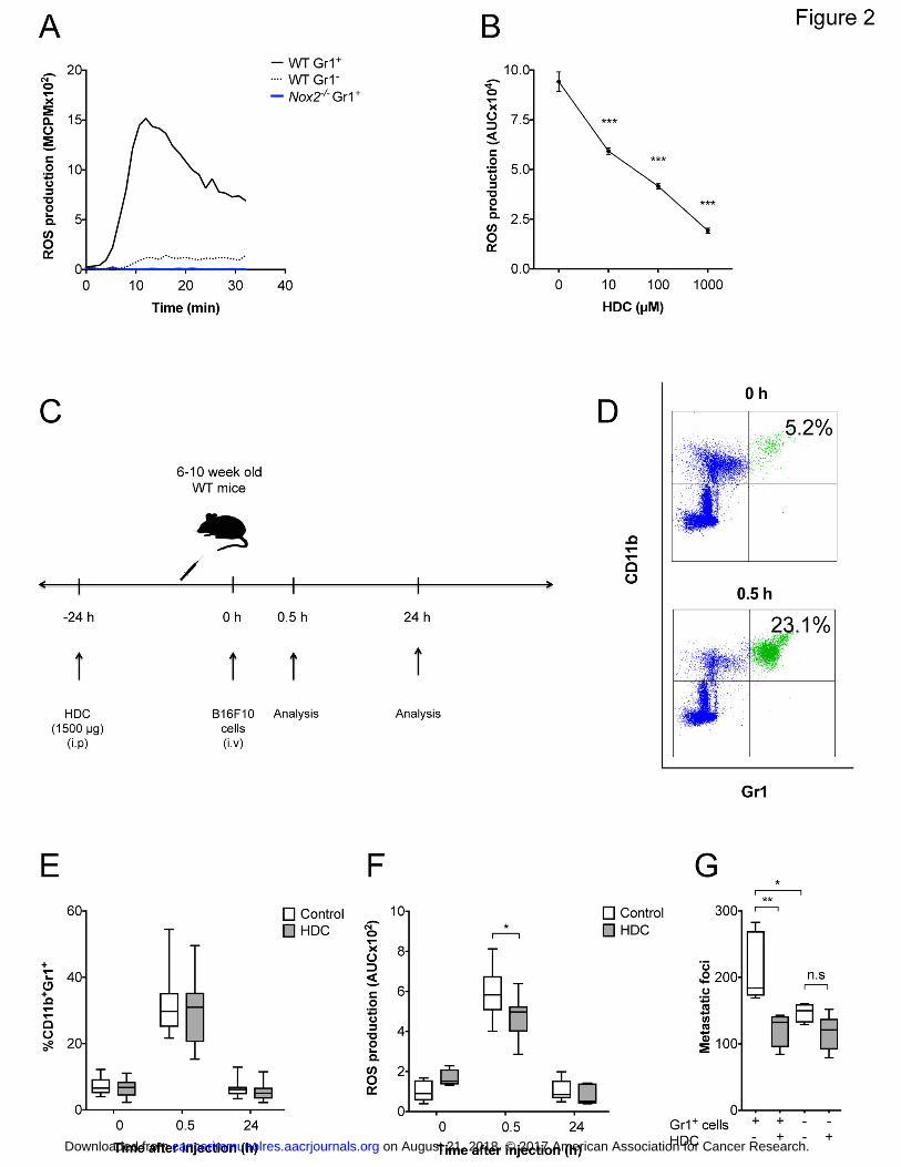
minute extracellular ROS (Fig. 2A). ROS formation from WT lung cells was dose-
dependently suppressed by HDC in vitro (Fig. 2B).
In experiments designed to assess the dynamics of ROS-producing myeloid cells in
lungs after B16F10 cell inoculation (Fig. 2C), we observed a pronounced and
transient influx of CD11b+Gr1
+ myeloid cells into lungs at 30 minutes after i.v
inoculation of tumor cells (Fig. 2D-E). Systemic treatment with HDC prior to
melanoma cell inoculation did not alter the degree of influx of myeloid cells into
lungs (Fig. 2E) but reduced the ROS formed ex vivo in lung cell suspensions (Fig.
2F). To further clarify the impact of CD11b+Gr1
+ cells on melanoma metastasis, Gr1
+
cells were depleted from WT mice before treatment of mice with HDC and i.v.
inoculation of B16F10 cells. The extent of lung metastasis was reduced in the absence
of Gr1+ cells. Systemic treatment with HDC did not affect metastasis in Gr1
+-depleted
mice (Fig. 2G).
Role of NK cells for melanoma metastasis in WT and Nox2-KO mice
on August 21, 2018. © 2017 American Association for Cancer Research. cancerimmunolres.aacrjournals.org Downloaded from
Author manuscripts have been peer reviewed and accepted for publication but have not yet been edited. Author Manuscript Published OnlineFirst on July 31, 2017; DOI: 10.1158/2326-6066.CIR-16-0382

11
NK cell function is reportedly critical to limit lung metastasis in experimental models
of murine melanoma (23, 28). To define the role of NK cells in the context of NOX2
inhibition, WT and Nox2-KO mice were depleted of NK cells by anti-NK1.1 antibody
treatment prior to melanoma cell inoculation. NK cell depletion more than doubled
metastasis formation in WT and Nox2-KO mice. HDC did not inhibit melanoma
metastasis in animals depleted of NK cells (Fig. 3A). In experiments designed to
clarify whether the reduced ROS levels in lungs following administration of HDC
translated into altered NK cell function at the site of tumor expansion, it was observed
that treatment of mice with HDC entailed increased NK cell counts in lungs, but not
in spleen, at 3 weeks after tumor cell inoculation (Fig. 3B). Unexpectedly, we
detected fewer NK cells in lungs and spleens of Nox2-KO mice than in WT animals
(Fig. 3B). Also, as shown in Fig. 3A, the degree of metastasis was strikingly enhanced
in NK cell-depleted Nox2-KO mice, which may point towards the possibility of
increased functionality of NK cells in the absence of NOX2.
NOX2 inhibition enhances the capacity of lung NK cells to produce IFN
As the antimetastatic functions of NK cells in the B16 model reportedly rely on the
formation of IFNγ (29, 30), we assessed the IFNγ production of pulmonary NK cells
from Nox2-KO and WT mice. Lung cells were isolated 30 min after B16F10 cell
inoculation and IFNγ production was then assessed upon co-culture of lung cells with
B16F10 cells in vitro. Only minor amounts of IFNγ (<25 pg/ml) were detected when
lung cells or B16 cells were cultured alone. Also, minute levels (<10 pg/ml) of IFNγ
were produced in co-cultures of lung cells and B16 cells after the depletion of NK
cells in vivo using anti-NK1.1, thus supporting that the IFN produced in these cell
cultures was contributed by NK cells (Fig. 3C). It was further observed that lung NK
on August 21, 2018. © 2017 American Association for Cancer Research. cancerimmunolres.aacrjournals.org Downloaded from
Author manuscripts have been peer reviewed and accepted for publication but have not yet been edited. Author Manuscript Published OnlineFirst on July 31, 2017; DOI: 10.1158/2326-6066.CIR-16-0382

12
cells from Nox2-KO mice produced significantly higher amount of IFNγ ex vivo at a
lung cell to melanoma cell ratio of 50:1 compared with lung NK cells from WT mice
(WT vs. Nox2-KO lungs; 297±81 vs 749±27 pg/ml, respectively; P = 0.004, t-test). A
similar experimental design was adopted to assess the impact of pharmacological
NOX2 inhibition by HDC on the formation of IFNγ in lungs. Lung cells were isolated
from HDC-treated or control WT mice at 30 min after B16F10 cell inoculation. When
lung cells were co-cultured with the B16 cells, higher concentrations of IFNγ were
produced ex vivo by lung NK cells isolated from mice treated with HDC in vivo (Fig.
3C).
Role of IFNin NOX2-mediated control of melanoma metastasis
We next assessed the capacity of B16F10 cells to form metastases in Ifng-KO vs. WT
mice. In accordance with earlier studies (29, 30) melanoma metastasis was enhanced
in IFN-deficient mice (Fig. 4A). Systemic treatment with HDC did not reduce
melanoma metastasis in Ifng-KO mice (Fig. 4B). The adoptive transfer of IFNγ-
producing WT NK cells, but not the transfer of Ifng-KO NK cells, to Ifng-KO mice
significantly restored the antimetastatic efficacy of HDC (Fig. 4B). Presence of cells
with Ifng+/+
genotype in blood of Ifng-KO mice was confirmed by PCR at 2 days after
the adoptive transfer of WT NK cells (Fig. 4C).
on August 21, 2018. © 2017 American Association for Cancer Research. cancerimmunolres.aacrjournals.org Downloaded from
Author manuscripts have been peer reviewed and accepted for publication but have not yet been edited. Author Manuscript Published OnlineFirst on July 31, 2017; DOI: 10.1158/2326-6066.CIR-16-0382

13
DISCUSSION
We report that genetic inhibition of NOX2, which mediates oxidative stress by
generating ROS from myeloid cells, reduced the capacity of two strains of murine
melanoma cells (B16F1 and B16F10) to form lung metastases after i.v. inoculation,
apparently by facilitating NK cell-mediated clearance of malignant cells. Also,
treatment of mice with the NOX2 inhibitor HDC reduced melanoma metastasis in WT
but not in Nox2-KO mice. Our results concur with a study showing that HDC reduces
the subcutaneous growth of EL-4 thymoma tumors in WT but not in Nox2-KO mice
(31), thus underscoring that the anti-neoplastic efficacy of HDC depends on the
availability of NOX2.
Our results also show that the establishment of melanoma metastases was associated
with a rapid and transient accumulation of ROS-forming CD11b+Gr1
+ myeloid cells
in the lung parenchyma and that the ROS-forming capacity of infiltrating myeloid
cells ex vivo was suppressed by the in vivo administration of HDC. Pharmacological
inhibition of NOX2 also entailed increased numbers of lung NK cells in tumor-
bearing mice. In agreement with earlier studies (29, 30), the availability of IFN was
critical for NK cell-mediated clearance of B16 melanoma cells from lungs.
Furthermore, the antimetastatic effect of HDC was absent in Ifng-KO mice but could
be reconstituted by the adoptive transfer of Ifng+/+
NK cells. Collectively, these
results imply that the antimetastatic properties of HDC rely on the availability of NK
cell-derived IFNγ. We observed that despite the more efficient NK cell-mediated
clearance of melanoma cells in Nox2-KO, rather than than in WT mice, higher counts
on August 21, 2018. © 2017 American Association for Cancer Research. cancerimmunolres.aacrjournals.org Downloaded from
Author manuscripts have been peer reviewed and accepted for publication but have not yet been edited. Author Manuscript Published OnlineFirst on July 31, 2017; DOI: 10.1158/2326-6066.CIR-16-0382

14
of NK cells were detected in the lung parenchyma of WT mice. This finding implies
that NK cells were more efficient effector cells on a per cell basis in Nox2-KO mice.
In agreement with this hypothesis, we observed that pulmonary NK cells from Nox2-
KO mice showed enhanced formation of IFN ex vivo.
From these results, we hypothesize that NOX2-derived ROS produced by myeloid
cells may exert oxidative stress with ensuing reduction of NK cell–mediated clearance
of melanoma cells and aggravation of metastasis. In agreement with our findings, the
subcutaneous growth of murine melanoma and lung carcinoma was reduced in Nox2-
KO mice (13). However, several studies report contradictory effects of ROS on the
course of experimental melanoma and other forms of cancer, in particular regarding
direct effects of ROS on tumor cells. Cancer cells thus display elevated ROS
concentrations due to enhanced metabolism and mutations that trigger oxidative
processes (32, 33, 34). The increased ROS may promote mutagenesis and may also
render tumor cells more prone to expand and produce distant metastases (34, 35, 36,
37). In agreement, over-expression of the antioxidant SOD3 inhibits murine breast
cancer cell metastasis (38), and the ROS scavenger N-acetyl-cysteine reduces the
tumorigenicity of murine melanoma cells (15).
High endogenous ROS concentrations in malignant cells may also render these cells
more vulnerable to further stresses (34, 39, 40). Hence, anticancer therapies that
trigger a further increase in ROS formation may induce cell death in cancer cells
compared with their non-malignant counterparts (34, 41, 42). In addition, ROS can
limit malignant growth by triggering activation of p53, whereas antioxidants
enhanced tumor progression in a p53-dependent manner (17). Antioxidants can
on August 21, 2018. © 2017 American Association for Cancer Research. cancerimmunolres.aacrjournals.org Downloaded from
Author manuscripts have been peer reviewed and accepted for publication but have not yet been edited. Author Manuscript Published OnlineFirst on July 31, 2017; DOI: 10.1158/2326-6066.CIR-16-0382

15
enhance lymph node metastasis in a model of genetically related melanoma (16). In
immunodeficient NOD-SCID-Il2rg-/-
mice, oxidative stress reduces the ability of
primary melanoma cells to metastasize, whereas treatment with antioxidants enhanced
metastasis (18).
We hypothesize that at least two mechanisms of relevance to melanoma metastasis
and ROS-mediated oxidant stress are operable in immunocompetent mice, i.e. direct
effects of ROS on tumor cells that may either inhibit or enhance melanoma cell
expansion, and oxidant-induced immunosuppression that may promote tumor growth
and metastasis. The relative significance of these partly opposing mechanisms may
relate to the sensitivity of melanoma cells to the growth-promoting or toxic effects of
ROS as well as to the sensitivity of melanoma cells to immune-mediated clearance.
This view may, at least in part, explain the contradiction that the in vivo
administration of ROS-scavenging antioxidants such as N-acetylcysteine promotes as
well as prevents murine melanoma metastasis (15, 16, 17).
The source of ROS may be critical for its capacity to promote or inhibit tumor
progression. In our model, only ROS derived from NOX2-sufficient myeloid cells
were targeted. In contrast, ROS scavengers such as N-acetyl-cysteine may also
neutralize ROS generated from other sources, including those formed in mitochondria
during cell respiration (16). The notion that NOX2+ myeloid cells may facilitate
melanoma metastasis is supported by the findings that neutrophil infiltration of human
primary melanomas heralds early metastatic spread (43) and that the exposure of
murine cutaneous melanomas to UV light or chemical carcinogens triggers
neutrophil-dependent inflammation that promotes metastasis (44, 45). Indeed, the
on August 21, 2018. © 2017 American Association for Cancer Research. cancerimmunolres.aacrjournals.org Downloaded from
Author manuscripts have been peer reviewed and accepted for publication but have not yet been edited. Author Manuscript Published OnlineFirst on July 31, 2017; DOI: 10.1158/2326-6066.CIR-16-0382

16
adoptive transfer of CD11b+Ly6G
+ neutrophilic granulocytes enhances the formation
of lung metastases after i.v. inoculation of murine carcinoma cells (46). The effect
was secondary to granulocyte-induced inhibition of NK cell function. Neutrophil
secretion of IL1β and matrix metalloproteinases contributed to tumor cell
extravasation but the detailed mechanism of NK cell inhibition was not defined (46).
Although further studies are required to clarify the detailed impact of granulocytes
and other NOX+ myeloid cells for the course of cancer, our results imply that the
release of NOX2-derived ROS from these cells may constitute a mechanism of NK-
cell inhibition during metastasis of relevance to these previous reports, and that
strategies to target NOX2-derived ROS may facilitate NK cell-mediated clearance of
metastatic cells. This assumption gains further support by the results of the present
study showing that the depletion of Gr1+ cells reduced melanoma metastasis, and that
NOX2 inhibition using HDC did not affect metastasis in Gr1+-depleted animals.
Additionally, the finding that IL15, an NK cell-activating cytokine, improved the
antimetastatic efficacy of pharmacological NOX2 inhibition in WT animals suggests
a combinatorial immunotherapy to reduce metastasis formation.
In summary, our results suggest that NOX2 function impacts NK cell-mediated
control of murine melanoma metastasis. We propose that pharmacological inhibition
of NOX2, alone or combined with immunostimulatory strategies, should be further
evaluated in preventing melanoma metastasis.
Author contribution
on August 21, 2018. © 2017 American Association for Cancer Research. cancerimmunolres.aacrjournals.org Downloaded from
Author manuscripts have been peer reviewed and accepted for publication but have not yet been edited. Author Manuscript Published OnlineFirst on July 31, 2017; DOI: 10.1158/2326-6066.CIR-16-0382

17
Authors E.A., K.H. and A.M. designed the study. Authors E.A., J.J. and F.H.N.
performed experiments. Authors E.A. and A.M. analyzed experiments. Authors E.A.,
K.H. and A.M. wrote the manuscript. All authors read and approved the manuscript
prior of submission.
Acknowledgements
We are grateful to Prof. Nils Lycke, MIVAC, University of Gothenburg, for
providing the B6.129S7-Ifngtm1Ts
/J mice.
Footnotes
Abbreviations used:
histamine dihydrochloride (HDC)
nicotinamide adenine dinucleotide phosphate (NADPH)
myeloid cell NADPH oxidase (NOX2)
reactive oxygen species (ROS)
on August 21, 2018. © 2017 American Association for Cancer Research. cancerimmunolres.aacrjournals.org Downloaded from
Author manuscripts have been peer reviewed and accepted for publication but have not yet been edited. Author Manuscript Published OnlineFirst on July 31, 2017; DOI: 10.1158/2326-6066.CIR-16-0382

18
References
1. Karlsson A, Dahlgren C. Assembly and activation of the neutrophil NADPH
oxidase in granule membranes. Antioxid Redox Signal. 2002;4(1):49-60.
2. Geiszt M. NADPH oxidases: new kids on the block. Cardiovasc Res.
2006;71(2):289-99.
3. Kusmartsev S, Gabrilovich DI. Inhibition of myeloid cell differentiation in
cancer: the role of reactive oxygen species. J Leukoc Biol. 2003;74(2):186-96.
4. Bedard K, Krause KH. The NOX family of ROS-generating NADPH
oxidases: physiology and pathophysiology. Physiol Rev. 2007;87(1):245-313.
5. Quinn MT, Gauss KA. Structure and regulation of the neutrophil respiratory
burst oxidase: comparison with nonphagocyte oxidases. J Leukoc Biol.
2004;76(4):760-81.
6. Hellstrand K, Asea A, Dahlgren C, Hermodsson S. Histaminergic regulation
of NK cells. Role of monocyte-derived reactive oxygen metabolites. J Immunol.
1994;153(11):4940-7.
7. Hansson M, Asea A, Ersson U, Hermodsson S, Hellstrand K. Induction of
apoptosis in NK cells by monocyte-derived reactive oxygen metabolites. J Immunol.
1996;156(1):42-7.
8. Kono K, Salazar-Onfray F, Petersson M, Hansson J, Masucci G, Wasserman
K, et al. Hydrogen peroxide secreted by tumor-derived macrophages down-modulates
signal-transducing zeta molecules and inhibits tumor-specific T cell-and natural killer
cell-mediated cytotoxicity. Eur J Immunol. 1996;26(6):1308-13.
on August 21, 2018. © 2017 American Association for Cancer Research. cancerimmunolres.aacrjournals.org Downloaded from
Author manuscripts have been peer reviewed and accepted for publication but have not yet been edited. Author Manuscript Published OnlineFirst on July 31, 2017; DOI: 10.1158/2326-6066.CIR-16-0382
19
9. Aurelius J, Martner A, Brune M, Palmqvist L, Hansson M, Hellstrand K, et al.
Remission maintenance in acute myeloid leukemia: impact of functional histamine
H2 receptors expressed by leukemic cells. Haematologica. 2012;97(12):1904-8.
10. Kiessling R, Wasserman K, Horiguchi S, Kono K, Sjoberg J, Pisa P, et al.
Tumor-induced immune dysfunction. Cancer Immunol Immunother. 1999;48(7):353-
62.
11. Hellstrand K, Brune M, Naredi P, Mellqvist UH, Hansson M, Gehlsen KR, et
al. Histamine: a novel approach to cancer immunotherapy. Cancer Invest.
2000;18(4):347-55.
12. Martner A, Thoren FB, Aurelius J, Hellstrand K. Immunotherapeutic
strategies for relapse control in acute myeloid leukemia. Blood Rev. 2013;27(5):209-
16.
13. Kelkka T, Pizzolla A, Laurila JP, Friman T, Gustafsson R, Kallberg E, et al.
Mice lacking NCF1 exhibit reduced growth of implanted melanoma and carcinoma
tumors. PLoS One. 2013;8(12):e84148.
14. Ligtenberg MA, Cinar O, Holmdahl R, Mougiakakos D, Kiessling R.
Methylcholanthrene-Induced Sarcomas Develop Independently from NOX2-Derived
ROS. PLoS One. 2015;10(6):e0129786.
15. De Flora S, D'Agostini F, Masiello L, Giunciuglio D, Albini A. Synergism
between N-acetylcysteine and doxorubicin in the prevention of tumorigenicity and
metastasis in murine models. Int J Cancer. 1996;67(6):842-8.
16. Le Gal K, Ibrahim MX, Wiel C, Sayin VI, Akula MK, Karlsson C, et al.
Antioxidants can increase melanoma metastasis in mice. Sci Transl Med.
2015;7(308):308re8.
on August 21, 2018. © 2017 American Association for Cancer Research. cancerimmunolres.aacrjournals.org Downloaded from
Author manuscripts have been peer reviewed and accepted for publication but have not yet been edited. Author Manuscript Published OnlineFirst on July 31, 2017; DOI: 10.1158/2326-6066.CIR-16-0382

20
17. Sayin VI, Ibrahim MX, Larsson E, Nilsson JA, Lindahl P, Bergo MO.
Antioxidants accelerate lung cancer progression in mice. Sci Transl Med.
2014;6(221):221ra15.
18. Piskounova E, Agathocleous M, Murphy MM, Hu Z, Huddlestun SE, Zhao Z,
et al. Oxidative stress inhibits distant metastasis by human melanoma cells. Nature.
2015;527(7577):186-91.
19. Dalton DK, Pitts-Meek S, Keshav S, Figari IS, Bradley A, Stewart TA.
Multiple defects of immune cell function in mice with disrupted interferon-gamma
genes. Science. 1993;259(5102):1739-42.
20. Dahlgren C, Karlsson A. Respiratory burst in human neutrophils. J Immunol
Methods. 1999;232(1-2):3-14.
21. Coffelt SB, Kersten K, Doornebal CW, Weiden J, Vrijland K, Hau CS, et al.
IL-17-producing gammadelta T cells and neutrophils conspire to promote breast
cancer metastasis. Nature. 2015;522(7556):345-8
22. Betten A, Dahlgren C, Hermodsson S, Hellstrand K. Histamine inhibits
neutrophil NADPH oxidase activity triggered by the lipoxin A4 receptor-specific
peptide agonist Trp-Lys-Tyr-Met-Val-Met. Scand J Immunol. 2003;58(3):321-6.
23. Hellstrand K, Asea A, Hermodsson S. Role of histamine in natural killer cell-
mediated resistance against tumor cells. J Immunol. 1990;145(12):4365-70.
24. Fehniger TA, Caligiuri MA. Interleukin 15: biology and relevance to human
disease. Blood. 2001;97(1):14-32.
25. Fidler IJ, Nicolson GL. Organ selectivity for implantation survival and growth
of B16 melanoma variant tumor lines. J Natl Cancer Inst. 1976;57(5):1199-202.
on August 21, 2018. © 2017 American Association for Cancer Research. cancerimmunolres.aacrjournals.org Downloaded from
Author manuscripts have been peer reviewed and accepted for publication but have not yet been edited. Author Manuscript Published OnlineFirst on July 31, 2017; DOI: 10.1158/2326-6066.CIR-16-0382

21
26. Corzo CA, Cotter MJ, Cheng P, Cheng F, Kusmartsev S, Sotomayor E, et al.
Mechanism regulating reactive oxygen species in tumor-induced myeloid-derived
suppressor cells. J Immunol. 2009;182(9):5693-701.
27. Maghzal GJ, Krause KH, Stocker R, Jaquet V. Detection of reactive oxygen
species derived from the family of NOX NADPH oxidases. Free Radic Biol Med.
2012;53(10):1903-18.
28. Saijo N, Ozaki A, Beppu Y, Takahashi K, Fujita J, Sasaki Y, et al. Analysis of
metastatic spread and growth of tumor cells in mice with depressed natural killer
activity by anti-asialo GM1 antibody or anticancer agents. J Cancer Res Clin Oncol.
1984;107(3):157-63.
29. Takeda K, Nakayama M, Sakaki M, Hayakawa Y, Imawari M, Ogasawara K,
et al. IFN-gamma production by lung NK cells is critical for the natural resistance to
pulmonary metastasis of B16 melanoma in mice. J Leukoc Biol. 2011;90(4):777-85.
30. Kakuta S, Tagawa Y, Shibata S, Nanno M, Iwakura Y. Inhibition of B16
melanoma experimental metastasis by interferon-gamma through direct inhibition of
cell proliferation and activation of antitumour host mechanisms. Immunology.
2002;105(1):92-100.
31. Martner A, Wiktorin HG, Lenox B, Ewald Sander F, Aydin E, Aurelius J, et
al. Histamine promotes the development of monocyte-derived dendritic cells and
reduces tumor growth by targeting the myeloid NADPH oxidase. J Immunol.
2015;194(10):5014-21.
32. Szatrowski TP, Nathan CF. Production of large amounts of hydrogen peroxide
by human tumor cells. Cancer Res. 1991;51(3):794-8.
33. Panieri E, Santoro MM. ROS homeostasis and metabolism: a dangerous liason
in cancer cells. Cell Death Dis. 2016;7(6):e2253.
on August 21, 2018. © 2017 American Association for Cancer Research. cancerimmunolres.aacrjournals.org Downloaded from
Author manuscripts have been peer reviewed and accepted for publication but have not yet been edited. Author Manuscript Published OnlineFirst on July 31, 2017; DOI: 10.1158/2326-6066.CIR-16-0382

22
34. Galadari S, Rahman A, Pallichankandy S, Thayyullathil F. Reactive oxygen
species and cancer paradox: To promote or to suppress? Free Radic Biol Med.
2017;104:144-64.
35. Nelson KK, Melendez JA. Mitochondrial redox control of matrix
metalloproteinases. Free Radic Biol Med. 2004;37(6):768-84.
36. Diaz B, Shani G, Pass I, Anderson D, Quintavalle M, Courtneidge SA. Tks5-
dependent, nox-mediated generation of reactive oxygen species is necessary for
invadopodia formation. Sci Signal. 2009;2(88):ra53.
37. Diaz B, Courtneidge SA. Redox signaling at invasive microdomains in cancer
cells. Free Radic Biol Med. 2012;52(2):247-56.
38. Teoh-Fitzgerald ML, Fitzgerald MP, Zhong W, Askeland RW, Domann FE.
Epigenetic reprogramming governs EcSOD expression during human mammary
epithelial cell differentiation, tumorigenesis and metastasis. Oncogene.
2014;33(3):358-68.
39. Schumacker PT. Reactive oxygen species in cancer: a dance with the devil.
Cancer Cell. 2015;27(2):156-7.
40. Nishikawa M. Reactive oxygen species in tumor metastasis. Cancer Lett.
2008;266(1):53-9.
41. Lee JH, Won YS, Park KH, Lee MK, Tachibana H, Yamada K, et al. Celastrol
inhibits growth and induces apoptotic cell death in melanoma cells via the activation
ROS-dependent mitochondrial pathway and the suppression of PI3K/AKT signaling.
Apoptosis. 2012;17(12):1275-86.
42. Tsai MH, Liu JF, Chiang YC, Hu SC, Hsu LF, Lin YC, et al. Artocarpin, an
isoprenyl flavonoid, induces p53-dependent or independent apoptosis via ROS-
on August 21, 2018. © 2017 American Association for Cancer Research. cancerimmunolres.aacrjournals.org Downloaded from
Author manuscripts have been peer reviewed and accepted for publication but have not yet been edited. Author Manuscript Published OnlineFirst on July 31, 2017; DOI: 10.1158/2326-6066.CIR-16-0382

23
mediated MAPKs and Akt activation in non-small cell lung cancer cells. Oncotarget.
2017; doi: 10.18632/oncotarget.16058
43. Jensen TO, Schmidt H, Moller HJ, Donskov F, Hoyer M, Sjoegren P, et al.
Intratumoral neutrophils and plasmacytoid dendritic cells indicate poor prognosis and
are associated with pSTAT3 expression in AJCC stage I/II melanoma. Cancer.
2012;118(9):2476-85.
44. Bald T, Quast T, Landsberg J, Rogava M, Glodde N, Lopez-Ramos D, et al.
Ultraviolet-radiation-induced inflammation promotes angiotropism and metastasis in
melanoma. Nature. 2014;507(7490):109-13.
45. Bald T, Landsberg J, Jansen P, Gaffal E, Tuting T. Phorbol ester-induced
neutrophilic inflammatory responses selectively promote metastatic spread of
melanoma in a TLR4-dependent manner. Oncoimmunology. 2016;5(2):e1078964.
46. Spiegel A, Brooks MW, Houshyar S, Reinhardt F, Ardolino M, Fessler E, et
al. Neutrophils Suppress Intraluminal NK Cell-Mediated Tumor Cell Clearance and
Enhance Extravasation of Disseminated Carcinoma Cells. Cancer Discov.
2016;6(6):630-49.
on August 21, 2018. © 2017 American Association for Cancer Research. cancerimmunolres.aacrjournals.org Downloaded from
Author manuscripts have been peer reviewed and accepted for publication but have not yet been edited. Author Manuscript Published OnlineFirst on July 31, 2017; DOI: 10.1158/2326-6066.CIR-16-0382

24
Figures and legends
Fig. 1. Impact of genetic and pharmacological inhibition of NOX2 on B16
melanoma metastasis. Panel (A) shows the experimental design. Panel (B) shows the
number of metastatic foci formed in lungs of WT and Nox2-KO (Nox2-/-
) mice at 3
weeks after i.v. inoculation of 50,000, 100,000 or 150,000 B16F10 cells. Medians and
quartiles are indicated by boxes. Error bars show the min to max values (n = 6 for
each group; t-test; two independent experiments). Panel (C) shows the number of
metastatic foci in lungs of WT or Nox2-KO mice after systemic treatment with HDC
and/or IL15. In these experiments, 100,000 B16F10 cells were injected into WT mice
and 150,000 cells into Nox2-/-
mice to achieve comparable levels of baseline
metastasis (n = 15 for all groups of WT mice; n = 8 for control, HDC and IL15 groups
of Nox2-/-
mice and n = 3 for HDC + IL15). Combined treatment with IL15 and HDC
was significantly more effective than IL15 alone to reduce metastasis formation in
WT mice, when analyzed by t-test, P = 0.01 (up to five independent experiments).
Panel (D) shows results from lung metastasis formation by the B16F1 melanoma cell
line. This cell line is less metastatic compared to B16F10 and therefore 200,000
B16F1 cells were injected into WT mice and 300,000 cells into Nox2-/-
mice. The
number of metastatic foci in lungs of WT and Nox2-KO mice after systemic treatment
with HDC or IL15 was determined after 3 weeks. (n = 4 for all groups except n = 3
for control group of WT mice). The results shown in Fig 1C-D were evaluated by
Repeated Measures ANOVA. Non-significant values where P > 0.05 represented with
‘n.s’ whereas ‘*’ represents P ≤ 0.05; ‘**’ represents P ≤ 0.01 and ‘***’ represents P
≤ 0.001.
on August 21, 2018. © 2017 American Association for Cancer Research. cancerimmunolres.aacrjournals.org Downloaded from
Author manuscripts have been peer reviewed and accepted for publication but have not yet been edited. Author Manuscript Published OnlineFirst on July 31, 2017; DOI: 10.1158/2326-6066.CIR-16-0382

25
Fig. 2. Effect of HDC on the ROS production in mouse lungs after melanoma cell
inoculation. Panel (A) shows the extracellular ROS production of PMA-stimulated
WT Gr1+ (black solid line), WT Gr1
- (black dashed line) or Nox2
-/- Gr1
+ mouse lung
cells (blue line) in a representative experiment out of three performed. Panel (B)
shows the ROS production of WT lung cells triggered by 10-7
M WKYMVm in
response to HDC at indicated final concentrations. The mean ROS production +/-
SEM is displayed (n = 3, t-test). Panel (C) represents the experimental design
designed to assess the dynamics of ROS-producing myeloid cells in lungs after B16
cell inoculation. Panel (D) shows a representative dot plot of CD11b+Gr1
+ cells out of
live CD45+ lung cells before (0 h) or 0.5 h after tumor cell inoculation. Panel (E)
shows the fraction of CD11b+Gr1
+ cells out of live CD45
+ cells in lungs at indicated
time points after tumor cell injection, with or without pre-treatment of mice with PBS
(control) or HDC 24 h before analysis, as determined in single cell lung suspensions
(n = 18 in each group; four independent experiments). Panel (F) shows the ROS
formation (area under curve, AUC) ex vivo in response to PMA stimulation of single
lung cell suspensions from mice pretreated with HDC or PBS on the day before tumor
cell inoculation. ROS production was determined at 30 min and 24 h after inoculation
of 100,000 B16 cells (n = 5-10, t-test; three independent experiments). Panel (G)
shows a reduced B16F10 metastasis formation and lack of effects of systemic
treatment with HDC on metastasis formation in animals depleted of Gr1+ cells prior to
melanoma cell inoculation (n = 4-5 for each group, one way ANOVA). Non-
significant values where P > 0.05 represented with ‘n.s’ whereas ‘*’ represents P ≤
0.05; ‘**’ represents P ≤ 0.01 and ‘***’ represents P ≤ 0.001.
on August 21, 2018. © 2017 American Association for Cancer Research. cancerimmunolres.aacrjournals.org Downloaded from
Author manuscripts have been peer reviewed and accepted for publication but have not yet been edited. Author Manuscript Published OnlineFirst on July 31, 2017; DOI: 10.1158/2326-6066.CIR-16-0382

26
Fig. 3. Antimetastatic effects of HDC rely on NK cells and NK cell-derived IFN.
Panel (A) shows the effects of systemic treatment with HDC on B16F10 metastasis
formation in WT and Nox2-/-
animals depleted of NK cells (n = 7 for untreated WT
mice with and without NK cells (two independent experiments); n = 3 for HDC-
treated WT mice with and without NK cells; n = 4 for each group of Nox2-/-
mice, one
way ANOVA). Panel (B) shows effects of systemic treatment with HDC on NK cell
numbers in lungs and spleens of WT and Nox-KO (Nox2-/-
) mice at 3 weeks after
tumor cell inoculation. The percentage of NK cells out of live CD45+ cells was
determined by flow cytometry (WT mice n = 9-11; Nox2-/-
mice n = 9-13, t-test; three
independent experiments). Panel (C) shows IFNγ levels produced in lung cells from
HDC-treated and control WT mice that were incubated with B16 melanoma cells at
indicated effector to target cell ratios. Mice received HDC or PBS (control) 24 hours
before i.v. inoculation of B16 cells. Lungs were recovered 30 min after tumor cell
inoculation (n = 11 for the control group, n = 6 for the other groups, two-way
ANOVA; two independent experiments). Non-significant values where P > 0.05
represented with ‘n.s’ whereas ‘*’ represents P ≤ 0.05; ‘**’ represents P ≤ 0.01 and
‘***’ represents P ≤ 0.001.
Fig. 4. Impact of IFNγ in B16 melanoma metastasis. Panel (A) shows box plots of
B16F10 metastasis at 3 weeks after i.v. inoculation of 50,000, 100,000 or 150,000
B16 melanoma cells into WT and Ifng-/-
mice (n = 6 for each group, t-test; two
on August 21, 2018. © 2017 American Association for Cancer Research. cancerimmunolres.aacrjournals.org Downloaded from
Author manuscripts have been peer reviewed and accepted for publication but have not yet been edited. Author Manuscript Published OnlineFirst on July 31, 2017; DOI: 10.1158/2326-6066.CIR-16-0382

27
independent experiments). The left part of panel (B) shows the lack of efficacy of
systemic treatment with HDC on metastasis formation at 3 weeks after inoculation of
B16 melanoma cells into Ifng -/-
mice (n = 19-21, t-test; four independent
experiments). The right part of panel (B) shows effects of systemic treatment with
HDC on metastasis formation (% of control) in Ifng-/-
mice that received the adoptive
transfer of purified NK cells from WT mice (n = 9 for each group, t-test) or purified
NK cells from Ifng-/-
mice (n = 6, t-test; two independent experiments). Panel (C)
shows the presence of WT Ifng in peripheral blood collected from six representative
Ifng -/-
mice who had received adoptive transfer of WT NK cells (lanes 1-3) or Ifng -/-
NK cells (lanes 4-6) 2 days earlier. PCR was performed for WT Ifng and the disrupted
IFNγ gene of Ifng-/-
mice. Non-significant values where P > 0.05 represented with
‘n.s’ whereas ‘*’ represents P ≤ 0.05; ‘**’ represents P ≤ 0.01 and ‘***’ represents P
≤ 0.001.
on August 21, 2018. © 2017 American Association for Cancer Research. cancerimmunolres.aacrjournals.org Downloaded from
Author manuscripts have been peer reviewed and accepted for publication but have not yet been edited. Author Manuscript Published OnlineFirst on July 31, 2017; DOI: 10.1158/2326-6066.CIR-16-0382

Published OnlineFirst July 31, 2017.Cancer Immunol Res Ebru Aydin, Junko Johansson, Faisal Hayat Nazir, et al. cell-mediated control of murine melanoma metastasisRole of NOX2-derived reactive oxygen species in NK
Updated version
10.1158/2326-6066.CIR-16-0382doi:
Access the most recent version of this article at:
Manuscript
Authorbeen edited. Author manuscripts have been peer reviewed and accepted for publication but have not yet
E-mail alerts related to this article or journal.Sign up to receive free email-alerts
Subscriptions
Reprints and
To order reprints of this article or to subscribe to the journal, contact the AACR Publications
Permissions
Rightslink site. Click on "Request Permissions" which will take you to the Copyright Clearance Center's (CCC)
.http://cancerimmunolres.aacrjournals.org/content/early/2017/07/29/2326-6066.CIR-16-0382To request permission to re-use all or part of this article, use this link
on August 21, 2018. © 2017 American Association for Cancer Research. cancerimmunolres.aacrjournals.org Downloaded from
Author manuscripts have been peer reviewed and accepted for publication but have not yet been edited. Author Manuscript Published OnlineFirst on July 31, 2017; DOI: 10.1158/2326-6066.CIR-16-0382


![Proposal [ Nk ] Kecil](https://img.pdfslide.us/doc/110x75/5571fd9149795991699965fa/proposal-nk-kecil.jpg)