Embed Size (px)
Citation preview

Articles
Role of Neighboring FMN Side Chains in the Modulation of Flavin ReductionPotentials and in the Energetics of the FMN:Apoprotein Interaction inAnabaena
Flavodoxin†
Isabel Nogue´s,‡ Luis Alberto Campos,‡ Javier Sancho,‡ Carlos Go´mez-Moreno,‡ Stephen G. Mayhew,§ andMilagros Medina*,‡
Departamento de Bioquı´mica y Biologı´a Molecular y Celular, Facultad de Ciencias and Institute of Biocomputation and Physicsof Complex Systems (BIFI), UniVersidad de Zaragoza, 50009-Zaragoza, Spain, and Department of Biochemistry,
Conway Institute for Biomolecular and Biomedical Research, UniVersity College Dublin, Belfield, Dublin 4, Ireland
ReceiVed August 5, 2004; ReVised Manuscript ReceiVed September 23, 2004
ABSTRACT: Flavodoxins (Flds) are electron transfer proteins that carry a noncovalently bound flavinmononucleotide molecule (FMN) as a redox active center. A distinguishing feature of these flavoproteinsis the dramatic change in theEsq/rd reduction potential of the FMN upon binding to the apoprotein (at pH8.0, from-269 mV when free in solution to-438 mV inAnabaenaFld). In this study, the contributionof three neighboring FMN residues, Thr56, Asn58, and Asn97, and of three negatively charged surfaceresidues, Glu20, Asp65, and Asp96, to modulate the redox properties of FMN upon its binding to theapoprotein has been investigated. Additionally, the role of these residues in the apoflavodoxin:FMNinteraction has been analyzed. Concerning the redox potentials, the most noticeable result was obtainedfor the Thr56Gly mutant. In this Fld variant, the increased accessibility of FMN leads to an increase of+63 mV in theEsq/rd value. On the other hand, a correlation between the electrostatic environment ofFMN and theEsq/rd has been observed. The more positive residues or the less negative residues presentin the surroundings of the FMN N(1) atom, then the less negative the value forEsq/rd. With regard toFMN binding to apoflavodoxin, breaking of hydrophobic interactions between FMN and residues 56, 58,and 97 seems to increase theKd values, especially in the Thr56Gly Fld. Such results suggest that theH-bond network in the FMN environment influences the FMN affinity.
Flavodoxins (Flds)1 are smallR/â flavoproteins involvedin electron-transfer (ET) reactions in microorganisms andcertain algae, which are either constitutively synthesized orinduced to replace ferredoxin (Fd) under iron stress condi-tions. Flds are noncovalent complexes between an apoprotein[apoflavodoxin (ApoFld)] and a low-potential flavin cofactor,the FMN, that confers redox properties to the protein.
Binding of ApoFld to FMN modifies the free flavin midpointreduction potentials, withEox/sq being displaced to a lessnegative value, whileEsq/rd is shifted to a more negative one(1, 2). These changes reflect a stabilization of the intermedi-ate flavin semiquinone by the protein. The role played bythe protein environment in modulating the properties ofbound FMN has been studied using Flds from severalsources. These studies have demonstrated the importance ofelectrostatic interactions (3-9), aromatic stacking interactions(3, 10, 11), flavin-sulfur interactions (12) and H bonds toeither flavin N(3) (13, 14) or flavin N(5) (7, 15-17).
Three-dimensional structures of a large number of Fldsare known in their oxidized state (18-23), with somestructures also known for the corresponding semiquinoneand/or hydroquinone states (2, 23-26). Structures are alsoavailable for mutants of some of these proteins (9, 17, 27-29) and for the ApoFld fromAnabaena(30). Although theoverall folding of the polypeptide is similar in all Flds, thereis considerable variation in specific interactions between theFMN cofactor and the apoprotein. In general, the isoallox-azine moiety of the flavin is locked in a nonpolar environ-ment and is stacked against at least one aromatic side chain.
† This work has been supported by Comisio´n Interministerial deCiencia y Tecnologı´a (CICYT, BQU2001-2520 to M.M. and BCM2001-252 to J.S.), by DGA (P120/2001 to J.S.) and by a FEBS TravelGrant (to I.N.).
* To whom correspondence should be addressed: Departamento deBioquımica y Biologıa Molecular y Celular, Facultad de Ciencias,Universidad de Zaragoza, 50009-Zaragoza, Spain. Phone: 34-976-762-476. Fax: 34-976-762-123. E-mail: [email protected].
‡ Universidad de Zaragoza.§ University College Dublin.1 Abbreviations: dRf, 5-deazariboflavin; dRfH‚, semiquinone form
of dRf; ET, electron transfer;Eox/sq, midpoint reduction potentials forthe ox/sq couple;Esq/rd, midpoint reduction potentials for the sq/rdcouple; WT, wild type;F, Faraday’s constant; Fld, flavodoxin; ApoFld,apoflavodoxin; Fldox, Fld in the oxidised state; Fldrd, Fld in thehydroquinone state; Fldsq, Fld in the semiquinone state;Kd, dissociationconstant; EPR, continuous wave electron paramagnetic resonance;ESEEM, electron spin-echo envelope modulation.
15111Biochemistry2004,43, 15111-15121
10.1021/bi0483256 CCC: $27.50 © 2004 American Chemical SocietyPublished on Web 11/06/2004

In some Flds, such as that fromAnabaena, the isoalloxazinering is stacked between a Tyr and a Trp (19, 21, 22, 27,31-33). In these structures, the Tyr residue is positioned ina nearly coplanar orientation with the outer face of theisoalloxazine ring, whereas the Trp forms a large portion ofthe inner surface of the flavin-binding pocket. Three seg-ments of ApoFld contribute to FMN binding: the phosphate-binding loop (residues 10-15 inAnabaenaFld) and the loopsconnectingâ3 and R3 (residues 56-62) andâ4 and R4(residues 90-99) (Figures 1 and 2A) (29). In particular, the56-62 and 90-99 segments contain residues in close contactwith the isoalloxazine ring system (parts A and B of Figure2) and are likely to influence the binding of FMN and theredox properties of the flavin (4, 16, 17, 34).
Parts A and B of Figure 2 summarize the interactions,either hydrophobic or H bonds, that might contribute to FMNstabilization in oxidizedAnabaenaFld. The Asp90 NH formsa bifurcated H bond with N(1) and O(2) of the flavin; theGln99 backbone NH makes a H bond to O(2); the Asn97main-chain CO is H-bonded to N(3); the Gly60 NH H-bondsto O(4); and the Thr56 CO H-bonds to N(1) of the flavin.The protein group closest to the key N(5) position of FMN
is the Ile59 NH (Figure 2B). The distance between thesetwo nitrogen atoms is 3.6 Å and therefore larger than usualfor an H bond. However, the groups are well-oriented,suggesting that a weak H bond may be established (21). Inaddition to these H-bonding interactions, the isoalloxazinemoiety makes nonpolar contacts with atoms from Asn58,Ile59, Gly60, Tyr94, and Asn97 (Figure 2A).
While the H-bond network observed inAnabaenaFld isessentially conserved in Fld structures from different species(26), the 56-62 segments vary considerably in both sequenceand conformation. Additionally, conformational changesupon FMN reduction have been reported in this region. Thus,the analysis of the oxidized and semiquinone structures ofAnacystis nidulans, Clostridium beijerinckii, and Desulfo-Vibrio VulgarisFlds indicates that semiquinone formation isaccompanied by a backbone rearrangement that allows amain-chain carbonyl O to form a H bond with the N(5)Hpresent in the neutral flavin semiquinone (15, 23, 24). In C.beijerinckii, D. Vulgaris, and D. desulfuricansFlds, thiscarbonyl O is contributed by a Gly, whereas inA. nidulansFld it comes from an Asn (corresponding to Asn58 inAnabaenaFld) (7, 15, 26). Moreover, the formation of this
FIGURE 1: Sequence alignment of flavodoxins fromAnabaena, A. nidualns, C. beijerinckii, D. Vulgaris, andD. desulfuricansat the regionsinvolved in FMN binding among residues 1-20, 51-70, and 80-100. Alignments have been obtained with CLUSTALW. Numerationcorresponds to that ofAnabaenaFld. Residues in bold and presenting an asterisk under sequence are those studied in the present paper.
FIGURE 2: (A) FMN environment withinAnabaenaFld. H bonds are denoted by dashed lines, and distances are in angstroms. The figurewas produced with LIGPLOT (56). (B) Relative position of Thr56, Asn58, and Asn97 with regard to the FMN inAnabaenaFld. H bondsare shown by blue lines. (C) Surface representation ofAnabaenaFld showing the localization of the negatively charged residues (in red)Glu20, Asp65, and Asp96 and the neutral residue Asn58 (in magenta). The FMN surface is shown in yellow. B and C were produced usingPyMOL (57). (D) Representation of theC. beijerinckiiFld isoalloxazine environment in the semiquinone and oxidized states.
15112 Biochemistry, Vol. 43, No. 48, 2004 Nogues et al.

H bond is accompanied in some Flds by the breakage of aweak H bond to FMN N(5) that appears in oxidized Fld [withVal59 NH in A. nidulans, equivalent to Ile59 NH inAnabaenaor with the side chain of Thr58 inChondruscrispusFld (35)]. Such backbone rearrangements appear toprovide a versatile device for reduction potential modulation.The fact that the semiquinone states ofA. nidulansandAnabaenaFlds are less stable than those of Flds from otherspecies has been related to the weaker H bond that is formedbetween the N(5)H of the flavin and the backbone CO ofAsn compared to the bond formed with the smaller Gly (7).The three-dimensional structure of theAnabaenaFld semi-quinone is not known, and it therefore remains to beconfirmed whether a similar conformational change occursin this protein.
In an attempt to further elucidate the roles played by theresidues in the 56-62 and 90-99 segments inAnabaenaFld, we have introduced different mutations at key residuesaround the isoalloxazine moiety so that the polarity and/orthe H-bond network is modified. The influence of theThr56Gly, Thr56Ser, Asn58Cys, Asn58Lys, and Asn97Lysmutations on the reduction potentials and binding affinitiesof FMN in its three redox states has been determined. Inaddition, we have studied the effects of mutating threenegatively charged residues on the surface of the protein thatare close to the FMN but have no direct contact with theflavin ring (mutants Glu20Lys, Asp65Lys, and Asp96Asn;Figure 2C).
MATERIALS AND METHODS
Biological Material.Site-directed mutagenesis to producethe Asp96Asn mutant ofAnabaenasp. PCC7119 Fld hasbeen previously described (36). The Glu20Lys, Thr56Gly,Thr56Ser, Asn58Lys, Asn58Cys, Asp65Lys, and Asn97Lysmutants were produced using the QuickChange mutagenesiskit (Stratagene) and the following synthetic oligonucleotides(base changes are in bold): 5’-CTGAATCAGTAGCGA-AAATCATTCGAGACGAGTTTGG-3′ for Glu20Lys, 5’-GATTATTGGCTGTCCTGGTTGGAATATTGGC-3′ forThr56Gly, 5’-GATTATTGGCTGTCCTAGCTGGAATAT-TGGC-3′ for Thr56Ser, 5’-CGCTTTGCAGTTCGCCAAT-TTTCCAAGTAGGACAGCC-3′ for Asn58Lys, 5′-GTTC-GCCAATACACCAAGTAGGA- 3′ for Asn58Cys, 5’-GGC-GAACTGCAAAGCAAATGGGAAGGACTCTATTCAG-3′ for Asp65Lys, and 5’-CCAAATAGGTTACGCAGATAA-ATTTCAGGATGCGATCGG-3′ for Asn97Lys.
Mutations were verified by DNA sequence analysis. Theexpression and purification of the Fld mutants was essentiallyas described previously (11, 30). The UV-visible absorptionspectrum and SDS-PAGE patterns were used as puritycriteria. ApoFld was obtained by treating the Fld withtrichloroacetic acid at 4°C in the presence of dithiothreithol.The precipitate of ApoFld was separated from FMN bycentrifugation (37).
Spectroscopic Analysis.UV-vis spectra of Flds wererecorded on a Kontron Uvikon 942 or a CARY 1 spectro-photometer. Extinction coefficients of the different Fldmutants in the fully oxidized state were determined in 50mM Tris-HCl at pH 8.0 as described by Mayhew and Massey(38) using, for released FMN, a corrected extinction coef-ficient of 12.02 mM-1 cm-1 at 445 nm (11). Extinction
coefficients for the neutral semiquinone state at 580 nm weredetermined from data obtained during anaerobic photo-reduction in the presence of 5-deazariboflavin (dRf) andEDTA (see below). The maximum semiquinone state sta-bilized for each Fld form was determined as the intercept ofthe two linear sections of a plot of the absorbance at 580nm versus the absorbance at 460 nm. Fluorescence spectrawere recorded at 25°C in a Kontron SFM25 spectro-fluorimeter in 50 mM Tris-HCl at pH 8.0. Circular dichroismspectra were recorded at 25°C in a Jasco 710 spectropola-rimeter in a 1 cmpath-length cuvette in 1 mM Tris-HCl atpH 8.0 and 25°C. The protein concentrations were 0.7µMfor the far-UV and 4µM for the near UV-vis regions ofthe spectrum. Photoreduction of protein-bound flavin wascarried out at 25°C in an anaerobic cuvette containing 15-25µM Fld in the desired buffer, which also contained 1 mMEDTA and 2µM dRf to initiate reduction via the highlyreductive dRfH‚radical. Reaction solutions were made anaer-obic by several cycles of evacuation and flushing with O2-free Ar. Absorption spectra were recorded after successiveperiods of irradiation with a 150 W light source and wereused to calculate the concentration of the different redoxstates of the protein-flavin complex throughout the reductionprocess.
To carry out electron paramagnetic resonance (EPR) andelectron spin-echo envelope modulation (ESEEM) spec-troscopic measurements the Fld mutants were reducedanaerobically to the semiquinone state at 4°C by lightirradiation with a 150 W light source in the presence of 20mM EDTA and 2.5µM dRf in 10 mM HEPES at pH 7.0, asdescribed previously (39, 40). A Bruker ESP380E spectrom-eter operating in X band (9-10 GHz) was used forcontinuous wave (cw)-EPR and pulsed-EPR measurements.Spectra were taken at 15 K. The field position, at the centerof the EPR signal, was selected to give a maximum echointensity (40). The microwave pulse sequence was (π/2 - τ- π/2 - t1 - π - t2 - π/2) for the 4-pulse 2D-ESEEM(HYSCORE) experiment. Appropriate phase cycling wasapplied to remove unwanted echoes. 1D- and 2D-ESEEMexperiments were recorded as indicated elsewhere (40).
Reduction Potential Determinations.Midpoint reductionpotentials of Glu20Lys, Thr56Gly, Thr56Ser, Asn58Cys,Asn58Lys, Asp65Lys, Asp96Asn, and Asn97Lys Flds weredetermined by anaerobic photoreduction in the presence of1 µM dRf and 20 mM EDTA at 25°C using a saturatedcalomel electrode as the reference (41). Stepwise Fldphotoreduction was achieved by irradiating the solution (withthe cell immersed in ice water) with light from a 250 Wslide projector for periods of approximately 1 min. Afterreduction, the cell was placed in a temperature-controlledholder in a Cary 1 spectrophotometer. The solution potentialwas monitored using a Sycopel Ministat potentiostat. Equili-bration of the system was considered established when themeasured potential remained stable for 10 min, and the UV-vis spectrum was then recorded. The concentrations of thedifferent redox species in equilibrium were determined fromthe absorbance spectra. The two one-electron steps inreduction could be analyzed separately because only oxidizedand semiquinone states were present during most of the firststep and only semiquinone and hydroquinone Flds werepresent during most of the second one. The concentrationof semiquinone was calculated from the absorbance of the
Modulation of FMN Properties by the Protein Environment in Fld Biochemistry, Vol. 43, No. 48, 200415113

580 nm band (maximum and extinction coefficient used foreach Fld form can be found in Table 1), and the concentrationof the other species was determined by subtraction of thesemiquinone concentration from the total concentration ofFld. The midpoint potentials for the redox couples werecalculated by linear regression analysis of the plots ofpotential versus logarithm of the concentration ratio (oxidized/semiquinone or semiquinone/hydroquinone) according to theNernst equation
Data points in the region of maximal semiquinone ac-cumulation were not included in the regression because, inthis region, all three redox species could be present. Themidpoint potentials are reported relative to the potential ofthe standard hydrogen electrode. Typical experimental solu-tions contained 25-40 µM protein, 1-3 µM mediator dyes,2 µM dRf, and 1 mM EDTA at 25°C. The following dyeswere used as mediators: 1µM anthraquinone-2,6-disulfonate(-184 mV) and 1µM anthraquinone-2-sulfonate (-225 mV)for the determination ofEox/sq and 1µM benzyl viologen(-359 mV) and 1µM methyl viologen (-446 mV) for thedetermination ofEsq/rd. When required, protein spectra werecorrected for the spectra of oxidized or reduced mediators.The buffer used for the determination of the reductionpotential at pH 8.0, 7.3, and 7.0 was 50 mM Tris-HCl,whereas determinations at pH 6.6, 5.9, and 5.7 were carriedout in 20 mM potassium phosphate. The error in theEox/sq
andEsq/rd determined was estimated to be(3 mV.Dissociation Constants.The dissociation constants of the
ApoFld:FMNox complexes were determined fluorometricallyin a SMF25 spectrofluorimeter at 25°C in the dark.Excitation was at 445 nm, and emission was recorded at 525nm. The FMN used was>95% pure according to reverse-phase HPLC. In a typical experiment, 1 mL of 200 nM FMNin 50 mM Tris-HCl at pH 8.0 was titrated with aliquots of10-100µM ApoFld solutions. Binding of the protein to thecofactor strongly quenches its fluorescence emission. Aftereach protein addition, the system was allowed to reachequilibrium for 2 min. The dissociation constants werecalculated by fitting the fluorescence emission to the fol-lowing equation (28):
whereF is the observed fluorescence emission intensity after
each addition,Ffinal is the remaining emission intensity atthe end of the titration,Fδ is the difference in emissionintensity between 1µM free FMN and 1µM Fld, CA is thetotal protein concentration after each addition,CF is thestarting concentration of FMN, andd is the dilution factorof this initial concentration (initial volume/total volume) aftereach addition. Error in the determination ofKd values wasestimated to be(10%.
Measurement of the CaVity Volume.On the basis of theAnabaenaFld three-dimensional structure (PDB accessioncode 1flv), models for the different mutants studied havebeen generated using the Swiss-PdbViewer by using thereplace routine (GlaxoSmithKline R&D). The cavity volumewas calculated for each all-atom Fld mutant model with aprobe radius of 1.4 Å using the method implemented in theSwiss-PdbViewer (42).
RESULTS
Expression, Purification, and Spectral Properties of theDifferent Fld Mutants.The levels of expression of Glu20,Thr56, Asn58, Asp65, Asp96, and Asn97 mutants and theirspectral properties were similar to those of the wild-type(WT) Fld (not shown), indicating that no major structuralperturbations had occurred. The Thr56Gly mutant lost afraction of its FMN during purification, an early indicationthat the flavin in this protein is bound rather weakly.
Although the absorption spectra of the mutants are similarto that of WT Fld, differences occur in the shapes of thespectra and also in the extinction coefficients at the maxima(Figure 3 and Table 1). In most cases, the extinctioncoefficients at the maxima around 460 nm are smaller thanthat of the WT, and the spectra of Asn58Lys and especiallythat of Thr56Gly show an important decrease of thecharacteristic shoulder found at 480 nm in WT Fld, indicatingthat the flavin environment has been modified. Photoreduc-tion under anoxic conditions showed that all of the mutantsstabilize the blue neutral semiquinone. However, the absorp-tion spectra of the semiquinone forms of the mutants differfrom that of the WT, both in the position of the maximumaround 580 nm and in the extinction coefficient at themaximum. An isosbestic point between the spectra of the
Table 1: UV-Visible Spectral Properties of WT and MutatedFlavodoxins in the Oxidized and Semiquinone Statesa
oxidized
semiquinoneλmax
(nm)εmax
(mM-1 cm-1)Fldform I II I II εI/εII
isosbestic pointsox/sq (nm)
λmax
(nm)εmax
(M-1 cm-1)
WT 463 374 9.4 8.6 1.1 516.3 578 5.0T56G 465 373 9.4 8.0 1.2 528.3 581 3.5T56S 464 372 8.7 7.6 1.1 520.3 580 4.6N58C 464 370 8.6 8.6 1.0 518.3 574 4.4N58K 463 370 8.2 7.6 1.1 524.3 580 3.5N97K 463 372 9.1 7.6 1.2 516.3 574 4.6
a Data obtained in 50 mM Tris-HCl at pH 8.0 and 25°C.
E ) Em + (0.059/n) log([ox]/[rd]) (1)
F ) Ffinal + Fδ(dCF - [(CA + Kd + dCF) -
[(CA + Kd + dCF)2 - 4CAdCF]
1/2]/2 (2)
FIGURE 3: Absorption spectra of WT (bold line), Thr56Gly (thinline), Thr56Ser (broken thin line), Asn58Cys (dotted thin line),Asn58Lys (broken bold line), and Asn97Lys (dotted bold line)AnabaenaFld forms in the visible region. The spectra were recordedin 50 mM Tris-HCl at pH 8.0 and room temperature. Differentprotein concentrations were used to clarify the figure.
15114 Biochemistry, Vol. 43, No. 48, 2004 Nogues et al.

oxidized and semiquinone forms of WT Fld occurs at 516.3nm; the corresponding isosbestic point for most of themutants is different (Table 1). The extent of semiquinonestabilization at half-reduction is less for the mutants thanfor WT Fld, and the decrease is especially marked forThr56Gly and Asn58Lys (Table 2, values determined fromthe reduction potentials).
The fluorescence emission of FMN bound to WT Fld isvery low (less than 5% of free FMN under our experimentalconditions) as a result of the strong quenching by theapoprotein (11). The flavin fluorescence of the mutants issimilar to that of the WT, except in the case of Thr56Glyand Asn58Lys, which display 29 and 9%, respectively, ofthe fluorescence of free FMN. The greater fluorescence ofthese two mutants suggests that the FMN in them has moreexposure to the external medium. The circular dichroism UVspectra of mutated Fld forms are very similar to that of theWT, again indicating that the global protein fold is notaffected by the introduced mutations.
The semiquinone forms of all of the Fld mutants that wereinvestigated gave the isotropic cw-EPR spectrum, centeredat g ) 2.005, that was described for WT Fld semiquinone(39). Similarly, the 1D- and 2D-ESEEM spectra of thedifferent Fld semiquinone mutants are very similar to thoseof the WT (40) (not shown). No significant differences couldbe discerned between the different Fld mutants and the WTprotein either in the14N hyperfine couplings of the flavinring nitrogens, in 1D or 2D experiments, or in the calculatedisotropic and anisotropic hyperfine coupling constants forH(5). This indicates that the individual residues modifiedaround the flavin ring do not influence the electron densitydistribution in the semiquinone form of the FMN.
Reduction Potentials.The average slopes of the Nernstplots for WT Fld and its mutants were 59.8( 2.0 and 60.8( 3.0 mV for Eox/sq andEsq/rd, respectively (Figure 4). Themidpoint reduction potentials calculated from the Nernst plotsfor the WT protein at pH 8.0 are similar to those reportedearlier (43). In general, although both midpoint reductionpotentials,Esq/rd and Eox/sq, change for the mutants, largereffects are seen inEsq/rd (∆E from -11 to+63 mV) than inEox/sq (∆E from -23 to +19 mV). The effects are greatestfor the Thr56Gly and Asn58Lys mutants. Thus, replacementof Thr56 by the smaller residue Gly makesEsq/rd less negative(∆E ) +63 mV), while the mutation has no effect onEox/sq.The potentials for the two steps tend to converge so that thesemiquinone that is stabilized at half-reduction decreasesfrom 97% for WT Fld to only 75% for the mutant (Table2). It is notable that, when Thr56 is replaced by Ser, a sidechain that is also able to form a H bond, only small changesin Esq/rd andEox/sq occur (Table 2). In contrast, when Asn58
is replaced by the charged residue Lys, bothEsq/rd andEox/sq
change, causingEox/sq to become more negative (∆E ) -23mV) and Esq/rd to become less negative (∆E ) +32 mV).Again, the decrease by 55 mV in the difference in reductionpotential between the two steps of reduction is consistentwith the decrease observed in the maximum semiquinonestabilized by this mutant (25% less than for WT Fld) (Table2). However, when Asn58 is replaced by Cys, the changesare smaller, with both potentials becoming slightly morenegative,∆Esq/rd) -11 mV and∆Eox/sq) - 6 mV. Changesalso occur in theEox/sq and Esq/rd values of the Asp65Lys,Asp96Asn, and Asn97Lys mutants (Table 2). In contrast,when the surface charge from the side chain of Glu20 isreversed in the Glu20Lys mutant, neither reduction potentialis affected.
The effects of pH on the reduction potentials weredetermined for WT Fld and for the Thr56Gly and Asn58Lysmutants in the pH range from 6 to 8 (Figure 5). Data obtainedfor WT Fld confirmed an earlier study (43), which showedthatEox/sq is pH-dependent with a slope of-51 mV/pH unitand thatEsq/rd is pH-independent at high pH but becomespH-dependent as the pH is decreased below about pH 7(Figure 5). The slope forEox/sq is smaller than might beexpected for the addition of one electron and a proton toform the neutral semiquinone (theoretical value-59 mV at25°C), possibly reflecting ionizable groups on the apoproteinthat might alter the FMN environment and modulate thereduction properties of the flavin-protein complex. The slopechange for Esq/rd has been ascribed to a redox-linkedprotonation of the hydroquinone complex with an apparentpKa of 6.1 (43). Similar pH-dependent changes inEsq/rdhave
Table 2: Midpoint Reduction Potentials and Percentage of MaximalSemiquinone Stabilized for the Different Fld Formsa
Fldform Eox/sq Esq/rd
%sq
Fldform Eox/sq Esq/rd
%sq
WT -266 -439 97 N58K -289 -407 72E20K -265 -430 94 D65K -247 -423 90T56G -264 -376 75 D96N -279 -426 88T56S -251 -445 93 N97K -282 -421 86N58C -272 -450 90 FMN freeb -269 -215 14
a Data obtained in 50 mM Tris-HCl at pH 8.0 and 25°C. b Datafrom refs54 and55.
FIGURE 4: UV-visible spectra obtained during photoreduction andredox titration of (A) WT, (B) Thr56Gly, and (C) Asn58Lys Fldforms. The insets show the Nernst plots for the different Fldforms: (A) oxidized/semiquinone of WT (4), Asp65Lys (O), andAsp96Asn (9) and semiquinone/hydroquinone of WT (2), Asp65Lys(b), and Asp96Asn (0); (B) oxidized/semiquinone of Glu20Lys(9), Thr56Gly (O), and Thr56Ser (4) and semiquinone/hydro-quinone of Glu20Lys (0), Thr56Gly (b), and Thr56Ser (2); (C)oxidized/semiquinone of Asn58Cys (O), Asn58Lys (9), andAsn97Lys (4) and semiquinone/hydroquinone Asn58Cys (b),Asn58Lys (0), and Asn97Lys (2).
Modulation of FMN Properties by the Protein Environment in Fld Biochemistry, Vol. 43, No. 48, 200415115
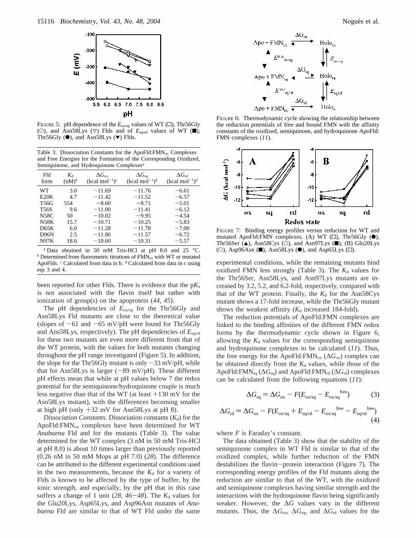
been reported for other Flds. There is evidence that the pKa
is not associated with the flavin itself but rather withionization of group(s) on the apoprotein (44, 45).
The pH dependencies ofEox/sq for the Thr56Gly andAsn58Lys Fld mutants are close to the theoretical value(slopes of-61 and-65 mV/pH were found for Thr56Glyand Ans58Lys, respectively). The pH dependencies ofEsq/rd
for these two mutants are even more different from that ofthe WT protein, with the values for both mutants changingthroughout the pH range investigated (Figure 5). In addition,the slope for the Thr56Gly mutant is only-33 mV/pH, whilethat for Asn58Lys is larger (-89 mV/pH). These differentpH effects mean that while at pH values below 7 the redoxpotential for the semiquinone/hydroquinone couple is muchless negative than that of the WT (at least+130 mV for theAsn58Lys mutant), with the differences becoming smallerat high pH (only+32 mV for Asn58Lys at pH 8).
Dissociation Constants.Dissociation constants (Kd) for theApoFld:FMNox complexes have been determined for WTAnabaenaFld and for the mutants (Table 3). The valuedetermined for the WT complex (3 nM in 50 mM Tris-HClat pH 8.0) is about 10 times larger than previously reported(0.26 nM in 50 mM Mops at pH 7.0) (28). The differencecan be attributed to the different experimental conditions usedin the two measurements, because theKd for a variety ofFlds is known to be affected by the type of buffer, by theionic strength, and especially, by the pH that in this casesuffers a change of 1 unit (28, 46-48). The Kd values forthe Glu20Lys, Asp65Lys, and Asp96Asn mutants ofAna-baenaFld are similar to that of WT Fld under the same
experimental conditions, while the remaining mutants bindoxidized FMN less strongly (Table 3). TheKd values forthe Thr56Ser, Asn58Lys, and Asn97Lys mutants are in-creased by 3.2, 5.2, and 6.2-fold, respectively, compared withthat of the WT protein. Finally, theKd for the Asn58Cysmutant shows a 17-fold increase, while the Thr56Gly mutantshows the weakest affinity (Kd increased 184-fold).
The reduction potentials of ApoFld:FMN complexes arelinked to the binding affinities of the different FMN redoxforms by the thermodynamic cycle shown in Figure 6,allowing theKd values for the corresponding semiquinoneand hydroquinone complexes to be calculated (11). Thus,the free energy for the ApoFld:FMNox (∆Gox) complex canbe obtained directly from theKd values, while those of theApoFld:FMNsq (∆Gsq) and ApoFld:FMNrd (∆Grd) complexescan be calculated from the following equations (11):
whereF is Faraday’s constant.The data obtained (Table 3) show that the stability of the
semiquinone complex in WT Fld is similar to that of theoxidized complex, while further reduction of the FMNdestabilizes the flavin-protein interaction (Figure 7). Thecorresponding energy profiles of the Fld mutants along thereduction are similar to that of the WT, with the oxidizedand semiquinone complexes having similar strength and theinteractions with the hydroquinone flavin being significantlyweaker. However, the∆G values vary in the differentmutants. Thus, the∆Gox, ∆Gsq, and ∆Grd values for the
FIGURE 5: pH dependence of theEox/sqvalues of WT (0), Thr56Gly(O), and Asn58Lys (3) Flds and ofEsq/rd values of WT (9),Thr56Gly (b), and Asn58Lys (1) Flds.
Table 3: Dissociation Constants for the ApoFld:FMNox Complexesand Free Energies for the Formation of the Corresponding Oxidized,Semiquinone, and Hydroquinone Complexesa
Fldform
Kd
(nM)b∆Gox
(kcal mol-1)c∆Gsq
(kcal mol-1)d∆Grd
(kcal mol-1)d
WT 3.0 -11.69 -11.76 -6.61E20K 4.7 -11.42 -11.52 -6.57T56G 554 -8.60 -8.71 -5.01T56S 9.6 -11.00 -11.41 -6.12N58C 50 -10.02 -9.95 -4.54N58K 15.7 -10.71 -10.25 -5.83D65K 6.0 -11.28 -11.78 -7.00D96N 2.5 -11.80 -11.57 -6.72N97K 18.6 -10.60 -10.31 -5.57
a Data obtained in 50 mM Tris-HCl at pH 8.0 and 25°C.b Determined from fluorometric titrations of FMNox with WT or mutatedApoFlds.c Calculated from data in b.d Calculated from data in c usingeqs 3 and 4.
FIGURE 6: Thermodynamic cycle showing the relationship betweenthe reduction potentials of free and bound FMN with the affinityconstants of the oxidized, semiquinone, and hydroquinone ApoFld:FMN complexes (11).
FIGURE 7: Binding energy profiles versus reduction for WT andmutated ApoFld:FMN complexes. (A) WT (0), Thr56Gly (b),Thr56Ser (2), Asn58Cys (O), and Asn97Lys (9); (B) Glu20Lys(O), Asp96Asn (9), Asn58Lys (b), and Asp65Lys (0).
∆Gsq ) ∆Gox - F(Eox/sq- Eox/sqfree) (3)
∆Grd ) ∆Gox - F(Eox/sq+ Esq/rd- Eox/sqfree - Esq/rd
free)(4)
15116 Biochemistry, Vol. 43, No. 48, 2004 Nogues et al.
ApoFld:FMN interactions of the Glu20Lys, Thr56Ser,Asp65Lys, and Asp96Asn mutants are similar to those ofWT Fld (Table 3 and Figure 7). When Thr56 is replaced bySer, a slight weakening of the oxidized and hydroquinonecomplexes occurs, whereas the affinity of the protein for theFMN semiquinone is unaffected. In contrast, the changesassociated with the other mutants are larger. Thus, thesubstitution of Asn58 or Asn97 by residues with a positivelycharged side chain clearly displaces the free energy profileto less negative values (Figure 7). For Asn58Lys, thedestabilization of the semiquinone and hydroquinone com-plexes is larger than that of the oxidized complex, whereasthe greater destabilization for the Asn97Lys mutant occurswith the hydroquinone complex. Moreover, when Asn58 isreplaced by a Cys, further destabilization of the interactionin all redox states is observed, again with the greatest effecton the hydroquinone complex. Finally, the largest overalleffect is observed when Thr56 is replaced by Gly. Complexesof this mutant with FMN in all redox states are much weakerthan in the WT protein, and the effects are greatest for theoxidized and semiquinone complexes.
DISCUSSION
The factors involved in modulation of reduction potentialsin flavoproteins have been an object of study for many years,and the molecular basis for differences in redox potentialsis still a matter of extensive discussion. Mutations have beenintroduced in the close environment of the flavin inAnabaenaFld to modify not only the electrostatic environment of theisoalloxazine ring but also the solvent accessibility of theflavin, as indicated by the differences found between theUV-vis absorbance and FMN fluorescence quenchingproperties of the different mutants (Table 1). The dataindicate that, when the electrostatic properties of amino acidside chains within a radius of 14 Å of the isoalloxazinesystem are modified, substantial changes occur in bothreduction potentials,Eox/sqandEsq/rd(Table 2). However, onlysmall effects occur when Glu20, a negatively charged aminoacid on the surface of Fld and more than 20 Å from theisoalloxazine moiety, is replaced by Lys. Additionally, forthe Thr56Gly mutant, onlyEsq/rd is modified (Table 2).
Modulation of the sq/rd Potential by the Protein EnViron-ment.Structural and functional studies have established thatelectrostatic interactions are a determinant factor in the sq/rd equilibrium in Flds. Because the hydroquinone form ofFMN in Fld is normally not protonated at N(1) (49) andbecause this position is in an environment of negative chargein the protein, repulsive forces are generated (4, 50). Acareful analysis of theEsq/rdvalues for the differentAnabaenaFld mutants indicates that either removal of negativelycharged side chains or introduction of positive ones shiftsEsq/rd to less negative values, with the magnitude of thechange depending on the distance and on the relative locationof the modified charge relative to the isoalloxazine ring.Thus, neutralization of the Asp96 side-chain charge, situatedon the Fld surface 10 Å away from the flavin ring, by anAsn, produces a change of+13 mV in Esq/rd, whereasintroduction of Lys in the direct environment of the FMNisoalloxazine ring at positions 58 or 97 (side chains around4 and 5 Å away from the ring, respectively) causesdisplacement ofEsq/rd to less negative values by+32 and
+18 mV, respectively. Moreover, theEsq/rd also shiftsmoderately to less negative values upon charge reversion ofGlu20 (by +9 mV) and Asp65 (by+16 mV), residuessituated on the Fld surface further away from the flavin ringby 20 and 13 Å, respectively. These observation mightindicate that the positive charges introduced in the flavinenvironment partially neutralize the negative charge thatinteracts with the negative charge at N(1) of the FMNhydroquinone. Similarly,Esq/rd also becomes less negativewhen Lys replaces either Asn58 or Asn97. The results clearlyshow that elimination of negative charges in the FMNsurroundings favors reduction to the hydroquinone, an effectthat is also produced by partially balancing the negativecharge with the positive charge. Our observations are inagreement with earlier ones that indicate that the generallynegative electrostatic environment generated by negativelycharged residues in the FMN surroundings contributes to thenegative Esq/rd value in Flds (4, 8, 9, 51). Theoreticalcalculations on the Fld system estimate a contribution of-4mV (at an ionic strength of 100 mM) per negatively chargedresidue to the shift of the free FMN potential (52), andexperimental neutralization of negatively charged residuesin D. Vulgaris Fld has shown an increase of+15 mV in theEsq/rd value per mutated residue (4, 10). A comparison ofthe crystal structures of the Asp95Asn mutant in its threeredox states with the corresponding structures of the WT ofthis protein revealed that the conformations of the two proteinloops that bind the flavin hydroquinone are different in themutant and that the phenol ring of Tyr98 rotates so that itmakes an edge-face interaction with the isoalloxazine ratherthan a face-face interaction (9). Similarly large structuralchanges occur in the oxidized forms of several Gly61 mutantsof this Fld (17), emphasising that mutations at the flavin sitecan have profound structural consequences. The importanceof electrostatic repulsion in the control of the sq/rd potentialsin Flds has also been demonstrated with the proteins ofC.beijerinckii, A. nidulans, andM. elsdenii(7, 8, 50).
Solvent accessibility is another important factor that hasbeen shown to contribute to modulatingEsq/rd. Thus, replace-ment of the aromatic side chain that stacks against the flavinring by a smaller amino acid leads to a displacement ofEsq/rd
to less negative values (3, 11). In addition, the more negativevalues forA. nidulansandD. Vulgaris Flds compared withthat for theC. beijerinckii protein can be explained by achannel for the solvent at the pyrimidine end of theisoalloxazine ring in theC. beijerinckiiFld that is blockedby three to five additional residues in the other two proteins(7). Among the mutations analyzed in the present study, wecan also observe how the accessibility of the flavin to thesolvent influencesEsq/rd. Thus, when Thr56 is replaced byGly, a +63 mV increase ofEsq/rd is observed, whereasreplacement by a more bulky residue such as Ser producesa value for this parameter that is similar to that of WT Fld.The theoretical calculation indicates that the volume of theinternal cavities increases only in the case of the Thr56Glymutant. In addition, the changes are only observed in theclose environment of the FMN and the mutated side chain,where a 20 Å3 cavity is present in WT Fld. In the case ofthe Thr56Gly mutant, the cavity is enlarged to 44 Å3 and asecond smaller cavity (16 Å3) appears next to it (Figure 8A).
Modulation of the ox/sq Potential by the Protein EnViron-ment.Transition from the oxidized to the semiquinone state
Modulation of FMN Properties by the Protein Environment in Fld Biochemistry, Vol. 43, No. 48, 200415117

of A. nidulans, C. beijerinckii, andD. VulgarisFlds has beenshown to be accompanied by a backbone peptide rearrange-ment (O-up conformation, Figure 2D). Thus, a comparisonof these oxidized and semiquinone 3D structures indicatesthat, upon reduction to the semiquinone state, formation ofa new H bond occurs between a carbonyl O of the twistedbackbone and the N(5)H position of the ring O58-N(5)H(A. nidulansnumbering) (7) and that the NH59 to N(5)contact that is found in the oxidized state is lost. Therefore,this peptide rearrangement appears to provide a versatiledevice for manipulation of the ox/sq potential. Although theamide group of Ile59 in oxidizedAnabaenaFld is known toestablish a weak H bond with the N(5) of the flavin ring(21), the structure of the semiquinone of this Fld has notbeen determined and there are no data to confirm theexistence of structural changes in the backbone uponreduction to the semiquinone Fld state. However, thestructural similarities betweenAnabaenaandA. nidulansFldsand the fact that the loop sequence Trp57-Asn58-Ile59-Gly60-Glu61 inAnabaenaFld is conserved inA. nidulansFld with only Ile59 substituting for a Val, suggest that theAsn58-Ile59 region in AnabaenaFld would similarlyexperience rearrangement in the transition from the oxidizedform to the semiquinone (Figure 1). Thus, previous studieshave shown that replacement of Ile59 by a Lys inAnabaenaFld produces a displacement ofEox/sqto more negative values(53). This proposed that conformational change might actin two ways, first, by altering flavin-protein contacts and,second, by modifying the conformational stability of theprotein itself. It has been demonstrated that replacement ofthe N(5)-HN59 interaction in the oxidized state by theN(5)H-O58 bond in the semiquinone state is energeticallyfavorable because N(5)H-O58 is the strongest of the twoH bonds (7). On the other hand, the conformation adoptedby the Asn58 side chain inA. nidulansFld has lower energyin the oxidized state than in the semiquinone state (7).However, this energy difference might be compensated bychanges at Ile59. One key difference betweenC. beijerinckiiFld and the proteins fromA. nidulansandAnabaenais thepresence of Asn at position 58 in the last two proteins instead
of Gly. This Asn residue raises the energy of the O-upsemiquinone conformer (7), thus making reduction moredifficult and producing a decrease in the ox/sq potential. Infact, the Asn58Gly substitution inA. nidulansFld producesthe expected increase inEox/sq(7). In this context, replacementof Asn58 of AnabaenaFld by a Lys produces a morenegativeEox/sq value (by-23 mV) compared to that of theWT. The effect might correspond to an increase in theconformational energy of the peptide Asn58-Ile59 in thesemiquinone state of this mutant by comparison with thatof WT Fld. However, changes in the interaction betweenresidue Asn58 and N(5)H cannot be discounted as secondarycontributors to the potential shifts. In fact, theAnabaenaFldstructure shows that this mutation might break hydrophobicinteractions between the side chain of Asn58 and C6, C7,and C(7)M atoms of FMN. Finally, the introduction of apositive-charged residue near the neutral semiquinone cancooperate in raising the energy for the conversion oxidizedsemiquinone.
Dependence of Reduction Potentials on pH.Although nomajor alterations are detected in theEox/sq values of themutants, changes in the slopes ofEox/sq versus pH for themutants compared to that of WT Fld and of the theoreticalvalue expected for the addition of one electron and oneproton to give the neutral semiquinone suggest that themutations introduced affect the redox-linked protonation ofsome ionizable group in the flavin environment that con-tributes to the modulation of theEox/sq potential.
Because no structural changes accompany reduction offlavodoxins from the semiquinone to the hydroquinone, ithas been difficult to establish the basis for their very lowEsq/rd potentials. However, it is accepted that in the Fld-reduced state the FMN ring remains unprotonated (anionic)and that the change in slope ofEsq/rd (with pKa ∼ 6-7)reflects a change in the protonation state of the apoprotein,either of a single nearby carboxylate side chain or to thecombined effects of several negatively charged side chainsaround the flavin-binding site (44, 45, 50).
The introduction of a Gly at the position of Thr56 or of aLys at Asn58 evidently increases the value of the apparent
FIGURE 8: Representation of the cavities formed around the isoalloxazine system in WT and Thr56Gly Flds. Thein silico Thr56Gly Fldmutation has been obtained using the Swiss-PdbViewer. The same program has also been used to add H atoms, minimize the structures,calculate the cavity volumes, and produce the figure. The protein backbone is represented in dark blue; FMN, in orange; and position 56,in green. One cavity is found in the close environment of WT Fld isoalloxazine (red). This cavity is enlarged in the case of the mutant (alsoin red), where a new cavity is also formed (gray).
15118 Biochemistry, Vol. 43, No. 48, 2004 Nogues et al.

pKa of AnabaenaFld. Although our data are insufficient forthe new values to be calculated, they appear to be greaterthan pKa ) 8. Moreover, because the amino acids that wereintroduced are completely different both in their physico-chemical properties and in their locations relative to the FMNring, the reasons for the changes are probably different forthe two mutants and related to conformational changes inthe flavin ring environment. Unfortunately, none of themutant proteins has crystallized in a form suitable forstructural determination by X-ray crystallography. However,our 3D models suggest that, when Thr56 is replaced by Gly,cavities are produced around the N(1) and N(3) atoms ofthe flavin ring (Figure 8). In solution, such cavities mighteither be filled by solvent molecules or by atoms of theproteins if small conformational changes are produced in thearea. It seems likely that these structural changes accountfor the shifts of Esq/rd to more positive values, for thedecreases in the slope for theEsq/rd pH dependence, and forthe displacement of the apparent pKa of the hydroquinoneto larger values. The shifts ofEsq/rd suggest that theunfavorable interactions between anionic semiquinone andthe protein environment are decreased in the mutants,possibly by an increase in the dielectric constant of the N(1)environment caused by the entrance of solvent molecules.
Role of Fld Residues in the ApoFld:FMN Interaction.Themost noticeable change inKd for the complexes ApoFld:FMN is for the Thr56Gly mutant with aKd 184-fold higherthan for WT Fld that corresponds to a change in∆G of 3kcal mol-1. Thr56 establishes different interactions withFMN. These include a H bond between the backbone O andO(2′) of FMN and hydrophobic interactions between the side-chain atoms and N(1), C(2), O(2), N(3), C(4), O(4), andC(4a) atoms of FMN. Furthermore, the OG1 atom is involvedin a H-bond network in the neighborhood of the FMN,interacting with Asp100 and Ala101 (21). All of theseinteractions stabilize the ApoFld:FMN complex in its threeredox states. The substitution of Gly for Thr breaks theinteractions established by the Thr side-chain atoms, causingthe ApoFld:FMN binding to be weaker. Furthermore, a Glyin this position makes FMN more accessible to the solvent.In the case of the Thr56Ser mutant, the H-bond network inthe vicinity of the FMN does not change, and although someof the hydrophobic interactions with FMN atoms maydisappear as a consequence of the longer distance betweenthem and Ser56, the ApoFld:FMN binding is not affected toa great extent. The O atom of Asn97 ofAnabaenaFld formsan H bond with N(3) of FMN, and Asn97 is also involvedin the H-bond network surrounding the FMN. It forms fourH bonds, two with Asp100, one with Tyr94, and anotherwith Gln99. Assuming that no structural changes occur thataffect position 97, at least one H bond must be broken bythe introduction of a Lys, the one with Asp100. Moreover,hydrophobic interactions with C(4) and O(4) of FMN shouldalso be broken. All of these considerations lead to an ApoFldthat forms a slightly weaker complex with FMN.
All three redox states of FMN are bound more weakly byAsn58Lys Fld than by WT Fld. This weaker binding mightbe a consequence of the different structural arrangements ofthe peptide Asn58-Ile59 and/or a different pattern of ApoFld:FMN interactions in the oxidized, semiquinone, and hydro-quinone states. In the oxidized state, the H bond betweenIle59 and N(5)H can be weaker than in WT Fld, whereas in
the complexes ApoFld:FMNsq and ApoFld:FMNrd, the Hbond between Asn58 and N(5)H is the one that is affected.
In conclusion, the data presented here indicate thatflavodoxin regulates the FMN redox properties by limitingthe solvent accessibility to the isoalloxazine ring, by control-ling the hydrophopicity and electrostatic properties of itsenvironment. In direct relationship with such observations,our data support the idea that the pKa values of proteinionizable groups and H-bond networks around the flavin ringare important determinants of the ApoFld:FMN interactionstrength.
ACKNOWLEDGMENT
We thank Drs. P. J. Alonso and J. I. Martı´nez from theDepartamento de Fı´sica de la Materia CondensadasI.C.M.A.,Universidad de Zaragoza, for the recording and interpretationof the EPR and HYSCORE spectra.
REFERENCES
1. Mayhew, S. G., Foust, G. P., and Massey, V. (1969) Oxidation-reduction properties of flavodoxin fromPeptostreptococcus els-denii, J. Biol. Chem. 244, 803-810.
2. Ludwig, M. L., and Luschinsky, C. L. (1992) Structure and redoxproperties of Clostridial flavodoxin, inChemistry and Biochemistryof FlaVoenzymes III(Muller, F., Ed.) pp 427-466, CRC Press,Boca Raton, FL.
3. Swenson, R. P., and Krey, G. D. (1994) Site-directed mutagenesisof tyrosine-98 in the flavodoxin fromDesulfoVibrio Vulgaris(Hildenborough): Regulation of oxidation-reduction propertiesof the bound FMN cofactor by aromatic, solvent and electrostaticinteractions,Biochemistry 33, 8505-8514.
4. Zhou, Z., and Swenson, R. P. (1995) Electrostatic effects of surfaceacidic amino acid residues on the oxidation-reduction potentialsof the flavodoxin fromDesulfoVibrio Vulgaris (Hildenborough),Biochemistry 34, 3183-3192.
5. Chang, F. C., and Swenson, R. P. (1997) Regulation of oxidation-reduction potentials through redox-linked ionization in the Y98Hmutant of theDesulfoVibrio Vulgaris [Hildenborough] flavo-doxin: Direct proton nuclear magnetic resonance spectroscopicevidence for the redox-dependent shift in the pKa of Histidine-98, Biochemistry 36, 9013-9021.
6. Walsh, M. A., McCarthy, A., O’Farrell, P. A, McArdle, P.,Cunningham, P. D., Mayhew, S. G., and Higgins, T. M. (1998)X-ray crystal structure of theDesulfoVibrio Vulgaris (Hildenbor-ough) apoflavodoxin-riboflavin complex,Eur. J. Biochem. 258,362-371.
7. Hoover, D. M., Drennan, C. L., Metzger, A. L., Osborne, C.,Weber, C. H., Pattridge, K. A., and Ludwig, M. L. (1999)Comparisons of wild-type and mutant flavodoxins fromAnacystisnidulans. Structural determinants of the redox potentials,J. Mol.Biol. 294, 725-743.
8. Geoghegan, S. M., Mayhew, S. G., Yalloway, G. N., and Butler,G. (2000) Cloning, sequencing, and expression of the gene forflavodoxin fromMegasphaera elsdeniiand the effects of removingthe protein negative charge that is closest to N(1) of the boundFMN, Eur. J. Biochem. 267, 4434-4444.
9. McCarthy, A. A., Walsh, M. A., Verma, C. S., O’Connell, D. P.,Reinhold, M., Yalloway, G. N, D’Arcy, D., Higgins, T. M,Voordouw, G., and Mayhew, S. G. (2002) Crystallographicinvestigation of the role of aspartate 95 in the modulation of theredox potentials ofDesulfoVibrio Vulgaris flavodoxin,Biochem-istry 41, 10950-10962.
10. Zhou, Z., and Swenson, R. P. (1996) The cumulative electrostaticeffect of aromatic stacking interactions and the negative electro-static environment of the flavin mononucleotide binding site is amajor determinant of the reduction potential for the flavodoxinfrom DesulfoVibrio Vulgaris [Hildenborough],Biochemistry 35,15980-15988.
11. Lostao, A., Gomez-Moreno, C., Mayhew, S. G., and Sancho, J.(1997) Differential stabilization of the three FMN redox formsby tyrosine 94 and tryptophan 57 in flavodoxin fromAnabaenaand its influence on the redox potentials,Biochemistry 36, 14334-14344.
Modulation of FMN Properties by the Protein Environment in Fld Biochemistry, Vol. 43, No. 48, 200415119

12. Druhan, L. J., and Swenson, R. P. (1998) Role of methionine 56in the control of the oxidation-reduction potentials of theClostridium beijerinckiiflavodoxin: Effects of substitutions byaliphatic amino acids and evidence for a role of sulfur-flavininteractions,Biochemistry 37, 9668-9678.
13. Bradley, L. H., and Swenson, R. P. (1999) Role of glutamate-59hydrogen bonded to N(3)H of the flavin mononucleotide cofactorin the modulation of the redox potentials of theClostridiumbeijerinckii flavodoxin. Glutamate-59 is not responsible for thepH dependency but contributes to the stabilization of the flavinsemiquinone,Biochemistry 38, 12377-12386.
14. Bradley, L. H., and Swenson, R. P. (2001) Role of hydrogenbonding interactions to N(3)H of the flavin mononucleotidecofactor in the modulation of the redox potentials of theClostridium beijerinckiiflavodoxin,Biochemistry 40, 8686-8695.
15. Ludwig, M. L., Pattridge, K. A., Metzger, A. L., Dixon, M. M.,Eren, M., Feng, Y., and Swenson, R. P. (1997) Control ofoxidation-reduction potentials in flavodoxin fromClostridiumbeijerinckii: The role of conformation changes,Biochemistry 36,1259-1280.
16. Chang, F. C., and Swenson, R. P. (1999) The midpoint potentialsfor the oxidized-semiquinone couple for Gly57 mutants of theClostridium beijerinckiiflavodoxin correlate with changes in thehydrogen-bonding interaction with the proton on N(5) of thereduced flavin mononucleotide cofactor as measured by NMRchemical shift temperature dependencies.Biochemistry 38, 7168-7176.
17. O′Farrell, P. A., Walsh, M. A., McCarthy, A. A., Higgins, T. M.,Voordouw, G., and Mayhew, S. G. (1998) Modulation of the redoxpotentials of FMN inDesulfoVibrio Vulgaris flavodoxin: Thermo-dynamic properties and crystal structures of glycine-61 mutants,Biochemistry 37, 8405-8416.
18. Smith, W. W., Pattridge, K. A., Ludwig, M. L., Petsko, G. A.,Tsernoglou, D., Tanaka, M., and Yasunobu, K. T. (1983) Structureof oxidized flavodoxin fromAnacystis nidulans, J. Mol. Biol. 165,737-753.
19. Fukuyama, K., Wakabayashi, S., Matsubara, H., and Rogers, L.J. (1990) Tertiary structure of oxidized flavodoxin from aneukaryotic red algaChondrus crispusat 2.35 Å resolution.Localization of charged residues and implication for interactionwith electron-transfer partners,J. Biol. Chem. 265, 15804-15812.
20. Burkhart, B., Ramakrisman, B., Yan, H., Reedstrom, R., Markley,J., Straus, N., and Sundaralingam, M. (1995) Structure of thetrigonal form of recombinant oxidized flavodoxin fromAnabaena710 at 1.4 Å resolution,Acta Crystallogr., Sect. D 51, 318-330.
21. Rao, S., Shaffie, F., Yu, C., Satyshur, K., Stokman, B., Markley,J. L., and Sundarlingam, M. (1992) Structure of the oxidized long-chain flavodoxin fom Anabaena 7120 at 2 Å resolution,ProteinSci. 1, 1413-1427.
22. Watenpaugh, K. D., Sieker, L. C., and Jensen, L. H. (1973) Thebinding of riboflavin-5′-phosphate in a flavoprotein: Flavodoxinat 2.0 Å resolution,Proc. Natl. Acad. Sci. U.S.A. 70, 3857-3860.
23. Watt, W., Tulinski, A., Swenson, R. P., and Watenpaugh, K. D.(1991) Comparison of the crystal structures of a flavodoxin in itsthree oxidation states at cryogenic temperatures,J. Mol. Biol. 218,195-208.
24. Smith, W. W., Burnett, R. M., Darling, G. D., and Ludwig, M. L.(1977) Structure of the semiquinone form of flavodoxin fromClostridum MP. Extension of 1.8 Å resolution and some com-parisons with the oxidized state,J. Mol. Biol. 117, 195-225.
25. Sharkey, C. T., Mayhew, S. G., Higgins, T. M., and Walsh, M.A. (1997) Crystallographic studies on flavodoxin fromMega-sphaera eldenii, in FlaVins and FlaVoproteins(Stephenson, K.J., Massey, V., and Williams, C. H., Jr., Eds.) pp 445-448,University of Calgary Press, Calgary, Canada.
26. Romero, A., Caldeira, J., Legall, J., Moura, I., Moura, J. J., andRomao, M. J. (1996) Crystal structure of flavodoxin fromDesulfoVibrio desulfuricansATCC 27774 in two oxidation states,Eur. J. Biochem. 239, 190-196.
27. Reynolds, R. A., Watt, W., and Watenpaugh, K. D. (2001)Structures and comparison of the Y98H (2.0 Å) and Y98W (1.5Å) mutants of flavodoxin (DesulfoVibrio Vulgaris), Acta Crystal-logr., Sect. D 57, 527-535.
28. Lostao, A., El Harrous, M., Daoudi, F., Romero, A., Parody-Morreales, A., and Sancho, J. (2000) Dissecting the energetics ofthe apoflavodoxin-FMN complex,J. Biol. Chem. 275, 9518-9516.
29. Lostao, A., Daoudi, F., Iru´n, M. P., Ramo´n, A., Ferna´ndez-Cabrera,C., Romero, A., Sancho, J. (2003) How FMN binds toAnabaena
apoflavodoxin: A hydrophobic encounter at an open binding site,J. Biol. Chem. 278, 24053-24061.
30. Genzor, C. G., Perales-Alcon, A., Sancho, J., and Romero, A.(1996) Closure of a tyrosine/tryptophan aromatic gate leads to acompact fold in apo flavodoxin,Nat. Struct. Biol. 3, 329-332.
31. Helms, L. R., and Swenson, R. P. (1991) Cloning and character-ization of the flavodoxin gene fromDesulfoVibrio desulfuricans,Biochim. Biophys. Acta 1089, 417-419.
32. Helms, L. R., and Swenson, R. P. (1992) The primary structuresof the flavodoxins from two strains ofDesulfoVibrio gigas.Cloning and nucleotide sequence of the structural genes,Biochim.Biophys. Acta 1131, 325-328.
33. Helms, L. R., Krey, G. D., and Swenson, R. P. (1990) Identifica-tion, sequence determination, and expression of the flavodoxingene from DesulfoVibrio salexigens, Biochem. Biophys. Res.Commun. 168, 809-817.
34. Kasim, M., and Swenson, R. P. (2001) Alanine-scanning of the50's loop in theClostridium beijerinckiiflavodoxin: Evaluationof additivity and the importance of interactions provided by themain chain in the modulation of the oxidation-reduction poten-tials, Biochemistry 40, 13548-13555.
35. Fukuyama, K., Matsubara, H., and Rogers, L. J. (1992) Crystalstructure of oxidized flavodoxin from a red algaChondrus crispusrefined at 1.8 Å resolution. Description of the flavin mononucleo-tide binding site,J. Mol. Biol. 225, 775-789.
36. Irun, M. P., Garcı´a-Mira, M. M., Sanchez-Ruiz, J. M., and Sancho,J. (2001) Native hydrogen bonds in a molten globule theApoflavodoxin thermal intermediate,J. Mol. Biol. 306, 877-888.
37. Edmondson, D. E., and Tollin, G. (1971) Chemical and physicalcharacterization of the Shethna flavoprotein and apoprotein andkinetics and thermodynamics of flavin analog binding to theapoprotein,Biochemistry 10, 124-132.
38. Mayhew, S. G., and Massey, V. (1969) Purification and charac-terization of flavodoxin fromPeptostreptococcus elsdenii, J. Biol.Chem. 244, 794-802.
39. Medina, M., Lostao, A., Sancho, J., Go´mez-Moreno, C., Cammack,R., Alonso, P. J., and Martı´nez, J. (1999) Electron-nuclear doubleresonance and hyperfine sublevel correlation spectroscopic studiesof flavodoxin mutants fromAnabaenasp. PCC7119,Biophys. J.77, 1712-1720.
40. Martınez, J. I., Alonso, P. J., Go´mez-Moreno, C., and Medina,M. (1997) One- and two-dimensional ESEEM spectroscopy offlavoproteins,Biochemistry 36, 15526-15537.
41. Mayhew, S. G. (1999) Potentiometric measurement of oxidation-reduction potentials, inFlaVoprotein Protocols(Chapman, S. K.,and Reid, G. A., Eds.) pp 49-59, Humana Press, Totowa, NJ.
42. Guex, N., and Peitsch, M. C. (1997) SWISS-MODEL and theSwiss-PdbViewer: An environment for comparative proteinmodeling,Electrophoresis 18, 2714-2723.
43. Pueyo, J. J., Gomez-Moreno, C., and Mayhew, S. G. (1991)Oxidation-reduction potencials of ferredoxin-NADP+ oxido-reductase and flavodoxin fromAnabaenaPCC7119 and theirelectrostatic complexes,Eur. J. Biochem. 202, 1065-1071.
44. Yalloway, G. N., Mayhew, S. G., Malthouse, J. P., Gallagher, M.E., and Curley, G. P. (1999) pH-dependent spectroscopic changesassociated with the hydroquinone of FMN in flavodoxins,Biochemistry 38, 3753-3762.
45. Yalloway, G. N., Mayhew, S. G., Boren, S. J. and Vervoort, J.(1999) Effects of pH on the13C and 15N NMR spectra of thehydroquinone ofDesulfoVibrio Vulgaris flavodoxin and its G61Amutant, inFlaVins and FlaVoproteins(Ghisla, S., Kroneck, P.,Macheroux, P., and Sund, H., Eds.) pp 187-190, Rudolf Weber,Berlin, Germany.
46. Gast, R., Valk, B. E., Muller, F., Mayhew, S. G., and Veeger, C.(1976) Studies on the binding of FMN by apoflavodoxin fromPeptostreptococcus elsdenii, pH and NaCl concentration depen-dence,Biochim. Biophys. Acta 28, 463-471.
47. Curley, G. P., Carr, M. C., Mayhew, S. G., and Voordouw, G.(1991) Redox and flavin-binding properties of recombinantflavodoxin from DesulfoVibrio Vulgaris, Eur. J. Biochem. 202,1091-1100.
48. Murray, A., and Swenson, R. P. (2003) Mechanism of flavinmononucleotide cofactor binding to theDesulfoVibrio Vulgarisflavodoxin. 1. Kinetic evidence for cooperative effects associatedwith the binding of inorganic phosphate and the 5′-phosphatemoiety of the cofactor,Biochemistry 42, 2307-2316.
49. Franken, H. D., Ru¨terjans, H., and Mu¨ller, F. (1984) Nuclearmagnetic resonance investigation of15N-labeled flavins, free and
15120 Biochemistry, Vol. 43, No. 48, 2004 Nogues et al.

bound toMegasphaera elsdeniiapoflavodoxin,Eur. J. Biochem.138, 481-489.
50. Ludwig, M. L., Schopfer, L. M., Metzger, A. L., Pattridge, K. A.,and Massey, V. (1990) Structure and oxidation-reduction be-havior of 1-deaza-FMN flavodoxins: Modulation of redoxpotentials in flavodoxins,Biochemistry 29, 10364-10375.
51. Feng, Y., and Swenson, R. P. (1997) Evaluation of the role ofspecific acidic amino acid residues in electron transfer betweenthe flavodoxin and cytochromec3 from DesulfoVibrio Vulgaris,Biochemistry 36, 13617-13628.
52. Moonen, C. T. W., Vervoort, J., and Mu¨ller, F. (1984) Some newideas about the possible regulation of redox potentials in flavo-proteins, with special reference to flavodoxin, inFlaVins andFlaVoproteins(Bray, R. C., Engel, P. C, and Mayhew, S. G., Eds.)pp 493-496, De Guyter, Berlin, Germany.
53. Casaus, J. L., Navarro, J. A., Hervas, M., Lostao, A., De la Rosa,M. A., Gomez-Moreno, C., Sancho, J., and Medina, M. (2002).
Anabaenasp. PCC7119 flavodoxin as electron carrier fromphotosystem I to ferredoxin-NADP+ reductase. Role of Trp(57)and Tyr(94),J. Biol. Chem. 277, 22338-22344.
54. Draper, R. D., and Ingraham, L. L. (1968) A potentiometric studyof the flavin semiquinone equilibrium,Arch. Biochem. Biophys.125, 802-808.
55. Mayhew, S. G. (1999) The effects of pH and semiquinoneformation on the oxidation-reduction potentials of flavin mono-nucleotide,Eur. J. Biochem. 265, 698-702.
56. Wallace, A. C., Laskowski, R. A., and Thornton, J. M. (1995)LIGPLOT: A program to generate schematic diagrams ofprotein-ligand interactions,Protein Eng. 8, 127-134.
57. DeLano, W. L. (2002) The PyMol molecular graphics system,DeLano Scientific, San Carlos, CA.
BI0483256
Modulation of FMN Properties by the Protein Environment in Fld Biochemistry, Vol. 43, No. 48, 200415121


![ANABAENA BERGII OSTENF. [F. MINOR (KISSELEV) KOSSINSK.] …serbiosoc.org.rs/arch_old/VOL61/SVESKA 4/39 Cvijan.pdf · 2015. 1. 13. · ANABAENA BERGII – tHe uNeXPected FIrSt record](https://img.pdfslide.us/doc/110x75/611ec3012662cd578b58eed5/anabaena-bergii-ostenf-f-minor-kisselev-kossinsk-439-cvijanpdf-2015.jpg)