Embed Size (px)
Citation preview
REVIEW
Role of Mammalian Lignans in the Preventionand Treatment of Prostate Cancer
Mark J. McCann, Chris I. R. Gill, Hugh McGlynn, and Ian R. Rowland
Abstract: Prostate cancer is poised to become the most prev-alent male cancer in the Western world. In Japan and China,incidence rates are almost 10-fold less those reported in theUnited States and the European Union. Epidemiologicaldata suggest that environmental factors such as diet can sig-nificantly influence the incidence and mortality of prostatecancer. The differences in lifestyle between East and West areone of the major risk factors for developing prostate cancer.Traditional Japanese and Chinese diets are rich in foods con-taining phytoestrogenic compounds, whereas the Westerndiet is a poor source of these phytochemicals. The lignanphytoestrogens are the most widely occurring of these com-pounds. In vitro and in vivo reports in the literature indicatethat lignans have the capacity to affect the pathogenesis ofprostate cancer. However, their precise mechanism of actionin prostate carcinogenesis remains unclear. This article out-lines the possible role of lignans in prostate cancer by re-viewing the current in vitro and in vivo evidence for theiranticancer activities. The intriguing concept that lignansmay play a role in the prevention and treatment of prostatecancer over the lifetime of an individual is discussed.
Incidence and Epidemiologyof Prostate Cancer
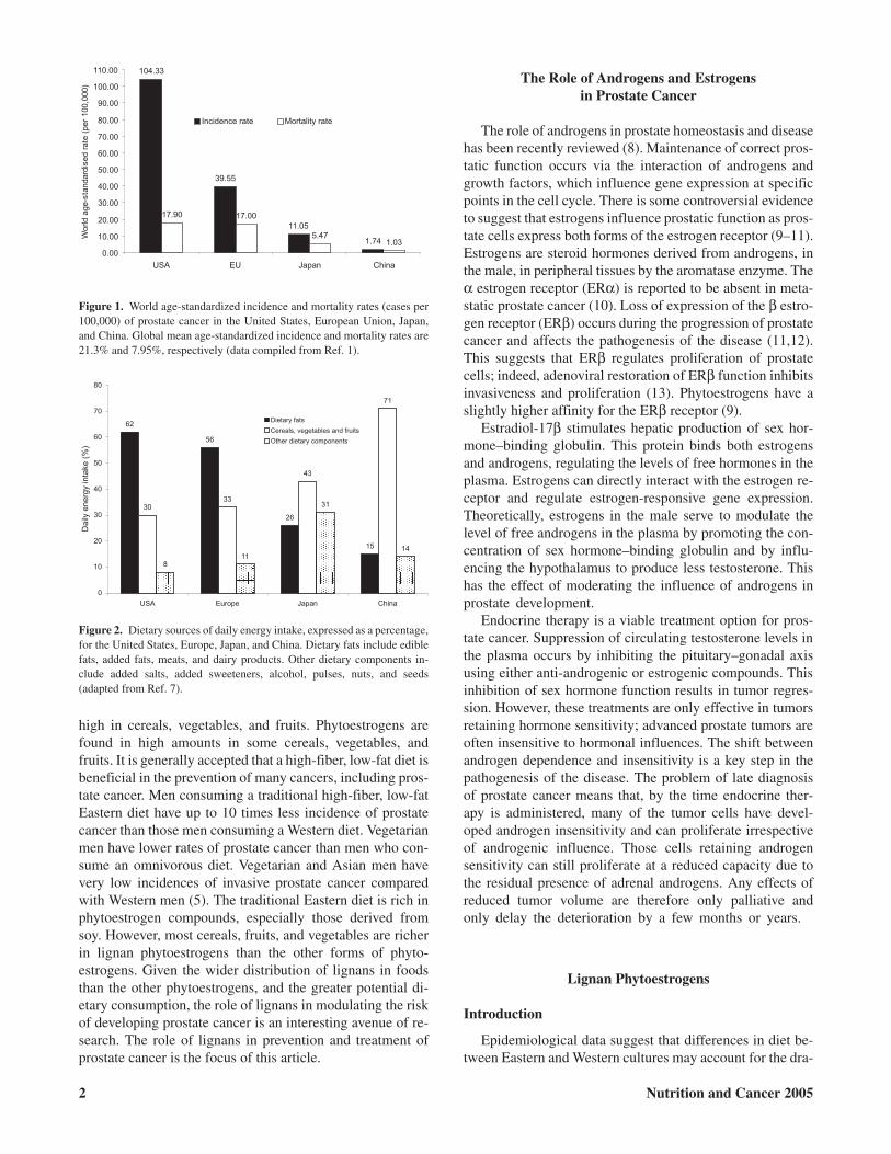
Globally, prostate cancer is the third most common cancerin men. The incidence of prostate cancer globally has beenincreasing by 1.7% per year for the last 15 yr (1). Marked dif-ferences in world age-standardized incidence and mortalityrates of prostate cancer exist among the United States, Euro-pean Union, Japan, and China (1). Figure 1 illustrates ap-proximately a 60-fold difference in incidence rates and a17-fold difference in mortality rates between the UnitedStates and China (1).
The etiology of prostate cancer is complex andmultifactorial. Primary risk factors are age, diet, and geneticdefects (2). Prostate cancer is usually only observed in malesover the age of 50 yr. The World Health Organization esti-mates that 30% of all cancers are primarily a result of dietaryfactors (2). Familial prostate cancer occurs in approximately10% of cases, twice the rate of hereditary breast cancer, and itdevelops at an earlier age than nonfamilial prostate cancer(2). Defects in the normal hormonal interactions with theprostate are a common feature of prostate cancer. The role ofgenes and growth factors in the development and progressionof prostate cancer is beyond the scope of this article and isdiscussed elsewhere (3,4). Epidemiological studies haveshown that, although the incidence of noninvasive prostatecancer in Asian societies is similar to that in Western society,the incidence of invasive cancer and associated mortality islower (5). Migrant populations are exposed to novel environ-mental factors such as diet. Incidence rates of prostate cancerin migrants from Asian countries living in the United Statestend to match those of the Caucasian and African-Americanpopulations over several generations (6). For example, Chi-nese male migrants from Shanghai to the United States reporta 27-fold increase in cases of prostate cancer, after severalgenerations, compared with those remaining in Shanghai (6).
These data strongly suggest that diet, not genetics, plays amore central role in the pathogenesis of prostate cancer.There is some evidence suggesting that the differences in dietbetween Eastern and Western cultures are responsible for thedramatic difference in the incidence of prostate cancer (5,6).There is a distinct difference in the dietary patterns betweenEastern and Western cultures as shown in Fig. 2. This figurehighlights the conspicuous differences in daily energysources between Eastern and Western cultures (7). The West-ern diet, typified by the United States and Europe, is high infat and low in cereals, vegetables, and fruit. The Japanese dietis much lower in daily fat intake and the Chinese diet is very
NUTRITION AND CANCER, 52(1), 1–14Copyright © 2005, Lawrence Erlbaum Associates, Inc.
All authors are affiliated with the Northern Ireland Centre for Diet and Health (NICHE), University of Ulster, Coleraine, Northern Ireland BT52 1SA.
high in cereals, vegetables, and fruits. Phytoestrogens arefound in high amounts in some cereals, vegetables, andfruits. It is generally accepted that a high-fiber, low-fat diet isbeneficial in the prevention of many cancers, including pros-tate cancer. Men consuming a traditional high-fiber, low-fatEastern diet have up to 10 times less incidence of prostatecancer than those men consuming a Western diet. Vegetarianmen have lower rates of prostate cancer than men who con-sume an omnivorous diet. Vegetarian and Asian men havevery low incidences of invasive prostate cancer comparedwith Western men (5). The traditional Eastern diet is rich inphytoestrogen compounds, especially those derived fromsoy. However, most cereals, fruits, and vegetables are richerin lignan phytoestrogens than the other forms of phyto-estrogens. Given the wider distribution of lignans in foodsthan the other phytoestrogens, and the greater potential di-etary consumption, the role of lignans in modulating the riskof developing prostate cancer is an interesting avenue of re-search. The role of lignans in prevention and treatment ofprostate cancer is the focus of this article.
The Role of Androgens and Estrogensin Prostate Cancer
The role of androgens in prostate homeostasis and diseasehas been recently reviewed (8). Maintenance of correct pros-tatic function occurs via the interaction of androgens andgrowth factors, which influence gene expression at specificpoints in the cell cycle. There is some controversial evidenceto suggest that estrogens influence prostatic function as pros-tate cells express both forms of the estrogen receptor (9–11).Estrogens are steroid hormones derived from androgens, inthe male, in peripheral tissues by the aromatase enzyme. Theα estrogen receptor (ERα) is reported to be absent in meta-static prostate cancer (10). Loss of expression of the β estro-gen receptor (ERβ) occurs during the progression of prostatecancer and affects the pathogenesis of the disease (11,12).This suggests that ERβ regulates proliferation of prostatecells; indeed, adenoviral restoration of ERβ function inhibitsinvasiveness and proliferation (13). Phytoestrogens have aslightly higher affinity for the ERβ receptor (9).
Estradiol-17β stimulates hepatic production of sex hor-mone–binding globulin. This protein binds both estrogensand androgens, regulating the levels of free hormones in theplasma. Estrogens can directly interact with the estrogen re-ceptor and regulate estrogen-responsive gene expression.Theoretically, estrogens in the male serve to modulate thelevel of free androgens in the plasma by promoting the con-centration of sex hormone–binding globulin and by influ-encing the hypothalamus to produce less testosterone. Thishas the effect of moderating the influence of androgens inprostate development.
Endocrine therapy is a viable treatment option for pros-tate cancer. Suppression of circulating testosterone levels inthe plasma occurs by inhibiting the pituitary–gonadal axisusing either anti-androgenic or estrogenic compounds. Thisinhibition of sex hormone function results in tumor regres-sion. However, these treatments are only effective in tumorsretaining hormone sensitivity; advanced prostate tumors areoften insensitive to hormonal influences. The shift betweenandrogen dependence and insensitivity is a key step in thepathogenesis of the disease. The problem of late diagnosisof prostate cancer means that, by the time endocrine ther-apy is administered, many of the tumor cells have devel-oped androgen insensitivity and can proliferate irrespectiveof androgenic influence. Those cells retaining androgensensitivity can still proliferate at a reduced capacity due tothe residual presence of adrenal androgens. Any effects ofreduced tumor volume are therefore only palliative andonly delay the deterioration by a few months or years.
Lignan Phytoestrogens
Introduction
Epidemiological data suggest that differences in diet be-tween Eastern and Western cultures may account for the dra-
2 Nutrition and Cancer 2005
Figure 1. World age-standardized incidence and mortality rates (cases per100,000) of prostate cancer in the United States, European Union, Japan,and China. Global mean age-standardized incidence and mortality rates are21.3% and 7.95%, respectively (data compiled from Ref. 1).
Figure 2. Dietary sources of daily energy intake, expressed as a percentage,for the United States, Europe, Japan, and China. Dietary fats include ediblefats, added fats, meats, and dairy products. Other dietary components in-clude added salts, added sweeteners, alcohol, pulses, nuts, and seeds(adapted from Ref. 7).
matic difference in incidence and mortality rates of prostatecancer (1,7). Traditional Asian diets are rich in foods such ascereals, fruits, and vegetables that contain high levels ofphytoestrogenic compounds such as isoflavones and lignans(7). The mammalian lignans enterolactone and enterodiolwere first characterized in both human and vervet monkey re-productive cycles over 25 yr ago (14–17). Lignans are pres-ent in a range of biological fluids in several diverse popula-tions, suggesting a biological role for these compounds(18–20). Morton et al. reported that the prostate fluid levelsof enterolactone of Chinese, Portuguese, and British menwere significantly higher than their plasma levels (19). Thissuggests that lignans accumulate in prostatic fluid and mayachieve significantly higher concentrations than those re-ported in plasma. The reason for this accumulation is un-known and implies that lignans could play a role in preven-tion and treatment of prostate cancer.
Chemistry and Analytical Methods
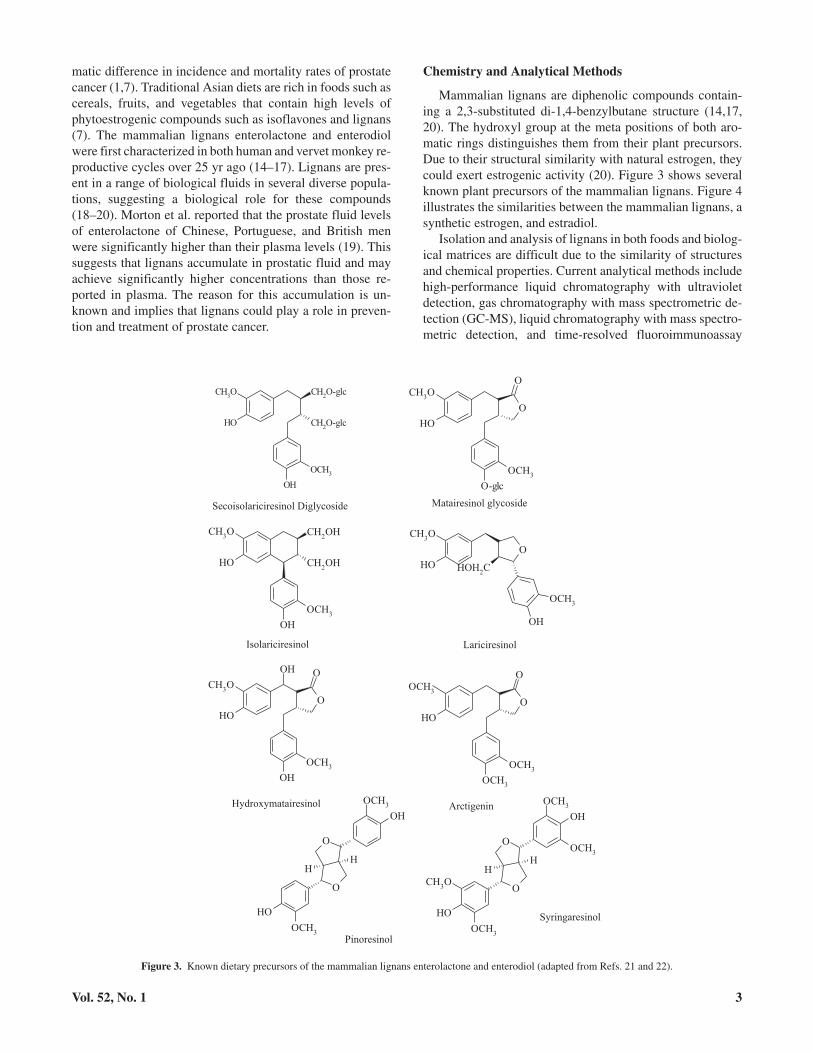
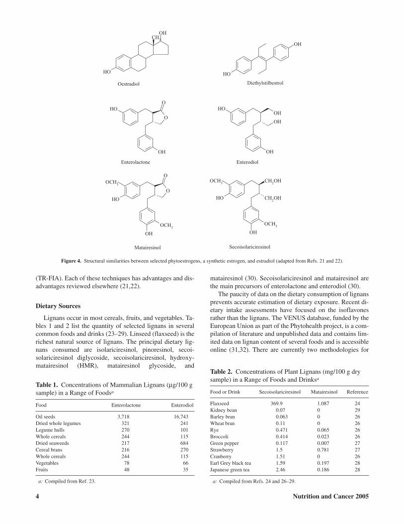
Mammalian lignans are diphenolic compounds contain-ing a 2,3-substituted di-1,4-benzylbutane structure (14,17,20). The hydroxyl group at the meta positions of both aro-matic rings distinguishes them from their plant precursors.Due to their structural similarity with natural estrogen, theycould exert estrogenic activity (20). Figure 3 shows severalknown plant precursors of the mammalian lignans. Figure 4illustrates the similarities between the mammalian lignans, asynthetic estrogen, and estradiol.
Isolation and analysis of lignans in both foods and biolog-ical matrices are difficult due to the similarity of structuresand chemical properties. Current analytical methods includehigh-performance liquid chromatography with ultravioletdetection, gas chromatography with mass spectrometric de-tection (GC-MS), liquid chromatography with mass spectro-metric detection, and time-resolved fluoroimmunoassay
Vol. 52, No. 1 3
Figure 3. Known dietary precursors of the mammalian lignans enterolactone and enterodiol (adapted from Refs. 21 and 22).
(TR-FIA). Each of these techniques has advantages and dis-advantages reviewed elsewhere (21,22).
Dietary Sources
Lignans occur in most cereals, fruits, and vegetables. Ta-bles 1 and 2 list the quantity of selected lignans in severalcommon foods and drinks (23–29). Linseed (flaxseed) is therichest natural source of lignans. The principal dietary lig-nans consumed are isolariciresinol, pinoresinol, secoi-solariciresinol diglycoside, secoisolariciresinol, hydroxy-matairesinol (HMR), matairesinol glycoside, and
matairesinol (30). Secoisolariciresinol and matairesinol arethe main precursors of enterolactone and enterodiol (30).
The paucity of data on the dietary consumption of lignansprevents accurate estimation of dietary exposure. Recent di-etary intake assessments have focused on the isoflavonesrather than the lignans. The VENUS database, funded by theEuropean Union as part of the Phytohealth project, is a com-pilation of literature and unpublished data and contains lim-ited data on lignan content of several foods and is accessibleonline (31,32). There are currently two methodologies for
4 Nutrition and Cancer 2005
Figure 4. Structural similarities between selected phytoestrogens, a synthetic estrogen, and estradiol (adapted from Refs. 21 and 22).
Table 1. Concentrations of Mammalian Lignans (µg/100 gsample) in a Range of Foodsa
Food Enterolactone Enterodiol
Oil seeds 3,718 16,743Dried whole legumes 321 241Legume hulls 270 101Whole cereals 244 115Dried seaweeds 217 684Cereal brans 216 270Whole cereals 244 115Vegetables 78 66Fruits 48 35
a: Compiled from Ref. 23.
Table 2. Concentrations of Plant Lignans (mg/100 g drysample) in a Range of Foods and Drinksa
Food or Drink Secoisolariciresinol Matairesinol Reference
Flaxseed 369.9 1.087 24Kidney bean 0.07 0 29Barley bran 0.063 0 26Wheat bran 0.11 0 26Rye 0.471 0.065 26Broccoli 0.414 0.023 26Green pepper 0.117 0.007 27Strawberry 1.5 0.781 27Cranberry 1.51 0 26Earl Grey black tea 1.59 0.197 28Japanese green tea 2.46 0.186 28
a: Compiled from Refs. 24 and 26–29.
determining the lignan content of foods. Thompson et al. useindirect in vitro fermentation with colonic microflora to as-sess production of enterolactone and enterodiol from plantlignans (23). Mazur et al. utilize isotope-dilution gas-chro-matography mass spectrometry (IDGCMS) to measure thelevels of plant lignans in foods (24). A review and compari-son of these methods are available (25). Measurements ofplasma levels and urinary excretion are more widely used toassess dietary exposure to lignans.
Pharmacokinetics
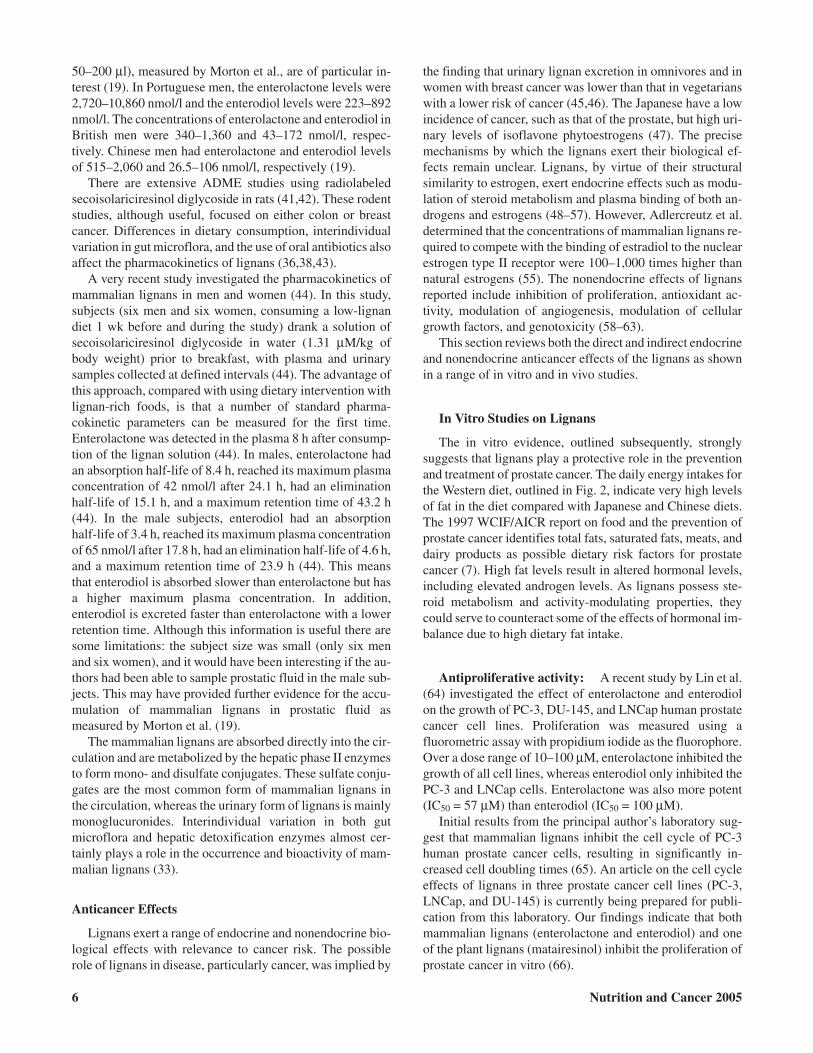
Enterolactone and enterodiol are the primary lignans ob-served in human biological fluids. The bioavailability of theselignans has been recently reviewed (33). Intestinal bacteriaproduce enterolactone and enterodiol from their plant precur-sors (34–36). Figure 5 illustrates a proposed schematic of theproduction of enterolactone and enterodiol by intestinalmicroflora. Other plant lignans, such as pinoresinol,syringaresinol, lariciresinol, arctigenin, and HMR, can also bemetabolized intoenterolactoneandenterodiolbutat lower lev-els than secoisolariciresinol or matairesinol (30). Due to thevirtuallynegligible levelsofplant lignans in theplasma, the in-testinal metabolism of these lignans into their mammalianformsseemsessential forabsorptionacross thegutwall (33).
There are very limited studies on the pharmacokinetics oflignans in men. Most studies have used foods containingknown precursors to assess the adsorption, distribution, me-tabolism, and excretion (ADME) profile of lignans (37–40).
The concentration of enterolactone and enterodiol in biologi-cal fluids varies considerably by geographical region andanalytical method. Kilkkinen et al. measured the serum lev-els of enterolactone in 2,380 Finnish men aged 25–64 yr, par-ticipating in a national nutrition survey, using TR-FIA andfound that the median serum enterolactone concentrationwas 13.8 nmol/l (37). Another study by Kilkkinen et al. re-ported mean enterolactone serum levels of 15.9 nmol/l in 214Finnish men in a nested case-control study, again usingTR-FIA (38). Jacobs et al. reported mean enterolactone con-centrations of 6.2 nmol/l, measured using TR-FIA, in fiveAmerican men fed a diet high in whole grains (39). Juntunenet al. fed a diet rich in rye bread, a rich source of lignans, to 18Finnish men aged 41–43 yr and measured (using TR-FIA) amean serum enterolactone concentration of 25.6 nmol/l (40).However, Morton et al. measured both serum and prostaticfluid levels of enterolactone and enterodiol in Portuguese,British, and Chinese men using GC-MS (19). There was nodifference in the mean plasma level of enterolactone amongPortuguese and British men (both 3.27 nmol/l); however,Chinese men had 5.2 nmol/l. Portuguese man had muchlower levels of enterodiol in their plasma than Chinese men(0.30 compared with 1.4 nmol/l). The plasma levels ofenterodiol for British men were not reported. The plasma lev-els measured by Morton et al. are lower than the other studiesreported here due to the different analytical technique usedand the samples were from men without any dietary interven-tion. The mean concentrations of enterolactone andenterodiol in prostatic fluid (based on a sample volume of
Vol. 52, No. 1 5
Figure 5. The production of enterolactone and enterodiol from secoisolariciresinol diglycoside and matairesinol by human intestinal bacteria (adapted fromRef. 36).
50–200 µl), measured by Morton et al., are of particular in-terest (19). In Portuguese men, the enterolactone levels were2,720–10,860 nmol/l and the enterodiol levels were 223–892nmol/l. The concentrations of enterolactone and enterodiol inBritish men were 340–1,360 and 43–172 nmol/l, respec-tively. Chinese men had enterolactone and enterodiol levelsof 515–2,060 and 26.5–106 nmol/l, respectively (19).
There are extensive ADME studies using radiolabeledsecoisolariciresinol diglycoside in rats (41,42). These rodentstudies, although useful, focused on either colon or breastcancer. Differences in dietary consumption, interindividualvariation in gut microflora, and the use of oral antibiotics alsoaffect the pharmacokinetics of lignans (36,38,43).
A very recent study investigated the pharmacokinetics ofmammalian lignans in men and women (44). In this study,subjects (six men and six women, consuming a low-lignandiet 1 wk before and during the study) drank a solution ofsecoisolariciresinol diglycoside in water (1.31 µM/kg ofbody weight) prior to breakfast, with plasma and urinarysamples collected at defined intervals (44). The advantage ofthis approach, compared with using dietary intervention withlignan-rich foods, is that a number of standard pharma-cokinetic parameters can be measured for the first time.Enterolactone was detected in the plasma 8 h after consump-tion of the lignan solution (44). In males, enterolactone hadan absorption half-life of 8.4 h, reached its maximum plasmaconcentration of 42 nmol/l after 24.1 h, had an eliminationhalf-life of 15.1 h, and a maximum retention time of 43.2 h(44). In the male subjects, enterodiol had an absorptionhalf-life of 3.4 h, reached its maximum plasma concentrationof 65 nmol/l after 17.8 h, had an elimination half-life of 4.6 h,and a maximum retention time of 23.9 h (44). This meansthat enterodiol is absorbed slower than enterolactone but hasa higher maximum plasma concentration. In addition,enterodiol is excreted faster than enterolactone with a lowerretention time. Although this information is useful there aresome limitations: the subject size was small (only six menand six women), and it would have been interesting if the au-thors had been able to sample prostatic fluid in the male sub-jects. This may have provided further evidence for the accu-mulation of mammalian lignans in prostatic fluid asmeasured by Morton et al. (19).
The mammalian lignans are absorbed directly into the cir-culation and are metabolized by the hepatic phase II enzymesto form mono- and disulfate conjugates. These sulfate conju-gates are the most common form of mammalian lignans inthe circulation, whereas the urinary form of lignans is mainlymonoglucuronides. Interindividual variation in both gutmicroflora and hepatic detoxification enzymes almost cer-tainly plays a role in the occurrence and bioactivity of mam-malian lignans (33).
Anticancer Effects
Lignans exert a range of endocrine and nonendocrine bio-logical effects with relevance to cancer risk. The possiblerole of lignans in disease, particularly cancer, was implied by
the finding that urinary lignan excretion in omnivores and inwomen with breast cancer was lower than that in vegetarianswith a lower risk of cancer (45,46). The Japanese have a lowincidence of cancer, such as that of the prostate, but high uri-nary levels of isoflavone phytoestrogens (47). The precisemechanisms by which the lignans exert their biological ef-fects remain unclear. Lignans, by virtue of their structuralsimilarity to estrogen, exert endocrine effects such as modu-lation of steroid metabolism and plasma binding of both an-drogens and estrogens (48–57). However, Adlercreutz et al.determined that the concentrations of mammalian lignans re-quired to compete with the binding of estradiol to the nuclearestrogen type II receptor were 100–1,000 times higher thannatural estrogens (55). The nonendocrine effects of lignansreported include inhibition of proliferation, antioxidant ac-tivity, modulation of angiogenesis, modulation of cellulargrowth factors, and genotoxicity (58–63).
This section reviews both the direct and indirect endocrineand nonendocrine anticancer effects of the lignans as shownin a range of in vitro and in vivo studies.
In Vitro Studies on Lignans
The in vitro evidence, outlined subsequently, stronglysuggests that lignans play a protective role in the preventionand treatment of prostate cancer. The daily energy intakes forthe Western diet, outlined in Fig. 2, indicate very high levelsof fat in the diet compared with Japanese and Chinese diets.The 1997 WCIF/AICR report on food and the prevention ofprostate cancer identifies total fats, saturated fats, meats, anddairy products as possible dietary risk factors for prostatecancer (7). High fat levels result in altered hormonal levels,including elevated androgen levels. As lignans possess ste-roid metabolism and activity-modulating properties, theycould serve to counteract some of the effects of hormonal im-balance due to high dietary fat intake.
Antiproliferative activity: A recent study by Lin et al.(64) investigated the effect of enterolactone and enterodiolon the growth of PC-3, DU-145, and LNCap human prostatecancer cell lines. Proliferation was measured using afluorometric assay with propidium iodide as the fluorophore.Over a dose range of 10–100 µM, enterolactone inhibited thegrowth of all cell lines, whereas enterodiol only inhibited thePC-3 and LNCap cells. Enterolactone was also more potent(IC50 = 57 µM) than enterodiol (IC50 = 100 µM).
Initial results from the principal author’s laboratory sug-gest that mammalian lignans inhibit the cell cycle of PC-3human prostate cancer cells, resulting in significantly in-creased cell doubling times (65). An article on the cell cycleeffects of lignans in three prostate cancer cell lines (PC-3,LNCap, and DU-145) is currently being prepared for publi-cation from this laboratory. Our findings indicate that bothmammalian lignans (enterolactone and enterodiol) and oneof the plant lignans (matairesinol) inhibit the proliferation ofprostate cancer in vitro (66).
6 Nutrition and Cancer 2005
Steroid metabolism and activity: Lignans inhibit theactivity of human aromatase, 17β-hydroxysteroid dehydro-genase, and 5α-reductase (48–57). The lignans appearunique among the phytoestrogens in their ability to modulatesteroid activity via the aromatase enzymes. In plasma, thephytoestrogens are transported to their target tissues by car-rier proteins, such as α-fetoprotein, sex hormone–bindingglobulin, and human sex steroid–binding protein. These pro-teins normally play a role in maintaining the levels of freehormone in plasma. Sex steroid–binding protein has beenfound in prostate cells (57).
Martin et al. investigated the ability of several phyto-estrogens, including enterolactone and enterodiol (doserange 2–200 nM), in affecting the binding of sex steroid–binding protein with either estradiol or testosterone (48). Inthe case of estradiol to sex steroid–binding protein,enterolactone was more potent at inhibition than enterodiolor genistein. Enterolactone was more effective at inhibitingthe binding of testosterone to sex steroid–binding proteinthan genistein, and enterodiol showed negligible inhibition(48). Schottner et al. reported that matairesinol, secoi-solariciresinol, enterolactone, and enterodiol inhibit thebinding of 5α-dihydrotestosterone with human sex hor-mone–binding globulin (49). Adlercreutz et al. reports thatserum levels of 6 µM enterolactone are sufficient to inhibitthe activity of human aromatase (50). Adlercreutz et al. re-port that serum levels of 0.5–50.0 µM enterolactone andenterodiol are sufficient to inhibit binding of testosterone andestrogen to sex hormone–binding globulin (51). Garreau etal. reported that enterolactone was more effective thanenterodiol at inhibiting the binding of α-fetoprotein to eitherestrogen or testosterone (52). Brooks and Thompson have in-vestigated the ability of enterolactone and enterodiol to in-hibit the activity of aromatase and 17β-hydroxysteroiddehydrogenase in MCF-7 human breast cancer cells (53).This has the effect of reducing the amount of estradiol pro-duced. A 50-µM concentration of enterolactone significantlyinhibits estradiol production (53). Evans et al. report that anenterolactone and enterodiol concentration of 100 µM signif-icantly inhibited the activity of 5α-reductase and 17β-hydroxysteroid dehydrogenase in genital skin fibroblasts(54). Adlercreutz et al. determined that enterolactoneconcentrations of 0.5–10 µM stimulated the synthesis of sexhormone–binding globulin (55).
Mammalian lignans compete with estrogens and testos-terone for binding of plasma proteins; in effect, they are di-etary modulators of the physiochemical activity of the sexhormones.
Antioxidant capacity: There is evidence in the litera-ture to suggest that several lignans display antioxidant prop-erties (58,59,67). Kangas et al. examined the antioxidant andantitumor properties of HMR in vitro and in vivo by investi-gating its ability to affect lipid peroxidation and superoxidescavenging compared with Trolox (a synthetic vitamin E de-rivative) and enterolactone (67). HMR was several orders ofmagnitude more effective at superoxide scavenging and at in-
hibiting lipid peroxidation than enterolactone. However,enterolactone was significantly more effective (IC50 = 1µmol/l) than HMR (IC50 = 10 µmol/l) at inhibiting an oxida-tive burst in human monocytes (67).
Prasad investigated the ability of several lignans and vita-min E to inhibit the chemiluminescence of zymosan-acti-vated polymorphonuclear leukocytes (58). In this model sys-tem, enterolactone was the most potent antioxidant (IC50 = 1mg/ml) compared with enterodiol (IC50 = 2.5 mg/ml),secoisolariciresinol diglycoside, secoisolariciresinol, and vi-tamin E (IC50 = 5 mg/ml) (58).
Kitts et al. studied the antioxidant activity ofsecoisolariciresinol diglycoside, enterodiol, and entero-lactone by assessing their ability to inhibit lipid peroxidationand both specific and nonspecific Fenton reactant–inducedhydroxyl scavenging (59). Ten- and 100 µM concentrationsof the lignans inhibited lipid peroxidation. The results of thisstudy indicate that, in the assay models used, enterolactoneand enterodiol are significantly more potent antioxidantsthan secoisolariciresinol diglycoside at both 10 and 100 µM.
Niemeyer and Metzler investigated the antioxidant poten-tial of enterolactone, enterodiol, matairesinol, and secoi-solariciresinol in vitro using the ferric reducing/antioxidantpower assay (60). Secoisolariciresinol and matairesinol(50–400 µmol/l) were found to be more potent than ascorbicacid (10–800 µmol/l) and significantly more potent thanenterolactone and enterodiol (1–2 µM) (60).
Antigenotoxic activity: Kulling et al. used cell-freemicrotubule assembly and various selected endpoints in cul-tured V79 Chinese hamster cells to assess the genotoxicity ofenterolactone, enterodiol, matairesinol, and secoiso-lariciresinol (61). Diethylstilbestrol (aneuploidogen) and4-nitroquinoline-N-oxide (clastogen) were used as positivecontrols. None of the four lignans had any activity in any ofthe assays used. In contrast, other phytoestrogens such asgenistein have been shown to have in vitro genotoxic activityin the V79 cell line (61). Therefore, it is concluded that thelignans tested do not possess any aneuploidogenic orclastogenic properties in the model system used. However,the authors of this article referred to the lack of informationon the genotoxicity of any oxidative metabolites of theselignans.
Anti-angiogenic activity: The process of tumorangiogenesis is a critical event in the pathogenesis of themany cancers, including prostate cancer. Vascular endothe-lial growth factor (VEGF) is associated with the angiogenicprocess and is the focus of much of the research intoanti-angiogenic treatments. A recent article has examined therole of angiogenesis in prostate cancer (62). Studies pertain-ing to the influence of phytoestrogens on angiogenesis havefocused on the isoflavones, particularly genistein. There havebeen no studies to date on the role of lignans in angiogenesisin prostate cancer.
Fotsis et al. investigated the phytoestrogen content of 24-hurine samples collected from subjects consuming soy-rich
Vol. 52, No. 1 7
vegetarian diet. Of the six fractions collected, one containedenterolactone, enterodiol, and matairesinol (63). Ente-rolactone, over a concentration range of 0–100 µM, wasfound to inhibit the growth of endothelial cells, supple-mented with basic fibroblast growth factor, derived from bo-vine brain tissue. However, this study focused on theanti-angiogenic potential of genistein, an isoflavone found insoy, and not that of enterolactone or the other lignans mea-sured in one of the fractions.
However, a recent study using MDA-MB-435 humanbreast cancer cells transplanted into mice fed a 10% flax-seed-supplemented diet for 8 wk found a significant decreasein the tumor volume, incidence of metastases, and levels ofVEGF (68).
Summary of in vitro studies: The studies reviewedpreviously suggest that both plant and mammalian lignanspossess beneficial anticancer effects in vitro. However, theeffective doses used in the in vitro studies are in themicromolar range, significantly higher than the plasma levelsreported by several authors (37–40,44). These supraphy-siological doses used may not be achievable in vivo throughdietary consumption of foods rich in lignans. The observa-tion by Morton et al. regarding the significantly higher pros-tatic fluid levels of mammalian lignans compared withplasma levels may, if confirmed in future studies, clarify therole of dietary consumption of lignans in prostate cancer. Theanticancer effects reported can only be indicative of possibleeffects of lignans in prostate cancer. Despite the evidence de-scribed previously, very few of the studies used prostate celllines. The validity of the in vitro effects reported with respectto prostate cancer requires clarification and support from invivo studies.
The evidence for antiproliferative effects of lignans inprostate cancer is limited to two published studies (64,65).Therefore, only possible antiproliferative properties can beascribed to lignans. Several reports suggest that lignans in-hibit the activity of critical aspects of sex steroid metabolismand activity (48–57). These studies are limited by the factthat they were mainly performed in breast cancer research orfemale subjects. As prostate cancer is also a hormone-de-pendent cancer, it is feasible to infer that the effects reportedmay be seen in this type of cancer. There is only one articleon lignans and steroid metabolism in prostate cancer (54). Inthe investigations into the antioxidant potential of lignans,half of the studies indicate that plant lignans are more potentthan mammalian lignans (60,67), whereas the others suggestthat it is the opposite situation (58,59). The antioxidant po-tency of the lignans tested appears to depend on which assayis chosen. Lignans were not genotoxic in the sole study inthis area (61). However, the authors did concede that the oxi-dative metabolites of enterolactone and enterodiol may ormay not possess genotoxicity (61). This study used Chinesehamster cells, a fibroblast cell line. The reason the authorschose this cell line is unclear and may be due to its suitabilityfor the endpoints selected. It is questionable as to the rele-vance of these data to prostate cancer. The anti-angiogenic
effects of the lignans reported by Fotsis et al. (63) were mea-sured in endothelial cells derived from bovine brain tissue.The validity of these results with respect to prostate cancer isa pertinent issue. The recent study by Dabrosin et al. showsthat dietary intervention with flaxseed affects several indict-ors of angiogenesis in breast cancer but not necessarily pros-tate cancer (68).
In Vivo Studies on Lignans
A number of limited animal and human studies supportthe reported in vitro anticancer effects of lignans outlinedpreviously. Lignans are not widely available in purified formand are difficult to synthesize; therefore, many of the in vivostudies have used flaxseed or rye, which are rich sources oflignans. It is difficult to ascribe any observed in vivo effectsof flaxseed and rye to the lignans alone as many other poten-tially bioactive compounds are present in these foods. Themajority of the in vivo studies have been in murine and ro-dent models; only a few dietary interventions with lignanshave used human subjects. Despite the limitations, in vivo re-search has provided experimental evidence for the potentialrole of lignans in the prevention and treatment of prostatecancer. Tables 3 and 4 provide a summary of the current invivo research into the anticancer effects of lignans.
Animal studies: Bylund et al. transplanted 70 athymicmice (BALB/cABom strain) with approximately 8 millionhuman LNCap prostate cancer cells 3 days before dietary in-tervention with 1 of 7 diets (69). The control diet (CC) con-sisted of cornstarch, sucrose, low-fat milk powder, corn oil,and lard supplemented with cellulose. The experimental ryebran diets consisted of the control diet supplemented with ryebran (RB diet), with rye bran extracted with ethyl acetate(EXRB diet), and with a higher fat content (HFRB diet). Ryebran is known to contain lignans such as secoisolariciresinoland matairesinol (26). In addition, a soy-based diet (SCC)consisted of the CC diet with soy replacing the milk powder.The remaining two diets were derived from the CC diet sup-plemented with ethyl acetate extracts from rye bran (CCEEdiet) and ethyl acetate extracts of alkylresorcinols extractedfrom rye bran (CCA diet) (69). Six of the diets were con-trolled for fat levels (12.7 g/100 g of diet) and energy density(377.5 kcal/100 g). The HFRB diet had a fat content 18.1g/100 g and an energy density of 399.4 kcal/100 g of diet. Af-ter 5 wk on dietary intervention, the RB and SCC diets weresignificantly different from the control diet in terms of tumorvolume. After 6 wk, these two diets also had lower prostatespecific antigen (PSA) levels than the control diet (69). Atsacrifice (9 wk) the mice fed the RB and CCEE diets had sig-nificantly lower tumor take rates, tumor volumes, and PSAlevels and higher necrotic and apoptotic levels comparedwith the control diet (69). The HFRB results were not signifi-cantly different from the control diet. This suggests that thephytoestrogens present in these diets (both lignans andisoflavones) could be responsible for the growth restrictionof the tumor observed. However, both rye-based and soy di-
8 Nutrition and Cancer 2005
ets contain other bioactive compounds in addition tophytoestrogens.
Lin et al. studied the effect of a 5% (by weight) flax-seed-supplemented diet on 135 male transgenic adenocar-cinoma mouse prostate (TRAMP) mice for 20 and 30 wk, re-spectively (70). These transgenic mice posses a rat probasinpromoter that regulates the prostate-specific expression ofthe SV40 T-antigen (71). The advantage of this study is thatTRAMP mice do not depend on exogenous hormones ortransplanted tumors to be a suitable animal model. The ex-perimental and control diets were equivalent in terms of calo-ries, carbohydrates, protein, and fat content. All of the con-trol mice and 97% of the intervention mice developedprostate cancer; however, there was a marked difference in
the tumor mass and characteristics of the tumors. The inter-vention mice had a significantly lower average tumor massand percentage tumor mass to body mass ratio than thosefound in the control mice after 20 and 30 wk of dietary inter-vention. Despite the fact that the Gleason grading system isused to assess the aggressiveness of human prostate cancers,the authors of the article used this system to measure the ag-gressiveness of the mouse tumors at 30 wk. The interventionmice had a significantly lower Gleason score than the controlmice. The level of cellular proliferation as measured by theKI-67 nuclear antigen decreased significantly. The apoptoticindex, measured using the deoxynucleotidyl transferase-mediated dUTP-digoxigenin nick end labeling (TUNEL)technique, increased significantly in the intervention mice.
Vol. 52, No. 1 9
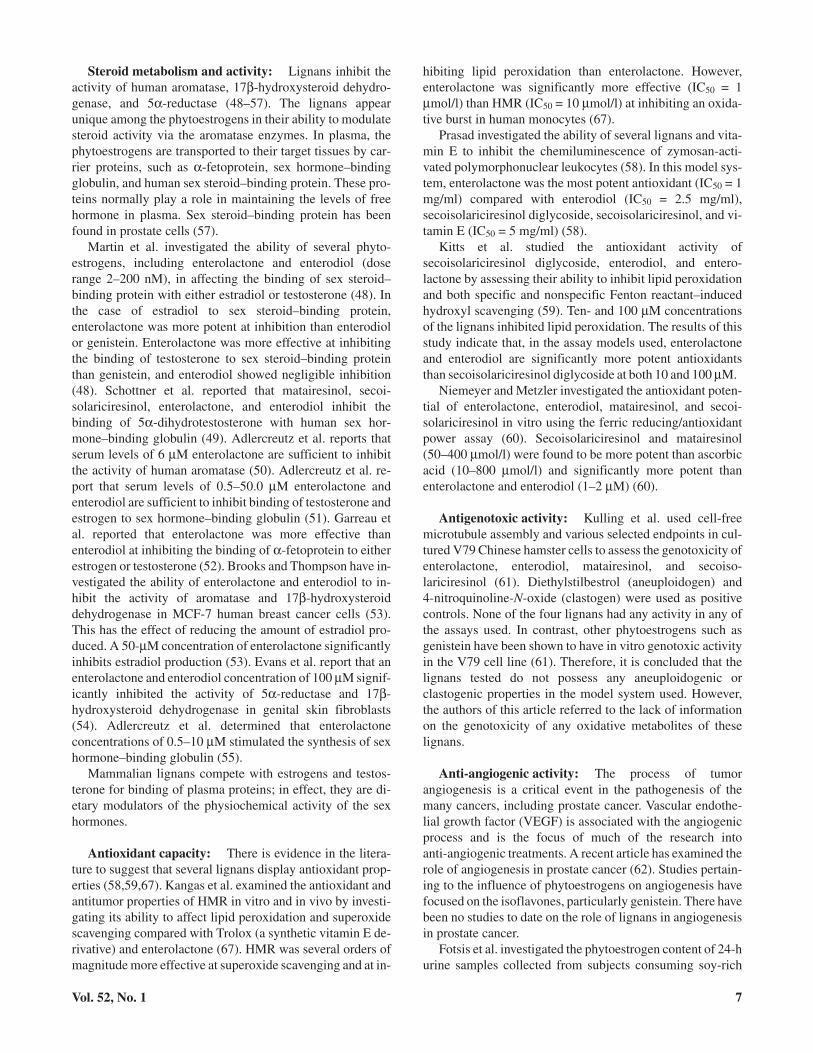
Table 3. Summary of the In Vivo Animal Studies Into the Effects of Lignans on Prostate Cancera,b
Model Intervention Duration (wk) Effect of Intervention Reference
BALB/cABom mice (n = 70) Transplant of LNCap cells with 1of 7 distinct rye- and soy-baseddiets
9 Reduced tumor take rates, tumormass, and PSA levels
Increased necrosis and apoptosis
69
TRAMP mice (n = 135) 5% (by weight)flaxseed-supplemented diet
20 and 30 Decreased tumor massLower Gleason grade observed
70
Dunning R3327 rat (n = 125) 33% soy flour diet33% rye bran diet33% heat-treated rye bran diet33% rye endosperm diet
24 Decreased tumor mass observed inall diets after 16 wk; mostsignificant decrease in tumormass was measured for the ryebran diet
72
BALB/cABom mice 0.15% or 0.30% HMR diet 9 Both diets had lower tumor takerates and tumor volumes thanthe control mice
73
a: Adapted from Refs. 69, 70, 72, and 73.b: Abbreviations are as follows: PSA, prostate specific antigen; TRAMP, transgenic adenocarcinoma mouse prostate; HMR, hydroxymatairesinol.
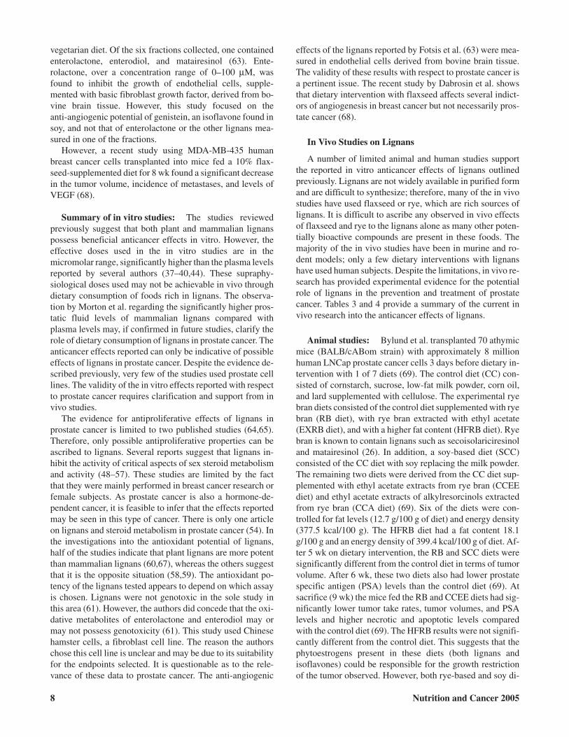
Table 4. Summary of the In Vivo Human Studies Into the Effects of Lignans on Prostate Cancera,b
Model Intervention Duration Effect of Intervention/Study Reference
Human (n = 25) 30 g/day flaxseed diet 3 wk Decreased total serum cholesterol, totaltestosterone, and free androgen index
Patients with a Gleason grade of 6 orless had reduced PSA levels
Apoptosis and proliferation ratesdependent on length of intervention
74
Human (n = 10) 295 g/day rye bran bread diet 3 wk Increase in serum plasma enterolactonelevels
Increase in apoptosis rates
75
Human (n = 26) 50 g soy bread diet50 g soy bread + 20 g linseed dietWheat bread diet
27.4 (±3.6) days Increase in total PSAIncrease in total androgen indexDecrease in free to total PSA ratio
76
Human (n = 214) Nested case-control study of Finnishmen involved in theAlpha-Tocopherol, Beta-Carotenestudy
6 yr No relationship between lignanconsumption and prostate cancer risk
No relationship between serumenterolactone concentration andprostate cancer risk
38
Human (n = 136) 105-item dietary questionnaire 10 yr No relationship between prostate cancerrisk and a diet of fruit and vegetablesor a diet of red meat and starch
77
a: Adapted from Refs. 38 and 74–77.b: Abbreviation is as follows: PSA, prostate specific antigen.
Zhang et al. have reported on the restriction of tumorgrowth in 125 Dunning R3327 rats transplanted with prostatetumors (72). The rats consumed one of the experimental dietsfor 24 wk. The control diet was fiber-free (FF). The experi-mental diets were FF diet with 33% soy flour (SD diet), 33%rye bran (RB diet), 33% heat-treated rye bran (HRB diet),and 33% rye endosperm (RE), respectively. The rats consum-ing the RB, HRB, and SD diets showed a significant decreasein tumor volume compared with rats on the FF diet (72). TheRB diet was the most effective at suppressing tumor growth,of all the diets, after 24 wk despite different energy intakesamong the diets. To account for differences in energy intakeby rats consuming the RB diet, a second experiment was per-formed. The same prostate tumors from the first experimentwere transplanted into 150 fresh rats, and tumor developmentwas examined with dietary intervention with six isocaloricdiets (72). The control diets were FF and cellulose supple-mented FF (FC). The RB diet from the first experiment wasused unchanged. The other diets were FF diet with 33% en-zyme-treated (0.2% β-glucanase and xylonase) rye bran (RSdiet), 33% rye bran plus 10% soy flour (RS diet), and 3.3%linseed (flaxseed) (LS diet). The rats were fed the diets for 18wk and the tumor volume was assessed at sacrifice. In this ex-periment, only the RB diet significantly inhibited the tumormass of the rats. Enzymatic treatment and the addition of soyflour negated the effect of rye bran. The LS diet was observedto have no effect on tumor growth.
Bylund et al. have recently reported the anticancer proper-ties of HMR on prostate cancer in mice (73). This is the firstin vivo study to use a purified lignan. HMR was chosen as itis available in sufficient quantities from the Norway spruce(Picea abies) and is structurally similar to matairesinol (73).In addition, HMR is metabolized into enterolactone (30,34).This study used 36 athymic male mice of the BALB/cABomstrain. These mice were implanted with human LNCap pros-tate cancer cells 3 days prior to dietary intervention. The in-terventions were a control diet, a 0.15% HMR diet, and a0.3% HMR diet for 9 wk. All diets were low fat andisocaloric. The effect of 0.15% and 0.30% HMR on the tu-mor take rates and growth of the implanted LNCap cells werethe primary endpoints utilized. The results show that both in-terventions with HMR have significant anticancer properties(73). At sacrifice, the mice fed the HMR-supplemented dietshad lower tumor take rates and tumor volumes than the con-
trol mice (73). An increase in the level of nongrowing tumorswas seen as well as an increase in the apoptotic index (mea-sured by the TUNEL technique) (73). The 0.30% HMR dietalso inhibited the proliferation of the LNCap tumors (mea-sured by the KI-67 nuclear antigen) (73). This study supportsthe hypothesis that lignans may play a role in the early stagesof prostate tumorigenesis.
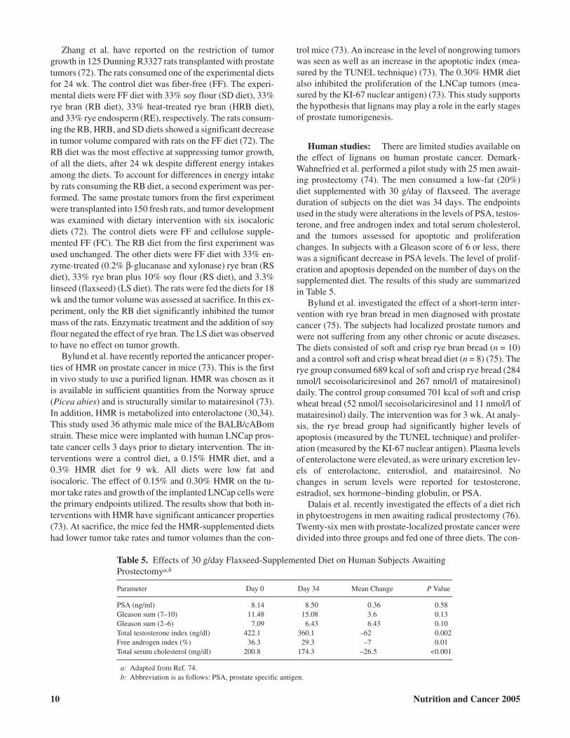
Human studies: There are limited studies available onthe effect of lignans on human prostate cancer. Demark-Wahnefried et al. performed a pilot study with 25 men await-ing prostectomy (74). The men consumed a low-fat (20%)diet supplemented with 30 g/day of flaxseed. The averageduration of subjects on the diet was 34 days. The endpointsused in the study were alterations in the levels of PSA, testos-terone, and free androgen index and total serum cholesterol,and the tumors assessed for apoptotic and proliferationchanges. In subjects with a Gleason score of 6 or less, therewas a significant decrease in PSA levels. The level of prolif-eration and apoptosis depended on the number of days on thesupplemented diet. The results of this study are summarizedin Table 5.
Bylund et al. investigated the effect of a short-term inter-vention with rye bran bread in men diagnosed with prostatecancer (75). The subjects had localized prostate tumors andwere not suffering from any other chronic or acute diseases.The diets consisted of soft and crisp rye bran bread (n = 10)and a control soft and crisp wheat bread diet (n = 8) (75). Therye group consumed 689 kcal of soft and crisp rye bread (284nmol/l secoisolariciresinol and 267 nmol/l of matairesinol)daily. The control group consumed 701 kcal of soft and crispwheat bread (52 nmol/l secoisolariciresinol and 11 nmol/l ofmatairesinol) daily. The intervention was for 3 wk. At analy-sis, the rye bread group had significantly higher levels ofapoptosis (measured by the TUNEL technique) and prolifer-ation (measured by the KI-67 nuclear antigen). Plasma levelsof enterolactone were elevated, as were urinary excretion lev-els of enterolactone, enterodiol, and matairesinol. Nochanges in serum levels were reported for testosterone,estradiol, sex hormone–binding globulin, or PSA.
Dalais et al. recently investigated the effects of a diet richin phytoestrogens in men awaiting radical prostectomy (76).Twenty-six men with prostate-localized prostate cancer weredivided into three groups and fed one of three diets. The con-
10 Nutrition and Cancer 2005
Table 5. Effects of 30 g/day Flaxseed-Supplemented Diet on Human Subjects AwaitingProstectomya,b
Parameter Day 0 Day 34 Mean Change P Value
PSA (ng/ml) 8.14 8.50 0.36 0.58Gleason sum (7–10) 11.48 15.08 3.6 0.13Gleason sum (2–6) 7.09 6.43 6.43 0.10Total testosterone index (ng/dl) 422.1 360.1 –62 0.002Free androgen index (%) 36.3 29.3 –7 0.01Total serum cholesterol (mg/dl) 200.8 174.3 –26.5 <0.001
a: Adapted from Ref. 74.b: Abbreviation is as follows: PSA, prostate specific antigen.
trol diet was wheat-based bread, and the experimental dietswere supplanted with either 50 g of soy grit or a mixture of 50g soy and 20 g of linseed (flaxseed). The subjects consumedfour slices of bread for 23–27 days. No changes in serum lev-els were reported with any of the diets for testosterone,dihydrotestosterone, or sex hormone–binding globulin.Those men fed a diet containing 50 g of soy had significantlydecreased total PSA and increased free androgen levels andfree to total PSA levels. However, these effects were not re-ported in the diet of 50 g of soy and 20 g of linseed. The al-tered free androgen and PSA levels observed agree with thefindings of Demark-Wahnefried (74).
Kilkkinen et al. investigated the hypothesis that entero-lactone is protective against prostate cancer risk in Finnishmen (38). They performed a 6-yr nested case-control study of214 men diagnosed with prostate cancer from the 29,000men involved in the Alpha-Tocopherol, Beta-Carotene Can-cer Prevention study. A slight negative correlation betweenserum enterolactone concentration and both smoking andbody mass index was found as well as a slight positive corre-lation with both age and number of smoking years. However,no correlation was found between either lignan consumptionor serum enterolactone levels and prostate cancer risk.
Tseng et al. examined dietary patterns among 136 men di-agnosed with prostate cancer that were involved in the UnitedStates National Health and Nutrition Examination Surveyand the Epidemiological Follow-up Study during the periods1971–1975 and 1982–1994 (77). The responses from the di-etary questionnaires were identified as one of three groups:vegetable-fruit, red meat-starch (red meats, potatoes, cheese,and salty snacks), and Southern (cornbread, grits, sweet pota-toes, beans, and rice). No link between the vegetable-fruitand red meat-starch groups and prostate cancer risk was re-ported. The Southern diet showed a slight reduction in pros-tate cancer risk. The authors speculate that this reductionmay be due to a higher exposure to sunlight and thereforehigher levels of vitamin D.
Summary of in vivo animal and human studies: Thein vivo animal studies seem to support the in vitro observa-tions of protective effects (48–67). All of the animal studiesreported a significant decrease in tumor mass after dietary in-tervention with lignan-rich foods. However, the few in vivohuman studies available are inconsistent. Two of the fivestudies reported beneficial results in men consuming alignan-rich diet (74,75) and another found that soy and lin-seed together had no effects (76), whereas the remaining twofound no link between serum enterolactone levels and pros-tate cancer risk (38,77). The other human studies found noassociation between serum enterolactone levels and prostatecancer risk in men consuming a more varied diet (38,75).
There are a number of discrepancies with the in vivo datawith respect to the effect of lignans in prostate cancer. Sev-eral of the dietary interventions used foods rich in lignans,such as flaxseed (70,72,74,76) and rye bread (69, 72, 75), orin isoflavones, such as soy (69,76). It is not possible to as-cribe the in vivo anticancer effects reported to the lignans
alone. There are many other bioactive components in thefoods used that may influence prostate pathogenesis eitheron their own or synergistically with the lignans. The exactnutritional and lignan content of the diets were not clear ormeasured in plasma. It is reported that lignans accumulate atmuch higher concentrations in prostatic fluid than in plasma(19). The level of fat in the diets affects the interpretation ofthe results. A high-fat diet is generally accepted to increasethe risk of prostate cancer by altering the balance of sex hor-mones such as testosterone (7). Reducing the level of fat inthe diet reduces the risk of prostate cancer (78). Only thestudy by Zhang et al. included a high-fat control (72). Thelack of a high-fat control in many of the diets also preventsany biological effects reported from being ascribed solely tothe lignans. Many of the studies used a small sample size anda short dietary intervention. It is conceivable that long-termexposure to lignans may be required for any protective ef-fects to be observed.
The serum enterolactone levels reported by Kilkkinen andothers are discrete measurements at specific time points(38,77). They did not measure the level of serum entero-lactone over a defined time. They also did not measure thelevels in prostatic fluid. If the hypothesis that lifetime con-sumption of lignan affects the pathogenesis of prostate can-cer is valid, then serum concentrations are not valid measure-ments of lignan consumption and risk of prostate cancer aslignans can achieve much higher prostatic levels than foundin the plasma (19).
Although the animal and human in vivo studies supportmany of the reported in vitro effects of lignans, there is anacute lack of knowledge regarding the anticancer effects ofpure lignan compounds.
Conclusions
Prostate cancer is becoming the most common male can-cer in the Western world, and epidemiological evidence sug-gests that dietary modification is a potential method for pre-vention and treatment of the disease. As with most cancers,an unacceptably high number of cases of prostate cancer arediagnosed at an advanced stage. With the possibility that di-etary intervention may not be sufficient to achieve the effec-tive biological levels reported in vitro, it may be that lignansare more suitable as a pharmacological treatment. Indeed,there is a lack of evidence that lignans may be preventativeagainst prostate cancer; it may be the case that lignans aremore suitable in conjunction with current treatments forprostate cancer. The side effects of the current treatments area source of considerable psychological stress for patients.The perceived “loss of masculinity” associated with the moreadvanced treatments almost certainly affects the quality oflife for the patient. Other more effective treatment options arerequired. It is theoretically possible that intervention withlignans in men with prostate cancer will reduce the need forradical procedures such as prostectomy or reduce the chanceof relapse.
Vol. 52, No. 1 11
However, the exact molecular mechanism of action of thelignans is unclear as our understanding is hampered by thelack of experimental evidence. Future research should focusspecifically on the in vitro effects of lignans, especially purelignans, on established prostate cancer cell lines. Thepharmacokinetics of the lignans requires considerable clari-fication prior to any meaningful in vivo work. Any future invivo studies should also use purified lignans, account for therole of fat in prostate cancer, include measurement of lignansin prostatic fluid, and be of sufficient sample size to be statis-tically significant. This will clarify if lignans possess anyvalue in the prevention and treatment of prostate cancer. Inaddition, this will determine whether dietary or pharmaco-logical intervention is the most effective method of usinglignans in the development of novel treatments for prostatecancer.
Acknowledgments and Notes
The principal author would like to acknowledge the University of Ulsterfor the Vice Chancellor’s Research Scholarship award. Address correspon-dence to Mark J. McCann, Northern Ireland Centre for Diet and Health(NICHE), Centre for Molecular Biosciences (CMB), University of Ulster,Coleraine, Northern Ireland BT52 1SA. Phone: (0044) 028 7032 3068.FAX: (0044) 028 7032 3023. E-mail: [email protected].
Submitted 6 May 2004; accepted in final form 4 May 2005.
References
1. Ferlay J, Bray P, and Parkin DM: GLOBOCAN 2000: Cancer inci-dence, mortality, and prevalence worldwide, Lyon, France: IARCPress, 2001.
2. World Health Organization: Cancers of the male reproductive tract. InWorld Cancer Report, Stewart BW and Kleihues P (eds). Lyon,France: IARC Press, 2003, pp 208–211.
3. Shulka S, MacLennan GT, Marengo SR, Seftel AD, Resnick MI, et al.:Genetic abnormalities in prostate cancer. Curr Genom 5, 67–83, 2004.
4. Abate-Shen C and Shen MM: Molecular genetics of prostate cancer.Genes Dev 14, 2410–2434, 2000.
5. Adlercreutz H and Mazur W: Phyto-oestrogens and Western diseases.Ann Med 29, 95–120, 1997.
6. Dhom G: Epidemiology of hormone-depending tumours. In Endo-crine Dependent Tumours, Voigt K and Knabbe C (eds). New York:Raven Press, 1991, pp 1–42.
7. Potter JD: Patterns of diet. In Food, Nutrition, and the Prevention ofCancer: A Global Perspective, Washington, DC: American Institutefor Cancer Research, 1997, pp 22–34.
8. Marker PC, Donjacour AA, Dahiya R, and Cunha GR: Hormonal, cel-lular, and molecular control of prostatic development. Dev Biol 253,165–174, 2003.
9. Morrissey C and Watson WG: Phytoestrogens and prostate cancer.Curr Drug Targets 4, 231–241, 2003.
10. Linja MJ, Savinainen KJ, Tammela TLJ, Isola JJ, and Visakorpi T: Ex-pression of ERα and ERβ in prostate cancer. Prostate 55, 180–186,2003.
11. Ji Q, Liu PI, Elshimali Y, and Stolz A: Frequent loss of estrogen andprogesterone receptors in human prostatic tumors determined by quan-titative real-time PCR. Mol Cell Endocrinol 229, 103–110, 2005.
12. Bardin A, Boulle N, Lazennec G, Vignon F, and Pujol P: Loss of ERβexpression as a common step in estrogen-dependent tumor progres-sion. Endo-Related Cancer 11, 537–551, 2004.
13. Cheng J, Lee EJ, Madison LD, and Lazennec G: Expression of estro-gen receptor b in prostate carcinoma cells inhibits invasion and prolif-eration and triggers apoptosis. FEBS Lett 566, 169–172, 2004.
14. Stitch SR, Toumba JK, Groen MB, Funke CW, Leemhuis J, et al.: Ex-cretion, isolation and structure of a new phenolic constituent of femaleurine. Nature 287, 738–740, 1980.
15. Setchell KDR and Adlercreutz H: The excretion of two new phenoliccompounds (Compound 180/442 and Compound 180/410) during thehuman menstrual cycle and in pregnancy. J Steroid Biochem 11,xv–xvi, 1979.
16. Setchell KDR, Bull R, and Adlercreutz H: Steroid excretion during thereproductive cycle and in pregnancy of the vervet monkey(Cercopithecus aethiopus pygerythrus). J Steroid Biochem 12,375–384, 1980.
17. Setchell KDR, Lawson AM, Conway E, Taylor NF, Kirk DN, et al.:The definitive identification of the lignans trans-2,3-bis(3-hydroxybenzyl)-gamma-butyrolactone and 2,3-bis(3-hydroxyben-zyl)butane-1,4-diol in human and animal urine. Biochem J 197,447–458, 1981.
18. Finlay EMH, Wilson DW, Adlercreutz H, and Griffith K: The identifi-cation and measurement of “phyto-oestrogens” in human saliva,plasma, breast aspirate or cyst fluid, and prostate fluid using gas chro-matography-mass spectrometry. J Endocr 129, S49, 1991.
19. Morton MS, Chan PS, Cheng C, Clacklock N, Matos-Ferreria A, et al.:Lignans and isoflavones in plasma and prostatic fluid in men: samplesfrom Portugal, Hong Kong, and the United Kingdom. Prostate 32,122–128, 1997.
20. Setchell KDR, Lawson AM, Mitchell FL, Adlercreutz H, Kirk DN, etal.: Lignans in man and animal species. Nature 287, 740–743, 1980.
21. Raffaelli B, Hoikkala A, Leppala E, and Wahala K: Enterolignans. JChromatogr B Analyt Technol Biomed Life Sci 777, 29–43, 2002.
22. Wang L-Q: Mammalian phytoestrogens: enterodiol and enterolactone.J Chromatogr B Analyt Technol Biomed Life Sci 777, 289–309, 2002.
23. Thompson LU, Robb P, Serraino M, and Cheung F: Mammalian lignanproduction from various foods. Nutr Cancer 16, 43–52, 1991.
24. Mazur WM, Fotsis T, Wahala K, Ojala S, Salakka A, et al.: Isotope-di-lution gas-chromatographic mass-spectrometric method for the deter-mination of isoflavonoids, coumestrol, and lignans in food samples.Anal Biochem 223, 169–180, 1996.
25. Meagher LP and Beecher GR: Assessment of data on the lignan con-tent of foods. J Food Comp Anal 13, 935–947, 2000.
26. Mazur W, Wahala K, Wang G, and Adlercreutz H: Dietaryphytoestrogens—from chemistry to chemoprevention. Kem Kemi 25,48–55, 1998.
27. Mazur WM: Phytoestrogen content in foods. Clin Endocrinol Metab12, 169–180, 1998.
28. Mazur WM, Wahala K, Rasku S, Salaka A, Hase T, et al.: Lignan andisoflavonoid concentrations in tea and coffee. Br J Nutr 79, 37–45,1998.
29. Mazur WM, Duke JA, Wahala K, Rasku S, and Adlercreutz H:Isoflavonoids and lignans in legumes: nutritional and health aspects inhumans. J Nutr Biochem 9, 193–200, 1998.
30. Heinonen S, Nurmi T, Liukkonen K, Poutanen K, Wahala K, et al.: Invitro metabolism of plant lignans: new precursors of mammalianlignans enterolactone and enterodiol. J Agric Food Chem 49, 3178–3186, 2001.
31. Kiely M, Faughnan M, Wahala K, Brasts H, and Mulligan A:Phyto-oestrogen levels in foods: the design and construction of theVENUS database. Br J Nutr 89 (Suppl 1), S19–S23, 2003.
32. VENUS database. Available online from at http://www.phytohealth.org (7 April 2005).
33. Rowland I, Faughnan M, Hoey L, Wahala K, Williamson G, et al.:Bioavailability of phyto-oestrogens. Br J Nutr 89 (Suppl), S45–S58,2003.
12 Nutrition and Cancer 2005
34. Axelson M and Setchell KDR: The excretion of lignans in rats—evi-dence for and intestinal bacterial source for this new group of com-pounds. FEBS Lett 123, 337–342, 1981.
35. Setchell KDR, Lawson AM, Borriello SP, Harkness R, Gordon H, etal.: Lignan formation in man—microbial involvement and possibleroles in relation to cancer. Lancet 11, 4–7, 1981.
36. Borriello SP, Setchell KDR, Axelson M, and Lawson AM: Productionand metabolism of lignans by the human faecal flora. J Appl Microbiol58, 37–43, 1985.
37. Kilkkinen A, Stumpf K, Pietinen P, Valsta LM, Tapanainen H, et al.:Determinants of serum enterolactone concentration. Am J Clin Nutr73, 1094–1100, 2001.
38. Kilkkinen A, Virtamo J, Virtanen MJ, Adlercreutz H, Albanes D, et al.:Serum enterolactone concentration is not associated with prostate can-cer risk in a nested case-control study. Cancer Epidemiol BiomarkersPrev 12, 1209–1212, 2003.
39. Jacobs DR, Pereira MA, Stumpf K, Pins JL, and Adlercreutz H:Whole grain food elevates serum enterolactone. Br J Nutr 88,111–116, 2002.
40. Juntunen KS, Mazur WM, Liukkonen KH, Uehara M, Poutanen KS, etal.: Consumption of wholemeal rye bread increases serum concentra-tions and urinary excretion of enterolactone compared with consump-tion of white wheat bread in healthy Finnish men and women. Br JNutr 84, 839–846, 2000.
41. Jenab M and Thompson L: The influence of flaxseed and lignans oncolon carcinogenesis and beta-glucuronidase activity. Carcinogenesis17, 1343–1348, 1996.
42. Rickard S and Thompson L: Chronic exposure to secoisolariciresinoldiglycoside alters lignan disposition in rats. J Nutr 128, 615–623,1998.
43. Kilkkinen A, Pietinen P, Klaukka T, Virtamo J, Korhonen P, andAdlercreutz H: Use of oral antimicrobials decreases serumenterolactone concentration. Am J Epidemiol 155, 472–477, 2002.
44. Kuijsten A, Arts ICW, Vree TB, and Hollman PCH: Pharmacokineticsof enterolignans in healthy men and women consuming a single doseof secoisolariciresinol diglucoside. J Nutr 135, 795–801, 2005.
45. Adlercreutz H, Fotsis T, Bannwart C, Wahala K, Makela T, et al.: De-termination of urinary lignans and phytoestrogen metabolites, poten-tial antiestrogens and anticarcinogens, in urine of women on varioushabitual diets. J Steroid Biochem 25, 791–797, 1986.
46. Adlercreutz CH, Goldin BR, Gorbach SL, Hockerstedt KA, WatanabeS, et al.: Soybean phytoestrogen intake and cancer risk. J Nutr 125,S757–S770, 1995.
47. Adlercreutz H, Honjo H, Higashi A, Fotsis T, Hamalainen E, et al.:Urinary excretion of lignans and isoflavonoid phytoestrogens in Japa-nese men and women consuming a traditional Japanese diet. Am J ClinNutr 54, 1093–1100, 1991.
48. Martin ME, Haourigui M, Pelissero C, Benassayag C, and Nunez EA:Interactions between phytoestrogens and human sex steroid bindingprotein. Life Sci 58, 429–436, 1996.
49. Schottner M, Spiteller G, and Ganseer D: Lignans interfering with 5alpha-dihydrotestosterone binding to human sex hormone-bindingglobulin. J Nat Prod 61, 119–121, 1998.
50. Adlercreutz H, Bannwart C, Wahala K, Makela T, Brunow G, et al.: In-hibition of human aromatase by mammalian lignans and isoflavonoidphytoestrogens. J Steroid Biochem Mol Biol 44, 147–153, 1993.
51. Adlercreutz H, Hockerstedt K, Bannwart C, Bloigu S, Hamalainen E,et al.: Effects of dietary components, including lignans andphytoestrogens, on enterohepatic circulation and liver metabolism ofoestrogens and on sex hormone binding globulin (SHBG). J SteroidBiochem 27, 1135–1144, 1987.
52. Garreau B, Vallette G, Adlercreutz H, Wahala K, Makela T, et al.:Phytoestrogens: new ligands for rat and human alpha-fetoprotein.Biochim Biophys Acta 1094, 339–345, 1991.
53. Brooks JD and Thompson LU: Mammalian lignans and genistein de-crease the activities of aromatase and 17β-hydroxysteroid dehydro-genase inMCF-7cells.JSteroidBiochemMolBiol94,461–467,2005.
54. Evans BAJ, Griffiths K, and Morton MS: Inhibition of5alpha-reductase in genital skin fibroblasts and prostate tissue by di-etary lignans and isoflavonoids. J Endocrinol 147, 295–302, 1995.
55. Adlercreutz H, Mousavi Y, Clark J, Hockerstedt K, Hamalainen E, etal.: Dietary phytoestrogens and cancer: in vitro and in vivo studies. JSteroid Biochem Mol Biol 41, 331–337, 1992.
56. Adlercreutz H: Western diet and Western diseases: some hormonal andbiochemical mechanisms and associations. Scand J Clin Lab Invest 50,3–23, 1990.
57. Mercier-Bodard C, Nivet V, and Baulieu E-E: Effects of hormones onSBP mRNA levels in human cancer cells. J Steroid Biochem Mol Biol40, 777–785, 1991.
58. Prasad K: Antioxidant activity of secoisolariciresinol diglycoside-de-rived metabolites, secoisolariciresinol, enterodiol, and enterolactone.Int J Angiol 9, 220–225, 2000.
59. Kitts DD, Yuan YV, Wijewickreme AN, and Thompson LU: Antioxi-dant activity of the flaxseed lignan secoisolariciresinol diglycosideand its mammalian lignan metabolites enterodiol and enterolactone.Mol Cell Biochem 202, 91–100, 1999.
60. Niemeyer HB and Metzler M: Differences in the antioxidant activity ofplant and mammalian lignans. J Food Eng 56, 255–256, 2003.
61. Kulling SE, Jacobs E, Pfeiffer E, and Metzler M: Studies on thegenotoxicity of the mammalian lignans enterolactone and enterodioland their metabolic precursors at various endpoints in vitro. Mutat Res416, 115–124, 1998.
62. Nicholson B and Theodorescu D: Angiogenesis and prostate cancer tu-mour growth. J Cell Biochem 91, 125–150, 2004.
63. Fotsis T, Pepper M, Adlercreutz H, Fleischmann G, Hase T, et al.:Genistein, a dietary-derived inhibitor of in vitro angiogenesis. ProcNatl Acad Sci USA 90, 2690–2694, 1993.
64. Lin X, Swtizer BR, and Demark-Wahnefried W: Effect of mammalianlignans on the growth of prostate cancer cell lines. Anti-cancer Res 21,3995–4000, 2001.
65. McCann MJ, Gill CI, McGlynn H, and Rowland IR: Effect of selectedlignans on the cell cycle of PC-3 human prostate cancer cells.Polyphenols Commun P93, 289–290, 2004.
66. McCann MJ, Gill CI, McGlynn H, and Rowland IR: Lignan phyto-oes-trogens delay the proliferation of prostate cancer in vitro via cell cycleinhibition. in preparation 2005.
67. Kangas L, Saarinen N, Mutanen M, Ahotupa M, Hirsinummi R, et al.:Antioxidant and antitumour effects of hydroxymatairesinol HM-3000,HMR, a lignan isolated from the knots of spruce. Eur J Cancer Prev11, S48–S57, 2002.
68. Dabrosin C, Chen J, Wang L, and Thompson LU: Flaxseed inhibitsmetastasis and decreases extracellular vascular endothelial growthfactor in human breast cancer xenografts. Cancer Lett 185, 31–37,2002.
69. Bylund A, Zhang JX, Bergh A, Damber JE, Widmark A, et al.: Ryebran and soy protein delay growth and increase apoptosis of humanLNCap prostate adenocarcinoma in nude mice. Prostate 42, 304–314,2000.
70. Lin X, Gingrich JR, Bao W, Li J, Haroon ZA, et al.: Effect of flaxseedsupplementation on prostatic carcinoma in transgenic mice. Urology60, 919–924, 2002.
71. Greenberg NM, DeMayo FJ, Sheppard PC, Barrios R, Lebovitz R,et al.: The rat probasin gene promoter directs hormonally anddevelopmentally regulated expression of a heterologous gene specifi-cally to the prostate in transgenic mice. Mol Endocrinol 8, 230–239,1994.
72. Zhang JX, Hallmans G, Landstrom M, Bergh A, Damber JE, et al.: Soyand rye diets inhibit the development of Dunning R3327 prostaticadenocarcinoma in rats. Cancer Lett 114, 313–314, 1997.
73. Bylund A, Saarinen N, Zhang JX, Bergh A, Widmark A, et al.:Anticancer effects of a plant lignan 7-hydroxymatairesinol on a pros-tate cancer model in vivo. Exp Biol Med 230, 217–223, 2005.
74. Demark-Wahnefried W, Price DT, Polascik TJ, Robertson CN, Ander-son EE, et al.: Pilot study of dietary fat restriction and flaxseed
Vol. 52, No. 1 13
supplementation in men with prostate cancer before surgery: exploringthe effects on hormonal levels, prostate-specific antigen, andhistopathologic features. Urology 58, 47–52, 2001.
75. Bylund A, Lundin E, Zhang JX, Nordin A, Kaaks R, et al.: Random-ised controlled short-term intervention pilot study on rye bran bread inprostate cancer. Eur J Cancer Prev 12, 407–415, 2003.
76. Dalais FS, Meliala A, Wattanapenpaiboon N, Frydenberg M, SuterDAI, et al.: Effects of a diet rich in phytoestrogens on prostate-specific
antigen and sex hormones in men diagnosed with prostate cancer.Urology 64, 510–515, 2004.
77. Tseng M, Breslow RA, DeVellis RF, and Ziegler RG: Dietary patternsand prostate cancer risk in the National Health and Nutrition Examina-tion Survey epidemiological follow-up study cohort. CancerEpidemiol Biomarkers Prev 13, 71–77, 2004.
78. Hamalainen EK, Adlercreutz H, Puska P, and Pietinen P: Decrease ofserum total and free testosterone during a low-fat high-fibre diet. J Ste-roid Biochem 18, 369–370, 1983.
14 Nutrition and Cancer 2005