Embed Size (px)
Citation preview
1
Role of Human Ribosomal RNA (rRNA) Promoter Methylation and of Methyl CpG Binding Protein
MBD2 in the Suppression of rRNA Gene Expression
Kalpana Ghoshal1†, Sarmila Majumder1†, Jharna Datta1, Tasneem Motiwala1, Shoumei Bai1, Sudarshana
M. Sharma1, Wendy Frankel2 and Samson T. Jacob1*
1Department of Molecular and Cellular Biochemistry and 2Department of Pathology, College of
Medicine, The Ohio State University, Columbus, Ohio.
†These authors contributed equally to the work
*Corresponding author: Samson T. Jacob, e-mail: [email protected]; tel#: 614-688-5494; Fax#: 614-688-
5600.
Running Title: Role DNA methylation in rRNA gene expression
Key words: DNA methylation, rRNA expression, human hepatocellular carcinomas, DNA methyltransferases,
methyl CpG binding proteins, chromatin immunoprecipitation, immunofluorescence, nucleolin.
Abbreviations used: rRNA, ribosomal RNA; IGS, intergenic spacer region; ICF, immunodeficiency,
centromeric instability and facial anomaly; ETS1, external transcribed spacer region 1; pol I, RNA polymerase
I; pol II, RNA polymerase II; UBF, upstream binding factors, MBD, methyl CpG binding protein, DNMT,
DNA methyltransferase; HDAC, histone deacetylase; ChIP, chromatin immunoprecipitation, HMT, histone
methyltransferase, Ado-Met, S-adenosyl-L-methionine, GAPDH, glyceraldehyde 3-phosphate dehydrogenase;
TBST, Tris-buffered saline containing Tween 20.
Copyright 2003 by The American Society for Biochemistry and Molecular Biology, Inc.
JBC Papers in Press. Published on November 10, 2003 as Manuscript M309393200 by guest on A
ugust 29, 2018http://w
ww
.jbc.org/D
ownloaded from
2
ABSTRACT
The methylation status of CpG island located within the ribosomal RNA (rRNA) promoter in human
hepatocellular carcinomas and pair-matched liver tissues was analyzed by bisulfite genomic sequencing.
Significant hypomethylation of methyl-CpGs in the rRNA promoter was observed in the tumor samples
compared to matching normal tissues, which was consistent with the relatively high level of rRNA synthesis in
rapidly proliferating tumors. To study the effect of CpG methylation on RNA polymerase I (pol I)-transcribed
rRNA genes, we constructed pHrD-IRES-Luc (human rRNA promoter-luciferase reporter). In this plasmid,
Kozak sequence of pGL3-basic vector was replaced by the internal ribosome entry site (IRES) of
encephalomyocarditis viral genome to optimize pol I driven reporter gene expression. Transfection of this
plasmid into HepG2 (human) cells revealed reduced pol I-driven luciferase activity with increase in methylation
density at the promoter. Markedly reduced luciferase activity in Hepa (mouse) cells compared to HepG2
(human) cells showed that pHrD-IRES-Luc is transcribed by pol I. Site-specific methylation of human rRNA
promoter demonstrated that methylation of CpG at the complimentary strands located in the promoter (-9, -102,
-345 with respect to +1 site) inhibited luciferase activity while symmetrical methylation of a CpG in the
transcribed region (+152) did not affect the promoter activity. Immunofluorescence studies showed that the
methyl CpG binding proteins, MBD1, MBD2, MBD3 and MeCP2, are localized both in the nuclei and nucleoli
of HepG2 cells. Transient overexpression of MBD2 suppressed luciferase activity specifically from the
methylated rRNA promoter whereas MBD1 and MBD3 inhibited rRNA promoter activity irrespective of the
methylation status. Chromatin immunoprecipitation analysis confirmed predominant association of MBD2 with
the endogenous methylated rRNA promoter, which suggests a selective role for MBD2 in the methylation-
mediated inhibition of ribosomal RNA gene expression.
by guest on August 29, 2018
http://ww
w.jbc.org/
Dow
nloaded from
3
INTRODUCTION
The transcriptional regulation of rRNA genes is a control point in the complex process of ribosome biogenesis.
Diploid somatic cells harbor 300-400 copies of the ribosomal RNA (rRNA) genes that code for the most
abundant cellular RNA. Only a fraction of these genes is transcribed, which depends on the growth stage of the
cells and extracellular stimuli [for review, see (1,2)]. In general, multiple copies of rRNA are found as repeated
clusters, usually arranged in head-to-tail fashion. The core promoter region spanning from -50 bp to +20 bp
with respect to the initiation site is necessary and sufficient for the initiation of basal transcription in most
species [for review, see (3-6)]. Another key element is the upstream control element (UCE) that extends
150–200 bp upstream of transcription start site. Apart from core promoter and UCE, upstream enhancers and
terminator also play critical role in rRNA transcription. While the transcription machineries of RNA
polymerase II (pol II) and RNA polymerase III (pol III) are often compatible with genes from widely different
species, RNA polymerase I (pol I) exhibits stringent (7), but not absolute (8), species specificity. This could
result from very little sequence similarity between rRNA promoters from different species despite the general
conservation of functional transactivation domains of the transcription factors from mouse to human (6,9).
Although considerable advances have been made in the identification and characterization of factors that
upregulate rRNA gene expression, the factors controlling its downregulation have not been fully characterized.
Methylation of DNA at 5 position of cytosine of CpG base pairs, particularly in the promoter region is the
predominant epigenetic modification of DNA in mammals and is known to suppress many RNA polymerase II
(pol II) genes (10-12). DNA methylation is essential for development (13,14). It regulates inactivation of X
chromsome in females, genomic imprinting and suppresses spurious transcription from promoters of
retroviruses and transposable elements integrated with the genome (15). In addition, aberrations in DNA
methylation cause activation of oncogenes, genomic instability and silencing of a variety of tumor suppressor
genes, e.g., P16, P15, P21, E-CAD, VHL etc., leading to uncontrolled cell proliferation [for review see (11,15-
by guest on August 29, 2018
http://ww
w.jbc.org/
Dow
nloaded from
4
17)]. This modification is initiated by de novo DNA methyltransferases (DNMT3A and DNMT3B) and is
propagated in successive cell divisions by the maintenance methyltransferase (DNMT1). DNMT1 transfers
methyl group from S-adenosyl methionine (Ado-Met) to the newly replicated strand using hemimethylated
strand as the template [for review, see ref (18,19)]. Aberrations in DNA methylation lead to a variety of
diseases. For example, ICF (immunodeficiency, centromeric instability and facial anomaly) syndrome is caused
by mutations in DNMT3B gene (20-22). The drugs inhibiting DNMT, 5-deoxyazacytidine, 5-fluorocytidine,
Zebularine alone or in combination with HDAC inhibitors are used clinically in certain types of cancer to
activate methylated tumor suppressor or differentiation inducing genes (23,24).
DNA methylation can impede the transcriptional activity of a pol II gene (25) directly by blocking the
access of a transcription factor, e.g., AP-2, NF-kB, E2F and c-MYC to their cognate sites (19). Most of the
methylated promoters are, however, recognized by a group of proteins called methyl-CpG binding proteins
(MBDs) by virtue of their conserved methyl-CpG binding domain. Five such MBDs with highly conserved
DNA binding domains have been identified (26). Among these proteins, MeCP2, MBD1, MBD2 and MBD4
can bind to methylated DNA. MBD4 is a uracil-DNA glycosylase involved in G-T mismatch repair (12,27).
MBDs repress transcription by recruiting a variety of proteins such asSin3a, HDACs (histone deacetylases),
HMTs (histone methyltransferases) and HP1α (heterochromatin protein 1α) as co-repressors [for review, see
(10,14,28,29)]. Kaiso, a partner of β-catenin, lacking the signature methyl CpG binding domain can also bind
to methyl CpGs and repress methylated promoters (30). Defect in functions of these proteins also leads to
various abnormalities. Rett syndrome, a prevalent X-linked neurological disorder among Caucasian females, is
caused by dominant negative mutations in MECP2 gene (31). Adult MBD1 knock-out mice, like MeCP2 null
mice, also exhibit neurological abnormalities (32) whereas MBD4 null mice are susceptible to cancer because
of enhanced CpG to TpG mutation in their genome (33).
by guest on August 29, 2018
http://ww
w.jbc.org/
Dow
nloaded from
5
Most of the studies to date have focussed on the upregulation of rRNA promoter activity. Since
methylation of DNA, particularly the promoter region, is known to silence many pol II genes (23-26), it was of
interest to investigate whether the methylation status of rRNA promoter modifies pol I directed rRNA
transcription. In the present study, we explored the methylation status of the CpG island that spans rRNA
promoter in human primary hepatocellular carcinomas and corresponding normal liver tissues, and elucidated a
potential molecular mechanism for the methylation-mediated alteration in rRNA promoter activity in human
cells.
MATERIALS AND METHODS
Construction of plasmids:
(a)pIRES-Luc: Internal ribosome entry site (IRES: 16-518bp) was amplified from pCITE vector (Novagen)
using T3 (5'-AATTAACCCTCACTAAAGGG-3') and T7 (5'-GTAATACGACTCACTATAGGGC-3')
oligonucleotides and digested with Nco I to generate the 503bp IRES (Internal Ribosome Entry Site) product.
The Kozak sequence [(GCC)GCCRCCATGG where R is a purine] that directs translation of pol II transcribed
mRNAs, was removed from pGL3-basic vector by digestion with Nco I and Hind III and was replaced by the
amplified IRES fragment to generate the plasmid, pIRES-Luc. The replacement of Kozak with IRES was
essential to minimize pol II-directed spurious expression from human rRNA promoter-luciferase reporter
constructs in transfected cells.
(b) Human rRNA-luciferase vector (pHrD-IRES-Luc): Human rRNA promoter spanning -410 to +314bp
(accession number K01105) with respect to transcription initiation site was amplified from ~2kb fragments of
HeLa genomic DNA digested with EcoR I. The following primer pairs with Kpn I (forward) and Bgl II
(reverse) restriction sites at the 5' ends were used for PCR:
Forward: 5'-gtggtacccCGCGATCCTTTCTGGAGAGTCCC-3'
by guest on August 29, 2018
http://ww
w.jbc.org/
Dow
nloaded from
6
Reverse: 5'-ggagatctGACGAGAACGCCTGACACGCAC-3'
The annealing temperature was 58°C. The resultant 730 bp PCR product was treated with Pfu polymerase
(Stratagene) to polish the ends following manufacturer’s protocol, digested with Kpn I and Bgl II and cloned
into the same sites of pIRES-Luc to generate pHrD-IRES-Luc.
(c) Expression vectors for MBD1, MBD2, MBD3 and MeCP2: These plasmids were constructed in
mammalian expression vectors pcDNA 3.1(+/-) (Invitrogen) to obtain pcMBD1, pcMBD2, pcMBD3, pcMBD4
and pcMeCP2. Briefly, rat MeCP2 cDNA in pBlueScript-SK(-) (a generous gift from Adrian Bird) was digested
with Not I–EcoR V enzymes and the resultant ~1.8 kb fragment was cloned into the same sites in pcDNA3.1(-)
to generate pcMeCP2. Similarly, human MBD1 cDNA from pET6H (a generous gift from Adrian Bird) was
excised with Nco I, the sticky 5'-end of the ~2kb insert fragment was filled in with dNTPs catalyzed by Klenow
and cloned in EcoR V site of pcDNA3.1(-). Amplification with the vector- and insert-specific primers
determined the correct orientation of MBD1. The mouse MBD2 cDNA in pBSK vector (a generous gift from
Brian Hendrich) was digested with EcoR I and Xho I and ligated to the same sites in pcDNA3.1(-) to generate
the mammalian expression vector. MBD3 cDNA was amplified from a mouse cDNA library and ligated to
BamH I-Hind III sites of pcDNA3.1(-) to generate the plasmid pcMBD3.
Cell culture and transfection assays:
HepG2 cells were grown in DMEM with 10% fetal bovine serum (FBS). For transfection assay, 2.5x10⁵ cells
were plated onto 60mm dishes 24 hr prior to transfection and then transfected using calcium phosphate co-
precipitation method (34,35). Unless mentioned otherwise, each transfection cocktail contains a maximum of
8.8µg of total plasmid DNA that includes the reporter plasmid (0.5 to 1.0µg), pRLTK [renila luciferase reporter
driven by HSV-tk promoter (Promega)] (50ng) as an internal control and the eukaryotic expression vectors
(4µg) harboring the gene of interest in a total volume of 500µl. Briefly, DNA was dissolved in 220µl 0.1X TE,
(1mM Tris HCl, pH8.0; 0.1mM EDTA pH8.0) and mixed with 250µl 2X Hepes buffered saline (280mM NaCl,
10mM KCl, 1.5mM Na2HPO4, 12 mM dextrose and 50mM HEPES). Next, 31µl of 2M CaCl2 was added slowly
by guest on August 29, 2018
http://ww
w.jbc.org/
Dow
nloaded from
7
to the DNA mixture and incubated 20 minute at room temperature before adding to the cell culture. The cells
were allowed to incubate with the transfection reagent in complete medium for 16h at 37°C, followed by
replacement with fresh medium. After 24-48 hr in the fresh medium, the cells were harvested in the lysis buffer
(Promega), and luciferase activity was measured using Dual luciferase assay kit (Promega) in a Luminometer
(Lumat LB 9507; EG&G Berthold, Oak Ridge, TN).
Bisulfite Genomic Sequencing:
Genomic DNA isolated form hepatocellular carcinomas and matching liver tissues from the same individuals
were treated with sodium bisulfite according to the protocol optimized in our lab (36,37). The rRNA promoter
spanning -377 to +51 bp was amplified using two sets of nested primers from the bisulfite-treated DNA. The
primers used were:
hrRNA BF1: 5'-AAT TTT TTTGGAGAGTTT TCGTG-3'
hrRNA BR1: 5'-GAG TCG GAG AGCGTT TTT TGAG-3'
Annealing temperature used was 50ºC.
The nested primers were
hrRNA BF2: 5'-GAG TCG GAG AGC GTT TTT TGAG-3'
hrRNA BR2: 5'-CATCCGAAAACCCAACCTCTCCAA-3'
Annealing temperature was 55°C.
To confirm complete conversion of unmethylated cytosines to uracils, the PCR products were digested with Taq
I, the restriction sites of which were generated only after bisulfite conversion. Completely converted PCR
products were then cloned into TA cloning vector (Invitrogen). Ten clones selected at random from each DNA
were sequenced in automated DNA sequencer.
by guest on August 29, 2018
http://ww
w.jbc.org/
Dow
nloaded from
8
DNA methylation in vitro with bacterial methylases:
Kpn I-Bgl II fragment of pHrD-IRES-Luc was methylated with M. Sss I methylase (NEB) or with M. Hha I
methylase (NEB) in the presence (methylated) or absence (mock-methylated) of 160µM S-adenosyl methionine
(Ado-Met) in manufacturer-supplied buffer at 37°C for 4hr. An additional 10U of enzyme and Ado-Met were
added to the same mixture and the methylation reaction was continued for an additional 4h. The completion of
the methylation reaction was determined by digestion of the fragment with BstU I, Hpa II or Hha I for M. Sss I,
M. Hpa II and M. Hha I methylases respectively. These enzymes cannot cleave DNA if their cognate restriction
sites are methylated. The methylated promoter fragment was then ligated to the same sites of pIRES-Luc. The
ligated plasmid was separated on an agarose gel and purified using gel extraction kit (Qiagen). Before
transfection the concentrations of methylated and mock-methylated plasmids were measured in a Beckman
spectrophotometer at 260 nm.
Site-Specific methylation:
Site-specific methylation was carried out following the methodology used for site-directed mutagenesis with
some modifications. Single-stranded DNA was obtained by infection of XL1-blue-MRF' bacteria harboring
HrDNA plasmid with helper phage (R408, Stratagene). In a typical reaction, positive strand HrD-Luc plasmid
(~0.05pmol) was annealed to 1.25 pmol of phosphorylated oligos (with a specific CpG either methylated
denoted as ‘M’ oligonucleotide or unmethylated control, depicted as ‘C’ oligonucleotide) spanning different
regions of the rRNA promoter or external transcribed spacer (ETS1). The annealing mixture (20µl) contained
20 mM Tris.HCl (pH 7.5), 10 mM MgCl2 and 50 mM NaCl. The reaction was carried out in a thermocycler
(Perkin Elmer) programmed for 5 min incubation at 96°C followed by a slow descent (with a ramp of 3
min/degree Celsius) to 660C (annealing temperature of the oligo) and 1hr incubation at 66°C. Annealed oligo
was then extended and ligated in a reaction mixture comprising of 3µl of 10X synthesis buffer [100 mM Tris
HCl, pH 7.5, 5mM dNTPs, 10mM ATP and 20mM DTT], 5U of T4 DNA polymerase and 1U of T4 DNA
ligase in a total volume of 30µl and was incubated at 37°C for 90 minutes. The circularized plasmids were
by guest on August 29, 2018
http://ww
w.jbc.org/
Dow
nloaded from
9
pooled, precipitated and ligated overnight at 16°C with high concentration (10U/µl) ligase (Invitrogen) in 10µl
of ligation mixture. These hemimethylated plasmids were methylated at the complementary CpG base pair with
Ado-Met catalyzed by DNMT1 (NEB) at 37°C. Methylated (with Ado-Met) or mock-methylated (without
Ado-Met), ligated plasmids were resolved by agarose gel electrophoresis, purified using gel extraction kit
(Qiagen) and concentration measured at 260 nm. Oligonucleotides used for sitespecific methylation were:
C(-347): 5´-CGGCCAGGCCGCGACCTCTC-3´
M(-347): 5´-CGGCCAGGmCGCGACCTCTC-3´
C(-102): 5´-GCGCG ACACGGACACCTGT-3´
M(-102): 5´-GCGCGACAmCGGACACCTGT-3´
C(-9): 5´-TCAGCAATAACCCGGCGGCC-3´
M(-9): 5´-TCAGCAATAACCmCGGCGGCC-3´
C(+152): 5´-CGGGAGTCGGGACGCTCGGA-3´
M(+152): 5´-CGGGAG TCGGGACGCTmCGGA-3´
Methylation at the respective sites were confirmed by sequencing the bisulfite-converted plasmids with the
primers 5'-TGTGTGGAGTTGGAGAGTG-3' and 5'-TTGGGGTTGATTAGAGGG-3' (for the positive strand)
and 5'-CCCTCCTACAACCAAAAC3' and 5'-TGTGTGGTTGTGATGGTG-3' (for the negative strand).
Western blot analysis:
Whole cell extract (200 µg) prepared from HepG2 cells overexpressing different MBDs were separated on a
7.5% SDS-polyacrylamide gel, transferred to nitrocellulose membrane, blocked in 5% milk in Tris buffered (pH
7.5) saline containing 0.1% Tween-20 (TBST). For the detection of the overexpressed proteins, the membrane
was subjected to immunoblot analysis with the respective antisera (37,38) at appropriate dilution and the HRP-
conjugated anti-rabbit IgG as the secondary antibody. The antigen-antibody complex was detected using
ECLTM kit (Amersham) following the manufacturer’s protocol.
by guest on August 29, 2018
http://ww
w.jbc.org/
Dow
nloaded from
10
Chromatin Immunoprecipitation:
HepG2 cells at a density of 2.5X10⁶ were plated on 150mm tissue culture dish 24 hours prior to the cross-
linking. These cells were treated with formaldehyde (1%, final concentration) at 37 C for 15 min to cross-link
proteins to DNA. The reaction was stopped by addition of 125mM glycine. Soluble chromatin with an average
size of 600 bp to 1000 bp was prepared following the protocol of Weinmann et al. (39). For ChIP analysis,
antisera raised against the methyl binding proteins MBD1, MBD3 and MeCP2 in our laboratory were used (37).
Anti-MBD2 antibody was from Upstate Biotechnology. The chromatin was first pre-cleared with preimmune
sera-coupled to protein A and protein G beads, followed by overnight incubation with preimmune or immune
sera. The immune complex was then captured by protein G (for anti-MBD2) and protein A (for all other
antibodies) beads, washed extensively as described (37,39). Immunoprecipitated DNA-protein complex was
eluted, de-crosslinked, treated with RNase A and proteinase K and purified as described (37). The
immunoprecipitated as well as the input DNA was digested with Hpa II or Msp I and the digests along with
equal amount of the undigested immunoprecipitated DNA were amplified using the radio-labeled primers
specific for human rRNA promoter region: HrDChIP-F: 5'-CTGCGATGGTGGCGTTTTTG-3' and HrDChIP-
R: 5'-ACAGCGTGTCAGCAATAA CC-3'. The primers used for GAPDH promoter (accession number:
AY340484) are GAPDH-F-: 5'-GTGCCCAGTTGAACCAG-3' and GAPDH-R: 5'-
AACAGGAGGAGCAGAGAGCGAAGC-3'. The annealing temperatures for rRNA and GAPDH primers were
58oC and 60oC respectively. The sizes of the PCR products for rRNA and GAPDH are 152 and 223 bp
respectively. The amplicons were run on polyacrylamide gels (6% acrylamide) and the dried gels were
subjected to autoradiography and phosphorimager analysis. 32P-labeled PCR products were quantified using
Imagequant software (Molecular dynamics) and results were depicted as the ratio of methylated and
unmethylated DNA precipitated with the antibodies to the input methylated and unmethylated DNA
respectively.
by guest on August 29, 2018
http://ww
w.jbc.org/
Dow
nloaded from
11
Indirect Immunofluorescence Assay:
HepG2 cells were grown overnight on Labtek chamber slides (2X10⁴ cells per chamber). Cells were fixed with
1:1 methanol and acetone mixture for 15 minutes at 4 C, washed with PBS, and permeabilized with 0.3%
Triton X-100 in PBS for 10 minutes at room temperature. Next the cells were incubated with 1% BSA for 1hr to
block nonspecific binding. Blocked chambers were subsequently washed and incubated overnight at 4 C with
mixture of anti-nucleolin monoclonal antibody (anti-C23) and antibodies raised against recombinant MBD1,
MBD3, MeCP2 in our laboratory or MBD2 (Upstate Biotechnology). MBD1, MBD3 and MeCP2 antibodies
were used at a dilution of 1:500 in PBS while MBD2 antibody was used at a dilution of 1:20. FITC-conjugated
anti-rabbit (for MBD1, MBD3 and MeCP2) and anti-sheep (for MBD2) were used for green channel detection
while monoclonal anti-C23 antibody was detected by TRITC-conjugated monoclonal anti-mouse antibody
(Sigma) for detection in red channel. Nuclei were stained using DAPI in the mounting fluid.
RESULTS
Ribosomal RNA promoter is hypomethylated in human hepatocellular carcinomas
The sequence analysis of rRNA transcriptional initiation region of the human rRNA gene showed that it is
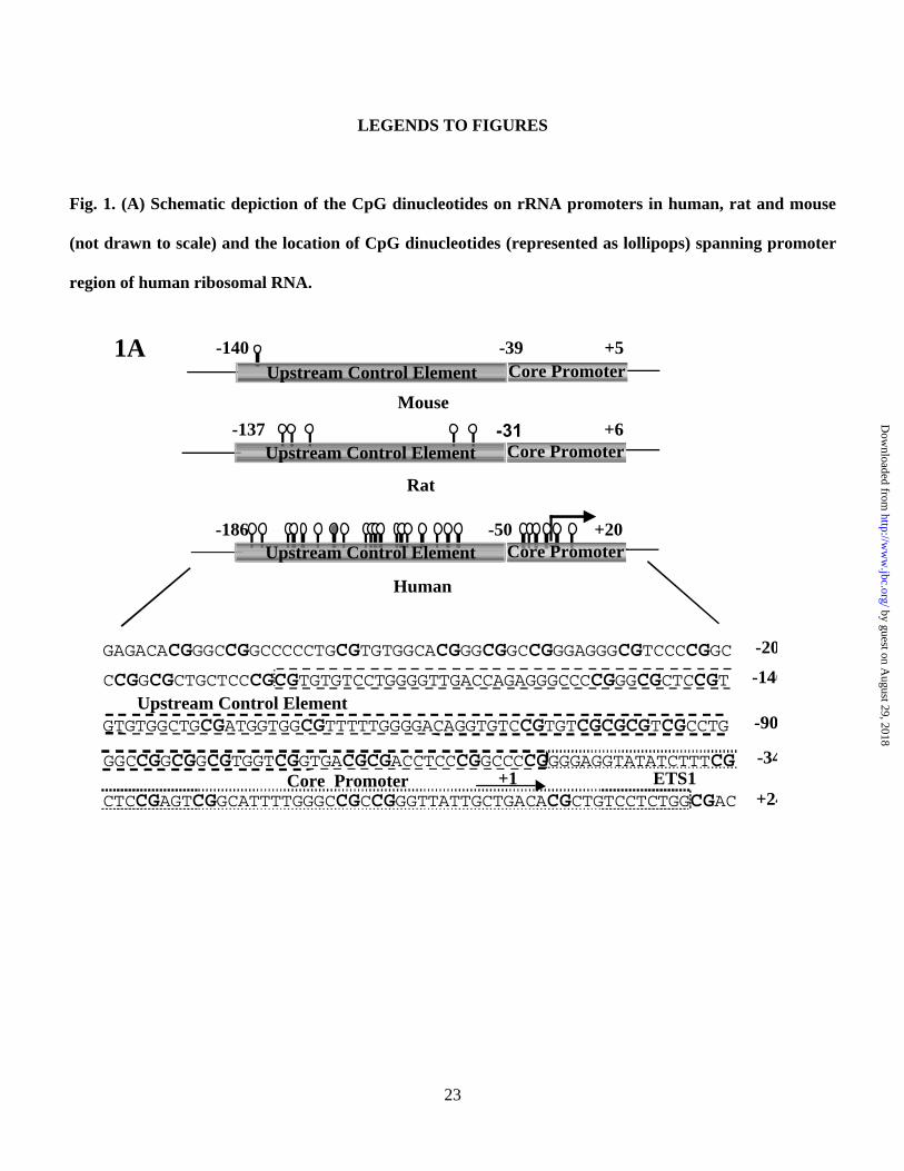
highly enriched in CpG and the promoter harbors a CpG island encompassing 19 CpGs in the upstream control
element (UCE) and 6 CpGs in the core promoter regions. On the other hand, the mouse and rat rRNA promoters
contain only one and five CpGs, respectively (Fig. 1A). Most of the CpG islands of house keeping genes
transcribed by RNA polymerase II are located in the promoter and exon 1 regions that are methylation-free in
normal somatic cells. Although ribosomal RNA genes are house keeping genes, they are highly reiterated and
by guest on August 29, 2018
http://ww
w.jbc.org/
Dow
nloaded from
12
only a fraction of these genes is transcribed by pol I. Therefore, we sought to investigate the methylation status
of each CpG within different cis elements (spanning from –200 to –9 bp) in the rRNA promoter in human
livers, and their potential alterations in hepatocellular carcinomas (HCCs). To determine the methylation status
of individual CpGs in the human rRNA promoter region, we performed bisulfite genomic sequencing (40) of
DNA isolated from hepatocellular carcinomas and the pair-matched liver tissues. Treatment of DNA with
bisulfite reagent converts unmethylated cytosine residues to uracils that are amplified as thymine during
subsequent PCR. On the other hand, the methylated cytosine residues remain unconverted during bisulfite
reaction and amplify as cytosines during subsequent PCR (41). The rRNA promoter region from bisulfite-
treated DNA was amplified with nested primers and the PCR product was cloned (see Methods for details). To
analyze quantitatively the methylation status of each CpGs spanning the core promoter and UCE, 8-10
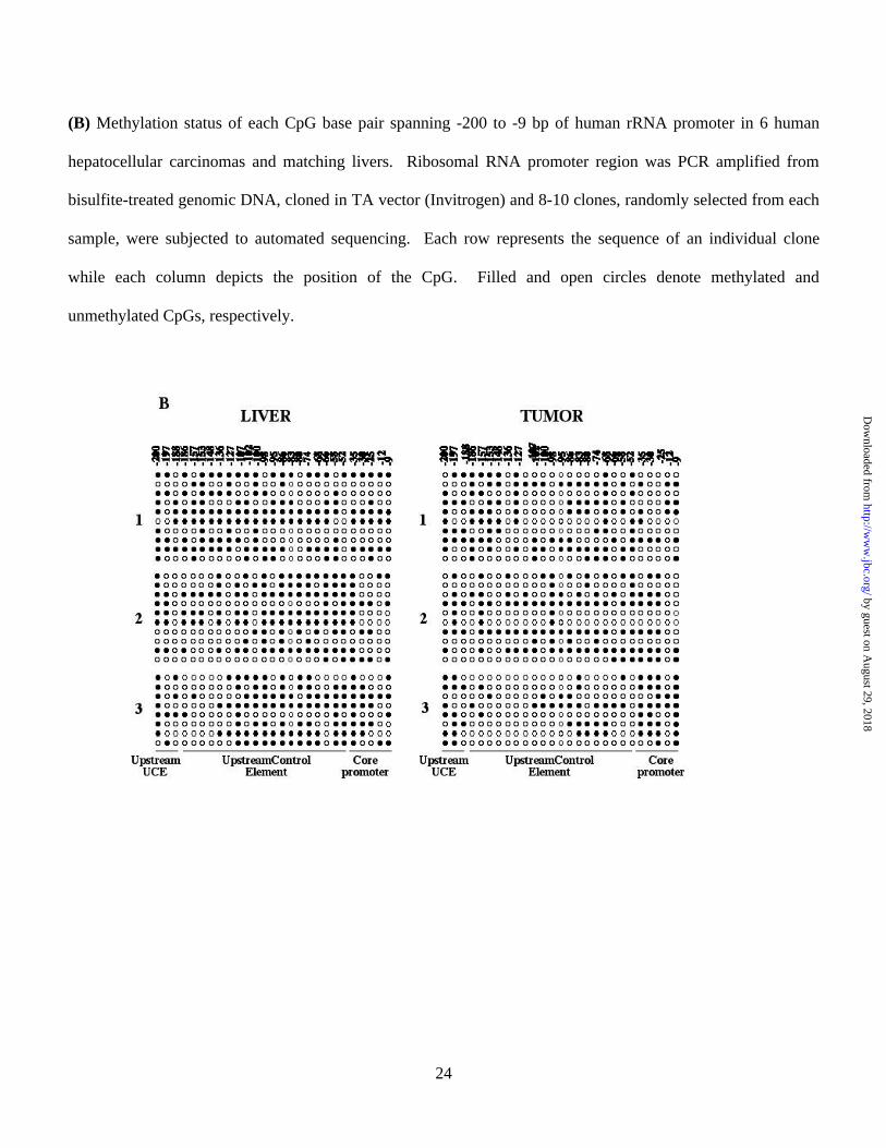
randomly selected clones from each HCC as well as the matching liver were sequenced. Alignment and analysis
of the methylation profiles of the rRNA promoter in HCC and liver from the same individual revealed a
differential methylation profile of CpGs between the two tissues (Fig. 1B). Comparison of methylation pattern
of the six individual livers revealed variations among them, probably due to polymorphism. For example, the
region spanning from -200 to -148 (with respect to +1 site) was poorly methylated in individuals 3 and 6, but
heavily methylated in individuals 1 and 5. However, the overall methylation density at this region was
comparable among the tumors and livers (Fig. 1B). Hypomethylation at UCE between positions -136 and -58
was more pronounced in all six tumors. Most CpGs within this region, e.g. -136, -107, -102, -100 -98, -95, -86,
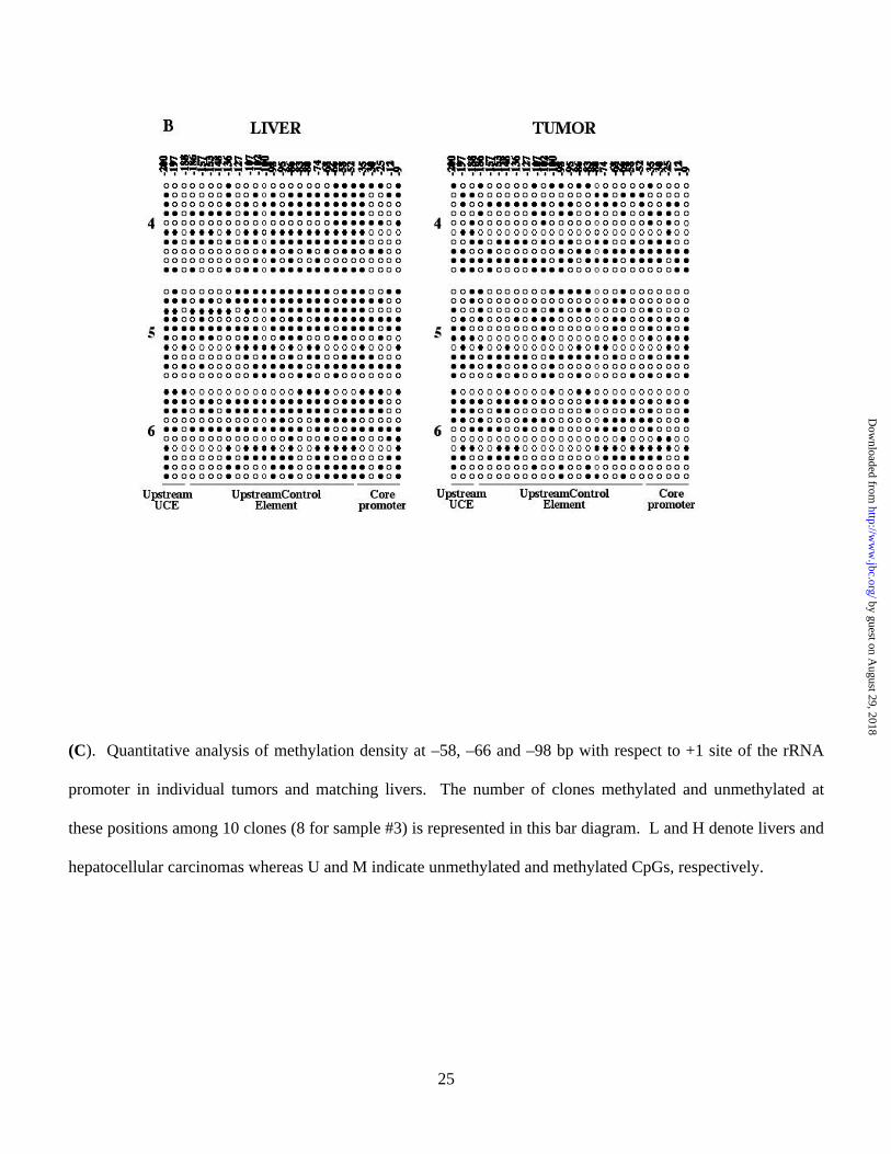
-66, and -58 were significantly hypomethylated in tumors. The quantitative analysis of methylated and
unmethylated cytosine at positions -58, -66 and -98 (Fig. 1C) showed that the differential level of methylation
in the liver DNA among the individuals. Further, in five out of six samples, clones containing unmethylated
cytosines were more abundant in HCCs than in corresponding livers. These data demonstrate that the rRNA
promoter, particularly in the UCE, is heavily methylated in human liver and is significantly hypomethylated in
hepatocellular carcinomas compared to the matched liver tissues.
by guest on August 29, 2018
http://ww
w.jbc.org/
Dow
nloaded from
13
rRNA promoter activity inversely correlates with the density of methylation
To explore whether methylation of CpG island in human rRNA promoter indeed suppresses its expression, we
cloned a region spanning from -410 to +314bp comprising the upstream control element, core promoter and part
of external transcribed spacer region 1 (ETS1) in pGL3-basic vector. The rRNA promoter-luciferase reporter
(pHrD-IRES-Luc) was constructed by replacing the Kozak sequence of pGL3 vector with the internal ribosome
entry site (IRES) (see Methods for detail). This reporter plasmid was used in transient transfection studies (Fig.
2A). The rationale for eliminating Kozak sequence was to minimize luciferase expression driven by spurious
pol II promoters within the plasmid as the 5′-trimethyl G-capped mRNAs (transcribed by pol II) exclusively
require Kozak sequence for initial ribosome binding and translation (42). The IRES sequence was introduced
immediate 5′ of the firefly luciferase cDNA for efficient translation of uncapped transcripts (pol I transcript and
some viral RNAs) which requires IRES (43). To confirm further the authenticity of the pol I promoter activity
in the transfection assay, we took advantage of the species-specificity of pol I transcription (44). Accordingly,
the human rRNA promoter will not be transcribed by mouse pol I transcription machinery and vice versa (7).
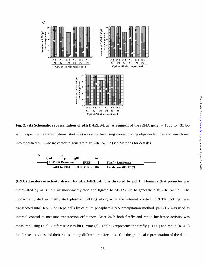
For this purpose, we transfected both Hepa (mouse) and HepG2 (human) cells with either pIRES-Luc
(promoter-less) or pHrD-IRES-Luc vector. In HepG2 cells the rRNA promoter-driven luciferase activity was
35 fold higher compared to pIRES-Luc whereas only 3 fold higher activity of pHrD-IRES-Luc was observed in
Hepa cells (Fig. 2B & 2C). If the reporter gene were transcribed by pol II in HepG2 (human) cells transfected
with pHrD-IRES-Luc, the luciferase activity would be comparable to that in Hepa (mouse) cells, as pol II
transcription is not species specific. Significantly higher (14 fold) activity of pHrD-IRES-Luc in HeG2 cells
compared to that in Hepa cells clearly demonstrates that the rRNA promoter directs the luciferase expression
from pHrD-IRES-Luc in HepG2 cells and is transcribed by pol I (Fig. 2C).
Next, we investigated the effect of methylation of the rRNA promoter on luciferase activity. To address
this issue, HepG2 and Hepa cells were transfected with pHrD-IRES-Luc harboring methylated or mock
by guest on August 29, 2018
http://ww
w.jbc.org/
Dow
nloaded from
14
methylated (with M. Hha I) rRNA promoter or pIRES-Luc. Luciferase activity was measured 24 hr post-
transfection. The rRNA promoter activity was significantly inhibited in HepG2 cells when methylated by M.
Hha I, compared with the mock-methylated control (Fig 2B, rows 5 & 6 and Fig. 2C). The inhibitory effect of
methylation was also robust in HepG2 cells (10 fold) compared to only 2 fold reduction in Hepa cells (Fig 2B,
rows 2 & 3 and Fig. 2C), attributing the minimal promoter activity observed in Hepa cells to some leaky
expression. These results further confirmed that the rRNA promoter driven luciferase activity in HepG2 cells
was sensitive to methylation. These results also demonstrate that rRNA promoter activity in human system is
sensitive to CpG methylation.
We next investigated whether the pol I-driven pHrD-IRES-Luc activity was sensitive to the density of
methylation at the promoter. This was accomplished by methylating the promoter and the ETS1 (external
transcribed spacer region 1) region (-410 to +314) with bacterial methylases that methylate densely (M. Sss I),
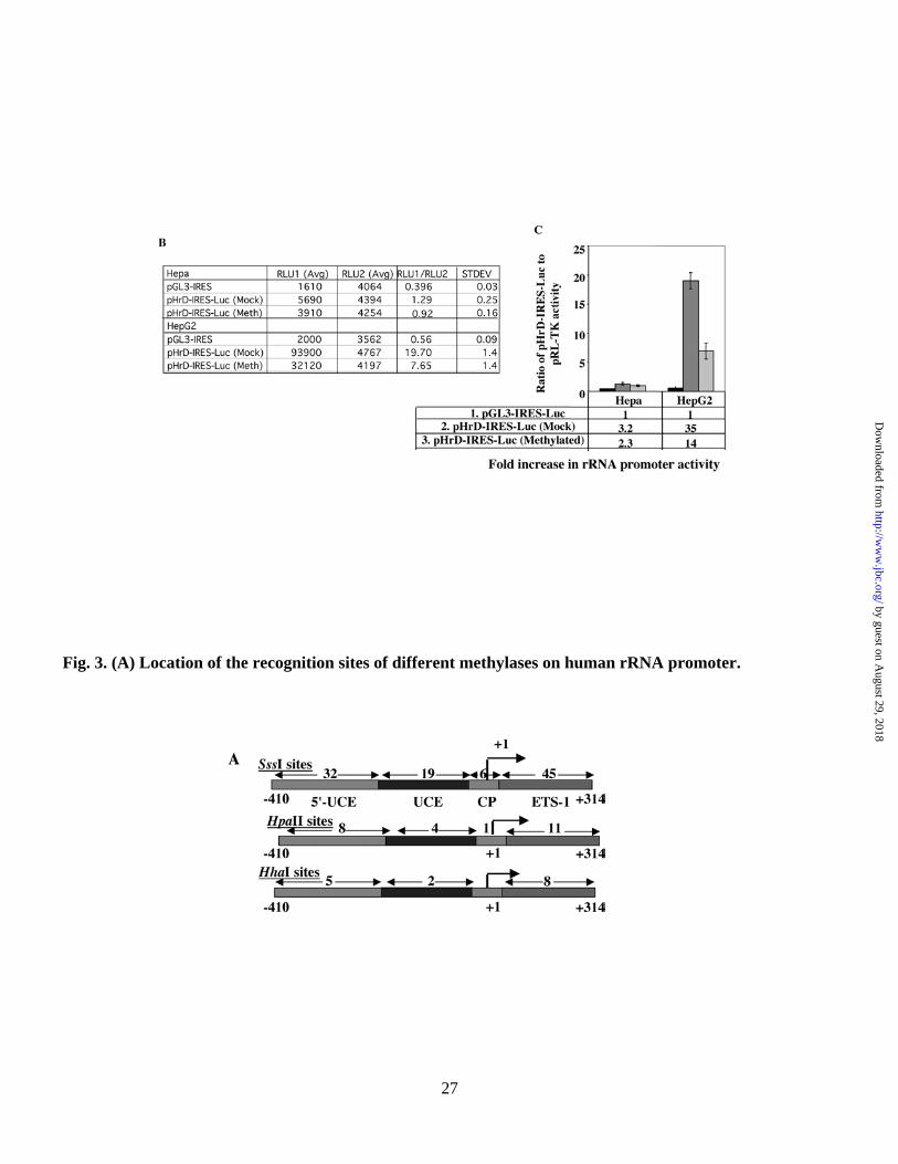
moderately (M. Hpa II) or sparsely (M. Hha I) (Fig. 3A). The promoter activity was indeed dependent on the
extent of methylation (Fig. 3B & 3C). Methylation of only 7 sites (5 in the upstream of UCE and 2 in UCE)
with M. Hha I resulted in significant (78%) inhibition of the luciferase activity compared to the mock-
methylated promoter (Fig. 3C). Methylation with Hpa II (which methylates 13 CpGs in the promoter region)
resulted in almost complete (94%) inhibition in rRNA transcription compared to the mock-methylated
promoter. As expected, the promoter activity abolished when all the CpGs located in the region spanning –410
to +314 were methylated with M. Sss I (Fig. 3C). These results were highly reproducible in different batches of
transfected cells. This data clearly demonstrate that the methylation density at the CpG island plays an
important role in repression of human rRNA transcription.
by guest on August 29, 2018
http://ww
w.jbc.org/
Dow
nloaded from
15
Methylation at single sites located on the promoter but not on the external transcribed spacer region 1
(ETS1) of human rRNA gene down regulates the promoter activity
The bacterial methylases can methylate specific sites not only in the promoter but also in ETS1. Effect of
methylation in the ETS1 on rRNA promoter activity can not be addressed by the previous experiment. To
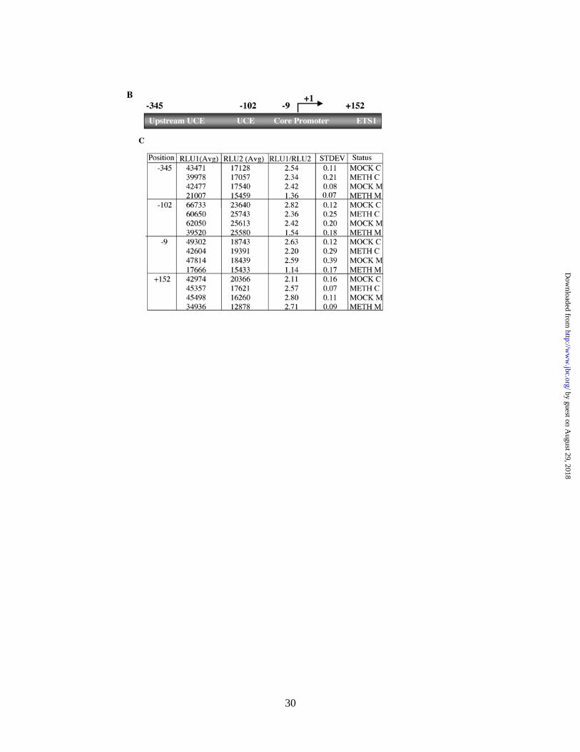
investigate if methylation at specific sites in the promoter or the coding region had differential effect on rRNA
promoter activity, we developed site-specific methylation technique. For this purpose, we designed
oligonucleotides containing either a methylated cytosine or unmethylated cytosine (as a control) at positions -
345, -102, -9 and +152 (Fig. 4B) of the rRNA gene and generated mock-, hemi- and fully-methylated circular
pHrD-IRES-Luc plasmids (Fig. 4A, see Methods for details). The promoter activity was measured in HepG2
cells 24 hr post-transfection and the result is expressed as the ratio of human rRNA promoter-directed firefly
luciferase activity to the internal control, thymidine-kinase promoter-driven renila luciferase activity (pRL-TK).
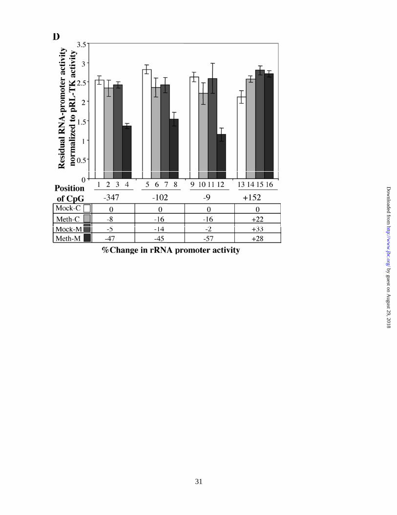
When CpG at position -345 (upstream UCE) was symmetrically methylated on both strands (Meth-M), the
promoter activity was inhibited by ~50% whereas methylation on individual strands (Meth-C, Mock-M) had no
significant effect (Fig. 4C, compare rows 2-4 with 1 and Fig. 4D, lanes 2-4). Comparable level of inhibition of
luciferase activity was attained when pHrD-IRES-Luc was methylated at -102 (located in UCE) or at –9
(located in the core promoter) on both strands (rows 8 & 12 in Fig. 4C and lanes 8 & 12 in Fig. 4D,
respectively). In contrast, methylation at +152 located on ETS1 has no inhibitory effect on the promoter
activity (Fig. 4C, rows 13-16 and Fig. 4D, lanes 13-16). These data clearly demonstrate that only symmetrical
methylation of CpG at single sites located within the promoter but not in the ETS1 down-regulates human
rRNA promoter activity.
by guest on August 29, 2018
http://ww
w.jbc.org/
Dow
nloaded from
16
Methyl CpG binding protein MBD2 specifically represses methylated rRNA promoter activity in HepG2
cells whereas MBD1, MBD3 and MeCP2 inhibit the promoter activity irrespective of its methylation
status
Methylation density-dependent inhibition of rRNA promoter activity implicates the role of methyl CpG binding
proteins (MBDs) in the repression of the methylated rRNA promoter. Transcription of rRNA precursor, its
processing to mature 28S, 18S and 5.8S rRNAs and their assembly into ribosomal subunits occur in the
nucleolus. As a first step to explore the role of the MBDs in rRNA gene transcription in humans we determined
whether any of these MBDs are localized in the nucleoli. To address this issue, we studied co-localization of
MBDs with nucleolin, a nucleolar marker protein (45). As expected, all the MBDs tested are localized in the
nucleus as demonstrated by their overlap with DAPI-stained nuclei (Fig. 5, panels 2 & 4). Merging of MBD
signals at two spots (panel 3) with that of nucleolin indicated that MBDs are localized also in the nucleolus.
These results provided us the impetus to explore the role of MBDs in rRNA expression.
Next, we co-transfected the methylated (M. Hha I) or mock-methylated rRNA-promoter in pIRES-Luc
into HepG2 cells, along with the expression vectors encoding different MBDs. Overexpression of MBDs in
HepG2 cells was confirmed by Western blot analysis with antibodies specific for MBD1-3 and MeCP2. These
antibodies were raised against C-terminal recombinant proteins that lack highly homologous N-terminal MBD
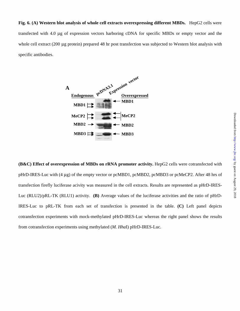
domains and do not cross-react (37,38). The results showed that MBD2 is the most abundant among these
MBDs in HepG2 cells and its level increased 2 to 3 fold after overexexpression (Fig. 6A). In contrast,
endogenous levels of MBD1, MBD3 and MeCP2 are low and increased at least 5-10 fold upon overexpression.
Five different alternatively spliced variants of MBD1 have been identified in different mammalian cells (46).
The endogenous variants of MBD1 expressed in HepG2 cells are ~55 and 60 kDa whereas that of the
overexpressed MBD1 is ~65 kDa. Overexpressed MeCP2 generated two polypeptides of ~75 and 66 kDa
detected by MeCP2-specific antibody. The lower polypeptide may arise either due to proteolysis or initiation
by guest on August 29, 2018
http://ww
w.jbc.org/
Dow
nloaded from
17
from an internal ATG site. Overexpressed MBD3 co-migrated with the larger of the two closely migrating
endogenous polypeptides.
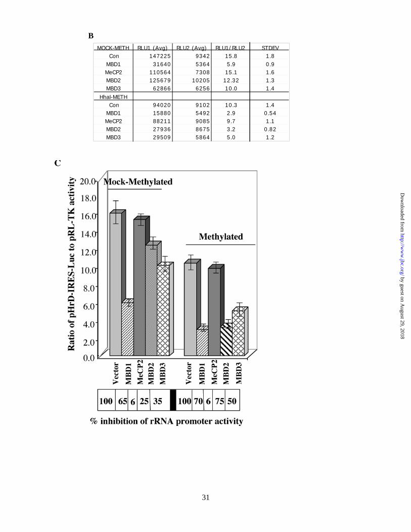
We then determined the effect of MBDs on rRNA promoter activity 48 hr post-transfection in HepG2
cells. MBD2 specifically suppressed the activity of the methylated (M. Hha I) promoter (75%) compared to
unmethylated promoter (25%). In contrast, MBD1 had profound repressive effect on the activity of both
methylated (70%) and mock-methylated rRNA promoter (65%) (Fig. 6B and 6C). A relatively small but
reproducible repression (35-40%) of both promoters was observed in cells overexpressing MBD3. On the other
hand, overexpression of MeCP2 had no significant effect on the rRNA promoter activity. This experiment was
repeated at least three times with different batches of cells and each transfection reaction was performed in
triplicate. These results demonstrate that methyl CpG binding proteins indeed differentially modulate human
rRNA promoter-driven reporter activity.
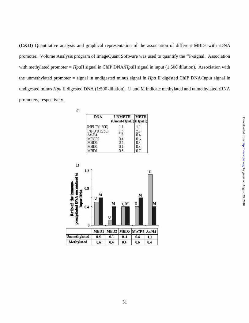
Chromatin immunoprecipitation (ChIP) assay demonstrates association of MBD2 specifically with the
endogenous methylated rRNA promoter in HepG2 cells
To confirm that the inhibitory effect of MBDs indeed occurs in vivo in the chromatin context, as observed in
transient transfection assay, we analyzed the association of the MBDs with endogenous rRNA promoter by
chromatin immunoprecipitation (ChIP) assay. In the first step, formaldehyde cross-linked chromatin from
growing HepG2 cells was immunoprecipitated with antibodies against specific MBDs. To distinguish between
the methylated and unmethylated promoter, the input and ChIP-DNAs were digested with the isoschizomers
Msp I (methylation insensitive) or Hpa II (methylation sensitive). The human rRNA promoter was then
amplified from the undigested and digested DNAs. The region of the rRNA promoter amplified by the
promoter-specific primers harbors 4 Hpa II/Msp I sites (Fig. 7A). The Hpa II resistant-PCR product generated
by guest on August 29, 2018
http://ww
w.jbc.org/
Dow
nloaded from
18
from the input DNA measures the level of the methylated rRNA promoter in HepG2 cells, whereas the
difference between mock-digested and Hpa II digested signal reflects the level of the unmethylated rRNA
promoters. Similarly, the Hpa II-resistant PCR product obtained from the DNA immunoprecipitated by
antibody for a specific MBD, indicates its association with the rRNA promoter methylated at all four sites in
HepG2 cells. The difference in the signal between the amplified product generated from the undigested and
Hpa II-digested DNA measures the association of the MBD with the unmethylated rRNA promoter. As
expected, the input and ChIP-DNAs were completely digested with the methylation insensitive enzyme Msp I
(3, 6, 9, 12, 15, 18, 21 & 24). Quantification of the signal from the input DNA showed methylation of
approximately half of 300-400 copies of rRNA promoters at all four Hpa II sites within the amplified region
(Fig. 7A) in HepG2 cells (Fig. 7B, lanes 1, 2 & 4, 5). All MBDs were associated with both methylated and
unmethylated rRNA promoters, albeit to different extent (lanes, 13 & 14, 16 & 17, 19 & 20 and 22 & 23). The
results suggest that MBD2 is predominantly associated with the methylated promoter, as the rRNA promoter
immunoprecipitated by anti-MBD2 antibody is mostly Hpa II-resistant (Fig. 7B, lanes 19 & 20 and Fig. 7C &
7D). In contrast, MBD3 bound to both promoters equally well (lanes 16, 17 and Fig. 7C & 7D). The amount of
the 32P-labeled, amplified rRNA promoter significantly decreased after Hpa II digestion of the DNA
immunoprecipitated with MBD1 (lanes 22 & 23 and 7C & 7D), MBD3 (lanes 16 & 17 and Fig. 7C & 7D) and
MeCP2 (lanes 13 & 14 and Fig. 7C & 7D) antibodies. Quantitative analysis demonstrated that MBD1 and
MeCP2 have higher affinity for the methylated rRNA promoter, whereas MBD3 bound to both promoters
equally well (Fig. 7C & 7D). As expected, acetylated histone H4 was predominantly associated with the
unmethylated promoter (Fig 7B, lanes 10 & 11). As a control we also amplified the promoter region of
GAPDH gene, a highly expressed, unmethylated, single copy gene, from the input and ChIP DNA. The
amplicon of GAPDH harbors 2 Hpa II sites. Amplification of GAPDH from the mock-digested, but not the
Hpa II digested input DNA, confirmed that this promoter is indeed methylation-free in HepG2 cells. Among
the proteins tested, only acetylated histone H4 and MeCP2 were associated with the GAPDH promoter (Fig. 7B,
lanes 10 & 13, respectively). The interaction of MeCP2 with unmethylated rRNA or GAPDH promoter is
by guest on August 29, 2018
http://ww
w.jbc.org/
Dow
nloaded from
19
probably mediated through a nonspecific DNA binding domain that resides in the transcriptional repressor
domain located at the C-terminus of MeCP2 (47). The lack of association of MBD2 with the unmethylated
GAPDH promoter further reinforces the notion that it is specifically targeted to the methylated promoters. This
experiment was performed at least twice with different chromatin preparation and the results were reproducible.
These results demonstrate the association of MBDs with the rRNA promoter in the chromatin context and
correlate well with their inhibitory effect on transcriptional activity of the promoter.
DISCUSSION
The molecular mechanisms of the methylation-mediated alteration in gene expression associated with neoplastic
transformation have not been completely elucidated. Multistage hepatocarcinogenesis is associated with
regional hypermethylation of tumor suppressor genes, e.g. hMLH1 (48), RASSF1A (49), p16 (50), O-6-MGMT
(51), PTPRO (52) and genome wide hypomethylation of repetitive elements (53) and oncogenes, e.g. RAS (54),
C-MYC (52). Recent studies with Dnmt1-deficient mice have shown that global hypomethylation causes
propensity towards tumorigenesis (55). A hallmark of cancer cells is the augmented transcription of ribosomal
RNA (rRNA) genes to meet the demand for increased production of ribosomes and for protein synthesis. We
hypothesized that the higher level of rRNA synthesis in the tumors could also result from hypomethylation of
rRNA promoters in addition to upregulation of pol I-specific transcription factor(s), e.g. UBF (56). To our
knowledge, the present study is the first report on the analysis of the methylation status of each CpG in the
promoter region of rRNA genes in human primary tumors and its relationship to the rRNA promoter activity.
Comparison of the assembled genomic sequences from seven different species has shown that the diversity
among animals lies not in the protein coding region, but rather in the cis regulatory elements, implicating more
complex regulation of gene expression in higher organisms (57). The existence of CpG island in the human, but
not in rodent rRNA promoters suggested that the underlying molecular mechanism of methylation-mediated
by guest on August 29, 2018
http://ww
w.jbc.org/
Dow
nloaded from
20
silencing among these two species may be distinct. This prompted us to explore whether the CpG island,
located on the rRNA promoter, is differentially methylated in human hepatocellular carcinomas, and the
potential role of its methylation on rRNA expression. Indeed, the present data have revealed an inverse
relationship between human rRNA promoter activity and CpG methylation status. Bisulfite genomic
sequencing also demonstrated polymorphic variations in this epigenetic modification in the liver DNA among
the six individuals analyzed. We have observed polymorphic variations in the liver DNA among the individuals
of the same rat strain (Fisher 344) at the Not I site of a CpG island on a protein tyrosine phosphatase (PTPRO)
promoter (52). Like the rRNA promoter, PTPRO promoter also showed tumor-specific methylation (52).
The significant hypomethylation specifically in the upstream control element (UCE) of the rRNA
promoter merits some explanation. This region is probably less accessible to DNA methyltransferases during
replication in the rapidly proliferating tumor tissues. Alternatively, CpGs in the UCE region may undergo
active demethylation by MBD2b, a variant of MBD2 that has been reported to exhibit demethylase activity in
vitro and in vivo (58). In this context, it is noteworthy that differential methylation of rRNA repeats has been
observed in other diseases, such as ATRX (59) and Werner syndrome (60). In ATRX syndrome, an X-linked
disorder leading to α-thalassaemia, rRNA arrays are hypomethylated due to mutation in ATRX gene (59). On
the contrary, rRNA repeats are hypomethylated in fibroblasts from patients with Werner syndrome (60). These
observations implicate that methylation of rRNA repeats are tightly regulated in normal cells.
In mouse cells, pol I transcription could be inhibited by methylation of a single CpG located at –133 in
the UCE region. This forms a repressive chromatin structure inhibiting access of the key transcription factor
UBF (61). The relatively larger number of CpGs in the human rRNA promoter region compared to the rodent,
promoter prompted us to investigate whether methylation density plays any role in repression of rRNA gene
transcription. To address this issue, we generated a rRNA-promoter-luciferase reporter plasmid where Kozak
was replaced by IRES to facilitate efficient translation of pol I-transcribed luciferase mRNA. Minimal
by guest on August 29, 2018
http://ww
w.jbc.org/
Dow
nloaded from
21
luciferase activity in mouse (Hepa) cells compared to that in human (HepG2) cells confirmed pol I-driven
transcription of the reporter gene. The same strategy was used earlier to link pol I promoter to chloramphenicol
acetyl transferase reporter (43). Inverse correlation of pol I promoter activity with methylation density (Fig. 3)
suggests that the mechanism of methylation-mediated silencing in humans may be distinct from that in mice.
We can not, however, rule out the potential role of MeCP2 that can bind to a symmetrically methylated CpG
(31), in silencing the rRNA promoter in mice, as methylation at -133 does not directly impede UBF binding
(61).
To study the effect of methylation at a single site within the promoter or coding region, we developed
site-specific methylation technique by modifying site-directed mutagenesis protocol. Here, a combination of a
methylated oligo (in place of a mutant oligo) and hemimethylase activity of Dnmt1 generated a plasmid DNA
with a single site methylated in one or both strands (Fig. 4A). Transfection studies using these plasmids have
shown that symmetrical methylation at single sites located within the core promoter, UCE or sequence upstream
of UCE, but not in the transcribed (ETS1) region, represses rRNA promoter activity. Inhibition of promoter
activity upon methylation of the cytosine residue at –314 indicates that methylation at sites outside of UBF
binding site can modulate rRNA promoter activity, probably by recruiting MBDs. The unabated activity of the
hemimethylated rRNA promoter also indicates the involvement of MBDs in the repression of RNA genes, as
recruitment of MBDs to the DNA requires symmetrically methylated CpGs (Fig. 4B). The technically
demanding site-specific methylation analysis, used in the present study, provided the most conclusive answer
regarding the effect of methylation at a single site on gene transcription.
Although all MBD proteins have highly homologous methyl CpG binding domains, immunolocalization
studies demonstrated that in vivo they are targeted to different regions of the genome by yet unidentified
mechanisms. Co-localization of MBDs with nucleolin implicated their role in rRNA expression. We used two
approaches to identify the MBDs regulating the rRNA promoter, (i) transient overexpression and its effect on
by guest on August 29, 2018
http://ww
w.jbc.org/
Dow
nloaded from
22
rRNA-luciferase activity, and (ii) ChIP assay to identify their interactions with the endogenous rRNA promoter.
Among these MBDs, MBD2 specifically repressed the methylated rRNA promoter activity whereas MeCP2
inhibited promoter activity irrespective of its methylation status only when expressed at a high level (data not
shown). MBD1 repressed the rRNA promoter irrespective of its methylation status. This observation is in
agreement with its ability to bind both methylated and unmethylated DNA (26,46). The ChIP data corroborates
well with the effect of MBDs on rRNA promoter activity in transient transfection assays. MBDs associate with
a variety of corepressors that include mSin3a, HDACs, HMTs (29,62,63). These repressor-corepressor
complexes are responsible for the ultimate suppression of pol II-directed gene expression. Identification of the
corepressors that are recruited by different MBDs to repress rRNA gene expression will be of considerable
interest. Further studies are needed to address this issue. Nevertheless, the present study has clearly
demonstrated hypomethylation of the rRNA promoter in human hepatocellular carcinomas, elucidated the role
of promoter methylation in the ribosomal RNA gene expression and the potential role of the methyl CpG
binding protein MBD2 in the methylation-mediated silencing of the rRNA promoter.
ACKNOWLEDGEMENT
We thank Drs. Adrian Bird and Brian Hendrich for MeCP2 and MBD2 cDNAs, respectively, Ram Reddy for
anti-nucleolin antibody, Arthur Burghes and Jill Rafael-Fortney for their generosity with the use of fluorescence
microscope, and Dennis Summers for excellent technical assistance. The present study was supported, in part,
by grants ES 10874, CA 81024 and CA 86978.
by guest on August 29, 2018
http://ww
w.jbc.org/
Dow
nloaded from
31
REFERENCES
1. McStay, B., Paule, M., Schultz, M. C., Willis, I., and Pikaard, C. S. (2002) Gene Expr 10: 263-269.
2. Leary, D. J., and Huang, S. (2001) FEBS Lett 509: 145-150.
3. Jacob, S. T. (1995) Biochem J 306: 617-626.
4. Hannan, K. M., Hannan, R. D., and Rothblum, L. I. (1998) Front Biosci 3: d376-398.
5. Grummt, I. (1999) Prog Nucleic Acid Res Mol Biol 62: 109-154.
6. Paule, M. R., and White, R. J. (2000) Nucleic Acids Res 28: 1283-1298.
7. Heix, J., and Grummt, I. (1995) Curr Opin Genet Dev 5: 652-656.
8. Ghosh, A. K., Niu, H., and Jacob, S. T. (1996) Biochem Biophys Res Commun 225: 890-895.
9. Reeder, R. H. (1984) Cell 38: 349-351.
10. Bird, A. P., and Wolffe, A. P. (1999) Cell 99: 451-454.
11. Baylin, S. B., Esteller, M., Rountree, M. R., Bachman, K. E., Schuebel, K., and Herman, J. G. (2001)
Hum Mol Genet 10: 687-692.
12. Wade, P. A. (2001) Bioessays 23: 1131-1137.
13. Okano, M., Bell, D. W., Haber, D. A., and Li, E. (1999) Cell 99: 247-257.
14. Jaenisch, R., and Bird, A. (2003) Nat Genet 33: 245-254.
15. Robertson, K. D., and Wolffe, A. P. (2000) Nat Rev Genet 1: 11-19.
16. Jones, P. A., and Baylin, S. B. (2002) Nat Rev Genet 3: 415-428.
17. Plass, C. (2002) Hum Mol Genet 11: 2479-2488.
18. Bestor, T. H. (2000) Hum Mol Genet 9: 2395-2402.
19. Jeltsch, A. (2002) Chembiochem 3: 274-293.
20. Hansen, R. S., Wijmenga, C., Luo, P., Stanek, A. M., Canfield, T. K., Weemaes, C. M., and Gartler, S.
M. (1999) Proc Natl Acad Sci U S A 96: 14412-14417.
by guest on August 29, 2018
http://ww
w.jbc.org/
Dow
nloaded from
31
21. Ehrlich, M., Buchanan, K. L., Tsien, F., Jiang, G., Sun, B., Uicker, W., Weemaes, C. M., Smeets, D.,
Sperling, K., Belohradsky, B. H., Tommerup, N., Misek, D. E., Rouillard, J. M., Kuick, R., and Hanash,
S. M. (2001) Hum Mol Genet 10: 2917-2931.
22. Xu, G. L., Bestor, T. H., Bourc'his, D., Hsieh, C. L., Tommerup, N., Bugge, M., Hulten, M., Qu, X.,
Russo, J. J., and Viegas-Pequignot, E. (1999) Nature 402: 187-191.
23. Zhu, W. G., and Otterson, G. A. (2003) Curr Med Chem Anti-Canc Agents 3: 187-199.
24. Cheng, J. C., Matsen, C. B., Gonzales, F. A., Ye, W., Greer, S., Marquez, V. E., Jones, P. A., and
Selker, E. U. (2003) J Natl Cancer Inst 95: 399-409.
25. Holliday, R. (1996) Cancer Surv 28: 103-115.
26. Hendrich, B., and Bird, A. (1998) Mol Cell Biol 18: 6538-6547.
27. Hendrich, B., Hardeland, U., Ng, H. H., Jiricny, J., and Bird, A. (1999) Nature 401: 301-304.
28. Hendrich, B., and Bird, A. (2000) Curr Top Microbiol Immunol 249: 55-74.
29. Wade, P. A. (2001) Oncogene 20: 3166-3173.
30. Prokhortchouk, A., Hendrich, B., Jorgensen, H., Ruzov, A., Wilm, M., Georgiev, G., Bird, A., and
Prokhortchouk, E. (2001) Genes Dev 15: 1613-1618.
31. Nan, X., and Bird, A. (2001) Brain Dev 23: S32-37.
32. Zhao, X., Ueba, T., Christie, B. R., Barkho, B., McConnell, M. J., Nakashima, K., Lein, E. S., Eadie, B.
D., Willhoite, A. R., Muotri, A. R., Summers, R. G., Chun, J., Lee, K. F., and Gage, F. H. (2003) Proc
Natl Acad Sci U S A 100: 6777-6782.
33. Millar, C. B., Guy, J., Sansom, O. J., Selfridge, J., MacDougall, E., Hendrich, B., Keightley, P. D.,
Bishop, S. M., Clarke, A. R., and Bird, A. (2002) Science 297: 403-405.
34. Ghosh, A. K., Kermekchiev, M., and Jacob, S. T. (1994) Gene 141: 271-275.
35. Majumder, S., Ghoshal, K., Gronostajski, R. M., and Jacob, S. T. (2001) Gene Expr 9: 203-215.
36. Majumder, S., Ghoshal, K., Li, Z., and Jacob, S. T. (1999) J Biol Chem 274: 28584-28589.
by guest on August 29, 2018
http://ww
w.jbc.org/
Dow
nloaded from
31
37. Ghoshal, K., Datta, J., Majumder, S., Bai, S., Dong, X., Parthun, M., and Jacob, S. T. (2002) Mol Cell
Biol 22: 8302-8319.
38. Majumder, S., Ghoshal, K., Datta, J., Bai, S., Dong, X., Quan, N., Plass, C., and Jacob, S. T. (2002) J
Biol Chem 277: 16048-16058.
39. Weinmann, A. S., Bartley, S. M., Zhang, T., Zhang, M. Q., and Farnham, P. J. (2001) Mol Cell Biol 21:
6820-6832.
40. Clark, S. J., Harrison, J., Paul, C. L., and Frommer, M. (1994) Nucleic Acids Res 22: 2990-2997.
41. Ghoshal, K., Majumder, S., and Jacob, S. T. (2002) Methods Enzymol 353: 476-486.
42. Kozak, M. (1986) Proc Natl Acad Sci U S A 83: 2850-2854.
43. Palmer, T. D., Miller, A. D., Reeder, R. H., and McStay, B. (1993) Nucleic Acids Res 21: 3451-3457.
44. Heix, J., Zomerdijk, J. C., Ravanpay, A., Tjian, R., and Grummt, I. (1997) Proc Natl Acad Sci U S A 94:
1733-1738.
45. Valdez, B. C., Henning, D., Busch, R. K., Srivastava, M., and Busch, H. (1995) Mol Immunol 32: 1207-
1213.
46. Nakao, M., Matsui, S., Yamamoto, S., Okumura, K., Shirakawa, M., and Fujita, N. (2001) Brain Dev
23: S174-176.
47. Cross, S. H., Charlton, J. A., Nan, X., and Bird, A. P. (1994) Nat Genet 6: 236-244.
48. Matsukura, S., Soejima, H., Nakagawachi, T., Yakushiji, H., Ogawa, A., Fukuhara, M., Miyazaki, K.,
Nakabeppu, Y., Sekiguchi, M., and Mukai, T. (2003) Br J Cancer 88: 521-529.
49. Schagdarsurengin, U., Wilkens, L., Steinemann, D., Flemming, P., Kreipe, H. H., Pfeifer, G. P.,
Schlegelberger, B., and Dammann, R. (2003) Oncogene 22: 1866-1871.
50. Pogribny, I. P., and James, S. J. (2002) Cancer Lett 187: 69-75.
51. Yu, J., Ni, M., Xu, J., Zhang, H., Gao, B., Gu, J., Chen, J., Zhang, L., Wu, M., Zhen, S., and Zhu, J.
(2002) BMC Cancer 2: 29.
by guest on August 29, 2018
http://ww
w.jbc.org/
Dow
nloaded from
31
52. Motiwala, T., Ghoshal, K., Das, A., Majumder, S., Weichenhan, D., Wu, Y. Z., Holman, K., James, S.
J., Jacob, S. T., and Plass, C. (2003) Oncogene 22: 6319-6331.
53. Lin, C. H., Hsieh, S. Y., Sheen, I. S., Lee, W. C., Chen, T. C., Shyu, W. C., and Liaw, Y. F. (2001)
Cancer Res 61: 4238-4243.
54. Zapisek, W. F., Cronin, G. M., Lyn-Cook, B. D., and Poirier, L. A. (1992) Carcinogenesis 13: 1869-
1872.
55. Gaudet, F., Hodgson, J. G., Eden, A., Jackson-Grusby, L., Dausman, J., Gray, J. W., Leonhardt, H., and
Jaenisch, R. (2003) Science 300: 489-492.
56. Huang, R., Wu, T., Xu, L., Liu, A., Ji, Y., and Hu, G. (2002) Faseb J 16: 293-301.
57. Levine, M., and Tjian, R. (2003) Nature 424: 147-151.
58. Detich, N., Bovenzi, V., and Szyf, M. (2003) J Biol Chem 278: 27586-27592.
59. Gibbons, R. J., McDowell, T. L., Raman, S., O'Rourke, D. M., Garrick, D., Ayyub, H., and Higgs, D. R.
(2000) Nat Genet 24: 368-371.
60. Machwe, A., Orren, D. K., and Bohr, V. A. (2000) Faseb J 14: 1715-1724.
61. Santoro, R., and Grummt, I. (2001) Mol Cell 8: 719-725.
62. Nan, X., Ng, H. H., Johnson, C. A., Laherty, C. D., Turner, B. M., Eisenman, R. N., and Bird, A. (1998)
Nature 393: 386-389.
63. Jones, P. L., Veenstra, G. J., Wade, P. A., Vermaak, D., Kass, S. U., Landsberger, N., Strouboulis, J.,
and Wolffe, A. P. (1998) Nat Genet 19: 187-191.
by guest on August 29, 2018
http://ww
w.jbc.org/
Dow
nloaded from
23
LEGENDS TO FIGURES
Fig. 1. (A) Schematic depiction of the CpG dinucleotides on rRNA promoters in human, rat and mouse
(not drawn to scale) and the location of CpG dinucleotides (represented as lollipops) spanning promoter
region of human ribosomal RNA.
1A
Mouse
+5-39Core PromoterUpstream Control Element
-140
Core Promoter
Upstream Control Element
+1 ETS1+24CTCCGAGTCGGCATTTTGGGCCGCCGGGTTATTGCTGACACGCTGTCCTCTGGCGAC
-34GGCCGGCGGCGTGGTCGGTGACGCGACCTCCCGGCCCCGGGGAGGTATATCTTTCG
-90GTGTGGCTGCGATGGTGGCGTTTTTGGGGACAGGTGTCCGTGTCGCGCGTCGCCTG
-146CCGGCGCTGCTCCCGCGTGTGTCCTGGGGTTGACCAGAGGGCCCCGGGCGCTCCGT
-202GAGACACGGGCCGGCCCCCTGCGTGTGGCACGGGCGGCCGGGAGGGCGTCCCCGGC
Rat
-31-137 +6Core PromoterUpstream Control Element
Human
-50 +20-186Core PromoterUpstream Control Element
by guest on August 29, 2018
http://ww
w.jbc.org/
Dow
nloaded from
24
(B) Methylation status of each CpG base pair spanning -200 to -9 bp of human rRNA promoter in 6 human
hepatocellular carcinomas and matching livers. Ribosomal RNA promoter region was PCR amplified from
bisulfite-treated genomic DNA, cloned in TA vector (Invitrogen) and 8-10 clones, randomly selected from each
sample, were subjected to automated sequencing. Each row represents the sequence of an individual clone
while each column depicts the position of the CpG. Filled and open circles denote methylated and
unmethylated CpGs, respectively.
by guest on August 29, 2018
http://ww
w.jbc.org/
Dow
nloaded from
25
(C). Quantitative analysis of methylation density at –58, –66 and –98 bp with respect to +1 site of the rRNA
promoter in individual tumors and matching livers. The number of clones methylated and unmethylated at
these positions among 10 clones (8 for sample #3) is represented in this bar diagram. L and H denote livers and
hepatocellular carcinomas whereas U and M indicate unmethylated and methylated CpGs, respectively.
by guest on August 29, 2018
http://ww
w.jbc.org/
Dow
nloaded from
26
Fig. 2. (A) Schematic representation of pHrD-IRES-Luc. A segment of the rRNA gene (–410bp to +314bp
with respect to the transcriptional start site) was amplified using corresponding oligonucleotides and was cloned
into modified pGL3-basic vector to generate pHrD-IRES-Luc (see Methods for details).
(B&C) Luciferase activity driven by pHrD-IRES-Luc is directed by pol I. Human rRNA promoter was
methylated by M. Hha I or mock-methylated and ligated to pIRES-Luc to generate pHrD-IRES-Luc. The
mock-methylated or methylated plasmid (500ng) along with the internal control, pRLTK (50 ng) was
transfected into HepG2 or Hepa cells by calcium phosphate-DNA precipitation method. pRL-TK was used as
internal control to measure transfection efficiency. After 24 h both firefly and renila luciferase activity was
measured using Dual Luciferase Assay kit (Promega). Table B represents the firefly (RLU1) and renila (RLU2)
luciferase activities and their ratios among different transfectants. C is the graphical representation of the data.
Firefly LuciferaseIRESHrDNA Promoter
-410 to +314 CITE (16 to 518) Luciferase (88-1737)
KpnI BglII NcoI+1A
by guest on August 29, 2018
http://ww
w.jbc.org/
Dow
nloaded from
27
Fig. 3. (A) Location of the recognition sites of different methylases on human rRNA promoter.
by guest on August 29, 2018
http://ww
w.jbc.org/
Dow
nloaded from
28
(B) Human rRNA promoter activity in HepG2 cells transfected with pHrD-IRES-Luc where rRNA promoter
region was methylated with M. SssI, M. HpaII or M. HhaI and Ado-Met. 1.5X10⁵ cells were transfected with
500ng of mock-methylated or methylated plasmid DNA and promoter activity was analyzed as described in Fig.
2B. Table B represents the average value of the luciferase activities and the ratio of RLU1 (pHrD-IRES-Luc) to
RLU2 (pRL-TK) from each set of transfected cells. (C) is the graphical representation of the data.
BRLU1 (Avg) RLU2 (Avg) RLU1/RLU2 STDEV
MOCK SssI 11797 46160 0.256 0.02MET/SssI 429 121956 0.004 0.0006Met HpaII 2255 161239 0.014 0.0015Met HhaI 6456 114884 0.056 0.0052
by guest on August 29, 2018
http://ww
w.jbc.org/
Dow
nloaded from
29
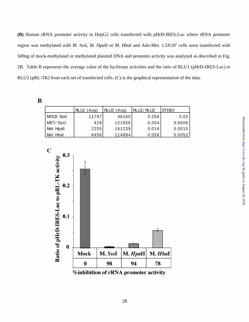
Fig. 4. (A). Schematic depiction of site-specific methylation technique.
(B) Location of CpGs on human rRNA gene subjected to site-specific methylation to study their effect on
the promoter activity. These sites were selected because of the ability to generate primers spanning these
regions. The construction of pHrD-IRES-Luc methylated at specific sites is described in Methods. (C&D)
Plasmids (500ng) methylated in both strands (Meth-M), methylated in the (-) strand (Mock-M) or non
methylated controls (Mock-C and Meth-C) along with pRL-TK (50ng) were transfected into 2X10⁵ HepG2 cells
and both firefly (RLU1) and renila (RLU2) luciferase activities were measured 24 hrs post-transfection using
Dual Luciferase assay kit. The data of a representative experiment is tabulated in C and graphically represented
in D.
by guest on August 29, 2018
http://ww
w.jbc.org/
Dow
nloaded from
31
Fig. 5. Colocalization of methyl binding proteins with nucleolin in HepG2 cells.
Co-localization studies were performed with antibodies specific for methyl binding proteins raised in rabbit
(except MBD2 which was raised in sheep) and monoclonal antibody (C23) against nucleolin, a nucleolus
specific protein. MBDs were recognized by FITC conjugated secondary antibody (green fluorescence) and
nucleolin was recognized by TRITC conjugated secondary antibody (red fluorescence). Yellow signal indicates
co-localization of MBDs with nucleolin.
FITC
TRITC
MERGE
DAPI
MBD2 PRE-IMMBD3 MECP2MBD1Nucleolin NucleolinNucleolin Nucleolin
MBD1 MBD3 MeCP2MBD2
by guest on August 29, 2018
http://ww
w.jbc.org/
Dow
nloaded from
31
Fig. 6. (A) Western blot analysis of whole cell extracts overexpressing different MBDs. HepG2 cells were
transfected with 4.0 µg of expression vectors harboring cDNA for specific MBDs or empty vector and the
whole cell extract (200 µg protein) prepared 48 hr post transfection was subjected to Western blot analysis with
specific antibodies.
(B&C) Effect of overexpression of MBDs on rRNA promoter activity. HepG2 cells were cotransfected with
pHrD-IRES-Luc with (4 µg) of the empty vector or pcMBD1, pcMBD2, pcMBD3 or pcMeCP2. After 48 hrs of
transfection firefly luciferase acivity was measured in the cell extracts. Results are represented as pHrD-IRES-
Luc (RLU2)/pRL-TK (RLU1) activity. (B) Average values of the luciferase activities and the ratio of pHrD-
IRES-Luc to pRL-TK from each set of transfection is presented in the table. (C) Left panel depicts
cotransfection experiments with mock-methylated pHrD-IRES-Luc whereas the right panel shows the results
from cotransfection experiments using methylated (M. HhaI) pHrD-IRES-Luc.
Expression vector
pcDNA3.1
MBD1
MeCP2
MBD2
MBD3
OverexpressedEndogenous
MBD1
MBD2
MBD3
MeCP2
A
by guest on August 29, 2018
http://ww
w.jbc.org/
Dow
nloaded from
31
MOCK-METH RLU1 (Avg) RLU2 (Avg) RLU1/RLU2 STDEVCon 147225 9342 15.8 1.8
MBD1 31640 5364 5.9 0.9MeCP2 110564 7308 15.1 1.6MBD2 125679 10205 12.32 1.3 MBD3 62866 6256 10.0 1.4
HhaI-METHCon 94020 9102 10.3 1.4
MBD1 15880 5492 2.9 0.54MeCP2 88211 9085 9.7 1.1MBD2 27936 8675 3.2 0.82 MBD3 29509 5864 5.0 1.2
B
by guest on August 29, 2018
http://ww
w.jbc.org/
Dow
nloaded from
31
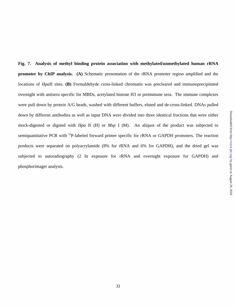
Fig. 7. Analysis of methyl binding protein association with methylated/unmethylated human rRNA
promoter by ChIP analysis. (A) Schematic presentation of the rRNA promoter region amplified and the
locations of HpaII sites. (B) Formaldehyde cross-linked chromatin was precleared and immunoprecipitated
overnight with antisera specific for MBDs, acetylated histone H3 or preimmune sera. The immune complexes
were pull down by protein A/G beads, washed with different buffers, eluted and de-cross-linked. DNAs pulled
down by different antibodies as well as input DNA were divided into three identical fractions that were either
mock-digested or digsted with Hpa II (H) or Msp I (M). An aliquot of the product was subjected to
semiquantitative PCR with 32P-labeled forward primer specific for rRNA or GAPDH promoters. The reaction
products were separated on polyacrylamide (8% for rRNA and 6% for GAPDH), and the dried gel was
subjected to autoradiography (2 hr exposure for rRNA and overnight exposure for GAPDH) and
phosphorimager analysis.
by guest on August 29, 2018
http://ww
w.jbc.org/
Dow
nloaded from
31
(C&D) Quantitative analysis and graphical representation of the association of different MBDs with rDNA
promoter. Volume Analysis program of ImageQuant Software was used to quantify the 32P-signal. Association
with methylated promoter = HpaII signal in ChIP DNA/HpaII signal in input (1:500 dilution). Association with
the unmethylated promoter = signal in undigested minus signal in Hpa II digested ChIP DNA/Input signal in
undigested minus Hpa II digested DNA (1:500 dilution). U and M indicate methylated and unmethylated rRNA
promoters, respectively.
by guest on August 29, 2018
http://ww
w.jbc.org/
Dow
nloaded from

Sudarshana M. Sharma, Wendy Frankel and Samson T. JacobKalpana Ghoshal, Sarmila Majumder, Jharna Datta, Tasneem Motiwala, Shoumei Bai,
binding protein MBD2 in the suppression of rRNA gene expressionRole of human Ribosomal RNA (rRNA) promoter methylation and of methyl CpG
published online November 10, 2003J. Biol. Chem.
10.1074/jbc.M309393200Access the most updated version of this article at doi:
Alerts:
When a correction for this article is posted•
When this article is cited•
to choose from all of JBC's e-mail alertsClick here
by guest on August 29, 2018
http://ww
w.jbc.org/
Dow
nloaded from