Role of epigenetic responses in susceptibility to liver ... 1B-2... · Role of epigenetic responses...
16
Role of epigenetic responses in susceptibility to liver toxicity Igor Koturbash
Role of epigenetic responses in susceptibility to liver ... 1B-2... · Role of epigenetic responses in susceptibility to liver toxicity ... a related promoter of GSGP-1, ... dCTP
Thank you, Dr. Seeff, for a kind introduction. Good morning ladies and gentlemen. First of all, I want to thank the organizing committee for a kind invitation to speak in here, and particularly Dr. John Senior. It's a very big honor and a pleasure for me. Today, I'll be talking about the role of epigenetic responses in susceptibility to liver toxicity.
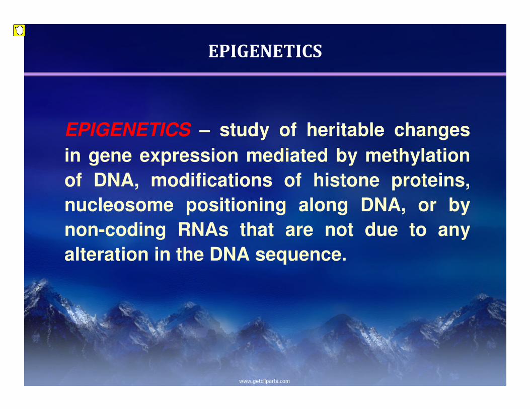
EPIGENETICS
EPIGENETICS – study of heritable changes
in gene expression mediated by methylation
of DNA, modifications of histone proteins,
nucleosome positioning along DNA, or by
non-coding RNAs that are not due to any
alteration in the DNA sequence.
Presenter
Sticky Note
First of all, I will start by saying what exactly epigenetics is, which is a study of heritable changes in gene expression that are not associated with any alterations in DNA sequence. However, they are mediated by DNA methylation, histone modification, nucleosome position in a lot of the DNA, or in non-coding RNAs.
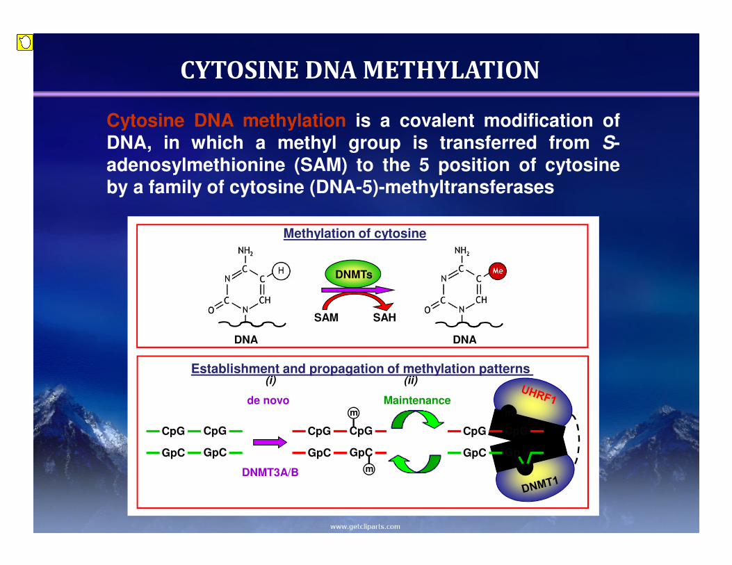
Cytosine DNA methylation is a covalent modification ofDNA, in which a methyl group is transferred from S-adenosylmethionine (SAM) to the 5 position of cytosineby a family of cytosine (DNA-5)-methyltransferases
CYTOSINE DNA METHYLATION
Methylation of cytosine
Establishment and propagation of methylation patterns
SAM SAH
DNMTs
DNA DNA
CpG
m
CpG
GpC
m
GpC
CpG
m
CpG
GpGpC
CpGCpG
GpCGpC
DNMT3A/B
de novo Maintenance
C
(i) (ii)
Presenter
Sticky Note
In particular, I will be talking today first about cytosine DNA methylation and about histone modifications. So what exactly is cytosine DNA methylation is? This is covalent modification of DNA, where the methyl groove that has been taken from a donor, usually it's S-adenosylmethionine or SAM, and being transferred to the first position of cytosine. This process is being facilitated by a family of so-called DNA methyltransferases.
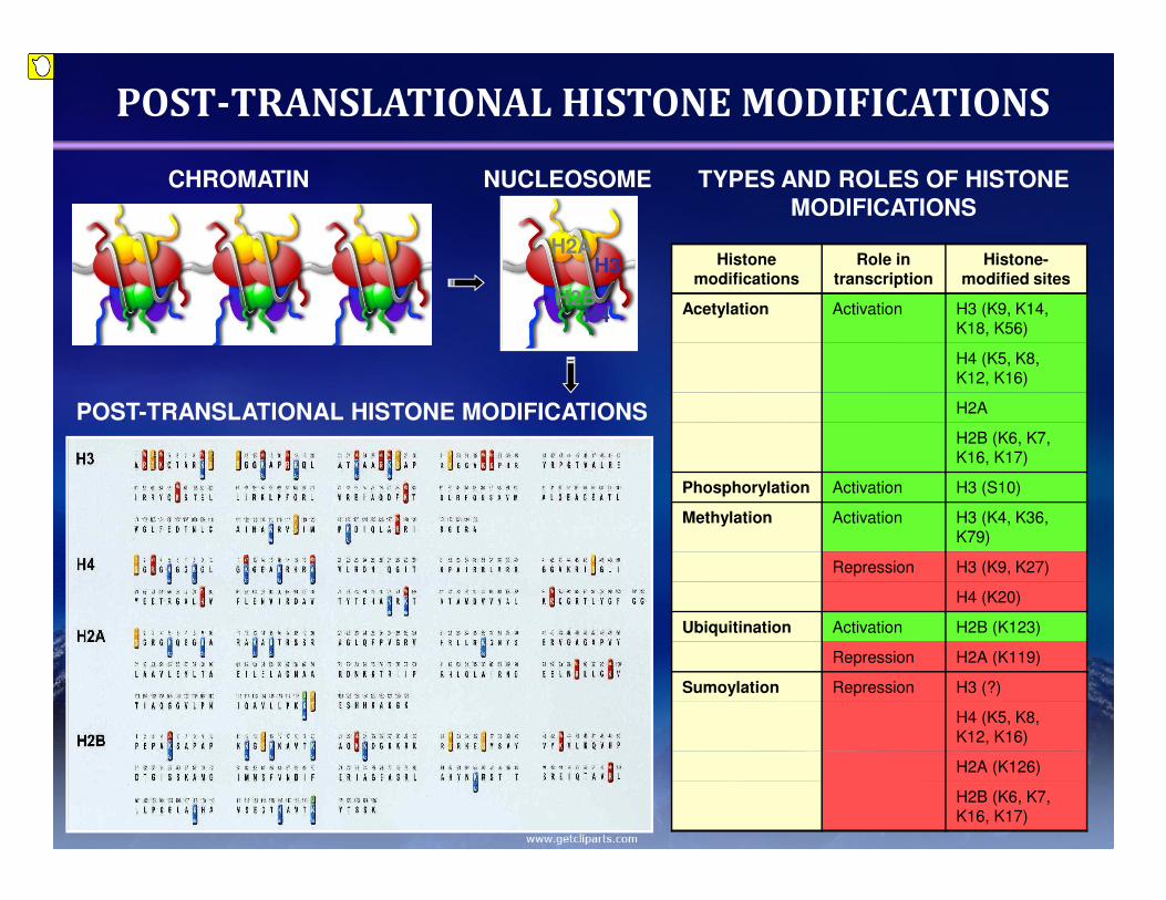
POST-TRANSLATIONAL HISTONE MODIFICATIONS
H2AH3
H4H2B
CHROMATIN NUCLEOSOME
POST-TRANSLATIONAL HISTONE MODIFICATIONS
Histone modifications
Role in transcription
Histone-modified sites
Acetylation Activation H3 (K9, K14, K18, K56)
H4 (K5, K8, K12, K16)
H2A
H2B (K6, K7, K16, K17)
Phosphorylation Activation H3 (S10)
Methylation Activation H3 (K4, K36, K79)
Repression H3 (K9, K27)
H4 (K20)
Ubiquitination Activation H2B (K123)
Repression H2A (K119)
Sumoylation Repression H3 (?)
H4 (K5, K8, K12, K16)
H2A (K126)
H2B (K6, K7, K16, K17)
TYPES AND ROLES OF HISTONE
MODIFICATIONS
Presenter
Sticky Note
Another mechanism is histone modifications. Up to today, there are about ten modifications described. However, our particular interests are on acetylation and methylation, as those that are mostly understood and mostly described. It is easier with histone acetylation, because usually acetylation of histones is associated with a relaxed status of chromatin, so-called U-chromatin, and activation of gene transfer, of gene transcription. More complex situation with methylation of histones, because depending on which histone residue is being methylated, the chromatin will be easily relaxed or condensed. I want to point your attention to those three histone marks that are responsible for formation of heterochromatin. This lies in 9 and 27 on histone-3, and lies in 20 on histone-4. So I'll be talking a little bit about them further on. So what exactly is the role of epigenetic modifications in our genome?
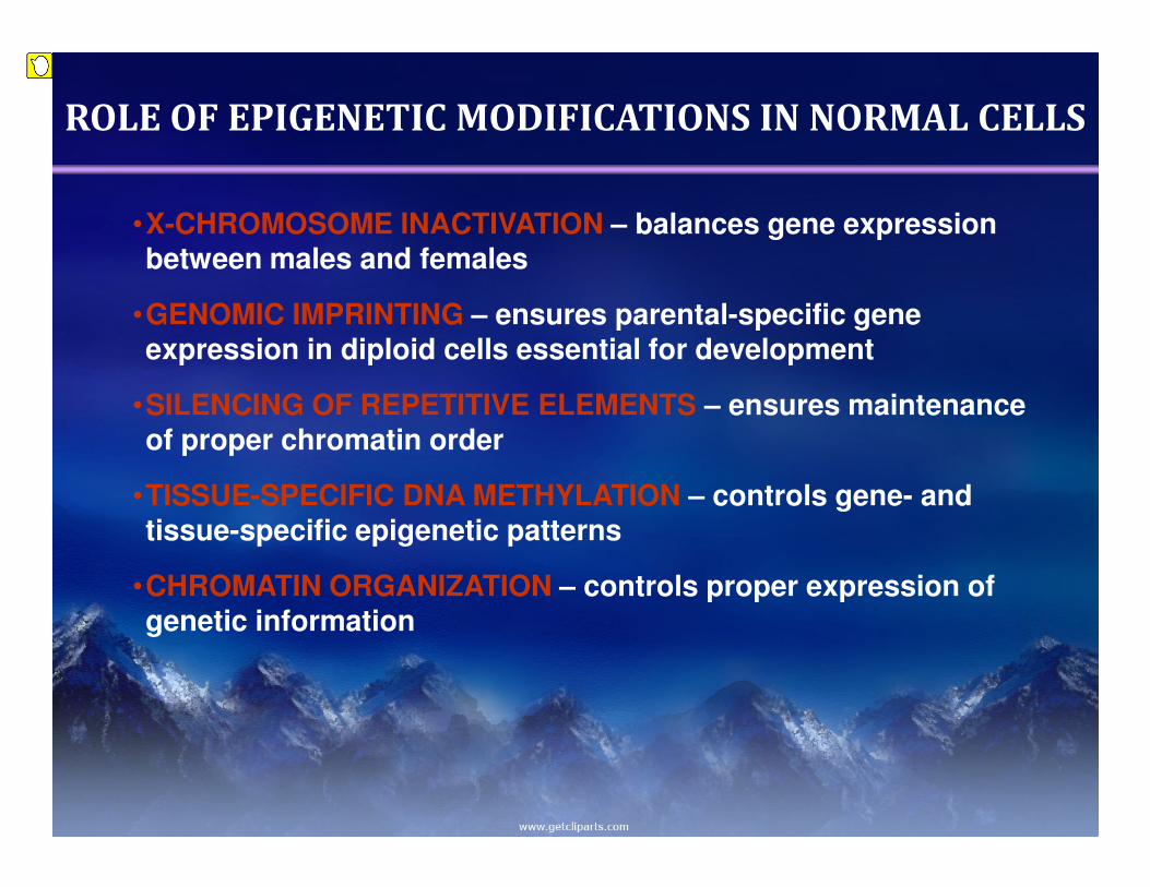
ROLE OF EPIGENETIC MODIFICATIONS IN NORMAL CELLS
•X-CHROMOSOME INACTIVATION – balances gene expression between males and females
•GENOMIC IMPRINTING – ensures parental-specific gene expression in diploid cells essential for development
•SILENCING OF REPETITIVE ELEMENTS – ensures maintenance of proper chromatin order
•TISSUE-SPECIFIC DNA METHYLATION – controls gene- and tissue-specific epigenetic patterns
•CHROMATIN ORGANIZATION – controls proper expression of genetic information
Presenter
Sticky Note
First of all, the most important role, of course, is the control of proper expression of genetic information. This control can be site-specific; this control can be tissue-specific, and it's being facilitated by epigenetics. Epigenetics is also responsible for silencing of foreign DNA, responsible for silencing of repetitive elements which comprise a large part of our genome. So what happens when epigenetic, when certain environmental or other toxicants disturb this balance in the cell?
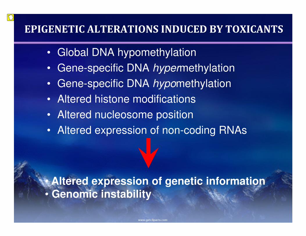
EPIGENETIC ALTERATIONS INDUCED BY TOXICANTS
• Global DNA hypomethylation
• Gene-specific DNA hypermethylation
• Gene-specific DNA hypomethylation
• Altered histone modifications
• Altered nucleosome position
• Altered expression of non-coding RNAs
• Altered expression of genetic information
• Genomic instability
Presenter
Sticky Note
Well, a list of different epigenetic alterations that are described up to now. The most studied are the global DNA hypermethylation, which is usually associated with changes in gene methylation, which can be either/or, hyper or hypomethylation. Also, it is associated with altered histone modifications, altered expression of non-coding RNAs. Altogether, those changes leads to altered expression of genetic information, and subsequently can result in genomic instability. Today, I will be talking a little bit about drugs, a little bit about environmental toxicants, and I will show you by example how epigenetics can underlie the inter-individual difference in response to certain toxicants.
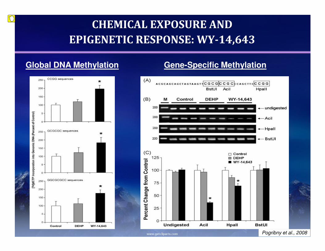
CHEMICAL EXPOSURE AND
EPIGENETIC RESPONSE: WY-14,643
Global DNA Methylation Gene-Specific Methylation
Pogribny et al., 2008
Presenter
Sticky Note
I will start talking about WY-14643, which is apparently a known drug and peroxisome profilerator. This drug is also known and considered to be a carcinogen in the rodent model, with a tumor formation site in liver. In a comparatible study where we used DHP, which is another known peroxisome proliferator, and WY, administration of dyes with those chemicals have led to development of global demethylation in liver tissue of animals that were treated with WY. For evaluation of global DNA methylation, we used cytosine extension method, which is based mainly on quantification of unmethylated CPG site. So exactly when the higher the bar is, the more pronounced demethylation in the genome is. On the right side of the slide, I wanted to show you the example of gene-specific alteration in methylation, a related promoter of GSGP-1, which is usually heavily methylated in rodent liver. However, treatment with WY resulted in significant loss of DNA methylation in promoter, in two CPG sites as a promoter of GSGP-1.
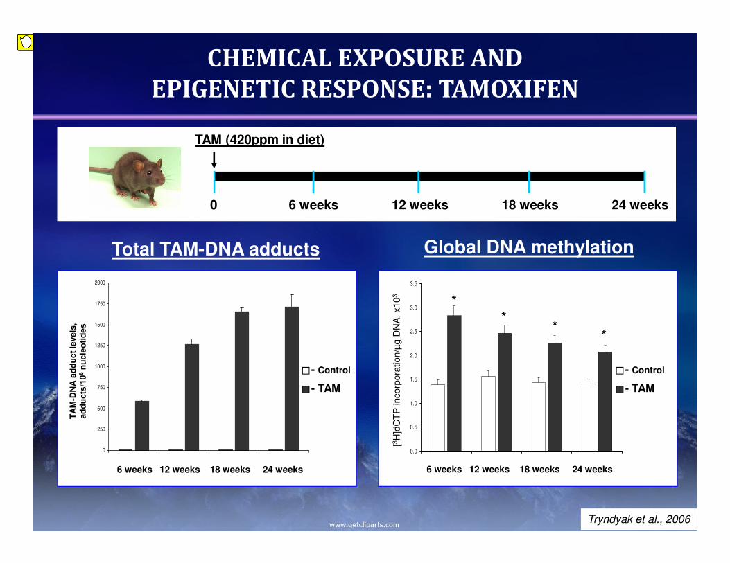
CHEMICAL EXPOSURE AND
EPIGENETIC RESPONSE: TAMOXIFEN
0
250
500
750
1000
1250
1500
1750
2000
Total TAM-DNA adducts
TA
M-D
NA
ad
du
ct
leve
ls,
ad
du
cts
/10
8n
uc
leo
tid
es
- Control
- TAM
6 weeks 12 weeks 18 weeks 24 weeks
TAM (420ppm in diet)
0 6 weeks 12 weeks 18 weeks 24 weeks
0.0
0.5
1.0
1.5
2.0
2.5
3.0
3.5
[3H
]dC
TP
incorp
ora
tion/µ
g D
NA
, x10
3
Global DNA methylation
*
**
*
- Control
- TAM
6 weeks 12 weeks 18 weeks 24 weeks
Tryndyak et al., 2006
Presenter
Sticky Note
Next, I'll be talking about tamoxifen, which is a well-known drug for treatment of breast cancer. It also has been used for as a preventive drug and preventive for recurrence of breast cancer. It is also known as a potent liver carcinogen in the rodent model, and it is also known to exert very strong genotoxic effects on rat liver. To evaluate the epigenetic alterations that can be associated with the administration of tamoxifen, we have used Fischer 344 rats, and they received a diet with tamoxifen for 6, 12, 18 and 24 weeks, to evaluate genotoxic potential of tamoxifen, with a related level of DNA adducts in liver tissue of these rats, and you can see significant accumulation of DNA adducts throughout the treatment. What was really very interesting to see is that in parallel to the genotoxicity that tamoxifen exerts, it possesses also very strong epigenotoxic function. As you can see, significant loss of global DNA methylation, also in all four time frames. Of course, you will ask me how do I know that this hypomethylation is not the cause of DNA damage, that it's not associated with tamoxifen DNA adducts? So to answer that question, we have performed the following study.
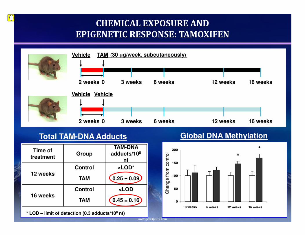
0.45 ± 0.16TAM
<LODControl16 weeks
0.25 ± 0.09TAM
<LOD*Control12 weeks
TAM-DNA adducts/108
ntGroup
Time of treatment
* LOD – limit of detection (0.3 adducts/108 nt)
- Control
- TAM
Total TAM-DNA Adducts Global DNA Methylation
Vehicle Vehicle
2 weeks 0 3 weeks 6 weeks 12 weeks 16 weeks
Vehicle TAM
2 weeks 0 3 weeks 6 weeks 12 weeks 16 weeks
CHEMICAL EXPOSURE AND
EPIGENETIC RESPONSE: TAMOXIFEN
0
50
100
150
200
8 17 38 46
Ch
an
ge
fro
m c
on
tro
l
**
3 weeks 6 weeks 12 weeks 16 weeks
(30 µg/week, subcutaneously)
Presenter
Sticky Note
We have the same Fischer 344 rats, who are treated with a very, very low dose of tamoxifen, just for two weeks, with a dose of 30 micrograms per week introduced subcutaneously. The table on the right side of the slide clearly depicts that accumulation of DNA adducts was really very low, just about the level of detection. However, significant and dynamic loss of global DNA methylation in the liver tissue of the threat would suggest that epigenetic alterations are not necessarily a cause of genetic alterations, but can exhibit their own independent pattern.
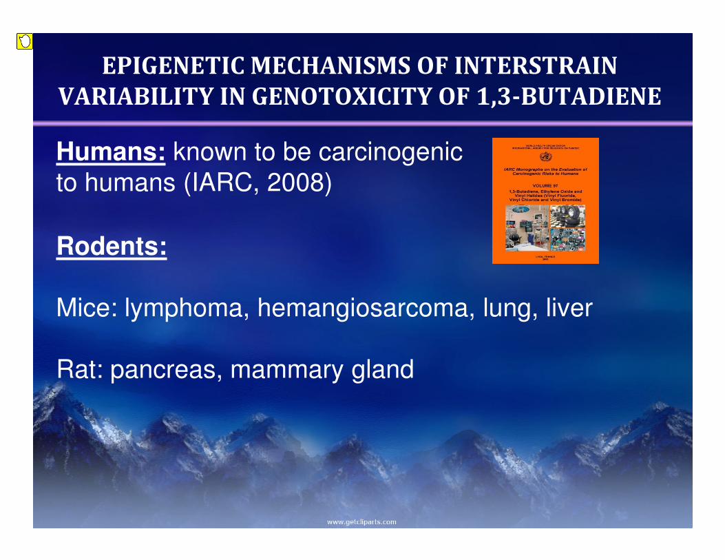
EPIGENETIC MECHANISMS OF INTERSTRAIN
VARIABILITY IN GENOTOXICITY OF 1,3-BUTADIENE
Humans: known to be carcinogenic
to humans (IARC, 2008)
Rodents:
Mice: lymphoma, hemangiosarcoma, lung, liver
Rat: pancreas, mammary gland
Presenter
Sticky Note
Of course it's interesting how epigenetic parameters, how epigenetics in general, can affect the inter-individual susceptibility to various chemicals or toxicants, and to study that, as a chemical choice we have chosen butadiene, which is a ubiquitous environmental toxicant. It is also known to be a carcinogen, both in humans and in rodents.
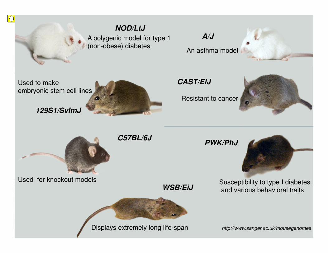
A/J
129S1/SvImJ
C57BL/6J
CAST/EiJ
NOD/LtJ
PWK/PhJ
WSB/EiJ
Displays extremely long life-span
A polygenic model for type 1
(non-obese) diabetes
Resistant to cancer
Used to make
embryonic stem cell lines
An asthma model
Used for knockout models Susceptibility to type I diabetes
and various behavioral traits
http://www.sanger.ac.uk/mousegenomes
Presenter
Sticky Note
To relate these differences, we have used the inter-strained panel of mice. This was exactly what Dr. Watkins was talking in the morning. This mice has progenitor strains for this collaborative cross. These mice were computationally predicted to have the most diverse genomes. As you can see, every mouse has some unique feature about it. Some are susceptible to development of cancer; some are resistant; some display real extremely long life span.
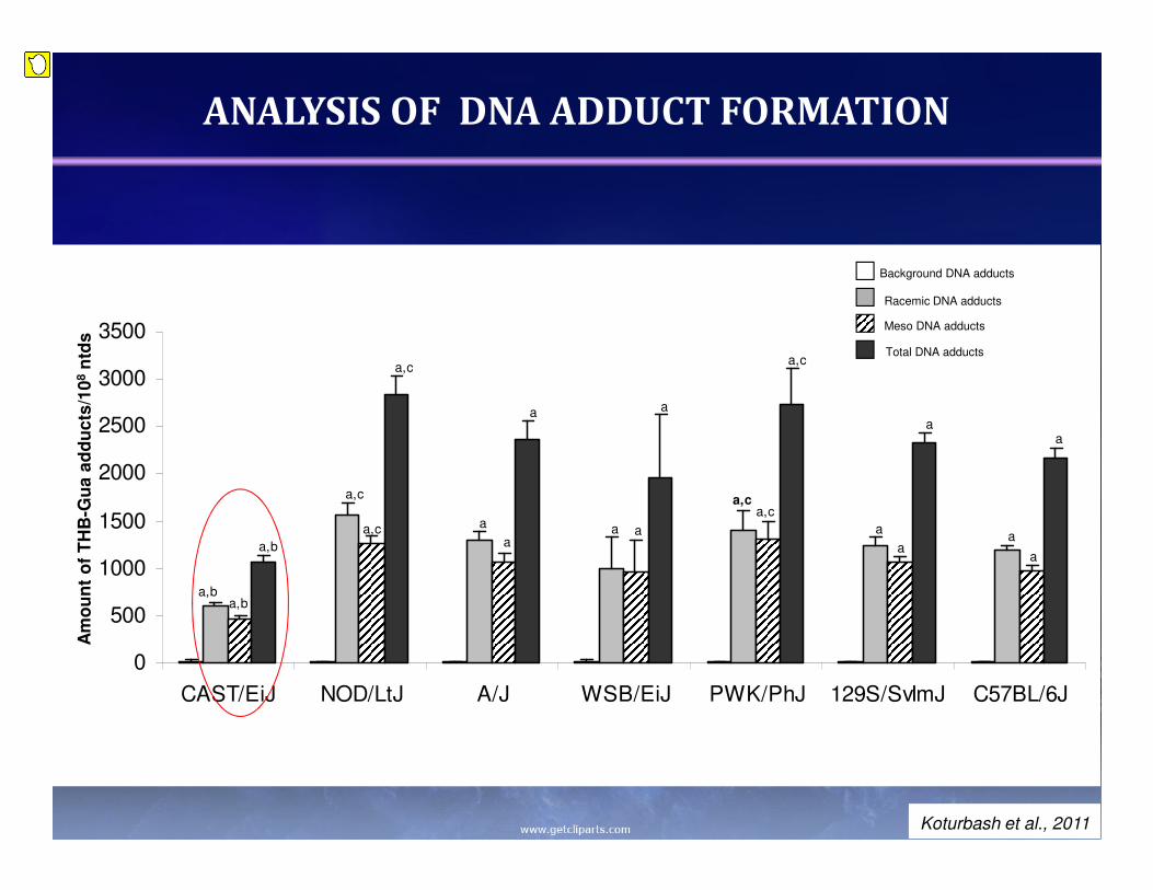
So we treated these animals with butadiene for a very short period of time, just for two weeks, and first of all, we analyzed the level of DNA adducts in the liver tissue of those animals. It was really very interesting to see that those different mice, even though they were treated with the same dose, exhibited different levels of accumulation of butadiene DNA adducts in the liver. Especially it was interesting that to see that test A/J mice developed two to three times less DNA adducts in comparison with other animals.
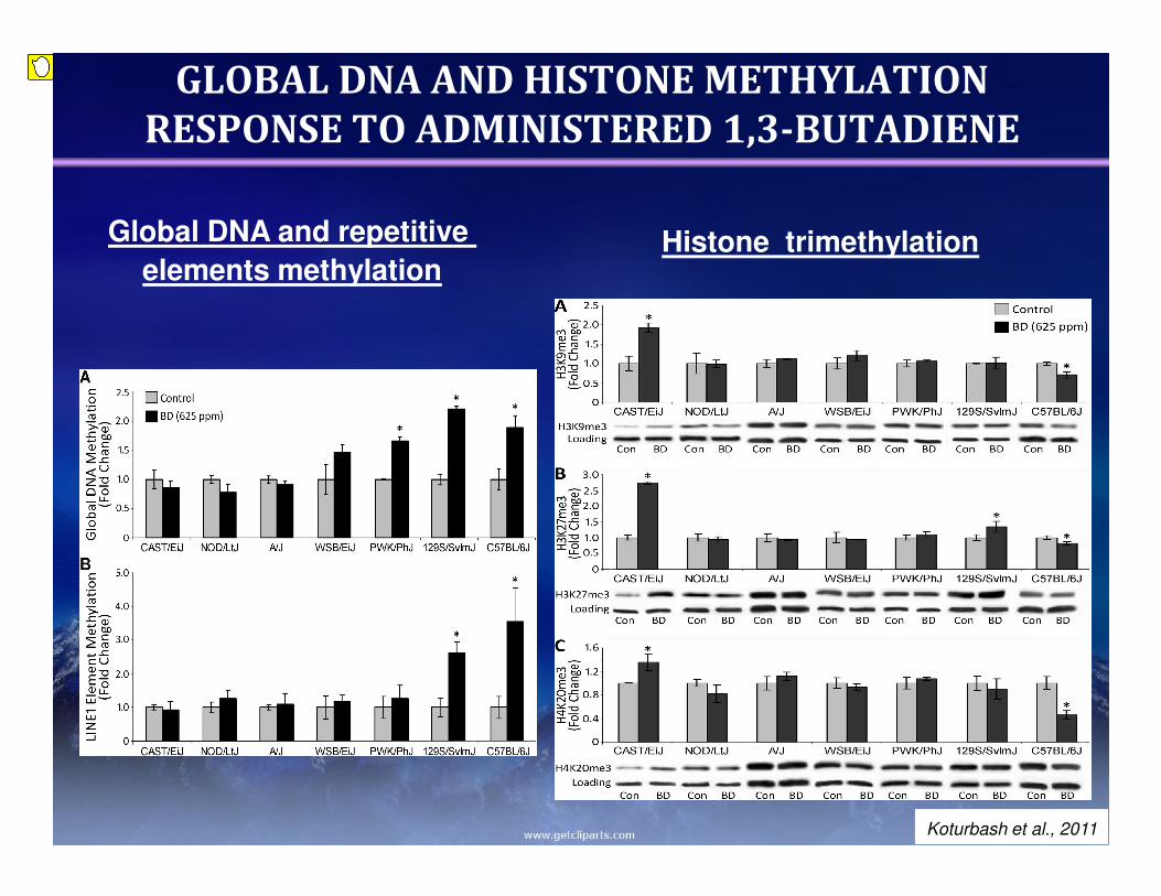
GLOBAL DNA AND HISTONE METHYLATION
RESPONSE TO ADMINISTERED 1,3-BUTADIENE
Global DNA and repetitive
elements methylationHistone trimethylation
Koturbash et al., 2011
Presenter
Sticky Note
To see how epigenetic goes along with this, we have related to the global DNA methylation and histone modification in livers of these animals. The panel on the right side shows changes, significant differences in DNA methylation patterns after exposure to butadiene. The top graph represents results obtained with using cytosine extension assay, and the bottom graph apparently evaluation of methylation profile of a line 1 repetitive element, which comprised about almost a quarter of the mouse genome, and therefore considered to be a good marker of genomic methylation. And finally, we have also related the methylation status of three histone marks that are responsible for formation of this silent heterochromatin, which is, lies in 9, and lies in 27 histone-3, and lies in 20 on histone-4. It was very interesting to see that this low number of DNA adducts in test A/J mice was correlated with a significant increase in trimethylation of all three histone marks, whereas C57BL/6 mice, that exhibited very significant increase in DNA adducts, they had ‑‑ their genome was severely demethylated. Their histones were also severely demethylated as well.
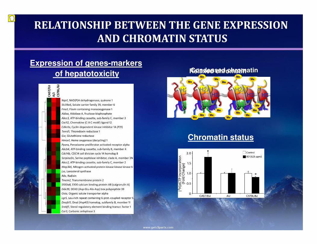
RELATIONSHIP BETWEEN THE GENE EXPRESSION
AND CHROMATIN STATUS
Relaxed chromatin
H2AH3
H4H2B
H2AH3
H4H2B
Condensed chromatin
H2AH3
H4H2B
H2AH3
H4H2B
Me Me
MeMeMe
Me
Me
MeMe
Me
Me
Me
Me
Me Me
Expression of genes-markers
of hepatotoxicity
Chromatin status
Presenter
Sticky Note
To further understand these mechanisms, we can show the test mice, C57BL/6 and A/J mice as a, let's say negative, a resistant strain, as far as we did not see any epigenetic changes in these animals. We evaluated expression of a panel of genes that are generally accepted as markers of hepatotoxicity, and you can see those clearly. A majority of genes are up-regulated in A/J mice. There is majority of genes down-regulated in C57BL/6 mice. However, there are really very minor changes in the livers of test A/J mice. So of course we clearly need to explain why it is such a big difference, and our initial hypothesis was that these changes can be attributed to formation, to the plasticity of chromatin. Because in the case of the relaxed chromatin, that what we expected to see in C57BL/6 mice, this chromatin does not have these trimethylation marks. Therefore, the chromatin is being relaxed and the DNA in there is open. It is open for any possible DNA-damaging agent. In the case when the histone trimethylation marks are present, the chromatin is getting more condensed, and therefore prevents this DNA from possible DNA damage. So to test this hypothesis, we have developed a new method for evaluation of chromatin status, which it will take about five minutes to explain. So for those who are interested, I will gladly show about it after the conference. But now I will just show you the end result, where you can see that the chromatin of test A/J mice was really condensed in response to butadiene, thus probably preventing it from the formation of those DNA adducts. However, the chromatin in C57BL/6 mice was reacting absolutely in opposite direction, being more decondensed, more to the U-chromatin status, suggesting the possible best way why there is way higher accumulation of DNA adducts in the livers of those mice.
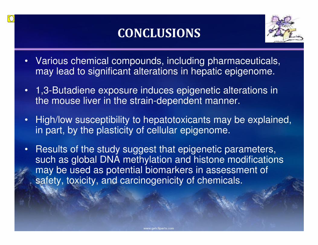
CONCLUSIONS
• Various chemical compounds, including pharmaceuticals, may lead to significant alterations in hepatic epigenome.
• 1,3-Butadiene exposure induces epigenetic alterations in the mouse liver in the strain-dependent manner.
• High/low susceptibility to hepatotoxicants may be explained, in part, by the plasticity of cellular epigenome.
• Results of the study suggest that epigenetic parameters, such as global DNA methylation and histone modifications may be used as potential biomarkers in assessment of safety, toxicity, and carcinogenicity of chemicals.
Presenter
Sticky Note
In conclusion, first of all it is clear that various chemical compounds, including pharmaceuticals, in parallel to the genotoxicity, can also exert that epigenotoxic effect on hepatic epigenome. Exposure to butadiene have shown that epigenetic alterations in the mouse liver are really strain-dependent. This high or low susceptibility to various chemicals can be explained, at least in part, by the plasticity of the epigenome, and in this case, particularly the plasticity of chromatin, to form either more or less condensed structure. And finally, the results of the study clearly suggest that in the future, epigenetic parameters, such as global DNA methylation and histone modifications, can be used as the biomarkers for assessment of potential safety to toxicity and carcinogenicity of chemicals.
Thank you!
Dr. Igor P. Pogribny
Dr. Frederick A. Beland
Dr. Volodymyr Tryndyak
Mona Churchwell
National Center for
Toxicological Research
University of North Carolina,
Chapel Hill
Dr. Ivan Rusyn
Dr. James A. Swenberg
Dr. Wanda Bodnar
Dr. Fernando Pardo-Manuel de Villena
Dr. Kenneth Sexton
Jessica SorrentinoUniversity of Minnesota
Dr. Danuta Malejka-Giganti
Oak Ridge Institute for Science and Education
Presenter
Sticky Note
I want to thank all the people who contributed to that research. Dr. Pogribny, Dr. Beland from NCTR, and the team of Dr. Ivan Rusyn from the University of North Carolina in Chapel Hill, and Oak Ridge Institute for Science and Education for financial support. Thank you for your attention.