Embed Size (px)
Citation preview

“ROLE OF DNMT IN CANCER”
A THESIS SUBMITTED IN PARTIAL FULFILLMENT OF THE REQUIREMENTS
FOR THE DEGREE OF
MASTER OF SCIENCE IN
LIFE SCIENCE
SUBMITTED TO
NATIONAL INSTITUTE OF TECHNOLOGY, ROURKELA
BY
SURYANARAYAN BISWAL ROLL NO. 409LS2034
UNDER THE SUPERVISION OF
Associate PROF. Samir k. patra
DEPARTMENT OF LIFE SCIENCE NATIONAL INSTITUTE OF TECHNOLOGY
ROURKELA-769 008, ODISHA, INDIA

i
DEPARTMENT OF LIFE SCIENCE
NATIONAL INSTITUTE OF TECHNOLOGY ROURKELA
CERTIFICATE This is to certify that the thesis entitled “Role of DNMT in Cancer” which is being submitted
by Mr. Suryanarayan Biswal, Roll No. 409LS2034, for the award of the degree of Master of
Science in Life Science from National Institute of Technology, Rourkela, is a record of
bonafide research work, carried out by him under my supervision. The results embodied in
this thesis are new and have not been submitted to any other university or institution for the
award of any degree or diploma.
To the best of my knowledge, Mr. Biswal bears a good moral character and is mentally and
physically fit to get the degree.
Dr. SAMIR KUMAR PATRA
Associate Professor and Head
Department of Life Science
National Institute of Technology
Rourkela – 769 008
Odisha, India
ii
DECLARATION
I, Suryanarayan Biswal hereby declare that this project report entitled “Role of DNMT in Cancer” is
the original work carried out by me under the supervision of Associate Prof. Samir K. Patra,
Department of Life Science, National Institute of Technology Rourkela (NITR), Rourkela and the
present work or any other part thereof has not been presented to any other University or Institution for
the award of any other degree.
Suryanarayan Biswal

iii
ACKNOWLEDGEMENTS
I wish to express my deepest sense of gratitude to my supervisor Dr. Samir K. Patra,
Associate Professor, Department of Life Science, National Institute of Technology, Rourkela
for his valuable guidance, assistance and time to time inspiration throughout my project.
I am very much grateful to Prof. P. C. Panda, Director, National Institute of Technology,
Rourkela for providing excellent facilities in the Institute for carrying out research.
I would like to take the opportunity to acknowledge quite explicitly with gratitude my debt to
all the Professors and Staff, Department of Life Science, National Institute of Technology,
Rourkela for his encouragement and valuable suggestions during my project work.
I would like to give heartfelt thanks to Ms. Moonmoon Deb, all the other PhD scholars and
second yr. friends for their inspirative support throughout my project work
Finally I was highly great full to my parent for their continued moral support.
And to all mighty, who made all things possible………..
(SURYANARAYAN BISWAL)
iv
CONTENTS Page No.
PREFACE-“DNA: THE SECRET OF LIFE”………………. 1
INTRODUCTION…………………………………………… 2
What is a Cancer? ..............................…........………... 2
REVIEW OF LITERATURE……………………………….. 3-8
Epigenetics …………………………………………………... 3
DNA Methylation …………………………………………..
3
DNA methylation as a gene silencing mechanism ….. 5
DNA Methyl-transferases (DNMTs) ………………....... 6
DNMTs & tumor suppressor genes (TSGs) in cancer 8
OBJECTIVES……………………………………………….. 9
MATERIALS AND METHODS…………………………… 10-12
RESULTS & DISCUSSON…………………………………. 13-16
CONCLUSION……………………………………………… 17
FUTURE WORK……………………………………………. 18
“Role of DNMT in Cancer”
ABSTRACT
DNA methylation is a biochemical process catalyzed by enzyme DNA Methyltransferases
(DNMTs). In most of the carcinogenesis DNMTs are over expressed and aberrant genomic
DNA methylation pattern (genome wide hypomethylation and regional hypermethylation) are
being observed. One of such consequences results in hypermethylation of Tumor Suppressor
Genes. In different cancerous, different DNMTs show significantly elevated expression than
their normal cells. In our study we have investigated relative expression of DNMTs and TSGs
in normal and cancerous tissue. We found that DNMT3A expression level is comparatively
more than the other DNMTs and it may cause the transcriptional repression of TSGs,
particularly in lymph Node cancer.
Key Words: DNA Methylation, DNA Methyltransferases, Tumor Suppressor Genes, Lymph
Node Cancer.

“Role of DNMT in Cancer”
Page 1
PREFACE-“DNA: THE SECRET OF LIFE”
On February the 28th 1953, James Watson and Francis Crick deciphered the structure of
deoxyribonucleic acid, DNA1, which was later published in the journal Nature [1]. The DNA
X-ray diffraction pictures made by Rosalind Franklin were essential for this discovery, as
they provided several of the vital helical parameters [2]. The four separate building blocks of
the DNA molecule, the nucleotides adenine, cytosine, guanine, and thymine, had been
isolated and characterized several years before this immensely important discovery. The
modified cytosine base, 5-methylcytosine, was first recognized in 1948 [3] and was later
identified as a central element in the field of epigenetics2. The DNA contains the genetic
instruction specifying how to assemble protein molecules, which are the building blocks of
each phenotype. Indeed, Crick described the DNA molecule as “the secret of life”, and today
several fields of research address DNA directly or indirectly. The most recent breakthrough
in the history of DNA research has been the sequencing of the human genome [4, 5], which
has heralded a new era for genetic as well as epigenetic research. The challenge now, is to
understand the molecular mechanisms that allow specific genes and gene families to be
selectively expressed in normal development and how aberrations in this process can lead to
disease. In addition to well-described genetic mechanisms, imbalances in the epigenetic
control of gene expression can profoundly alter this finely tuned machinery. Epigenetic
changes are now recognized to have a lead role in cancer development [6]. Simultaneously,
such changes have been hypothesized to be a master key to more effective ways of
diagnosing, monitoring, and treating cancer [7]. On our way to molecular assisted medicine,
we need to explore this in detail in order to get a better understanding of the role of epigenetic
in cancer development, which is necessary to fully master these new tools.

“Role of DNMT in Cancer”
Page 2
INTRODUCTION
What is a Cancer?:
Organisms are maintained by homeostasis, a finely tuned balance between cell proliferation
and cell death. When the homeostasis is disturbed, either by an increased proliferation rate or
a decrease in cell death, a tumor might occur, which can further progress into a cancer.
Tumor development is most commonly described as natural selection followed by clonal
expansion, resulting in monoclonal tumors originating from the progeny of a single cell [8].
Aberrations that confer growth advantages to the cell will accumulate during the clonal
selection process. These changes are consequences of several processes: 1) activation of
proto-oncogenes, rendering the gene constitutively active or active under conditions in which
the wild type gene is not, 2) inactivation of tumor suppressor genes, reducing or abolishing
the activity of the gene product, 3) alteration of repair genes, which normally keep genetic
alterations to a minimum. Genomic analyses focusing on structural and numerical aberrations
of chromosomes have long suggested that cancer is, in essence, a genetic disease [9].
The first cancer-specific genetic aberration described was the Philadelphia chromosome in
patients with chronic myeloid leukemia. This was initially identified in 1960 by Nowell and
Hungerford and was later demonstrated to be the result of a translocation between
chromosomes 9 and 22 [10]. Today, numerous mutations at the chromosome and DNA level
have been described in hematological as well as solid tumors [10, 11]. The Mitelman
Database of Chromosome Aberrations in Cancer lists the chromosomal aberrations of more
than 47,000 tumors [12], and the IARC mutation database have recorded 21,587 somatic
mutations of the tumor suppressor gene TP53 [13].
During the last decades, several lines of evidence have proven the importance also of
epigenetic modifications in tumorigenesis. Indeed, epigenetic changes are now recognized to
be at least as common as genetic changes in cancer [6]. Moreover, epigenetic changes often
precede and appear to be essential for several genetic events that drive tumor progression.

“Role of DNMT in Cancer”
Page 3
REVIEW OF LITRETURE
Epigenetics:
The term „epigenetic‟ was coined by Conrad Waddigton in 1940s to describe “the
interactions of genes with their environment, which bring the phenotype into being” [14].
This early usage of the term has been effectively displaced during the last decades and today
epigenetic inheritance is defined as cellular information, other than the DNA sequence itself,
that is heritable during cell division i.e. “The sum of the alterations to the chromatin template
that collectively establish and propagate different patterns of gene expression (transcription)
and silencing from the same genome.” [15]. Epigenetics affect the transcription in the cell,
thereby controlling gene expression and abnormal epigenetic changes can have serious
effects for the organism. Most epigenetic changes only occur within the course of one
individual organism‟s lifetime, but, if a mutation in the DNA has been caused in gamete that
result in fertilization, then some epigenetic changes are inherited from one generation to the
next - “Lamarckism”. We can very roughly divide epigenetics into three substantially
overlapping categories: DNA methylation, genomic imprinting, and histone modification.
Among these mechanisms, DNA methylation is the most studied, and is the main focus of
this thesis.
DNA Methylation:
DNA methylation is a covalent modification of nucleotides and the most frequently
methylated nucleotide in the human genome is cytosine subsequently followed by a guanine
in the DNA sequence, constituting a CpG dinucleotide. The cytosine is methylated in the C-5
position by a family of DNA (cytosine-5) methyltransferases (DNMTs) using the universal
methyl donor Sadenosyl- L-methionine (SAM) (Figure.1).
Figure.1: Methylation of cytosine catalyzed by DNMTs.
“Role of DNMT in Cancer”
Page 4
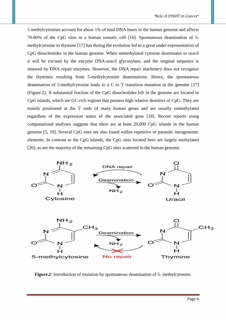
5 methylcytosines account for about 1% of total DNA bases in the human genome and affects
70-80% of the CpG sites in a human somatic cell [16]. Spontaneous deamination of 5-
methylcytosine to thymine [17] has during the evolution led to a great under-representation of
CpG dinucleotides in the human genome. When unmethylated cytosine deaminates to uracil
it will be excised by the enzyme DNA-uracil glycosylase, and the original sequence is
restored by DNA repair enzymes. However, the DNA repair machinery does not recognize
the thymines resulting from 5-methylcytosine deaminations. Hence, the spontaneous
deamination of 5-methylcytosine leads to a C to T transition mutation in the genome [17]
(Figure.2). A substantial fraction of the CpG dinucleotides left in the genome are located in
CpG islands, which are GC-rich regions that possess high relative densities of CpG. They are
mainly positioned at the 5' ends of many human genes and are usually unmethylated
regardless of the expression status of the associated gene [18]. Recent reports using
computational analyses suggests that there are at least 29,000 CpG islands in the human
genome [5, 19]. Several CpG sites are also found within repetitive or parasitic intragenomic
elements. In contrast to the CpG islands, the CpG sites located here are largely methylated
[20], as are the majority of the remaining CpG sites scattered in the human genome.
Figure.2: Introduction of mutation by spontaneous deamination of 5- methylcytosine.

“Role of DNMT in Cancer”
Page 5
DNA methylation as a gene silencing mechanism:
The first connection between DNA methylation and gene expression was published more
than 25 years ago [21]. Two mechanisms have been proposed to account for transcriptional
repression via DNA methylation. In the first mechanism, DNA methylation directly inhibits
the binding of transcription factors (TFs) such as AP-2, c-Myc/Myn, E2F and NFkB to their
binding sites within promoter sequence. In this mechanism, CpG dinucleotides have to be
present within the binding site of TFs, which are sensitive to methylation of CpG
dinucleotides (Figure.3: A & B).
Figure.3: Repression of transcription via CpG dinucleotide methylation. (Fujita N et.al.2000)
The second mode of repression includes a binding of proteins specific for m5CpG
dinucleotides to methylated DNA. Methylated DNA recruits m5CpG-binding (MeCP) and
m5CpG-binding domain (MBD) proteins. MeCP1 and MeCP2 bind specifically to
methylated DNA in whole genome and form spatial obstacle that unable binding of TFs to
promoter sequences (Figure.3: C). MeCP1 represses transcription of specific genes, which are
controlled by densely methylated promoters containing more than ten m5CpG dinucleotides.
MeCP2 can bind to a single symmetrically located m5CpG pair in two DNA strands [22].
These changes were inherited by the next generation of cells and it became obvious that
reducing DNA methylation reactivated certain genes, allowing the development of new cells
from the original embryo. Today, two different pathways have been described for the
inactivation of gene transcription by DNA methylation:
“Role of DNMT in Cancer”
Page 6
1) Methyl-CpGs can repel transcription factors directly by being present in the
transcription factor binding sequence. Although regulation by such a mechanism in vivo is
relatively rare, some transcription factors, like Ets-1 [23] and the boundary element factor
CTCF [24] are unable to bind DNA if the cytosines in their recognition sites are methylated.
2) DNA methylation can recruit proteins that bind methylated CpGs and subsequent
inhibit transcription by remodelling the chromatin structure.
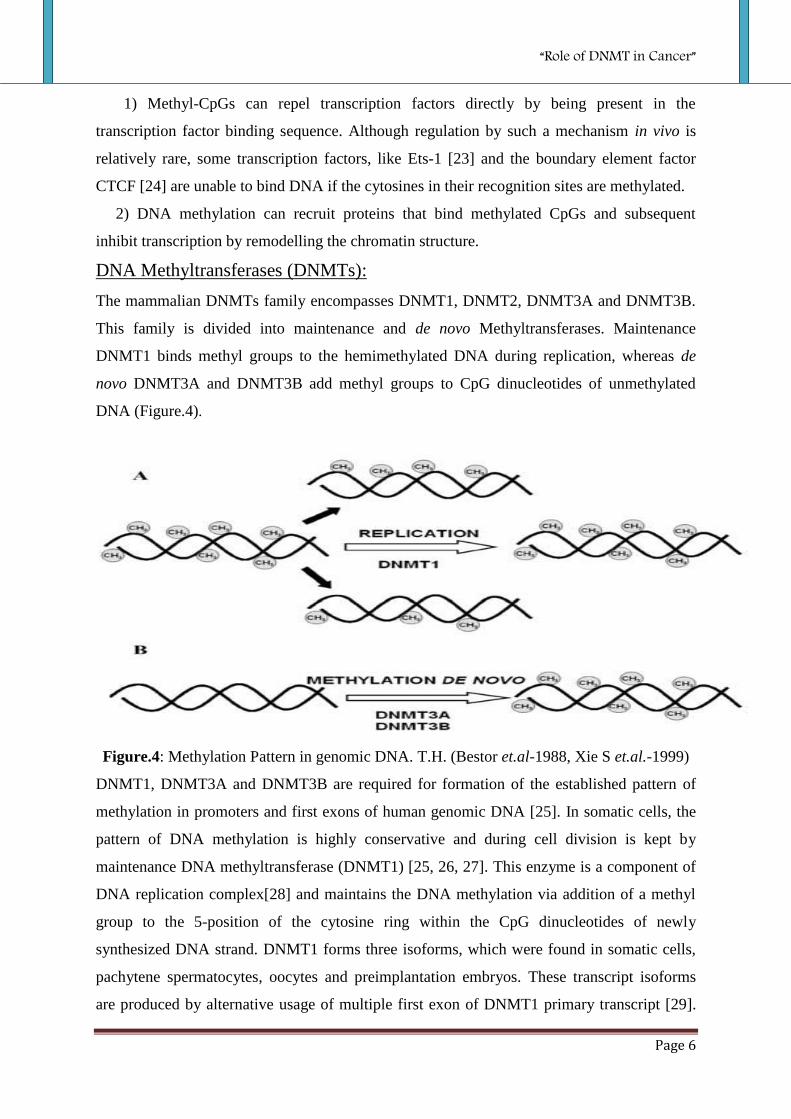
DNA Methyltransferases (DNMTs):
The mammalian DNMTs family encompasses DNMT1, DNMT2, DNMT3A and DNMT3B.
This family is divided into maintenance and de novo Methyltransferases. Maintenance
DNMT1 binds methyl groups to the hemimethylated DNA during replication, whereas de
novo DNMT3A and DNMT3B add methyl groups to CpG dinucleotides of unmethylated
DNA (Figure.4).
Figure.4: Methylation Pattern in genomic DNA. T.H. (Bestor et.al-1988, Xie S et.al.-1999)
DNMT1, DNMT3A and DNMT3B are required for formation of the established pattern of
methylation in promoters and first exons of human genomic DNA [25]. In somatic cells, the
pattern of DNA methylation is highly conservative and during cell division is kept by
maintenance DNA methyltransferase (DNMT1) [25, 26, 27]. This enzyme is a component of
DNA replication complex[28] and maintains the DNA methylation via addition of a methyl
group to the 5-position of the cytosine ring within the CpG dinucleotides of newly
synthesized DNA strand. DNMT1 forms three isoforms, which were found in somatic cells,
pachytene spermatocytes, oocytes and preimplantation embryos. These transcript isoforms
are produced by alternative usage of multiple first exon of DNMT1 primary transcript [29].

“Role of DNMT in Cancer”
Page 7
DNMT3A and DNMT3B enzymes are responsible for establishment of new methylation
pattern in genomic DNA (Figure.4.B) [30, 31 & 25].
Mammalian DNMT1, DNMT3A and DNMT3B are composed of the N-terminal regulatory
and the C-terminal catalytic domains that are linked by a short fragment of repeated GK
dipeptides (Figure.5). The N-terminal domains of DNMT1 and DNMT3B do not exhibit
extensive homolog of primary structure. These differences are responsible for distinct
functions of N-regions in these enzymes. The DNMT1 requires interaction between the N-
and C-terminal domains for catalytic activity. Separated C-terminal domain of DNMT1 is
catalytically inactive despite the presence of the highly conserved sequence motifs typical of
active DNMTs. In contrast to DNMT1, C-terminal domain of DNMT3A and DNMT3B is
active without interaction with their N-regulatory regions. These differences between
DNMT1 and de novo DNMTs indicate significantly disparate mechanism that regulate
methylation activity of these enzymes.
Figure.5: Members of mammalian DNMTs family. (Weisenberger DJ, et.al.-2004)
The N-terminal domain possesses nuclear localization signal sequence (NLS) responsible for
localization of DNMTs in the nucleus. The N-fragment of DNMTs also contains proliferating
cell nuclear antigen binding domain (PDB), a cysteine rich zinc finger DNA binding motif
(ATRX), and polybromo homology domain (PHD) targeting DNMTs to the replication foci.
However, PWWP tetrapeptide is only present in N-terminal domains of DNMT3A and
DNMT3B and interact with histones [32]. The C-terminal domain contains six conservative
motifs I, IV, VI, VIII, IX and X. Motifs I and X form S-adenosylomethionine binding site,
motif IV binds cytosine at the active site, motif VI possesses glutamyl residue donating
protons, and motif IX maintains the structure of the target recognition domain (TRD) usually
located between motifs VIII and IX, that makes base-specific contacts in the major groove of
DNA [33, 34, 35, 36].
“Role of DNMT in Cancer”
Page 8
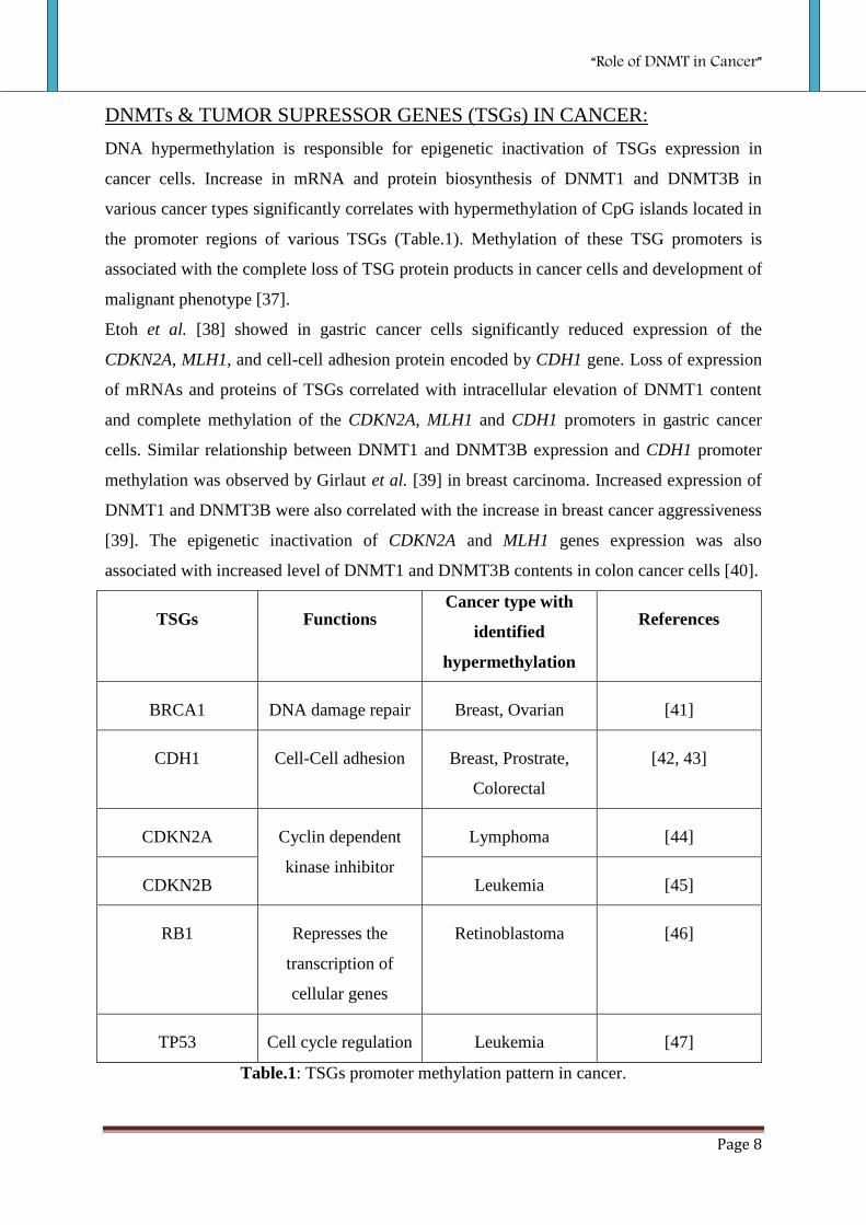
DNMTs & TUMOR SUPRESSOR GENES (TSGs) IN CANCER:
DNA hypermethylation is responsible for epigenetic inactivation of TSGs expression in
cancer cells. Increase in mRNA and protein biosynthesis of DNMT1 and DNMT3B in
various cancer types significantly correlates with hypermethylation of CpG islands located in
the promoter regions of various TSGs (Table.1). Methylation of these TSG promoters is
associated with the complete loss of TSG protein products in cancer cells and development of
malignant phenotype [37].
Etoh et al. [38] showed in gastric cancer cells significantly reduced expression of the
CDKN2A, MLH1, and cell-cell adhesion protein encoded by CDH1 gene. Loss of expression
of mRNAs and proteins of TSGs correlated with intracellular elevation of DNMT1 content
and complete methylation of the CDKN2A, MLH1 and CDH1 promoters in gastric cancer
cells. Similar relationship between DNMT1 and DNMT3B expression and CDH1 promoter
methylation was observed by Girlaut et al. [39] in breast carcinoma. Increased expression of
DNMT1 and DNMT3B were also correlated with the increase in breast cancer aggressiveness
[39]. The epigenetic inactivation of CDKN2A and MLH1 genes expression was also
associated with increased level of DNMT1 and DNMT3B contents in colon cancer cells [40].
TSGs Functions Cancer type with
identified
hypermethylation
References
BRCA1 DNA damage repair Breast, Ovarian [41]
CDH1 Cell-Cell adhesion Breast, Prostrate,
Colorectal
[42, 43]
CDKN2A Cyclin dependent
kinase inhibitor
Lymphoma [44]
CDKN2B Leukemia [45]
RB1 Represses the
transcription of
cellular genes
Retinoblastoma [46]
TP53 Cell cycle regulation Leukemia [47]
Table.1: TSGs promoter methylation pattern in cancer.

“Role of DNMT in Cancer”
Page 9
OBJECTIVE
To clarify the role of DNMTs in the aberrant promoter hypermethylation of TSGs in various
human cancers, the expression levels of a number of DNMTs in cancer tissues were
examined and were correlated with the findings of the promoter methylation status of p53
TSGs which is commonly involved in cellular regulatory pathways. The expressions of the
DNMTs, DNMT1, DNMT2, DNMT3A and DNMT3B were examined, since these enzymes
have a reported role in the maintenance of the genome methylation status integrity and have
been implicated in the transcriptional regulatory changes of human cancer.
So our main objective was “Comparative Analysis of DNA Methyltransferase (DNMTs)
Expression and Promoter Hypermethylation of Tumor Suppressor (TSGs) Genes in
Normal and various Human Cancer Tissues”.
“Role of DNMT in Cancer”
Page 10
MATERIALS AND METHODS
SAMPLE COLLECTION:
To achieve our objective, blood was collected from CWS Hospital, Rourkela as normal
human tissue and cancer tissues (Gall Bladder and Lymph Node) from CMC, Kolkata.
TOTAL RNA ISOLATION:
Chemical Reagents and Buffer:-
TRIzol Reagents (Sigma),
Choloroform,
Isopropanol,
Ethanol (70%),
Denaturation Buffer- 50 % deionized formamide,
, 2.2 M formaldehyde,
MOPS buffer (pH 7.0),
6.6 % glycerol,
0.5 % bromphenol,
Ethidium Bromide,
Agarose,
Protocol:-
Transfered 50-100 mg of frozen tissue in a 2 ml tube with 1 ml TRIzol.
Homogenized for 60 sec in the polytron
Added 200 l chloroform
Mixed by inverting the tube for 15 sec
Incubated for 3 min at room temperature
Centrifuged at 12.000 g for 15 min
Transfered the aqueous phase into a fresh Eppi tube
Added 500 l isopropanol
Centrifuged at max. 12.000 g for 10 min in the cold room
“Role of DNMT in Cancer”
Page 11
Washed the pellet with 500 l 70 % ethanol
Centrifuged at max. 7.500 g for 5 min in the cold room
Dried the pellet on air for 10 min
Disolved the pellet in 50-100 l DEPC-H2O
Incubated for 10 min at 60 C
Took spectrophotometer reading
And analysed the RNA on a MOPS gel:
-Disolved 1-3 g RNA in 11 l denaturation buffer
- Added 1 l ethidium bromide (1mg/ml) and denaturate at 65 C for 15 min
- Loaded a 1 % agarose gel in MOPS buffer plus 5 % formaldehyde
- Run the gel at 40 V for 4 h
cDNA SYNTHESIS:
Chemical Regents and Buffer:-
5X First Strand Buffer
10mM dNTP Set
0.1M DTT
Random Primers
RNaseOUT Ribonuclease Inhibitor
SuperScript II RNase H- Reverse Transcriptase
Protocol:-
Took 8µl of total RNA.
Then added 3 μl Random Primers.
Added 1 μl dNTP mix.
Vortex and then spin down tube.
Incubated at 65°C for 5 min.
Placed tube on ice.
“Role of DNMT in Cancer”
Page 12
Added 4 μl 5X Buffer, 2 μl DTT and 1μl RNAseOut.
Vortex and then spin down tube.
Incubated at 42°C for 1 min.
Added 1μl SuperScript II RNase H- Reverse Transcriptase.
Incubated at 42°C for 60 min.
Incubated at 70°C for 15 min.
Added 180 μl molecular grade water.
Store at -80°C.
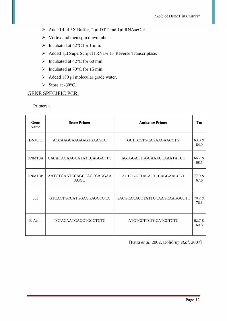
GENE SPECIFIC PCR:
Primers:-
Gene
Name
Sense Primer Antisense Primer Tm
DNMT1 ACCAAGCAAGAAGTGAAGCC GCTTCCTGCAGAAGAACCTG 63.3 &
64.0
DNMT3A CACACAGAAGCATATCCAGGAGTG AGTGGACTGGGAAACCAAATACCC 66.7 &
68.5
DNMT3B AATGTGAATCCAGCCAGCCAGGAA
AGGC
ACTGGATTACACTCCAGGAACCGT 77.9 &
67.6
p53 GTCACTGCCATGGAGGAGCCGCA GACGCACACCTATTGCAAGCAAGGGTTC 78.2 &
76.1
Β-Actin TCTACAATGAGCTGCGTGTG ATCTCCTTCTGCATCCTGTC 62.7 &
60.8
[Patra et.al, 2002. Dolidrup et.al, 2007]

“Role of DNMT in Cancer”
Page 13
PCR Mixture:- (Total 25µl)
0.2 µM dNTP- 0.5µl
1.5 mM MgCl2- 1.5µl
1x PCR Buffer- 2.5µl
Taq Polymearse (5U/µl)- 0.5µl
Primers (0.2µM)- 0.5µl & 0.5µl
cDNA- 2µl
MQ Water- 17µl
PCR Condition:-
94oC1:00[94
o C0:20; 57˚C0:20; 72
oC 0.30]30; 72
oC 5:00 for DNMT3B.
94oC1:00[94
o C0:20; 65˚C0:20; 72
oC 0.30]30; 72
oC 5:00 for DNMT3A.
94oC1:00[94
o C0:20; 58˚C0:20; 72
oC 0.30]30; 72
oC 5:00 for DNMT1.
94oC1:00[94
o C0:30; 60˚C0:45; 72
oC 1.30]30; 72
oC 5:00 for p53.
“Role of DNMT in Cancer”
Page 14
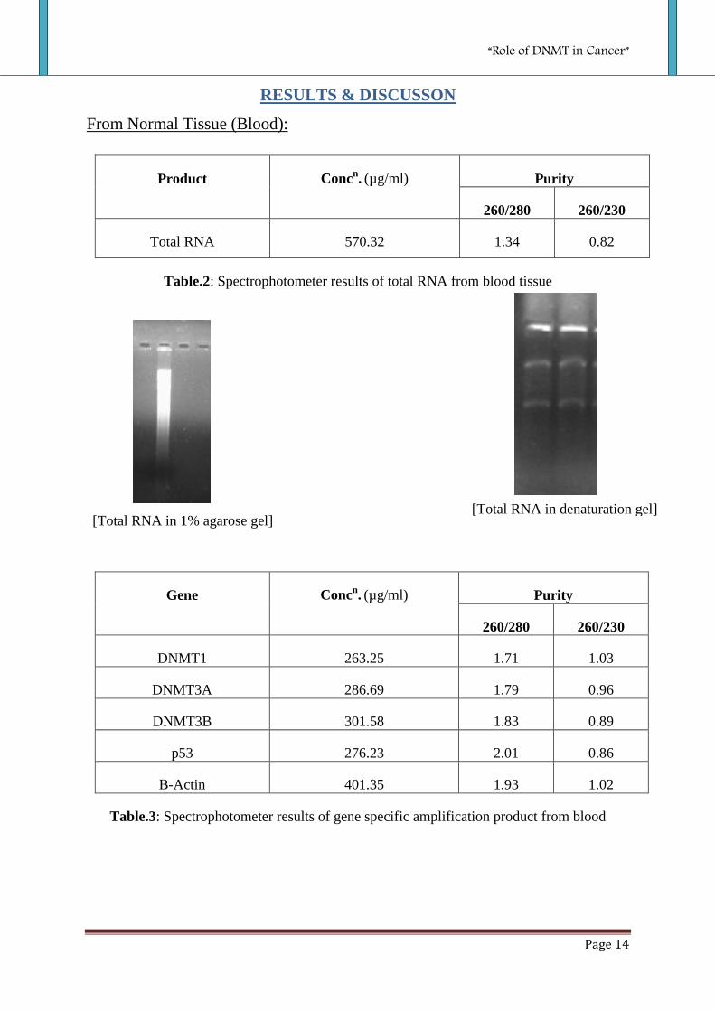
RESULTS & DISCUSSON
From Normal Tissue (Blood):
Product Concn. (µg/ml) Purity
260/280 260/230
Total RNA 570.32 1.34 0.82
Table.2: Spectrophotometer results of total RNA from blood tissue
Gene Concn. (µg/ml) Purity
260/280 260/230
DNMT1 263.25 1.71 1.03
DNMT3A 286.69 1.79 0.96
DNMT3B 301.58 1.83 0.89
p53
276.23 2.01 0.86
Β-Actin 401.35 1.93 1.02
Table.3: Spectrophotometer results of gene specific amplification product from blood
[Total RNA in 1% agarose gel] [Total RNA in denaturation gel]
“Role of DNMT in Cancer”
Page 15
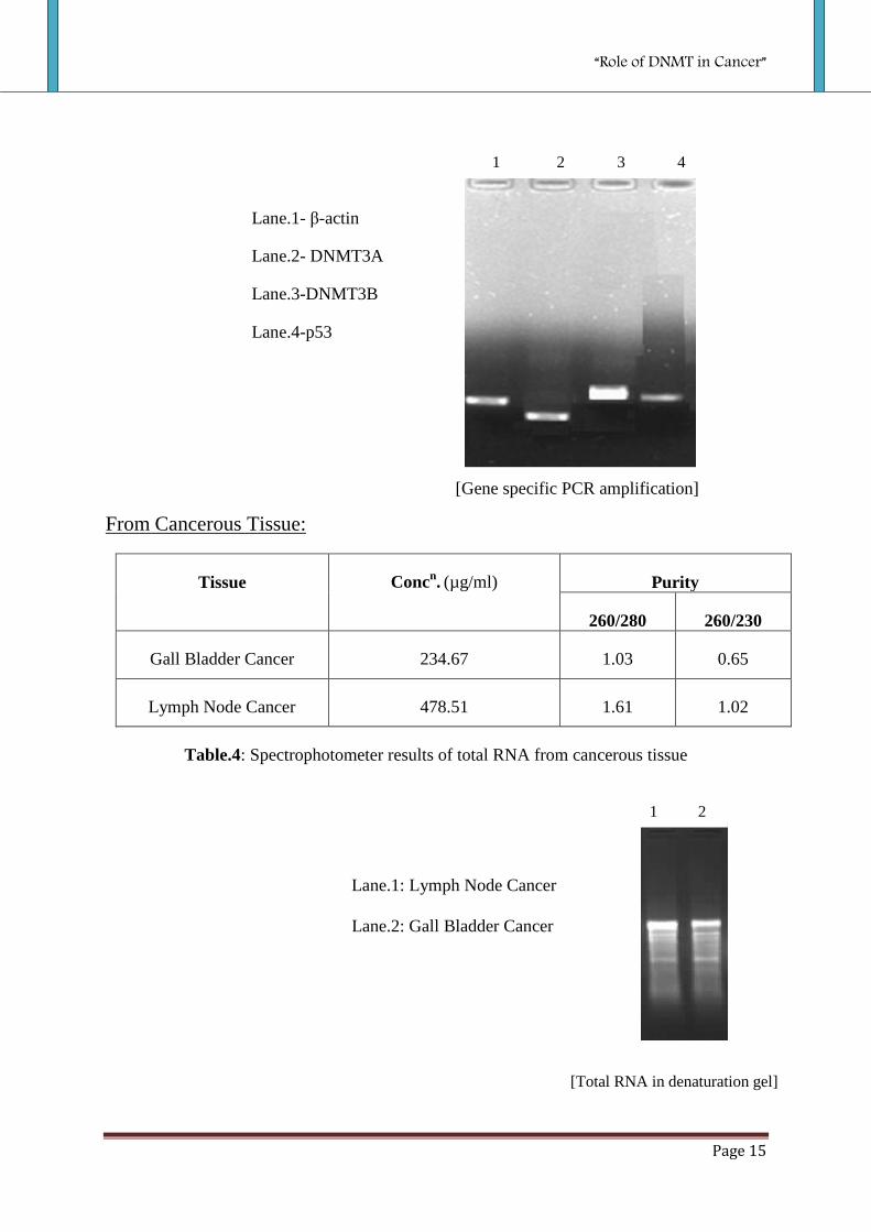
From Cancerous Tissue:
Tissue Concn. (µg/ml) Purity
260/280 260/230
Gall Bladder Cancer 234.67 1.03 0.65
Lymph Node Cancer 478.51 1.61 1.02
Table.4: Spectrophotometer results of total RNA from cancerous tissue
Lane.1: Lymph Node Cancer
Lane.2: Gall Bladder Cancer
1 2
[Total RNA in denaturation gel]
1 2 3 4
[Gene specific PCR amplification]
Lane.1- β-actin
Lane.2- DNMT3A
Lane.3-DNMT3B
Lane.4-p53
“Role of DNMT in Cancer”
Page 16
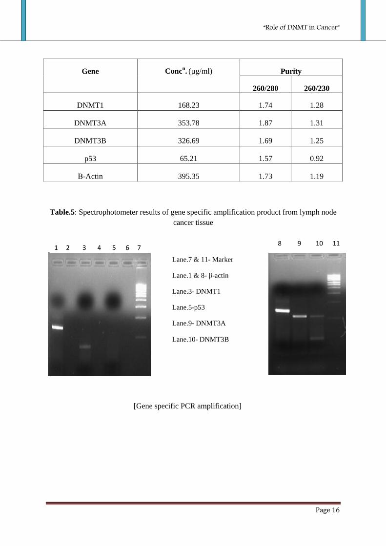
Table.5: Spectrophotometer results of gene specific amplification product from lymph node
cancer tissue
Gene Concn. (µg/ml) Purity
260/280 260/230
DNMT1 168.23 1.74 1.28
DNMT3A 353.78 1.87 1.31
DNMT3B 326.69 1.69 1.25
p53
65.21 1.57 0.92
Β-Actin 395.35 1.73 1.19
[Gene specific PCR amplification]
1 2 3 4 5 6 7 8 9 10 11
Lane.7 & 11- Marker
Lane.1 & 8- β-actin
Lane.3- DNMT1
Lane.5-p53
Lane.9- DNMT3A
Lane.10- DNMT3B
“Role of DNMT in Cancer”
Page 17
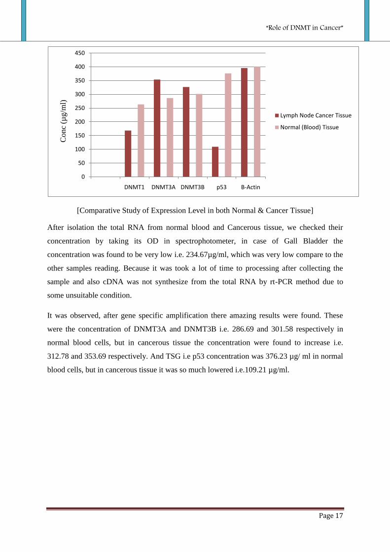
[Comparative Study of Expression Level in both Normal & Cancer Tissue]
After isolation the total RNA from normal blood and Cancerous tissue, we checked their
concentration by taking its OD in spectrophotometer, in case of Gall Bladder the
concentration was found to be very low i.e. 234.67µg/ml, which was very low compare to the
other samples reading. Because it was took a lot of time to processing after collecting the
sample and also cDNA was not synthesize from the total RNA by rt-PCR method due to
some unsuitable condition.
It was observed, after gene specific amplification there amazing results were found. These
were the concentration of DNMT3A and DNMT3B i.e. 286.69 and 301.58 respectively in
normal blood cells, but in cancerous tissue the concentration were found to increase i.e.
312.78 and 353.69 respectively. And TSG i.e p53 concentration was 376.23 µg/ ml in normal
blood cells, but in cancerous tissue it was so much lowered i.e.109.21 µg/ml.
0
50
100
150
200
250
300
350
400
450
DNMT1 DNMT3A DNMT3B p53 Β-Actin
Lymph Node Cancer Tissue
Normal (Blood) Tissue
Conc
(µg/m
l)

“Role of DNMT in Cancer”
Page 18
CONCLUSION
As observed from the above results, DNMTs are over express in Lymph Node cancer tissue.
So from this we can hypothesize that these over expression of DNMTs leads to
hypermethylation of TSG (for example, p53), which in turn diminish the expression of p53
and causing the cancer.

“Role of DNMT in Cancer”
Page 19
FUTURE WORK
The role of DNMTs in causing cancer through its effects on TSGs can be further confirmed
by carrying out by bisulphate modification and methylation specific-PCR (MS-PCR) of the
respective genes. This can help to locate the exact methylation site on the TSGs promoters.

“Role of DNMT in Cancer”
REFERENCES
1. JD Watson, FH Crick: Molecular structure of nucleic acids: a structure for deoxyribose
nucleic acid. Nature 1953, 171: 737-738.
2. JD Watson: The double helix, 1 edn. Weidenfeld & Nicolson; 1968.
3. RD Hotchkiss: The quantitative separation of purines, pyrimidines, and nucleosides by
paper chromatography. J Biol Chem 1948, 175: 315-332.
4. JD McPherson, M Marra, L Hillier, RH Waterston, A Chinwalla, J Wallis, M Sekhon, K
Wylie, ER Mardis, RK Wilson et al.: A physical map of the human genome. Nature 2001,
409: 934-941.
5. JC Venter, MD Adams, EW Myers, PW Li, RJ Mural, GG Sutton, HO Smith, M Yandell,
CA Evans, RA Holt et al.: The sequence of the human genome. Science 2001, 291: 1304-
1351.
6. PA Jones, SB Baylin: The fundamental role of epigenetic events in cancer. Nat Rev
Genet 2002, 3: 415 428.
7. PW Laird: The power and the promise of DNA methylation markers. Nat Rev Cancer
2003, 3: 253- 266.
8. PC Nowell: The clonal evolution of tumor cell populations. Science 1976, 194: 23-28.
9. B Vogelstein, KW Kinzler: Cancer genes and the pathways they control. Nat Med 2004,
10: 789-799.
10. JD Rowley: Letter: A new consistent chromosomal abnormality in chronic
myelogenous leukaemia identified by quinacrine fluorescence and Giemsa staining.
Nature 1973, 243: 290-293.
11. MR Teixeira, S Heim: Multiple numerical chromosome aberrations in cancer: what
are their causes and what are their consequences? Semin Cancer Biol 2005, 15: 3-12.
“Role of DNMT in Cancer”
12. F Mitelman, B Johansson, F Mertens. Mitelman Database of Chromosome Aberrations in
Cancer. http://cgap.nci.nih.gov/Chromosomes/Mitelman. 2005. Ref Type: Electronic Citation
13. M Olivier, R Eeles, M Hollstein, MA Khan, CC Harris, P Hainaut: The IARC TP53
database: new online mutation analysis and recommendations to users. Hum Mutat 2002,
19: 607-614.
14. C Waddington: The Epigenotype. Endeavour 1942, 1: 18-20.
15. AP Feinberg, B Tycko: The history of cancer epigenetics. Nat Rev Cancer 2004, 4: 143-
153.
16. M Ehrlich, MA Gama-Sosa, LH Huang, RM Midgett, KC Kuo, RA McCune, C Gehrke:
Amount and distribution of 5 methylcytosine in human DNA from different types of
tissues of cells. Nucleic Acids Res 1982, 10: 2709-2721.
17. C Coulondre, JH Miller, PJ Farabaugh, W Gilbert: Molecular basis of base substitution
hotspots in Escherichia coli. Nature 1978, 274: 775-780.
18. AP Bird: CpG-rich islands and the function of DNA methylation. Nature 1986, 321:
209-213.
19. ES Lander, LM Linton, B Birren, C Nusbaum, MC Zody, J Baldwin, K Devon, K Dewar,
M Doyle, W FitzHugh et al.: Initial sequencing and analysis of the human genome. Nature
2001, 409: 860-921.
20. F Larsen, G Gundersen, R Lopez, H Prydz: CpG islands as gene markers in the human
genome. Genomics 1992, 13: 1095-1107.
21. SM Taylor, PA Jones: Multiple new phenotypes induced in 10T1/2 and 3T3 cells
treated with 5- azacytidine. Cell 1979, 17: 771-779.
22. Hendrich B, Bird A: Identification and characterizationof a family of mammalian
methyl-CpG binding proteins. Mol Cell Biol 1998, 18: 6538-6547.

“Role of DNMT in Cancer”
23. H Maier, J Colbert, D Fitzsimmons, DR Clark, J Hagman: Activation of the early B-cell-
specific mb-1 (Ig-alpha) gene by Pax-5 is dependent on an unmethylated Ets binding
site. Mol Cell Biol 2003, 23: 1946-1960.
24. AC Bell, G Felsenfeld: Methylation of a CTCF-dependent boundary controls
imprinted expression of the Igf2 gene. Nature 2000, 405: 482-485.
25. Das PM, Singal R: DNA methylation and cancer. J Clin Oncol 2004, 22: 4632-4642.
26. Momparler RL: Cancer epigenetics. Oncogene 2003, 22: 6479-6483.
27. Robertson KD, Jones PA: DNA methylation: past, present and future directions.
Carcinogenesis 2000, 21: 461-467.
28. Szyf M, Pakneshan P, Rabbani SA: DNA methylation and breast cancer. Biochem
Pharmacol 2004, 68: 1187-1197.
29. Mertineit C, Yoder JA, Taketo T, Laird DW, Trasler JM, Bestor TH: Sex-specific exons
control DNA methyltransferase in mammalian germ cells. Development 1998, 125: 889-
897.
30. Brown R, Strathdee G: Epigenomics and epigenetic therapy of cancer. Trends Mol
Med 8, 2002, Suppl 4: S43-S48.
31. Wang YM, Wang R, Wen DG, Li Y, Guo W, Wang N, Wei LZ, He YT, Chen ZF, Zhang
XF, Zhang JH: Single nucleotide polymorphism in DNA methyltransferase 3B promoter
and its association with gastric cardiac adenocarcinoma in North China. World J
Gastroenterol 2005, 11: 3623-3627.
32. Hermann A, Gowher H, Jeltsch A: Biochemistry and biology of mammalian DNA
methyltransferases. Cell Mol Life Sci 2004, 61: 2571-2587.
33. Bestor TH: The DNA methyltransferases of mammals. Hum Mol Genet 2000, 9: 2395-
2402.

“Role of DNMT in Cancer”
34. Robertson KD, Uzvolgyi E, Liang G, Talmadge C, Sumegi J, Gonzales FA, Jones PA:
The human DNA Methyltransferases (DNMTs) 1, 3a and 3b: coordinate mRNA
expression in normal tissues and overexpression in tumors. Nucleic Acids Res 1999, 27:
2291-2298.
35. Weisenberger DJ, Velicescu M, Cheng JC, Gonzales FA, Liang G, Jones PA: Role of the
DNA methyltransferase variant DNMT3b3 in DNA methylation. Mol Cancer Res 2004, 2:
62-72.
36. Xie S, Wang Z, Okano M, Nogami M, Li Y, He WW, Okumura K, Li E: Cloning,
expression and chromosome locations of the human DNMT3 gene family. Gene 1999,
236: 87-95.
37. Mizuno S, Chijiwa T, Okamura T, Akashi K, Fukumaki Y, Niho Y, Sasaki H: Expression
of DNA Methyltransferases DNMT1 , 3A, and 3B in normal hematopoiesis and in acute
and chronic myelogenous leukemia. Blood 2001, 97: 1172-1179.
38. Etoh T, Kanai Y, Ushijima S, Nakagawa T, Nakanishi Y, Sasako M, Kitano S, Hirohashi
S: Increased DNA methyltransferase 1 (DNMT1) protein expression correlates
significantly with poorer tumor differentiation and frequent DNA hypermethylation of
multiple CpG islands in gastric cancers. Am J Pathol 2004, 164: 689-699.
39. Girault I, Tozlu S, Lidereau R, Bieche I: Expression analysis of DNA
methyltransferases 1, 3A, and 3B in sporadic breast carcinomas. Clin Cancer Res 2003,
9: 4415-4422.
40. Kanai Y, Ushijima S, Kondo Y, Nakanishi Y, Hirohashi S: DNA methyltransferase
expression and DNA methylation of CpG islands and peri-centromeric satellite regions
in human colorectal and stomach cancers. Int J Cancer 2001, 91: 205-212.
41. Esteller M, Silva JM, Dominguez G, Bonilla F, Matias-Guiu X, Lerma E, Bussaglia E,
Prat J, Harkes IC, Repasky EA, Gabrielson E, Schutte M, Baylin SB, Herman JG: Promoter

“Role of DNMT in Cancer”
hypermethylation and BRCA1 inactivation in sporadic breast and ovarian tumors. J
Natl Cancer Inst 2000, 92: 564-569.
42. Darwanto A, Kitazawa R, Maeda S, Kitazawa S: MeCP2 and promoter methylation
cooperatively regulate E-cadherin gene expression in colorectal carcinoma. Cancer Sci
2003, 94: 442-447.
43. Graff JR, Herman JG, Lapidus RG, Chopra H, Xu R, Jarrard DF, Isaacs WB, Pitha PM,
Davidson NE, Baylin SB: E-cadherin expression is silenced by DNA hypermethylation in
human breast and prostate carcinomas. Cancer Res 1995, 55: 5195- 5199.
44. Herman JG, Merlo A, Mao L, Lapidus RG, Issa JP, Davidson NE, Sidransky D, Baylin
SB: Inactivation of the CDKN2/p16/MTS1 gene is frequently associated with aberrant
DNA methylation in all common human cancers. Cancer Res 1995, 55: 4525-4530.
45. Melki JR, Vincent PC, Clark SJ: Concurrent DNA hypermethylation of multiple genes
in acute myeloid leukemia. Cancer Res 1999, 59: 3730 3740.
46. Stirzaker C, Millar DS, Paul CL, Warnecke PM, Harrison J, Vincent PC, Frommer M,
Clark SJ: Extensive DNA methylation spanning the Rb promoter in retinoblastoma
tumors. Cancer Res 1997, 57: 2229-2237.
47. Agirre X, Novo FJ, Calasanz MJ, Larrayoz MJ, Lahortiga I, Valganon M, Garcia-
Delgado M, Vizmanos JL: TP53 is frequently altered by methylation, mutation, and/or
deletion in acute lymphoblastic leukaemia. Mol Carcinog 2003, 38: 201-208.
48. Patra S. K, Patra A, Zhao H, Dahiya R: DNA Methyltransferase and Demethylase in
Human Prostate Cancer. Mol Carcinog 2002, 33: 163-171.
49. Luczak M. W, Jagodzinski P. P: The role of DNA methylation in cancer development.
Folia Histo Et Cyto 2006, 44 (3): 143-154.


![[Frontiers in Bioscience 11, 2179-2192, September 1, 2006]](https://img.pdfslide.us/doc/110x75/62356e1e67d47524e43cfd5f/frontiers-in-bioscience-11-2179-2192-september-1-2006.jpg)