Embed Size (px)
Citation preview

1Scientific RepoRts | 6:27517 | DOI: 10.1038/srep27517
www.nature.com/scientificreports
Role of Cystathionine Gamma-Lyase in Immediate Renal Impairment and Inflammatory Response in Acute Ischemic Kidney InjuryLajos Markó1,2, István A. Szijártó1, Milos R. Filipovic3, Mario Kaßmann1, András Balogh1,2, Joon-Keun Park4, Lukasz Przybyl1, Gabriele N’diaye1, Stephanie Krämer5, Juliane Anders1, Isao Ishii6, Dominik N. Müller1,2,* & Maik Gollasch1,7,*
Hydrogen sulfide (H2S) is known to act protectively during renal ischemia/reperfusion injury (IRI). However, the role of the endogenous H2S in acute kidney injury (AKI) is largely unclear. Here, we analyzed the role of cystathionine gamma-lyase (CTH) in acute renal IRI using CTH-deficient (Cth−/−) mice whose renal H2S levels were approximately 50% of control (wild-type) mice. Although levels of serum creatinine and renal expression of AKI marker proteins were equivalent between Cth−/− and control mice, histological analysis revealed that IRI caused less renal tubular damage in Cth−/− mice. Flow cytometric analysis revealed that renal population of infiltrated granulocytes/macrophages was equivalent in these mice. However, renal expression levels of certain inflammatory cytokines/adhesion molecules believed to play a role in IRI were found to be lower after IRI only in Cth−/− mice. Our results indicate that the systemic CTH loss does not deteriorate but rather ameliorates the immediate AKI outcome probably due to reduced inflammatory responses in the kidney. The renal expression of CTH and other H2S-producing enzymes was markedly suppressed after IRI, which could be an integrated adaptive response for renal cell protection.
Hydrogen sulfide (H2S) has been recognized as a toxic gas for many years until Warenycia et al. discovered the endogenous production of H2S in the rat brain1. Endogenous H2S is mainly produced by enzymes of the transsul-furation pathway, cystathionine gamma-lyase (CTH or CSE) and cystathionine beta-synthase (CBS). The third enzyme, 3-mercaptopyruvate sulfurtransferase (MPST or MST), can also contribute to endogenous H2S produc-tion in the presence of reductants using 3-mercaptopyruvate as a substrate2–4. Although the physiological role of MPST in mammalian tissues is less well characterized, MPST might contribute to H2S production in the brain or modulation of cardiovascular functions5,6.
Several studies demonstrated that H2S could exert protective effects in the cardiovascular system. In particu-lar, H2S has emerged as potential therapeutics3 for ischemia/reperfusion injury (IRI) of different organs2. This knowledge mainly relies on the results from animal studies using H2S/H2S donor molecules or pharmacological inhibitors of H2S-producing enzymes7. However, the physiological levels of H2S in tissues have been a matter of debate because of methodological difficulties in measuring accurate, reliable, and reproducible H2S levels in
1Experimental and Clinical Research Center, a joint cooperation between the Charité Medical Faculty and the Max-Delbrück Center (MDC) for Molecular Medicine, Berlin, Germany. 2Max-Delbrück Center (MDC) for Molecular Medicine, Berlin, Germany. 3Friedrich-Alexander-University of Erlangen-Nürnberg, Department of Chemistry and Pharmacy, Erlangen, Germany. 4Hannover Medical School, Hannover, Germany. 5German Institute of Human Nutrition, Potsdam-Rehbrücke, Germany. 6Department of Biochemistry, Graduate School of Pharmaceutical Sciences, Keio University, Tokyo, Japan. 7Charité Campus Virchow, Nephrology/Intensive Care, Berlin, Germany. *These authors jointly supervised this work. Correspondence and requests for materials should be addressed to L.M. (email: [email protected]) or M.G. (email: [email protected])
Received: 25 September 2015
Accepted: 20 May 2016
Published: 08 June 2016
OPEN

www.nature.com/scientificreports/
2Scientific RepoRts | 6:27517 | DOI: 10.1038/srep27517
biological samples. In addition, pharmacological CTH/CBS inhibitors have been used at suspicious high concen-trations in vivo and in vitro8, which might cause the lack of specificity in enzyme inhibition2,9. Therefore, trans-genic animals have been recently developed to elucidate the role of H2S in health and disease10,11.
CTH is highly expressed in the kidney and several studies demonstrated that H2S could exert protective effects in renal IRI. Han et al. found that NaHS administration to IRI mice accelerated the recovery from renal dysfunc-tion and impaired tubular morphology, whereas the administration of dl-propargylglycine (PAG), an inhibitor of CTH, delayed it12. NaHS administration had beneficial effects on renal IRI13 and gentamicin-induced acute kidney injury (AKI) in rats14. Tan et al. suggested that the beneficial effects of endogenously produced H2S in AKI are, at least in part, mediated by toll-like receptors (TLRs)-mediated inflammatory response and apoptosis15. Chen et al. showed that exogenous H2S reduces kidney injury from urinary-derived sepsis in rabbits, which is associated with decreased TNF-α expression in the kidneys16. On a conceptual level, these results are consistent with recent findings by Bos et al. who reported that their CTH-deficient (Cth−/−) mice display aggravated renal IRI10,17 when our study was underway. The authors concluded that CTH protects against renal IRI, likely by mod-ulating oxidative stress through the production of H2S. Autosomal-recessive cystathioninuria (OMIM 219500), which is considered as a benign biochemical anomaly, is caused by homozygous or compound heterozygous mutations in CTH (OMIM 607657) and has a relatively high prevalence (1 per 14,000 live births)18 though with somewhat lower incidence in other reports19,20. Therefore, both the findings of Bos et al. and ours could have important implications for humans.
Here, we used our Cth−/− mice on a C57BL/6J background11 to elucidate the function of CTH in renal IRI. We found that the systemic CTH loss does not deteriorate the immediate outcome of AKI. Instead Cth−/− mice displayed reduced renal damage and renal expression of inflammatory cytokines/adhesion molecules after IRI, compared to littermate control (wild-type; Cth+/+) and heterozygous (Cth+/−) mice.
ResultsThe mRNA Expression of H2S-Generating Enzymes in the Kidney. Levels of renal Cth mRNA in heterozygous (Cth+/−) mice were approximately half of those in Cth+/+ mice (Fig. 1A). In both Cth+/+ and Cth+/− mice subjected to IRI, renal Cth mRNA levels declined to one third of their initial expression levels (Fig. 1A). No Cth mRNA was detectable in the kidneys of Cth−/− mice (Fig. 1A). Renal mRNA levels of two other
Figure 1. Renal expression of H2S-producing enzymes and H2S. Gene expression levels of (A) cystathionine gamma -lyase (Cth), (B) cystathionine beta-synthase (Cbs) and (C) 3-mercaptopyruvate sulfurtransferase (Mpst) in sham- and ischemia/reperfusion (I/R)-injured kidneys of wild-type (Cth+/+), heterozygous (Cth+/−) and CTH-deficient (Cth−/−) mice. Values plotted are mean ± SEM (n = 4 in sham-operated groups, n = 8 in I/R-injured groups). *P < 0.05 vs. sham-operated Cth+/+ and sham-operated Cth+/−; #P < 0.05 vs. I/R-injured Cth+/+ and sham-operated Cth+/−; $P < 0.05 vs. sham-operated Cth+/+, Cth+/− and Cth−/− mice. (D) Relative CTH protein levels of sham- and I/R-injured kidneys of Cth+/+, Cth+/− and Cth−/− mice. Values plotted are mean ± SEM (n = 4 each). Mean CTH density of two Cth+/+ kidneys on each gel was set to 100% and relative density was calculated for the rests. (E) Levels of H2S in intact kidneys of Cth+/+, Cth+/− and Cth−/− mice. Values plotted are mean ± SEM (n = 11 for Cth+/+ and Cth−/−, and n = 4 for Cth+/−). &P < 0.01 vs. Cth+/+ and Cth+/−; §P < 0.05 vs. Cth+/+ and Cth−/−. AU, arbitrary units. n.d., not detectable.
www.nature.com/scientificreports/
3Scientific RepoRts | 6:27517 | DOI: 10.1038/srep27517
H2S-producing enzymes, CBS and MPST, were not different among all three Cth genotypes. These levels declined markedly after IRI and the levels after IRI were also not different among Cth genotypes (Fig. 1B,C).
Protein Expression of CTH and Endogenous H2S Levels in the Kidney. Levels of renal CTH protein in heterozygous (Cth+/−) mice were ~40% of those in Cth+/+ mice (Fig. 1D) as previously reported11. Similar to its mRNA level changes, renal CTH protein levels declined after IRI in both Cth+/+ and Cth+/− mice and no CTH protein was detectable in the kidneys of Cth−/− mice (Fig. 1D). Next, we measured endogenous H2S levels in the kidney to assess the impact of systemic CTH deletion on H2S production. Kidneys of Cth+/− and Cth−/− mice dis-played approximately 30% and 50% reduced H2S levels, respectively, compared to those of Cth+/+ mice (Fig. 1E).
The impact of CTH loss in Renal Damage after IRI. To get insights into (patho)physiological roles of CTH in renal IRI, we performed comparative in vivo studies using Cth+/+, Cth+/− and Cth−/− mice. Twenty-four
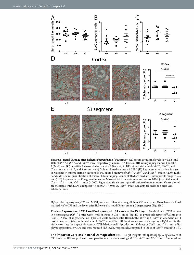
Figure 2. Renal damage after ischemia/reperfusion (I/R) injury. (A) Serum creatinine levels (n = 12, 8, and 10 for Cth+/+, Cth+/−, and Cth−/− mice, respectively) and mRNA levels of (B) kidney injury marker lipocalin 2 (Lcn2) and (C) hepatitis A virus cellular receptor 1 (Havcr1) in I/R-injured kidneys of Cth+/+, Cth+/−, and Cth−/− mice (n = 8, 7, and 8, respectively). Values plotted are mean ± SEM. (D) Representative cortical images of Masson’s trichrome stain on sections of I/R-injured kidneys of Cth+/+, Cth+/−, and Cth−/− mice (× 200). Right hand side is semi-quantification of cortical tubular injury. Values plotted are median ± interquartile range (n = 6 each). (E) Representative S3 segment images of Masson’s trichrome stain on sections of I/R-injured kidneys of Cth+/+, Cth+/−, and Cth−/− mice (× 200). Right hand side is semi-quantification of tubular injury. Values plotted are median ± interquartile range (n = 6 each). *P < 0.05 vs. Cth+/− mice. Red dots are red blood cells. AU, arbitrary units.

www.nature.com/scientificreports/
4Scientific RepoRts | 6:27517 | DOI: 10.1038/srep27517
hours after ischemia, Cth−/− mice showed somewhat lower serum creatinine levels (136 μ mol/l average) com-pared to Cth+/− mice (152 μ mol/l average) and Cth+/+ mice (155 μ mol/l average) although the differences were not statistically significant (overall ANOVA P = 0.30; Fig. 2A). Renal mRNA levels of two sensitive AKI markers, lipocalin 2 (Lcn2; also known as neutrophil gelatinase-associated lipocalin [Ngal]) and hepatitis A virus cel-lular receptor 1 (Havcr1; also known as kidney injury molecule 1 [Kim1]), were not different between Cth+/+, Cth+/−, and Cth−/− mice (Fig. 2B,C). To assess the degree of tubular damage after ischemic AKI, kidney sections were stained and examined by an experienced renal pathologist who was unaware of Cth genotypes. Histological analyses and semi-quantitative scoring revealed a moderate amelioration in cortical tubular damage after renal IRI in Cth−/− mice compared to Cth+/− or Cth+/+ mice (Fig. 2D). Histological analysis of the S3 segments of the proximal tubules in the outer stripe of outer medulla, especially vulnerable loci against renal IRI, identified that Cth−/− mice exhibit less tubular necrosis and less occlusions of tubular lumen with cellular debris, compared to Cth+/− or Cth+/+ mice (Fig. 2E). These parameters were comparable among sham-operated mice with all Cth genotypes (Supplementary Figure 1A–C). It is notable that all Cth+/+, Cth+/− and Cth−/− mice that underwent surgery survived in this study.
The impact of CTH loss in Cellular Infiltration to the Kidneys after IRI. Renal IRI is known to associate with infiltration of granulocytes, monocytes/macrophages and other immune cells immediately after reperfusion, which contributes to inflammation and subsequent repair in injured kidneys21. Therefore, we characterized granulocytes and macrophages in renal IRI by flow cytometry. Whole kidney cell suspensions were immunolabelled for Ly6G and F4/80 as markers for granulocytes and macrophages, respectively. Among pre-gated singlet live cells (Fig. 3A), Ly6G-positive & F4/80-negative granulocytes as well as Ly6G-negative & F4/80-positive macrophages were detected (Fig. 3B). There were no significant differences in both granulocyte and macrophage populations between Cth+/+, Cth+/−, and Cth−/− kidneys at 24 h after IRI (Fig. 3C and D) or after sham surgery (Supplementary Figure 2A–C). We next performed immunohistochemistry to detect IRI-induced granulocyte infiltration (Supplementary Figure 3A)22. In the outer medulla after IRI, average Ly6B-positive cell numbers per view field were 12 in Cth−/− mice while 13 and 19 in Cth+/− and Cth+/+ mice, respectively, although the differences were not statistically significant (P = 0.501, Supplementary Figure 3B). Furthermore, renal levels of S100a8/a9 mRNAs for calprotectin, a heterodimeric protein that was recently found to co-localize with Ly6G in granulocytes after AKI and playing a crucial part in controlling M2 macrophage-mediated renal repair following IRI23, were also not significantly different (Supplementary Figure 3C,D).
The impact of CTH loss in Expression of Cytokines, Chemokines, and Adhesion Molecules. Production of inflammatory molecules is maintained low in the normal kidney but is markedly increased under pathophysiological conditions such as ischemia21. We measured mRNA levels of several molecules involved in long-term outcome/repair after renal IRI. Renal expression of interleukin 1-beta (Il1b) and vascular cell adhesion
Figure 3. Flow cytometric analysis of renal granulocyte and macrophage infiltration. (A) Pre-gating on live cells using Fixable Viability Dye eFluor 660 and further gating on single cells. (B) Representative flow cytometry data of infiltrating Ly6G-positive cells (granulocytes) and F4/80-positive cells (macrophages) in I/R-injured kidneys of Cth+/+, Cth+/−, and Cth−/− mice. Quantification of infiltrating (C) Ly6G-positive cells and (D) F4/80-positive cells. Values plotted are mean ± SEM (n = 6, 3 and 5 for Cth+/+, Cth+/−, and Cth−/− mice, respectively).

www.nature.com/scientificreports/
5Scientific RepoRts | 6:27517 | DOI: 10.1038/srep27517
molecule 1 (Vcam1) after IRI was significantly lower in Cth−/− mice compared to Cth+/− mice (Fig. 4A,B). Also, renal expression of tumor necrosis factor-alpha (Tnf) and vascular cell adhesion molecule 1 (Vcam1) was sim-ilarly lower in Cth−/− mice compared to Cth+/− mice (overall ANOVA P = 0.099 and P = 0.088, respectively) (Fig. 4C,D). Renal expression of other important cytokines/chemokines such as interleukin 6 (Il6), chemokine (C-X-C motif) ligand 2 (Cxcl2), and chemokine (C-C motif) ligand 2 (Ccl2), were not altered among Cth geno-types (Supplementary Figure 4A–C).
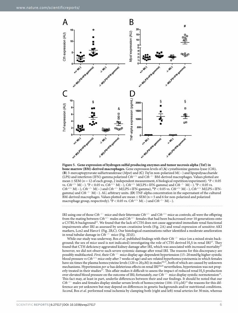
The impact of CTH loss in In Vitro Macrophage Polarization. Although the proportion of infil-trating macrophages after IRI was not significantly different (Fig. 3D), renal mRNA expression of IL1-beta and TNF-alpha, the two major inflammatory cytokines of macrophage origin, was lower or in Cth−/− mice (Fig. 4A,C). We hypothesized that macrophage polarization is disturbed by the lack of CTH, and thus investi-gated Tnf induction by the lipopolysaccharide (LPS)/interferon (IFN)-gamma in vitro treatment of bone marrow (BM)-derived macrophages from Cth+/+ and Cth−/− mice. Cth expression was induced while Mpst expression was not altered by LPS/IFN-gamma treatment in BM-derived macrophages from Cth+/+ mice (Fig. 5A,B). In contrast, Mpst expression was significantly induced by the same treatment in macrophages from Cth−/− mice (Fig. 5B), and Cbs expression was not detectable in macrophages from either mice (data not shown). Under such conditions, Tnf expression was markedly induced by LPS/IFN-gamma treatment of both Cth+/+ and Cth−/− macrophages, and the levels were significantly lower in Cth−/− macrophages (Fig. 5C), although the supernatant TNF-alpha concen-trations of activated macrophages were comparable between Cth+/+ and Cth−/− mice (Fig. 5D).
DiscussionA number of studies have demonstrated the cytoprotective effects of H2S in myocardial, liver, brain, pulmonary, and renal IRI (reviewed by Nicholson and Calvert)2. Most of these studies utilized Na2S/NaHS as exogenous H2S donors and PAG as a non-specific CTH inhibitor. To overcome pharmacokinetic problems in H2S donor applica-tions and specificity issues of PAG, two research groups have independently generated mice in which Cth genes have been differentially deleted10,11. In our study, we investigated the pathophysiological roles of CTH in renal
Figure 4. Renal gene expression of cytokines and adhesion molecules. Gene expression levels of (A) interleukin (Il))1-beta, (B) vascular cell adhesion molecule (Vcam)1, (C) tumor necrosis factor alpha (Tnf) and (D) intercellular adhesion molecule (Icam)1 in ischemia/reperfusion-injured kidneys of Cth+/+, Cth+/−, and Cth−/− mice. Values plotted are mean ± SEM (n = 8, 7, and 8 for Cth+/+, Cth+/−, and Cth−/− mice, respectively). *P < 0.05 vs. Cth+/− mice. AU, arbitrary units.
www.nature.com/scientificreports/
6Scientific RepoRts | 6:27517 | DOI: 10.1038/srep27517
IRI using one of those Cth−/− mice and their littermate Cth+/− and Cth+/+ mice as controls; all were the offspring from the mating between Cth+/− males and Cth+/− females that had been backcrossed over 10 generations onto a C57BL/6 background11. We found that the lack of CTH does not cause aggravated immediate renal functional impairments after IRI as assessed by serum creatinine levels (Fig. 2A) and renal expression of sensitive AKI markers, Lcn2 and Havcr1 (Fig. 2B,C). Our histological examinations rather identified a moderate amelioration in renal tubular damage in Cth−/− mice (Fig. 2D,E).
While our study was underway, Bos et al. published findings with their Cth−/− mice (on a mixed strain back-ground; the sex of mice used is not indicated) investigating the role of CTH-derived H2S in renal IRI17. They found that CTH deficiency aggravated kidney damage after IRI, which was associated with increased mortality17 however, we did not observe such severe systemic damage after renal IRI. The reasons for this discrepancy are possibly multifaceted. First, their Cth−/− mice display age-dependent hypertension (15–20 mmHg higher systolic blood pressure vs Cth+/+ mice only after 7 weeks of age) and sex-related hyperhomocysteinemia in which females have six times the plasma homocysteine levels (120 vs 20 μ M) in males10,17, both of which are caused by unknown mechanisms. Hypertension per se has deleterious effects on renal IRI24,25 nevertheless, hypertension was not prop-erly treated in their studies17. This affair makes it difficult to assess the impact of reduced renal H2S production over elevated blood pressure on the outcome of IRI; fortunately, our Cth−/− mice display systolic normotension11. This fact may, at least in part, underlie differences between their and our findings. It should be noted that our Cth−/− males and females display similar serum levels of homocysteine (104–151 μ M)11 the reasons for this dif-ference are yet unknown but may depend on differences in genetic backgrounds and/or nutritional conditions. Second, Bos et al. performed renal ischemia by clamping both (right and left) renal arteries for 30 min, whereas
Figure 5. Gene expression of hydrogen sulfid producing enzymes and tumor necrosis alpha (Tnf) in bone marrow (BM)-derived macrophages. Gene expression levels of (A) cystathionine gamma -lyase (Cth), (B) 3-mercaptopyruvate sulfurtransferase (Mpst) and (C) Tnf in non-polarized M(− ) and lipopolysaccharide (LPS) and interferon (IFN)-gamma polarized Cth+/+ and Cth−/− BM-derived macrophages. Values plotted are mean ± SEM (n = 12 of each group, 2 independent measurement, 6 biological repetition/experiment). *P < 0.05 vs. Cth+/+ M(− ). #P < 0.05 vs. Cth+/+ M(− ), Cth+/+ M(LPS+ IFN-gamma) and Cth−/− M(−). $P < 0.05 vs. Cth+/+ M(− ), Cth−/− M(− ) and Cth−/− M(LPS+ IFN-gamma), &P < 0.05 vs. Cth+/+ M(− ), Cth+/+ M(LPS+ IFN-gamma) and Cth−/− M(− ). AU, arbitrary units. (D) TNF-alpha concentration in the supernatant of the cultured BM-derived macrophages. Values plotted are mean ± SEM (n = 5 and 6 for non-polarized and polarized macrophage group, respectively). §P < 0.05 vs. Cth+/+ M(− ) and Cth−/− M(− ).
www.nature.com/scientificreports/
7Scientific RepoRts | 6:27517 | DOI: 10.1038/srep27517
we performed uninephrectomy by clamping the renal artery of the remnant left kidney for 20 min17. Despite the differences in surgical protocols, serum creatinine levels at 24 h after IRI were equivalent. But, importantly, all mice that underwent surgery survived in our study while Bos et al. observed 35% mortality only in Cth−/− mice17. Third, we used a temperature controller with heating pads to maintain a stable core temperature (which was meas-ured continuously during surgery by a rectal probe) whereas Bos et al. used only heating pads and lamps17. It is well known that fluctuations in core body temperature contribute to variability in IRI and the way of maintaining body temperature during ischemia has a major impact on the outcome of IRI26. Fourth, their Cth−/− mice showed a massive (91%) reduction in renal H2S production compared to Cth+/+ mice17 while our Cth−/− mice showed only 50% reduction (Fig. 1E). Although the methods used for H2S measurement substantially differ between the two studies and this precludes the direct comparison, > 90% reduction is surprising per se, considering the facts that (i) Cth−/− kidney still expresses CBS and MPST, (ii) (increased/activated) CBS could compensate for H2S pro-duction when CTH is inhibited or abrogated (though we did not observe compensatory Cbs mRNA induction; (Fig. 1B) and (iii) renal Cbs/Mpst expression was markedly down-regulated by ischemia/reperfusion irrespective of Cth genotypes (Fig. 1B,C)11,12,17,27,28. A previous study mentioned that the reduction in CBS (rather than CTH) activity may serve as the major contributor for endogenous H2S level reduction during renal IRI29.
Despite such differences, we also found some agreement with previous studies by Bos et al.17 and others27,30. First, renal expression (either gene or protein) of both CTH and CBS were suppressed after renal IRI (Fig. 1A,B). It might be noteworthy that the partial or complete loss of CTH did not cause compensatory induction (or reduced repression of expression) of CBS (or MPST) during IRI (Fig. 1B,C) at least on mRNA level. Second, both Bos et al. and we did not find significant differences between Cth genotypes in the numbers of granulocyte infil-trated into injured kidneys of IRI mice (Fig. 3C and Supplementary Figure 3A–C)17. We also counted the numbers of F4/80-positive macrophages infiltrated into injured kidneys of IRI mice and found that macrophages behave similar to granulocytes (Fig. 3D). In contrast, renal expression of Tnf, Il1b, Icam1, and Vcam1 after IRI were lower (though overall ANOVA was just P = 0.099 and 0.088 for Tnf and Icam1, respectively) in Cth−/− mice compared to Cth+/− mice (Fig. 4A–D). TNF-alpha was initially discovered as a LPS-induced macrophage product31. It is also released during IRI and acts as a potent pro-inflammatory cytokine32, and in line, the blockade of TNF-alpha sig-naling is a novel promising therapeutic target in renal IRI33. Although intrinsic renal cells also secret TNF-alpha upon injury, monocytes/classically activated macrophages are considered as the main source of TNF-alpha in early renal IRI34. We found that CTH deficiency alters Tnf expression in LPS/IFN-gamma-stimulated BM-derived macrophages that intrinsically differ from LPS-stimulated peritoneal macrophages10,35. However, the supernatant TNF-alpha concentrations did not differ between both groups, which questions the physiological relevance of this finding. Meanwhile, renal expression of other cytokines that are known to play a role in renal IRI36 (Il6, Cxcl2, and Ccl2) were not distinguishable between Cth genotypes (Supplementary Figure 4A–C).
Our findings are in contrast to previous results by others who use PAG for CTH inhibition. Tripatara et al. found that single intraperitoneal administration of PAG (50 mg/kg, 1 h before ischemia) prevented the renal recovery from IRI (45-min ischemia/72-h reperfusion) in a rat bilateral ischemia model37. More recently Han et al. showed similar deteriorative effects of PAG in renal IRI (50 mg/kg daily (i.p.), beginning 2 days after ischemia) in mice12. However, PAG (5 mg/kg (i.p.), twice a day for 4 successive days) exhibited nephroprotective effects in the cisplatin model of AKI in rats38. Similar protective effects of PAG (50 mg/kg (i.p.) at 2 h after adri-amycin injection) have been observed in adriamycin-induced nephrotoxicity in rats39. Whereas these kidney injury models differ, they point out that PAG treatment can have multiple effects depending on the renal injury models. Moreover, the specificity of this widely used CTH inhibitor and relatively late time points after reperfu-sion are a matter of concern. Our model is of particular interest because we used a genetic approach to abrogate CTH specifically and investigated acute renal post-ischemic injury after 24 h, a time point where serum creatinine levels are the highest and renal Cth/Cbs expression levels are the lowest17.
Numerous studies have revealed cytoprotective/anti-oxidative/anti-inflammatory roles of H2S, but some studies also have identified pro-inflammatory roles of H2S that accelerate inflammatory responses; for exam-ple, Ang et al. previously reported that caerulein-induced acute pancreatic damage as well as its associated lung injury was ameliorated in Cth−/− mice compared to Cth+/+ 40. It is possible that CTH-produced H2S may act as a pro-inflammatory factor in renal IRI. In addition, further studies should also clarify the impact of high levels of cystathionine and homocysteine and low levels of taurine that are common in Cth−/− mice11 on the outcome of renal IRI41–43. In conclusion, the systemic loss of CTH in mice caused approximately 50% reduction in renal H2S levels but did not influence immediate outcomes of ischemic AKI; however, it reduced tubular damage mod-erately and suppressed the renal expression of inflammatory cytokines. Future studies should clarify the role of CTH on the long-term outcome of renal impairment in AKI.
MethodsMice. Cth+/− and Cth−/− mice were generated and characterized earlier11. In this study, Cth+/− males and females were bred to obtain Cth+/+, Cth+/−, and Cth−/− littermates. Mice were allowed free access to stand-ard chow and water. The mice were kept in a 12:12-h light-dark cycle. All works involving animals have been approved by the Berlin Animal Review Board in 2012 (No. G 0444/12) and conducted in accordance with the American Physiological Society standards.
Renal IRI Model. Male mice (age between 12–15 weeks) were used. Anesthesia was performed with isoflu-rane (2.3%) in air (350 ml/min)44. Each mouse was operated separately to ensure similar exposure to isoflurane (35.7 ± 2.3 min, mean ± SD)45. In order to keep body temperature stable at 37 °C and monitor it during surgery, a temperature controller with heating pad (TCAT-2, Physitemp Instruments) was used. Rectal body temperature was continuously monitored during surgery using a sensor-based thermistor (36.9 ± 0.4 °C at beginning of the surgery, 37.0 ± 0.4 °C after uninephrectomy, 37.1 ± 0.3 °C five minutes after clamping the left renal pedicle and

www.nature.com/scientificreports/
8Scientific RepoRts | 6:27517 | DOI: 10.1038/srep27517
37.1 ± 0.1 °C at the end of surgery). After right-sided uninephrectomy, ischemia was induced by clipping the pedi-cles of the left kidney for 20 minutes with non-traumatic aneurysm clips (FE690K, Aesculap). Reperfusion was confirmed visually. After surgery, mice had free access to water and chow. We applied body-warm sterile physio-logical saline solutions and preemptive analgesia with tramadol (1 mg/kg) for every mouse. Sham operation was performed in a similar manner, except for clamping the renal pedicle. Mice with bleeding during surgery, with incomplete renal reperfusion, with excessive exposure of isoflurane of any reason, with significant temperature fluctuation during surgery, or with signs for infection 24 h after IRI, were immediately euthanized and were not used for further analysis. After 24 h of reperfusion, mice were sacrificed, and kidney and blood samples were collected for further analysis. The kidneys were divided into three portions. One third of the kidney was placed in optimum cutting temperature (OCT) compound for immunohistochemistry, one third was immersed in 4% phosphate-buffered saline (PBS)-buffered formalin for histology, and the rest was snap-frozen in liquid nitrogen for RNA preparation.
Quantitative Real-Time (qRT)-PCR. Total RNA from snap-frozen kidneys were isolated using RNeasy RNA isolation kit (Qiagen) according to manufacturer’s instruction after homogenization with a Precellys 24 homogenizator (Peqlab). RNA concentration and quality was determined by NanoDrop-1000 spectrophotom-eter (Thermo Fisher Scientific). Two micrograms of RNA were transcribed to cDNA (Applied Biosystems). Quantitative analysis of target mRNA expression was performed with qRT-PCR using the relative standard curve method. TaqMan and SYBR green analysis was conducted using an Applied Biosystems 7500 Sequence Detector (Applied Biosystems). The expression levels were normalized to 18S or to beta-actin. Primer sequences are pro-vided in Supplementary Table 1.
Western Blot. Sham and IRI-damaged kidneys were lysed with RIPA buffer (Sigma) supplemented with Complete® protease inhibitor (Roche), 1 mM phenylmethylsulfonyl fluoride (PMSF), phosphatase inhibitor cocktail 3 (Sigma) and were homogenized using a Precellys 24 homogenizator. Fifty micrograms of protein samples were separated by 12% SDS-PAGE. After wet transfer, non-specific binding sites of the nitrocellulose membrane were blocked with 5% non-fat skim milk in Tris-buffered saline containing 0.1% Tween (TBST). The membrane was then incubated with primary antibody (anti-CTH, 1:500 (ab80643) Abcam or anti-CTH carboxyl terminus rabbit polyclonal antibody that recognizes amino acids 194–398 of a rat 398-amino acid CTH protein, 1:1,00046 and anti-beta-actin, 1:2,000 (4970) Cell Signaling). Secondary antibody was from LI-COR Biosciences (anti-rabbit, 1:5,000). Images were acquired by Odyssey infrared imaging system (LI-COR Biosciences). Beta-actin was used as a loading control. Membranes were first probed with anti-CTH antibody and detected for their signals, and then stripped for re-probing with anti-beta-actin antibody (as loading controls). Successive stripping was confirmed by the absence of signals in the stripped membranes.
TNF-alpha Measurement. TNF-alpha levels in the supernatants of macrophages (that were used for qRT-PCR analyses) were measured using the Mouse TNF alpha ELISA Ready-SET-Go!® Kit (eBioscience).
H2S Measurement. To detect H2S production in the kidneys, Cth+/+ and Cth−/− mice were euthanized and freshly isolated kidneys were incubated in PBS containing 50 μ M of a recently developed fluorescent probe (Washington State Probe-1 [WSP-1], Cayman Chemical) for H2S47. After 45 min of incubation the samples were snap-frozen. Thawed samples were homogenized and centrifuged, and the supernatants were analyzed for fluo-rescence signals using Ex. 465 nm/Em. 525 nm using a spectrofluorometer48. Full spectrum was also analyzed to ensure that the measured fluorescence is indeed the product of the reaction between the probe and H2S. Further experiments with spiking the samples with H2S donor NaHS (10 and 50 μ M) were performed to determine the accuracy of our measurements.
Serum Creatinine. Blood samples were taken from left ventricle at the time of termination. After clotting on room temperature for at least 15 min blood was centrifuged at 2,000 × g for 10 min to obtain serum. Serum creatinine was measured by external clinical laboratory (Labor 28 GmbH, Berlin).
Histology. Formalin-fixed, paraffin-embedded sections (2 μ m) of kidneys were subjected to Masson’s tri-chrome stain using standard protocols. The severity of tubular injury was assessed by a renal pathologist who is blinded to the genotype of the mice. Tubular necrosis was evaluated in a semi-quantitative manner by determin-ing the percentage of tubules in the cortex where epithelial necrosis, loss of the brush border, cast formation, and tubular dilation was observed. A five-point scale was used: 1, normal kidney; 2: 1 to 25%; 3: 25 to 50%; 4: 50 to 75%; and 5, 75 to 100% tubular necrosis.
Immunofluorescence. Five-μ m thick cryosections of IRI-injured kidneys were post-fixed in ice-cold ace-tone, air-dried, rehydrated and blocked with 10% normal donkey serum (Jackson ImmunoResearch) for 30 min. Then sections were incubated in a humid chamber overnight at 4 °C with rat anti-Ly6B.2 (Gr1) (1:300; MCA771G; AbD Serotec). The bound anti-Ly6B.2 antibody were visualized using Cy3-conjugated secondary antibody (1:500; Jackson ImmunoResearch) by incubating the sections for 1 h in a humid chamber at room temperature. Positive cells were counted in the outer medulla on five non-overlapping view fields at 200 × magnification and mean cell numbers were taken for analysis.
Flow cytometry. To assess granulocyte and macrophage infiltration in sham-operated in IRI-injured kid-neys, single cell suspension was created with GentleMacs C-tubes (Miltenyi Biotec) in the presence of 10 mg/mL collagenase IV (Sigma) and 200 U/mL DNase I (Roche) dissolved in Hank’s balanced salt solution. Dead cells were excluded from the analysis using Fixable Viability Dye eFluor 660 (eBioscience). Granulocytes and macrophages

www.nature.com/scientificreports/
9Scientific RepoRts | 6:27517 | DOI: 10.1038/srep27517
were stained with PE-conjugated anti-Ly6G (clone: 1A8, Beckton Dickinson) and eFluor450-conjugated anti-F4/80 (clone: BM8, eBioscience) antibodies, respectively. Samples were analyzed on FACSCanto II flow cytometer (Becton Dickinson). Data analysis was conducted by FlowJo (TreeStar) software.
Preparation and activation of BM-derived macrophages. Cells were isolated from the femur and tibia of freshly euthanized Cth+/+ and Cth−/− mice, by flushing with approximately 10 ml of activation media (RPMI1640 containing l-glutamine (Gibco), 10% (v/v) fetal calf serum (FCS), 10 mM HEPES, 50 μ M beta-mercaptoethanol, 1% (v/v) penicillin/streptomycin (P/S), without colony stimulating factor (CSF)-1). Cells were then pelleted, and resuspended in monocyte differentiation media (DMEM (Gibco), 10% (v/v) FCS, 5% (v/v) adult horse serum (Cell Concepts), 1:100 non-essential amino acids (Sigma), 50 μ M beta-mercaptoethanol (Sigma), with 20% (v/v) L929 conditioned media containing CSF-1 Gibco® RPMI. Conditioned media containing CSF-1 was generated by collecting the media from L929 cells (ATCC) cultured for 14 days in DMEM containing 10% (v/v) FCS, 1:100 non-essential amino acids, 10 mM HEPES and 1% (v/v) P/S. For macrophage differentiation, 107 bone-marrow cells were cultivated in 50 ml of differentiation media for 7 days in sealed, hydrophobic Teflon® bags (FT FEP 100 C (DuPont), American Durafilm) at 37 °C and 10% CO2. The yield of BM-derived M(−) macrophages (also known as M0) from one bag was consistently approximately 7–10 × 107cells with a purity of > 95% (determined as F4/80+ CD11b+ cells by flow cytometry). For activation of M(− ) into M(LPS+ IFN-gamma) (also known as M1), BM-derived M(− ) were harvested from Teflon bags, pelleted and resuspended into activation media containing LPS (100 ng/ml) and recombinant mouse IFN-gamma (20 ng/ml). For qRT-PCR analysis, 2 × 10⁶ BMD-derived M(− ) and BM-derived M(LPS+ IFNgamma) were plated per well of 6-well plates. In all cases, the M(− ) mac-rophages were first allowed to rest and adhere for 2 h. Then cells were activated for 24 h at 37 °C and 5% CO2.
Statistics. Statistical analysis was performed using GraphPad 5.04 (GraphPad Software) and SPSS 13.0 (SPSS) softwares. Normality of the data was evaluated by Kolmogorov-Smirnov test. To test the presence of an outlier, Grubbs’ test was used. Study groups were analyzed by one-way ANOVA using Tukey’s post-hoc test or by Games-Howell post-hoc test if homogeneity of variances was violated, with the exception of tubular necrosis data. Those were ana-lyzed using Kruskal-Wallis test and Mann Whitney U-test. Data are presented as mean ± SEM, or median and inter-quartile range in case of tubular necrosis data. P values < 0.05 were considered as statistically significant.
References1. Warenycia, M. W. et al. Acute hydrogen sulfide poisoning. Demonstration of selective uptake of sulfide by the brainstem by
measurement of brain sulfide levels. Biochem Pharmacol 38, 973–981 (1989).2. Nicholson, C. K. & Calvert, J. W. Hydrogen sulfide and ischemia-reperfusion injury. Pharmacol Res 62, 289–297 (2010).3. Szabo, C. Hydrogen sulphide and its therapeutic potential. Nat Rev Drug Discov 6, 917–935 (2007).4. Kabil, O. & Banerjee, R. Enzymology of H2S biogenesis, decay and signaling. Antioxid Redox Signal 20, 770–782 (2014).5. Vandiver, M. & Snyder, S. H. Hydrogen sulfide: a gasotransmitter of clinical relevance. J Mol Med (Berl) 90, 255–263 (2012).6. Nagahara, N. Regulation of mercaptopyruvate sulfurtransferase activity via intrasubunit and intersubunit redox-sensing switches.
Antioxid Redox Signal 19, 1792–1802 (2013).7. Whiteman, M. & Winyard, P. G. Hydrogen sulfide and inflammation: the good, the bad, the ugly and the promising. Expert Rev Clin
Pharmacol 4, 13–32 (2011).8. Schleifenbaum, J. et al. Systemic peripheral artery relaxation by KCNQ channel openers and hydrogen sulfide. J Hypertens 28,
1875–1882 (2010).9. Kolluru, G. K., Shen, X., Bir, S. C. & Kevil, C. G. Hydrogen sulfide chemical biology: pathophysiological roles and detection. Nitric
Oxide 35, 5–20 (2013).10. Yang, G. et al. H2S as a physiologic vasorelaxant: hypertension in mice with deletion of cystathionine gamma-lyase. Science 322,
587–590 (2008).11. Ishii, I. et al. Cystathionine gamma-lyase-deficient mice require dietary cysteine to protect against acute lethal myopathy and
oxidative injury. J Biol Chem 285, 26358–26368 (2010).12. Han, S. J., Kim, J. I., Park, J. W. & Park, K. M. Hydrogen sulfide accelerates the recovery of kidney tubules after renal ischemia/
reperfusion injury. Nephrol Dial Transplant 30, 1497–1506 (2015).13. Azizi, F., Seifi, B., Kadkhodaee, M. & Ahghari, P. Administration of hydrogen sulfide protects ischemia reperfusion-induced acute
kidney injury by reducing the oxidative stress. Ir J Med Sci, doi: 10.1007/s11845-015-1328-z (2015).14. Otunctemur, A. et al. Protective effect of hydrogen sulfide on gentamicin-induced renal injury. Ren Fail 36, 925–931 (2014).15. Tan, Z. et al. Impact of endogenous hydrogen sulfide on toll-like receptor pathway in renal ischemia/reperfusion injury in rats. Ren
Fail 37, 727–733 (2015).16. Chen, X. et al. Hydrogen sulfide reduces kidney injury due to urinary-derived sepsis by inhibiting NF-kappaB expression, decreasing
TNF-alpha levels and increasing IL-10 levels. Exp Ther Med 8, 464–470 (2014).17. Bos, E. M. et al. Cystathionine gamma-lyase protects against renal ischemia/reperfusion by modulating oxidative stress. J Am Soc
Nephrol 24, 759–770 (2013).18. Wong, L. T., Hardwick, D. F., Applegarth, D. A. & Davidson, A. G. Review of Metabolic Screening Program of Children’s Hospital,
Vancouver, British Columbia. 1971–1977. Clin Biochem 12, 167–172 (1979).19. Wilcken, B., Smith, A. & Brown, D. A. Urine screening for aminoacidopathies: is it beneficial? Results of a long-term follow-up of
cases detected bny screening one millon babies. J Pediatr 97, 492–497 (1980).20. Lemieux, B., Auray-Blais, C., Giguere, R., Shapcott, D. & Scriver, C. R. Newborn urine screening experience with over one million
infants in the Quebec Network of Genetic Medicine. J Inherit Metab Dis 11, 45–55 (1988).21. Bonventre, J. V. & Zuk, A. Ischemic acute renal failure: an inflammatory disease? Kidney Int 66, 480–485 (2004).22. Awad, A. S. et al. Compartmentalization of neutrophils in the kidney and lung following acute ischemic kidney injury. Kidney Int 75,
689–698 (2009).23. Dessing, M. C. et al. The calcium-binding protein complex S100A8/A9 has a crucial role in controlling macrophage-mediated renal
repair following ischemia/reperfusion. Kidney Int 87, 85–94 (2015).24. Aronson, S., Fontes, M. L., Miao, Y. & Mangano, D. T. Risk index for perioperative renal dysfunction/failure: critical dependence on
pulse pressure hypertension. Circulation 115, 733–742 (2007).25. Kakoki, M. et al. Effects of vasodilatory antihypertensive agents on endothelial dysfunction in rats with ischemic acute renal failure.
Hypertens Res 23, 527–533 (2000).

www.nature.com/scientificreports/
1 0Scientific RepoRts | 6:27517 | DOI: 10.1038/srep27517
26. Delbridge, M. S., Shrestha, B. M., Raftery, A. T., El Nahas, A. M. & Haylor, J. L. The effect of body temperature in a rat model of renal ischemia-reperfusion injury. Transplant Proc 39, 2983–2985 (2007).
27. Wang, P., Isaak, C. K. & Siow, Y. L. & O, K. Downregulation of cystathionine beta-synthase and cystathionine gamma-lyase expression stimulates inflammation in kidney ischemia-reperfusion injury. Physiol Rep 2, doi: 10.14814/phy2.12251 (2014).
28. Wu, N. & Siow, Y. L. & O, K. Ischemia/reperfusion reduces transcription factor Sp1-mediated cystathionine beta-synthase expression in the kidney. J Biol Chem 285, 18225–18233 (2010).
29. Xu, Z. et al. Ischemia-reperfusion reduces cystathionine-beta-synthase-mediated hydrogen sulfide generation in the kidney. Am J Physiol Renal Physiol 297, F27–35 (2009).
30. Kim, J. I. et al. Protective role of methionine sulfoxide reductase A against ischemia/reperfusion injury in mouse kidney and its involvement in the regulation of trans-sulfuration pathway. Antioxid Redox Signal 18, 2241–2250 (2013).
31. Carswell, E. A. et al. An endotoxin-induced serum factor that causes necrosis of tumors. Proc Natl Acad Sci USA 72, 3666–3670 (1975).32. Donnahoo, K. K., Shames, B. D., Harken, A. H. & Meldrum, D. R. Review article: the role of tumor necrosis factor in renal ischemia-
reperfusion injury. J Urol 162, 196–203 (1999).33. Choi, D. E. et al. Pretreatment with the tumor nerosis factor-alpha blocker etanercept attenuated ischemia-reperfusion renal injury.
Transplant Proc 41, 3590–3596 (2009).34. Lee, S. et al. Distinct macrophage phenotypes contribute to kidney injury and repair. J Am Soc Nephrol 22, 317–326 (2011).35. Shirozu, K. et al. Cystathionine gamma-lyase deficiency protects mice from galactosamine/lipopolysaccharide-induced acute liver
failure. Antioxid Redox Signal 20, 204–216 (2014).36. Chung, A. C. & Lan, H. Y. Chemokines in renal injury. J Am Soc Nephrol 22, 802–809 (2011).37. Tripatara, P. et al. Generation of endogenous hydrogen sulfide by cystathionine gamma-lyase limits renal ischemia/reperfusion
injury and dysfunction. Lab Invest 88, 1038–1048 (2008).38. Della Coletta Francescato, H. et al. Inhibition of hydrogen sulphide formation reduces cisplatin-induced renal damage. Nephrol Dial
Transplant 26, 479–488 (2011).39. Francescato, H. D. et al. Role of endogenous hydrogen sulfide on renal damage induced by adriamycin injection. Arch Toxicol 85,
1597–1606 (2011).40. Ang, A. D., Rivers-Auty, J., Hegde, A., Ishii, I. & Bhatia, M. The effect of CSE gene deletion in caerulein-induced acute pancreatitis
in the mouse. Am J Physiol Gastrointest Liver Physiol 305, G712–721 (2013).41. Zhang, F. & Siow, Y. L. & O, K. Hyperhomocysteinemia activates NF-kappaB and inducible nitric oxide synthase in the kidney.
Kidney Int 65, 1327–1338 (2004).42. Maclean, K. N. et al. Cystathionine protects against endoplasmic reticulum stress-induced lipid accumulation, tissue injury, and
apoptotic cell death. J Biol Chem 287, 31994–32005 (2012).43. Michalk, D. V., Hoffmann, B. & Minor, T. Taurine reduces renal ischemia/reperfusion injury in the rat. Adv Exp Med Biol 526, 49–56 (2003).44. Chen, L. et al. Role of TRPV1 channels in ischemia/reperfusion-induced acute kidney injury. PLoS One 9, e109842 (2014).45. Lee, H. T. et al. Isoflurane protects against renal ischemia and reperfusion injury and modulates leukocyte infiltration in mice. Am J
Physiol Renal Physiol 293, F713–722 (2007).46. Ishii, I. et al. Murine cystathionine gamma-lyase: complete cDNA and genomic sequences, promoter activity, tissue distribution and
developmental expression. Biochem J 381, 113–123 (2004).47. Liu, C. et al. Capture and visualization of hydrogen sulfide by a fluorescent probe. Angew Chem Int Ed Engl 50, 10327–10329 (2011).48. Dux, M., Will, C., Vogler, B., Filipovic, M. R. & Messlinger, K. Meningeal blood flow is controlled by H S-NO crosstalk activating a
HNO-TRPA1-CGRP signalling pathway. Br J Pharmacol, 173, 431–445 (2015).
AcknowledgementsThis work was supported by the Deutsche Forschungsgemeinschaft (DFG) (FOR 1368 to M.G. and D.N.M.), DAAD and Dr. Werner Jackstädt-Stiftung (to M.G.). We thank Sabine Bartel PhD, May-Britt Köhler, Jana Czychi and Petra Berkefeld for help and expert technical assistance.
Author ContributionsL.M. designed the experiments, performed Western blots, analyzed data and drafted the manuscript. I.A.Sz. contributed substantially to conception of the study and revised the manuscript critically for important intellectual content. M.R.F. performed H2S measurements and revised the manuscript. M.K. performed genotyping of the mice and revised the manuscript. A.B. performed the flow-cytometry experiments. J.-K. P. performed the histological evaluation of kidneys. L.P. performed all experiments with bone marrow-derived macrophages. G.N. performed ischemia-reperfusion surgery and collected blood and tissues. S.K. contributed to the drafting of the manuscript and revised the manuscript. J.A. performed the histological evaluation of granulocyte influx in kidneys. I.I. supplied the animals and revised the manuscript critically for important intellectual content. D.N.M. designed experiments with macrophages, supervised the experimental work, revised the manuscript critically for important intellectual content. M.G. supervised the experimental work, contributed to the drafting of the manuscript and revised the manuscript critically for important intellectual content. Final approval of the version to be published was obtained. All authors agree to be accountable for all aspects of the work in ensuring that questions related to the accuracy or integrity of any part of the work are appropriately investigated and resolved.
Additional InformationSupplementary information accompanies this paper at http://www.nature.com/srepCompeting financial interests: The authors declare no competing financial interests.How to cite this article: Markó, L. et al. Role of Cystathionine Gamma-Lyase in Immediate Renal Impairment and Inflammatory Response in Acute Ischemic Kidney Injury. Sci. Rep. 6, 27517; doi: 10.1038/srep27517 (2016).
This work is licensed under a Creative Commons Attribution 4.0 International License. The images or other third party material in this article are included in the article’s Creative Commons license,
unless indicated otherwise in the credit line; if the material is not included under the Creative Commons license, users will need to obtain permission from the license holder to reproduce the material. To view a copy of this license, visit http://creativecommons.org/licenses/by/4.0/


![The hyperthermophilic cystathionine [gamma]-synthase from ... · dence of the activity was measured using 3 or 0.3 mg purified enzyme at 353 K in 60 ml 100 mM buffer [acetate (pH](https://img.pdfslide.us/doc/110x75/5f3a754218331565233da505/the-hyperthermophilic-cystathionine-gamma-synthase-from-dence-of-the-activity.jpg)