Embed Size (px)
Citation preview
For antibody staining, the 3XFlag epitope (from p3XFlag-CMV-14; Sigma), fused at theend of coding sequences of mec-6 genomic DNA, was expressed from the strong, touch-cell-specific mec-18 promoter (G. Gu and M.C., unpublished data).
Expression in CHO cells, immunoprecipitation and immunostainingConstructs encoding C-terminally tagged proteins were expressed in CHO-K1 cells. Wild-type mec-4 (ref. 17) and mec-10 (ref. 3) cDNAs were cloned in the vectors p3XFlag-CMV-14 (Sigma) and pCDNA3.1/Myc-His(þ) (Invitrogen), respectively, and grown inEscherichia coli strain SMC4 (ref. 11). Wild-type mec-2 cDNA was cloned in pCDNA3.1/Myc-His(þ) and a PCR primer containing the entire sequence of the nine-amino-acidhaemagglutinin (HA) peptide was used to add the HA-tag at the end of the codingsequence of mec-6 cDNA in pCDNA3.1. mec-6 cDNA was inserted into p3XFlag-CMV-14to make a mec-6::3XFlag construct. These other constructs were propagated in XL-2 Blue(Stratagene). CHO cells were cultured in F12 nutrient medium with 10% bovine fetalserum. Cells, transiently transfected using Lipofectamine2000 (Invitrogen), were collectedafter 24 h and lysed in IP buffer (20 mM Tris, pH 7.5, 150 mM NaCl, 10% glycerol, 0.5%Triton X-100 and protease inhibitor cocktail (Roche)). The lysate was incubated withagarose-conjugated antibodies (Santa Cruz Biotechnology) in the same buffer for 16 h at4 8C, and the precipitated proteins were analysed on western blot using peroxidase-conjugated antibodies (Santa Cruz Biotechnology) and the ECL plus chemiluminescencekit (Amersham). Membrane-localized CFP, used as a control in the immunoprecipitationreaction, was expressed from pECFP-Mem (Clontech). For surface immunostaining, theMEC-6::HA transfected cells were fixed in 4% paraformaldehyde in PBS and incubatedwith anti-HA rat monoclonal antibody (Roche) followed by Texas-red-conjugated anti-ratsecondary antibodies.
Oocyte electrophysiology and surface expressionConstruction of mec-2, mec-4(d) and mec-10(d) plasmids in oocyte expression vectorpGEM-HE, mec-4(d) and mec-10(d) propagation in SMC4, electrophysiological methods,and analysis of the surface expressed proteins have been described11. Wild-type mec-6cDNA was cloned in pGEM-HE and the G235R mutation of mec-6(u3) was introduced byin vitro mutagenesis. Oocytes were collected from female Xenopus laevis frogs (Xenopus Ior Nasco) and injected with 10 ng of capped RNA encoding MEC-4(d), MEC-10(d) andMEC-2, and 1 ng of capped RNA encoding MEC-6. Oocytes were maintained in L-15oocyte medium containing 100 mg ml21 gentamicin (Cell & Molecular Technologies) and300 mM amiloride (Sigma). Membrane current was measured 4–10 d after RNA injection.Relative ion permeability and amiloride K i
0were determined as described11. Relative ion
permeabilities were measured from the reversal potentials of amiloride difference currentsexcept for MEC-4(d) þ MEC-2, which was measured from the reversals of total current.The K i
0and ion selectivities of oocytes expressing both MEC-2 and MEC-6 with either
MEC-4(d) or MEC-4(d) þ MEC-10(d) were measured 2–4 d after RNA injection.
Received 7 August; accepted 20 August 2002; doi:10.1038/nature01205.
1. Ernstrom, G. G. & Chalfie, M. Genetics of sensory mechanotransduction. Annu. Rev. Genet. (in the
press).
2. Driscoll, M. & Chalfie, M. The mec-4 gene is a member of a family of Caenorhabditis elegans genes that
can mutate to induce neuronal degeneration. Nature 349, 588–593 (1991).
3. Huang, M. & Chalfie, M. Gene interactions affecting mechanosensory transduction in Caenorhabditis
elegans. Nature 367, 467–470 (1994).
4. Chalfie, M. & Wolinsky, E. The identification and suppression of inherited neurodegeneration in
Caenorhabditis elegans. Nature 345, 410–416 (1990).
5. Shreffler, W., Magardino, T., Shekdar, K. & Wolinsky, E. The unc-8 and sup-40 genes regulate ion
channel function in Caenorhabditis elegans motorneurons. Genetics 139, 1261–1272 (1995).
6. Garcıa-Anoveros, J., Ma, C. & Chalfie, M. Regulation of Caenorhabditis elegans degenerin proteins by
a putative extracellular domain. Curr. Biol. 5, 441–448 (1995).
7. Liu, J., Schrank, B. & Waterston, R. H. Interaction between a putative mechanosensory membrane
channel and a collagen. Science 273, 361–364 (1996).
8. Tavernarakis, N., Shreffler, W., Wang, S. & Driscoll, M. unc-8, a DEG/ENaC family member, encodes a
subunit of a candidate mechanically gated channel that modulates C. elegans locomotion. Neuron 18,
107–119 (1997).
9. Durrington, P. N., Mackness, B. & Mackness, M. I. Paraoxonase and atherosclerosis. Arterioscler.
Thromb. Vasc. Biol. 21, 473–480 (2001).
10. Huang, M., Gu, G., Ferguson, E. L. & Chalfie, M. A stomatin-like protein necessary for
mechanotransduction in C. elegans. Nature 378, 292–295 (1995).
11. Goodman, M. B. et al. MEC-2 regulates C. elegans DEG/ENaC channels needed for
mechanosensation. Nature 415, 1039–1042 (2002).
12. Primo-Parmo, S. L., Sorenson, R. C., Teiber, J. & La Du, B. N. The human serum paraoxonase/
arylesterase gene (PON1) is one member of a multigene family. Genomics 33, 498–507 (1996).
13. Draganov, D. I., Stetson, P. L., Watson, C. E., Billecke, S. S. & La Du, B. N. Rabbit serum paraoxonase 3
(PON3) is a high density lipoprotein-associated lactonase and protects low density lipoprotein
against oxidation. J. Biol. Chem. 275, 33435–33442 (2000).
14. Ng, C. J. et al. Paraoxonase-2 is an ubiquitously expressed protein with antioxidant properties, and is
capable of preventing cell-mediated oxidative modification of low-density lipoprotein. J. Biol. Chem.
28, 44444–44449 (2001).
15. Harbinder, S. et al. Genetically targeted cell disruption in Caenorhabditis elegans. Proc. Natl Acad. Sci.
USA 94, 13128–13133 (1997).
16. The C. elegans Sequencing Consortium. Genome sequence of the nematode C. elegans: a platform for
investigating biology. Science 282, 2012–2018 (1998).
17. Lai, C. C., Hong, K., Kinnell, M., Chalfie, M. & Driscoll, M. Sequence and transmembrane topology of
MEC-4, an ion channel subunit required for mechanotransduction in Caenorhabditis elegans. J. Cell.
Biol. 133, 1071–1081 (1996).
18. Canessa, C. M. et al. Amiloride-sensitive epithelial Naþ channel is made of three homologous
subunits. Nature 367, 463–467 (1994).
19. Gu, G., Caldwell, G. A. & Chalfie, M. Genetic interactions affecting touch sensitivity in Caenorhabditis
elegans. Proc. Natl Acad. Sci. USA 93, 6577–6582 (1996).
20. Galbiati, F., Razani, B. & Lisanti, M. P. Emerging themes in lipid rafts and caveolae. Cell 106, 403–411
(2001).
21. Hill, W. G., An, B. & Johnson, A. P. Endogenously expressed epithelial sodium channel is present in
lipid rafts in A6 cells. J. Biol. Chem. 277, 33541–33544 (2002).
22. Price, M. P. et al. The mammalian sodium channel BNC1 is required for normal touch sensation.
Nature 407, 1007–1011 (2000).
23. Garcıa-Anoveros, J., Samad, T. A., Zuvela-Jelaska, L., Woolf, C. J. & Corey, D. P. Transport and
localization of the DEG/ENaC ion channel BNaC1a to peripheral mechanosensory terminals of dorsal
root ganglia neurons. J. Neurosci. 21, 2678–2686 (2001).
24. Okkema, P. G. & Fire, A. The Caenorhabditis elegans NK-2 class homeoprotein CEH-22 is involved in
combinatorial activation of gene expression in pharyngeal muscle. Development 120, 2175–2186 (1994).
25. Fire, A., Harrison, S. W. & Dixon, D. A modular set of lacZ fusion vectors for studying gene expression
in Caenorhabditis elegans. Gene 93, 189–198 (1990).
26. Miller, D. M. III et al. Two-colour GFP expression system for C. elegans. Biotechniques 26, 914–921
(1999).
Supplementary Information accompanies the paper on Nature’s website
(ç http://www.nature.com/nature).
Acknowledgements We thank J. Kong for technical assistance in isolating mec-6 cDNAs. This
work was supported by the National Institutes of Health (M.C.), a Human Frontiers Science
Program postdoctoral fellowship (D.S.C.), and a postdoctoral fellowship from the National
Institute of Deafness and other Communication Disorders (M.B.G.).
Competing interests statement The authors declare that they have no competing financial
interests.
Correspondence and requests for materials should be addressed to M.C.
(e-mail: [email protected]).
..............................................................
Role for Slimb in the degradationof Drosophila Period proteinphosphorylated by DoubletimeHyuk Wan Ko*, Jin Jiang† & Isaac Edery‡
* Graduate Program in Physiology and Neurobiology, and ‡ Department ofMolecular Biology and Biochemistry, Rutgers University, Center for AdvancedBiotechnology and Medicine, 679 Hoes Lane, Piscataway, New Jersey 08854, USA† Center for Developmental Biology and Department of Pharmacology, Universityof Texas Southwestern Medical Center, Dallas, Texas 75390, USA.............................................................................................................................................................................
Protein phosphorylation has a key role in modulating thestabilities of circadian clock proteins in a manner specific tothe time of day1. A conserved feature of animal clocks is thatPeriod (Per) proteins undergo daily rhythms in phosphorylationand levels2,3, events that are crucial for normal clock progress-ion4–7. Casein kinase Ie (CKIe) has a prominent role in regulatingthe phosphorylation and abundance of Per proteins in animals8.This was first shown in Drosophila with the characterization ofDoubletime (Dbt), a homologue of vertebrate casein kinase Ie4,6.However, it is not clear how Dbt regulates the levels of Per. Herewe show, using a cell culture system, that Dbt promotes theprogressive phosphorylation of Per, leading to the rapid degrad-ation of hyperphosphorylated isoforms by the ubiquitin–proteasome pathway. Slimb, an F-box/WD40-repeat proteinfunctioning in the ubiquitin–proteasome pathway9,10 inter-acts preferentially with phosphorylated Per and stimulates itsdegradation. Overexpression of slimb or expression in clock cellsof a dominant-negative version of slimb disrupts normal rhyth-mic activity in flies. Our findings suggest that hyperphosphory-lated Per is targeted to the proteasome by interactions withSlimb.
There have been no reports of Dbt-dependent phosphorylationof Per in a heterologous system, which restricts the ability to address
letters to nature
NATURE | VOL 420 | 12 DECEMBER 2002 | www.nature.com/nature 673© 2002 Nature Publishing Group
mechanistic issues. To circumvent this limitation we sought toestablish a simplified system by using transfected cultured Droso-phila S2 cells. Expression of per was controlled from a constitutivepromoter (pAct-per), whereas the copper inducible metallothionein(pMT) promoter regulated dbt expression (pMT-dbt). In somecases expressed proteins contained either the V5 or the haemagglu-tinin (HA) epitope tag to facilitate detection.
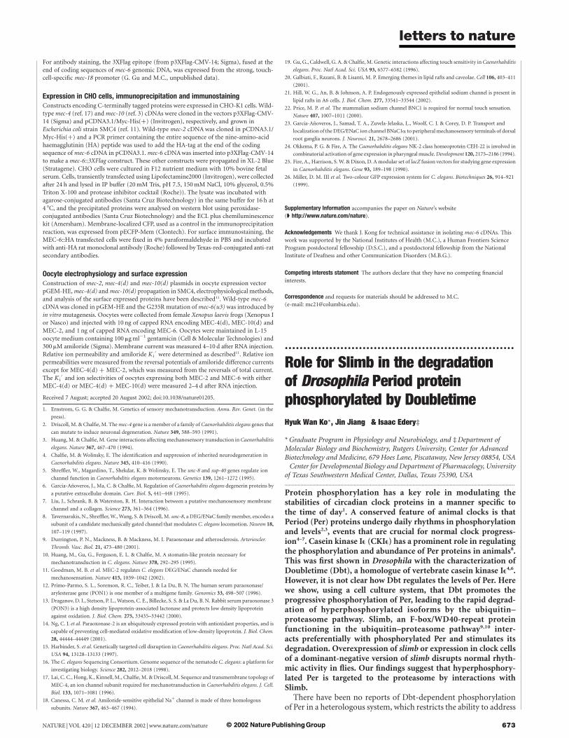
Induction of dbt-V5 led to progressive decreases in the electro-phoretic mobility and abundance of Per (Fig. 1a) similar to thoseobserved for native Per protein in heads from wild-type Drosophila2
(Fig. 1b; compare lanes 1–3 with lanes 4–6). Daily changes in theapparent molecular mass of native Per are mainly or solelyaccounted for by differential phosphorylation2. Similarly, phospha-tase treatment of Per isolated from S2 cells showed that theDbt-induced changes in mobility were caused by phosphorylation(Fig. 1c). The kinetics of Per hyperphosphorylation is tightlycorrelated with the formation of stable interactions between Perand Dbt (Fig. 1d). Increasing the levels of dbt resulted in steadilyfaster rates of Per hyperphosphorylation and degradation (data notshown). These results indicate that Dbt interacts stably with Per andfunctions, at least partly, in a stoichiometric manner, consistentwith recent findings in flies11. In the absence of Dbt there is little orno phosphorylation of Per (Fig. 1a; compare lanes 6 and 7).
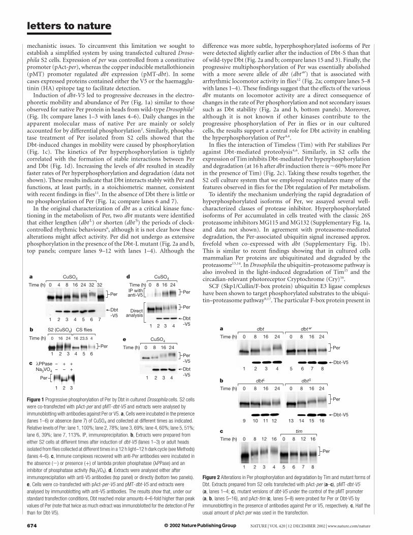
In the original characterization of dbt as a critical kinase func-tioning in the metabolism of Per, two dbt mutants were identifiedthat either lengthen (dbt L) or shorten (dbt S) the periods of clock-controlled rhythmic behaviours6, although it is not clear how thesealterations might affect activity. Per did not undergo as extensivephosphorylation in the presence of the Dbt-L mutant (Fig. 2a and b,top panels; compare lanes 9–12 with lanes 1–4). Although the
difference was more subtle, hyperphosphorylated isoforms of Perwere detected slightly earlier after the induction of Dbt-S than thatof wild-type Dbt (Fig. 2a and b; compare lanes 15 and 3). Finally, theprogressive multiphosphorylation of Per was essentially abolishedwith a more severe allele of dbt (dbt ar) that is associated witharrhythmic locomotor activity in flies12 (Fig. 2a; compare lanes 5–8with lanes 1–4). These findings suggest that the effects of the variousdbt mutants on locomotor activity are a direct consequence ofchanges in the rate of Per phosphorylation and not secondary issuessuch as Dbt stability (Fig. 2a and b, bottom panels). Moreover,although it is not known if other kinases contribute to theprogressive phosphorylation of Per in flies or in our culturedcells, the results support a central role for Dbt activity in enablingthe hyperphosphorylation of Per4,6.
In flies the interaction of Timeless (Tim) with Per stabilizes Peragainst Dbt-mediated proteolysis4,6. Similarly, in S2 cells theexpression of Tim inhibits Dbt-mediated Per hyperphosphorylationand degradation (at 16 h after dbt induction there is ,60% more Perin the presence of Tim) (Fig. 2c). Taking these results together, theS2 cell culture system that we employed recapitulates many of thefeatures observed in flies for the Dbt regulation of Per metabolism.
To identify the mechanism underlying the rapid degradation ofhyperphosphorylated isoforms of Per, we assayed several well-characterized classes of protease inhibitor. Hyperphosphorylatedisoforms of Per accumulated in cells treated with the classic 26Sproteasome inhibitors MG115 and MG132 (Supplementary Fig. 1a,and data not shown). In agreement with proteasome-mediateddegradation, the Per-associated ubiquitin signal increased approx.fivefold when co-expressed with dbt (Supplementary Fig. 1b).This is similar to recent findings showing that in cultured cellsmammalian Per proteins are ubiquitinated and degraded by theproteasome13,14. In Drosophila the ubiquitin–proteasome pathway isalso involved in the light-induced degradation of Tim15 and thecircadian-relevant photoreceptor Cryptochrome (Cry)16.
SCF (Skp1/Cullin/F-box protein) ubiquitin E3 ligase complexeshave been shown to target phosphorylated substrates to the ubiqui-tin–proteasome pathway9,17. The particular F-box protein present in
Figure 2 Alterations in Per phosphorylation and degradation by Tim and mutant forms of
Dbt. Extracts prepared from S2 cells transfected with pAct-per (a–c), pMT-dbt-V5
(a, lanes 1–4; c), mutant versions of dbt-V5 under the control of the pMT promoter
(a, b, lanes 5–16), and pAct-tim (c, lanes 5–8) were probed for Per or Dbt-V5 by
immunoblotting in the presence of antibodies against Per or V5, respectively. c, Half the
usual amount of pAct-per was used in the transfection.
Figure 1 Progressive phosphorylation of Per by Dbt in cultured Drosophila cells. S2 cells
were co-transfected with pAct-per and pMT-dbt-V5 and extracts were analysed by
immunoblotting with antibodies against Per or V5. a, Cells were incubated in the presence
(lanes 1–6) or absence (lane 7) of CuSO4 and collected at different times as indicated.
Relative levels of Per: lane 1, 100%; lane 2, 78%; lane 3, 69%; lane 4, 60%; lane 5, 51%;
lane 6, 39%; lane 7, 113%. IP, immunoprecipitation. b, Extracts were prepared from
either S2 cells at different times after induction of dbt-V5 (lanes 1–3) or adult heads
isolated from flies collected at different times in a 12 h light–12 h dark cycle (see Methods)
(lanes 4–6). c, Immune complexes recovered with anti-Per antibodies were incubated in
the absence (2) or presence (þ) of lambda protein phosphatase (lPPase) and an
inhibitor of phosphatase activity (Na3VO4). d, Extracts were analysed either after
immunoprecipitation with anti-V5 antibodies (top panel) or directly (bottom two panels).
e, Cells were co-transfected with pAct-per-V5 and pMT-dbt-V5 and extracts were
analysed by immunoblotting with anti-V5 antibodies. The results show that, under our
standard transfection conditions, Dbt reached molar amounts 4–6-fold higher than peak
values of Per (note that twice as much extract was immunoblotted for the detection of Per
than for Dbt-V5).
letters to nature
NATURE | VOL 420 | 12 DECEMBER 2002 | www.nature.com/nature674 © 2002 Nature Publishing Group
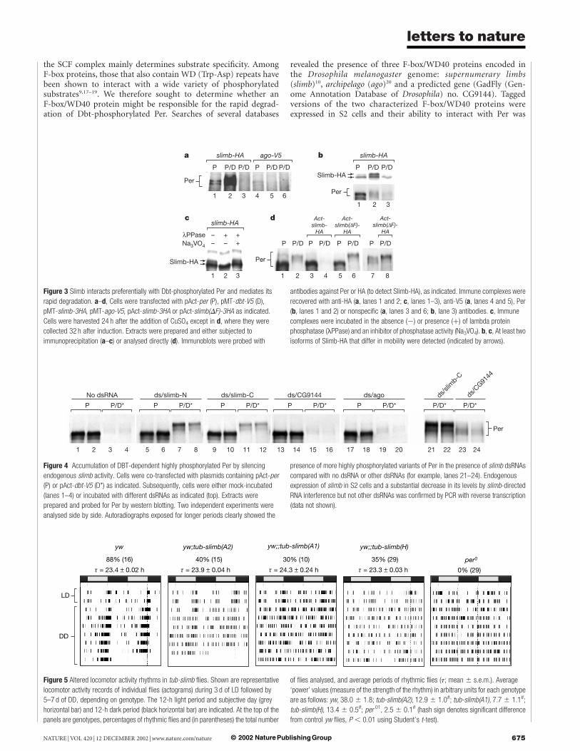
the SCF complex mainly determines substrate specificity. AmongF-box proteins, those that also contain WD (Trp-Asp) repeats havebeen shown to interact with a wide variety of phosphorylatedsubstrates9,17–19. We therefore sought to determine whether anF-box/WD40 protein might be responsible for the rapid degrad-ation of Dbt-phosphorylated Per. Searches of several databases
revealed the presence of three F-box/WD40 proteins encoded inthe Drosophila melanogaster genome: supernumerary limbs(slimb)10, archipelago (ago)20 and a predicted gene (GadFly (Gen-ome Annotation Database of Drosophila) no. CG9144). Taggedversions of the two characterized F-box/WD40 proteins wereexpressed in S2 cells and their ability to interact with Per was
Figure 4 Accumulation of DBT-dependent highly phosphorylated Per by silencing
endogenous slimb activity. Cells were co-transfected with plasmids containing pAct-per
(P) or pAct-dbt-V5 (D*) as indicated. Subsequently, cells were either mock-incubated
(lanes 1–4) or incubated with different dsRNAs as indicated (top). Extracts were
prepared and probed for Per by western blotting. Two independent experiments were
analysed side by side. Autoradiographs exposed for longer periods clearly showed the
presence of more highly phosphorylated variants of Per in the presence of slimb dsRNAs
compared with no dsRNA or other dsRNAs (for example, lanes 21–24). Endogenous
expression of slimb in S2 cells and a substantial decrease in its levels by slimb-directed
RNA interference but not other dsRNAs was confirmed by PCR with reverse transcription
(data not shown).
Figure 5 Altered locomotor activity rhythms in tub-slimb flies. Shown are representative
locomotor activity records of individual flies (actograms) during 3 d of LD followed by
5–7 d of DD, depending on genotype. The 12-h light period and subjective day (grey
horizontal bar) and 12-h dark period (black horizontal bar) are indicated. At the top of the
panels are genotypes, percentages of rhythmic flies and (in parentheses) the total number
of flies analysed, and average periods of rhythmic flies (t; mean ^ s.e.m.). Average
‘power’ values (measure of the strength of the rhythm) in arbitrary units for each genotype
are as follows: yw, 38.0 ^ 1.8; tub-slimb(A2), 12.9 ^ 1.0#; tub-slimb(A1), 7.7 ^ 1.1#;
tub-slimb(H), 13.4 ^ 0.5#; per 01, 2.5 ^ 0.1# (hash sign denotes significant difference
from control yw flies, P , 0.01 using Student’s t-test).
Figure 3 Slimb interacts preferentially with Dbt-phosphorylated Per and mediates its
rapid degradation. a–d, Cells were transfected with pAct-per (P), pMT-dbt-V5 (D),
pMT-slimb-3HA, pMT-ago-V5, pAct-slimb-3HA or pAct-slimb(DF)-3HA as indicated.
Cells were harvested 24 h after the addition of CuSO4 except in d, where they were
collected 32 h after induction. Extracts were prepared and either subjected to
immunoprecipitation (a–c) or analysed directly (d). Immunoblots were probed with
antibodies against Per or HA (to detect Slimb-HA), as indicated. Immune complexes were
recovered with anti-HA (a, lanes 1 and 2; c, lanes 1–3), anti-V5 (a, lanes 4 and 5), Per
(b, lanes 1 and 2) or nonspecific (a, lanes 3 and 6; b, lane 3) antibodies. c, Immune
complexes were incubated in the absence (2) or presence (þ) of lambda protein
phosphatase (lPPase) and an inhibitor of phosphatase activity (Na3VO4). b, c, At least two
isoforms of Slimb-HA that differ in mobility were detected (indicated by arrows).
letters to nature
NATURE | VOL 420 | 12 DECEMBER 2002 | www.nature.com/nature 675© 2002 Nature Publishing Group
determined by immunoprecipitation. Slimb preferentially inter-acted with Dbt-phosphorylated Per, whereas there was little or nointeraction with Ago (Fig. 3a). The preference of Slimb for Dbt-phosphorylated Per cannot be accounted for simply by grossvariations in the levels of Per or the different F-box/WD40 proteins(Supplementary Fig. 2). When extracts were incubated with anti-Per antibodies significantly more Slimb co-purified with Dbt-phosphorylated Per (Fig. 3b, top panel), even though higher levelsof Per are present in immune complexes recovered from cells nottransfected with dbt (Fig. 3b, bottom panel; compare lanes 1 and 2).
We noted that Slimb migrates as a doublet (Fig. 3b and c, andSupplementary Fig. 2), which is consistent with recent findings forSlimb21 and its vertebrate homologue b-TrCP22. The slower-
migrating isoform of Slimb interacts preferentially with phosphory-lated Per (note that a portion of the faster-migrating Slimbco-purified non-specifically) (Fig. 3b). It has been suggested thatphosphorylation might underlie the isoforms of Slimb/b-TrCP22
with different mobilities. Indeed, treatment with phosphataseindicated that the slower-migrating isoform of Slimb is phosphory-lated (Fig. 3c). Our results raise the possibility that the phosphory-lated state of Slimb/b-TrCP modulates its ability to interact withtarget proteins.
High-level expression of slimb from a constitutive promoterspecifically enhanced the degradation of Dbt-phosphorylated Per(Fig. 3d; compare lanes 4 and 2). The F-box mediates interactionswith other core members of the SCF but is not required for the
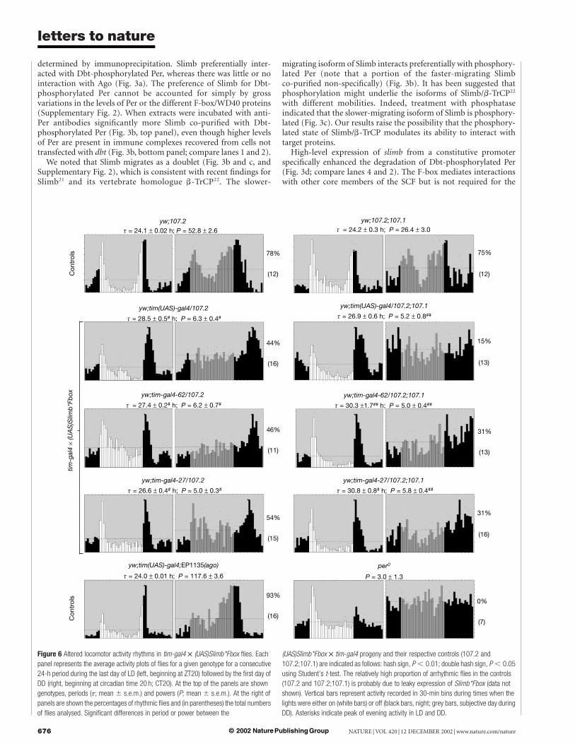
Figure 6 Altered locomotor activity rhythms in tim-gal4 £ (UAS)Slimb*Fbox flies. Each
panel represents the average activity plots of flies for a given genotype for a consecutive
24-h period during the last day of LD (left, beginning at ZT20) followed by the first day of
DD (right, beginning at circadian time 20 h; CT20). At the top of the panels are shown
genotypes, periods (t; mean ^ s.e.m.) and powers (P; mean ^ s.e.m.). At the right of
panels are shown the percentages of rhythmic flies and (in parentheses) the total numbers
of flies analysed. Significant differences in period or power between the
(UAS)Slimb*Fbox £ tim-gal4 progeny and their respective controls (107.2 and
107.2;107.1) are indicated as follows: hash sign, P , 0.01; double hash sign, P , 0.05
using Student’s t-test. The relatively high proportion of arrhythmic flies in the controls
(107.2 and 107.2;107.1) is probably due to leaky expression of Slimb*Fbox (data not
shown). Vertical bars represent activity recorded in 30-min bins during times when the
lights were either on (white bars) or off (black bars, night; grey bars, subjective day during
DD). Asterisks indicate peak of evening activity in LD and DD.
letters to nature
NATURE | VOL 420 | 12 DECEMBER 2002 | www.nature.com/nature676 © 2002 Nature Publishing Group
specific recognition of phosphorylated substrates9,17. Expression of aSlimb mutant lacking the F-box (Slimb(DF)) leads to the accumu-lation of highly phosphorylated isoforms of Per but only in thepresence of Dbt (Fig. 3d, lanes 5–8), strongly suggesting thatSlimb(DF) is functioning as a dominant-negative inhibitor toblock endogenous slimb activity. A similar F-box deletion ofb-TrCP blocked IkBa degradation and NF-kB activation22.
We also used RNA interference23 to silence any endogenous slimbthat might be present in our cultured cells (Fig. 4). Incubation of S2cells with either of two different slimb double-stranded RNAs(dsRNAs) efficiently blocked the degradation of phosphorylatedPer, whereas dsRNAs directed against the other two DrosophilaF-box/WD40 proteins had no noticeable effects on Per metabolism.Furthermore, highly phosphorylated isoforms of Per—not normallydetected—accumulated in the presence of slimb dsRNAs (Fig. 4,lanes 21–24). The addition of slimb dsRNAs did not affect the levelsof Per in the absence of Dbt, which is consistent with the preferentialinteraction of Slimb with Dbt-phosphorylated Per (Fig. 3a and b).
To obtain evidence in vivo that slimb has a role in circadianfunction we used wild-type flies bearing a transgene in which slimbexpression was driven from the constitutive tubulin promoter(tub-slimb). Three independent tub-slimb lines (S102.H, S102.A1and S102.A2) were examined. In standard 12 h light–12 h darkcycles (LD, where zeitgeber time 12 (ZT12) is when the lights areturned off), normal flies exhibit a bimodal distribution of activitycentred on the lights-on (morning peak) and lights-off (eveningpeak) transitions with a robust evening activity rhythm continuingfor many days in constant dark conditions (DD). Although all threestrains of tub-slimb flies entrained (synchronized) to daily light–dark cycles, between 60% and 70% of these flies displayed arrhyth-mic locomotor activity under constant dark conditions (Fig. 5).Expression of several control genes under the regulation of thetubulin promoter did not affect locomotor activity rhythms (datanot shown).
In addition, we used the GAL4–UAS binary system (whichexploits the yeast GAL4 transcriptional activator and the upstreamactivating sequence (UAS))24 to drive the production of a mutantform of Slimb lacking the F-box (Slimb*Fbox) in tim-expressingcells. We used two independent lines of UAS-Slimb*Fbox (107.1 and107.2) and the transheterozygote combination (107.1/107.2), andseparately crossed each one to three different tim-gal4 strains(tim-gal4-27, tim-gal4-62 and tim(UAS)-gal4)25,26. Flies expressingSlimb*Fbox under the control of the different gal4 drivers mani-fested abnormal locomotor activity rhythms in LD (Fig. 6). Notabledifferences were little or no rise in activity before the beginning ofthe 12-h dark phase and a delayed downswing in evening activity.More severe differences were observed in DD, in which the majorityof the tim-gal £ UAS-Slimb*Fbox flies were arrhythmic or had weakrhythms with long periods, consistent with the delayed eveningactivity in LD (Fig. 6). These behavioural defects were generallymore severe with the stronger tim(UAS)-gal4 driver25,26 and withtwo copies of the UAS-Slimb*Fbox transgene (Fig. 6). All thesemutant phenotypes require tim-gal4 drivers, because flies carryingthe UAS-Slimb*Fbox transgene alone showed normal rhythms(Fig. 6). Furthermore, normal activity rhythms were observed inflies in which the expression of ago was regulated by the sametim-gal4 drivers, indicating that the altered rhythm phenotypes arenot due simply to overexpression of any F-box/WD40 protein(Fig. 6). Consistent with the behavioural defects was the westernblot analysis, which showed that the temporal regulation of Perabundance and phosphorylation is delayed in flies expressingSlimb*Fbox under the control of the tim promoter (data notshown).
We suggest that Dbt-mediated phosphorylation of Per is a keyrecognition signal that enables Slimb to recruit phosphorylated Perselectively and target it for rapid degradation by the 26S protea-some. Given the highly conserved nature of circadian clocks in
Drosophila and vertebrates it is tempting to speculate that b-TrCPhas a similar role in regulating the levels of mammalian Perproteins. A
MethodsConstructs and transfectionThe pAct-per27, pAct-tim27 and pCasper-hs-Ub/HA16 constructs were describedpreviously. To generate an expression construct in which ubiquitin (Ub) was under thecontrol of an inducible promoter, the Ub/HA octamer from pCasper-hs-Ub/HA wassubcloned into the plasmid pMT/V5-His (Invitrogen), generating pMT-Ub/HA. For thedbt expression vectors, the coding region of dbt was amplified by polymerase chainreaction (PCR) in the presence of the plasmid pGEM-5Zf/dbt28 and the relevant fragmentwas subcloned into either the pAc5.1/V5-His (Invitrogen) or pMT/V5-His vectors to yieldpAct-dbt-V5, pAct-dbt (by introducing an in-frame stop codon upstream of plasmidsequences encoding V5) and pMT-dbt-V5. The dbt mutant constructs (dbt S, dbtL anddbt ar) were generated by site-directed mutagenesis with the QuickChange site-directedmutagenesis kit (Stratagene) and subcloned into the pMT/V5-His vector. Codingsequences for the F-box proteins were amplified by PCR in the presence of the appropriateexpressed sequence tag clones (LD08669 for slimb and LD21322 for ago) and subclonedinto the pMT/V5-His vector. The slimb open reading frame (ORF) was fused in frame atthe carboxy terminus with sequences encoding three tandem HA epitope tags (3HA)followed by a stop codon, yielding pMT-slimb-3HA. slimb-3HA was also subcloned intothe pAct vector to yield pAct-slimb-3HA. The plasmid pJM1534 (S. Brill, personalcommunication) was used in a PCR of the fragment containing the 3HA sequence. Inaddition, pAct-slimb(DF)-3HA was generated by site-directed mutagenesis by deletingsequences coding for the region containing amino-acid residues 92–136. The agoORF was fused in-frame with sequences encoding the V5 epitope tag, generatingpMT-ago-V5. Relevant parts from all final expression vectors were sequenced beforeuse.
The S2 cells and DES expression medium were purchased from Invitrogen, and allprocedures were performed in accordance with the manufacturer’s instructions.Transfections were performed with Cellfectin (Gibco-BRL) in accordance with themanufacturer’s instructions. Induction of genes under the control of the pMT promoterwas initiated by adding 500 mM CuSO4 to the medium, beginning at 36 h aftertransfection.
RNA-mediated interferenceRNA-mediated interference was applied to S2 cells as described previously23. After theaddition of dsRNA, cells were left to recover for 2 d, transfected with pAct-per andpAct-dbt-V5 plasmids and incubated for a further 1.5 d before cells were harvested. ThedsRNAs used were (numbering of nucleotide sequence begins with start ATG):ds/slimb-N, 49–756; ds/slimb-C, 404–1102; ds/CG9144, 685–1384; and ds/ago, 1–505.
Western blot analysisTransfected cells were washed with serum-free medium and collected by freezing at theindicated times. For each time point, extracts were prepared and immunoblotted asdescribed previously28. Gels differing in polyacrylamide concentration were used toresolve different proteins (6% for Per and Ago; 12% for Slimb; 8% for experimentsinvolving Ub-HA). Primary antibodies were used at the following dilutions: 12CA5anti-HA (Boehringer Mannheim) 1:2000, anti-V5 (Invitrogen) 1:6000, and GP73 anti-Per1:3000 (ref. 29). The intensities of relevant bands on autoradiographs were quantified witha charge-coupled device camera (Alpha Innotech Corporation). Scanned images ofautoradiographs were manipulated with Adobe Photoshop.
Immunoprecipitation and treatment with phosphataseClarified cell extracts (1 mg total protein in a final volume of 0.5 ml) were subjected toimmunoprecipitation with Gammabind Plus (Pharmacia) as described previously28. Weadded 3 ml of anti-V5, anti-HA (12CA5) or 5 ml of anti-Per (GP73) antibodies to theprecleared cell extracts. Treatment of immune complexes with phosphatase wasperformed as described previously28. The phosphatase inhibitor Na3VO4 (40 mM finalconcentration) and/or 200 U of lambda (l) protein phosphatase (NEB) were added asindicated.
Transgenic flies and locomotor activity rhythmsyw, per 01, tim-gal4-27, tim-gal4-62 and tim-(UAS)-gal4 were described previously25,26. TheEP line 1135 (Berkeley Drosophila Genome Project) was used to overexpress ago. Togenerate the tub-slimb construct, cDNA encoding full-length slimb was subcloned into thepCS2 vector between XhoI and XbaI, yielding a slimb with six copies of Myc-tag fused atthe N-terminus. A ClaI–XbaI fragment containing the Myc-tagged slimb coding sequencewas then subcloned into a Casper vector containing the 2.6-kilobase tubulin promoter. Togenerate the Slimb*Fbox construct, sequences encoding an amino-terminal region ofSlimb (residues 1–89) and a C-terminal region (residues 144–510) were amplified by PCRwith pBluscript-Myc-slimb as a template. The two fragments were rejoined and subclonedinto the pUAST vector between the KpnI and XbaI sites to generate pUAST-Myc-Slimb*Fbox. Slimb*Fbox lacks sequences from residues 90–143. All plasmids were injectedinto yw embryos to generate transformants.
For the analysis of locomotor activity, young adult flies were placed in incubators at25 8C, exposed to 3 d of 12 h light followed by 12 h dark (12:12LD) and subsequently keptin constant dark conditions (DD) for 5–8 d. Locomotor activity was analysed with theBrandeis Rhythm Package as described previously30. Power is a quantification of thestrength of the rhythm during DD25,26. Flies with a power $5 and a ‘width’ (number of
letters to nature
NATURE | VOL 420 | 12 DECEMBER 2002 | www.nature.com/nature 677© 2002 Nature Publishing Group
period values in 30-min increments above that line) $2 were considered to berhythmic25,26.
Received 11 September; accepted 5 November 2002; doi:10.1038/nature01272.
Published online 20 November 2002.
1. Edery, I. Role of posttranscriptional regulation in circadian clocks: lessons from Drosophila.
Chronobiol. Int. 16, 377–414 (1999).
2. Edery, I., Zwiebel, L. J., Dembinska, M. E. & Rosbash, M. Temporal phosphorylation of the Drosophila
period protein. Proc. Natl Acad. Sci. USA 91, 2260–2264 (1994).
3. Lee, C., Etchegaray, J. P., Cagampang, F. R., Loudon, A. S. & Reppert, S. M. Posttranslational
mechanisms regulate the mammalian circadian clock. Cell 107, 855–867 (2001).
4. Kloss, B. et al. The Drosophila clock gene double-time encodes a protein closely related to human
casein kinase I1. Cell 94, 97–107 (1998).
5. Lowrey, P. L. et al. Positional syntenic cloning and functional characterization of the mammalian
circadian mutation tau. Science 288, 483–492 (2000).
6. Price, J. L. et al. double-time is a novel Drosophila clock gene that regulates PERIOD protein
accumulation. Cell 94, 83–95 (1998).
7. Toh, K. L. et al. An hPer2 phosphorylation site mutation in familial advanced sleep phase syndrome.
Science 291, 1040–1043 (2001).
8. Eide, E. J. & Virshup, D. M. Casein kinase I: another cog in the circadian clockworks. Chronobiol. Int.
18, 389–398 (2001).
9. Kipreos, E. T. & Pagano, M. The F-box protein family. Genome Biol. 1, 30021–30027 (2000).
10. Jiang, J. & Struhl, G. Regulation of the Hedgehog and Wingless signalling pathways by the F-box/
WD40-repeat protein Slimb. Nature 391, 493–496 (1998).
11. Kloss, B., Rothenfluh, A., Young, M. W. & Saez, L. Phosphorylation of PERIOD is influenced by
cycling physical associations of DOUBLE-TIME, PERIOD, and TIMELESS in the Drosophila clock.
Neuron 30, 699–706 (2001).
12. Rothenfluh, A., Abodeely, M. & Young, M. W. Short-period mutations of per affect a double-time-
dependent step in the Drosophila circadian clock. Curr. Biol. 10, 1399–1402 (2000).
13. Akashi, M., Tsuchiya, Y., Yoshino, T. & Nishida, E. Control of intracellular dynamics of mammalian
period proteins by casein kinase I1 (CKI1) and CKId in cultured cells. Mol. Cell. Biol. 22, 1693–1703
(2002).
14. Yagita, K. et al. Nucleocytoplasmic shuttling and mCRY-dependent inhibition of ubiquitylation of the
mPER2 clock protein. EMBO J. 21, 1301–1314 (2002).
15. Naidoo, N., Song, W., Hunter-Ensor, M. & Sehgal, A. A role for the proteasome in the light response of
the timeless clock protein. Science 285, 1737–1741 (1999).
16. Lin, F. J., Song, W., Meyer-Bernstein, E., Naidoo, N. & Sehgal, A. Photic signaling by cryptochrome in
the Drosophila circadian system. Mol. Cell. Biol. 21, 7287–7294 (2001).
17. Craig, K. L. & Tyers, M. The F-box: a new motif for ubiquitin dependent proteolysis in cell cycle
regulation and signal transduction. Prog. Biophys. Mol. Biol. 72, 299–328 (1999).
18. Koepp, D. M. et al. Phosphorylation-dependent ubiquitination of cyclin E by the SCFFbw7 ubiquitin
ligase. Science 294, 173–177 (2001).
19. Lassot, I. et al. ATF4 degradation relies on a phosphorylation-dependent interaction with the
SCFbTrCP ubiquitin ligase. Mol. Cell. Biol. 21, 2192–2202 (2001).
20. Moberg, K. H., Bell, D. W., Wahrer, D. C., Haber, D. A. & Hariharan, I. K. Archipelago regulates cyclin
E levels in Drosophila and is mutated in human cancer cell lines. Nature 413, 311–316 (2001).
21. Bocca, S. N., Muzzopappa, M., Silberstein, S. & Wappner, P. Occurrence of a putative SCF ubiquitin
ligase complex in Drosophila. Biochem. Biophys. Res. Commun. 286, 357–364 (2001).
22. Spencer, E., Jiang, J. & Chen, Z. J. Signal-induced ubiquitination of IkBa by the F-box protein Slimb/
b-TrCP. Genes Dev. 13, 284–294 (1999).
23. Worby, C. A., Simonson-Leff, N. & Dixon, J. E. RNA interference of gene expression (RNAi) in
cultured Drosophila cells. Sci. STKE 2001, PL1 (2001).
24. Brand, A. H. & Perrimon, N. Targeted gene expression as a means of altering cell fates and generating
dominant phenotypes. Development 118, 401–415 (1993).
25. Kaneko, M., Park, J. H., Cheng, Y., Hardin, P. E. & Hall, J. C. Disruption of synaptic transmission or
clock-gene-product oscillations in circadian pacemaker cells of Drosophila cause abnormal behavioral
rhythms. J. Neurobiol. 43, 207–233 (2000).
26. Blau, J. & Young, M. W. Cycling vrille expression is required for a functional Drosophila clock. Cell 99,
661–671 (1999).
27. Ceriani, M. F. et al. Light-dependent sequestration of TIMELESS by CRYPTOCHROME. Science 285,
553–556 (1999).
28. Bae, K., Lee, C., Hardin, P. E. & Edery, I. dCLOCK is present in limiting amounts and likely mediates
daily interactions between the dCLOCK-CYC transcription factor and the PER—TIM complex.
J. Neurosci. 20, 1746–1753 (2000).
29. Sidote, D., Majercak, J., Parikh, V. & Edery, I. Differential effects of light and heat on the Drosophila
circadian clock proteins PER and TIM. Mol. Cell. Biol. 18, 2004–2013 (1998).
30. Kim, E. Y. et al. Drosophila CLOCK protein is under posttranscriptional control and influences light-
induced activity. Neuron 34, 69–81 (2002).
Supplementary Information accompanies the paper on Nature’s website
(ç http://www.nature.com/nature).
Acknowledgements We thank S. Kay for the pAct-per and pAct-tim constructs, A. Seghal for the
pCasper-hs-Ub/HA construct, J. Hall for the tim-gal4-27 and tim-gal4-62 lines, and M. Young for
the tim-(UAS)-gal4 line. The work was supported by the Searle Scholar Program and the UTSW
Endowed Scholar Program to J.J., and by a grant from the National Institutes of Health to I.E.
Competing interests statement The authors declare that they have no competing financial
interests.
Correspondence and requests for materials should be addressed to I.E.
(e-mail: [email protected]).
..............................................................
HIV-1 evades antibody-mediatedneutralization throughconformational maskingof receptor-binding sitesPeter D. Kwong*†, Michael L. Doyle‡§, David J. Casper‡, Claudia Cicalak,Stephanie A. Leavitt{, Shahzad Majeed*†, Tavis D. Steenbekek,Miro Venturi*, Irwin Chaiken#, Michael Fungq, Hermann Katinger**,Paul W. I. H. Parren††, James Robinson‡‡, Donald Van Ryk#,Liping Wang§§, Dennis R. Burton††, Ernesto Freire{, Richard Wyatt*§§,Joseph Sodroski§§kk, Wayne A. Hendrickson†{{ & James Arthosk
* Vaccine Research Center, and kNational Institute of Allergy and InfectiousDiseases, National Institutes of Health, Bethesda, Maryland 20892, USA† Department of Biochemistry and Molecular Biophysics, and{{Howard Hughes Medical Institute, Columbia University, New York, New York10032, USA‡ Department of Structural Biology, GlaxoSmithKline Pharmaceuticals, King ofPrussia, Pennsylvania 19406, USA{Department of Biology, Johns Hopkins University, Baltimore, Maryland 21218,USA# Department of Medicine, University of Pennsylvania, Philadelphia,Pennsylvania 19104, USAq Cell Biology, Tanox, Houston, Texas 77025, USA** Institute for Applied Microbiology, University of Agriculture and Forestry,A-1190 Vienna, Austria†† Departments of Immunology and Molecular Biology, Scripps ResearchInstitute, La Jolla, California 92037, USA‡‡ Department of Pediatrics, Tulane University Medical Center, New Orleans,Louisiana 70112, USA§§ Department of Cancer Immunology and AIDS, Dana-Farber Cancer Institute,and Department of Pathology, Division of AIDS, Harvard Medical School, andkkDepartment of Immunology and Infectious Diseases, Harvard School of PublicHealth, Boston, Massachusetts 02115, USA.............................................................................................................................................................................
The ability of human immunodeficiency virus (HIV-1) to persistand cause AIDS is dependent on its avoidance of antibody-mediated neutralization. The virus elicits abundant, envelope-directed antibodies that have little neutralization capacity1. Thislack of neutralization is paradoxical, given the functional con-servation and exposure of receptor-binding sites on the gp120envelope glycoprotein, which are larger than the typical antibodyfootprint2 and should therefore be accessible for antibody bind-ing. Because gp120–receptor interactions involve conformationalreorganization3, we measured the entropies of binding for 20gp120-reactive antibodies. Here we show that recognition byreceptor-binding-site antibodies induces conformational change.Correlation with neutralization potency and analysis of recep-tor–antibody thermodynamic cycles suggested a receptor-bind-ing-site ‘conformational masking’ mechanism of neutralizationescape. To understand how such an escape mechanism would becompatible with virus–receptor interactions, we tested a solubledodecameric receptor molecule and found that it neutralizedprimary HIV-1 isolates with great potency, showing that simul-taneous binding of viral envelope glycoproteins by multiplereceptors creates sufficient avidity to compensate for such mask-ing. Because this solution is available for cell-surface receptorsbut not for most antibodies, conformational masking enablesHIV-1 to maintain receptor binding and simultaneously to resistneutralization.
The gp120 envelope glycoprotein binds sequentially to thecellular receptors CD4 and a member of the chemokine receptor
§ Present address: Biopharmaceuticals Department H13-07, Bristol-Myers Squibb PRI, Pharmaceutical
Research Institute, P.O. Box 4000, Princeton, New Jersey 08543-4000, USA.
letters to nature
NATURE | VOL 420 | 12 DECEMBER 2002 | www.nature.com/nature678 © 2002 Nature Publishing Group