Embed Size (px)
Citation preview
Roco kinase structures give insights into themechanism of Parkinson disease-relatedleucine-rich-repeat kinase 2 mutationsBernd K. Gilsbacha,b, Franz Y. Hoc, Ingrid R. Vetterb, Peter J. M. van Haasterta, Alfred Wittinghoferb,1,and Arjan Kortholta,b,1
aDepartment of Cell Biochemistry, University of Groningen, 9747 AG, Groningen, The Netherlands; bStructural Biology Group, Max Planck Institut fürMolekulare Physiologie, 44227 Dortmund, Germany; and cDepartment of Neurobiology, University of Eastern Finland, 70211 Kuopio, Finland
Edited by Tony Hunter, The Salk Institute for Biological Studies, La Jolla, CA, and approved May 21, 2012 (received for review February 25, 2012)
Mutations in human leucine-rich-repeat kinase 2 (LRRK2) have beenfound to be the most frequent cause of late-onset Parkinsondisease. Here we show that Dictyostelium discoideum Roco4 isa suitable model to study the structural and biochemical character-istics of the LRRK2 kinase and can be used for optimization of cur-rent and identification of new LRRK2 inhibitors. We have solved thestructure of Roco4 kinase wild-type, Parkinson disease-relatedmutants G1179S and L1180T (G2019S and I2020T in LRRK2) andthe structure of Roco4 kinase in complex with the LRRK2 inhibitorH1152. Taken together, our datagive important insight in the LRRK2activation mechanism and, most importantly, explain the G2019S-related increase in LRRK2 kinase activity.
Leucine-rich-repeat kinase 2 (LRRK2) belongs to the Rocofamily of proteins, which are characterized by the presence of
leucine-rich repeats, a Ras-like G-domain (called “Roc”), a Cterminal of ROC (COR) domain, and a kinase domain (1). Re-cently, missense mutations in LRRK2 have been linked to auto-somal-dominant, late-onset Parkinson disease (PD) (2, 3). PD isa common neurodegenerative disorder characterized by a pro-gressive loss of dopaminergic neurons of the substantia nigra, as-sociated with the formation of fibrillar aggregates composed ofα-synuclein and other proteins. PD is characterized clinically bytremor, bradykinesia, rigidity, and postural instability. The iden-tification of missense mutations in LRRK2 has redefined the roleof genetic variation in PD susceptibility. LRRK2 mutations initi-ate a penetrant phenotype with complete clinical and neuro-chemical overlap with idiopathic disease (4–6). The variousmutations that have been identified in PD are concentrated in thecentral region of the protein; one residue mutated in the LRRregion, one in theRoc domain (withmultiple substitutions), one inthe COR domain, and two in the kinase domain (7). The muta-tions are found in 5–6% of patients with familial PD and, im-portantly, also have been implicated in sporadic PD (8, 9).Although much progress has been made during the last few years,the exact pathogenic role and associated biochemical pathwaysresponsible for LRRK2-linked disease are emerging only slowly(10). The multiple disease-linked mutations in LRRK2 repre-sent a unique opportunity to explore the pathogenicity ofLRRK2 biochemically and to identify therapeutic targets for thisneurodegenerative disorder.In the absence of suitable amounts of purified mammalian
LRRK2 protein, and because recombinantly expressed full-lengthprotein or any fragment thereof turned out to be unstable, insoluble,or permanently bound to chaperones, structural understanding ofLRRK2 is very limited (11). Therefore, we used related proteins toinvestigate the complex structural regulatory mechanism ofLRRK2. Previously we elucidated the structure of the RocCORtandem ofChlorobium tepidum, which shows that theRoc domain isa Ras-like G domain tightly coupled to the COR domain as a di-merization device. Mutations analogous to Parkinson mutationswere shown to be located in the Roc–COR interface. RocCORproteins thus seem to belong to the G proteins activated by the
nucleotide-dependent dimerization (GAD) class of molecularswitches. PD-analogous mutations in Roc and COR alter the Roc–COR interface and result in decreasedGTPase activity (12, 13). Thestructure of the Roc domain of human LRRK2 showed a domain-swapped dimeric G domain whose significance for the native pro-tein is unclear (11, 13).LRRK2 kinase activity is linked critically to clinical effects, and
several pathogenic mutations in LRRK2 result in enhanced kinaseactivity, suggesting a possible PD-related gain of abnormal ortoxic function (14–16). However, because of the lack of sufficientrecombinant protein and physiological substrate, the publisheddata regarding kinase activity of the PD-related mutants areconflicting (except for G2019S, which is associated consistentlywith an increased kinase activity) (17, 18).Here we use Dictyostelium discoideum Roco4 as model to study
the structural and biochemical characteristics of the LRRK2 kinasedomain. We have solved the structure of Roco4 kinase wild-typeand PD-relatedmutants G1179S and L1180T (G2019S and I2020Tin LRRK2). A comparison of wild-type and mutant structuresrevealed that the PD mutants have different effects and, mostimportantly, explains the G2019S-related increase in LRRK2 ki-nase activity. Identifying small-molecule inhibitors of the kinaseactivity that specifically counteract the effect in vivo will be animportant step towards finding a treatment for PD. The structureof Roco4 kinase in complex with the LRRK2 inhibitor H1152shows that Roco4 is a suitable model system to obtain insight intothe binding mechanism and to optimize current and identify newLRRK2 inhibitors.
Results and DiscussionVertebrates possess four Roco proteins, LRRK1, LRRK2,DAPK1, and MFHAS1. Remarkably, the social amoeba Dictyos-telium contains 11 Roco family members that contain a large va-riety of domains and have been studied in detail (19–21). In thisstudy we used Dictyostelium Roco4, which has the same domaintopology as LRRK2 (Fig. 1A) but is biochemically more tractable.The kinase domain is well conserved in the Roco family of pro-teins, and the Roco4 kinase domain (amino acids 1018–1292) hasa similarity of 47% to LRRK2. Unlike LRRK2, the Roco4 kinasedomain could be expressed in Escherichia coli and isolated as
Author contributions: P.J.M.v.H., A.W., and A.K. designed research; B.K.G., F.Y.H., andA.K. performed research; B.K.G., F.Y.H., I.R.V., and A.K. analyzed data; and B.K.G., P.J.M.v.H.,A.W., and A.K. wrote the paper.
The authors declare no conflict of interest.
This article is a PNAS Direct Submission.
Data deposition: The structures reported in this article have been deposited in the ProteinData Bank, www.pdb.org [PDB code 4F0F (wild type active), 4F0G (wild type inactive),4F1M (G1179S), 4F1O (L1180T), and 4F1T (wild type in complex with H1152)].1To whom correspondence may be addressed. E-mail: [email protected] or [email protected].
This article contains supporting information online at www.pnas.org/lookup/suppl/doi:10.1073/pnas.1203223109/-/DCSupplemental.
10322–10327 | PNAS | June 26, 2012 | vol. 109 | no. 26 www.pnas.org/cgi/doi/10.1073/pnas.1203223109
Dow
nloa
ded
by g
uest
on
Dec
embe
r 17
, 202
1
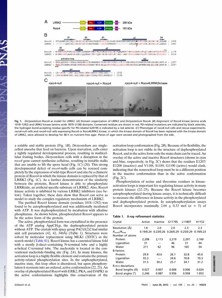
a soluble and stable protein (Fig. 1B). Dictyostelium are single-celled amoeba that feed on bacteria. Upon starvation, cells entera tightly regulated developmental process, resulting in multicel-lular fruiting bodies. Dictyostelium cells with a disruption in theroco4 gene cannot synthesize cellulose, resulting in instable stalksthat are unable to lift the spore head (Fig. 1C) (20). This strongdevelopmental defect of roco4-nulls cells can be rescued com-pletely by the expression of wild-typeRoco4 and also by a chimericprotein of Roco4 in which the kinase domain is replaced by that ofLRRK2 (Fig. 1C). As a further demonstration of the similaritybetween the proteins, Roco4 kinase is able to phosphorylateLRRKtide, an artificial specific substrate of LRRK2. Also, Roco4kinase activity is inhibited by various LRRK2 inhibitors (see be-low). Taken together, these data show that Roco4 can serve asmodel to study the complex regulatory mechanism of LRRK2.The purified Roco4 kinase domain (residues 1018–1292) was
found to be autophosphorylated and was additionally incubatedwith ATP. It was dephosphorylated by incubation with alkalinephosphatase. As shown below, phosphorylated Roco4 appears tobe the active form of the protein.The active, phosphorylated form was crystallized in the presence
of the ATP analog AppCH2p, the dephosphorylated proteinwithout ATP. The crystals with space group P4(3)3(2)2 had similarunit cell parameters (42, 42, 340Å) (Table 1). Structures weresolved by molecular replacement using MLK1 (3DTC) as thesearch model (Table S1). Roco4 kinase has a canonical kinase foldwith a mostly β-sheet–containing N-terminal lobe and a highlyα-helical C-terminal lobe. The nucleotide is located in the con-ventional nucleotide-binding site (Fig. 2A). In many kinases, theactivation loop is a highly flexible element and contains the primaryactivity-related phosphorylation sites. In the unphosphorylated,inactive state, this loop often is disordered, but upon phosphory-lation it reorients into an ordered, active conformation (22, 23). Anoverlay of phosphorylatedRoco4 with ERK2, PKA, andDAPK1 inthe active conformations highlights this conservation of the
activation loop conformation (Fig. 2B). Because of its flexibility, theactivation loop is not visible in the structure of dephosphorylatedRoco4, and in the active form only themain chain can be traced. Anoverlay of the active and inactive Roco4 structures (shown in cyanand blue, respectively, in Fig. 2C) shows that the residues E1207/E1208 (inactive) and V1188, S1189, G1190 (active) would clash,indicating that the nonresolved loop must be in a different positionin the inactive conformation than in the active conformation(Fig. 2C).Phosphorylation of serine and threonine residues in kinase-
activation loops is important for regulating kinase activity in manyprotein kinases (22–25). Because the Roco4 kinase becomesautophosphorylated during kinase assays, it is technically difficultto measure the difference in kinase activity in the phosphorylatedand dephosphorylated protein. In autophosphorylation assaysRoco4 incorporates maximally 2.04 ± 0.33 mol (n = 5) of
Fig. 1. Dictyostelium Roco4 as model for LRRK2. (A) Domain organization of LRRK2 and Dictyostelium Roco4. (B) Alignment of Roco4 kinase (amino acids1018–1292) and LRRK2 kinase (amino acids 1879–2138) domains. Conserved residues are shown in red, PD-related mutations are indicated by black asterisks,the hydrogen bond-accepting residue specific for PD-related G2019S is indicated by a red asterisk. (C) Phenotype of roco4-null cells and rescue experiments.roco4-null cells and roco4-null cells expressing Roco4 or Roco4LRRK2 kinase, in which the kinase domain of Roco4 has been replaced with the kinase domainof LRRK2, were allowed to develop for 48 h on nutrient-free agar. Pieces of agar were excised and photographed from the side.
Table 1. X-ray refinement statistics
Crystal Active Inactive G1179S L1180T H1152
Resolution (Å) 1.8 2.0 2.0 2.3 2.3Rwork/Rfree 0.19/0.24 0.23/0.26 0.26/0.29 0.25/0.29 0.19/0.23Number of atoms
Protein 2,208 2,113 2,219 2,207 2,160Ligand/ion 31 — 46 31 44Water 211 62 96 165 54
B-factorsProtein 29.9 43.6 26.1 32.8 45.6Ligand/ion 55.3 — 24.6 70.8 70.3Water 31.5 30.2 25.7 34.1 31.2
R.m.s deviationsBond lengths (Å) 0.027 0.007 0.006 0.006 0.024Bond angles (°) 2.246 0.987 0.956 0.958 1.953
Gilsbach et al. PNAS | June 26, 2012 | vol. 109 | no. 26 | 10323
BIOCH
EMISTR
Y
Dow
nloa
ded
by g
uest
on
Dec
embe
r 17
, 202
1

phosphate per mole of protein. Roco4 kinase contains four pu-tative phosphorylation sites in the activation loop: S1181, S1184,S1187, and S1189 (Figs. 1B and 2A). To characterize the Roco4activation mechanism, we reexpressed Roco4 constructs with S-to-A mutations in roco4-null cells and analyzed them for de-velopment (Fig. 2D). By reexpressing single mutants and doublemutants, we find that the double mutant S1181/1184, but notS1187/1189, rescues the developmental phenotype. Consistently,the purified S1187 and S1189 single mutants and the doublemutant S1187/1189 have hardly any kinase activity, whereas thedouble mutant S1181/1184 shows wild-type activity (Fig. S1). Thedata show that Roco4 incorporates approximately two moles ofphosphate and that serine 1187 and serine 1189 are essential forkinase activity. This finding supports the notion that autophos-phorylation in the activation loop is required to induce the activeconformation of the kinase.The putative autophosphorylation sites are not conserved be-
tween Roco4 and LRRK2 (Fig. 1B). LRRK2 contains T2031/S2032/T2035 (Fig. 1B), three potential phosphorylation sites inthe activation loop. Studies using phosphospecific antibodies haveshown that all three sites are phosphorylated, but as in Roco4, onlythe latter two sites, S2032 and T2035, are important for LRRK2activity in vivo (26, 27).
PD-linked mutations have been identified throughout theLRRK2 gene; one residue is mutated in the LRR region, one inthe Roc domain (with multiple substitutions), one in the CORdomain, and two in the kinase domain (7). The most prevalent PDmutation is G2019S in the kinase domain, which enhances kinaseactivity, whereas the PD-related mutation I2020T shows slightlydecreased activity (15, 16, 28–30). The LRRK2 G2019 and I2020residues are conserved in Roco4 and correspond to G1179 andL1180, respectively (Fig. 1B). We have created the correspondingmutations in Roco4 and determined their biochemical andstructural properties.Active phosphorylatedRoco4 kinase phosphorylates LRRKtide
with a rate constant of 1.5·103 s−1 (± 427) (Fig. 3A). Like LRRK2,the Roco4 G1179S mutant showed a 1.5 ± 0.13-fold increasedactivity in autophosphorylation, relative to wild-type kinase,whereas the L1180T mutant shows a slightly but significantly de-creased activity (0.8 ± 0.02) (Fig. 3B). The structures of the Rocomutants L1180T and G1179S were solved by molecular re-placement to a resolution of 2.3 and 2.04 Å, respectively. Com-parison of wild-type and mutant structures did not show largedifferences in the overall structure (rmsd 0.6) (Fig. 3C andD). Anoverlay of wild-type and themost prevalent PDhomologmutation,G1179S, revealed an additional hydrogen bond between the mu-tated S1179 and an R1077 from the regulatory αC-helix, (Fig. 3C,
Fig. 2. (A) Ribbon diagram of Roco4 kinase in the active statewith AppCH2p (red ball and stick model) bound in the nucle-otide-binding pocket. The activation loop and the regulatoryαC-helix are shown in green and blue, respectively. Relevantside chains are indicated. In the enlarged view, residues ho-mologous to relevant PD mutations (in blue/red) and putativephosphorylation sites within the activation loop (in red) arehighlighted. (B) Overlay of active phosphorylated Roco4 (cyan)with several kinase structures in the active conformation: ERK2(2ERK) (green), PKA (2CPK) (orange), and DAPK1 (3GU5) (yel-low). The enlarged view highlights the activation loops. (C)Overlay of Roco4 in the active (cyan) and inactive (blue) forms.The enlarged view highlights the clash of the residues 1207/1208 (inactive) with 1188–1190 (active). (D) Analysis of Roco4autophosphorylation sites in vivo. Roco4-null cells expressingthe indicated serine mutants were analyzed for developmentas described in Fig. 1C. Pictures were taken from above. Insetsshow a side view of a fruiting body. The double-mutantS1187A/S1189A is not able to rescue the developmental defectof roco4-null cells.
10324 | www.pnas.org/cgi/doi/10.1073/pnas.1203223109 Gilsbach et al.
Dow
nloa
ded
by g
uest
on
Dec
embe
r 17
, 202
1
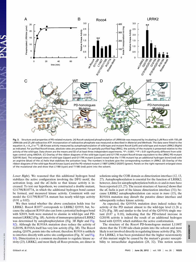
Lower Right). We reasoned that this additional hydrogen bondstabilizes the active configuration involving the DFG motif, theactivation loop, and the αC-helix so that kinase activity is in-creased. To test our hypothesis, we constructed a double mutant,G1179S/R1077A, in which the additional hydrogen bond cannotbe formed, and measured kinase activity. Consistent with ourmodel the G1179S/R1077A mutant has nearly wild-type activity(0.91 ± 0.02).We then tested whether the above conclusion holds true for
LRRK2. Roco4 R1077 corresponds to LRRK2 Q1919, but, be-cause the adjacent R1918 also can form a potential hydrogen bondwith S2019, both were mutated to alanine in wild-type and PD-mutant LRRK2 (Fig. 1B). Activity of immunoprecipitated LRRK2was determined by autophosphorylation (Fig. 3B Right and Fig.S2). Although the R1918A mutation does reduce the activity ofG2019S, R1918A itself has very low activity (Fig. 3B). The Roco4analog, Q1076, points into the solvent; therefore R1918 is unlikelyto interfere directly with active site configuration (Fig. 3C and Fig.S3). Dimerization is a common mechanism to regulate kinase ac-tivity (23). LRRK2, and most likely all Roco proteins, are dimer in
solutions using the COR domain as dimerization interface (12, 13,27). Autophosphorylation is essential for the function of LRRK2;however, data for autophosphorylation both in cis and in trans havebeen reported (15, 27). The recent structure of Aurora2 shows thatthe αC-helix is part of the kinase dimerization interface (31); be-cause LRRK2 autophosphorylation can occur in trans (15), theR1918A mutation may disturb the putative dimer interface andsubsequently reduce kinase activity.As expected, the Q1919A mutation does indeed reduce the
activity of the PD mutant almost to the wild-type level (1.26 ±0.25) (Fig. 3B) and similar to the level of the Q1919A single mu-tant (0.87 ± 0.18), indicating that the PD-related increase inG2019S activity is indeed the result of an additional hydrogenbond with Q1919, which stabilizes the active conformation.The structure of the Roco4 PD-homologous mutant L1180T
shows that the T1180 side-chain points into the solvent and mostlikely is not involved directly in regulating kinase activity (Fig. 3D).For LRRK2, it has been postulated that the higher neurotoxicityof this mutant might be caused by the mutant’s greater suscepti-bility to intracellular degradation (28, 32). This notion seems
Fig. 3. Structure and properties of PD-related mutants. (A) Roco4-catalyzed phosphorylation of LRRKtide was measured by incubating 3 μM Roco with 150 μMLRRKtide and 25 μM radioactive ATP. Incorporation of radioactive phosphate was measured as described inMaterial and Methods. The data were fitted to theequation At = A∞(1-e
−kt). (B) Kinase activity measured by autophosphorylation of wild-type and mutant Roco4 (Left) and wild-type and mutant LRRK2 (Right)as indicated. For purified Roco4 kinase, absolute rates are presented. For partially purified flag-LRRK2, the activity of the mutants is presented relative to theactivity of the wild type. Data shown are the means and SD of at least three independent experiments. *P < 0.001; **P < 0.01 significantly different from wild-type control using ANOVA. (C) Overlay of the ribbon diagrams of the wild-type (cyan) and G1179S mutant Roco4 kinase equivalent to the LRRK2 PD-mutantG2019S (tan). The enlarged views of wild type (Upper) and G1179S mutant (Lower) reveal that the 1179S mutant has an additional hydrogen bond (red) withan arginine (blue) of the αC-helix that stabilizes the activation loop. The numbers in brackets give the corresponding numbers in LRRK2. (D) Overlay of theribbon diagrams of the wild-type Roco4 kinase (cyan) and the PD-related mutant L1180T (LRRK2 I2020T) (green). Panels on the right represent enlarged viewsof the mutational site and show that L1180 (cyan) and T1180 (red) point into the solvent.
Gilsbach et al. PNAS | June 26, 2012 | vol. 109 | no. 26 | 10325
BIOCH
EMISTR
Y
Dow
nloa
ded
by g
uest
on
Dec
embe
r 17
, 202
1
unlikely, given that the disease phenotype is autosomal dominantand caused by a gain of function. Although the Roco4 structuredoes not reveal the exact mechanism, we speculate that, in analogyto the lower activity of B-Raf mutations, which are complementedby interaction with c-Raf, the kinase domains in LRRK2 work intandem so that the interaction between wild-type and LRRK2-T1180 increases kinase activity (33). More importantly, the datado show that the PD-related effect of LRRK2 mutations resultsfrom different defects in the LRRK2 activation mechanism andsuggest that the different LRRK2 mutations, such as S2019 andI2020, might require different methods of inhibition for the pur-pose of drug development.To date, several relatively nonspecific kinase inhibitors, such as
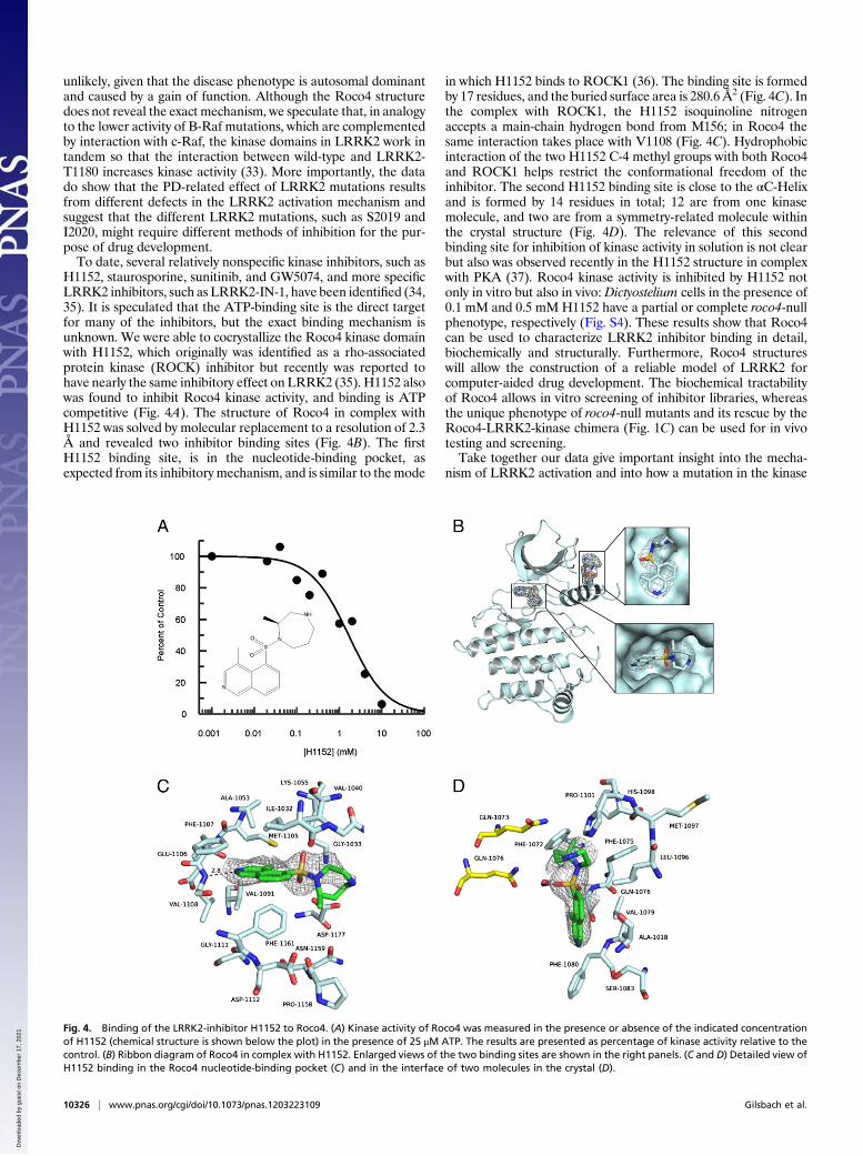
H1152, staurosporine, sunitinib, and GW5074, and more specificLRRK2 inhibitors, such as LRRK2-IN-1, have been identified (34,35). It is speculated that the ATP-binding site is the direct targetfor many of the inhibitors, but the exact binding mechanism isunknown. We were able to cocrystallize the Roco4 kinase domainwith H1152, which originally was identified as a rho-associatedprotein kinase (ROCK) inhibitor but recently was reported tohave nearly the same inhibitory effect on LRRK2 (35). H1152 alsowas found to inhibit Roco4 kinase activity, and binding is ATPcompetitive (Fig. 4A). The structure of Roco4 in complex withH1152 was solved by molecular replacement to a resolution of 2.3Å and revealed two inhibitor binding sites (Fig. 4B). The firstH1152 binding site, is in the nucleotide-binding pocket, asexpected from its inhibitory mechanism, and is similar to themode
in which H1152 binds to ROCK1 (36). The binding site is formedby 17 residues, and the buried surface area is 280.6 Å2 (Fig. 4C). Inthe complex with ROCK1, the H1152 isoquinoline nitrogenaccepts a main-chain hydrogen bond from M156; in Roco4 thesame interaction takes place with V1108 (Fig. 4C). Hydrophobicinteraction of the two H1152 C-4 methyl groups with both Roco4and ROCK1 helps restrict the conformational freedom of theinhibitor. The second H1152 binding site is close to the αC-Helixand is formed by 14 residues in total; 12 are from one kinasemolecule, and two are from a symmetry-related molecule withinthe crystal structure (Fig. 4D). The relevance of this secondbinding site for inhibition of kinase activity in solution is not clearbut also was observed recently in the H1152 structure in complexwith PKA (37). Roco4 kinase activity is inhibited by H1152 notonly in vitro but also in vivo: Dictyostelium cells in the presence of0.1 mM and 0.5 mM H1152 have a partial or complete roco4-nullphenotype, respectively (Fig. S4). These results show that Roco4can be used to characterize LRRK2 inhibitor binding in detail,biochemically and structurally. Furthermore, Roco4 structureswill allow the construction of a reliable model of LRRK2 forcomputer-aided drug development. The biochemical tractabilityof Roco4 allows in vitro screening of inhibitor libraries, whereasthe unique phenotype of roco4-null mutants and its rescue by theRoco4-LRRK2-kinase chimera (Fig. 1C) can be used for in vivotesting and screening.Take together our data give important insight into the mecha-
nism of LRRK2 activation and into how a mutation in the kinase
Fig. 4. Binding of the LRRK2-inhibitor H1152 to Roco4. (A) Kinase activity of Roco4 was measured in the presence or absence of the indicated concentrationof H1152 (chemical structure is shown below the plot) in the presence of 25 μM ATP. The results are presented as percentage of kinase activity relative to thecontrol. (B) Ribbon diagram of Roco4 in complex with H1152. Enlarged views of the two binding sites are shown in the right panels. (C and D) Detailed view ofH1152 binding in the Roco4 nucleotide-binding pocket (C) and in the interface of two molecules in the crystal (D).
10326 | www.pnas.org/cgi/doi/10.1073/pnas.1203223109 Gilsbach et al.
Dow
nloa
ded
by g
uest
on
Dec
embe
r 17
, 202
1
domain increases kinase activity. Although mutants spread over allparts of the multidomain protein LRRK2 produce a similar path-ogenic output signal, i.e., PD, our structures show that differentmutations have different defects in the activation mechanism. Fora further understanding of other mutations, it will be important tocharacterize fully the intramolecular regulation of LRRK2 andshow how the Roc domain might regulate kinase activity, the rolethat COR plays in this process, and how PD-linked missensemutations alter the interactions between the different domains.Our work shows that structures of the more tractable Roco4 andpossibly other Roco proteins could be important in this enterprise.
Materials and MethodsProtein Purification and Radiometric Assays. Roco4 kinase (amino acids 1018–1292) was cloned into a Gateway-compatible pGEX4T1 plasmid containingan N-terminal TEV cleavage side. Proteins were purified in the presence of1 mM ATP by GSH affinity, cleavage, and size-exclusion chromatography.Dephosphorylated Roco4 kinase was obtained by incubating 1 mg isolatedprotein with 100 U alkaline phosphate for 1 h at 4 °C. Roco4 kinase activitywas determined at 30 °C in kinase buffer consisting of 20 mM Tris (pH 7.5),10 mM MgCl2, 1 mM EGTA, 1 mM sodium orthovanadate, 1 mM NaF, 5 mMβ-glycerolphosphate, 0.02% Triton X-100, and 2 mM DTT. Autophosphor-ylation was measured with 1 mg/mL purified protein. The reaction wasstarted by adding 50 μM (2 Ci/mmol) ATP-γ-32P (Perkin-Elmer) and wasstopped by adding 100 mM ice-cold EDTA. Samples were spotted on nitro-cellulose filters, washed with 50 mM phosphoric acid, and dried beforescintillation counting (Perkin-Elmer). For LRRKtide, 32P incorporation assayssimilar to those described above were performed with 0.05 mg/mL kinase,25 μM (2 Ci/mmol) ATP-γ-32P, and 150 μM LRRKtide. Kinase inhibition was
determined by varying the concentration of H1152 (Tocris Bioscience). Hu-man Flag-LRRK2 was expressed and isolated from HEK293T cells by immu-noprecipitation using anti-Flag (M5) antibody (Sigma), and kinase activitywas measured as previously described (30).
Crystallography. Roco4 crystals were obtained in 100 mM 1,3-bis(tris(hydroxymethyl)methylamino)propane (pH 8,5), 200 mM Na/K tartrate, and11% (wt/vol) PEG 3350 using the hanging drop/vapor diffusion method. Fordata collection, crystals were cryoprotected in reservoir solution containing35% (wt/vol) PEG3350 as cryoprotectant. Datasets were collected on beamline ×10SA at the Swiss Light Source (Paul Scherrer Institut, Villigen, Swit-zerland) and were indexed, integrated, and scaled with the XDS package(38). The model was built in COOT (39) and refined with REFMAC5 using TLS-refinement (CCP4 suite) (40). Figures were generated using PYMOL (DeLanoScientific LLC).
Dictyostelium Aggregation Assays. Development of Dictyostelium mutantswas tested as previously described (20). Briefly, cells were harvested inphosphate buffer (11 mM KH2PO4, 2.8 mM Na2HPO4), plated at a density of1 × 106 cells/cm2, and allowed to develop for 48 h on nutrient-free platescontaining phosphate buffer supplemented with 15 g/L agar. Pieces of agarwere excised and photographed from the top and side.
ACKNOWLEDGMENTS. We thank Matthieu Bosman for his input in theproject, Patricia Stege and Ineke Keizer-Gunnink for technical assistance, theX-ray communities of Max Planck Institutes Dortmund and Heidelberg,Eckhard Hofmann (University Bochum) and the beamline staff of X10SA (SwissLight Source, Paul Scherrer Institut, Villigen, Switzerland) for support and datacollection, and Maarten Linskens for reading the manuscript. Funding wasprovided by the Michael J. Fox Foundation for Parkinson’s Research.
1. Bosgraaf L, Van Haastert PJ (2003) Roc, a Ras/GTPase domain in complex proteins.Biochim Biophys Acta 1643:5–10.
2. Paisán-Ruíz C, et al. (2004) Cloning of the gene containing mutations that causePARK8-linked Parkinson’s disease. Neuron 44:595–600.
3. Zimprich A, et al. (2004) Mutations in LRRK2 cause autosomal-dominant parkinsonismwith pleomorphic pathology. Neuron 44:601–607.
4. Aasly JO, et al. (2005) Clinical features of LRRK2-associated Parkinson’s disease incentral Norway. Ann Neurol 57:762–765.
5. Hernandez DG, et al. (2005) Clinical and positron emission tomography of Parkinson’sdisease caused by LRRK2. Ann Neurol 57:453–456.
6. Khan NL, et al. (2005) Mutations in the gene LRRK2 encoding dardarin (PARK8) causefamilial Parkinson’s disease: Clinical, pathological, olfactory and functional imagingand genetic data. Brain 128:2786–2796.
7. Cookson MR (2010) The role of leucine-rich repeat kinase 2 (LRRK2) in Parkinson’sdisease. Nat Rev Neurosci 11:791–797.
8. Gilks WP, et al. (2005) A common LRRK2 mutation in idiopathic Parkinson’s disease.Lancet 365:415–416.
9. Nalls MA, et al.; International Parkinson Disease Genomics Consortium (2011) Impu-tation of sequence variants for identification of genetic risks for Parkinson’s disease:A meta-analysis of genome-wide association studies. Lancet 377:641–649.
10. Cookson MR, Bandmann O (2010) Parkinson’s disease: Insights from pathways. HumMol Genet 19(R1):R21–R27.
11. Deng J, et al. (2008) Structure of the ROC domain from the Parkinson’s disease-as-sociated leucine-rich repeat kinase 2 reveals a dimeric GTPase. Proc Natl Acad Sci USA105:1499–1504.
12. Gasper R, Meyer S, Gotthardt K, Sirajuddin M, Wittinghofer A (2009) It takes two totango: Regulation of G proteins by dimerization. Nat Rev Mol Cell Biol 10:423–429.
13. Gotthardt K, Weyand M, Kortholt A, Van Haastert PJ, Wittinghofer A (2008) Structureof the Roc-COR domain tandem of C. tepidum, a prokaryotic homologue of the hu-man LRRK2 Parkinson kinase. EMBO J 27:2239–2249.
14. Greggio E, et al. (2006) Kinase activity is required for the toxic effects of mutantLRRK2/dardarin. Neurobiol Dis 23:329–341.
15. Luzón-Toro B, Rubio de la Torre E, Delgado A, Pérez-Tur J, Hilfiker S (2007) Mecha-nistic insight into the dominant mode of the Parkinson’s disease-associated G2019SLRRK2 mutation. Hum Mol Genet 16:2031–2039.
16. West AB, et al. (2005) Parkinson’s disease-associated mutations in leucine-rich repeatkinase 2 augment kinase activity. Proc Natl Acad Sci USA 102:16842–16847.
17. Anand VS, Braithwaite SP (2009) LRRK2 in Parkinson’s disease: Biochemical functions.FEBS J 276:6428–6435.
18. Greggio E, CooksonMR (2009) Leucine-rich repeat kinase 2 mutations and Parkinson’sdisease: Three questions. ASN Neuro 1:1.
19. van Egmond WN, et al. (2008) Intramolecular activation mechanism of the Dictyos-telium LRRK2 homolog Roco protein GbpC. J Biol Chem 283:30412–30420.
20. van Egmond WN, van Haastert PJ (2010) Characterization of the Roco protein familyin Dictyostelium discoideum. Eukaryot Cell 9:751–761.
21. Kicka S, et al. (2011) The LRRK2-related Roco kinase Roco2 is regulated by Rab1A andcontrols the actin cytoskeleton. Mol Biol Cell 22:2198–2211.
22. Huse M, Kuriyan J (2002) The conformational plasticity of protein kinases. Cell 109:275–282.
23. Taylor SS, Kornev AP (2011) Protein kinases: Evolution of dynamic regulatory proteins.Trends Biochem Sci 36:65–77.
24. Adams JA (2003) Activation loop phosphorylation and catalysis in protein kinases: Isthere functional evidence for the autoinhibitor model? Biochemistry 42:601–607.
25. Kornev AP, Haste NM, Taylor SS, Eyck LF (2006) Surface comparison of active andinactive protein kinases identifies a conserved activation mechanism. Proc Natl AcadSci USA 103:17783–17788.
26. Li X, Moore DJ, Xiong Y, Dawson TM, Dawson VL (2010) Reevaluation of phosphor-ylation sites in the Parkinson disease-associated leucine-rich repeat kinase 2. J BiolChem 285:29569–29576.
27. Greggio E, et al. (2008) The Parkinson disease-associated leucine-rich repeat kinase 2(LRRK2) is a dimer that undergoes intramolecular autophosphorylation. J Biol Chem283:16906–16914.
28. Jaleel M, et al. (2007) LRRK2 phosphorylates moesin at threonine-558: Characteriza-tion of how Parkinson’s disease mutants affect kinase activity. Biochem J 405:307–317.
29. Smith WW, et al. (2006) Kinase activity of mutant LRRK2 mediates neuronal toxicity.Nat Neurosci 9:1231–1233.
30. Anand VS, et al. (2009) Investigation of leucine-rich repeat kinase 2 : Enzymologicalproperties and novel assays. FEBS J 276:466–478.
31. Fancelli D, et al. (2005) Potent and selective Aurora inhibitors identified by the ex-pansion of a novel scaffold for protein kinase inhibition. J Med Chem 48:3080–3084.
32. Ohta E, Kubo M, Obata F (2010) Prevention of intracellular degradation of I2020Tmutant LRRK2 restores its protectivity against apoptosis. Biochem Biophys Res Com-mun 391:242–247.
33. Wan PT, et al.; Cancer Genome Project (2004) Mechanism of activation of the RAF-ERK signaling pathway by oncogenic mutations of B-RAF. Cell 116:855–867.
34. Deng X, et al. (2011) Characterization of a selective inhibitor of the Parkinson’s dis-ease kinase LRRK2. Nat Chem Biol 7:203–205.
35. Nichols RJ, et al. (2009) Substrate specificity and inhibitors of LRRK2, a protein kinasemutated in Parkinson’s disease. Biochem J 424:47–60.
36. Jacobs M, et al. (2006) The structure of dimeric ROCK I reveals the mechanism forligand selectivity. J Biol Chem 281:260–268.
37. Breitenlechner C, et al. (2003) Protein kinase A in complex with Rho-kinase inhibitorsY-27632, Fasudil, and H-1152P: Structural basis of selectivity. Structure 11:1595–1607.
38. Kabsch W (2010) XDS. Acta Crystallogr D Biol Crystallogr 66:125–132.39. Emsley P, Lohkamp B, Scott WG, Cowtan K (2010) Features and development of Coot.
Acta Crystallogr D Biol Crystallogr 66:486–501.40. Murshudov GN, Vagin AA, Dodson EJ (1997) Refinement of macromolecular struc-
tures by the maximum-likelihood method. Acta Crystallogr D Biol Crystallogr 53:240–255.
Gilsbach et al. PNAS | June 26, 2012 | vol. 109 | no. 26 | 10327
BIOCH
EMISTR
Y
Dow
nloa
ded
by g
uest
on
Dec
embe
r 17
, 202
1