Embed Size (px)
Citation preview

Development/Plasticity/Repair
Robo2–Slit and Dcc–Netrin1 Coordinate Neuron AxonalPathfinding within the Embryonic Axon Tracts
Changwen Zhang,* Jingxia Gao,* Hefei Zhang, Liu Sun, and Gang PengInstitutes of Brain Science and State Key Laboratory of Medical Neurobiology, Fudan University, Shanghai 200032, China
In the embryonic vertebrate brain, early born neurons establish highly stereotyped embryonic axonal tracts along which the neuronalinterconnections form. To understand the mechanism underlying neuron axonal pathfinding within the embryonic scaffold of axontracts, we studied zebrafish anterior dorsal telencephalic (ADt) neuron development. While previous studies suggest the ADt neuronalaxons extend along a commissural tract [anterior commissure (AC)] and a descending ipsilateral tract [supraoptic tract (SOT)], it isunclear whether individual ADt neuronal axons choose specific projection paths at the intersection between the AC and the SOT. Welabeled individual ADt neurons using a forebrain-specific promoter to drive expression of fluorescent proteins. We found the ADt axonalprojection patterns were heterogeneous and correlated with their soma positions. Our results suggest that cell intrinsic differences alongthe dorsal ventral axis of the telencephalon regulate the axonal projection choices. Next, we determined that the guidance receptorsroundabout2 (Robo2) and deleted in colorectal cancer (Dcc) were differentially expressed in the ADt neurons. We showed that knockingdown Robo2 function by injecting antisense morpholino oligonucleotides abolished the ipsilateral SOT originating from the ADt neu-rons. Knocking down Dcc function did not prevent formation of the AC and the SOT. In contrast, the AC was specifically reduced whenNetrin1 function was knocked down. Further mechanistic studies suggested that Robo2 responded to the repellent Slit signals andsuppressed the attractive Netrin signals. These findings demonstrate how Robo2–Slit and Dcc–Netrin coordinate the axonal projectionchoices of the developing neurons in the vertebrate forebrain.
IntroductionThe vertebrate brain contains many distinct types of neurons.Multiple transcription factors and guidance signals are neededfor the specification and axonal pathfinding of the diverse neu-ronal populations (Polleux et al., 2007; Molyneaux et al., 2007).Groups of different types of neurons often form discrete nucleiand project axonal tracts over long distance to specific regions ofthe brain. The organization of vertebrate brain into nuclei andaxonal tracts has its foundation in the embryonic development ofthe brain (Chedotal and Richards, 2010). In the embryonic brain,neurons are specified as clusters in different regions of the brain.Axons originating from these neuronal clusters migrate in de-fined directions and establish highly stereotyped embryonic scaf-fold of axonal tracts (Chitnis and Kuwada, 1990; Wilson et al.,1990; Easter et al., 1993, 1994). Growth cones of these neurons areknown to choose their appropriate projection pathways at theintersection between different axonal tracts (Chitnis and Ku-
wada, 1990). Transplantation experiments, ablation studies, andmutation analyses have shown that axonal pathfinding within theembryonic scaffold of axonal tracts are highly regulated (Chitnisand Kuwada, 1991; Patel et al., 1994; Kanki and Kuwada, 2000).
In the embryonic zebrafish and Xenopus forebrains, the telen-cephalic neurons extend axons along a commissural tract [ante-rior commissure (AC)] and a descending ipsilateral tract[telencephalon tract or supraoptic tract (SOT)] by the pharyn-gula period (Chitnis and Kuwada, 1990; Wilson et al., 1990; An-derson and Key, 1996). Multiple guidance signals are involved inthe formation of the AC and the SOT (Wolman et al., 2004;Wilson and Key, 2006). However, few studies have examined theheterogeneity of the telencephalic neurons, and it is unclearwhether the individual axons choose specific projection paths atthe intersection between the AC and the SOT. To understand themechanism underlying neuron axonal pathfinding within theembryonic scaffold of axon tracts, we studied zebrafish anteriordorsal telencephalic (ADt) neuron development. We examinedthe axonal projection pattern of individual ADt neurons by label-ing the cell using a forebrain-specific promoter to drive expres-sion of fluorescent proteins. Our results suggested that the ADtneurons were composed of distinct population of projection neu-rons and cell intrinsic differences may regulate the developmentof the ADt neurons. We next determined that the guidance re-ceptors roundabout2 (Robo2) and deleted in colorectal cancer(Dcc) were differentially expressed in the ADt neurons. Weshowed that knocking down Robo2 function by injecting anti-sense morpholino (MO) oligonucleotides abolished the descend-ing SOT labeled by fluorescent proteins in transgenic zebrafish
Received Dec. 30, 2011; revised July 10, 2012; accepted July 20, 2012.Author contributions: C.Z., J.G., and G.P. designed research; C.Z., J.G., H.Z., L.S., and G.P. performed research; C.Z.,
J.G., and G.P. analyzed data; G.P. wrote the paper.This work was supported by the National 985 Program sponsored by the Ministry of Education of China, National
Natural Science Foundation of China (Grants 30900861, 30970948), the Pujiang Talent Project (Grant 09PJ1401900),and the Innovation Program of Shanghai Education Commission (Grant 11ZZ05). We thank M. Westerfield for adviceand support. We thank C. Zhang, L. Shen, and L. Gao for fish care and Y. Shi, M. Zhou, and Q. Huang for technicalassistance.
*C.Z. and J.G. contributed equally to this work.Correspondence should be addressed to Gang Peng, Institutes of Brain Science, Fudan University, 138 Yi Xue Yuan
Road, Shanghai 200032, China. E-mail: [email protected]:10.1523/JNEUROSCI.6518-11.2012
Copyright © 2012 the authors 0270-6474/12/3212589-14$15.00/0
The Journal of Neuroscience, September 5, 2012 • 32(36):12589 –12602 • 12589

embryos. Whereas knocking down Dcc function did not affectthe AC and the SOT formation, the AC was specifically reducedwhen Netrin1 function was knocked down. Further mechanisticstudies showed that Robo2 responded to the repellent Slit signaland suppressed the attractive Netrin1 signal. These findings dem-onstrate how Robo2–Slit and Dcc–Netrin signaling pathways co-ordinate the axonal projection choices of the developing neuronsin the vertebrate forebrain.
Materials and MethodsFish maintenance. Zebrafish were maintained in a recirculating watersystem according to standard protocol (Westerfield, 2007). Embryoswere staged by hours postfertilization (hpf) as described previously(Kimmel et al., 1995). The Fudan University Institutional Animal Careand Use Committee approved all work with zebrafish animals.
Morpholino oligonucleotides and synthetic mRNA. Morpholinos were syn-thesized by Gene Tools: standard control, 5�-CCTCTTACCTCAGTTACAATTTATA (4 ng or 6 ng); dcc-MO1, 5�-GAATATCTC CAGTGACGCAGCCCAT [6 ng; described previously by Suli et al. (2006)]; dcc-MO2, 5�-GCGAAATCCGCTGCTAATCAATCAA [6 ng; described previously by Suli et al.(2006)]; dcc-MO3, 5�-GAGCAGCACTGACCGTGTGTGTAGA [4 ng; de-scribed previously by Suli et al. (2006)]; ntn1a-MO, 5�-CCAAAGCATCAGAGACTCTCAACAT (4 ng); ntn1b-MO, 5�-CGCACGTTACCAAAATCCTTATCAT [4 ng; described previously by Suli et al. (2006)]; robo2translationblockingMO(robo2-tMO),5�-AAGGACCCATCCTGTCATAGTCCAC (4 ng); robo2 splice blocking MO (robo2-sMO), GTAGCGCAACTCACCATCACTTGG (4 ng); slit1a-MO, 5�-GAAATAAACTCACAGCCTCTCGGTG [2 ng; described previously by Zolessi et al. (2006) and Campbellet al. (2007)]; slit1b-MO, GCTCGGTGTCCGGCATCTCCAAAAG [4 ng;described previously by Zolessi et al. (2006) and Kastenhuber et al. (2009)];slit2-MO, CATCACCGCTGTTTCCTCAAGTTCT [2 ng; described previ-ously by Barresi et al. (2005) and Kastenhuber et al. (2009)]; slit3-MO, TATATCCTCTGAGGCTGATAGCAGC [2 ng; described previously by Barresiet al. (2005) and Kastenhuber et al. (2009)]. The sources for morpholinodescriptions and the dosages for morpholino injections (per embryo) wereindicated in parentheses.
Primers used in RT-PCR analysis of robo2-sMO effects were as follows:ZW30, 5�-TCAAAGAGGAGAAACGTGTTCTGG (robo2 forward pri-mer); ZW31, 5�-GTGGGATCGAGGGTCATCTTTG (robo2 reverseprimer). The primers used for housekeeping gene odc1 were describedpreviously by Peng and Westerfield (2006).
Zebrafish robo2 cDNAs were amplified by RT-PCR with the followingprimers: ZW43, 5�-GGGTTGAGAACTGAGGTGTGGATGTGGAC (robo2cDNA forward); ZW44, 5�-ACTGCACCAAGCAGGTGAGGCATTTCCAG (robo2 cDNA reverse). To construct a robo2 cDNA that was resistant tomorpholino antisense blocking effects (robo2-R mRNA), silent mutationswere introduced into the robo2 cDNA sequence with forward primer robo2-MO-resistant (5�-GATCTCGAGTATCACACGATGGGACCATTAACACACCTTTTACTCTGTGG; the morpholino targeting site is in bold and themutated nucleotides are underlined). Silent mutations were introduced intothe dcc cDNA sequence with primer dcc-MO1-resistant (5�-CTCAAGCTTCGAATTCTCCACCATGGGGTGTGTGACAGGTGACATACGCCGACTTTCCGCGCTCCTC) using a full length dcc cDNA clone as PCR template(the morpholino targeting site is in bold and the mutated nucleotides areunderlined). Capped mRNA was synthesized with the mMESSAGE mMA-CHINE kit (Ambion). The dosage for injection was 250 pg of synthetic robo2or 350 pg of synthetic dcc mRNA per embryo.
Genetic single-cell labeling. A mixture of two plasmids �5.2emx3:Gal4FF and 5�UAS:EGFP or 5�UAS:tdTomato (10 ng/�l each) wasinjected into one-cell-stage embryos. Injected embryos were screened byfluorescent dissection microscopy at 24 hpf and then by confocal micros-copy at 36 hpf to identify animals with axons that could be traced andassigned to labeled single cells. To label cells in robo2-tMO- or dcc-MO-injected animals, robo2-tMO or dcc-MO morpholino was injected at theone- to four-cell stage after the plasmid injection.
Whole-mount in situ hybridization. Fluorescent in situ hybridization wasperformed as described previously (Talbot et al., 2010). Digoxigenin-labeleddcc and fluorescein-labeled robo2 antisense RNA probes were detected by
Tyramide Signal Amplification kits (Invitrogen). The dcc clone was de-scribed previously (Suli et al., 2006). To make the robo2 clone, RT-PCR wasperformed with primers robo2 forward (5�-CAAGATGAATGGCTCCAACTGGGC) and robo2 reverse (5�-AGCTGCTTTCACCTTGTTCCGTGG).Whole-mount in situ hybridization was performed as described previously(Peng and Westerfield, 2006). Digoxigenin-labeled antisense RNA probeswere detected with alkaline phosphatase-conjugated digoxigenin antibodyFab fragment (1:7500; Roche) and alkaline phosphatase substrate NBT/BCIP (1:80; Roche). The Netrin1 clones used in this study were as follows:ntn1a (GenBank accession number, BC114259) and ntn1b (GenBank acces-sion number, CK684895). To make the Slit clones, RT-PCRs were per-formed with primers slit1a forward (5�-GACTGC CAGAACAACGCCCCAT), slit1a reverse (5�-GGTTTTCTCCACCTCCTCGCTG), slit1b forward(5�-CTGCTGCTGAGAGATGCTACACAAT), slit1b reverse (5�-CTTTCCACAGCCA CACTCCACTG), slit2 forward (5�-CCGATGGTCCTGCCAAGAACAAG), slit2 reverse (5�-GATGGACATTTTGTGCAGCCACAC), slit3forward (5�-CTGGTTACTTCGGCACTAA GTGTC), and slit3 reverse(5�-GCAGCCACACTCCACAGTGATC). All clones were verified bysequencing.
Photoconversion of Kaede. Photoconversion of Kaede was performedwith a laser scanning confocal microscope (Olympus FV1000) equippedwith a 405 nm laser. Live embryos were mounted with 1.2% low-melting-temperature agarose, and the ADt region on one hemisphere of the tel-encephalon was marked with the polygon selection tool of the FluoViewsoftware (Olympus). Ten pulses of focused 405 nm laser light werethen applied to the selected region. Each pulse lasted 500 ms and used60% of the light output of the 405 nm laser. Confocal image stackswere acquired 30 min later after the converted Kaede diffused into theneuronal processes.
Whole-mount antibody staining. To stain Kaede and acetylate tubulin,fish embryos were fixed with 2% trichloroacetic acid (w/v) in 0.1%Tween-20 in PBS at room temperature for 1 h. Fixed embryos wereblocked with 1� blocking solution (Roche) at room temperature for 3 h.Blocked embryos were incubated with acetylated tubulin antibodies (1:800; Sigma) and Kaede antibodies (1:100; MBL International) in 1�blocking solution at 4°C overnight, followed by incubation with Alexa488- and Alexa 546-conjugated secondary antibodies (1:100; Invitrogen)at room temperature for 3 h. To stain tdTomato-labeled cells, postfluo-rescent in situ hybridization embryos were incubated with anti-DsRedantibodies (1:100; Clontech) followed by incubation with Alexa 635-conjugated secondary antibodies (1:100; Invitrogen).
Microscopy and image analysis. For confocal microscopy, live orfixed embryos were mounted with 1.2% low-melting-temperatureagarose and imaged on an Olympus FV1000 confocal laser scanningsystem with a 40� water-immersion objective. For fluorescence insitu results, image stack projections or single image slices were ex-ported from the FluoView software (Olympus). For other results, thecaptured image stacks were converted by ImageJ for reconstructionwith FluoRender software (http://www.sci.utah.edu/software.html).Reconstructed three-dimensional images were projected to standard-ized views for figure presentation. To register images to commonreference space, manually segmented images were automatically pro-cessed by custom written MATLAB scripts (MathWorks; scripts availableupon request). The image registration method was based on a nonrigidB-spline function (Klein et al., 2011). To quantify the expression level ofdcc and robo2, the confocal data were batch converted by ImageJ Macroand then batch processed by MATLAB scripts (MathWorks). The MAT-LAB scripts used manually segmented cell body masks to index the dccand robo2 image channels and calculated the sum of the pixel intensityvalues corresponding to the indexed cell body positions (macros andscripts available upon request). To quantify the SOT/AC ratio, regions(20 � 40 pixels) of the SOT or the AC at locations proximate to theSOT–AC junction were selected (see Fig. 5G, boxes) and then batchprocessed by MATLAB scripts (MathWorks). The MATLAB scripts usedautomated thresholding methods to index the selected axon images andcalculated the sum of the pixel intensity values corresponding to theindexed axonal positions (scripts available upon request). Statistical testswere performed with SPSS software.
12590 • J. Neurosci., September 5, 2012 • 32(36):12589 –12602 Zhang, Gao et al. • Dcc, Robo2 and Axon Pathfinding in Forebrain

ResultsMosaic labeling of forebrain neurons reveals axonalprojection patterns of individual neuronsIn the embryonic zebrafish telencephalon, the early born neuronsare specified as a cluster of cells in the dorsal rostral region of theneural tube. By the pharyngula period, the telencephalic neuronsextended axons into the AC and the SOT (Fig. 1A) (Chitnis andKuwada, 1990; Wilson et al., 1990). To examine the axonal pro-jection patterns of single neurons, we labeled the forebrain neu-rons with a transient transgenic method (Fig. 1B). We coinjecteda plasmid carrying emx3:Gal4FF and a plasmid carrying UAS:EGFP into embryos at the one-cell stage. The emx3:Gal4FF plas-mid carries a Gal4 transcriptional activator variant (Asakawa et
al., 2008) under control of a forebrain-specific enhancer elementfrom the emx3 gene (Viktorin et al., 2009). The UAS:EGFP plas-mid carries fluorescent protein EGFP under control of five copiesof the Gal4 recognition element UAS. Gal4FF acts on the UASelements to drive EGFP expression in the forebrain. Because plas-mids injected into the early stage zebrafish embryos were mosa-ically distributed among the dividing cells, mosaic expression offluorescence protein encoded by the injected plasmids may resultin random labeling of single or small numbers of cells in theinjected embryos (Westerfield et al., 1992; Miyasaka et al., 2009).By screening the injected embryos for axons that can be tracedand assigned to single-labeled neurons with confocal micros-copy, we found that some of the labeled axons crossed the mid-
Figure 1. Distinct axonal projection patterns of individually labeled neurons. A–E, Heterogeneous axonal projection patterns of forebrain neurons. A, Acetylated tubulin antibodies label the ACand the SOT in a pharyngula period (36 hpf) embryo (frontal view, dorsal is to the top). B, Single-cell labeling via mosaic expression of fluorescent protein. Forebrain specific emx3 enhancer drivesexpression of transcriptional activator Gal4FF in forebrain. Gal4FF binds onto the UAS elements to drive EGFP or tdTomato expression. C–E, Individual forebrain neurons labeled by EGFP showdifferent axonal projection patterns. F, Schematic view of ADt neurons and their axonal projections along the AC and the SOT. G–J, Distinct axonal projection patterns of individual ADt neurons. G,The lhx5:Kaede transgenic line labels the neurons located at the anterior dorsal region of the telencephalon. The Kaede expression cassette replaced the first exon of the lhx5 gene. A live 36hpflhx5:Kaede transgenic embryo was mounted in a tilted frontal view (with a sideways rotation from the frontal view) to reveal the AC and the SOT simultaneously. The white circle marks the left ADtregion. D, Dorsal; A, anterior. H–J, Mosaic expression of tdTomato show three distinct axonal projection patterns of individually labeled ADt neurons. Images of the tdTomato channel are shown inthe top panels. Merged images of tdTomato and Kaede channels are shown in the bottom panels. Scale bar: (in J ) A, C–E, G–J, 50 �m.
Zhang, Gao et al. • Dcc, Robo2 and Axon Pathfinding in Forebrain J. Neurosci., September 5, 2012 • 32(36):12589 –12602 • 12591
line likely along the AC (n � 3; Fig. 1C), and some axonsprojected exclusively on the ipsilateral side likely along the SOT(n � 4; Fig. 1D), whereas other labeled neuronal axons bifurcatedand projected into both the AC and the SOT (n � 4; Fig. 1E).
Axonal projection patterns of the ADt neuronsare heterogeneousTo better examine the heterogeneity of the forebrain neurons, wecoinjected the emx3:Gal4FF plasmid and a plasmid carrying UAS:tdTomato into embryos of a transgenic line Tg(lhx5BAC:kaede) atthe one-cell stage (Fig. 1G–J). The Tg(lhx5BAC:kaede) line carriesa modified bacteria artificial chromosome (BAC) as a transgene.The modified BAC transgene contains an �200 kb sequence sur-rounding the zebrafish lhx5 gene and in which the first exon ofthe zebrafish lhx5 gene is replaced by an expression cassette of afluorescent protein Kaede (referred to as lhx5:Kaede hereafter;Fig. 1G) (Gao et al., 2012). The lhx5:Kaede line labels the neuronslocated at the anterior dorsal region of the telencephalon (Fig. 1G,white circle). Thus the Kaede fluorescence labeling provided po-sitional landmarks for localizing the tdTomato-labeled cells. Wescreened the injected embryos for single-labeled neurons thathad somata located in the Kaede fluorescence-marked ADt re-gion and had an axonal projection path clearly labeled. Amongthese labeled ADt neurons (N � 52), 12 neurons projected axons
into the AC exclusively (Fig. 1H), 14 projected axons in the ipsi-lateral SOT exclusively ( I), and 26 neurons projected axons intoboth the AC and the ipsilateral SOT ( J). These results suggest thatthe ADt neurons are likely composed of three distinct popula-tions of projection neurons based on their axonal projectionpatterns.
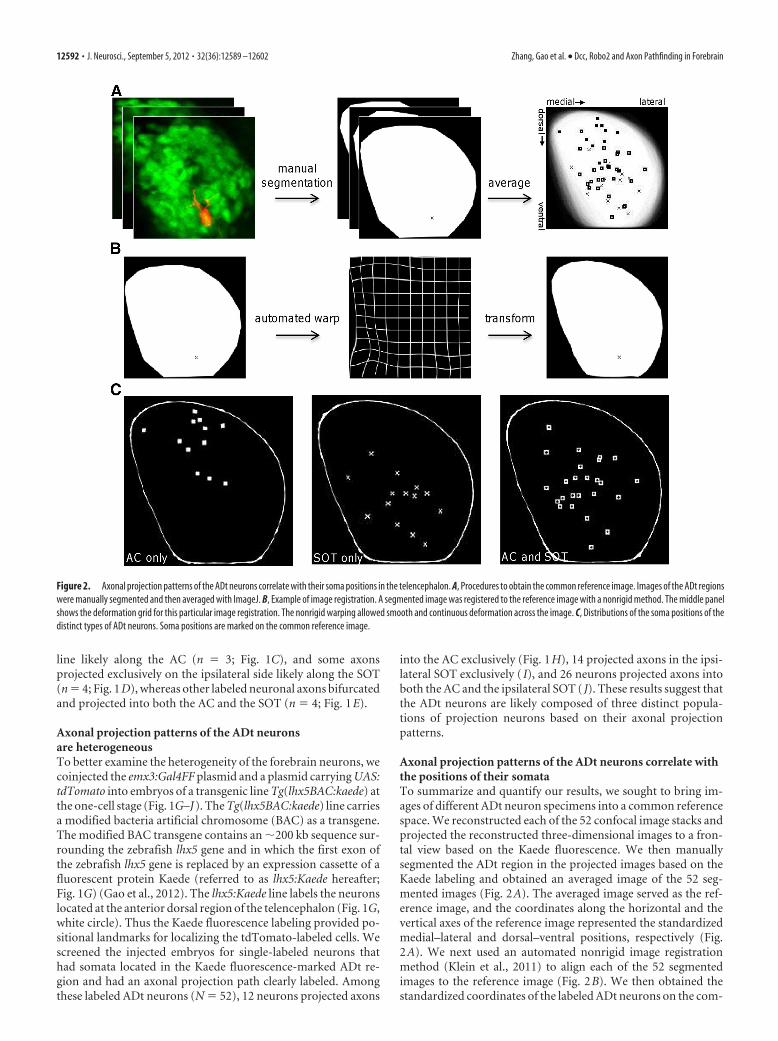
Axonal projection patterns of the ADt neurons correlate withthe positions of their somataTo summarize and quantify our results, we sought to bring im-ages of different ADt neuron specimens into a common referencespace. We reconstructed each of the 52 confocal image stacks andprojected the reconstructed three-dimensional images to a fron-tal view based on the Kaede fluorescence. We then manuallysegmented the ADt region in the projected images based on theKaede labeling and obtained an averaged image of the 52 seg-mented images (Fig. 2A). The averaged image served as the ref-erence image, and the coordinates along the horizontal and thevertical axes of the reference image represented the standardizedmedial–lateral and dorsal–ventral positions, respectively (Fig.2A). We next used an automated nonrigid image registrationmethod (Klein et al., 2011) to align each of the 52 segmentedimages to the reference image (Fig. 2B). We then obtained thestandardized coordinates of the labeled ADt neurons on the com-
Figure 2. Axonal projection patterns of the ADt neurons correlate with their soma positions in the telencephalon. A, Procedures to obtain the common reference image. Images of the ADt regionswere manually segmented and then averaged with ImageJ. B, Example of image registration. A segmented image was registered to the reference image with a nonrigid method. The middle panelshows the deformation grid for this particular image registration. The nonrigid warping allowed smooth and continuous deformation across the image. C, Distributions of the soma positions of thedistinct types of ADt neurons. Soma positions are marked on the common reference image.
12592 • J. Neurosci., September 5, 2012 • 32(36):12589 –12602 Zhang, Gao et al. • Dcc, Robo2 and Axon Pathfinding in Forebrain

mon reference space (Fig. 2C). Our results showed that the so-mata of the ADt neurons with exclusive AC-projecting axonswere clustered at more dorsal and medial positions (Fig. 2C, AConly), the somata of the ADt neurons with exclusive SOT-projecting axon were clustered at more ventral and lateral posi-tions (Fig. 2C, SOT only), and the ones with both AC and SOTprojections were more widely distributed (Fig. 2C, AC and SOT).ANOVA statistical tests confirmed these differential distributionswere significantly different along the dorsal–ventral axis (F(2,49)
� 18.22; p � 1.21 � 10�6). The distributions along the medial–lateral axis, however, were not significantly different (F(2,49) �1.44; p � 0.248). Post hoc analyses (with Bonferroni’s correction)revealed that the somata of the ADt neurons that projected axonsexclusively into the AC were located at more dorsal positionsthan those ADt neurons that projected axons into the SOT only(p � 1.89 � 10�6) or into both the AC and the SOT (p � 2.27 �10�5). Similar statistics results were obtained with soma positiondata determined from the pretransformed images (F(2,49) � 18.46p � 1.06 � 10�6) (data not shown). The correlation between theaxonal projection patterns and the positions of the somata ofthe ADt neurons suggested that cell intrinsic differences reg-ulate the development of the ADt neurons. These results fur-ther indicated intrinsic differences along the dorsal–ventralaxis determined the ADt axonal projection choice at the inter-section between the AC and the SOT.
Dcc and Robo2 are differentially expressed in theADt neuronsTo begin to define the molecular identity of the intrinsic differ-ences along the dorsal–ventral axis of the ADt region, we exam-ined expression profiles of two guidance receptors Dcc andRobo2. Both Dcc and Robo2 are expressed in the CNS duringzebrafish embryonic development (Lee et al., 2001; Fricke andChien, 2005). Double labeling by fluorescent in situ hybridizationshowed that dcc and robo2 were differentially expressed in thedorsal telencephalon (Fig. 3). At the beginning of the pharyngulaperiod, when the ADt neuronal axons started to extend ventrally(24hpf), dcc transcripts were more enriched in the dorsal andmedial regions in the dorsal telencephalon. In contrast, robo2transcripts were located in more ventral and lateral positions tothe dcc transcripts (Fig. 3A–C). At 36 hpf after the ADt neuronalaxons extended into the AC and the SOT pathway, dcc and robo2were similarly differentially expressed in dorsal telencephalon(Fig. 3D–F). Together with our cell-labeling results above, theseexpression patterns suggested that the ADt neurons with exclu-sively AC-projecting axons likely expressed Dcc, while those ADtneurons with SOT-projecting axons likely expressed Robo2.
To further examine whether the ADt axon projections corre-lated with Dcc and Robo2 expression, we performed single-cell-labeling experiments and determined the Dcc and Robo2expression with in situ hybridizations. Because the tdTomato flu-orescence did not survive the in situ hybridization procedure, themosaically labeled single cells and axonal projections werestained with an anti-DsRed antibody, which recognized the td-Tomato protein (Fig. 3H–P). Triple-labeling results showed thatADt axon projections correlated with Dcc and Robo2 expressionprofiles [Fig. 3Q; �(4)
2 � 40.49, n � 43; p � 5.06 � 10�9]. PairwiseFisher’s exact tests showed the Dcc and Robo2 expression profileswere significantly different between the three types of ADt neu-rons (AC vs AC–SOT, p � 6.35 � 10�4; AC vs SOT, p � 1.68 �10�5; AC–SOT vs SOT, p � 4.19 � 10�4). Thus, 60% of thelabeled ADt neurons with AC-projecting axons expressed dcc se-lectively (6 of 10; Fig. 3H–J), and none expressed robo2 exclu-
sively. In contrast, most (83%) of the labeled ADt neurons withboth AC- and SOT-projecting axons expressed both dcc androbo2 (15 of 18; Fig. 3K–M), and most (80%) of the labeled ADtneurons with SOT-projecting axons expressed robo2 selectively(12 of 15; Fig. 3N–P). In addition, none of the AC–SOT- or theSOT-type ADt neurons expressed dcc exclusively.
Knocking down Dcc function does not prevent formation ofthe AC and the SOT originating from the ADt neuronsWe next examined whether inhibition of Dcc or Robo2 functionmay affect the axonal projection patterns of the ADt neurons. Tofacilitate our investigation, we took advantage of the lhx5:Kaedetransgenic line and the photoconvertible property of the Kaedefluorescence protein (Ando et al., 2002). We photoconvertedKaede protein in the ADt neuronal somata from the green lightemission form to the red light emission form. Diffusion of thephotoconverted, red-colored Kaede from the ADt somata intothe axons marked the AC and the SOT axonal tracts that origi-nated from the ADt neurons (Fig. 4A,B).
To inhibit Dcc function, we injected a translation-blockingmorpholino antisense oligonucleotides [described by Suli et al.(2006); referred to as dcc-MO1 hereafter] into zebrafish embryosat one- to four-cell stages. Photoconversion of Kaede in dcc-MO1-injected lhx5:Kaede animals showed that both the AC andthe SOT were formed (n � 60; Fig. 4D). In addition to the AC andthe SOT, an aberrant axon tract was also observed in the injectedembryos (Fig. 4D, arrow).
We examined the effectiveness of the dcc morpholino knock-down with a rabbit anti-zebrafish Dcc antibody. By Western blot-ting, this antibody detected the endogenous Dcc protein as aband of �170 kb in the whole embryo protein extract, and thespecificity of the antibody was corroborated by detection of theMyc-tagged and GFP-tagged Dcc protein (data not shown). Inembryos injected with the dcc-MO1, the expression of the Dccwas reduced to levels below the detection limits of the Westernblots (Fig. 4D, inset). This antibody did not work on whole-mount embryos or frozen sections of embryos.
We further examined the ADt neuronal axons with single-cell-labeling when Dcc expression was knocked down with thedcc-MO1 injection. Among the labeled ADt neurons in dcc-MO-injected embryos (n � 50), �48% of the labeled neurons eitherfailed to extend axons ventrally or had axons stalled at the junc-tion between the AC and the SOT (Fig. 4E–H). The rest of thelabeled ADt neurons extended axons into the AC and the SOT inpatterns similar to those of the wild-type controls (Fig. 4 I, J). Thesomata of the ADt neurons with exclusive AC- or SOT-projectingaxons were clustered at more dorsal (Fig. 4 I, AC only) or at moreventral (Fig. 4 I, SOT only) positions, respectively, and the oneswith both AC and SOT projections were more widely distributed(Fig. 4 J, AC and SOT). ANOVA and Student’s t test resultsshowed that these distribution patterns were not significantdifferent from those of wild-type controls (ANOVA test alongthe dorsal–ventral axis, F(2, 24) � 4.861, p � 0.017; alongmedial–lateral axis, F(2, 24) � 1.354, p � 0.277; t test for the AConly type, p � 0.106; SOT only type, p � 0.439; AC and theSOT type, p � 0.073).
To address the specificity of the phenotypes caused by themorpholino knockdown of Dcc, we tested for rescue of the aber-rant axon tract by coinjection of a dcc-MO1-resistant mRNAcontaining silent mutations in the morpholino target site (Fig.4K). After coinjection of the dcc-MO1 and the morpholino-resistant dcc mRNA, the percentage of the embryos with aberrantaxon tracts was significantly reduced (from 64 � 12%, n � 60 to
Zhang, Gao et al. • Dcc, Robo2 and Axon Pathfinding in Forebrain J. Neurosci., September 5, 2012 • 32(36):12589 –12602 • 12593

Figure 3. Dcc and Robo2 are differentially expressed in the dorsal telencephalon. A–C, Dcc and Robo2 expression patterns in the telencephalon at 24 hpf. The embryo was counterstained withDAPI to show cell nuclei (frontal view, dorsal to the top). The dorsal telencephalon region in A is marked with a white square, and the details within the square are shown in B and C. A projection ofthe image stack is shown in A; images of single confocal planes are shown in B and C. The confocal plane of B is anterior to that of C. White dash lines in B and C demarcate the border between thetelencephalon and the olfactory epithelium (OE). D–F, Dcc and Robo2 expression patterns in the telencephalon at 36 hpf (frontal view, dorsal to the top). A projection of the image stack is shownin D; images of single confocal planes are shown in E and F. The confocal plane of E is anterior to the F plane. G, Schematic view of Dcc and Robo2 expression patterns in the ADt neurons. Transcriptsfor dcc are enriched in the dorsal and medial regions, and robo2 transcripts are distributed more in the ventral and lateral positions in the dorsal telencephalon. H–Q, Correlations of ADt axonalprojections and Dcc and Robo2 expression profiles. Single-cell-labeled embryos were triple stained with dcc/robo2 in situ probes and anti-DsRed antibodies. The anti-DsRed show labeled neurons andtheir axon projections (H, K, N ). Arrow marks the cell body position of the labeled neuron. Corresponding merged panels (I, L, O) show projected images of confocal image stacks of the triple labelingin the ADt regions. Labeled cell panels (J, M, P) show four (1– 4) consecutive single slices of confocal image stacks that go through the labeled cell bodies. Q, Labeled cells are counted as expressingDcc selectively (open bar), expressing both Dcc and Robo2 (gray bar), or expressing Robo2 selectively (black bar). Every single slice of confocal image stacks was inspected for presence of Dcc andRobo2 signals within the anti-DsRed-labeled cell bodies. A-S, AC and SOT. See Results for detailed statistical test results. Scale bar: (in P) A, H, K, N, 50 �m; B, C, I, L, O, 20 �m; in D–F, 25 �m; J,M, P, 12 �m.
12594 • J. Neurosci., September 5, 2012 • 32(36):12589 –12602 Zhang, Gao et al. • Dcc, Robo2 and Axon Pathfinding in Forebrain

24 � 8%, n � 41; p � 0.005). To further examine the specificity ofthe morpholino knockdowns, we tested a second nonoverlappingtranslation-blocking morpholino (dcc-MO2) and a splice-blocking morpholino (dcc-MO3) that have been described previ-ously (Suli et al., 2006). Similar to dcc-MO1-injected embryos,aberrant axon tracts were observed in the dorsal telencephalon indcc-MO2- or dcc-MO3-injected embryos (36 � 6%, n � 64 or50 � 9% n � 68, respectively; Fig. 4L). We also performed single-cell-labeling in dcc-MO2- or dcc-MO3-injected embryos. Similarto dcc-MO1-injected embryos, �24% of the labeled neurons in
dcc-MO2-injected embryos (5 of 21) or 41% of the labeled neu-rons in dcc-MO3-injected embryos (16 of 39) either failed toextend axons ventrally or had stalled axons (Fig. 4M–P).
Knocking down Robo2 function abolishes the SOToriginating from the ADt neuronsTo inhibit Robo2 function, we injected a morpholino that targetsthe translation start site of the robo2 mRNA (robo2-tMO) into thelhx5:Kaede embryos (Fig. 5A–C). Photoconversion of Kaede inrobo2-tMO-injected embryos (n � 36) showed that the SOTs
Figure 4. AC and SOT are present in dcc-MO-injected embryos. A, B, Photoconversion of Kaede in an lhx5:Kaede transgenic embryo labels the AC and the SOT originating from the ADt neurons.A live 36hpf lhx5:Kaede transgenic embryo was mounted in the tilted frontal view, and the region of the left telencephalon was selected for photoconversion. C, D, Knocking down Dcc function doesnot prevent the formation of the AC and the SOT. The arrow in D marks an aberrant axon tract after knockdown of Dcc function. The inset in D shows expression of the Dcc protein was effectivelyreduced by the morpholino knockdown (k.d.). ctl, Uninjected control embryos. Tubulin labels served as a loading control. E, F, ADt neuronal axon fails to reach the ventral positions in adcc-MO1-injected embryo. G, H, ADt neuronal axon stalls at the junction between the AC and the SOT in a dcc-MO1-injected embryo. I, J, Distributions of the soma positions of the distinct types ofADt neurons in embryos injected with dcc-MO1. The distribution patterns are similar to those of the standard controls. See Results section for statistic test results. K, Rescue of dcc-MO1 knockdownphenotype by a morpholino-resistant dcc mRNA. *p � 0.05. MO1, dcc-MO1 injected; rescue, dcc-MO1 and morpholino-resistant dcc mRNA coinjected. L, dcc-MO2 or dcc-MO3 causes similarphenotypes as dcc-MO1 injection. M, N, ADt neuronal axon extends dorsally in a dcc-MO3-injected embryo. O, P, ADt neuronal axon stalls at the junction between the AC and the SOT in adcc-MO3-injected embryo. Scale bar: (in P) A–D, M–P, 50 �m; E–H, 42 �m.
Zhang, Gao et al. • Dcc, Robo2 and Axon Pathfinding in Forebrain J. Neurosci., September 5, 2012 • 32(36):12589 –12602 • 12595

were significantly affected in most of theinjected embryos (97%); the SOTs wereeither absent (61%; Fig. 5B) or greatly re-duced (36%; Fig. 5C). To validate thespecificity of the robo2-tMO knockdowns,we examined phenotypes caused by injec-tion of a standard control morpholinoand a splice-blocking morpholino againstthe first exon–intron junction of the robo2transcripts (robo2-sMO). None of the em-bryos injected with the standard controlmorpholino showed defects in the SOT orthe AC (n � 55; Fig. 5A). In embryos in-jected with the splice-blocking robo2-sMO (n � 34), the SOTs were eitherseverely reduced (50%, Fig. 5F) or absent(44%, n � 36; Fig. 5E). RT-PCR analysisshowed that the wild-type robo2 tran-scripts were lost and replaced by shorterrobo2 transcripts in the robo2-sMO-injected embryos (Fig. 5D). Sequencinganalysis indicated that the shorter robo2transcripts missed 81 bp and were due tousage of a cryptic splice donor site. Be-cause the aberrantly spliced RNA lackedthe wild-type start codon, it was not ex-pected to encode functional proteins(data not shown). A previous study usinga different morpholino against the first
Figure 5. Knocking down Robo2 abolishes formation of SOT originating from the ADt neurons. A, Photoconverted Kaede labelsthe AC and the SOT in a standard control morpholino-injected embryo (std-ctl). B, C, The SOT is lost (B) or severely reduced (C) in
4
robo2-tMO-injected embryos. Arrows point to positions of ex-pected SOTs. Live embryos were mounted as in Figure 4A. D,RT-PCR results show an aberrant splice of robo2 transcripts inthe splice-blocking robo2-sMO-injected embryos. The aber-rantly spliced robo2 transcripts are smaller and lack the startcodon in the wild-type transcripts. The amplification of odc1gene served as a loading control. Lanes marked 2� are fromreactions using twice the amount of cDNA template as lanesmarked by 1�. ctl, Control; M, size marker; e1i1, robo2-sMOinjected. E, F, The SOT is lost (E) or severely reduced (F) in thesplice-blocking robo2-sMO-injected embryos. Arrows point topositions of expected SOTs. Live embryos were mounted as inFigure 4A. G, A control embryo injected with water. Boxes in-dicate the selected AC and SOT regions for quantification. H,Coinjection of a robo2 morpholino-resistant mRNA (robo2-rescue) restores the SOT formation in embryos injected withthe robo2-tMO. I, Robo2 overexpression (robo2-OE) causes re-duction of the AC and enhancement of the SOT. J, Quantifica-tion of the AC, SOT and SOT/AC ratios. Axon intensity values arepresented in arbitrary units. Error bars indicate SEs. Numbersof animals analyzed for each treatment group are n � 42, 60,36, 34, 30, and 30, respectively. *p � 0.05 versus standardcontrols by Student’s t test. n.s., Not significant, p � 0.05 ver-sus standard controls. #p�0.05 between the robo2-tMO and therobo2-rescue group (and bracket). Detailed statistic test results arereported in Results. K–M, Double staining of acetylated tubulinand Kaede in a standard control morpholino-injected embryo. Theacetylated tubulin-labeled axon tracts are broader than those la-beled by Kaede antibodies. The arrow in M points to the SOT la-beled by the Kaede antibodies. N–P, Double staining of acetylatedtubulin and Kaede in a robo2-tMO-injected embryo. The acety-lated tubulin-labeled SOT axons are present (O). The Kaede-labeled axons are absent at the expected position (arrow) for theSOT (P). Scale bar: (in P) K–P, 50 �m.
12596 • J. Neurosci., September 5, 2012 • 32(36):12589 –12602 Zhang, Gao et al. • Dcc, Robo2 and Axon Pathfinding in Forebrain
exon–intron junction of the robo2 gene showed the same effectson the splice of robo2 transcripts (Wyatt et al., 2010). These re-sults confirmed that the high penetrance (�90%) loss or severereduction of the SOT in robo2-MO-injected embryos was due tospecific knockdowns of Robo2 function.
To further address the specificity of the morpholino knock-downs, we tested for rescue of the robo2 knockdown phenotypesby coinjection of a robo2-tMO-resistant mRNA harboring sevensilent mutations in the morpholino recognition site (Fig. 5H,robo2-rescue). After coinjection of the robo2-tMO and themorpholino-resistant robo2 mRNA, the SOT was restored in�67% of the injected embryos (n � 30). Interestingly, overex-pression of Robo2 by injection of robo2 mRNA alone causedreduction of the AC and enhancement of the SOT (Fig. 5I). Toquantify these loss-of-function and gain-of-function phenotypesabove, we quantified the intensity of the SOT and the AC tracts,and the intensity ratios between the SOT and the AC tract in eachembryo (Fig. 5G, boxes) (see Materials and Methods). Thus,
compared with the controls (Fig. 5J), therobo2 morpholino knockdowns caused anapproximately threefold to fourfold re-duction in the SOT intensity, an �75–100% increase of the AC tract intensity,and an approximately sevenfold reduc-tion in the mean value of the SOT/AC ra-tios (p � 1.64 � 10�15 and p � 3.68 �10�15). Overexpression of Robo2 bymRNA injection caused an �40% in-crease of the SOT intensity, 25% decreaseof the AC tract intensity, and a more thantwofold increase of the mean value of theSOT/AC ratios (p � 0.001). The meanvalue of the SOT/AC ratios of the robo2-rescue group increased approximately four-fold compared to the group injected withthe robo2-tMO alone (p � 1.76 � 10�6),and the SOT and the AC tract intensitieswere restored to levels comparable to thosein the control embryos (p � 0.823 and p �0.129, respectively).
A previous study showed that the SOTlabeled by the acetylated tubulin appearedunaffected after the function of Robo2was inhibited by morpholino injections(Devine and Key, 2008). We thus fixed thestandard control morpholino or robo2-MO-injected embryos and costained thefixed embryos with acetylated tubulin andKaede antibodies. Our results showed thatin the robo2-tMO-injected embryos, theacetylated tubulin-labeled axonal tractswere present between the telencephalonand the diencephalon (Fig. 5 L, O). Incontrast, the Kaede-labeled axonaltracts were missing or dramatically re-duced (Fig. 5 M, P). Similar results wereobtained with the injection of the splice-blocking robo2-sMO (data not shown).Therefore, our results showed thatRobo2 function was specifically re-quired for the ADt neurons to extendthe axons along the SOT pathway.
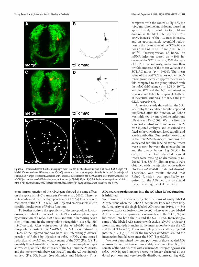
ADt neurons project axons into the AC when Robo2 functionis inhibitedWe examined the axonal projection patterns of singly labeledADt neurons when the Robo2 function was knocked down (Fig.6). A majority of the singly labeled ADt neurons (91%, n � 34)projected axons exclusively into the AC, whereas very few labeledADt neuronal axons projected exclusively into the SOT (3%) orbifurcated into both the AC and the SOT (6%). Interestingly,some of the labeled ADt neurons with exclusively AC-projectingaxons had multiple branches at the intersection between the ACand the SOT (n � 10). Those multiple processes either projectedinto the AC (Fig. 6A,B), or the branches wandered around theintersection but failed to enter the SOT (Fig. 6C,D).
We next determined the soma positions of those labeled ADtneurons. In contrast to results in wild-type animals (Fig. 2C), thesomata of the ADt neurons with exclusive AC-projecting axons inrobo2-tMO-injected embryos were no longer clustered at thedorsal positions and were broadly distributed instead (Fig. 6E).
Figure 6. Individually labeled ADt neurons project axons into the AC when Robo2 function is inhibited. A, B, A single-cell-labeled ADt neuronal axon bifurcates at the AC–SOT junction, and both branches project into the AC in a robo2-tMO-injectedembryo. C, D, A single-cell-labeled ADt neuron with one axonal branch projects into the AC, and the other branch wanders at theAC–SOT junction in a robo2-tMO-injected embryo. Scale bar: (in D) A–D, 50 �m. E, F, Distribution of soma positions of distincttypes of ADt neurons in robo2-tMO-injected embryos. Most labeled ADt neurons project axons exclusively into the AC.
Zhang, Gao et al. • Dcc, Robo2 and Axon Pathfinding in Forebrain J. Neurosci., September 5, 2012 • 32(36):12589 –12602 • 12597

ANOVA tests confirmed that the somata positions of the ADtneurons with exclusive AC-projecting axons were significantlydifferent between the wild-type and the robo2-MO-injected em-bryos (F(1,40) � 23.00; p � 2.26 � 10�5). Additional ANOVAsshowed that the distribution of the AC-projecting ADt neurons inrobo2-MO-injected embryos were similar to those of the SOT-projecting or AC- and SOT-projecting ADt neurons in the wild-typeanimals (F(2,67) � 1.06; p � 0.352). Thus these single-cell-labelingresults mirrored the group-labeling results with Kaede conver-sion. Together, these results suggested that the ADt axons thatshould extend along the SOT projected into the AC instead whenthe Robo2 function was inhibited.
Inhibition of Slit or Netrin function affects the SOT or ACformation, respectivelyThe Robo receptor ligands Slits are chemorepellents that preventthe axons from extending toward the midline (Bagri et al., 2002).We investigated whether inhibition of Slit function similarly af-fected the SOT formation as did the inhibition of Robo2 func-tion. There are four Slit homologs in zebrafish, and they aredifferentially expressed in the CNS during zebrafish embryonicdevelopment (Yeo et al., 2001; Hutson et al., 2003). We examinedthe expression of these four Slit homologs in the forebrain re-gions with whole-mount in situ hybridizations. The resultsshowed that Slit1a was expressed in both the dorsal and ventraltelencephalon in the early pharyngula period (24 hpf), Slit1b wasexpressed in a restricted region between the telencephalon andthe diencephalon at 36 hpf. Morpholino knockdown of Slit1a orSlit1b function did not affect the SOT formation. Similar to re-sults from a previous report (Barresi et al., 2005), the AC wasvariably reduced in some Slit1a morpholino-injected embryos(data not shown).
We focused our study on Slit2 and Slit3. Transcripts of Slit2and Slit3 were enriched in the ventral telencephalons and close tothe midline in the early pharyngula period (Fig. 7A,B, arrows)(data not shown) (see also Hutson and Chien, 2002). Inhibitionof Slit2 function by injecting the slit2 morpholino (Barresi et al.,2005) caused a twofold reduction in the SOT formation (Fig. 7E)and a twofold reduction in the mean value of the SOT/AC ratios(Fig. 7I; p � 1.38 � 10�4 vs standard control). Inhibition of Slit3function alone by injecting the slit3 morpholino (Barresi et al.,2005) did not affect the SOT formation or the SOT/AC ratio (Fig.7I; p � 0.64 and p � 0.10, respectively). Coinjection of both theslit2 and the slit3 morpholinos caused a further reduction in themean value of the SOT/AC ratios (Fig. 7F; p � 3.65 � 10�7 vsstandard control). The penetrance and severity of the SOT defectscaused by the Slit morpholino knockdowns were lower thanthose in the Robo2 morpholino knockdowns (a penetrance of68% in slit2 and slit3 double knockdowns vs 97% in robo2-tMOknockdown, and a threefold reduction of the SOT/AC ratio inslit2 and slit3 double knockdowns vs a sevenfold reduction inrobo2-tMO knockdown; Fig. 7I). Increasing the injection dosagesfor Slit morpholinos caused morphological alterations of the em-bryos and significant reduction of the Kaede fluorescence signal,which impeded the analysis of the ADt axon phenotypes (datanot shown).
We next examined whether knocking down the function ofthe Dcc receptor ligand Netrin affected the AC or the SOT for-mation. There are four Netrin homologs in zebrafish (Lauderdaleet al., 1997; Strahle et al., 1997; Park et al., 2005). At 24 hpf,netrin1a and netrin1b are expressed in the ventral and midlineregions of the forebrain (Fig. 7C,D) (Lakhina et al., 2012). Theother two Netrins, Netrin2 and Netrin4, are not expressed in the
forebrain regions before 36 hpf (Park et al., 2005). Inhibition ofeither Netrin1a or Netrin1b function by injection of the netrin1aor the netrin1b morpholinos (Suli et al., 2006) caused small re-ductions of the AC axon tract (20 or 25%) and almost no changesof the SOT axon tract (3% increase or 8% decrease, respectively).The mean value of the SOT/AC ratios was increased 30 or 17%compared to standard controls. Inhibition of Netrin1 function bycoinjecting the netrin1a and the netrin1b morpholinos togethercaused a larger reduction of the AC (35%) and an increase of theSOT (30%), and the mean value of the SOT/AC ratios was in-creased threefold compared to standard controls (Fig. 7G; p �0.008). Similar to Dcc knockdowns, inhibition of the Netrin1function also caused the ADt neurons to extend aberrant axons inthe dorsal telencephalon (Fig. 7G, arrow).
Our results showed that inhibition of the Robo2–Slit2/Slit3pathway caused reduction in the SOT formation, while inhibitionof the Netrin1 function caused reduction in the AC tract. To testhow the Robo2–Slit2/Slit3 pathway interacted with the Netrin1,we inhibited both Robo2 and Netrin1 function with morpholinoinjections. The results showed that the SOT was missing or se-verely reduced. In contrast, the AC was formed in these embryos(Fig. 7H). Quantification results showed the mean SOT intensitywas reduced approximately fourfold, while the AC was increased�60% (p � 1.50 � 10�9 and p � 0.007 vs standard control,respectively). The mean value of the SOT/AC ratios of the robo2/netrin1 double knockdown embryos was reduced to a level com-parable to that of the robo2 single knockdown embryos (Fig. 7I;p � 0.815). Thus, the robo2 morpholino knockdown was epistaticto the netrin1 morpholino knockdown in terms of the SOT/ACprojection pathway choices of the ADt neurons. In the robo2/netrin1 double knockdown embryos, the ADt neurons also proj-ect aberrant axons into the dorsal telencephalon regions (Fig. 7H,arrow).
Finally, to examine the effects of morpholino knockdowns onthe specification of the ADt neurons, we determined the expres-sion patterns of dcc and robo2 mRNA in the ADt region inmorpholino-injected embryos. We found the dcc transcripts wereenriched in the dorsal region, and the robo2 transcripts were en-riched in the ventral region in morpholino-injected embryos(dcc-MO, n � 8; robo2-tMO, n � 8; netrin1a,b-MO, n � 5; slit2,3-MO, n � 5). Thus, the expression patterns of dcc and robo2 weresimilar to those in the wild-type control embryos (Fig. 7J--M).Together with our single-cell-labeling and Kaede conversion re-sults, these results suggested that the incorrect axonal projectionsobserved in our morpholino knockdown experiments were notlikely due to abnormal specification of the ADt neurons, becausethe neurons differentiated along their normal time course andexpressed regional markers at appropriate locations.
DiscussionThe results of the present study indicate that intrinsic differencesalong the dorsal–ventral axis of the telencephalon regulate ADtneuron axonal pathfinding. Further mechanistic studies suggestthat Robo2 responds to the repellent Slit signals and suppressesthe attractive Netrin1 signals, allowing the ADt neurons to extendaxons along the SOT pathway (Fig. 8A). Our studies also revealthat in the absence of the attractive Netrin, the ADt neurons canextend axons into the AC pathway if the repellent Robo2 is alsoremoved (Fig. 8E). These findings demonstrate how multipleguidance receptors and cues interact to determine the axonalprojection choices in the developing vertebrate forebrain.
12598 • J. Neurosci., September 5, 2012 • 32(36):12589 –12602 Zhang, Gao et al. • Dcc, Robo2 and Axon Pathfinding in Forebrain

Axonal projection patterns of ADt neurons are heterogeneousOur studies demonstrate that ADt neuronal axons choose spe-cific projection paths at the intersection between the AC and theSOT (Fig. 1). These results suggest that ADt neurons are com-posed of distinct population of neurons based on their axon pro-jection patterns. In adult rat, the AC is a major commissuralprojection, and it connects bilateral olfactory structures and basaltelencephalic regions including the piriform cortex andamygadala (Jouandet and Hartenstein, 1983). The AC similarlyconnects the two telencephalic hemispheres in adult fish (Meekand Nieuwenhuys, 1998; Correa et al., 1998). The SOT likelyconnects the olfactory bulb and the nucleus posterior tuberis in
adult fish, and it may be a precursor of the lateral forebrain bun-dle system (Meek and Nieuwenhuys, 1998). Currently it is notknown what neuronal populations the ADt neurons may give riseto in the adult fish. Further studies are needed to determine whatdistinct functional role the ADt neurons may have.
Robo2 and Dcc signaling pathways are differentially requiredfor the axonal pathfinding within the AC and the SOTBecause the ADt axon growth cones are exposed to the sameextrinsic environment at the intersection between the AC and theSOT, the specific axonal projection choices of the ADt neurons(Fig. 2) are likely due to cell intrinsic differences among the ADt
Figure 7. Inhibition of Slit or Netrin function affects the SOT or the AC formation. A–D, Expression patterns of Slits and Netrins. The probes used for whole-mount in situ hybridization are listedin the top right corner of each panel (animals mounted in lateral view, rostral to the left). The arrow in A labels the dorsal-to-ventral axis in the lateral mount view. The short arrow in each panelindicates the position of the ventral telencephalon. E, F, The SOT is reduced in slit2-MO-injected (E) or slit2-MO- and slit3-MO-injected (F) embryos. G, The AC is reduced in ntn1a,b-MO-injectedembryos. H, Robo2 morpholino knockdown is epistatic to the Netrin1a,b morpholino knockdown. The arrows in G and H mark aberrant axons located in the dorsal telencephalon. I, Quantificationof the AC, the SOT, and the SOT/AC ratios. Axon intensity values are presented in arbitrary units. Error bars indicate SEs. Numbers of animals analyzed for each treatment group are n � 42, 33, 18,25, 36, 28, and 36, respectively. *p � 0.05 versus standard controls by Student’s t test. n.s., Not significant, p � 0.05 versus standard controls. J–M, Morpholino knockdowns do not affect theexpression patterns of dcc and robo2 in the ADt region (frontal view, dorsal to the top). Scale bar: (in M) A–D, 150 �m; E–H, 50 �m; J–M, 20 �m.
Zhang, Gao et al. • Dcc, Robo2 and Axon Pathfinding in Forebrain J. Neurosci., September 5, 2012 • 32(36):12589 –12602 • 12599

neurons. Our results show that the guidance receptors Dcc andRobo2 are respectively enriched in the dorsal region and the ven-tral region in the dorsal telencephalon, and the ADt axon projec-tions correlate with the Dcc and Robo2 expression profiles (Fig.3). Together these results suggest that Dcc and Robo2 may regu-late the ADt axonal projection patterns.
The Dcc–Netrin signaling pathway is known to mediate at-traction of the axonal growth cones. In zebrafish, inhibition ofNetrin or Dcc functions causes some olfactory sensory axons toproject aberrantly and away from netrin-expressing regions(Lakhina et al., 2012). Previous studies in knock-out miceshowed that DCC is required for the guidance of commissuralaxons to the midline (Fazeli et al., 1997). Our results show thatknocking down Dcc function causes some of the labeled ADtaxons to grow into aberrant dorsal positions or to stall at the ACand the SOT junction (Fig. 4E–H), while in the rest of labeledADt neurons, Dcc knockdown does not prevent formation of theAC or the SOT (Fig. 4 I, J). These results suggest that Dcc is re-quired in the ADt neurons before the AC–SOT guidance selectionchoice. Formation of aberrant axons in the forebrain regions inDcc morpholino-injected embryos has been reported previously(Gaudin et al., 2012). Examination of the aberrant dorsal ADtaxons suggests that Dcc is likely involved in the asymmetric out-growth of the ADt neurons (Gao et al. 2012). Previous studieshave implicated other guidance factors such as neuropilins andsemaphorin in the guidance of the anterior commissural axon tothe midline (Wolman et al., 2004). These and other factors suchas Neogenin, a guidance receptor related to Dcc (Wilson and Key,2007; De Vries and Cooper, 2008), may partially compensate forthe loss of the Dcc function to mediate the AC formation.
In contrast to the Dcc knockdowns, inhibition of Robo2 func-tion abolishes the SOT originating from the ADt neurons (Fig.5A–F). In addition, the Kaede-labeled AC tracts appear enhanced(Fig. 5J). Consistent with the group labeling by photoconversionof Kaede, single-cell-labeling results show that most ADt neuronsproject axons into the AC when Robo2 function is inhibited (Fig.6). These results suggest that in the absence of Robo2 function,those SOT-projecting ADt axons extend into the AC projectionpathway instead.
The Robo–Slit signaling has a conserved role in midline guid-ance (Ypsilanti et al., 2010). Previous studies showed that Roboproteins are highly expressed in the ipsilateral axons and preventthem from crossing the midline of Drosophila (Seeger et al., 1993;Kidd et al., 1998; Rajagopalan et al., 2000). A previous studyshowed that in Robo1 and Robo2 double knock-out mice, axonsfrom the internal capsule fail to avoid approaching the midline(Lopez-Bendito et al., 2007). Similarly, in Slit1 and Slit2 doubleknock-out mice, the corticofugal projections abnormally ap-proach the midline and cross it (Bagri et al., 2002). In the Slit1 andSlit2 double knock-out or other compound Slit knock-out mice(Bagri et al., 2002; Unni et al., 2012), the Robo1 knock-out mice(Andrews et al., 2006), and the Robo1 and Robo2 double knock-out mice (Lopez-Bendito et al., 2007), the corpus callosum isreduced, and some of the corticocortical axons are directedventrally. In zebrafish, Robo2 functions to prevent and correctRGC axon pathfinding errors before and after the axon mid-line crossing (Fricke et al., 2001; Hutson and Chien, 2002).Reducing Slit functions causes defasciculation of the postopticcommissure (Barresi et al., 2005). We show that inhibition ofSlit2 and Slit3 function in zebrafish has similar effects as inhi-
Figure 8. Schematic view of intrinsic factors and extrinsic cues that coordinate the ADt neuronal axon pathfinding in the AC and the SOT. A, Our data indicate Dcc and Robo2 are differentiallyexpressed in the ADt neurons along the dorsal–ventral axis of the dorsal telencephalon. Robo2 responds to the repellent Slit signals and suppresses the attractive Netrin signals, allowing the moreventrally located ADt neurons to extend axons along the SOT pathway. B–E, Summary of functional roles of the intrinsic factors and extrinsic cues. B, In the absence of Dcc, some of the ADt neuronsproject axons dorsally or fail to extend axons into the AC and the SOT. The rest of the ADt neurons project axons normally into the AC and the SOT, and likely use other guidance receptors (questionmark) to sense the attractive cues at the AC. C, In the absence of Robo2, the ADt axons are no longer repelled by the Slits, and most ADt axons are attracted by the Netrin1 cue and extend along theAC. D, Removal of the attractive Netrin1 cue causes ADt neurons to extend axons along the SOT pathway. Some ADt neurons also extend axons in the dorsal telencephalon regions. E, Removal of theRobo2 function is epistatic to the removal of the Netrin1 function in terms of the SOT/AC projection pathway choices. In the absence of both the Robo2 and Netrin1 functions, the ADt neurons fail toproject axons into the SOT. These ADt neuronal axons that extend along the AC likely respond to attractive cues (question mark) other than Netrin1.
12600 • J. Neurosci., September 5, 2012 • 32(36):12589 –12602 Zhang, Gao et al. • Dcc, Robo2 and Axon Pathfinding in Forebrain

bition of Robo2 function, and it reduces the SOT originatingfrom the ADt neurons (Fig. 7 E, F ). Thus, our results are con-sistent with these previous results and suggest that Robo–Slitsare essential for the axons to project along ipsilateral pathwaysin vertebrate forebrains.
Previous reports have implicated Slit1a as the ligand for theRobo2 receptor in the positioning of the longitudinal axons(Devine and Key, 2008) and in the arborization and synaptogen-esis of the retinal ganglion cell axons (Campbell et al., 2007). Ourstudies show that Slit1a is not involved in the SOT formation.These results are likely due to particular combinations of intrinsicreceptors and extracellular cues experienced by different types ofdeveloping neurons.
Interaction between the Robo2–Slit and the Dcc–Netrin1signaling pathwaysOur mechanistic studies of the ADt neuronal axon developmentshow that Robo2–Slit2/Slit3-mediated repulsion is required forthe SOT formation, while the Netrin1 mediate attraction is re-quired for the AC formation. These results suggest that Robo2responds to the repellent Slit signals and suppresses the attractiveNetrin signal (Fig. 8A). Our data support this model, as overex-pression of Robo2 is able to reduce the AC and appears to en-hance the SOT originating from the ADt neurons (Fig. 5 I, J).Thus, the competitive actions between Slits and Netrin1 are in-terpreted by ADt neurons expressing different levels of Robo2,Dcc, and likely other guidance receptors, allowing the appropri-ate guidance choices of the ADt neuronal axons based upon theirsomata positions within the dorsal telencephalon. Interestingly,our studies also show that in the absence of the attractive Netrin1signaling, the ADt neurons can extend axons into the AC pathwayonly if the repellent Robo2 signaling is also removed (Fig. 7G,H).This result suggests that other attractive guidance cues exist in theventral telencephalon. It also highlights the requirement of Ne-trin1 function in the presence of the Robo2-mediated repulsion.Finally, it is interesting to note that a previous study showed thatDcc promotes ventral growth of zebrafish spinal cord commis-sural primary ascending neuron axons through inhibition ofRobo2 (Bonner et al., 2012). Therefore, the interaction betweenthe Robo2–Slit and the Dcc–Netrin1 signaling pathways may playroles in a variety of guidance choice mechanisms in the CNS.
Intrinsic factors and the specification of the ADt neuronsOur studies focused on the function of the guidance factors thatact directly on the development of axon projections. Multipletranscription factors and guidance receptors regulate the specifi-cation and axonal pathfinding of the diverse neuronal popula-tions (Polleux et al., 2007; Molyneaux et al., 2007). Previousstudies showed that the axonal tracts in zebrafish and mousebrain were perturbed when the expression of the transcriptionfactor, such as Pax6 (Mastick et al., 1997) or Pax2 (Macdonald etal., 1997), was disrupted. A previous study showed that the tran-scription factors Lhx2 and Lhx9 regulate directly the expressionof the receptor Rig-1 to mediate the guidance of the dl1c axons tocross the midline in mouse spinal cord (Wilson et al., 2008). It hasalso been reported that Nkx2.9 may regulate Robo2 expression inthe spinal accessory motor neurons (SACMNs) for the SACMNaxon exit from the spinal cord (Bravo-Ambrosio et al., 2012). Itshould be interesting to examine how transcription factors mayregulate the spatial and temporal distribution of Robo2 and otherguidance receptors in the anterior dorsal telencephalon.
ReferencesAnderson RB, Key B (1996) Expression of a novel N-CAM glycoform
(NOC-1) on axon tracts in embryonic Xenopus brain. Dev Dyn207:263–269.
Ando R, Hama H, Yamamoto-Hino M, Mizuno H, Miyawaki A (2002) Anoptical marker based on the UV-induced green-to-red photoconversionof a fluorescent protein. Proc Natl Acad Sci U S A 99:12651–12656.
Andrews W, Liapi A, Plachez C, Camurri L, Zhang J, Mori S, Murakami F,Parnavelas JG, Sundaresan V, Richards LJ (2006) Robo1 regulates thedevelopment of major axon tracts and interneuron migration in the fore-brain. Development 133:2243–2252.
Asakawa K, Suster ML, Mizusawa K, Nagayoshi S, Kotani T, Urasaki A,Kishimoto Y, Hibi M, Kawakami K (2008) Genetic dissection of neuralcircuits by Tol2 transposon-mediated Gal4 gene and enhancer trapping inzebrafish. Proc Natl Acad Sci U S A 105:1255–1260.
Bagri A, Marín O, Plump AS, Mak J, Pleasure SJ, Rubenstein JL, Tessier-Lavigne M (2002) Slit proteins prevent midline crossing and determinethe dorsoventral position of major axonal pathways in the mammalianforebrain. Neuron 33:233–248.
Barresi MJF, Hutson LD, Chien C-B, Karlstrom RO (2005) Hedgehog reg-ulated Slit expression determines commissure and glial cell position in thezebrafish forebrain. Development 132:3643–3656.
Bonner J, Letko M, Nikolaus OB, Krug L, Cooper A, Chadwick B, Conklin P,Lim A, Chien CB, Dorsky RI (2012) Midline crossing is not required forsubsequent pathfinding decisions in commissural neurons. Neural Dev7:18.
Bravo-Ambrosio A, Mastick G, Kaprielian Z (2012) Motor axon exit fromthe mammalian spinal cord is controlled by the homeodomain proteinNkx2.9 via Robo-Slit signaling. Development 139:1435–1446.
Campbell DS, Stringham SA, Timm A, Xiao T, Law MY, Baier H, Nonet ML,Chien CB (2007) Slit1a inhibits retinal ganglion cell arborization andsynaptogenesis via Robo2-dependent and -independent pathways. Neu-ron 55:231–245.
Chedotal A, Richards LJ (2010) Wiring the brain: the biology of neuronalguidance. Cold Spring Harb Perspect Biol 2:a001917.
Chitnis AB, Kuwada JY (1990) Axonogenesis in the brain of zebrafish em-bryos. J Neurosci 10:1892–1905.
Chitnis AB, Kuwada JY (1991) Elimination of a brain tract increases errorsin pathfinding by follower growth cones in the zebrafish embryo. Neuron7:277–285.
Correa SA, Grant K, Hoffmann A (1998) Afferent and efferent connectionsof the dorsocentral telencephalon in an electrosensory teleost, Gymnotuscarapo. Brain Behav Evol 52:81–98.
Devine CA, Key B (2008) Robo-Slit interactions regulate longitudinal axonpathfinding in the embryonic vertebrate brain. Dev Biol 313:371–383.
De Vries M, Cooper HM (2008) Emerging roles for neogenin and its ligandsin CNS development. J Neurochem 106:1483–1492.
Easter SS Jr, Ross LS, Frankfurter A (1993) Initial tract formation in themouse brain. J Neurosci 13:285–299.
Easter SS Jr, Burrill J, Marcus RC, Ross LS, Taylor JS, Wilson SW (1994)Initial tract formation in the vertebrate brain. Prog Brain Res 102:79 –93.
Fazeli A, Dickinson SL, Hermiston ML, Tighe RV, Steen RG, Small CG,Stoeckli ET, Keino-Masu K, Masu M, Rayburn H, Simons J, Bronson RT,Gordon JI, Tessier-Lavigne M, Weinberg RA (1997) Phenotype of micelacking functional Deleted in colorectal cancer (Dcc) gene. Nature386:796 – 804.
Fricke C, Chien CB (2005) Cloning of full-length zebrafish dcc and expres-sion analysis during embryonic and early larval development. Dev Dyn234:732–739.
Fricke C, Lee JS, Geiger-Rudolph S, Bonhoeffer F, Chien CB (2001) astray, azebrafish roundabout homolog required for retinal axon guidance. Sci-ence (New York) 292:507–510.
Gao J, Zhang C, Yang B, Sun L, Zhang C, Westerfield M, Peng G (2012) Dccregulates asymmetric outgrowth of forebrain neurons in zebrafish. PloSOne 7:e36516.
Gaudin A, Hofmeister W, Key B (2012) Chemoattractant axon guidancecues regulate de novo axon trajectories in the embryonic forebrain ofzebrafish. Dev Biol 367:126 –139.
Hutson LD, Chien CB (2002) Pathfinding and error correction by retinalaxons: the role of astray/robo2. Neuron 33:205–217.
Hutson LD, Jurynec MJ, Yeo SY, Okamoto H, Chien CB (2003) Two diver-gent slit1 genes in zebrafish. Dev Dyn 228:358 –369.
Zhang, Gao et al. • Dcc, Robo2 and Axon Pathfinding in Forebrain J. Neurosci., September 5, 2012 • 32(36):12589 –12602 • 12601

Jouandet ML, Hartenstein V (1983) Basal telencephalic origins of the ante-rior commissure of the rat. Exp Brain Res 50:183–192.
Kanki JP, Kuwada JY (2000) Growth cones utilize both widespread and localdirectional cues in the zebrafish brain. Dev Biol 219:364 –372.
Kastenhuber E, Kern U, Bonkowsky JL, Chien CB, Driever W, Schweitzer J(2009) Netrin-DCC, Robo-Slit, and heparan sulfate proteoglycans coor-dinate lateral positioning of longitudinal dopaminergic diencephalospi-nal axons. J Neurosci 29:8914 – 8926.
Kidd T, Brose K, Mitchell KJ, Fetter RD, Tessier-Lavigne M, Goodman CS,Tear G (1998) Roundabout controls axon crossing of the CNS midlineand defines a novel subfamily of evolutionarily conserved guidance recep-tors. Cell 92:205–215.
Kimmel CB, Ballard WW, Kimmel SR, Ullmann B, Schilling TF (1995)Stages of embryonic development of the zebrafish. Dev Dyn 203:253–310.
Klein A, Kroon DJ, Hoogeveen Y, Kool LJ, Renema WK, Slump CH (2011)Multimodal image registration by edge attraction and regularization us-ing a B-spline grid. Proc SPIE 7962:796220.
Lakhina V, Marcaccio CL, Shao X, Lush ME, Jain RA, Fujimoto E, BonkowskyJL, Granato M, Raper JA (2012) Netrin/DCC signaling guides olfactorysensory axons to their correct location in the olfactory bulb. J Neurosci32:4440 – 4456.
Lauderdale JD, Davis NM, Kuwada JY (1997) Axon tracts correlate withnetrin-1a expression in the zebrafish embryo. Mol Cell Neurosci9:293–313.
Lee JS, Ray R, Chien CB (2001) Cloning and expression of three zebrafishroundabout homologs suggest roles in axon guidance and cell migration.Dev Dyn 221:216 –230.
Lopez-Bendito G, Flames N, Ma L, Fouquet C, Di Meglio T, Chedotal A,Tessier-Lavigne M, Marín O (2007) Robo1 and Robo2 cooperate tocontrol the guidance of major axonal tracts in the mammalian forebrain.J Neurosci 27:3395–3407.
Macdonald R, Scholes J, Strahle U, Brennan C, Holder N, Brand M, WilsonSW (1997) The Pax protein Noi is required for commissural axon path-way formation in the rostral forebrain. Development 124:2397–2408.
Mastick GS, Davis NM, Andrew GL, Easter SS (1997) Pax-6 functions inboundary formation and axon guidance in the embryonic mouse fore-brain. Development 124:1985–1997.
Meek J, Nieuwenhuys R (1998) Holosteans and teleosts. In: The central ner-vous system of vertebrates, Vol 1. (Nieuwenhuys R, Donkelaar HJ, Nich-olson C, eds), pp 759 –938. Heidelberg: Springer.
Miyasaka N, Morimoto K, Tsubokawa T, Higashijima S, Okamoto H, Yoshi-hara Y (2009) From the olfactory bulb to higher brain centers: geneticvisualization of secondary olfactory pathways in zebrafish. J Neurosci29:4756 – 4767.
Molyneaux BJ, Arlotta P, Menezes JRL, Macklis JD (2007) Neuronal sub-type specification in the cerebral cortex. Nat Rev Neurosci 8:427– 437.
Park KW, Urness LD, Senchuk MM, Colvin CJ, Wythe JD, Chien CB, Li DY(2005) Identification of new netrin family members in zebrafish: devel-opmental expression of netrin 2 and netrin 4. Dev Dyn 234:726 –731.
Patel CK, Rodriguez LC, Kuwada JY (1994) Axonal outgrowth within theabnormal scaffold of brain tracts in a zebrafish mutant. J Neurobiol25:345–360.
Peng G, Westerfield M (2006) Lhx5 promotes forebrain development andactivates transcription of secreted Wnt antagonists. Development133:3191–3200.
Polleux F, Ince-Dunn G, Ghosh A (2007) Transcriptional regulation of ver-tebrate axon guidance and synapse formation. Nat Rev Neurosci8:331–340.
Rajagopalan S, Nicolas E, Vivancos V, Berger J, Dickson BJ (2000) Crossingthe midline: roles and regulation of Robo receptors. Neuron 28:767–777.
Seeger M, Tear G, Ferres-Marco D, Goodman CS (1993) Mutations affect-ing growth cone guidance in Drosophila: genes necessary for guidancetoward or away from the midline. Neuron 10:409 – 426.
Strahle U, Fischer N, Blader P (1997) Expression and regulation of a netrinhomologue in the zebrafish embryo. Mech Dev 62:147–160.
Suli A, Mortimer N, Shepherd I, Chien CB (2006) Netrin/DCC signalingcontrols contralateral dendrites of octavolateralis efferent neurons. J Neu-rosci 26:13328 –13337.
Talbot JC, Johnson SL, Kimmel CB (2010) hand2 and Dlx genes specifydorsal, intermediate and ventral domains within zebrafish pharyngealarches. Development 137:2507–2517.
Unni DK, Piper M, Moldrich RX, Gobius I, Liu S, Fothergill T, Donahoo AL,Baisden JM, Cooper HM, Richards LJ (2012) Multiple Slits regulate thedevelopment of midline glial populations and the corpus callosum. DevBiol 365:36 – 49.
Viktorin G, Chiuchitu C, Rissler M, Varga ZM, Westerfield M (2009) Emx3is required for the differentiation of dorsal telencephalic neurons. DevDyn 238:1984 –1998.
Westerfield M (2007) The zebrafish book: a guide for the laboratory use ofzebrafish Danio rerio, Ed 5. Eugene, OR: Oregon UP.
Westerfield M, Wegner J, Jegalian BG, DeRobertis EM, Puschel AW (1992)Specific activation of mammalian Hox promoters in mosaic transgeniczebrafish. Genes Dev 6:591–598.
Wilson NH, Key B (2006) Neogenin interacts with RGMa and netrin-1 toguide axons within the embryonic vertebrate forebrain. Dev Biol296:485– 498.
Wilson NH, Key B (2007) Neogenin: one receptor, many functions. IntJ Biochem Cell Biol 39:874 – 878.
Wilson SI, Shafer B, Lee KJ, Dodd J (2008) A molecular program for con-tralateral trajectory: Rig-1 control by LIM homeodomain transcriptionfactors. Neuron 59:413– 424.
Wilson SW, Ross LS, Parrett T, Easter SS (1990) The development of a sim-ple scaffold of axon tracts in the brain of the embryonic zebrafish, Brachy-danio rerio. Development 108:121–145.
Wolman MA, Liu Y, Tawarayama H, Shoji W, Halloran MC (2004) Repul-sion and attraction of axons by semaphorin3D are mediated by differentneuropilins in vivo. J Neurosci 24:8428 – 8435.
Wyatt C, Ebert A, Reimer MM, Rasband K, Hardy M, Chien CB, Becker T,Becker CG (2010) Analysis of the astray/robo2 zebrafish mutant revealsthat degenerating tracts do not provide strong guidance cues for regener-ating optic axons. J Neurosci 30:13838 –13849.
Yeo SY, Little MH, Yamada T, Miyashita T, Halloran MC, Kuwada JY, HuhTL, Okamoto H (2001) Overexpression of a slit homologue impairsconvergent extension of the mesoderm and causes cyclopia in embryoniczebrafish. Dev Biol 230:1–17.
Ypsilanti AR, Zagar Y, Chedotal A (2010) Moving away from the midline:new developments for Slit and Robo. Development 137:1939 –1952.
Zolessi FR, Poggi L, Wilkinson CJ, Chien CB, Harris WA (2006) Polariza-tion and orientation of retinal ganglion cells in vivo. Neural Dev 1:2.
12602 • J. Neurosci., September 5, 2012 • 32(36):12589 –12602 Zhang, Gao et al. • Dcc, Robo2 and Axon Pathfinding in Forebrain