Embed Size (px)
Citation preview

RNase P without RNA: Identification andFunctional Reconstitution of the HumanMitochondrial tRNA Processing EnzymeJohann Holzmann,1 Peter Frank,2 Esther Loffler,1 Keiryn L. Bennett,3 Christopher Gerner,2 and Walter Rossmanith1,*1Center for Anatomy & Cell Biology, Medical University of Vienna2Institute of Cancer Research, Department of Medicine I, Medical University of Vienna3Research Center for Molecular Medicine of the Austrian Academy of Sciences1090 Vienna, Austria*Correspondence: [email protected] 10.1016/j.cell.2008.09.013
SUMMARY
tRNAs are synthesized as immature precursors, andon their way to functional maturity, extra nucleotidesat their 50 ends are removed by an endonucleasecalled RNase P. All RNase P enzymes characterizedso far are composed of an RNA plus one or more pro-teins, and tRNA50 endmaturation is considered a uni-versal ribozyme-catalyzedprocess.Usingacombina-torial purification/proteomics approach, we identifiedthe components of human mitochondrial RNase Pand reconstituted theenzymaticactivity fromthree re-combinantproteins.We therebydemonstrate that hu-man mitochondrial RNase P is a protein enzyme thatdoes not require a trans-acting RNA component forcatalysis. Moreover, the mitochondrial enzyme turnsout to be an unexpected type of patchwork enzyme,composedof a tRNAmethyltransferase, a short-chaindehydrogenase/reductase-familymember,andapro-tein of hitherto unknown functional and evolutionaryorigin, possibly representing the enzyme’smetallonu-clease moiety. Apparently, animal mitochondria lostthe seemingly ubiquitous RNA world remnant afterreinventing RNase P from preexisting components.
INTRODUCTION
tRNAs are synthesized as immature precursors. Extra nucleo-tides at their 50 termini are removed by an endonuclease calledribonuclease P (RNase P, EC 3.1.26.5; Robertson et al., 1972).All RNase P enzymes that have been characterized in moleculardetail so far are composed of an RNA plus one or more proteins(reviewed in Evans et al., 2006; Hartmann and Hartmann, 2003;Walker and Engelke, 2006; or Willkomm and Hartmann, 2007).While RNase P protein moieties exhibit diversity not only in num-ber but also in structural homology, RNase P RNAs from the dif-ferent domains of life appear to have a similar core structure,suggesting their evolution from a common primordial RNA worldprogenitor. Twenty-five years ago, the RNA moiety of bacterial
RNasePwas shown tobe the catalytic subunit of the enzyme, ca-pable of catalyzing 50 leader removal from tRNA precursors evenin the absence of the protein subunit at elevatedMg2+ concentra-tions (Guerrier-Takada et al., 1983). More recently, RNase PRNAs from archaea and eukarya were found capable of mediat-ing cleavage in the absence of protein, too (Kikovska et al., 2007;Pannucci et al., 1999). RNase P is therefore not only generallyconsidered tobe a ribonucleoprotein, but tRNA50 endmaturationin addition to ribosomal protein synthesis considered the only ri-bozyme-catalyzed cellular process universally persistent sincethe hypothetical prebiotic RNA world (Gesteland et al., 2006).Notwithstanding this general view, studies on RNase P activi-
ties from the organelles of some eukarya are at odds with a gen-eral ribonucleoprotein nature of the enzyme. Human mitochon-drial and spinach chloroplast RNase P were both reported tohave a protein-like density, to be insensitive to micrococcal nu-clease treatment, and, if sufficiently purified, to be devoid of pu-tative RNase P RNAs (Rossmanith and Karwan, 1998a; Rossma-nith et al., 1995; Thomas et al., 1995; Wang et al., 1988); similarfindings were reported for trypanosomal mitochondrial RNase P(Salavati et al., 2001). However, because none of the proteincomponents of one of these enzymes had been identified, all ev-idence remained essentially indirect, and so the idea of an RNA-free, protein-only RNase P has always been met with consider-able skepticism (reviewed in Walker and Engelke, 2006).To finally resolve the enigmaof oneof these controversial RNA-
free RNase P enzymes we identified the components of humanmitochondrial RNase P (mtRNase P), expressed the three identi-fied proteins in E. coli, affinity-purified them to homogeneity, andreconstituted the enzymatic activity from the purified subunits.Therebywe demonstrate that in contrast to all hitherto character-ized RNase P enzymes, human mtRNase P does not requirea trans-acting RNA for catalysis but is composed of protein only.
RESULTS
Identification of Human Mitochondrial RNase PCandidate Proteins by Establishing a ‘‘PartialPurification Overlap Proteome’’Initial attempts to purify human mtRNase P were reproduciblyassociated with a rapid loss of activity and accordingly poor
462 Cell 135, 462–474, October 31, 2008 ª2008 Elsevier Inc.

recovery. In order to avoid this apparent obstacle for a conven-tional purification-to-homogeneity approach we designeda strategy based on partial purification only, using a minimalnumber of purification steps and requiring only moderateamounts of starting material. Outlined in brief, this strategy iden-tifies the overlap proteome of different partially purified enzymepreparations by determining which proteins they have in com-mon as a basis for the selection of candidate proteins. Accordingto the underlying rationale, even if the composition of the individ-ual preparations is still complex, the ‘‘partial purification overlapproteome’’ (PPOP) should contain only a limited number of pro-teins in addition to the components of mtRNase P, thereby mak-ing screening and verifying of candidates by reverse geneticmethods a feasible task. Other combinatorial proteomics ap-proaches have previously been successfully used to define awk-ward biochemical preparations, and the PPOP principle can beviewed as a reversal of the concept of subtractive proteomics(Andersen et al., 2003; Schirmer et al., 2003).To avoid the potential confusion of mtRNase P activity with
that of nuclear RNase P (nRNase P), a minor but persistent con-taminant of mitochondrial preparations (Rossmanith and Potu-schak, 2001; Rossmanith et al., 1995), we used the precursorof mitochondrial tRNATyr ((mt)pre-tRNATyr) as a substrate formtRNase P throughout all purification steps. Notably, (mt)pre-tRNATyr is not a substrate for nRNase P (Rossmanith et al., 1995).Mitochondria were isolated from HeLa cells and purified by
density gradient centrifugation and digitonin treatment. Cleareddetergent lysates or sonicates were subjected to chromatogra-phy on anion exchange, hydrophobic interaction, or chromatofo-cusing media (Figure 1A). Fractions containing mtRNase P activ-ity were pooled and further purified by chromatography on
heparin affinity matrix or by rate-zonal sedimentation in glycerolgradients. The five different two-step purification procedures(Figure 1A) increased the specific activity of the peak fractionof the second step between 5- and 30-fold (Figure 1B). In linewith the rationale of our PPOP strategy the SDS polyacrylamidegel electrophoresis (SDS-PAGE) patterns of the five preparationswere basically dissimilar (Figure 1C).The proteome of each mtRNase P preparation was size-
fractionated by SDS-PAGE (Figure 1C), gel slices subjected toin-gel tryptic digestion, and peptides identified by mass spec-trometry. The entire proteome analysis of each preparationwas independently performed on two different mass spectrom-etry platforms. Proteins identified by the two analyses werepooled, resulting in a total number of distinct proteins of between166 and 362 per preparation (Figure 1D and Tables S1–S7 avail-able online). Overall, in the five partially purifiedmtRNase P prep-arations, 884 distinct proteins were identified (Tables S7 and S8),none of which was a previously described RNase P protein orhomologous to a previously described RNase P protein.From these five proteome sets, the PPOP was compiled: 24 of
the 884 proteins were identified in all five preparations (Table 1),29 in four, 81 in three, 164 in two of the five preparations, and 586were unique to one of the sets (Tables S6–S9). All PPOP proteinswere examined for their known or predicted biological function,biochemical pathway, catalytic role, and subcellular localization;information retrieved from various public databases was supple-mented by domain/motif analyses and a prediction of mitochon-drial targeting potential. Moreover, prospective mtRNase P can-didate proteins were reasoned to score better in a ranking basedon the number of peptides per protein in preparations withamore pronounced increase in relative specific activity (Figure 1;
Figure 1. Partial Purification of HumanmtRNase P(A) Schematic overview of the five different purification
procedures employed.
(B) Specific activity of the partially purified mtRNase P
preparations relative to the starting material in each
case.
(C) mtRNase P preparations separated by SDS-PAGE
and stained by Coomassie Brilliant Blue (gels shown
were subsequently processed for ion-trap mass spec-
trometry). Preparations run in different gels, from left to
right: molecular weight standards and mitochondrial
extract (detergent lysate), 10% gel; Mono Q/glycerol
gradient-purifiedmtRNase P, 8%gel; MonoQ/heparin
sepharose-purified mtRNase P, 10% gel; phenyl
sepharose/heparin sepharose-purified mtRNase P,
10% gel; Mono P/glycerol gradient-purified mtRNase
P, 10% gel; Mono P/heparin sepharose-purified
mtRNase P, 8% gel. Molecular weight standards
comigrated in each gel and indicated by short bars
are the same as in the leftmost gel; the migration front
is indicated by the long bar.
(D) Number of proteins identified in above indicated
preparations (see also Table S7).
Cell 135, 462–474, October 31, 2008 ª2008 Elsevier Inc. 463
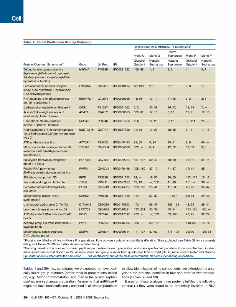
Tables 1 and S9); i.e., candidates were expected to have basi-cally lower group numbers (better rank) in preparations basedon, e.g., Mono P chromatofocusing than in the phenyl sephar-ose/heparin sepharose preparation. Assuming that mtRNase Pmight not have been sufficiently enriched in all five preparations
to allow identification of its components, we extended the anal-ysis to the proteins identified in four and three of five prepara-tions (Tables S8 and S9).Based on these analyses three proteins fulfilled the following
criteria: (1) they were found to be potentially involved in RNA
Table 1. Partial Purification Overlap Proteome
Protein (Common Synonyms)a Gene UniProt IPI
Rank (Group #) in mtRNase P Preparationsb
Mono Q Mono Q
Phenyl
Sepharose Mono P Mono P
Glycerol
Gradient
Heparin
Sepharose
Heparin
Sepharose
Glycerol
Gradient
Heparin
Sepharose
Trifunctional enzyme subunit a
(hydroxyacyl-CoA dehydrogenase/
3-ketoacyl-CoA thiolase/enoyl-CoA
hydratase subunit a)
HADHA P40939 IPI00031522 106; 98 1; 2 2; 6 1; 1 4; 7
Peroxisomal bifunctional enzyme
(enoyl-CoA hydratase/3-hydroxyacyl
CoA dehydrogenase)
EHHADH Q08426 IPI00216164 46; 168 2; 3 3; 5 2; 6 1; 2
RNA (guanine-9-)methyltransferase
domain containing 1
RG9MTD1 Q7L0Y3 IPI00099996 14; 19 13; 14 17; 15 5; 2 2; 3
Carbamoyl-phosphate synthetase 1 CPS1 P31327 IPI00011062 3; 2 35; 58 16; 42 11; 34 7; —
Acetyl-CoA acetyltransferase 1
(acetoacetyl CoA thiolase)
ACAT1 P24752 IPI00030363 183; 87 17; 18 8; 10 12; 9 12; 10
Heat-shock 70 kDa protein 9
(stress-70 protein, mortalin)
HSPA9 P38646 IPI00007765 4; 6 14; 23 9; 12 —; 111 25; —
Hydroxysteroid (17-b) dehydrogenase
10 (3-hydroxyacyl-CoA dehydrogenase
type 2)
HSD17B10 Q99714 IPI00017726 21; 36 12; 29 18; 35 7; 15 11; 15
ATP synthase subunit a ATP5A1 P25705 IPI00440493 29; 66 8; 24 29; 61 8; 8 66; —
Mitochondrial transcription factor B2
(mitochondrial dimethyladenosine
transferase 2)
TFB2M Q9H5Q4 IPI00034069 102; — 6; 4 42; 52 39; 39 9; 8
Eukaryotic translation elongation
factor 1 a-like 3
EEF1AL3 Q5VTE0 IPI00472724 137; 147 30; 49 70; 39 29; 31 34; 11
Poly(A) RNA polymerase
(PAP-associated domain containing 1)
PAPD1 Q9NVV4 IPI00470416 205; 160 37; 19 71; 37 17; 11 85; —
40S ribosomal protein S3 RPS3 P23396 IPI00011253 84; — 18; 55 36; 55 152; 136 10; 16
Translation elongation factor Tu TUFM P49411 IPI00027107 13; 16 —; 162 41; 49 151; — 96; —
Peroxisomal trans-2-enoyl-CoA
reductase
PECR Q9BY49 IPI00744627 123; 152 23; 41 118; 89 46; 75 36; 67
Mitochondrial histidyl-tRNA
synthetase 2
HARS2 P49590 IPI00027445 114; — 27; 39 —; 267 63; 64 29; 48
Uncharacterized protein C17orf42 C17orf42 Q96QE5 IPI00170503 172; — 56; 47 203; 198 43; 54 39; 44
Leucine-rich repeat containing 59 LRRC59 Q96AG4 IPI00396321 133; 221 33; 37 59; 34 163; 125 106; —
ATP-dependent RNA helicase DEAD
box 5
DDX5 P17844 IPI00017617 223; — —; 103 89; 136 73; 45 32; 22
peptidyl-prolyl cis-trans isomerase B
(cyclophilin B)
PPIB P23284 IPI00646304 226; — 68; 122 212; — 140; 84 13; 14
Mitochondrial single-stranded
DNA-binding protein
SSBP1 Q04837 IPI00029744 111; 137 57; 60 178; 124 95; 70 103; 54
a Proteins identified in all five mtRNase P preparations. Four obvious contaminants/artifacts (Keratins, Titin) excluded (see Table S8 for a complete
listing and Tables S1–S9 for further details not listed here).b Ranking based on the number of shared peptides per protein for each preparation and mass spectrometry analysis. Group number from ion-trap
mass spectrometer and Spectrum Mill analysis listed first; group number from quadrupole time-of-flight (QTOF) mass spectrometer and Mascot/
EpiCenter analysis listed after the semicolon; —, not identified by one of the mass spectrometry platforms (depending on position).
464 Cell 135, 462–474, October 31, 2008 ª2008 Elsevier Inc.

metabolism (database annotation and/or specific domains/motifs), (2) they were predicted to be mitochondrial, and(3) they showed a rank distribution in the five subproteomesroughly consistent with the relative specific activity of mtRNaseP in the respective preparations. The three proteins selected forfurther testing were RNA (guanine-9-)methyltransferase domaincontaining 1 (RG9MTD1; UniProt Q7L0Y3), the uncharacterizedprotein C17orf42 (Q96QE5), and the uncharacterized proteinKIAA0391 (Q8N5L5), the latter not identified in the two prepara-tions based on Mono Q chromatography (Tables S8 and S9).
Overexpression of Candidate Genes in 293 Cellsand Affinity Purification of mtRNase PAssuming that mtRNase P is a multisubunit enzyme, we decidedto test the selected candidate-protein subunits by tagged over-expression in human cells to enable them to assemble with en-dogenous components. A fraction of mtRNase P should therebybecome tagged and amenable to affinity purification. cDNAs ofthe three proteins were cloned in frame with C-terminal affinitytags, and 293 cell lines capable of inducible transgene expres-sion were generated. Mitochondria were prepared from expres-sion-induced cells and extracts assayed for mtRNase P activityor subjected to affinity purification. Overexpression ofRG9MTD1resulted in an increase in mtRNase P activity in crude mitochon-drial extracts when compared to cells overexpressing C17orf42or to control cells (Figures 2A and 2C, compare lanes 1 and 2 ineach panel). Moreover, upon affinity purification only RG9MTD1,but not C17orf42, was associated with mtRNase P activity (Fig-ures 2A and 2B). Cloning of KIAA0391 lagged behind that of theother two cDNAs, and the generation of cell lines expressing
Figure 2. Affinity Purification of PresumptivemtRNase P Proteins Overexpressed in 293Cells(A) mtRNase P activity of mitochondrial extracts from
293 cells overexpressing His-tagged RG9MTD1 (I) or
C17orf42 (II) and immobilized metal affinity batch
chromatography of those; substrate, (mt)pre-tRNATyr.
(B) 8% SDS-PAGE and silver staining of samples as-
sayed in (A).
(C) mtRNase P activity of mitochondrial extracts from
293 control cells (c) or cells overexpressing FLAG-
tagged RG9MTD1 (I) and fractions of an anti-FLAG im-
munoprecipitation; substrate, (mt)pre-tRNATyr.
(D) 10% SDS-PAGE and silver staining of samples as-
sayed in (C).
KIAA0391 was stopped when the associa-tion of RG9MTD1 with mtRNase P wasidentified.
A protein of approximately 26 kDa copuri-fied with RG9MTD1 (Figure 2B, lanes 11 and13). The apparent association was con-firmed by overexpression of RG9MTD1with a FLAG- instead of a 63 His-tag andsubsequent anti-FLAG immunoprecipitationinstead of immobilized metal affinity chro-matography (IMAC) (Figures 2C and 2D).
Mass spectrometry revealed the 26 kDa protein to be hydroxys-teroid (17-b) dehydrogenase 10 (HSD17B10), in the literaturemore frequently referred to as 3-hydroxyacyl-CoA dehydroge-nase type 2 (HADH2). In fact HSD17B10 was a constituent ofthe PPOP (Table 1), but although its rank distribution in the fivesubproteomes was compatible with the relative specific activityof mtRNase P, we did not initially consider it as amtRNase P pro-tein candidate because of its attributed catalytic activities. How-ever, in agreement with our data, an interaction of theDrosophilamelanogaster homologs of RG9MTD1 and HSD17B10 was pre-viously found in a two-hybrid screen of the fly proteome (Giotet al., 2003). Thus, since both proteins were consistently associ-ated with mtRNase P activity, we termed them mitochondrialRNase P protein (MRPP) 1 (RG9MTD1) andMRPP2 (HSD17B10).
Identification of a Third Protein Subunit by FunctionalReconstitution of mtRNase P from Recombinant MRPP1and MRPP2 and Mitochondrial FractionsRecombinant MRPP1 and MRPP2, expressed in E. coli, did notshow mtRNase P activity, neither alone nor when assayed incombination (see also below). Yet, when both proteins wereadded to mitochondrial extract, mtRNase P activity was stimu-lated several-fold (Figure 3C, compare lanes 1 and 13), indicatingthat in vitro reconstitution of mtRNase P from bacterial-expressed recombinant MRPP1 and MRPP2 is possible, appar-ently requiring only one or more additional components. Sizefractionation/reconstitution experiments suggested an addi-tional protein component of approximately 60 kDa: mtRNase Pactivity could be reconstituted from recombinant MRPP1and MRPP2 and protein(s) eluting from a gel slice of
Cell 135, 462–474, October 31, 2008 ª2008 Elsevier Inc. 465
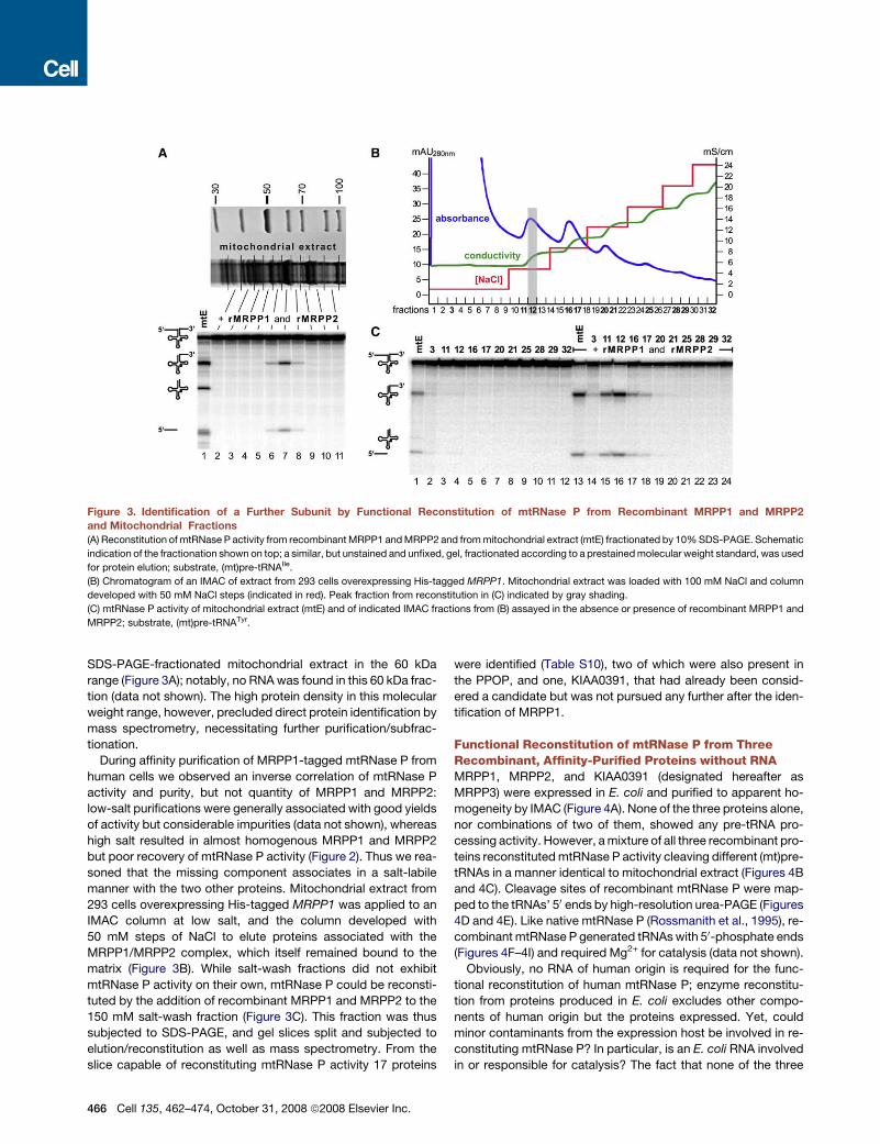
SDS-PAGE-fractionated mitochondrial extract in the 60 kDarange (Figure 3A); notably, no RNAwas found in this 60 kDa frac-tion (data not shown). The high protein density in this molecularweight range, however, precluded direct protein identification bymass spectrometry, necessitating further purification/subfrac-tionation.
During affinity purification of MRPP1-tagged mtRNase P fromhuman cells we observed an inverse correlation of mtRNase Pactivity and purity, but not quantity of MRPP1 and MRPP2:low-salt purifications were generally associated with good yieldsof activity but considerable impurities (data not shown), whereashigh salt resulted in almost homogenous MRPP1 and MRPP2but poor recovery of mtRNase P activity (Figure 2). Thus we rea-soned that the missing component associates in a salt-labilemanner with the two other proteins. Mitochondrial extract from293 cells overexpressing His-tagged MRPP1 was applied to anIMAC column at low salt, and the column developed with50 mM steps of NaCl to elute proteins associated with theMRPP1/MRPP2 complex, which itself remained bound to thematrix (Figure 3B). While salt-wash fractions did not exhibitmtRNase P activity on their own, mtRNase P could be reconsti-tuted by the addition of recombinant MRPP1 and MRPP2 to the150 mM salt-wash fraction (Figure 3C). This fraction was thussubjected to SDS-PAGE, and gel slices split and subjected toelution/reconstitution as well as mass spectrometry. From theslice capable of reconstituting mtRNase P activity 17 proteins
were identified (Table S10), two of which were also present inthe PPOP, and one, KIAA0391, that had already been consid-ered a candidate but was not pursued any further after the iden-tification of MRPP1.
Functional Reconstitution of mtRNase P from ThreeRecombinant, Affinity-Purified Proteins without RNAMRPP1, MRPP2, and KIAA0391 (designated hereafter asMRPP3) were expressed in E. coli and purified to apparent ho-mogeneity by IMAC (Figure 4A). None of the three proteins alone,nor combinations of two of them, showed any pre-tRNA pro-cessing activity. However, amixture of all three recombinant pro-teins reconstitutedmtRNase P activity cleaving different (mt)pre-tRNAs in a manner identical to mitochondrial extract (Figures 4Band 4C). Cleavage sites of recombinant mtRNase P were map-ped to the tRNAs’ 50 ends by high-resolution urea-PAGE (Figures4D and 4E). Like native mtRNase P (Rossmanith et al., 1995), re-combinant mtRNase P generated tRNAswith 50-phosphate ends(Figures 4F–4I) and required Mg2+ for catalysis (data not shown).Obviously, no RNA of human origin is required for the func-
tional reconstitution of human mtRNase P; enzyme reconstitu-tion from proteins produced in E. coli excludes other compo-nents of human origin but the proteins expressed. Yet, couldminor contaminants from the expression host be involved in re-constituting mtRNase P? In particular, is an E. coli RNA involvedin or responsible for catalysis? The fact that none of the three
Figure 3. Identification of a Further Subunit by Functional Reconstitution of mtRNase P from Recombinant MRPP1 and MRPP2and Mitochondrial Fractions(A) Reconstitution of mtRNase P activity from recombinantMRPP1 andMRPP2 and frommitochondrial extract (mtE) fractionated by 10%SDS-PAGE. Schematic
indication of the fractionation shown on top; a similar, but unstained and unfixed, gel, fractionated according to a prestainedmolecular weight standard, was used
for protein elution; substrate, (mt)pre-tRNAIle.
(B) Chromatogram of an IMAC of extract from 293 cells overexpressing His-tagged MRPP1. Mitochondrial extract was loaded with 100 mM NaCl and column
developed with 50 mM NaCl steps (indicated in red). Peak fraction from reconstitution in (C) indicated by gray shading.
(C) mtRNase P activity of mitochondrial extract (mtE) and of indicated IMAC fractions from (B) assayed in the absence or presence of recombinant MRPP1 and
MRPP2; substrate, (mt)pre-tRNATyr.
466 Cell 135, 462–474, October 31, 2008 ª2008 Elsevier Inc.

proteins alone showed any traceable mtRNase P activity ap-pears to rule out any contamination of functional significance(Figures 4B and 4C). Still the possibility remained that a copurify-ing E. coli RNA acted in concert with all three proteins only. Infact, the purified proteins still contained low amounts of RNA(Figure 4K, lane 4; !10 ng per mg protein). However, treatmentwith micrococcal nuclease completely removed any RNA with-out sacrificingmtRNase P activity (Figures 4J and 4K). Moreover,deliberate addition of either M1 RNA, the RNA component ofE. coli RNase P, or E. coli total RNA both inhibited instead ofstimulatedmtRNase P activity (Figures 4J and 4K). M1 RNA con-centrations above 20 nM, and E. coli total RNA above 2.5 ng/ml,became increasingly inhibitory under standard assay conditions,yet concentrations below did not affect the enzymatic activity,i.e., neither inhibited nor stimulated (full range tested: 0.1–100 nM M1 RNA, 0.05–50 ng/ml E. coli total RNA; data notshown). And though the mechanism of inhibition is not known,the lack of stimulation confirms that no RNA from the expressionhost was involved in reconstituting mtRNase P activity from thethree recombinant proteins. In summary, all data are incompat-ible with the involvement of any kind of trans-acting RNA, humanor bacterial, inferring that human mtRNase P is composed ofthree proteins but no RNA.
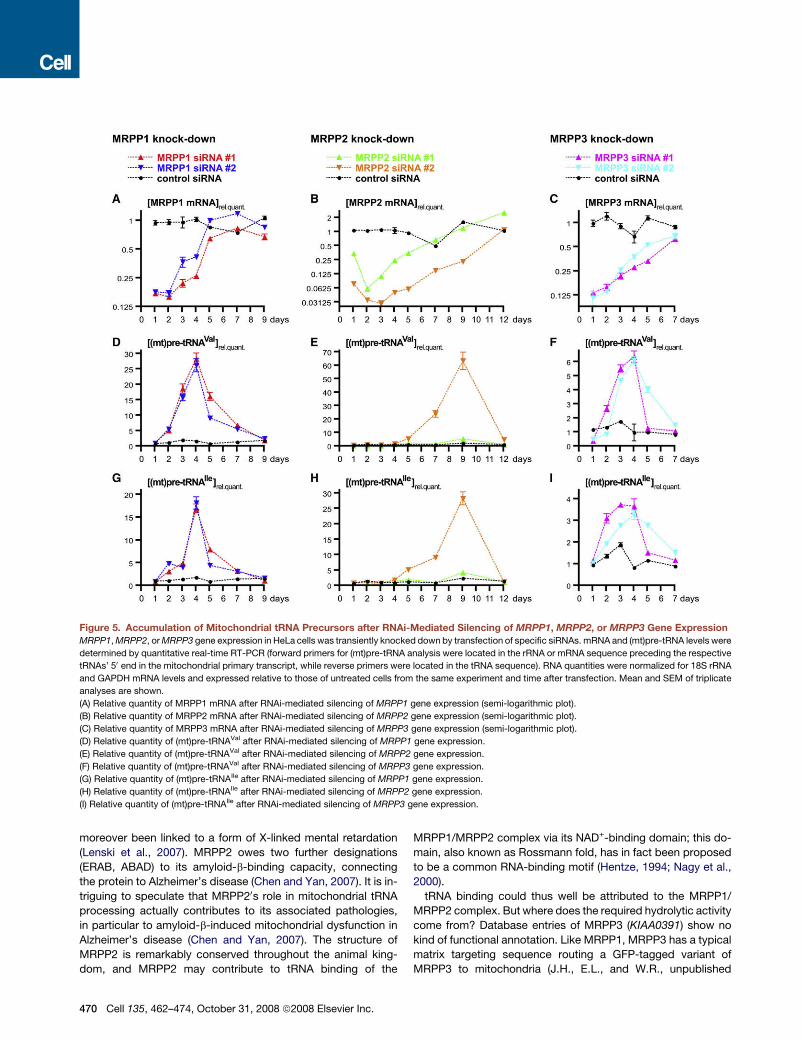
Accumulation of Mitochondrial tRNA Precursorsand Loss of mtRNase P Activity after RNAi-MediatedSilencing of MRPP1, MRPP2, or MRPP3 GeneExpressionTo substantiate the inferred physiological role of MRPP1,MRPP2, and MRPP3 in mitochondrial tRNA 50 end maturationin vivo we subjected their expression to a transient knockdownby RNAi. The supposed deficiency in mtRNase P activity wasreasoned to give rise to an increase in the cellular steady-statelevels of (mt)pre-tRNAs. Two different siRNAs were used foreach gene. All of them caused a decrease of the targetedmRNA (Figures 5A–5C) but no change in the steady-state levelsof the other, nontargeted MRPP mRNAs (data not shown). Witha delay of 2 to 6 days relative to the respective mRNA minima,(mt)pre-tRNAVal levels increased to temporarily peak at 6- to60-fold above those of untreated cells (Figures 5D–5F);(mt)pre-tRNAIle accumulated concurrently, but its accumulationwas less pronounced (Figures 5G–5I). Consistent with the tran-sient nature of siRNA transfections, specific mRNA reductionand concomitant (mt)pre-tRNA accumulation reversed to normalwithin a few days. The variation of the relativemRNA and (mt)pre-tRNA levels of control siRNA-treated cells was less than 2-fold(Figure 5) and thereby within the fluctuation range of untreatedcell samples (data not shown).Extent and kinetics of (mt)pre-tRNA accumulation are not di-
rectly related to the RNAi-induced mRNA reduction but dependalso on the initial cellular concentration, the stability, and the re-synthesis rate of the respective protein. It is thus not surprisingthat the degree of (mt)pre-tRNA accumulation varied betweenthe threeMRPP genes and was not a direct function of the max-imally achieved mRNA reduction; e.g., (mt)pre-tRNAVal in-creased almost 30-fold after MRPP1 silencing but only 6-foldin the case of MRPP3, although mRNA reduction of the latterwas even slightly more pronounced. Likewise, the offset be-
tweenmRNAminimumand (mt)pre-tRNAmaximumwas specificfor the target gene. Consistently, the two different siRNAs usedto silence the expression of a particular gene gave rise to thesame kinetics of (mt)pre-tRNA accumulation. For a given targetgene the extent of (mt)pre-tRNA accumulation nevertheless de-pended on the knockdown capacity of the respective siRNA.This was most obvious in the case of MRPP2, where the muchstronger and more persistent knockdown effect of siRNA #2caused a more than 60-fold increase of (mt)pre-tRNAVal, whilethe less potent siRNA #1 elicited only a 6-fold increase (Fig-ure 5E); in both cases (mt)pre-tRNAs nevertheless peaked atday 9 after transfection.Since we had no means to determine the levels of the three
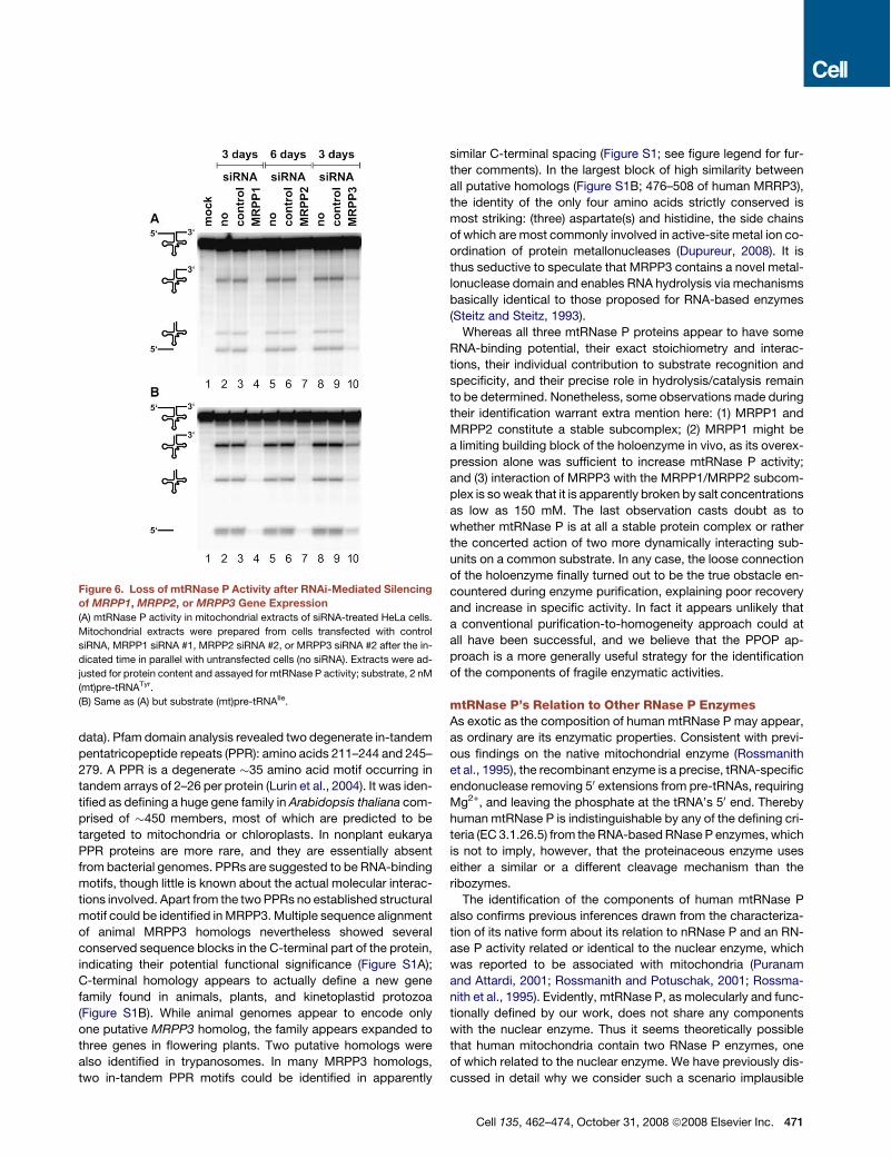
proteins directly, we scaled up and prepared and assayedmtRNase P activity from siRNA-treated cells. Reductions inmtRNase P activity should roughly correlate with the extent of(mt)pre-tRNA accumulation, if the latter is due to the RNAi-medi-ated loss of a genuine component of mtRNase P. A potent siRNAand a point in time between mRNA minimum and (mt)pre-tRNAmaximumwere selected for each gene, reasoning that the great-est loss of mtRNase P activity would be delayed tomRNA reduc-tion but precede the peak of (mt)pre-tRNA accumulation. Whilecontrol siRNA transfections did not alter mtRNase P activity, siR-NAs targeting any of the three MRPP genes caused a reductionin mtRNase P activity (Figure 6). Consistent with the lower(mt)pre-tRNA accumulation potency of MRPP3 siRNAs, moremtRNase P activity remained afterMRPP3 knockdown than afterthose of the other two mtRNase P genes. In conclusion, RNAi-mediated knockdown of any of the three MRPPs caused a tran-sient mtRNase P deficiency with the concomitant accumulationof (mt)pre-tRNAs. Thus all three proteins that are required andsufficient to reconstitute an enzymatic activity capable of specif-ically cleaving (mt)pre-tRNAs at their 50 end in vitro (without theneed for any kind of trans-acting RNA) are also directly involvedin (mt)pre-tRNA 50 end maturation in vivo.
DISCUSSION
Molecular identification of an RNase P devoid of RNA is withoutprecedent so far. Since the first demonstration that the prototypeE. coli enzyme contains an essential RNA moiety (Stark et al.,1978), every RNase P characterized in molecular detail hadbeen found to contain a structurally related RNA, too. Moreover,the recent demonstration that eukaryal nRNase P RNA is capa-ble of mediating pre-tRNA cleavage in the absence of protein likeits bacterial or archaeal counterparts (Kikovska et al., 2007) ap-peared to add further support to the prevailing view that theRNA-based catalytic mechanism of pre-tRNA 50 cleavage hasbeen universally preserved during evolution.An RNase P composed entirely of protein was actually first
proposed 20 years ago (Wang et al., 1988), but none of the com-ponents of this chloroplast enzyme have been identified sincethen. Now, the identification of the components of humanmtRNase P, proposed to be purely proteinaceous 10 yearsago (Rossmanith and Karwan, 1998a), and the functional recon-stitution of its enzymatic activity from recombinant proteins addproof to the idea of nonribozymal 50 pre-tRNAmaturation and re-veal how the seemingly universal RNA world remnant has been
Cell 135, 462–474, October 31, 2008 ª2008 Elsevier Inc. 467
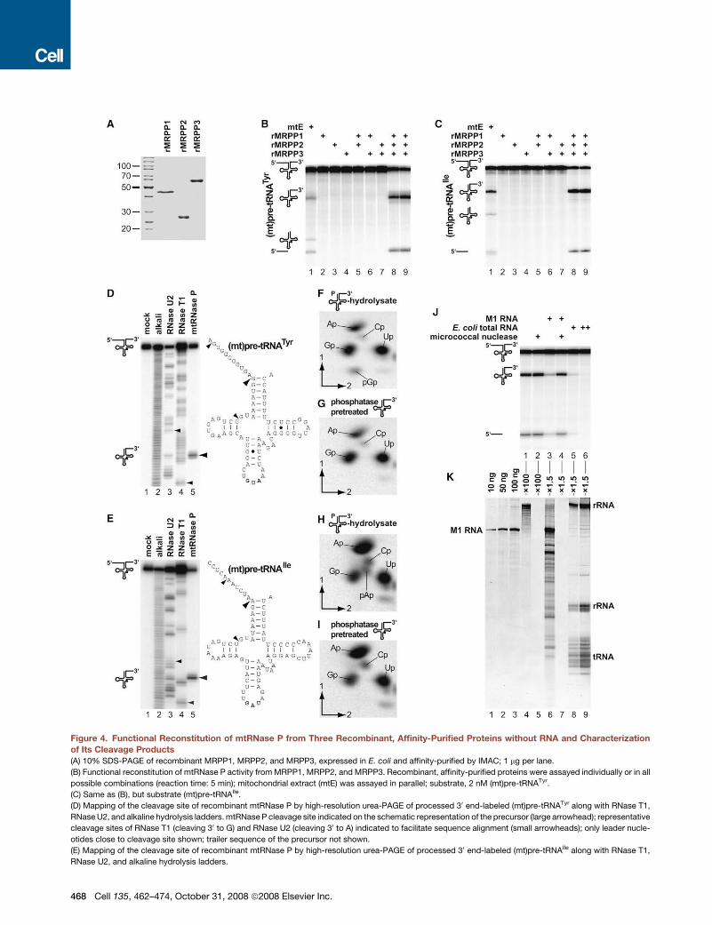
Figure 4. Functional Reconstitution of mtRNase P from Three Recombinant, Affinity-Purified Proteins without RNA and Characterizationof Its Cleavage Products(A) 10% SDS-PAGE of recombinant MRPP1, MRPP2, and MRPP3, expressed in E. coli and affinity-purified by IMAC; 1 mg per lane.
(B) Functional reconstitution of mtRNase P activity from MRPP1, MRPP2, and MRPP3. Recombinant, affinity-purified proteins were assayed individually or in all
possible combinations (reaction time: 5 min); mitochondrial extract (mtE) was assayed in parallel; substrate, 2 nM (mt)pre-tRNATyr.
(C) Same as (B), but substrate (mt)pre-tRNAIle.
(D) Mapping of the cleavage site of recombinant mtRNase P by high-resolution urea-PAGE of processed 30 end-labeled (mt)pre-tRNATyr along with RNase T1,
RNase U2, and alkaline hydrolysis ladders.mtRNase P cleavage site indicated on the schematic representation of the precursor (large arrowhead); representative
cleavage sites of RNase T1 (cleaving 30 to G) and RNase U2 (cleaving 30 to A) indicated to facilitate sequence alignment (small arrowheads); only leader nucle-
otides close to cleavage site shown; trailer sequence of the precursor not shown.
(E) Mapping of the cleavage site of recombinant mtRNase P by high-resolution urea-PAGE of processed 30 end-labeled (mt)pre-tRNAIle along with RNase T1,
RNase U2, and alkaline hydrolysis ladders.
468 Cell 135, 462–474, October 31, 2008 ª2008 Elsevier Inc.

replaced in the animal mitochondrial lineage. Not surprisingly,none of the threemtRNase Pproteins displays any apparent sim-ilarity to a known RNase P protein, including the yeast mitochon-drial one (Dang andMartin, 1993). Evidently, evolution built a newenzyme, and any remains of the ribozyme originally inheritedfrom the a-proteobacterial ancestor were subsequently lost.However, animal mtRNase P was not built simply from compo-nents of a preexisting nucleolytic pathway but by combiningcomponents from different, essentially unrelated biochemicalpathways. Yet, at least two of these components, MRPP1 andMRPP2, were apparently not recruited by gene duplication andsubsequent functional transformation, but the new functionrather seems to be an add-on to their preexisting role. AnimalmtRNase P thereby appears like a patchwork composed oftwo proteins involved in other biochemical processes too andone protein of hitherto unknown functional and evolutionaryorigin.
The Components of Human mtRNase PMRPP1 (RG9MTD1) is one of three vertebrate homologs to yeastTRM10. Trm10p is a tRNA m1G methyltransferase responsiblefor modification at position 9 (Jackman et al., 2003), a commonmodification of eukaryal tRNAs (Sprinzl and Vassilenko, 2005).A TRM10 homolog is widely found in eukaryal and archaeal ge-nomes, but not in eubacteria. Onlymetazoan genomes appear toencode two or three homologs, one of which ismitochondrial ac-cording to its conserved targeting sequence (J.H. and W.R., un-published data; Pagliarini et al., 2008). Notably, m1G9 is fre-quently found in animal but not in yeast or plant mitochondrialtRNAs (Sprinzl and Vassilenko, 2005), and preliminary evidenceindeed suggests that MRPP1 functions in the m1G9 methylationof humanmitochondrial tRNAs (C. Nachbagauer, J.H., andW.R.,unpublished data). The putative mitochondrial TRM10 homologswere reported to branch separately from those that group withyeast and plant in a phylogenetic reconstruction of the family’sevolution (Jackman et al., 2003). Yet, the implied phylogeneticplacement could well be the artifactual result of a duplicationearly in the branching of metazoans with a subsequently morerapid evolution of the mitochondrial lineage to cope with itsnewly acquired function as well as with the structural changes
of animal mitochondrial tRNAs, instead of a selective retentionof two lineages in animals and loss of (the mitochondrial) onein protists, fungi, and plants, as suggested by the authors.Such a scenario could also explain the apparently even earlierbranching of the nematode homolog as a result of adaptationto the bizarre structure of nematode mitochondrial tRNAs (Wol-stenholme et al., 1987).It appears reasonable to assume thatMRPP1 contributes spe-
cific tRNA-binding capacity to mtRNase P. A G9 methyltransfer-ase as a putative specificity component, however, impliesamode of substrate recognition distinct from that of other knownRNase P enzymes. The purine ring of residue 9 appears buried inthe core of the tRNA structure and is involved in a triple interac-tion with base pair 12"23 in the crystal structure of yeast tRNAPhe
(Sussman and Kim, 1976). tRNA core and hence neighborhoodof position 9 are built by D domain, variable loop, and anticodonstem, which thereby represent plausible contact points for anm1G9methyltransferase. Consistently, alterations of these struc-tural elements were previously observed to affect processing bymtRNase P but not by nRNase P (Rossmanith et al., 1995; Ross-manith, 1997; Rossmanith and Karwan, 1998b). Since G9
appears inaccessible for methylation enzymes in the core ofthe L-shaped tRNA structure, binding (and methylation) byMRPP1 and consequently processing by mtRNase P might beassociated with a transient reorganization of the tRNA structure.Such disruption of tertiary interactions and remolding of tRNAwas in fact reported for a G15 transglycosylase (Ishitani et al.,2003).While MRPP1 appears to contribute tRNA-binding capacity to
mtRNase P, the role of its binding partnerMRPP2 is less obvious.MRPP2 (HSD17B10) is a member of the ubiquitous short-chaindehydrogenase/reductase (SDR) family (Jornvall et al., 1995). Aplethora of names refers to the wide variety of fatty acid, steroid,and alcohol substrates oxidized by the enzyme in vitro (commonacronyms: HSD17B10, HADH2, ABAD, SCHAD, MHBD), thoughits actual biological role is unclear (reviewed in Yang et al., 2005).Mutations in its gene on Xp11.2 have been linked to 2-methyl-3-hydroxybutyryl-CoA dehydrogenase (MHBD) deficiency, involv-ing the protein in isoleucine metabolism and neurodegeneration(Ofman et al., 2003). Recently, a reduction of its expression has
(F) Characterization of the 50 nucleotide of (mt)tRNATyr as processed by recombinant mtRNase P. The tRNA precursor, labeled with [a32P]GTP, was incubated
with recombinant mtRNase P, and the cleavage product was isolated and subjected to complete alkaline hydrolysis; nucleoside phosphates were resolved by
two-dimensional thin layer chromatography (TLC). Guanosine 30,50-bisphosphate (pGp) spot from the tRNA’s 50 end indicated.
(G) Phosphatase sensitivity of the 50 nucleotide of (mt)tRNATyr as processed by recombinant mtRNase P. The tRNA precursor, labeled with [a32P]GTP, was in-
cubated with recombinant mtRNase P; the cleavage product was isolated, treated with alkaline phosphatase, and subsequently subjected to complete alkaline
hydrolysis; nucleoside phosphates were resolved by two-dimensional TLC. pGp spot visible in (F) was removed by phosphatase pretreatment of the mtRNase P
cleavage product.
(H) Characterization of the 50 nucleotide of (mt)tRNAIle as processed by recombinant mtRNase P. Same procedure as described for (F); Adenosine 30,50-bisphos-
phate (pAp) spot from the tRNA’s 50 end indicated.
(I) Phosphatase sensitivity of the 50 nucleotide of (mt)tRNAIle as processed by recombinant mtRNase P. Same procedure as described for (G); pAp spot visible in
(H) was removed by phosphatase pretreatment of the mtRNase P cleavage product.
(J) mtRNase P activity of reconstitutedmtRNase P treated with micrococcal nuclease (lane 2) and/or supplemented with 12.5 ng/ml (100 nM) M1 RNA (lanes 3 and
4) or supplemented with 10 or 50 ng/ml E. coli total RNA (lanes 5 and 6); substrate, 2 nM (mt)pre-tRNAIle.
(K) RNA isolated from reconstituted, micrococcal nuclease-treated or RNA-supplemented mtRNase P preparations. Six percent urea-PAGE stained with SYBR
Green I; full gel range from loading slots to the migration front at !35 nucleotides shown; representative RNA species indicated for approximate sizing. Lanes 1
to 3, indicated amounts of M1 RNA, each carried through the same RNA isolation procedure (it was derived from this part of the experiment that the procedure
would have allowed the detection of less than 10 ng of a certain RNA species); lanes 4 and 5, RNA isolated from!25 mg of recombinant mtRNase P, the 100-fold
amount of reconstituted, micrococcal nuclease-treated mtRNase P employed in (J); lanes 6 to 9, RNA isolated from !375 ng of recombinant mtRNase P, the
1.5-fold amount of reconstituted, micrococcal nuclease-treated or RNA-supplemented mtRNase P employed in (J).
Cell 135, 462–474, October 31, 2008 ª2008 Elsevier Inc. 469
moreover been linked to a form of X-linked mental retardation(Lenski et al., 2007). MRPP2 owes two further designations(ERAB, ABAD) to its amyloid-b-binding capacity, connectingthe protein to Alzheimer’s disease (Chen and Yan, 2007). It is in-triguing to speculate that MRPP20s role in mitochondrial tRNAprocessing actually contributes to its associated pathologies,in particular to amyloid-b-induced mitochondrial dysfunction inAlzheimer’s disease (Chen and Yan, 2007). The structure ofMRPP2 is remarkably conserved throughout the animal king-dom, and MRPP2 may contribute to tRNA binding of the
MRPP1/MRPP2 complex via its NAD+-binding domain; this do-main, also known as Rossmann fold, has in fact been proposedto be a common RNA-binding motif (Hentze, 1994; Nagy et al.,2000).tRNA binding could thus well be attributed to the MRPP1/
MRPP2 complex. But where does the required hydrolytic activitycome from? Database entries of MRPP3 (KIAA0391) show nokind of functional annotation. Like MRPP1, MRPP3 has a typicalmatrix targeting sequence routing a GFP-tagged variant ofMRPP3 to mitochondria (J.H., E.L., and W.R., unpublished
Figure 5. Accumulation of Mitochondrial tRNA Precursors after RNAi-Mediated Silencing of MRPP1, MRPP2, or MRPP3 Gene ExpressionMRPP1,MRPP2, orMRPP3 gene expression in HeLa cells was transiently knocked down by transfection of specific siRNAs. mRNA and (mt)pre-tRNA levels were
determined by quantitative real-time RT-PCR (forward primers for (mt)pre-tRNA analysis were located in the rRNA or mRNA sequence preceding the respective
tRNAs’ 50 end in the mitochondrial primary transcript, while reverse primers were located in the tRNA sequence). RNA quantities were normalized for 18S rRNA
and GAPDH mRNA levels and expressed relative to those of untreated cells from the same experiment and time after transfection. Mean and SEM of triplicate
analyses are shown.
(A) Relative quantity of MRPP1 mRNA after RNAi-mediated silencing of MRPP1 gene expression (semi-logarithmic plot).
(B) Relative quantity of MRPP2 mRNA after RNAi-mediated silencing of MRPP2 gene expression (semi-logarithmic plot).
(C) Relative quantity of MRPP3 mRNA after RNAi-mediated silencing of MRPP3 gene expression (semi-logarithmic plot).
(D) Relative quantity of (mt)pre-tRNAVal after RNAi-mediated silencing of MRPP1 gene expression.
(E) Relative quantity of (mt)pre-tRNAVal after RNAi-mediated silencing of MRPP2 gene expression.
(F) Relative quantity of (mt)pre-tRNAVal after RNAi-mediated silencing of MRPP3 gene expression.
(G) Relative quantity of (mt)pre-tRNAIle after RNAi-mediated silencing of MRPP1 gene expression.
(H) Relative quantity of (mt)pre-tRNAIle after RNAi-mediated silencing of MRPP2 gene expression.
(I) Relative quantity of (mt)pre-tRNAIle after RNAi-mediated silencing of MRPP3 gene expression.
470 Cell 135, 462–474, October 31, 2008 ª2008 Elsevier Inc.
data). Pfam domain analysis revealed two degenerate in-tandempentatricopeptide repeats (PPR): amino acids 211–244 and 245–279. A PPR is a degenerate !35 amino acid motif occurring intandem arrays of 2–26 per protein (Lurin et al., 2004). It was iden-tified as defining a huge gene family in Arabidopsis thaliana com-prised of !450 members, most of which are predicted to betargeted to mitochondria or chloroplasts. In nonplant eukaryaPPR proteins are more rare, and they are essentially absentfrom bacterial genomes. PPRs are suggested to be RNA-bindingmotifs, though little is known about the actual molecular interac-tions involved. Apart from the two PPRs no established structuralmotif could be identified in MRPP3. Multiple sequence alignmentof animal MRPP3 homologs nevertheless showed severalconserved sequence blocks in the C-terminal part of the protein,indicating their potential functional significance (Figure S1A);C-terminal homology appears to actually define a new genefamily found in animals, plants, and kinetoplastid protozoa(Figure S1B). While animal genomes appear to encode onlyone putative MRPP3 homolog, the family appears expanded tothree genes in flowering plants. Two putative homologs werealso identified in trypanosomes. In many MRPP3 homologs,two in-tandem PPR motifs could be identified in apparently
similar C-terminal spacing (Figure S1; see figure legend for fur-ther comments). In the largest block of high similarity betweenall putative homologs (Figure S1B; 476–508 of human MRRP3),the identity of the only four amino acids strictly conserved ismost striking: (three) aspartate(s) and histidine, the side chainsof which are most commonly involved in active-site metal ion co-ordination of protein metallonucleases (Dupureur, 2008). It isthus seductive to speculate that MRPP3 contains a novel metal-lonuclease domain and enables RNA hydrolysis via mechanismsbasically identical to those proposed for RNA-based enzymes(Steitz and Steitz, 1993).Whereas all three mtRNase P proteins appear to have some
RNA-binding potential, their exact stoichiometry and interac-tions, their individual contribution to substrate recognition andspecificity, and their precise role in hydrolysis/catalysis remainto be determined. Nonetheless, some observations made duringtheir identification warrant extra mention here: (1) MRPP1 andMRPP2 constitute a stable subcomplex; (2) MRPP1 might bea limiting building block of the holoenzyme in vivo, as its overex-pression alone was sufficient to increase mtRNase P activity;and (3) interaction of MRPP3 with the MRPP1/MRPP2 subcom-plex is soweak that it is apparently broken by salt concentrationsas low as 150 mM. The last observation casts doubt as towhether mtRNase P is at all a stable protein complex or ratherthe concerted action of two more dynamically interacting sub-units on a common substrate. In any case, the loose connectionof the holoenzyme finally turned out to be the true obstacle en-countered during enzyme purification, explaining poor recoveryand increase in specific activity. In fact it appears unlikely thata conventional purification-to-homogeneity approach could atall have been successful, and we believe that the PPOP ap-proach is a more generally useful strategy for the identificationof the components of fragile enzymatic activities.
mtRNase P’s Relation to Other RNase P EnzymesAs exotic as the composition of human mtRNase P may appear,as ordinary are its enzymatic properties. Consistent with previ-ous findings on the native mitochondrial enzyme (Rossmanithet al., 1995), the recombinant enzyme is a precise, tRNA-specificendonuclease removing 50 extensions from pre-tRNAs, requiringMg2+, and leaving the phosphate at the tRNA’s 50 end. Therebyhuman mtRNase P is indistinguishable by any of the defining cri-teria (EC 3.1.26.5) from the RNA-basedRNase P enzymes, whichis not to imply, however, that the proteinaceous enzyme useseither a similar or a different cleavage mechanism than theribozymes.The identification of the components of human mtRNase P
also confirms previous inferences drawn from the characteriza-tion of its native form about its relation to nRNase P and an RN-ase P activity related or identical to the nuclear enzyme, whichwas reported to be associated with mitochondria (Puranamand Attardi, 2001; Rossmanith and Potuschak, 2001; Rossma-nith et al., 1995). Evidently, mtRNase P, as molecularly and func-tionally defined by our work, does not share any componentswith the nuclear enzyme. Thus it seems theoretically possiblethat human mitochondria contain two RNase P enzymes, oneof which related to the nuclear enzyme. We have previously dis-cussed in detail why we consider such a scenario implausible
Figure 6. Loss of mtRNase P Activity after RNAi-Mediated Silencingof MRPP1, MRPP2, or MRPP3 Gene Expression(A) mtRNase P activity in mitochondrial extracts of siRNA-treated HeLa cells.
Mitochondrial extracts were prepared from cells transfected with control
siRNA, MRPP1 siRNA #1, MRPP2 siRNA #2, or MRPP3 siRNA #2 after the in-
dicated time in parallel with untransfected cells (no siRNA). Extracts were ad-
justed for protein content and assayed for mtRNase P activity; substrate, 2 nM
(mt)pre-tRNATyr.
(B) Same as (A) but substrate (mt)pre-tRNAIle.
Cell 135, 462–474, October 31, 2008 ª2008 Elsevier Inc. 471
and a functional association of nRNase P components with mito-chondria not sufficiently supported by the available evidence(Rossmanith and Potuschak, 2001). Although the current workalso cannot finally resolve this issue, two results of our study nev-ertheless deserve mention in this context: (1) (mt)pre-tRNA accu-mulation after RNAi silencing of MRPP genes excludes a full re-dundancy with a hypothetical alternative mtRNase P; (2) none ofthe 884 proteins identified in the five partially purified mtRNase Ppreparations is a component of nRNase P. Likewise, no nRNase Pprotein is currently listed in any of the comprehensive, experi-mentally derived mitochondrial proteome inventories (Pagliariniet al., 2008 and references therein; Prokisch and Ahting, 2007).In contrast, the designation of the enzyme originally identifiedin 1995 and molecularly characterized in this work, as ‘‘mito-chondrial’’ is warranted by (1) the subcellular distribution of itsactivity (Rossmanith et al., 1995), (2) the predicted subcellular lo-calization of its three subunits by subcellular sorting algorithms,(3) the reported subcellular localization of MRPP1 (RG9MTD1;Pagliarini et al., 2008) and MRPP2 (HSD17B10; Yang et al.,2005) and the mitochondrial localization of GFP-taggedMRPP3 (J.H., E.L., andW.R., unpublished data), (4) its specificityfor (mt)pre-tRNAs (Rossmanith et al., 1995), and (5) the increasein steady-state levels of (mt)pre-tRNAs after knockdown of anyof its three components by RNAi.
The Evolution of Animal mtRNase PKnowing that RNase P is a pure protein enzyme in human mito-chondria, it appears evenmore surprising now that an RNAworldremnant has been kept in most principal genetic systems forsuch a seemingly straightforward reaction and has not been re-placed more often by a protein enzyme during evolution, whilemost other putative enzymatic players of the former RNA worlddisappeared without a trace. But, why and when has the RNA-based enzyme, inherited from the a-proteobacterial ancestorand still kept in fungi, been displaced by its protein successorin animal mitochondria, and where else? Addressing this last is-sue first, plant chloroplast and trypanosomal mitochondrial RN-ase P are obvious candidates that might have found an indepen-dent solution for RNA-free tRNA processing during evolution(Gegenheimer, 1996; Salavati et al., 2001). The presence of pu-tative MRPP3 homologs in these organelles, however, suggeststhat at least some kind of relation to the animal mitochondrial en-zyme might exist. Intriguingly, plant MRPP3 proteins are pre-dicted to localize to chloroplasts as well as to mitochondria;and the composition of plant mitochondrial RNase P is com-pletely unknown. Sequencing and analysis of the genome ofthe thermophilic bacterium Aquifex aeolicus revealed the lackof rnpA and rnpB, the bacterial genes encoding the proteinand RNA component of bacterial RNase P (Swanson, 2001),and an RNase P activity in A. aeolicus extracts was reported tobe resistant to micrococcal nuclease (Marszalkowski et al.,2008). However, like other bacterial genomes, theA. aeolicus ge-nome contains no homologs to MRPP1 or MRPP3, suggestingyet an entirely different type of RNase P in this bacterium.
The peculiar structure and evolution of animal mitochondrialgenomes and their general lack of an RNase P RNA gene sug-gest that the switch from RNA to protein-based RNase P hasnot happened late in vertebrate evolution but rather early in the
diversification of metazoans, if not at all at their root. The evolu-tion of the three protein components appears basically consis-tent with such a hypothesis. However, sequence information iscurrently heavily biased toward vertebrate and insect genomes,and no comprehensive genomic information is available frommore ancient metazoan taxa.Yet, why of all genetic systems has RNA-based catalysis been
abandoned in animal mitochondria? Possibly, the RNA worldplayer failed to coevolve fast enough with the degeneratingstructures of animal mitochondrial tRNAs, leaving room fora patchwork assembly of three enzymes, including a tRNAmeth-yltransferase as a putative tRNA specificity factor, to jump intothe breach. Genome compaction could be another driving force,and the long leader and trailer sequences, which are mRNA orrRNAprecursors, themselves indirectly relying on tRNAprocess-ing for their maturation too (Ojala et al., 1981) are also an unusualfeature compared to nuclear encoded or bacterial pre-tRNAs.However, genome compaction and tRNA punctuation of mRNAsare also found in yeast mitochondria, the genomes of which haveretained an RNase P RNA gene (Schafer, 2005). Presumably,only a thorough comparison of protein and RNA enzymes orthe identification of further proteinaceous enzymes will show ifwhat we have described herewith is just a further freak of naturefound in the strange animal mitochondrial genetic system or re-flects specific evolutionary constraints that forced animal mito-chondria to abandon their RNA-based enzyme for a patchworkof three proteins.
EXPERIMENTAL PROCEDURES
Precursor tRNA Substrates, M1 RNAPre-tRNA substrates were synthesized essentially as previously described
(Rossmanith et al., 1995). See Supplemental Experimental Procedures for de-
tails. M1RNAwas transcribed frompJA2 (kindly provided by Leif A. Kirsebom).
mtRNase P Activity AssayPre-tRNA processing reactions were carried out essentially as previously de-
scribed (Rossmanith et al., 1995). For mtRNase P reconstitution, recombinant
MRPP1, MRPP2, and MRPP3 were preincubated for 30 min at room temper-
ature. See Supplemental Experimental Procedures for details.
Partial Purification of HeLa Cell mtRNase PMitochondria were prepared and purified essentially as previously described
(Rossmanith et al., 1995). Cleared detergent lysates or sonicates were sub-
jected to column chromatography using commercially available columns on
an AKTAexplorer FPLC system (GE Healthcare) according to the column-
suppliers’ instructions. Rate-zonal sedimentation in glycerol gradients was
carried out as previously described (Rossmanith and Karwan, 1998a). See
Supplemental Experimental Procedures for details.
Protein Identification and Overlap AnalysismtRNase P peak-fractions of the partially purified preparations were concen-
trated by ultrafiltration and 15–40 mg total protein separated by SDS-PAGE.
Entire gel lanes were cut into slices, digested in situ with trypsin, and analyzed
by nanoflow liquid chromatography tandemmass spectrometry. The complete
analysis (from gel tomass spectrometry) was performed using both an ion-trap
and a quadrupole time-of-flight (QTOF) mass spectrometer. Proteins were
identified by automated database searching against the human IPI database
using Spectrum Mill (Agilent) and Mascot (Matrix Science) search engines.
See Supplemental Experimental Procedures for details.
As the output formats of the two mass spectrometry platforms were not
compatible, the data could not be integrated using commercially available
472 Cell 135, 462–474, October 31, 2008 ª2008 Elsevier Inc.

mass spectrometry analysis software. Therefore, proteome comparison and
compilation of the PPOPwere based on the IPI accession numbers of the iden-
tified proteins and performed by Excel (Microsoft) spreadsheet analysis (see
Tables S1–S9 for details).
Bioinformatic Protein AnalysisWe used InterProScan for domain/motif analyses (Mulder et al., 2007) and
Mitoprot and ChloroP for the prediction of mitochondrial and chloroplast tar-
geting sequences, respectively (Claros and Vincens, 1996; Emanuelsson
et al., 1999).
Cloning and Overexpression of Putative mtRNase P Genesin 293 CellsComplete coding sequences of candidate genes including their native initia-
tion codon context were cloned by PCR (Table S11) in frame with a C-terminal
myc-63 His or FLAG-tag. T-Rex-293 cells (Invitrogen) were transfected and
stable cell lines selected; see Supplemental Experimental Procedures for de-
tails. For overexpression, cell lines were induced at 50% confluence with
0.5 mg/ml tetracycline and hyperconfluent cells harvested 4 days later.
Affinity Chromatography and Immunoprecipitation of PutativemtRNase P ProteinsFor immobilized metal affinity chromatography (IMAC) and immunoprecipita-
tion, commercially available affinity gels and columns were used according
to the supplier’s instructions. See Supplemental Experimental Procedures
for details.
Protein Elution/Renaturation from SDS-PAGsSlices from unstained and unfixed SDS-PAGs were washed thrice with H2O
and once with buffer ER (20 mM Tris"Cl [pH 7.4], 150 mM NaCl, 15% glycerol,
1 mM DTT, 0.1% proteinase inhibitor cocktail, 0.1% Tween 20), and proteins
diffusion-eluted with buffer ER at 16#C overnight.
Cloning and Bacterial Expression of MRPP1, MRPP2, and MRPP3
The coding sequences of the putative mature mitochondrial peptides were
cloned in frame with a 63His-tag (Table S11); see Supplemental Experimental
Procedures for details. Expression in E. coli BL21(DE3) was induced at an op-
tical density (600 nm) of 0.8 by 1 mM IPTG and continued overnight at 16#C.
Affinity Chromatography of Recombinant MRPP1, MRPP2,and MRPP3Washed bacteria were broken by sonication in buffer H (20 mM Tris"Cl [pH7.4], 15% glycerol, 0.1 mM DTT, 0.1% proteinase inhibitor cocktail, 0.02%
Tween 20) containing 150 mM NaCl and 30 mM imidazole, cleared as de-
scribed for mitochondrial extracts (see Supplemental Experimental Proce-
dures), and applied to a 1 ml HisTrap HP column (GE Healthcare) equilibrated
and washed with the same buffer. After washing at 100 to 125 mM imidazole,
recombinant proteins were eluted by a linear gradient to 500 mM imidazole.
Micrococcal Nuclease TreatmentReconstituted mtRNase P was incubated with 30 units micrococcal nuclease
per mg recombinant protein and 10mMCaCl2 for 30min at 30#C. The digestion
was stopped with 30 mM EGTA and mtRNase P activity assays carried out in
the presence of 0.25 mg/ml poly(A).
Analysis of the Cleavage Products of Recombinant mtRNase PCleavage site mapping and analysis of the 50 end of cleavage products were
carried out as previously described (Rossmanith et al., 1995).
RNAiHeLa cells were reverse transfected with commercially available (Ambion),
predesigned, chemically synthesized siRNAs (Table S12) at 30 nM using 1 ml
Lipofectamine 2000 (Invitrogen) per 7.5 pmole siRNA according to the manu-
facturers’ instructions. Transfection mediumwas replaced by standard culture
medium 24 hr after transfection.
RNA AnalysisRNA from reconstituted (micrococcal nuclease treated) mtRNase P prepara-
tions was prepared by phenol:chloroform extraction/ethanol precipitation (us-
ing glycogen as carrier), resolved by urea-PAGE, and visualized by SYBR
Green I staining.
RNA from RNAi-subjected cells was prepared by acidic guanidinium-phenol
extraction. cDNAs were synthesized using oligo d(T)18 or PCR reverse primers
and subjected to relative quantitation by SYBR green I real-time PCR (Table
S13). See Supplemental Experimental Procedures for details.
SUPPLEMENTAL DATA
Supplemental Data include Supplemental Experimental Procedures, one fig-
ure, and thirteen tables and can be found with this article online at http://
www.cell.com/supplemental/S0092-8674(08)01135-5.
ACKNOWLEDGMENTS
We thank Maria Selmer for comments on the manuscript and Philippe Giege
for pointing out a third Arabidopsis homolog of MRPP3. This work was sup-
ported by Austrian Science Fund grant P17453 to W.R.
Received: May 2, 2008
Revised: July 17, 2008
Accepted: September 2, 2008
Published: October 30, 2008
REFERENCES
Andersen, J.S., Wilkinson, C.J., Mayor, T., Mortensen, P., Nigg, E.A., and
Mann, M. (2003). Proteomic characterization of the human centrosome by
protein correlation profiling. Nature 426, 570–574.
Chen, J.X., and Yan, S.D. (2007). Amyloid-b-induced mitochondrial dysfunc-
tion. J. Alzheimers Dis. 12, 177–184.
Claros, M.G., and Vincens, P. (1996). Computational method to predict mito-
chondrially imported proteins and their targeting sequences. Eur. J. Biochem.
241, 779–786.
Dang, Y.L., and Martin, N.C. (1993). Yeast mitochondrial RNase P. Sequence
of theRPM2 gene and demonstration that its product is a protein subunit of the
enzyme. J. Biol. Chem. 268, 19791–19796.
Dupureur, C.M. (2008). Roles of metal ions in nucleases. Curr. Opin. Chem.
Biol. 12, 250–255.
Emanuelsson, O., Nielsen, H., and von Heijne, G. (1999). ChloroP, a neural
network-based method for predicting chloroplast transit peptides and their
cleavage sites. Protein Sci. 8, 978–984.
Evans, D., Marquez, S.M., and Pace, N.R. (2006). RNase P: interface of the
RNA and protein worlds. Trends Biochem. Sci. 31, 333–341.
Gegenheimer, P. (1996). Structure, mechanism and evolution of chloroplast
transfer RNA processing systems. Mol. Biol. Rep. 22, 147–150.
Gesteland R.F., Cech T.R., and Atkins J.F., eds. (2006). The RNA World: The
Nature of Modern RNA Suggests a Prebiotic RNA World, Third Edition (Cold
Spring Harbor, N.Y.: Cold Spring Harbor Laboratory Press).
Giot, L., Bader, J.S., Brouwer, C., Chaudhuri, A., Kuang, B., Li, Y., Hao, Y.L.,
Ooi, C.E., Godwin, B., Vitols, E., et al. (2003). A protein interaction map of Dro-
sophila melanogaster. Science 302, 1727–1736.
Guerrier-Takada, C., Gardiner, K., Marsh, T., Pace, N., and Altman, S. (1983).
The RNA moiety of ribonuclease P is the catalytic subunit of the enzyme. Cell
35, 849–857.
Hartmann, E., and Hartmann, R.K. (2003). The enigma of ribonuclease P evo-
lution. Trends Genet. 19, 561–569.
Hentze, M.W. (1994). Enzymes as RNA-binding proteins: a role for (di)nucleo-
tide-binding domains? Trends Biochem. Sci. 19, 101–103.
Cell 135, 462–474, October 31, 2008 ª2008 Elsevier Inc. 473

Ishitani, R., Nureki, O., Nameki, N., Okada, N., Nishimura, S., and Yokoyama,
S. (2003). Alternative tertiary structure of tRNA for recognition by a posttran-
scriptional modification enzyme. Cell 113, 383–394.
Jackman, J.E., Montange, R.K., Malik, H.S., and Phizicky, E.M. (2003). Identi-
fication of the yeast gene encoding the tRNA m1G methyltransferase respon-
sible for modification at position 9. RNA 9, 574–585.
Jornvall, H., Persson, B., Krook, M., Atrian, S., Gonzalez-Duarte, R., Jeffery, J.,
and Ghosh, D. (1995). Short-chain dehydrogenases/reductases (SDR). Bio-
chemistry 34, 6003–6013.
Kikovska, E., Svard, S.G., and Kirsebom, L.A. (2007). Eukaryotic RNase P RNA
mediates cleavage in the absence of protein. Proc. Natl. Acad. Sci. USA 104,
2062–2067.
Lenski, C., Kooy, R.F., Reyniers, E., Loessner, D., Wanders, R.J.A.,
Winnepenninckx, B., Hellebrand, H., Engert, S., Schwartz, C.E., Meindl, A.,
et al. (2007). The reduced expression of the HADH2 protein causes X-linked
mental retardation, choreoathetosis, and abnormal behavior. Am. J. Hum.
Genet. 80, 372–377.
Lurin, C., Andres, C., Aubourg, S., Bellaoui, M., Bitton, F., Bruyere, C.,
Caboche, M., Debast, C., Gualberto, J., Hoffmann, B., et al. (2004). Genome-
wide analysis of Arabidopsis pentatricopeptide repeat proteins reveals their
essential role in organelle biogenesis. Plant Cell 16, 2089–2103.
Marszalkowski, M., Willkomm, D.K., and Hartmann, R.K. (2008). 50-End matu-
ration of tRNA in Aquifex aeolicus. Biol. Chem. 389, 395–403.
Mulder, N.J., Apweiler, R., Attwood, T.K., Bairoch, A., Bateman, A., Binns, D.,
Bork, P., Buillard, V., Cerutti, L., Copley, R., et al. (2007). New developments in
the InterPro database. Nucleic Acids Res. 35, D224–D228.
Nagy, E., Henics, T., Eckert, M., Miseta, A., Lightowlers, R.N., and
Kellermayer, M. (2000). Identification of the NAD+-binding fold of glyceralde-
hyde-3-phosphate dehydrogenase as a novel RNA-binding domain. Biochem.
Biophys. Res. Commun. 275, 253–260.
Ofman, R., Ruiter, J.P.N., Feenstra, M., Duran, M., Poll-The, B.T., Zschocke,
J., Ensenauer, R., Lehnert, W., Sass, J.O., Sperl, W., et al. (2003). 2-Methyl-
3-hydroxybutyryl-CoA dehydrogenase deficiency is caused by mutations in
the HADH2 gene. Am. J. Hum. Genet. 72, 1300–1307.
Ojala, D., Montoya, J., and Attardi, G. (1981). tRNA punctuation model of RNA
processing in human mitochondria. Nature 290, 470–474.
Pagliarini, D.J., Calvo, S.E., Chang, B., Sheth, S.A., Vafai, S.B., Ong, S.-E.,
Walford, G.A., Sugiana, C., Boneh, A., Chen, W.K., et al. (2008). A mitochon-
drial protein compendium elucidates complex I disease biology. Cell 134,
112–123.
Pannucci, J.A., Haas, E.S., Hall, T.A., Harris, J.K., and Brown, J.W. (1999). RN-
ase P RNAs from some Archaea are catalytically active. Proc. Natl. Acad. Sci.
USA 96, 7803–7808.
Prokisch, H., and Ahting, U. (2007). MitoP2, an integrated database for mito-
chondrial proteins. Methods Mol. Biol. 372, 573–586.
Puranam, R.S., and Attardi, G. (2001). The RNase P associated with HeLa cell
mitochondria contains an essential RNA component identical in sequence to
that of the nuclear RNase P. Mol. Cell. Biol. 21, 548–561.
Robertson, H.D., Altman, S., and Smith, J.D. (1972). Purification and properties
of a specific Escherichia coli ribonuclease which cleaves a tyrosine transfer
ribonucleic acid presursor. J. Biol. Chem. 247, 5243–5251.
Rossmanith, W. (1997). Processing of human mitochondrial tRNASer(AGY):
a novel pathway in tRNA biosynthesis. J. Mol. Biol. 265, 365–371.
Rossmanith, W., and Karwan, R.M. (1998a). Characterization of human mito-
chondrial RNase P: novel aspects in tRNA processing. Biochem. Biophys.
Res. Commun. 247, 234–241.
Rossmanith, W., and Karwan, R.M. (1998b). Impairment of tRNA processing
by point mutations in mitochondrial tRNALeu(UUR) associated with mitochon-
drial diseases. FEBS Lett. 433, 269–274.
Rossmanith, W., and Potuschak, T. (2001). Difference between mitochondrial
RNase P and nuclear RNase P. Mol. Cell. Biol. 21, 8236–8237.
Rossmanith, W., Tullo, A., Potuschak, T., Karwan, R., and Sbisa, E. (1995).
Human mitochondrial tRNA processing. J. Biol. Chem. 270, 12885–12891.
Salavati, R., Panigrahi, A.K., and Stuart, K.D. (2001). Mitochondrial ribonucle-
ase P activity of Trypanosoma brucei. Mol. Biochem. Parasitol. 115, 109–117.
Schafer, B. (2005). RNA maturation in mitochondria of S. cerevisiae and
S. pombe. Gene 354, 80–85.
Schirmer, E.C., Florens, L., Guan, T., Yates, J.R., III, and Gerace, L. (2003).
Nuclear membrane proteins with potential disease links found by subtractive
proteomics. Science 301, 1380–1382.
Sprinzl, M., and Vassilenko, K.S. (2005). Compilation of tRNA sequences and
sequences of tRNA genes. Nucleic Acids Res. 33, D139–D140.
Stark, B.C., Kole, R., Bowman, E.J., and Altman, S. (1978). Ribonuclease P: an
enzyme with an essential RNA component. Proc. Natl. Acad. Sci. USA 75,
3717–3721.
Steitz, T.A., and Steitz, J.A. (1993). A general two-metal-ion mechanism for
catalytic RNA. Proc. Natl. Acad. Sci. USA 90, 6498–6502.
Sussman, J.L., and Kim, S.-H. (1976). Three-dimensional structure of a transfer
RNA in two crystal forms. Science 192, 853–858.
Swanson, R.V. (2001). Genome of Aquifex aeolicus. Methods Enzymol. 330,
158–169.
Thomas, B.C., Gao, L., Stomp, D., Li, X., and Gegenheimer, P.A. (1995). Spin-
ach chloroplast RNase P: a putative protein enzyme. Nucleic Acids Symp. Ser.
33, 95–98.
Walker, S.C., and Engelke, D.R. (2006). Ribonuclease P: the evolution of an an-
cient RNA enzyme. Crit. Rev. Biochem. Mol. Biol. 41, 77–102.
Wang, M.J., Davis, N.W., and Gegenheimer, P. (1988). Novel mechanisms for
maturation of chloroplast transfer RNA precursors. EMBO J. 7, 1567–1574.
Willkomm, D.K., and Hartmann, R.K. (2007). An important piece of the RNase P
jigsaw solved. Trends Biochem. Sci. 32, 247–250.
Wolstenholme, D.R., Macfarlane, J.L., Okimoto, R., Clary, D.O., and
Wahleithner, J.A. (1987). Bizarre tRNAs inferred from DNA sequences of mito-
chondrial genomes of nematode worms. Proc. Natl. Acad. Sci. USA 84,
1324–1328.
Yang, S.-Y., He, X.-Y., and Schulz, H. (2005). Multiple functions of type 10 17b-
hydroxysteroid dehydrogenase. Trends Endocrinol. Metab. 16, 167–175.
474 Cell 135, 462–474, October 31, 2008 ª2008 Elsevier Inc.


![RAYK HOLZMANN - VIVA ModelsRAYK HOLZMANN Height: 186 cm [6' 1"] B/W/H: 101 90 97 [40" 35" 38"] Size: 50 Shoes: 44 Hair: brown Eyes: blue](https://img.pdfslide.us/doc/110x75/61212c5e2f111510c84d9687/rayk-holzmann-viva-models-rayk-holzmann-height-186-cm-6-1-bwh-101.jpg)