Embed Size (px)
Citation preview

Assessment and Managementof Venous Leg Ulcers
Nursing Best Practice GuidelineShaping the future of Nursing
March 2004

Greetings from Doris Grinspun Executive DirectorRegistered Nurses Association of Ontario
It is with great excitement that the Registered Nurses Association of Ontario (RNAO)
disseminates this nursing best practice guideline to you. Evidence-based practice supports
the excellence in service that nurses are committed to deliver in our day-to-day practice.
We offer our endless thanks to the many institutions and individuals that are making
RNAO’s vision for Nursing Best Practice Guidelines (NBPGs) a reality. The Ontario Ministry
of Health and Long-Term Care recognized RNAO’s ability to lead this project and is providing multi-year
funding. Tazim Virani – NBPG project director – with her fearless determination and skills, is moving the
project forward faster and stronger than ever imagined. The nursing community, with its commitment and
passion for excellence in nursing care, is providing the knowledge and countless hours essential to the creation
and evaluation of each guideline. Employers have responded enthusiastically to the request for proposals
(RFP), and are opening their organizations to pilot test the NBPGs.
Now comes the true test in this phenomenal journey: Will nurses utilize the guidelines in their day-to-day practice?
Successful uptake of these NBPGs requires a concerted effort of four groups: nurses themselves, other
healthcare colleagues, nurse educators in academic and practice settings, and employers. After lodging
these guidelines into their minds and hearts, knowledgeable and skillful nurses and nursing students need
healthy and supportive work environments to help bring these guidelines to life.
We ask that you share this NBPG, and others, with members of the interdisciplinary team. There is much to
learn from one another. Together, we can ensure that Ontarians receive the best possible care every time they
come in contact with us. Let’s make them the real winners of this important effort!
RNAO will continue to work hard at developing and evaluating future guidelines. We wish you the
best for a successful implementation!
Doris Grinspun, RN, MScN, PhD (candidate)
Executive Director
Registered Nurses Association of Ontario

How to Use this Document
This nursing best practice guideline is a comprehensive document providing
resources necessary for the support of evidence-based nursing practice. The document
needs to be reviewed and applied based on the specific needs of the organization or practice
setting/environment, as well as the needs and wishes of the client. Guidelines should not be
applied in a “cookbook” fashion but used as a tool to assist in decision making for individualized
client care, as well as ensuring that appropriate structures and supports are in place to
provide the best possible care.
Nurses, other healthcare professionals and administrators who are leading and facilitating
practice changes will find this document valuable for the development of policies, procedures,
protocols, educational programs, assessment and documentation tools. It is recommended
that the nursing best practice guidelines be used as a resource tool. It is not necessary, nor
practical that every nurse have a copy of the entire guideline. Nurses providing direct client
care will benefit from reviewing the recommendations, the evidence in support of the
recommendations and the process that was used to develop the guidelines. However, it is
highly recommended that practice settings/environments adapt these guidelines in formats
that would be user-friendly for daily use. This guideline has some suggested formats for such
local adaptation and tailoring.
Organizations wishing to use the guideline may decide to do so in a number of ways:
� Assess current nursing and healthcare practices using the recommendations in the
guideline.
� Identify recommendations that will address identified needs or gaps in services.
� Systematically develop a plan to implement the recommendations using associated
tools and resources.
RNAO is interested in hearing how you have implemented this guideline. Please contact
us to share your story. Implementation resources will be made available through the
RNAO website at www.rnao.org/bestpractices to assist individuals and organizations to
implement best practice guidelines.
1
N u r s i n g B e s t P r a c t i c e G u i d e l i n e

Kathryn Kozell, RN, BA, BScN, MScN,ACNP/CNS, ET (Co-Team Leader)
GI Surgery Ostomy/Wound
St. Joseph’s Healthcare London
St. Joseph’s Site
London, Ontario
Susan Mills-Zorzes, RN, BScN, CWOCN(Co-Team Leader)
Enterostomal Therapy Nurse
St. Joseph’s Care Group
Thunder Bay, Ontario
Patti Barton, RN, PHN, ETOstomy, Wound and Skin Consultant
Specialty ET Services
Toronto, Ontario
Marion Chipman, RNONA Representative
Staff Nurse
Shaver Rehabilitation Hospital
St. Catharines, Ontario
Patricia Coutts, RNWound Care & Clinical Trials Coordinator
The Mississauga Dermatology Centre
Office of Dr. R. Gary Sibbald
Mississauga, Ontario
Diane Gregoire, RN, ET, MScN Spina Bifida Service Coordinator
Coordinatrice des Services de Spina Bifida
Ottawa, Ontario
Margaret Harrison, RN, PhDAssociate Professor
School of Nursing
Queen’s University
Kingston, Ontario
Nurse Scientist
Clinical Epidemiology Program
Ottawa Health Research Institute
Ottawa, Ontario
Staff Nurse
St. Joseph’s Healthcare London
Parkwood Site
London, Ontario
Karen Lorimer, RN, MScN (candidate)Clinical Leader
Ottawa-Carleton Regional Leg Ulcer Project
Ottawa, Ontario
Sheri Oliver, RPNProject Coordinator
Nursing Education Initiative
Registered Practical Nurses Association
of Ontario
Mississauga, Ontario
Nancy Parslow, RN, ETEnterostomal/Wound Care Consultant
Calea
Toronto, Ontario
Josephine Santos, RN, MNFacilitator, Project Coordinator
Nursing Best Practice Guidelines Project
Registered Nurses Association of Ontario
Toronto, Ontario
Guideline Development Panel Members
Terri Labate, RN, CRRN, GNC(C), BScN (candidate)
2
Assessment and Management of Venous Leg Ulcers

Assessment & Managementof Venous Leg Ulcers
Project team:
Tazim Virani, RN, MScN
Project Director
Josephine Santos, RN, MN
Project Coordinator
Heather McConnell, RN, BScN, MA(Ed)
Project Manager
Jane Schouten, RN, BScN, MBA
Project Coordinator
Stephanie Lappan-Gracon, RN, MN
Coordinator – Best Practice Champions Network
Carrie ScottProject Assistant
Elaine Gergolas, BA
Project Coordinator – Advanced Clinical/Practice
Fellowships
Melissa Kennedy, BA
Project Assistant
Keith Powell, BA, AIT
Web Editor
Registered Nurses Association of Ontario
Nursing Best Practice Guidelines Project
111 Richmond Street West, Suite 1100
Toronto, Ontario M5H 2G4
www.rnao.org/bestpractices
N u r s i n g B e s t P r a c t i c e G u i d e l i n e
3

Acknowledgement
Stakeholders representing diverse perspectives were solicited for their feedbackand the Registered Nurses Association of Ontario wishes to acknowledge the following for their contribution in reviewing this Nursing Best Practice Guideline.
Marlene Allen Physiotherapist
Oshawa, Ontario
Lucy Cabico Nurse Practitioner/Clinical Nurse Specialist
Baycrest Centre for Geriatric Care
Toronto, Ontario
Karen Campbell Nurse Practitioner/Clinical Nurse Specialist
Parkwood Hospital
London, Ontario
Dawn-Marie Clarke Chiropodist
Shaver Rehabilitation Hospital
St. Catharines, Ontario
Debra ClutterbuckRegistered Practical Nurse
Cambridge, Ontario
Nicole Denis Enterostomal Therapy Nurse
The Ottawa Hospital
Ottawa, Ontario
Elaine Diebold Enterostomal Therapy Nurse
Durham, Ontario
Geneviève GrégoireDietetic Intern
Moncton, New Brunswick
Connie HarrisEnterostomal Therapist/Consultant
Kitchener, Ontario
Cheri Hernandez Associate Professor
Faculty of Nursing
University of Windsor
Windsor, Ontario
Dr. Pamela HoughtonAssociate Professor
School of Physiotherapy
University of Western Ontario
London, Ontario
Madge LegraceRegistered Nurse
Unionville, Ontario
Dr. Ronald Mahler Dermatologist
Thunder Bay Medical Centre
Thunder Bay, Ontario
Stephanie McIntosh Consumer
Marie-Andre Meloche Victorian Order of Nurses – Peel
Mississauga, Ontario
Beverly MonetteClinical Nurse Consultant
Dell Pharmacy, Home Health Care Centre
Hamilton, Ontario
4
Assessment and Management of Venous Leg Ulcers

Sue Morrell-DeVries Nurse Coordinator, Vascular Surgery
Toronto General Hospital
Toronto, Ontario
Dr. Gary SibbaldDirector of Dermatology Day Care and
Wound Healing Clinic
Sunnybrook & Women’s College Health
Sciences Centre
Associate Professor & Director
Continuing Education
Department of Medicine
University of Toronto
Toronto, Ontario
The Mississauga Dermatology Centre
Mississauga, Ontario
Jennifer SkellyAssociate Professor
McMaster University
Hamilton, Ontario
Louise SpenceHamilton-Wentworth Community Care
Access Centre
Hamilton, Ontario
Dr. Terry Trusdale Varicose & Spider Vein Treatment
Kakabeka Falls, Ontario
Hélène Villeneuve Dietitian
Sarsfield, Ontario
Claire Westendorp Enterostomal Therapist
Kingston General Hospital
Kingston, Ontario
Meta Wilson Consumer
A special acknowledgement also goes
to Barbara Willson, RN, MSc, and
Anne Tait, RN, BScN, who served as
Project Coordinators at the onset of the
guideline development.
5
N u r s i n g B e s t P r a c t i c e G u i d e l i n e

Principal Investigators:Nancy Edwards, RN, PhDBarbara Davies, RN, PhDUniversity of Ottawa
Evaluation Team:Maureen Dobbins, RN, PhDJenny Ploeg, RN, PhDJennifer Skelly, RN, PhDMcMaster University
Patricia Griffin, RN, PhDUniversity of Ottawa
Project Staff:University of Ottawa
Barbara Helliwell, BA(Hons); Marilynn Kuhn, MHA; Diana Ehlers, MA(SW), MA(Dem);Lian Kitts, RN; Elana Ptack, BA; Isabelle St-Pierre, BScN, MScN(cand.)
As well, RNAO sincerely acknowledges the leadership and dedication of theresearchers who have directed the evaluation phase of the Nursing Best PracticeGuidelines Project. The Evaluation Team is comprised of:
Contact Information Registered Nurses Association of OntarioNursing Best Practice Guidelines Project
111 Richmond Street West, Suite 1100
Toronto, Ontario
M5H 2G4
Registered Nurses Association of OntarioHead Office
438 University Avenue, Suite 1600
Toronto, Ontario
M5G 2K8
RNAO also wishes to acknowledge the following organizations for their role inpilot testing this guideline:
Pilot Project Sites� Saint Elizabeth Health Care
Toronto, Ontario� St. Peter’s Hospital
Hamilton, Ontario
6
Assessment and Management of Venous Leg Ulcers

Disclaimer
These best practice guidelines are related only to nursing practice and not intended to take into
account fiscal efficiencies. These guidelines are not binding for nurses and their use should be
flexible to accommodate client/family wishes and local circumstances. They neither constitute
a liability or discharge from liability. While every effort has been made to ensure the accuracy
of the contents at the time of publication, neither the authors nor RNAO give any guarantee as
to the accuracy of the information contained in them, nor accept any liability, with respect to
loss, damage, injury or expense arising from any such errors or omissions in the contents of this
work. Any reference throughout the document to specific pharmaceutical products as examples
does not imply endorsement of any of these products.
Copyright
With the exception of those portions of this document for which a specific prohibition or
limitation against copying appears, the balance of this document may be produced, reproduced
and published in its entirety, in any form, including in electronic form, for educational or
non-commercial purposes only, without requiring the consent or permission of the Registered
Nurses Association of Ontario, provided that an appropriate credit or citation appears in the
copied work as follows:
Registered Nurses Association of Ontario (2004). Assessment and Management of Venous
Leg Ulcers. Toronto, Canada: Registered Nurses Association of Ontario.
Assessment and Management of Venous Leg Ulcers
7
N u r s i n g B e s t P r a c t i c e G u i d e l i n e
table of contents
Summary of Recommendations . . . . . . . . . . . . . . . . . . . . . . . . . . . . . . . . . . . . . . . . . . .10
Interpretation of Evidence . . . . . . . . . . . . . . . . . . . . . . . . . . . . . . . . . . . . . . . . . . . . . . .18
Responsibility for Development . . . . . . . . . . . . . . . . . . . . . . . . . . . . . . . . . . . . . . . . . . .19
Purpose and Scope . . . . . . . . . . . . . . . . . . . . . . . . . . . . . . . . . . . . . . . . . . . . . . . . . . . .19
Guideline Development Process . . . . . . . . . . . . . . . . . . . . . . . . . . . . . . . . . . . . . . . . . . .20
Definition of Terms . . . . . . . . . . . . . . . . . . . . . . . . . . . . . . . . . . . . . . . . . . . . . . . . . . . .24
Background Context . . . . . . . . . . . . . . . . . . . . . . . . . . . . . . . . . . . . . . . . . . . . . . . . . . .26
Guiding Principles/Assumptions in Venous Leg Ulcers Care . . . . . . . . . . . . . . . . . . . . . . .27
Interactive Guiding Principles of Venous Leg Ulcers Care . . . . . . . . . . . . . . . . . . . . . . . .28
Practice Recommendations . . . . . . . . . . . . . . . . . . . . . . . . . . . . . . . . . . . . . . . . . . . . . .29
Education Recommendations . . . . . . . . . . . . . . . . . . . . . . . . . . . . . . . . . . . . . . . . . . . . .53
Organization & Policy Recommendations . . . . . . . . . . . . . . . . . . . . . . . . . . . . . . . . . . . .55
Evaluation & Monitoring . . . . . . . . . . . . . . . . . . . . . . . . . . . . . . . . . . . . . . . . . . . . . . . .56
Implementation Tips . . . . . . . . . . . . . . . . . . . . . . . . . . . . . . . . . . . . . . . . . . . . . . . . . . .58
Process for Update/Review of Guideline . . . . . . . . . . . . . . . . . . . . . . . . . . . . . . . . . . . . .60
References . . . . . . . . . . . . . . . . . . . . . . . . . . . . . . . . . . . . . . . . . . . . . . . . . . . . . . . . . .61
Bibliography . . . . . . . . . . . . . . . . . . . . . . . . . . . . . . . . . . . . . . . . . . . . . . . . . . . . . . . . .64
8
Assessment and Management of Venous Leg Ulcers

Appendix A – Search Strategy for Existing Evidence . . . . . . . . . . . . . . . . . . . . . . . . . . .70
Appendix B – Glossary of Terms . . . . . . . . . . . . . . . . . . . . . . . . . . . . . . . . . . . . . . . . . .74
Appendix C – Different Types of Leg Ulcers and Their Causes . . . . . . . . . . . . . . . . . . . .88
Appendix D – Leg Ulcer Assessment Form . . . . . . . . . . . . . . . . . . . . . . . . . . . . . . . . . .89
Appendix E – Leg Ulcer Measurement Tool . . . . . . . . . . . . . . . . . . . . . . . . . . . . . . . . . .95
Appendix F – Quality of Life Assessment Tool . . . . . . . . . . . . . . . . . . . . . . . . . . . . . . .101
Appendix G – Pain Assessment Tools . . . . . . . . . . . . . . . . . . . . . . . . . . . . . . . . . . . . .103
Appendix H – Cleansing Agents and Their Associated Toxicities . . . . . . . . . . . . . . . . . .106
Appendix I – Potential Allergens . . . . . . . . . . . . . . . . . . . . . . . . . . . . . . . . . . . . . . . .107
Appendix J – Topical Antimicrobial Agents . . . . . . . . . . . . . . . . . . . . . . . . . . . . . . . . .108
Appendix K – Classes of Compression Bandages . . . . . . . . . . . . . . . . . . . . . . . . . . . .110
Appendix L – Description of the Toolkit . . . . . . . . . . . . . . . . . . . . . . . . . . . . . . . . . . .111
9
N u r s i n g B e s t P r a c t i c e G u i d e l i n e
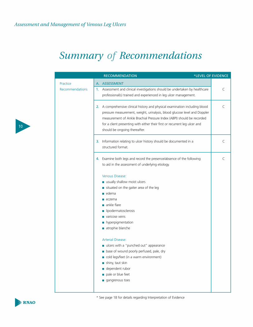
Summary of Recommendations
RECOMMENDATION *LEVEL OF EVIDENCE
Practice A. ASSESSMENT
Recommendations 1. Assessment and clinical investigations should be undertaken by healthcare C
professional(s) trained and experienced in leg ulcer management.
2. A comprehensive clinical history and physical examination including blood C
pressure measurement, weight, urinalysis, blood glucose level and Doppler
measurement of Ankle Brachial Pressure Index (ABPI) should be recorded
for a client presenting with either their first or recurrent leg ulcer and
should be ongoing thereafter.
3. Information relating to ulcer history should be documented in a C
structured format.
4. Examine both legs and record the presence/absence of the following C
to aid in the assessment of underlying etiology.
Venous Disease:
� usually shallow moist ulcers
� situated on the gaiter area of the leg
� edema
� eczema
� ankle flare
� lipodermatosclerosis
� varicose veins
� hyperpigmentation
� atrophie blanche
Arterial Disease:
� ulcers with a “punched out” appearance
� base of wound poorly perfused, pale, dry
� cold legs/feet (in a warm environment)
� shiny, taut skin
� dependent rubor
� pale or blue feet
� gangrenous toes
* See page 18 for details regarding Interpretation of Evidence
10
Assessment and Management of Venous Leg Ulcers
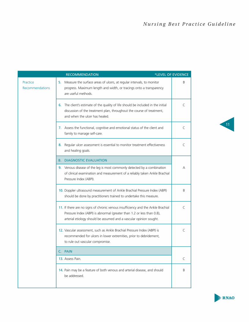
RECOMMENDATION *LEVEL OF EVIDENCE
Practice 5. Measure the surface areas of ulcers, at regular intervals, to monitor B
Recommendations progress. Maximum length and width, or tracings onto a transparency
are useful methods.
6. The client’s estimate of the quality of life should be included in the initial C
discussion of the treatment plan, throughout the course of treatment,
and when the ulcer has healed.
7. Assess the functional, cognitive and emotional status of the client and C
family to manage self-care.
8. Regular ulcer assessment is essential to monitor treatment effectiveness C
and healing goals.
B. DIAGNOSTIC EVALUATION
9. Venous disease of the leg is most commonly detected by a combination A
of clinical examination and measurement of a reliably taken Ankle Brachial
Pressure Index (ABPI).
10. Doppler ultrasound measurement of Ankle Brachial Pressure Index (ABPI) B
should be done by practitioners trained to undertake this measure.
11. If there are no signs of chronic venous insufficiency and the Ankle Brachial C
Pressure Index (ABPI) is abnormal (greater than 1.2 or less than 0.8),
arterial etiology should be assumed and a vascular opinion sought.
12. Vascular assessment, such as Ankle Brachial Pressure Index (ABPI) is C
recommended for ulcers in lower extremities, prior to debridement,
to rule out vascular compromise.
C. PAIN
13. Assess Pain. C
14. Pain may be a feature of both venous and arterial disease, and should B
be addressed.
11
N u r s i n g B e s t P r a c t i c e G u i d e l i n e

12
Assessment and Management of Venous Leg Ulcers
RECOMMENDATION *LEVEL OF EVIDENCE
Practice 15. Prevent or manage pain associated with debridement. Consult with a C
Recommendations physician and pharmacist as needed.
D. VENOUS ULCER CARE
16. Choose the technique of debridement, considering the type, quantity C
and location of non-viable tissue, the depth of the wound, the amount of
wound fluid and the general condition and goals of the client.
17. Cleansing of the ulcer should be kept simple; warm tap water or saline C
is usually sufficient.
18. Dressings must be simple, low adherent, acceptable to the client and A
should be low cost.
19. Avoid products that commonly cause skin sensitivity, such as those C
containing lanolin, phenol alcohol, or topical antibiotics.
20. Choose a type of dressing depending on the amount of exudate and C
the phase of healing.
21. No specific dressing has been demonstrated to encourage ulcer healing. A
22. In contrast to drying out, moist wound conditions allow optimal A
cell migration, proliferation, differentiation and neovascularization.
23. Refer clients with suspected sensitivity reactions to a dermatologist for B
patch testing. Following patch testing, identified allergens must be
avoided, and medical advice on treatment should be sought.
24. Venous surgery followed by graduated compression hosiery is an option C
for consideration in clients with superficial venous insufficiency.
25. Biological wound coverings and growth factor treatments should not be C
applied in cases of wound infection.

RECOMMENDATION *LEVEL OF EVIDENCE
Practice 26. Optimal nutrition facilitates wound healing, maintains immune B
Recommendations competence, and decreases the risk of infection.
E. INFECTION
27. Assess for infection. A
28. An infection is indicated when > 105 bacteria/gram tissue is present. B
29. The treatment of infection is managed by debridement, wound cleansing A
and systemic antibiotics.
30. Antibiotics should only be considered if the ulcer is clinically cellulitic C
(presence of some of the following signs and symptoms: pyrexia;
increasing pain; increasing erythema of surrounding skin; purulent exudate;
rapid increase in ulcer size).
31. Do not use topical antiseptics to reduce bacteria in wound tissue, B
e.g., povidone iodine, iodophor, sodium hypochlorite, hydrogen peroxide,
or acetic acid.
32. Topical antibiotics and antibacterial agents are frequent sensitizers and B
should be avoided.
F. COMPRESSION
33. The treatment of choice for clinical venous ulceration uncomplicated A
by other factors, is graduated compression bandaging, properly applied,
and combined with exercise. Graduated compression is the main
treatment for venous eczema.
34. High compression increases venous ulcer healing and is more effective A
than low compression, but should only be used where ABPI ≥ 0.8 and
ulcer is clinically venous.
35. Compression bandages should only be applied by a suitably trained and B
experienced practitioner.
13
N u r s i n g B e s t P r a c t i c e G u i d e l i n e

14
Assessment and Management of Venous Leg Ulcers
RECOMMENDATION *LEVEL OF EVIDENCE
Practice 36. Venous ulceration should be treated with high compression bandaging C
Recommendations to achieve a pressure between 35-40 mm Hg. at the ankle, graduating
to half at calf in the normally shaped limb, as per La Place’s Law.
37. Use protective padding over bony prominences when applying C
high compression.
38. Arterial insufficiency is a contraindication to the use of high compression. C
A modified form of compression may be used under specialist supervision.
39. Use compression with caution in clients with diabetes, those with C
connective tissue disease and the elderly.
40. Compression therapy should be modified until clinical infection is treated. C
41. Bandages should be applied according to manufacturer’s recommendations. C
42. When using elastic systems such as “high compression” bandages, the C
ankle circumference must be more than or padded to equal 18 cms.
43. Ankle circumference should be measured at a distance of 2.5 cm (one inch) C
above the medial malleolus.
44. The concepts, practice, and hazards of graduated compression should A
be fully understood by those prescribing and fitting compression stockings.
45. Graduated compression hosiery should be measured and fitted by a C
certified fitter.
46. To maintain a therapeutic level of compression, stockings should be C
cared for as per manufacturer’s instructions, and replaced every six months.
47. Graduated compression hosiery should be prescribed for life. B
48. External compression applied using various forms of pneumatic compression A
pumps is indicated for individuals with chronic venous insufficiency.

15
N u r s i n g B e s t P r a c t i c e G u i d e l i n e
RECOMMENDATION *LEVEL OF EVIDENCE
Practice 49. The client should be prescribed regular vascular exercise by means A
Recommendations of intensive controlled walking and exercises to improve the function of
the upper ankle joint and calf muscle pump.
G. COMPLEMENTARY THERAPIES
50. Consider electrical stimulation in the treatment of venous leg ulcers. B
51. Hyperbaric oxygen may reduce ulcer size in non-diabetic, A
non-atherosclerotic leg ulcers.
52. Therapeutic ultrasound may be used to reduce the size of chronic A
venous ulcers.
H. REASSESSMENT
53. With no evidence of healing, a comprehensive assessment should be carried C
out at three-month intervals, or sooner if clinical condition deteriorates.
54. For resolving and healing venous leg ulcers, routine assessment at six-month C
intervals should include: physical assessment; Ankle Brachial Pressure Index
(ABPI); replacement of compression stockings; and reinforcement of teaching.
I. SECONDARY PREVENTION
55. Measures to prevent recurrence of a venous leg ulcer include: wearing C
compression stockings, regular follow-up to monitor Ankle Brachial
Pressure Index (ABPI), discouragement of self-treatment with over-the-counter
preparations, and avoidance of accidents or trauma to legs.
56. Inform the client after the ulcer has healed regarding: wearing and C
maintenance of compression stockings; elevation of affected limb above
level of heart when at rest; early referral at first sign of skin breakdown or
trauma to limb; need for exercise and ankle-joint mobility; appropriate skin
care; avoidance of products likely to be sensitizers; and life-long use
of compression.

16
Assessment and Management of Venous Leg Ulcers
RECOMMENDATION *LEVEL OF EVIDENCE
Education 57. Guidelines are more likely to be effective if they take into account local C
Recommendations circumstances and are disseminated by an ongoing education and
training program.
58. Develop educational programs that target appropriate healthcare providers, C
clients, family members, and caregivers. Develop programs that maximize
retention, ensure carryover into practice, and support lifestyle changes.
Present information at an appropriate level for the target audience using
principles of adult learning.
59. Design, develop, and implement educational programs that reflect a C
continuum of care. The program should begin with a structured,
comprehensive, and organized approach to prevention and should
culminate in effective treatment protocols that promote healing as well
as prevent recurrence.
60. All healthcare professionals should be trained in leg ulcer assessment C
and management.
61. Education programs for healthcare professionals should include: C
� pathophysiology of leg ulceration
� leg ulcer assessment
� need for Doppler ultrasound to measure Ankle Brachial Pressure Index (ABPI)
� normal and abnormal wound healing
� compression therapy theory, management, and application
� dressing selection
� principles of debridement
� principles of cleansing and infection control
� skin care of the lower leg
� peri-wound skin care and management
� psychological impact of venous stasis disease
� quality of life
� pain management
� teaching and support for care provider
� health education
� preventing recurrence
� principles of nutritional support with regard to tissue integrity
� mechanisms for accurate documentation and monitoring of pertinent
data, including treatment interventions and healing progress
� criteria for referral for specialized assesment

17
N u r s i n g B e s t P r a c t i c e G u i d e l i n e
RECOMMENDATION *LEVEL OF EVIDENCE
Education 62. Healthcare professionals with recognized training in leg ulcer care should C
Recommendations cascade their knowledge and skills to local healthcare teams.
63. The knowledge and understanding of the healthcare professional is a C
major factor in adherence to treatment regimens.
Organization 64. Successful implementation of a venous ulcer treatment C
& Policy policy/strategy requires:
Recommendations � dedicated funding
� integration of healthcare services
� support from all levels of government
� management support
� human resources
� financial resources
� functional space
� commitment
� collection of baseline information about vulnerable populations
� resources and existing knowledge
� interpretation of above data and identification of
organizational problems
65. Nursing best practice guidelines can be successfully implemented only C
where there are adequate planning, resources, organizational and
administrative support, as well as appropriate facilitation. Organizations
may wish to develop a plan for implementation that includes:
� An assessment of organizational readiness and barriers to education.
� Involvement of all members (whether in a direct or indirect supportive
function) who will contribute to the implementation process.
� Dedication of a qualified individual to provide the support needed for
the education and implementation process.
� Ongoing opportunities for discussion and education to reinforce the
importance of best practices.
� Opportunities for reflection on personal and organizational experience
in implementing guidelines.
In this regard, RNAO (through a panel of nurses, researchers and
administrators) has developed the Toolkit: Implementation of Clinical
Practice Guidelines, based on available evidence, theoretical perspectives
and consensus. The RNAO strongly recommends the use of this Toolkit for
guiding the implementation of the best practice guideline on Assessment
and Management of Venous Leg Ulcers.

Interpretation of EvidenceThis RNAO guideline is a synthesis of a number of source guidelines. In order to fully
inform the reader, every effort has been made to maintain the original level of evidence
cited in the source document. No alterations have been made to the wording of the
source documents involving recommendations based on randomized controlled trials or
research studies. Where a source document has demonstrated an “expert opinion” level
of evidence, wording may have been altered and the notation of RNAO Consensus Panel
2004 has been added.
In the guidelines reviewed, the panel assigned each recommendation a rating of A, B, or
C level of evidence (LOE), to indicate the strength of the evidence supporting the
recommendation. It is important to clarify that these ratings represent the strength of
the supporting research evidence to date.
LEVEL OF EVIDENCE A: Evidence obtained from at least one randomized controlled trial or
meta-analysis of randomized controlled trials.
LEVEL OF EVIDENCE B: Evidence from well designed clinical studies but no randomized
controlled trials.
LEVEL OF EVIDENCE C: Evidence from expert committee reports or opinion and/or clinical
experience or respected authorities. Indicates absence of directly applicable studies of
good quality.
18
Assessment and Management of Venous Leg Ulcers

Responsibility for DevelopmentThe Registered Nurses Association of Ontario, with funding from the Ontario Ministry of
Health and Long-Term Care, has embarked on a multi-year project of nursing best practice
guideline development, pilot implementation, evaluation and dissemination. Assessment
and management of venous leg ulcers is one of six nursing best practice guidelines developed
in the third cycle of the project. The RNAO convened a panel to develop this guideline,
conducting its work independent of any bias or influence from the Ministry of Health and
Long-Term Care.
Purpose and Scope The purpose of this guideline is to:
� improve outcomes for venous leg ulcer clients;
� assist practitioners to apply the best available research evidence to clinical decisions;
and
� promote the responsible use of healthcare resources.
Best practice guidelines are systematically developed statements to assist practitioners and
clients’ decisions about appropriate healthcare (Field & Lohr, 1990; McKibbon, Eady & Marks, 1999).
This best practice guideline is intended to provide direction to practicing nurses in all care
settings, both institutional and community, in the assessment and management of venous
leg ulcers, including prevention of recurrence wherever possible.
The guideline focuses on:
1. Practice Recommendations: directed at the nurse to guide practice regarding assessment,
planning and interventions.
2. Education Recommendations: directed at educational institutions and organizations in
which nurses work to support its implementation.
3. Organization and Policy Recommendations: directed at practice settings and environment
to facilitate nurses’ practice.
4. Evaluation and monitoring indicators.
19
N u r s i n g B e s t P r a c t i c e G u i d e l i n e

This nursing best practice guideline contains recommendations for Registered Nurses (RNs)
and Registered Practical Nurses (RPNs). Although these guidelines are written for the nurse,
venous leg ulcer care is an interdisciplinary endeavour. Many settings have formalized
interdisciplinary teams and the panel strongly supports this structure. Collaborative assessment
and treatment planning with the client is essential. The recommendations made are not
binding for nurses and should accommodate client/family wishes and local circumstances.
It is the intention of this guideline to identify best nursing practices in the treatment of
venous leg ulcers. It is acknowledged that the individual competency of nurses varies
between nurses and across categories of nursing professionals (RNs and RPNs) and is based
on the knowledge, skills, attitudes and judgment enhanced over time by experience
and education.
It is expected that individual nurses will perform only those aspects of venous leg ulcer
assessment and management for which they have appropriate education and experience.
Further, it is expected that nurses, both RNs and RPNs, will seek consultation in instances
where the client’s care needs surpass the individual nurse’s ability to act independently. It is
acknowledged that effective client care depends on a coordinated interdisciplinary approach
incorporating ongoing communication between health professionals and clients, ever
mindful of the personal preferences and unique needs of each individual client.
Guideline Development ProcessIn February of 2001, a panel of nurses with expertise in the practice and research related to
venous leg ulcers, from community and academic settings, was convened under the auspices
of the RNAO. At the onset the panel discussed and came to consensus on the scope of the
best practice guideline.
A search of the literature for systematic reviews, clinical practice guidelines, relevant articles
and websites was conducted. See Appendix A for a detailed outline of the search strategy
employed.
20
Assessment and Management of Venous Leg Ulcers

The panel identified a total of eleven clinical practice guidelines related to venous leg
ulcers. An initial screening was conducted with the following criteria:
� Guideline was in English.
� Guideline was dated no earlier than 1998 as significant changes in venous leg ulcer
management occurred in that year.
� Guideline was strictly about the topic area.
� Guideline was evidence-based (e.g., contained references, description of evidence,
sources of evidence).
� Complete guideline was available and accessible for retrieval.
Eight guidelines were short-listed for critical appraisal using the “Appraisal Instrument for
Clinical Practice Guidelines” (Cluzeau et al., 1997). This appraisal tool allowed for evaluation
in three key dimensions: rigour, content and context, and application.
The panel, following the appraisal process, identified the following guidelines, and related
updates, to adapt and modify recommendations:
Clement, D. L. (1999). Venous ulcer reappraisal: Insights from an international task force.
Journal of Vascular Research, 36(Suppl.1), 42-47.
Clinical Resource Efficiency Support Team (CREST) (1998a). Guidelines for the assessment
and management of leg ulceration. CREST, Belfast, Northern Ireland [On-line].
Available: http://www.ni-nhs.uk/crest/index.htm
Compliance Network Physicians/Health Force Initiative, Inc. (1999). Guideline for the out-
patient treatment – venous and venous-arterial mixed leg ulcer. Compliance Network
Physicians/Health Force Initiative, Inc., Berlin, Germany [On-line]. Available:
http://www.cnhfi.de/index-engl.html
Kunimoto, B., Cooling, M., Gulliver, W., Houghton, P., Orsted, H., & Sibbald, R. G. (2001).
Best practices for the prevention and treatment of venous leg ulcers. Ostomy/Wound
Management, 47(2), 34-50.
New Zealand Guidelines Group (NZGG) (1999). Care of people with chronic leg ulcers:
An evidence based guideline. New Zealand Guidelines Group [On-line]. Available:
http://www.nzgg.org.nz/library.cfm
21
N u r s i n g B e s t P r a c t i c e G u i d e l i n e

Ottawa-Carleton Community Care Access Centre Leg Ulcer Care Protocol Task Force (2000).
Ottawa-Carleton Community Care Access Centre (CCAC) venous leg ulcer care protocol:
Development, methods, and clinical recommendations. Ottawa, Ontario: Ottawa-Carleton
CCAC Leg Ulcer Protocol Task Force.
Royal College of Nursing (RCN) (1998). Clinical practice guideline: The management of
patients with venous leg ulcers. RCN Institute, Centre for Evidence-Based Nursing, University
of York and the School of Nursing, Midwifery and Health Visiting, University of Manchester
[On-line]. Available: http://www.rcn.org.uk
Scottish Intercollegiate Guidelines Network (SIGN) (1998). The care of patients with chronic
leg ulcers: A national clinical guideline. Scottish Intercollegiate Guidelines Network
[On-line]. Available: http://www.show.scot.nhs.u.k/sign/home.htm
The Ottawa-Carleton Community Care Access Centre Venous Leg Ulcer Care Protocol (2000)
is a synthesis guideline that was based on all of the above noted guidelines with the exception
of the Care of People with Chronic Leg Ulcers: An Evidence Based Guideline which was
developed by the New Zealand Guidelines Group (1999).
A critique of systematic review articles and pertinent literature was conducted to update the
existing guidelines. Through a process of evidence gathering, synthesis and consensus, a
draft set of recommendations was established. This draft document was submitted to a set
of external stakeholders for review and feedback – an acknowledgement of these reviewers
is provided at the front of this document. Stakeholders represented various healthcare
professional groups, clients and families, as well as professional associations. External
stakeholders were provided with specific questions for comment, as well as the opportunity
to give overall feedback and general impressions. The results were compiled and reviewed by
the development panel – discussions and consensus resulted in revisions to the draft document
prior to pilot testing.
22
Assessment and Management of Venous Leg Ulcers

A pilot implementation practice setting was identified through a “Request for Proposal” (RFP)
process. Practice settings in Ontario were asked to submit a proposal if they were interested in
pilot testing the recommendations of the guideline. These proposals were then subjected to a
review process, from which a successful practice setting was identified. A nine-month pilot
implementation was undertaken to test and evaluate the recommendations. The evaluation
took place in a chronic care hospital and community care organization in Southern Ontario.
An acknowledgement of these organizations is included at the front of this document. The
development panel reconvened after the pilot implementation in order to review the experiences
of the pilot site, consider the evaluation results, and review any new literature published since
the initial development phase. All these sources of information were used to update/revise
the document prior to publication.23
N u r s i n g B e s t P r a c t i c e G u i d e l i n e

Definition of Terms An additional Glossary of Terms related to clinical aspects of the document is located in
Appendix B.
Clinical Practice Guidelines or Best Practice Guidelines: Systematically
developed statements (based on best available evidence) to assist practitioner and client
decisions about appropriate healthcare for specific clinical (practice) circumstances
(Field & Lohr, 1990).
Consensus: A process for making policy decisions, not a scientific method for creating
new knowledge. At its best, consensus development merely makes the best use of available
information, be that of scientific data or the collective wisdom of the participants (Black et al., 1999).
Education Recommendations: Statements of educational requirements and
educational approaches/strategies for the introduction, implementation and sustainability
of the best practice guideline.
Evidence: “An observation, fact or organized body of information offered to support or
justify inferences or beliefs in the demonstration of some proposition or matter at issue”
(Madjar & Walton, 2001, p.28).
Meta-analysis: The use of statistical methods to summarize the results of independent
studies, thus providing more precise estimates of the effects of healthcare than those derived
from the individual studies included in a review (Clarke & Oxman, 1999).
24
Assessment and Management of Venous Leg Ulcers

Organization & Policy Recommendations: Statements of conditions required for
a practice setting that enable the successful implementation of the best practice guideline.
The conditions for success are largely the responsibility of the organization, although they
may have implications for policy at a broader government or societal level.
Practice Recommendations: Statements of best practice directed at the practice of
healthcare professionals that are ideally evidence-based.
Randomized Controlled Trial: For the purposes of this guideline, a study in which
subjects are assigned to conditions on the basis of chance, and where at least one of the
conditions is a control or comparison condition.
Stakeholder: A stakeholder is an individual, group or organization with a vested interest
in the decisions and actions of organizations who may attempt to influence decisions and
actions (Baker et al., 1999). Stakeholders include all individuals or groups who will be directly or
indirectly affected by the change or solution to the problem. Stakeholders can be of various
types, and can be divided into opponents, supporters, and neutrals (Ontario Public Health
Association, 1996).
Systematic Review: Application of a rigorous scientific approach to the preparation of
a review article (National Health and Medical Research Council, 1998). Systematic reviews establish
where the effects of healthcare are consistent and research results can be applied across
populations, settings, and differences in treatment (e.g., dose); and where effects may vary
significantly. The use of explicit, systematic methods in reviews limits bias (systematic errors)
and reduces chance effects, thus providing more reliable results upon which to draw conclusions
and make decisions (Clarke & Oxman, 1999).
25
N u r s i n g B e s t P r a c t i c e G u i d e l i n e

Background Context Leg ulcer disease is typically cyclical and chronic, with periods of healing followed by recurrence.
It is not uncommon for leg ulcers to persist for years, with recurrence rates as high as 76 percent
within one year (Nelzen, Bergquist & Lindhagen, 1995). Leg ulcers are a major cause of morbidity,
suffering and high health service costs. The negative impact on the sufferer’s quality of life is
significant, as individuals may experience mobility loss, chronic pain, fear, anger, depression,
and social isolation (Phillips, Stanton, Provan & Lew, 1994; Pieper, Szczepaniak & Templin, 2000; Price & Harding, 1996).
International studies on leg ulcer prevalence from all etiologies have demonstrated rates of
between 1 and 6 per 1, 000 population in Western countries (Baker, Stacy, Jopp-McKay & Thompson,
1991; Callam, Ruckley, Harper & Dale, 1985; Cornwall, Dore & Lewis, 1986; Nelzen et al., 1995). A one-month
prevalence study in one large Canadian region found a prevalence rate of 1.8 per 1,000 for
the population over the age of 25 (Harrison, Graham, Friedberg, Lorimer & Vandervelde-Coke, 2001). The
care of this population is compounded by the fact that the condition is highly associated with
age, with the prevalence rate reported in the 2 percent range for those over age 65 (Callam et
al., 1985; Cornwall et al., 1986). Reports on the percentage of lower limb ulcerations that result
predominantly from a venous etiology range from 37 to 62 percent (Baker et al., 1991; Callam et
al., 1985; Cornwall et al., 1986; Nelzen, Bergquist, Lindhagen & Halbrook, 1991; Nelzen et al., 1995). Some
studies found venous leg ulcers had a longer duration and a higher recurrence rate than those
of a non-venous etiology (Baker et al., 1991; Nelzen et al., 1995).
Surveys have shown wide variation in the clinical management of leg ulcers. Numerous types
of wound dressings, bandages and stocking are used in the treatment and prevention of
recurrence (Lees & Lambert, 1992; Stevens, Franks & Harrington, 1997). In leg ulcer care, using treatments
with known efficacy leads to improvements in both healing rates and quality of life for the leg ulcer
sufferer (Cullum, Nelson, Fletcher & Sheldon, 2000; Franks et al., 1995a). Despite the evidence supporting
effective leg ulcer management, many clients are not receiving this care (Harrison et al., 2001; Hickie,
Ross & Bond, 1998).
The cost of caring for individuals with leg ulcers is significant. Reports from the United
Kingdom and France indicate that the cost of venous diseases of the leg accounts for 2 percent
of their total national health budgets (Laing, 1992). One study in the UK estimated that district
nurses spend as much as 30 to 50 percent of their time with clients in leg ulcer care (Lees &
Lambert, 1992). Over 80 percent of the ongoing management of chronic wounds such as leg
ulcers occurs mainly in the community (Callam et al., 1985; Lees & Lambert, 1992; Lindholm, Bjellerup,
Christensen & Zederfeldt, 1992). As the prevalence of leg ulcers increases with age, the swell in the
elderly population with the advance of the “boomer” generation, and an anticipated increment
in longevity will result in higher resource demand for community leg ulcer care.
26
Assessment and Management of Venous Leg Ulcers
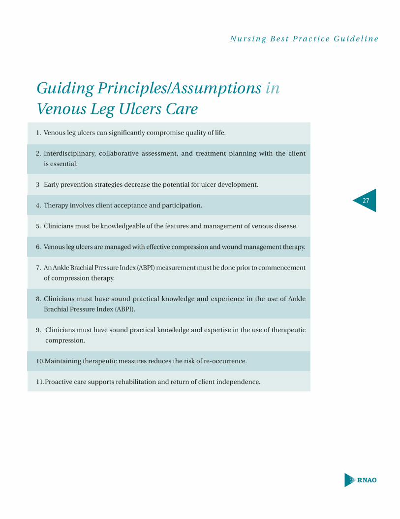
Guiding Principles/Assumptions in Venous Leg Ulcers Care1. Venous leg ulcers can significantly compromise quality of life.
2. Interdisciplinary, collaborative assessment, and treatment planning with the client
is essential.
3 Early prevention strategies decrease the potential for ulcer development.
4. Therapy involves client acceptance and participation.
5. Clinicians must be knowledgeable of the features and management of venous disease.
6. Venous leg ulcers are managed with effective compression and wound management therapy.
7. An Ankle Brachial Pressure Index (ABPI) measurement must be done prior to commencement
of compression therapy.
8. Clinicians must have sound practical knowledge and experience in the use of Ankle
Brachial Pressure Index (ABPI).
9. Clinicians must have sound practical knowledge and expertise in the use of therapeutic
compression.
10.Maintaining therapeutic measures reduces the risk of re-occurrence.
11.Proactive care supports rehabilitation and return of client independence.
27
N u r s i n g B e s t P r a c t i c e G u i d e l i n e

Interactive Guiding Principles of Venous Leg Ulcers CareA graphic depiction of the previously listed guiding principle statements can be visualized in
the following diagram:
Client with
Venous Leg Ulcers
Quality of Life
Early
Prevention
Proactive
Care
Interdisciplinary
Collaboration
Therapeutic
Compression
Knowledgeable
Clinicians
Maintenance of
therapeutic
measures
Client
acceptance and
participation
ABPI prior to
compression
28
Assessment and Management of Venous Leg Ulcers

Practice RecommendationsA. ASSESSMENT OF VENOUS LEG ULCERS
Recommendation • 1Assessment and clinical investigations should be undertaken by healthcare professional(s)
trained and experienced in leg ulcer management.
(Level of Evidence = C – RNAO Consensus Panel, 2004)
A complete client assessment precedes evaluation of the limb and ulcer characteristics. A
comprehensive assessment is essential to determine the underlying ulcer etiology and
appropriate treatment approaches.
Discussion of Evidence:Although little guidance is given, the literature strongly supports the importance of assessment
and clinical investigations for venous leg ulcers. Recognizing significant arterial insufficiency
is important, as no healing will occur in the presence of severe occlusive arterial disease of the
affected limb. Kunimoto et al. (2001) caution that the high levels of compression necessary to
correct venous hypertension will be potentially dangerous in this situation. Keast & Orsted
(1998) add that a chronic wound should prompt a search for underlying causes.
According to Zink, Rousseau & Holloway (2000), twenty-one percent of individuals with venous
ulcers experience concomitant arterial disease, with the risk of co-existing arterial dysfunction
increasing with age, which again supports the importance of a thorough assessment.
Research repeatedly confirms the necessity of trained healthcare professionals in leg ulcer
management. Surveys of reported practice by nurses demonstrate that knowledge of nurses
in wound care often falls short of what is ideal (RCN, 1998). Providers of healthcare recognize
that the mismanagement of wounds is both costly and unnecessary. Kerstein, van Rijswijk &
Betiz (1998), among others, maintain that providing optimal cost-effective wound care
requires extensive skills, as well as knowledge, and that classroom teaching alone will not
meet the needs of our aging population.
While findings as to what constitutes adequate training levels for nurses involved in leg ulcer
care are inconclusive, the essential point is that the person conducting the assessment must
29
N u r s i n g B e s t P r a c t i c e G u i d e l i n e

be trained and experienced. The RNAO guideline development panel found no trials assessing
and comparing reliability and accuracy based on levels of training.
Recommendation • 2A comprehensive clinical history and physical examination including blood pressure
measurement, weight, urinalysis, blood glucose level and Doppler measurement of Ankle
Brachial Pressure Index (ABPI) should be recorded for a client presenting with either their
first or recurrent leg ulcer and should be ongoing thereafter.
(Level of Evidence = C – RNAO Consensus Panel, 2004)
An assessment for a history of venous insufficiency also includes:
� Family history of venous disease.
� Client history of deep vein thrombosis (DVT).
� Lower leg fracture or other major leg injury, previous vein surgery, varicose veins, or prior
history of ulceration with/without use of compression stockings.
� History of episodes of chest pain, hemoptysis, or history of a pulmonary embolus.
� Lifestyle factors (e.g., sedentary lifestyle, chair-bound), obesity, poor nutrition.
An assessment for signs indicative of Non-Venous Disease also includes:
� Family history of non-venous etiology.
� Heart disease, stroke, transient ischemic attack.
� Diabetes mellitus.
� Peripheral vascular disease (PVD)/intermittent claudication.
� Smoking.
� Rheumatoid arthritis.
� Ischemic rest pain.
A combination of the features described above may be indicative of mixed arterial/venous
disease (RCN, 1998).
Discussion of Evidence:Several clinical studies show strong support for the need for thorough history taking for
assessment of venous insufficiency (NZGG, 1999; RCN, 1998). The New Zealand Guidelines
Group (1999) further suggests assessing the history of the ulcer, the mechanism of injury, and
previous methods of treatment.
30
Assessment and Management of Venous Leg Ulcers
Zink et al. (2000) recommend a guided interview to obtain the history most pertinent to the
cause of the ulcer, explaining that while the client may be able to relate important symptoms,
living with a chronic disease often blunts the ability to be discriminatory. Zink et al. (2000)
further adds that the initial encounter with the client is critical to establishing a positive,
therapeutic relationship. Establishing trust is instrumental for successful client outcomes,
particularly as venous leg ulcers often involve a lengthy healing time.
Misdiagnosis of ulcers can cause harm or lead to long periods of inappropriate treatment. It
is therefore important to have an accurate diagnosis of ulcer etiology (NZGG, 1999). Despite this,
there is only one population study that has systematically investigated and published data
on the etiology of identified ulcers.
Recommendation • 3Information relating to ulcer history should be documented in a structured format.
(Level of Evidence = C – RNAO Consensus Panel, 2004)
An ulcer history should include:
� The year first ulcer occurred.
� Site of ulcer and of any previous ulcers.
� Number of previous episodes of ulceration.
� Length of time taken to heal in previous episodes.
� Length of time with no recurrence of ulcers.
� Past treatment methods (both successful and unsuccessful).
� Previous operations on venous system.
� Previous and current use of compression hosiery.
Discussion of Evidence:Although no specific evidence was cited, the Royal College of Nursing (1998) supports the
theory that collection of data in a structured format will enable consideration of clinical factors
that may have an impact on treatment and healing progress, as well as provide baseline
information on ulcer history. They cautioned, however, that a diagnosis of ulcer type should
not be made solely on this information.
31
N u r s i n g B e s t P r a c t i c e G u i d e l i n e

32
Assessment and Management of Venous Leg Ulcers
The literature also stresses the importance of clear and comprehensive documentation of
information during history taking, and suggests several examples of leg ulcer assessment
forms. The RNAO guideline development panel does not consider one assessment form to
be superior to another. (For examples of leg ulcer assessment forms, see Appendices D and E).
Recommendation • 4Examine both legs and record the presence/absence of the following to aid in the
assessment of underlying etiology.
Venous Disease Arterial Disease
� usually shallow moist ulcers � ulcers with a “punched out” appearance
� situated on the gaiter area of the leg � base of wound poorly perfused, pale, dry
� edema � cold legs/feet (in a warm environment)
� eczema � shiny, taut skin
� ankle flare � dependent rubor
� lipodermatosclerosis � pale or blue feet
� varicose veins � gangrenous toes
� hyperpigmentation
� atrophie blanche
(Level of Evidence = C – RNAO Consensus Panel, 2004)
Discussion of Evidence:Research strongly recommends that the person conducting the assessment should be aware
that ulcers may result from many different causes, such as arterial insufficiencies, diabetes,
rheumatoid arthritis, or malignancy. Where there is mixed venous/arterial etiology, this
condition will have the features of venous ulcer in combination with signs of arterial
impairment (RCN, 1998).
Several studies confirm that malignancy can cause and may be a sequel of leg ulceration
(NZGG, 1999). The RNAO guideline development panel supports the practice of checking for a

history of skin cancers, although little guidance is offered in the literature. Signs suggestive
of malignancy include:
� irregular nodular appearance of the surface ulcer
� raised or rolled edge
� rapid increase in ulcer size
� failure to respond to treatment.
Any unusual appearance or signs of malignancy should be documented, and if present, refer
to a physician or to a dermatologist for a biopsy.
For characteristics specific to ulcer types, see:
� Different Types of Leg Ulcers and Their Causes (Appendix C)
Recommendation • 5Measure the surface areas of ulcers, at regular intervals, to monitor progress. Maximum
length and width, or tracings onto a transparency are useful methods.
(Level of Evidence = B)
Discussion of Evidence:The New Zealand Guidelines Group (1999) confirms that surface area and volume measurement
are indicators of ulcer healing. Common reproducible techniques, such as those described in this
recommendation, closely correlate to wound area determined by computerized planimetry of
photographs (a reliable and valid objective measure, but not widely available).
During an assessment, the following characteristics should be observed and recorded:
� location � pain
� depth � infection
� size (mm, cm) � appearance of the wound bed
� odour (eschar, slough, fibrin, granulation
� sinus tracts tissue, epithelial tissue)
� undermining � condition of the surrounding skin
� tunneling (peri-wound) and wound edges
� exudate
33
N u r s i n g B e s t P r a c t i c e G u i d e l i n e
Recommendation • 6The client’s estimate of the quality of life should be included in the initial discussion of the
treatment plan, throughout the course of treatment, and when the ulcer has healed.
(Level of Evidence = C – RNAO Consensus Panel, 2004)
Discussion of Evidence:Issues relating to quality of life for leg ulcer clients have been well documented in the literature,
with several studies confirming that the negative impact of venous ulcers on quality of life
is significant (Phillips et al.,1994; Pieper et al., 2000; Price & Harding, 1996). Healing the ulcer and
normalizing the clients’ lives can and should form the basis of care (Husband, 2001a).
Increased sensitivity to, and understanding of the impact of painful venous ulcers on quality of
life may lead to more effective intervention strategies and improved outcomes for these clients
(Krasner, Sibbald & Coutts, 2001). Although it is widely accepted among healthcare professionals that
the individual needs of the client should be considered, and that a positive management
outcome will likely be influenced by client insight into the severity of the venous disorder, there
has been little conclusive research done in this area. One qualitative study cited by Krasner
(1998), focused on understanding and interpreting the meaning of living with a painful venous
leg ulcer, and the resulting quality of life.
From the point of view of the client, quality of life is crucial in assessing the effectiveness of
medical treatments (Phillips et al., 1994). Compliance Network Physicians (1999) add that
compliance in clients may be enhanced as a result of regular communication in the
physician-client and nurse-client interaction.
In one study conducted in Sweden, where standard questionnaires were distributed to clients,
results showed that chronic leg ulcers had a marked impact on the client’s subjectively
perceived health. Males exhibited elevated scores, while for women the impact of leg ulcer
disease, although obvious, seemed much less marked (Lindholm et al., 1992). Lindholm et al.
(1992) further added that the impact of chronic disease on health is closely related to
personal, social, and environmental factors.
Research also indicates that quality of life is impacted if clients attend a leg ulcer clinic. Liew,
Law & Sinha (2000) found an improvement in three quality of life indicators – pain, sleep and
34
Assessment and Management of Venous Leg Ulcers
mobility, over an average of one to three visits to the clinic, and home visits by primary care
nurses. There is also some evidence demonstrating an improvement in the quality of life
resulting from healing of leg ulcers, but again, results are inconclusive.
See Appendix F for a Quality of Life Assessment Tool.
Recommendation • 7Assess the functional, cognitive and emotional status of the client and family to manage
self-care. (Level of Evidence = C – RNAO Consensus Panel, 2004)
Communicate with the client, family and caregivers to establish realistic expectations for the
healability of the venous leg ulcers. The basis for a treatment plan begins with the client when
the individual aims of the overall treatment are defined and agreed upon.
The RNAO guideline development panel believes that the presence or absence of a social
support system is important for the treatment and prevention of venous leg ulcers.
Discussion of Evidence:Pieper, Rossi & Templin (1998) describe how persons with leg ulcers describe interferences
in their functional status and psychological well-being. They experience more pain, less vitality,
more restrictions in physical and social functioning, and poorer general health and limitations
in their physical and emotional roles compared with age-matched cohorts.
Pain and increased sensitivity can serve as a constant reminder of the presence of an ulcer,
and contribute to sleep disturbances and decreased mobility (Liew et al., 2000). In a study where
62 individuals with chronic leg ulcers were interviewed, Phillips et al.
(1994) found the leg ulcer was associated with altered mobility
(81 percent of cases), burdensome care (58 percent), negative
emotional impact on life such as fear, isolation, anger,
depression, and negative depression (60 percent). Pieper et al.
(2000) documented similar findings.
35
N u r s i n g B e s t P r a c t i c e G u i d e l i n e

Recommendation • 8Regular ulcer assessment is essential to monitor treatment effectiveness and healing
goals. (Level of Evidence = C – RNAO Consensus Panel, 2004)
Common features of venous ulcers include:
� Irregular borders that are flat and slope into a shallow crater.
� Loss of epidermis with a dermal base.
� Base may be covered with yellow fibrin, or ruddy granulation.
� Ulcer located often over medial malleolus where long saphenous vein is most superficial
and has the greatest curvature. In severe cases, ulcers may extend over the circumference
of the ankle.
� Exudate evident and may be minimal to copious.
� Periwound skin that may be dry, scaly, irritated (stasis dermatitis) or macerated.
� Edema that may be pitting or firm.
B. DIAGNOSTIC EVALUATION
Recommendation • 9Venous disease of the leg is most commonly detected by a combination of clinical examination
and measurement of a reliably taken Ankle Brachial Pressure Index (ABPI).
(Level of Evidence = A)
Recommendation • 10Doppler ultrasound measurement of Ankle Brachial Pressure Index (ABPI) should be done
by practitioners trained to undertake this measure. (Level of Evidence = B)
Recommendation • 11If there are no signs of chronic venous insufficiency and the Ankle Brachial Pressure Index
(ABPI) is abnormal (greater than 1.2 or less than 0.8), arterial etiology should be assumed
and a vascular opinion sought.
(Level of Evidence = C – RNAO Consensus Panel, 2004)
Recommendation • 12Vascular assessment, such as Ankle Brachial Pressure Index (ABPI) is recommended for
ulcers in lower extremities, prior to debridement, to rule out vascular compromise.
(Level of Evidence = C – RNAO Consensus Panel, 2004)
36
Assessment and Management of Venous Leg Ulcers

Discussion of Evidence:The importance of making an objective etiological diagnosis by measuring the Ankle Brachial
Pressure Index (ABPI), in addition to visual inspection of the ulcer, pedal pulse palpation and
a thorough clinical and physical assessment, is highlighted in a number of studies (CREST, 1998a;
Moffatt, Oldroyd, Greenhalgh & Franks, 1994).
Expert opinion recommends that the ABPI is used to rule out arterial disease and to determine
the safe use of therapeutic compression therapy (RNAO Consensus Panel, 2004). The Royal College
of Nursing (1998) also notes that all clients should be given the benefit of Doppler ultrasound
management to ensure detection of arterial insufficiency, which could result in commencement
of inappropriate or even dangerous therapy.
According to Zink et al. (2000), the Trendelenburg test also assists in the physical evaluation
of venous valve competence in the perforators and saphenous system.
Research evidence cautions that Doppler ultrasound measurements of ABPI can be unreliable
if operators have not undergone training, adding that reliability can be considerably
improved if operators have received instruction and training to undertake this measure
(Cornwall et al.,1986).
Based on available research from the New Zealand Guidelines Group (1999), Doppler
ultrasound measurement of ABPI should be repeated when:
� a leg ulcer deteriorates
� an ulcer is not fully healed within three months
� clients present with recurrence (of whichever leg)
� there is a sudden increase in pain
� colour and/or temperature of foot changes (RCN, 1998).
In addition, the New Zealand Guidelines Group (1999) recommended that:
� The presence of palpable foot pulses alone are insufficient to rule out arterial disease.
� All ulcers should be screened for arterial disease using Doppler ultrasound to determine
the Ankle Brachial Pressure Index (ABPI). A single measure of ABPI < 0.8 makes the presence
of peripheral arterial occlusive disease (PAOD) highly likely.
� Further tests should be considered prior to initiating compression bandaging if a client has
an ABPI > 0.8 in the presence of signs and symptoms of PAOD, rheumatoid arthritis,
diabetes mellitus or systemic vasculitis.
� Clients with ABPI < 0.6 should be considered for referral to a vascular surgeon.
37
N u r s i n g B e s t P r a c t i c e G u i d e l i n e

A Specialist medical referral may be appropriate for:
� treatment of underlying medical problems
� ulcers of non-venous etiology (rheumatoid; diabetic; arterial; mixed etiology)
� suspected malignancy
� diagnostic uncertainty
� reduced ABPI (e.g., <0.8 – routine vascular referral; 0.5 – urgent vascular referral)
� increased ABPI (> 1.2 as in calcification of vessels)
� rapid deterioration of ulcers
� newly diagnosed diabetes mellitus
� signs of contact dermatitis (spreading eczema; increased itch)
� cellulitis
� consideration for venous surgery
� ulcers which have received adequate treatment, and have not improved for three months
� recurring ulceration
� ischemic foot
� infected foot
� pain management (LOE = C – RCN, 1998; RNAO Consensus Panel, 2004)
� clients with suspected sensitivity reactions (should be referred to a dermatologist for patch
testing). Following patch testing, identified allergens must be avoided and medical advice
on treatment should be sought (RCN, 1998)
� a non-healing or atypical leg ulcer which should be considered for biopsy (CREST, 1998a)
In the case of clients with diabetes, some studies note that there may be a higher risk of
peripheral vascular disease, and as a result ABPI readings may be unreliable (greater than 1.2)
due to arterial calcification. As results are inconclusive, further investigation is required.
38
Assessment and Management of Venous Leg Ulcers

C. PAIN
Recommendation • 13Assess Pain (Level of Evidence = C – RNAO Consensus Panel, 2004 )
Recommendation • 14Pain may be a feature of both venous and arterial disease, and should be addressed.
(Level of Evidence = B)
Recommendation • 15Prevent or manage pain associated with debridement. Consult with a physician and
pharmacist as needed. (Level of Evidence = C – RNAO Consensus Panel, 2004)
Discussion of Evidence:Research results consistently indicate that clients with venous leg ulcers can experience
considerable pain (RCN, 1998), and that a significant proportion of clients with venous leg
ulcers report moderate to severe pain. Sibbald (1998a) reports that 76 percent of severe
venous ulcers are painful. In a study cited by Kunimoto et al. (2001), pain in three distinct
locations was reported by clients – within ulcers, around ulcers, and elsewhere in the leg. Pain
often increases when the limb is in a dependent position.
Assessment of pain is complex, but a structured discussion and frequent re-assessment are
important (CREST, 1998a; SIGN, 1998). The importance of pain management in venous leg ulcer
clients is often cited in the literature, yet in one particular study, 55 percent of district nurses
did not assess the clients’ pain.
Pieper et al. (1998) identified a need for better control of venous leg ulcer pain so people felt
more confident and positive about treatment and could reduce activity restrictions,
complementing the observation by Liew et al. (2000), that pain can significantly reduce
clients’ quality of life (see Recommendation 6).
Although utilization of a pain assessment tool is strongly recommended in the literature, no
research evidence could be identified that examined the use of a pain assessment method
specifically designed for clients with venous leg ulcers, or compared different methods of
39
N u r s i n g B e s t P r a c t i c e G u i d e l i n e

relief. There are several samples of pain assessment tools currently available being used;
the RNAO guideline development panel does not consider one tool superior to others.
(See Appendix G for examples of Pain Assessment Tools).
Although other pain relief strategies may be considered, there is little conclusive research on
interventions such as exercise or leg elevation (RCN, 1998). However, Johnson (1995) observed
that increased pain on mobility may be associated with poorer healing rates.
The presence of severe pain does not necessarily indicate arterial disease or infection, and
Krasner (1998) observes that pain is often inadequately controlled in these clients. According
to Scottish Intercollegiate Guidelines Network (1998), “the pain associated with a dressing
change can be reduced by adequate soakage before the dressing is removed. In two trials, one
of a hydrocolloid and the other of a foam dressing, the ulcer pain was less when compared
with a non-adherent dressing” (p.8).
The RNAO guideline development panel has found that there is very limited guidance in the
literature on how best to manage pain associated with debridement.
D. VENOUS ULCER CARE
Recommendation • 16Choose the technique of debridement, considering the type, quantity and location of non-
viable tissue, the depth of the wound, the amount of wound fluid and the general condition
and goals of the client. (Level of Evidence = C – RNAO Consensus Panel, 2004)
Recommendation • 17Cleansing of the ulcer should be kept simple; warm tap water or saline is usually sufficient.
(Level of Evidence = C – RNAO Consensus Panel, 2004)
Recommendation • 18Dressings must be simple, low adherent, acceptable to the client and should be low cost.
(Level of Evidence = A)
40
Assessment and Management of Venous Leg Ulcers

Recommendation • 19Avoid products that commonly cause skin sensitivity, such as those containing lanolin,
phenol alcohol, or topical antibiotics. (Level of Evidence = C – RNAO Consensus Panel, 2004)
Recommendation • 20Choose a type of dressing depending on the amount of exudate and the phase of healing.
(Level of Evidence = C – RNAO Consensus Panel, 2004)
Recommendation • 21No specific dressing has been demonstrated to encourage ulcer healing.
(Level of Evidence = A)
Recommendation • 22In contrast to drying out,moist wound conditions allow optimal cell migration,proliferation,
differentiation and neovascularization. (Level of Evidence = A)
Recommendation • 23Refer clients with suspected sensitivity reactions to a dermatologist for patch testing.
Following patch testing, identified allergens must be avoided, and medical advice on
treatment should be sought. (Level of Evidence = B)
Recommendation • 24Venous surgery followed by graduated compression hosiery is an option for consideration
in clients with superficial venous insufficiency.
(Level of Evidence = C – RNAO Consensus Panel, 2004)
Recommendation • 25Biological wound coverings and growth factor treatments should not be applied in cases
of wound infection. (Level of Evidence = C – RNAO Consensus Panel, 2004)
Recommendation • 26Optimal nutrition facilitates wound healing, maintains immune competence and
decreases the risk of infection. (Level of Evidence = B)
41
N u r s i n g B e s t P r a c t i c e G u i d e l i n e

Debridement is necessary to remove devitalized tissue and exudate, reduce the risk of infection,
prepare the wound bed and promote healing. Debridement can be:
� Autolytic, the natural self-clearance of debris in the wound bed by phagocytosis and
proteolytic enzymes
� Mechanical, the use of wet-to-dry dressings, hydrotherapy or irrigation with saline solution
� Enzymatic
� Sharp, using a scalpel or scissors (Fowler, 1992)
Select the method of debridement most appropriate to the client’s condition and goals.
Sharp debridement is a high-risk procedure. Debridement with a scalpel should be undertaken
with caution and performed by specially trained and experienced healthcare professionals.
Subcutaneous debridement with a scalpel is a controlled act that must be carried out by a
physician or the delegate.
Discussion of Evidence:There is no evidence to favour any one method of debridement, whether mechanical, autolytic,
enzymatic/chemical or sharp (NZGG, 1999). Fowler (1992) states that debridement of non-viable
tissue in open wounds is clearly an overlapping function of medicine and nursing, and nurses
who are trained to perform this function are practicing within the scope of nursing.
There is a body of research showing a wide variation in the clinical management of venous
leg ulcers through the use of dressings, however it is unlikely that a single type of dressing will
be appropriate for all types of wounds (Bryant, 2001). Bryant (2001)also explains that if the
dressing material transmits less moisture than the wound loses, then the wound will remain
moist. Several articles confirm that there are numerous types of wound dressings, bandages,
and stockings used in the treatment and prevention of recurrence (Lees & Lambert, 1992; Stevens,
Franks & Harrington, 1997). The RNAO guideline development panel, however, found insufficient
evidence to determine whether any particular dressing increases healing or reduces the pain
of venous leg ulcers. The most important factor, according to the Royal College of Nursing
(1998), is that the dressings be simple, low adherent, acceptable to the client, and low in cost.
The New Zealand Guidelines Group (1999) cautions that a number of cleansing agents
currently on the market may commonly cause skin sensitivity, and that some antiseptic and
42
Assessment and Management of Venous Leg Ulcers
chemical agents have been shown to damage cells. (See Appendix H for a list of Cleansing
Agents and Their Associated Toxicities). Health professionals should also be aware that clients
can become sensitized to elements of their treatment at any time (RCN, 1998). (See Appendix I
for a list of Potential Allergens).
Wound cleansing can be accomplished by showering and ensuring peri-wound skin is dried
carefully.
The nutritional status of clients appears to be a key factor in the management of venous leg
ulcers. Several studies show a strong link between deteriorating nutritional status and the
development and healing of chronic, non-healing wounds (Himes, 1999; Whitney & Heirkemper,
1999; Wissing, Unosson, Lennernas & Ek, 1997).
Himes (1999) observed that clients with chronic wounds require higher intake of protein and
calories, suggesting that aggressive intervention may be required to prevent malnutrition.
While the importance of specific nutrients to wound healing, including ascorbic acid,
Vitamin A and zinc is well documented, the actual understanding of the precise nutritional
requirements needed for tissue repair is still developing (Whitney & Heirkemper, 1999).
Kunimoto et al. (2001) state that a nutritionist or dietitian should be consulted if nutritional
deficiency is thought to be significant enough to possibly impair wound healing. Wipke-Tevis
& Stotts (1998) note that there are many factors that can contribute to inadequate dietary
intake:
� financial constraints
� mobility limitations
� social isolation
� coexistent medical problems
� inadequate cooking facilities
� poor eating habits
� lack of nutritional knowledge.
In clinical practice, Wipke-Tevis & Stotts (1998) recommend that a multidisciplinary
approach is desirable, as the factors listed above are quite different in nature.
43
N u r s i n g B e s t P r a c t i c e G u i d e l i n e

E. INFECTION
Recommendation • 27Assess for infection. (Level of Evidence = A)
Recommendation • 28An infection is indicated when > 105 bacteria/gram tissue is present.
(Level of Evidence = B)
Recommendation • 29The treatment of infection is managed by debridement, wound cleansing and systemic
antibiotics. (Level of Evidence = A)
Recommendation • 30Antibiotics should only be considered if the ulcer is clinically cellulitic (presence of some
of the following signs and symptoms: pyrexia; increasing pain; increasing erythema of
surrounding skin; purulent exudate; rapid increase in ulcer size).
(Level of Evidence = C – RNAO Consensus Panel, 2004)
Recommendation • 31Do not use topical antiseptics to reduce bacteria in wound tissue, e.g., povidone iodine,
iodophor, sodium hypochlorite, hydrogen peroxide, or acetic acid. (Level of Evidence = B)
Recommendation • 32Topical antibiotics and antibacterial agents are frequent sensitizers and should be avoided.
(Level of Evidence = B)
Discussion of Evidence:Avoidance of wound infection is a primary concern in the healing of acute injuries and
wounds (Whitney & Heirkemper, 1999), and wound care regimens should be aimed at preventing
infection, which delays healing (Mureebe & Kerstein, 1998). All wounds are colonized by a colony
of bacteria, but most do not become infected. In chronic wounds, an infection should be
suspected if the wound does not begin to show signs of healing.
44
Assessment and Management of Venous Leg Ulcers

There are numerous strategies cited in the literature regarding assessment of infection, but
the Royal College of Nursing (1998) recommends that routine bacteriological swabbing is
unnecessary unless there is evidence of clinical infection such as:
� inflammation/redness/cellulitis
� increased pain
� purulent exudate
� rapid deterioration of the ulcer
� pyrexia
When a client with an ulcer develops sudden pain, a bacterial infection may be present
(Kunimoto et al., 2001). An infection may be indicated based on the presence of bacteria/gram
tissue (Compliance Network Physicians, 1999). The presence of bacteria in a leg ulcer, however, does
not mean that it is infected as all chronic ulcers can be colonized by microorganisms which
are not producing any inflammatory reaction. A diagnosis of infection should therefore be
made on clinical evidence, e.g., cellulitis. Odour or increased exudate also does not necessarily
indicate infection and can be managed with selective dressings. Again, a clinical assessment
is recommended if infection is suspected.
There is strong support in the literature, citing clinical studies, stressing the avoidance of
topical antibiotics and antiseptics to treat infections, as they are frequent sensitizers or
cytotoxics (CREST, 1998a; Compliance Network Physicians, 1999; NZGG, 1999). (See Appendix H for
Cleansing Agents and Their Associated Toxicities; and Appendix J for a list of Topical
Antimicrobial Agents).
F. COMPRESSION
Recommendation • 33The treatment of choice for clinical venous ulceration uncomplicated by other factors, is
graduated compression bandaging, properly applied, and combined with exercise.
Graduated compression is the main treatment for venous eczema.
(Level of Evidence = A)
45
N u r s i n g B e s t P r a c t i c e G u i d e l i n e

Recommendation • 34High compression increases venous ulcer healing and is more effective than low
compression, but should only be used where ABPI ≥ 0.8 and ulcer is clinically venous.
(Level of Evidence = A)
Recommendation • 35Compression bandages should only be applied by a suitably trained and experienced
practitioner. (Level of Evidence = B)
Recommendation • 36Venous ulceration should be treated with high compression bandaging to achieve a
pressure between 35-40 mm Hg. at the ankle, graduating to half at calf in the normally
shaped limb, as per La Place’s Law. (Level of Evidence = C – RNAO Consensus Panel, 2004)
Recommendation • 37Use protective padding over bony prominences when applying high compression.
(Level of Evidence = C – RNAO Consensus Panel, 2004)
Recommendation • 38Arterial insufficiency is a contraindication to the use of high compression. A modified
form of compression may be used under specialist supervision.
(Level of Evidence = C- RNAO Consensus Panel, 2004)
Recommendation • 39Use compression with caution in clients with diabetes, those with connective tissue
disease and the elderly. (Level of Evidence = C – RNAO Consensus Panel, 2004)
Recommendation • 40Compression therapy should be modified until clinical infection is treated.
(Level of Evidence = C – RNAO Consensus Panel, 2004)
Recommendation • 41Bandages should be applied according to manufacturer’s recommendations.
(Level of Evidence = C – RNAO Consensus Panel, 2004)
46
Assessment and Management of Venous Leg Ulcers
Recommendation • 42When using elastic systems such as “high compression” bandages, the ankle circumference
must be more than or padded to equal 18 cms.
(Level of Evidence = C – RNAO Consensus Panel, 2004)
Recommendation • 43Ankle circumference should be measured at a distance of 2.5 cm (one inch) above the
medial malleolus. (Level of Evidence = C – RNAO Consensus Panel, 2004)
Recommendation • 44The concepts, practice, and hazards of graduated compression should be fully understood
by those prescribing and fitting compression stockings. (Level of Evidence = A)
Recommendation • 45Graduated compression hosiery should be measured and fitted by a certified fitter.
(Level of Evidence = C – RNAO Consensus Panel, 2004)
Recommendation • 46To maintain a therapeutic level of compression, stockings should be cared for as per
manufacturer’s instructions, and replaced every six months.
(Level of Evidence = C – RNAO Consensus Panel, 2004)
Recommendation • 47Graduated compression hosiery should be prescribed for life. (Level of Evidence = B)
Recommendation • 48External compression applied using various forms of pneumatic compression pumps is
indicated for individuals with chronic venous insufficency. (Level of Evidence = A)
Recommendation • 49The client should be prescribed regular vascular exercise by means of intensive controlled
walking and exercises to improve the function of the upper ankle joint and calf muscle
pump. (Level of Evidence = A)
47
N u r s i n g B e s t P r a c t i c e G u i d e l i n e

Discussion of Evidence:Results of at least one randomized controlled trial recommends the use of graduated compression
bandaging as the treatment of choice for clinical venous ulceration, uncomplicated by other
factors (CREST, 1998a). Cotton (1996) notes that compression therapy increases skin perfusion
pressure and decreases interstitial fluid volume, thereby increasing tissue oxygenation, thus
promoting tissue healing.
Graduated compression may be achieved by prescribed bandages or by compression stockings.
Tensor bandages and post-operative anti-embolic stockings, e.g., TEDS™, do not provide
therapeutic compression for treatment and management of venous stasis disease. Thomas
(1999) explains that these materials have limited elasticity and tend to “lock out” at relatively
low levels of extension, further adding that they are not suitable for significant levels of pressure.
Compression systems must be applied correctly so that sufficient (but not excessive)
pressure is applied. Incorrectly applied systems may be harmful or ineffective. CREST (1998a)
in recommending the use of graduated compression bandaging, cautions that compression
bandages should only be applied by a suitably trained and experienced practitioner.
Practitioners without training in compression bandages apply bandages at inappropriate
and wide varying pressures (NZGG, 1999; RCN, 1998). More research is needed to see what training
strategies improve compression techniques, and if the effects of training are maintained over
time (RCN, 1998).
It is important to note, however, that no controlled studies have compared recurrence rates
of venous ulceration achieved with or without compression hosiery, nor are there conclusive
studies to indicate which high compression system (3 layer, 4 layer, or short stretch) is most
effective (NZGG, 1999). The systematic review by Nelson, Bell-Syer and Cullum (2003) has found
that there is no evidence to indicate that high compression hosiery is more effective than
moderate compression in the prevention of ulcer recurrence. Compliance is lower in people
wearing high compression stockings. From the same systematic review, it was suggested
that clients should be prescribed the highest grade stocking they are able to wear.
Kunimoto et al. (2001) note that recognition of significant arterial insufficiency is important,
and that no healing will occur in the presence of severe occlusive arterial disease of the affected
limb. Kunimoto et al. (2001) also add that the high levels of compression necessary to correct venous
hypertension will be potentially dangerous in this situation.
48
Assessment and Management of Venous Leg Ulcers

External compression using various forms of pneumatic compression pumps (PCPs) are
indicated for individuals with chronic venous insufficiency. However, there is no strong evidence
about the effects of intermittent pneumatic compression (IPC) on venous leg ulcer. In the
review by Mani, Vowden and Nelson (2003), they found that there is conflicting evidence
whether or not IPC can help heal venous leg ulcers.
Note: At time of publishing, this recommendation is not inclusive of elastic and non-elastic
bandages (RNAO Consensus Panel, 2004).
Findings from several studies indicate that high compression bandaging should be used for
venous leg ulceration, as per La Place’s Law (NZGG, 1999):
La Place’s Law:The theoretical pressure produced beneath a bandage can be calculated as follows:
P = 4630 x N x T
C x W
Where P = sub-bandage pressure (mmHg)
N = number of layers
T = tension within bandage (Kgforce)
C = limb circumference (cm)
W = width of bandage (cm)
It can be seen that sub-bandage pressure is directly proportional to the tension in the bandage
during application and the number of layers applied, but inversely proportional to the radius
of curvature of the limb (Logan, Thomas, Harding & Collyer, 1992).
The treatment of venous stasis disease demands the life-long use of therapeutic compression.
The concepts, practice, and hazards of graduated compression should be fully understood by
those prescribing and fitting (SIGN, 1998). Graduated compression hosiery should be prescribed
for life (CREST, 1998a).
Provided the client has been measured by a certified stocking fitter for the correct size, elasticized
stockings are a safe alternative to bandages. They can be full-length, but generally
49
N u r s i n g B e s t P r a c t i c e G u i d e l i n e

below-knee stockings are the most frequently used, and are more acceptable to the client.
There are three classes of compression stockings. The stockings are unsuitable in clients with
a high level of exudate, and are prescribed for use after compression therapy with bandages has
reduced the edema. When edema and exudate are controlled, the use of therapeutic compression
stockings may be considered. (See Appendix K for Classes of Compression Bandages).
The client should be prescribed regular vascular exercise by means of intensive controlled
walking and exercises to improve the function of the upper ankle joint and calf muscle pump
(Compliance Network Physicians, 1999; Kan & Delis, 2001).
Loss of ankle joint movement can accompany venous ulceration. Good calf muscle pump
function is an important aspect of ulcer healing. Walking and passive ankle exercises should
be encouraged. The immobility of the ankle joint is thought to influence ambulatory venous
hypertension and may be a factor in causing venous ulceration. Exercise is necessary to
enhance compression therapy. These can be modified to accommodate the needs of
non-ambulatory and obese individuals. A physical and/or occupational therapist should be
consulted. When resting, elevation of the limb above chest level is beneficial.
G. COMPLEMENTARY THERAPIES
Recommendation • 50Consider electrical stimulation in the treatment of venous leg ulcers. (Level of Evidence = B)
Recommendation • 51Hyperbaric oxygen may reduce ulcer size in non-diabetic, non-atherosclerotic leg ulcers.
(Level of Evidence = A)
Recommendation • 52Therapeutic ultrasound may be used to reduce the size of chronic venous ulcers.
(Level of Evidence = A)
Discussion of Evidence:The clinical research evidence for complementary therapies to treat chronic venous leg
ulcers is the subject of an article by Kunimoto et al. (2001).
50
Assessment and Management of Venous Leg Ulcers

The New Zealand Guidelines Group (1999) reports that there is sufficient evidence to conclude
that hyperbaric oxygen may reduce ulcer size in non-diabetic, non-atherosclerotic leg ulcers,
and should be considered as a complementary therapy for venous leg ulcer clients.
There have been several randomized controlled trials examining the effect of ultrasound on
chronic venous leg ulcers. In addition, a meta-analysis published by Johannsen, Gam &
Karlsmark (1998) found a significant effect of ultrasound on wound size of chronic venous
leg ulcers.
In a search of the literature, the RNAO guideline development panel also found that there is
insufficient evidence to give clear direction on the use of laser therapy, maggot therapy, sugar,
honey, vitamins, hormones, Vacuum Assisted Closure (VAC) Therapy ™, growth factors,
mineral elements, and normalthermic therapies in the treatment of venous ulcers.
H. REASSESSMENT
Recommendation • 53With no evidence of healing, a comprehensive assessment should be carried out at three-
month intervals, or sooner if clinical condition deteriorates.
(Level of Evidence = C – RNAO Consensus Panel, 2004)
Recommendation • 54For resolving and healing venous leg ulcers, routine assessment at six-month intervals
should include:
� physical assessment
� Ankle Brachial Pressure Index (ABPI)
� replacement of compression stockings
� reinforcement of teaching
(Level of Evidence = C – RNAO Consensus Panel, 2004)
Discussion of Evidence:Refer to a specialist (Dermatologist or Vascular physician) if there is a deterioration of the
ulcer status, client status or if non-venous etiology is identified or suspected (e.g., rheumatoid
disease; suspected malignancy; acute congestive heart failure (CHF); renal failure; diagnostic
uncertainty; rapid deterioration of ulcers; new diagnosis of diabetes; lack of healing; recurrent
ulceration; ischemic limb or foot infection; pain management; or for potential surgery).
51
N u r s i n g B e s t P r a c t i c e G u i d e l i n e

The active management of leg ulcers may be required over many months or years and may
be carried out by several different healthcare professionals. It is important to reassess
progress 12 weeks after the institution of treatment. This should involve a comprehensive
review, similar to the initial assessment. Likewise, when an ulcer recurs, a full assessment
should be carried out even though the client may be well known to the nurse or doctor.
I. SECONDARY PREVENTION
Recommendation • 55Measures to prevent recurrence of a venous leg ulcer include:
� wearing compression stockings
� regular follow-up to monitor Ankle Brachial Pressure Index (ABPI)
� discouragement of self-treatment with over-the-counter preparations
� avoidance of accidents or trauma to legs.
(Level of Evidence = C – RNAO Consensus Panel, 2004)
Recommendation • 56Inform the client after the ulcer has healed regarding:
� wearing and maintenance of compression stockings
� elevation of affected limb above level of heart when at rest
� early referral at first sign of skin breakdown or trauma to limb
� need for exercise and ankle-joint mobility
� appropriate skin care
� avoidance of products likely to be sensitizers
� life-long use of compression.
(Level of Evidence = C – RNAO Consensus Panel, 2004)
Discussion of Evidence:The majority of venous leg ulcers recur. The rates of recurrence vary, but several large
population studies found 59 to 76 percent of all identified ulcers were recurrent (NZGG, 1999).
Secondary prevention must be put into place and maintained in order to prevent this recurrence.
Secondary prevention presently takes the form of graduated compression, surgery, or drugs.
52
Assessment and Management of Venous Leg Ulcers

With graduated compression, it is important to take note that not all stockings produce an
adequate pressure. Stockings are more effective when client adherence is taken into account.
The hazards of incorrectly fitting stockings are the same as those of improperly applied
compression bandages.
In order to maintain a proper level of compression, stockings should be replaced every 3 to
6 months as per manufacturer’s instructions. If a stocking is uncomfortable a change of the
brand of stocking within the same class of stocking may be beneficial to the comfort and
compliance of the client.
Education RecommendationsRecommendation • 57Guidelines are more likely to be effective if they take into account local circumstances and
are disseminated by an ongoing education and training program.
(Level of Evidence = C – RNAO Consensus Panel, 2004)
Recommendation • 58Develop educational programs that target appropriate healthcare providers, clients, family
members, and caregivers. Develop programs that maximize retention, ensure carryover
into practice, and support lifestyle changes. Present information at an appropriate level
for the target audience using principles of adult learning.
(Level of Evidence = C – RNAO Consensus Panel, 2004)
Recommendation • 59Design, develop, and implement educational programs that reflect a continuum of care.
The program should begin with a structured, comprehensive, and organized approach to
prevention and should culminate in effective treatment protocols that promote healing as
well as prevent recurrence. (Level of Evidence = C – RNAO Consensus Panel, 2004)
Recommendation • 60All healthcare professionals should be trained in leg ulcer assessment and management.
(Level of Evidence = C – RNAO Consensus Panel, 2004)
53
N u r s i n g B e s t P r a c t i c e G u i d e l i n e

Recommendation • 61Education programs for healthcare professionals should include:
� pathophysiology of leg ulceration
� leg ulcer assessment
� need for Doppler ultrasound to measure Ankle Brachial Pressure Index (ABPI)
� normal and abnormal wound healing
� compression therapy theory, management, and application
� dressing selection
� principles of debridement
� principles of cleansing and infection control
� skin care of the lower leg
� peri-wound skin care and management
� psychological impact of venous stasis disease
� quality of life
� pain management
� teaching and support for care provider
� health education
� preventing recurrence
� principles of nutritional support with regard to tissue integrity
� mechanisms for accurate documentation and monitoring of pertinent data, including
treatment interventions and healing progress
� criteria for referral for specialized assessment.
(Level of Evidence = C – RNAO Consensus Panel, 2004)
Recommendation • 62Healthcare professionals with recognized training in leg ulcer care should cascade their
knowledge and skills to local healthcare teams.
(Level of Evidence = C – RNAO Consensus Panel, 2004)
Recommendation • 63The knowledge and understanding of the healthcare professional is a major factor in
adherence to treatment regimens.
(Level of Evidence = C – RNAO Consensus Panel, 2004)
54
Assessment and Management of Venous Leg Ulcers

Discussion of Evidence:Research using non-randomized comparison groups on pre- and post-test designs have
shown that community nurses’ knowledge of leg ulcer management is often inadequate, but
that knowledge can be improved by provision of training (RCN, 1998).
In an article by Ruane-Morris (1995), programs of continuing support and education offer an
alternative to discharge and loss of contact for clients in the United Kingdom. All clients with
a healed ulcer and an ABPI of 0.8 or greater, are invited to participate in Healed Ulcer Groups
(HUGS). The program focuses on daily life activities, exercise and movement, skin care, and the
reasons for developing ulcers, with a goal to shifting clients from nurse-led to self-directed care.
Organization & PolicyRecommendations Recommendation • 64Successful implementation of a venous ulcer treatment policy/strategy requires:
� dedicated funding
� integration of healthcare services
� support from all levels of government
� management support
� human resources
� financial resources
� functional space
� commitment
� collection of baseline information about vulnerable populations
� resources and existing knowledge
� interpretation of above data and identification of organizational problems.
(Level of Evidence = C – RNAO Consensus Panel, 2004)
55
N u r s i n g B e s t P r a c t i c e G u i d e l i n e
Recommendation • 65Nursing best practice guidelines can be successfully implemented only where there are
adequate planning, resources, organizational and administrative support, as well as
appropriate facilitation. Organizations may wish to develop a plan for implementation
that includes:
� An assessment of organizational readiness and barriers to education.
� Involvement of all members (whether in a direct or indirect supportive function) who
will contribute to the implementation process.
� Dedication of a qualified individual to provide the support needed for the education
and implementation process.
� Ongoing opportunities for discussion and education to reinforce the importance of best
practices.
� Opportunities for reflection on personal and organizational experience in implementing
guidelines.
In this regard, RNAO (through a panel of nurses, researchers and administrators) has
developed the Toolkit: Implementation of Clinical Practice Guidelines, based on available
evidence, theoretical perspectives and consensus. The RNAO strongly recommends the
use of this Toolkit for guiding the implementation of the best practice guideline on
Assessment and Management of Venous Leg Ulcers.
(Level of Evidence = C – RNAO Consensus Panel, 2004)
Evaluation & MonitoringOrganizations implementing the recommendations in this nursing best practice guideline
are advised to consider how the implementation and its impact will be monitored and
evaluated. The following table, based on the framework outlined in the RNAO Toolkit:
Implementation of Clinical Practice Guidelines (2002), illustrates some suggested indicators
for monitoring and evaluation.
56
Assessment and Management of Venous Leg Ulcers

Indicator
Objectives
Organization/Unit
Provider
ClientEligibility criteria:
• new ulcer or• recurrent venous
leg ulcer
Exclusion: • arterial• diabetic• mixed• primary
lymphadema• vasculitis
Financial Costs
Process
• To evaluate the changes inpractice that lead towardsassessment and managementof venous leg ulcers.
• Development of forms ordocumentation systems thatencourage documentation ofassessment and managementof venous leg ulcers.
• Concrete procedures formaking referrals to internaland external resources andservices.
• Nurses’ self-assessed knowledgeof assessment and managementof venous leg ulcers.
• Nurses’ average self-reportedawareness levels of communityreferral sources for clientswith venous leg ulcers.
• Percentage of clients withvenous leg ulcers who have a Doppler assessment completed and recorded by a trained professional.
• Percentage of clients withvenous leg ulcers where compression is appropriatelyused.
• Cost related to implementingguideline:• Education and access to on
the job supports.• New documentation systems.• Support systems.
Outcome
• To evaluate the impact of implementing the recommendations.
• Incorporation of assessmentand management of venousleg ulcers in staff orientationprogram.
• Referrals internally andexternally.
• Evidence of documentationin the client’s record consistent with the guidelinerecommendations:a) Referral to the following
services or resources withinthe community or withinthe organization as necessary– Wound Care Clinic,Wound Care Specialist orEnterostomal Nurse,Dermatologist, InfectiousDisease Specialist, VascularSurgeon, Plastic Surgeon,Family Physician,Dietitian, OccupationalTherapist, Physiotherapist,Chiropodist/Podiatrist,Certified CompressionStocking Fitter.
b) Provision of education andsupport to client and familymembers.
c) Client/family satisfaction.
• Percentage of clients adheringto treatment plan at threemonths post discharge.
• Percentage of clients reportingreduced leg ulcer pain at threemonths post discharge.
• Percentage of clients withulcers partially or fully healedat three months post discharge.
• Percentage of clients accessingreferral sources in community.
• Percentage of clients seen orto be seen for referral.
• Cost for treatments.
Structure
• To evaluate the supportsavailable in the organizationthat allow for nurses to integrateinto their practice the assessment and managementof venous leg ulcers.
• Review of best practice recommendations by organizational committee(s)responsible for policies andprocedures.
• Availability of client educationresources that are consistentwith best practice recommendations.
• Provision of accessibleresource people for nurses toconsult for ongoing supportduring and after initialimplementation period.
• Percentage of nurses attendingthe best practice guidelineeducation sessions on assessment and managementof venous leg ulcers.
• Percentage of clients admittedto unit/facility or seen at theclinic with venous leg ulcers.
• Cost related to equipmentand products (e.g., Doppler,bandages).
57
N u r s i n g B e s t P r a c t i c e G u i d e l i n e

Implementation TipsThis best practice guideline was pilot tested in a chronic care hospital and community care
setting. There were many strategies that the pilot sites found helpful during the implementation,
and those who are interested in implementing this guideline may consider these strategies
or implementation tips. A summary of these strategies follows:
� Have a dedicated person such as a clinical resource nurse who will provide support, clinical
expertise and leadership. The individual should also have good interpersonal, facilitation
and project management skills.
� Establishment of a steering committee comprised of key stakeholders and members
committed to leading the initiative. A work plan can assist as a means of keeping track of
activities, responsibilities and timelines.
� Provide educational sessions and ongoing support for implementation. At the pilot sites,
training was set up for maximum flexibility to respond to the various levels of experience
of the nurses and accommodate their work schedule. The key resource for training nurses
was a manual produced by the pilot sites, which is based on this RNAO best practice guideline.
It contained two modules available in both print and electronic format: (1) basic wound
care; and (2) care of venous leg ulcers. The training consisted of four phases. In the first phase,
nurses were given the training manual, both in hard copy and on a CD, as a self-directed
learning package. The nurses were given four to six weeks to review the material. The second
phase was a two-hour, face-to-face session where participants had a short quiz on the
training manual, an opportunity to ask questions, and a demonstration and practice
session on bandaging. The third phase involved training a group of consultants who were
available to support the further learning of staff. The final phase was support through a
series of monthly newsletters. Each newsletter included an update of the project and
focused on a specific group of recommendations from the guideline, such as product
assessment, exercise, or nutrition.
Samples of these implementation tools developed by the pilot sites can be found at the
RNAO website, www.rnao.org/bespractices.
� Organizational support, such as having the structures in place to facilitate the implementation.
For examples, hiring of replacement staff so participants would not be distracted by concerns
about work, and having an organizational philosophy that reflects the values of best practices
through policies and procedures and documentation tools.
58
Assessment and Management of Venous Leg Ulcers

� Organizations implementing this guideline should look at a range of self-learning, group
learning, mentorship and reinforcement strategies that will, over time, build the knowledge
and confidence of nurses in implementing this guideline.
� Beyond skilled nurses, the infrastructure required to implement this guideline includes
access to specialized equipment and treatment materials. A vigilant monitoring of the
most effective pressure bandage products in the market needs to be established. A formal
allocation of this monitoring activity to an appropriate staff or team is required.
� Timely access to Doppler ultrasound measurements is essential to proper assessment.
Staff using the Doppler must have appropriate training and make frequent use of their
skills in order to maintain a high standard of quality.
� Orientation of the staff to the use of specific products must be provided and regular
refresher training planned.
� Teamwork, collaborative assessment and treatment planning with the client and family
and through interdisciplinary work are beneficial in implementing guidelines successfully.
Referral should be made as necessary to the following services or resources in the
community or within the organization: Wound Care Clinic, Wound Care Specialist or
Enterostomal Nurse, Dermatologist, Infectious Disease Specialist, Vascular Surgeon,
Plastic Surgeon, Other healthcare professionals who provide care to clients with venous
leg ulcers such as family physician, dietitian, occupational therapist, physiotherapist,
chiropodist/podiatrist, and Certified Compression Stocking fitter.
� The RNAO’s Advanced/Clinical Practice Fellowships (ACPF) Project is another way that
registered nurses in Ontario may apply for a fellowship and have an opportunity to work
with a mentor who has expertise in venous leg ulcer management. With the ACPF, the
nurse fellow will have the opportunity to hone their skills in assessing and managing
venous leg ulcers.
In addition to the tips mentioned above, the RNAO has developed resources that are available
on the website. A toolkit for implementing guidelines can be helpful if used appropriately.
A brief description of this Toolkit can be found in Appendix L. A full version of the document, in
pdf file, is also available at the RNAO website, www.rnao.org/bestpractices.
59
N u r s i n g B e s t P r a c t i c e G u i d e l i n e

Process For Update/ Review of GuidelineThe Registered Nurses Association of Ontario proposes to update the Best
Practice Guidelines as follows:
1. Following dissemination, each nursing best practice guideline will be reviewed by a team of
specialists (Review Team) in the topic area every three years following the last set of revisions.
2. During the three-year period between development and revision, RNAO Nursing Best
Practice Guideline project staff will regularly monitor for new systematic reviews, meta-
analysis and randomized controlled trials (RCT) in the field.
3. Based on the results of the monitor, project staff may recommend an earlier revision period.
Appropriate consultation with a team of members, comprised of original panel members
and other specialists in the field, will help inform the decision to review and revise the best
practice guideline earlier than the three year milestone.
4. Three months prior to the three year milestone, the project staff will commence the planning
of the review process as follows:
a) Invite specialists in the field to participate in the Review Team. The Review Team will be
comprised of members from the original panel, as well as other recommended specialists.
b) Compilation of feedback received, questions encountered during the dissemination
phase, as well as other comments and experiences of implementation sites.
c) Compilation of new clinical practice guidelines in the field, systematic reviews,
meta-analysis papers, technical reviews and randomized controlled trial research.
d) Detailed work plan with target dates for deliverables will be established.
The revised guideline will undergo dissemination based on established structures and processes.
60
Assessment and Management of Venous Leg Ulcers

ReferencesBaker, S., Stacy, M., Jopp-McKay, A., & Thompson,P. (1991). Epidemiology of chronic venous ulcers.British Journal of Surgery, 78(7), 864-867.
Baker, C., Ogden, S., Prapaipanich, W., Keith, C. K., Beattie, L.C., Nickleson, L. (1999). Hospitalconsolidation: Applying stakeholder analysis tomerger life-cycle. Journal of NursingAdministration, 29(3), 11-20.
Black, N., Murphy, M., Lamping, D., McKee, M., Sanderson, C., Ashkam, J. et. al. (1999).Consensus development methods: Review of bestpractice in creating clinical guidelines. Journal ofHealth Services Research & Policy, 4(4), 236-248.
Blair, S. D., Wright, D. D. I., Backhouse, C. M.,Riddle, E., & McCollum, C. (1988). Sustained compression and healing of chronic venous ulcers.British Medical Journal, 297, 1159-1161.
Brignell, A. (2000). Guidelines for developing apain management program – A resource guide forlong term facilities. [On-line]. Available:http://www.lambtoncounty.com/hospice/manual-main.htm
Bryant, R. A. (2001). Acute and chronic wounds:Nursing management. (2nd ed.) St. Louis: Mosby.
Callam, M. J., Ruckley, C. V., Harper, D. R., & Dale,J. J. (1985). Chronic ulceration of the leg: Extent ofthe problem and provision of care. British MedicalJournal, 290(6485), 1855-1857.
Clinical Resource Efficiency Support Team (CREST)(1998a). Guidelines for the assessment and management of leg ulceration. CREST, Belfast,Northern Ireland [On-line].Available: http://www.ni-nhs.uk/crest/index.htm
Clarke M. & Oxman, A. D. (1999). CochraneReviewers’ Handbook 4.0 (updated July 1999)(Version 4.0) [Computer software]. Oxford: ReviewManager (RevMan).
Clement, D. L. (1999). Venous ulcer reappraisal:Insights from an international task force. Journal ofVascular Research, 36(Suppl. 1), 42-47.
Cluzeau, F., Littlejohns, P., Grimshaw, J., et al.(1997). Appraisal instrument for clinical guidelines.St.Georges’s Hospital Medical School, England [On-line]. Available: http://sghms.ac.uk/phs/hceu/
Compliance Network Physicians/Health ForceInitiative, Inc. (1999). Guideline for the outpatienttreatment – venous and venous-arterial mixed legulcer. Compliance Network Physicians/Health ForceInitiative, Inc., Berlin, Germany [On-line]. Available: http:www.cnhfi.de/index-engl.html
Cornwall, J., Dore, C., & Lewis, J. (1986). Legulcers: Epidemiology and aetiology. British Journal of Surgery, 73(9), 693-696.
Cotton, C. (1996). Compression therapy in themanagement of leg ulcers. Canadian Associationfor Enterostomal Therapy (CAET) Journal, 15(1), 8-9.
Cullum, N., Nelson, E. A., Fletcher, A., & Sheldon,T. (2000). Compression bandaging and stockingsfor venous leg ulcers. (Cochrane Review). In TheCochrane Library, Issue 3. Oxford: Update Software.
Field, M. J. & Lohr, K. N. (1990). Guidelines forClinical Practice: Directions for a new program.Washington, DC: Institute of Medicine, NationalAcademy Press.
Fowler, E. (1992). Instrument/sharp debridement of non-viable tissue in wounds. Ostomy/WoundManagement, 38(8),26, 28-33.
Franks, P.J., Moffatt, C.J., Connolly, M., Bosanquet,N., Oldroyd, M.I. & Greenhalgh, R.M. (1995a).Factors associated with healing leg ulceration withhigh compression. Age and Ageing, 24(5), 407-410.
Gilchrist, B. (1997). Shoud iodine be reconsideredin wound management? Journal of Wound Care,6(3), 148-150
Harrison, M. B., Graham, I. D., Friedberg, E.,Lorimer, K., & Vandervelde-Coke, S. (2001).Assessing the population with leg and foot ulcers.Canadian Nurse, 97(2), 18-23.
61
N u r s i n g B e s t P r a c t i c e G u i d e l i n e

Hickie, S., Ross, S., & Bond, C. (1998). A survey ofthe management of leg ulcers in primary care settings in Scotland. Journal of Clinical Nursing, 7(1),45-50.
Himes, D. (1999). Protein-calorie malnutrition andinvoluntary weight loss: The role of aggressivenutritional intervention in wound healing.Ostomy/Wound Management, 45(3), 46-55.
Husband, L. L. (2001a). Shaping the trajectory ofpatients with venous ulceration in primary care.Health Expectations, 4, 189-198.
Johannsen, F., Gam, A. N., & Karlsmark, T. (1998).Ultrasound therapy in chronic leg ulceration: Ameta-analysis. Wound Repair & Regeneration, 6,121-126.
Johnson, M. (1995). Patient characteristics andenvironmental factors in leg ulcer healing. Journalof Wound Care, 4(6), 277-282.
Kan, Y. M. & Delis, K. T. (2001). Hemodynamiceffects of supervised calf muscle exercise inpatients with venous leg ulceration: A prospectivecontrolled study. Archives of Surgery, 136, 1364-1369.
Keast, D. H. & Orsted, H. (1998). The basic principlesof wound care. Ostomy/Wound Management, 44(8),24-31.
Kerstein, M. D., van Rijswijk, L., & Betiz, J. M.(1998). Improved coordination: The wound carespecialist. Ostomy/Wound Management, 44(5), 42-53.
Krasner, D. (1998). Painful venous ulcers: Themesand stories about their impact on quality of life.Ostomy/Wound Management, 44(9), 38-49.
Krasner, D., Sibbald, G., & Coutts, P. (2001). Best practices for a new millennium: Options forcompression therapy. Toronto, Ontario, DumexMedical 2001. Ref Type: Video Recording
Kunimoto, B., Cooling, M., Gulliver, W., Houghton,P., Orsted, H., & Sibbald, R. G. (2001). Best practices for the prevention and treatment of venousleg ulcers. Ostomy/Wound Management, 47(2),34-50.
Laing, W. (1992). Chronic venous diseases of theleg. London, England: Office of Health Economics,United Kingdom.
Lees, T. & Lambert, D. (1992). Prevalence of lowerlimb ulceration in an urban district. British Journalof Surgery, 79(10), 269-274.
Liew, I. H., Law, A. K., & Sinha, S. N. (2000). Doleg ulcer clinics improve patients’ quality of life?Journal of Wound Care, 9(9), 423-426.
Lindholm, C., Bjellerup, M., Christensen, O. B., &Zederfeldt, B. (1992). Leg and foot ulcers. Nursingcare in Malmo, Sweden. Acta Derm Venereol, 72(3),224-226.
Logan, R. A., Thomas, S., Harding, E. F., & Collyer,G. J. (1992). A comparison of sub-bandage pressuresproduced by experienced and inexperienced bandagers. Journal of Wound Care, 1(3), 23-26.
Madjar, I. & Walton, J. A. (2001). What is problematicabout evidence? Thousand Oaks: Sage.
Mani, R., Vowden, K., Nelson, E. A. (2003).Intermittent pneumatic compression for treatingvenous leg ulcers. (Cochrane Review). In TheCochrane Library, Volume 3. Oxford: Update Software.
McKibbon, A., Eady, A., & Marks, S. (1999).Secondary publications: Clinical practice guidelines.In PDQ Evidence-Based Principles And Practice(pp. 153-172). Hamilton, Ontario: B. C. Decker Inc.
Moffatt, C. J., Oldroyd, M. I., Greenhalgh, R. M., &Franks, P. J. (1994). Palpating ankle pulses is insufficientin detecting arterial insufficiency in patients withleg ulceration. Phlebology, 9, 170-172.
Mureebe, L. & Kerstein, M. D. (1998). Woundinfection: A physician’s perspective. Ostomy/WoundManagement, 44(8), 56-64.
National Health and Medical Research Council (1998). A guide to the development,implementation and evaluation of clinical practice guidelines. National Health and MedicalResearch Council [On-line]. Available: www.ausinfo.gov.au/general/gen_hottobuy.htm
62
Assessment and Management of Venous Leg Ulcers

Nelson, E. A., Bell-Syer, S. E. M., & Cullum, N. A.(2003). Compression for preventing recurrence ofvenous ulcers. (Cochrane Review). In The Cochrane Library, Issue 4. Oxford: Update Software.
Nelzen, O., Bergquist, D., Lindhagen, A., &Halbrook, T. (1991). Chronic leg ulcers: An underestimated problem in primary health careamong elderly patients. Journal of Epidemiologyand Community Health, 45, 184-187.
Nelzen, O., Bergquist, D., & Lindhagen, A. (1995).The prevalence of chronic lower-limb ulceration has been underestimated: Results of a validatedpopulation study. British Journal of Surgery, 83,255-258.
New Zealand Guidelines Group (NZGG) (1999).Care of people with chronic leg ulcers: An evidence based guideline. New ZealandGuidelines Group [On-line]. Available: http://www.nzgg.org.nz/library.cfm.
Ontario Public Health Association (1996). Making adifference! A workshop on the basics of policychange. Toronto, Ontario: Government of Ontario.
Ottawa-Carleton Community Care Access CentreLeg Ulcer Protocol Task Force (2000). Ottawa-Carleton Community Care Access Centre (CCAC)venous leg ulcer care protocol: Development,methods, and clinical recommendations. Ottawa, Ontario: Ottawa-Carleton CCAC Leg UlcerProtocol Task Force.
Phillips, T., Stanton, B., Provan., A. & Lew, R. (1994).A study of the impact of leg ulcers on quality oflife: Financial, social, and psychological implications.Journal of the Academy of Dermatology, 31, 49-53.
Pieper, B., Szczepaniak, K., & Templin, T. (2000).Psychosocial adjustment, coping, and quality of lifein persons with venous ulcers and a history ofintravenous drug use. Journal of Wound, Ostomy,and Continence, 27(4), 227-237.
Pieper, B., Rossi, R., & Templin, T. (1998). Pain associated with venous ulcers in injecting drugusers. Ostomy/Wound Management, 44(11), 54-67.
Price, P. & Harding, K. (1996). Measuring health-related quality of life in patients with chronic leg ulcers. Wounds: A Compendium ofClincial Research and Practice, 8(3), 91-94.
Registered Nurses Association of Ontario (2002).Toolkit: Implementation of Clinical Practice Guidelines. Toronto, Canada: Registered NursesAssociation of Ontario.
Rodeheaver, G. T. (2001). Wound cleansing, woundirrigation, wound disinfection. In D. L. Krasner, G. T. Rodeheaver & R. G. Sibbald (Eds.), Chronicwound care: A clinical source book for healthcareprofessionals, Third Edition. (pp. 369-383). Wayne,PA: HMP Communications.
Royal College of Nursing (RCN) (1998). Clinicalpractice guideline: The management of patientswith venous leg ulcers. RCN Institute, Centre forEvidence-Based Nursing, University of York and theSchool of Nursing, Midwifery and Health Visiting,University of Manchester [On-line]. Available: http://www.rcn.org.uk
Ruane-Morris, M. (1995). Supporting patients withhealed leg ulcers. Professional Nurse,10(12), 765-770.
Scottish Intercollegiate Guidelines Network (SIGN).(1998). The care of patients with chronic leg ulcer:A national clinical guideline. Scottish Intercollegiate Guidelines Network [On-line]. Available: http://www.show.scot.nhs.u.k/sign/home.htm
Sibbald, R. G. (1998a). An approach to leg andfoot ulcers: A brief overview. Ostomy/WoundManagement, 44(9), 28-35.
Stevens, J., Franks, P., & Harrington, M. (1997). Acommunity/hospital leg ulcer service. Journal ofWound Care, 6(2), 62-68.
Thomas, S. (1999). Bandages and bandaging: Thescience behind the art. Care, Science and Practice,8(2), 56-60.
Whitney, J. D. & Heirkemper, M. M. (1999). Issuesin wound healing–Modifying perfusion, nutrition,and stress to promote wound healing in patientswith acute wounds. Heart & Lung, 28, 124-133.
Wipke-Tevis, D. D. & Stotts, N. A. (1998). Nutrition,tissue oxygenation, and healing of venous legulcers. Journal of Vascular Nursing, 14(3), 48-56.
Wissing, U., Unosson, M., Lennernas, M.A.-C. &Ek, A.-C. (1997). Nutritional intake and physicalactivity in leg ulcer patients. Journal of AdvancedNursing, 25(3), 57-8.
63
N u r s i n g B e s t P r a c t i c e G u i d e l i n e

Zink, M., Rousseau, P., & Holloway, G. A. Jr. (2000).Lower extremity ulcers. In R. Bryant (Ed.), Chronicwounds: Nursing management. (pp. 64-212). St. Louis: Mosby.
BibliographyAdams, C., Munasinghe, I., & Bryar, R. (1999).Clinical effectiveness information bulletin. CPHVA:Clinical Effectiveness Information Bulletin 1, 1-4.
Adams, C., Munasinghe, I., & Bryar, R. (2000).Clinical effectiveness information bulletin. CPHVA:Clinical Effectiveness Information Bulletin 2, 1-4.
American Society of Plastic and ReconstructiveSurgeons. (1998). Lower extremity ulceration.National Guideline Clearinghouse[On-line].Available: www.guidelines.gov/index.asp
Anderson, E. & Ippen, H. (1998). Report on experience with a new hydroactive wound dressingfor treating leg ulcers. Compression Aided WoundCare (CAWC), 1(3), 1-4.
Baharestani, M. M. (1999). International woundcare ambassadorship: Exploring healthcare systemparadigms and wound care practices in France.Ostomy/Wound Management, 45(2), 46-54.
Balaji, P. & Mosley, G. J. (1995). Evaluation of vascular and metabolic deficiency in patients withlarge leg ulcers. Annals of the Royal College ofSurgeons of England, 77, 270-272.
Bauer, N. (2000). Limitations of the ankle: Brachialindex (ABI). World Council Enterostomal Therapist(WCET) Journal, 20(4), 33-35.
Bergan, J. J. & Sparks, S. R. (2000). Non-elasticcompression: An alternative in management of chronicvenous insufficiency. Journal of Wound, Ostomy &Continence Nursing (JWOCN), 27(2), 83-89.
Bolton, L. L., Monte, K., & Pirone, L. A. (2000).Moisture and healing: Beyond the jargon.Ostomy/Wound Management, 46(Suppl. 1A), 51S-64S.
Bosanquet, N. (1992). Costs of venous ulcers: Frommaintenance therapy to investment programmes.Phlebology, (Suppl. 1), 44-46.
Bosanquet, N., Franks, P., Moffatt, C., Connolly,M., Oldroyd, M., Brown, P., et al. (1993).Community leg ulcer clinics: Cost-effectiveness.Health Trends, 25(4), 146-148.
Bradbury, A. W. (2003). Modern management ofchronic venous insufficiency. Asian Journal ofSurgery, 26(3), 129-132.
Briggs, M. and Nelson, E. A. (2003). Topical agentsor dressings for pain in venous leg ulcers (CochraneReview). In The Cochrane Library, Issue 3. Oxford:Update Software.
Bucknole, M. W. (1996). Treating venous leg ulcersin the community. Journal of Wound Care, 5(6),258-260.
Callam, M. (1992). Prevalence of chronic leg ulceration and severe chronic venous disease inWestern Countries. Phlebology, Suppl. 1(6), 2-8.
Callam, M. J., Harper, D. R., Dale, J. J., Brown, D.,Gibson, B., Prescott, J. R., & Ruckley, C. V. (1992).Lothian and forth valley leg ulcer healing trial, Part 1:Elastic versus non-elastic bandaging in the treatmentof chronic leg ulceration. Phlebology, 7, 136-141.
Callam, M. J., Dale, J. J., Harper, D. R. & Ruckley, C. V.(1987). A controlled trial of weekly ultrasound therapyin chronic leg ulceration. The Lancet, 204-205.
64
Assessment and Management of Venous Leg Ulcers

Callam, M. J., Ruckley, C. V., Dale, J. J., & Harper,D. R. (1987). Hazard of compression treatment ofthe leg: An estimate from Scottish surgeons. British Medical Journal, 295, 1392.
Campbell, L. V., Graham, A. R., Kidd, R. M., Molloy,H. F., O’Rourke, S. R., & Colagiuri, S. (2000). The lower limb in people with diabetes: Positionstatement of the Australian Diabetes Society. EMJA [On-line]. Available:http://www.mja.com.au/public/issues/173_07_021000/campbell/campbell.html
Charles, H., Callicott, C., Mathuri, D., Hart, J., &Ballard, K. (1998). Short stretch compression: Aneffective and cost-saving system for venous ulcermanagement. Compression Aided Wound Care(CAWC), 1(1), 1-4.
Cherry, G. W. & Wilson, J. (1999). The treatment ofambulatory venous ulcer patients with warmingtherapy. Ostomy/Wound Management, 45(9), 65-70.
Clinical Resource Efficiency Support Team (CREST).(1998b). Wound management guidelines:Summary of conclusions and key recommendations.CREST, Belfast, Northern Ireland [On-line]. Available:http://www.n-i.nhs.uk.crest/Wound/summary
Cluzeau, F., Littlejohns, P., Grimshaw, J., Feder, G.,& Moran, S. (1999). Development and applicationof a generic methodology to assess the quality ofclinical guidelines. International Journal for Qualityin Health Care, 11, 21-28.
Connell-Gifford, O. E. (2000). Contact dermatitiscomplicated by environment. Ostomy/WoundManagement, 46(9), 16-17.
Cullum, N., Nelson, E. A., Fletcher, A. W., &Sheldon, T. A. (1999). Compression bandages andstockings for venous leg ulcers. (Cochrane Review).In The Cochrane Library, Issue 4. Oxford: UpdateSoftware.
Declair, V. (1999). The importance of growth factorsin wound healing. Ostomy/Wound Management, 45(4),64-80.
Devlin, H. R. (1998). Bacteria for the nineties.Ostomy/Wound Management, 44(8), 32-40.
Dolynchuk, K., Hull, P., Gunether, L., Sibbald, R. G.,Brassard, A., Cooling, M., et al. (1999). The role ofapligraf in the treatment of venous leg ulcers.Ostomy/Wound Management, 45(1), 34-43.
Douglas, V. (2001). Living with a chronic leg ulcer:An insight into patients’ experiences and feelings.Journal of Wound Care, 10(9), 355-360.
Dow, G., Browne, A., & Sibbald, R. G. (1999).Infection in chronic wounds: Controversies in diagnosisand treatment. Ostomy/Wound Management, 45(8),23-40.
Duby, T., Hoffman, D., Cameron, J., Doblhoff-Brown,D., Cherry, G., & Ryan, T. (1993). A randomized trialin the treatment of venous leg ulcers comparingshort stretch bandages, four layer bandage system,and a long stretch-paste bandage system. Wounds: ACompendium of Clincial Research and Practice,5(6), 276-278.
Ennis, W. J. & Meneses, P. (2000). Wound healingat the local level: The stunned wound.Ostomy/Wound Management, 46(Suppl 1A), 39S-48S.
Eriksson, S. V., Lundeberg, T., & Malm, M. (1991).A placebo controlled trial of ultrasound therapy inchronic leg ulceration. Scandinavian Journal ofRehabilitative Medicine, 23(4), 211-213.
Falanga, V. (1999). Care of venous leg ulcers.Ostomy/Wound Management, 45(Suppl 1A), 33S-43S.
Faller, N. A. (1999). Clean versus sterile: A reviewof the literature. Ostomy/Wound Management, 45(5),56-68.
Federman, D. G., Trent, J. T., Forelich, C. W.,Demirovic, J., & Kirsner, R. S. (1998). Epidemiologyof peripheral vascular disease: A predictor of systemic vascular disease. Ostomy/WoundManagement, 44(5), 58-69.
65
N u r s i n g B e s t P r a c t i c e G u i d e l i n e

Ferris, F., Kenshole, A., Krasner, D., & Sibbald, G.(2001). Understanding Venous Ulcers. Toronto,Ontario, Dumex Medical 2001. Ref Type: VideoRecording
Fleming, K. & Cullum, N. (2003). Electromagnetictherapy for treating venous leg ulcers. (CochraneReview). In The Cochrane Library, Volume 3.Oxford: Update Software.
Fleming, K. & Cullum, N. (2001). Laser therapy forvenous leg ulcers. (Cochrane Review). In TheCochrane Library, Issue 4. Oxford: Update Software.
Fleming, K. & Cullum, N. (2000). Therapeutic ultrasound for venous leg ulcers. (Cochrane Review).In The Cochrane Library, Issue 4. Oxford: UpdateSoftware.
Franks, P. J., Oldroyd, M. I., Dickson, D., Sharp, E. J.,& Moffatt, C. J. (1995b). Risk factors for leg ulcerrecurrence: A randomized trial of two types ofcompression stocking. Age and Ageing, 24(6),490-494.
Furlong, W. (2001). Venous disease treatment andcompliance: The nursing role. British Journal ofNursing, 10(11), S18-S20, S25-S35.
Gaber, Y., Siemens, H.-J., & Schmeller, W. (2001).Resistance to activated protein C due to factor VLeiden mutation: High prevalence in patients withpost-thrombotic leg ulcers. British Journal ofDermatology, 144, 546-548.
Gallagher, S. M. (1999). The beneficent nature ofchronic wound care. Ostomy/Wound Management,45(5), 16-17.
Gardner, S. E., Frantz, R. A., Troia, C., Eastman, S., MacDonald, M., Buresh, K., et al. (2001). A toolto assess clinical signs and symptoms of localizedinfection in chronic wounds: Development andreliability. Ostomy/Wound Management, 47(1), 40-47.
Gardon-Mollard, C. (2000). Tubular compression inthe treatment of venous ulcers of the leg: A newgraduated tubular device. Phlebology, 15, 169-174.
Gaylarde, P. M., Sarkany, I. & Dodd, H. J. (1993).The effect of compression on venous stasis. BritishJournal of Dermatology, 128(3), 255-8.
Gilchrist, B. (1998). Innovations in leg ulcer care.Journal of Wound Care, 7(3), 151-152.
Graham, I. D., Lorimer, K., Harrison, M. B., &Pierscianowski, T. (2001). Evaluating the quality andcontent of international clinical practice guidelines forleg ulcers: Preparing for Canadian adaptation.Canadian Association for Enterostomal Therapy(CAET) Journal, 19(3), 15-31.
Groeneveld, A., Allen, S., Goodhelpsen, S., &Bressmer, S. (2000). A wound decision tree forpressure ulcer management. Abstracts 2000, 122.
Hafner, J., Schadd, I., Schneider, E., Seifert, B.,Burg, G., & Cassina, P. C. (2000). Leg ulcers inperipheral arterial disease (arterial leg ulcers):Impaired healing above the threshold of chroniccritical limb ischemia. Journal of the AmericanAcademy of Dermatolgy, 43(6), 1001-1008.
Heit, J. A., Rooke, T. W., Silverstein, M. C., Mohr,D. N., Lohse, M., Petterson, T. M. et al. (2001).Trends in the incidence of venous stasis syndromeand venous ulcer: A 25-year population-basedstudy. Journal of Vascular Surgery, 33(5), 1022-1027.
Hendricks, W. M. & Swallow, R. T. (1985).Management of statis leg ulcers with Unna’s bootsversus elastic support stockings. Journal of theAmerican Academy of Dermatology, 12(1), 90-98.
Hoffman, D. (1995). Intermittent compression treatment for venous leg ulcers. Journal of WoundCare, 4(4), 163-165.
Houghton, P. E., Kincaid, C. B., Campbell, K. E.,Woodbury, M. G., & Keast, D. H. (2000).Photographic assessment of the appearance ofchronic pressure and leg ulcers. Ostomy/WoundManagement, 46(4), 20-30.
Husband, L. L. (2001b). Venous ulceration: Thepattern of pain and the paradox. ClinicalEffectiveness in Nursing, 5, 35-40.
Jennings, B. M. & Loan, L. A. (2001).Misconceptions among nurses about evidence-basedpractice. Journal of Nursing Scholarship, 33(2),121-126.
Jones, L. & Carroll, M. (1999a). The art of bandaging:Unna’s boot – cut and smooth technique.Compression Aided Wound Care (CAWC), 2(5), 1-4.
66
Assessment and Management of Venous Leg Ulcers

Jones, L. & Carroll, M. (1999b). The art of bandaging:Unna’s boot. Compression Aided Wound Care(CAWC), 2(4), 1-4.
Kerstein, M. D. & Gahtan, V. (2000). Outcomes ofvenous ulcer care: Results of a longitudinal study.Ostomy/Wound Management, 46(6), 22-29.
Kohr, R. (2001). Moist healing versus wet-to-dry.Canadian Nurse, 97(1), 17-19.
Kolari, P. I. & Pekanmaki, K. (1986). Intermittentpneumatic compression in healing of venousulcers. Lancet, 2(8515),1108.
Krasner, D. (2000). Painful venous ulcers: Themesand stories about living with the pain and suffering.Journal of Wound, Ostomy, and ContinenceNursing (JWOCN), 25(3), 158-168.
Kunimoto, B. T. (2001). Assessment of venous legulcers: An in-depth discussion of a literature-guidedapproach. Ostomy/Wound Management, 47(5),38-49.
Lagan, K. M., McKenna, T., Witherow, A., Johns,J., McDonough, S. M., & Baxter, G. D. (2002). Low-intensity laser therapy/combined phototherapyin the management of chronic venous ulceration:A placebo-controlled study. Journal of Clinical LaserMedicine & Surgery, 20(3), 109-116.
Lait, M. E. & Smith, L. N. (1998). Wound management:A literature review. Journal of Clinical Nursing, 7,11-17.
Lauder, W. (1999). A survey of self-neglect inpatients living in the community. Journal of ClinicalNursing 8(1), 95-102.
Limova, M. & Toyer-Caudle, J. (2002). Controlled,randomized clinical trial of 2 hydrocolloid dressingsin the management of venous insufficiency ulcers.Journal of Vascular Nursing, 20(1), 22-34.
Lindholm, C., Bjellerup, M., Christensen, O. B., &Zederfeldt, B. (1993). Quality of life in chronic legulcer patients. Acta Derm Venereol (Stockh), 73,440-443.
Lundeberg, T., Nordstrom, F., Brodda-Jansen, G.,Eriksson, S. V., Kjartansson, J., & Samuelson, U. E.(1990). Pulsed ultrasound does not improve healingof venous ulcers. Scandinavian Journal ofRehabilitation Medicine, 22, 195-197.
Mahan, L. K. & Escott-Stump, S. (2001). Krause’sfood nutrition and diet therapy. (10th ed.) Toronto:W. B. Saunders Co.
Marshall, J. L., Mead, P., Jones, K., Kaba, E., &Roberts, A. P. (2001). The implementation ofvenous leg ulcer guidelines: Process analysis of theintervention used in a multi-centre, pragmatic, randomized, controlled trial. Journal of ClinicalNursing, 10, 758-766.
McCulloch, J. (1991). Intermittent compression inthe treatment of a chronic stasis ulceration. Physical Therapy, 61(10),1452-1455.
McGuckin, M., Kerstein, M. D., McGann, L., &Turnbull, G. (1997). Venous leg ulcer video.Pittsburgh, PA, University of Pennsylvania School ofMedicine. Ref Type: Video Recording.
McGuckin, M., Stineman, M. G., Goin, J. E., &Williams, S. V. (1997). Venous leg ulcer guideline.Pittsburgh, PA: University of Pennsylvania School of Medicine.
McGuckin, M., Williams, L., Brooks, J., & Cherry, G.(2001). Guidelines in practice: The effect on healingof venous ulcers. Advances in Skin and WoundCare, 14(1), 33-36.
Moffatt, C., Harper, P., & Marks-Maren, D. (1997).Leg ulcers. New York: Churchill-Livingstone.
Neil, J. A. & Munjas, B. A. (2000). Living with achronic wound: The voices of sufferers.Ostomy/Wound Management, 46(5), 28-38.
Nelson, E., Cullum, N., & Jones, J. (2000). Venousleg ulcers. In BMJ Publishing Group (Ed.), ClinicalEvidence (pp. 1167-1178). London, England: BMJPublishing Group.
Nelson, E. A. (1998). The evidence in support ofcompression bandaging. Journal of Wound Care,7(3), 148-150.
67
N u r s i n g B e s t P r a c t i c e G u i d e l i n e

O’Brien, S. P., Mureebe, L., Lossing, A., & Kerstein,M. D. (1998). Epidemiology, risk factors, and management of peripheral vascular disease.Ostomy/Wound Management, 44(9), 68-75.
O’Meara, S. & Ovington, L. (2003). Antibiotics and antiseptics for venous leg ulcers. (CochraneReview). In The Cochrane Library, Volume 3.Oxford: Updated Software.
Orsted, H. (1996). Venous disease in the agedrelated to decreased mobility. Canadian Associationfor Enterostomal Therapy (CAET) Journal, 15(3), 7-10.
Ostomy Wound Management (1999). Wounds: Acompendium of clinical research and practice – Legcompression & wrapping systems. Ostomy/WoundManagement, 11(1), 8, 114-116.
Palfreyman, S. J., Michaels, J. A., Lochiel, R. &Nelson, E. A. (2003). Dressings for venous legulcers. (Cochrane Review). In The Cochrane Library,Volume 3. Oxford: Updated Software.
Partsch, H. & Horakova, M. A. (1994).Compression stockings for the treatment of venousleg ulcers. Wiener Medizinische Wochenschrift,144, 242-249.
Pediani, R. (2001). What has pain relief to do withacute surgical wound healing? World WideWounds [On-line]. Available: http://www.worldwidewounds.com/2001/march/Pediani/Pain-relief-surgical-wounds.html
Penn, E. (2002). Nurses’ education and skills inbandaging the lower limb. British Journal ofNursing, 11(3), 164-170.
Peschen, M., Weichenthal, M., Schopf, E. &Vanscheidt, W. (1997). Low-frequency ultrasoundtreatment of chronic venous leg ulcers in an outpatient therapy. Acta Derm Venereol (Stockh), 77,311-314.
Peters, J. (1998). A review of the factors influencingnonrecurrence of venous leg ulcers. Journal ofClinical Nursing, 7(1), 3-9.
Phillips, T. J., Machado, F., Trout, R., Porter, J., Olin,J., Falanga, V., et al. (2001). Prognostic indicatorsin venous ulcers. Journal of the American Academyof Dermatology, 43(4), 627-630.
Reynolds, S. (1999). The impact of a bandagetraining programme. Journal of Wound Care, 8(2),55-60.
Roche C. & West J. A. (1984). Controlled trialinvestigating the effect of ultrasound on venousulcers referred from general practitioners.Physiotherapy, 70, 475-477.
Rudolph, D. (2001). Standards of care for venousleg ulcers: Compression therapy and moist woundhealing. Journal of Vascular Diseases, 19(1), 20-27.
Seaman, S. (2000). Considerations for the globalassessment and treatment of patients with recalcitrantwounds. Ostomy/Wound Management, 46(Suppl 1A),10S-29S.
Sibbald, R. G. (1998b). Venous leg ulcers.Ostomy/Wound Management, 44(9), 52-64.
Sibbald, R. G., Falanga, V., Kunimoto, B.,Parenteau, N., Sabolinski, M., & Brassard, A. (1997).A new era in wound healing: The evolution of skinsubstitutes and the development of apligraf(human skin equivalent) in the treatment of venousstatis ulcers. Montreal: Novartis Pharma Canada Inc.
Sibbald, R. G. & Krasner, D. (1998). Introduction:Leg and foot ulcers. Ostomy/Wound Management,44(9), 24-25.
Sibbald, R. G., Williamson, D., Orsted, H.,Campbell, K., Keast, D., Krasner, D., et al. (2000).Preparing the wound bed – debridement, bacterialbalance, and moisture balance. Ostomy/WoundManagement, 46(11), 14-35.
Siegel, D. M. (2000). Contact sensitivity and recalcitrant wounds. Ostomy/Wound Management,46(Suppl 1A), 65S-74S.
68
Assessment and Management of Venous Leg Ulcers
Snowball, R. (1999). Critical appraisal of clinicalguidelines. In M. Dawes, P. Davies, A. Gray, J. Mant,K. Seers, & R. Snowball (Eds.), Evidence-basedpractice: A primer for health care professionals (pp.127-131). Toronto, Ontario: Churchill-Livingstone.
Steins, A., Junger, M., Zuder, D., & Rassner, G.(1999). Microcirculation in venous leg ulcers duringhealing: Prognostic impact. WOUNDS: A Compendiumof Clinical Research and Practice, 11(1), 6-12.
Tarlo, S. M. (1998). Latex allergy: A problem forboth healthcare professionals and patients.Ostomy/Wound Management, 44(8), 80-88.
Taylor, A. D., Taylor, R. J., & Said, S. S. (1998).Using a bandage pressure monitor as an aid inimproving bandaging skills. Journal of WoundCare, 7(3), 131-133.
Thomas, S. (1996a). High-compression bandages.Journal of Wound Care, 5(1), 40-43.
Thomas, S. (1996b). Sure Press High-CompressionBandage. Development and testing of a new bandagedesigned to provide appropriate tension levelswhen applied to limbs of different size. Wales, UK:Bridgend Hospital.
Thomson, P. D. (2000). Immunology, microbiology,and the recalcitrant wound. Ostomy/WoundManagement, 46(Suppl 1A), 77S-82S.
Thorne, E. (2000). Community clinics versus homemanagement for leg ulcer treatment. (CochraneReview). In The Cochrane Library, Issue 1. Oxford:Update Software.
Tinkler, A., Hotchkiss, J., Nelson, E. A., & Edwards,L. (1999). Implementing evidence-based leg ulcermanagement. Evidence-Based Nursing, 2(1), 6-8.
Valdes, A., Angderson, C., & Giner, J. (1999). Amultidisciplinary, therapy-based, team approach forefficient and effective wound healing: A retrospectivestudy. Ostomy/Wound Management, 45(6), 30-36.
Valencia, I. C., Falabella, A., Kirsner, R. S., &Eaglstein, W. (2001). Chronic venous insufficiencyand venous leg ulceration. Journal of the AmericanAcademy of Dermatology, 44(3), 401-421.
Vowden, P. (1998). The investigation and assessment of venous disease. Journal of WoundCare, 7(3), 143-147.
Vowden, P. & Vowden, K. (2001). Doppler assessment and ABPI: Interpretation in the management of leg ulceration. World Wide Wounds[On-line]. Available: http://www.worldwidewounds.com/2001/march/vowden/Doppler-assessment-and-ABPI.html
Weichenthal, M., Mohr, P., Stegmann, W., &Breitbart, E.W. (1997). Low-frequency ultrasoundtreatment of chronic venous ulcers. Wound Repairand Regeneration, 5,18-22.
Williams, C. (1999). The management of patientswith venous leg ulcers: New guidelines. BritishJournal of Nursing, 8(8), 489, 492, 494-495.
World Wide Wounds (2001). The premier onlineresource for dressing materials and practical woundmanagement information. The Electronic Journal ofWound Management Practice [On-line]. Available:http://www.worldwidewounds.com/
69
N u r s i n g B e s t P r a c t i c e G u i d e l i n e

Appendix A:Search Strategy for Existing Evidence
STEP 1 – Database SearchAn initial database search for existing guidelines was conducted in early 2001 by a company
that specializes in searches of the literature for health related organizations, researchers and
consultants. A subsequent search of the MEDLINE, CINAHL and Embase databases, for articles
published from January 1, 1998 to February 28, 2001, was conducted using the following search
terms and keywords: “leg ulcer”, “leg ulcers”, “venous leg ulcer(s)”, “practice guidelines”,
“practice guideline”, “clinical practice guideline”, “clinical practice guidelines”, “standards”,
“consensus statement(s)”, “consensus”, “evidence based guidelines” and “best practice
guidelines”. In addition, a search of the Cochrane Library database for systematic reviews was
conducted using the above search terms.
STEP 2 – Internet SearchA metacrawler search engine (metacrawler.com), plus other available information provided
by the project team, was used to create a list of 42 websites known for publishing or storing
clinical practice guidelines. The following sites were searched in early 2001.
� Agency for Healthcare Research and Quality: www.ahrq.gov
� Alberta Clinical Practice Guidelines Program:
www.amda.ab.ca/general/clinical-practice-guidelines/index.html
� American Medical Association: http://www.ama-assn.org/
� Best Practice Network: www.best4health.org
� British Columbia Council on Clinical Practice Guidelines:
www.hlth.gov.bc.ca/msp/protoguide/index.html
� Canadian Centre for Health Evidence: www.cche.net
� Canadian Institute for Health Information (CIHI): www.cihi.ca/index.html
� Canadian Medical Association Guideline Infobase: www.cma.ca/eng-index.htm
� Canadian Task Force on Preventative Health Care: www.ctfphc.org/
� Cancer Care Ontario: www.cancercare.on.ca
� Centre for Clinical Effectiveness – Monash University, Australia:
http://www.med.monash.edu.au/publichealth/cce/evidence/
70
Assessment and Management of Venous Leg Ulcers

� Centre for Disease Control and Prevention: www.cdc.gov
� Centre for Evidence-Based Child Health: http://www.ich.bpmf.ac.uk/ebm/ebm.htm
� Centre for Evidence-Based Medicine: http://cebm.jr2.ox.ac.uk/
� Centre for Evidence-Based Mental Health: http://www.psychiatry.ox.ac.uk/cebmh/
� Centre for Evidence-Based Nursing: www.york.ac.uk/depts/hstd/centres/evidence/ev-intro.htm
� Centre for Health Services Research: www.nci.ac.uk/chsr/publicn/tools/
� Core Library for Evidenced-Based Practice: http://www.shef.ac.uk/~scharr/ir/core.html
� Clinical Resource Efficiency Support Team (CREST): http://www.n-i.nhs.uk/crest/index.htm
� Evidence-Based Nursing: http://www.bmjpg.com/data/ebn.htm
� Health Canada: www.hc-sc.gc.ca
� Healthcare Evaluation Unit: Health Evidence Application and Linkage Network (HEALNet):
http://healnet.mcmaster.ca/nce
� Institute for Clinical Evaluative Sciences (ICES): www.ices.on.ca/
� Institute for Clinical Systems Improvement (ICSI): www.icsi.org
� Journal of Evidence-Base Medicine: http://www.bmjpg.com/data/ebm.htm
� McMaster University EBM site: http://hiru.hirunet.mcmaster.ca/ebm
� McMaster Evidence-Based Practice Centre: http://hiru.mcmaster.ca/epc/
� Medical Journal of Australia: http://mja.com.au/public/guides/guides.html
� Medscape Multispecialty: Practice Guidelines:
www.medscape.com/Home/Topics/multispecialty/directories/dir-MULT.PracticeGuide.html
� Medscape Women’s Health:
www.medscape.com/Home/Topics/WomensHealth/directories/dir-WH.PracticeGuide.html
� National Guideline Clearinghouse: www.guideline.gov/index.asp
� National Library of Medicine: http://text.nim.nih.gov/ftrs/gateway
� Netting the Evidence: A ScHARR Introduction to Evidence-Based Practice on the Internet:
www.shef.ac.uk/uni/academic/
� New Zealand Guideline Group (NZGG): http://www.nzgg.org.nz/library.cfm
� Primary Care Clinical Practice Guideline: http://medicine.ucsf.educ/resources/guidelines/
� Royal College of Nursing (RCN): www.rcn.org.uk
� The Royal College of General Practitioners: http://www.rcgp.org.uk/Sitelis3.asp
� Scottish Intercollegiate Guidelines Network (SIGN): www.show.scot.nhs.uk/sign/home.htm
� TRIP Database: www.tripdatabase.com/publications.cfm
� Turning Research into Practice: http://www.gwent.nhs.gov.uk/trip/
� University of California: www.library.ucla.edu/libraries/biomed/cdd/clinprac.htm
� www.ish.ox.au/guidelines/index.html
71
N u r s i n g B e s t P r a c t i c e G u i d e l i n e
One individual searched each of these sites. The presence or absence of guidelines was noted
for each site searched – at times it was indicated that the website did not house a guideline,
but re-directed to another website or source for guideline retrieval. A full version of the document
was retrieved for all guidelines.
STEP 3 – Hand Search/Panel ContributionsPanel members were asked to review personal archives to identify guidelines not previously
found through the above search strategy. In a rare instance, a guideline was identified by
panel members and not found through the database or internet search. These were guidelines
that were developed by local groups and had not been published to date.
STEP 4 – Core Screening CriteriaThe search method described above revealed eleven guidelines, several systematic reviews
and numerous articles related to venous leg ulcer assessment and management. The final
step in determining whether the clinical practice guideline would be critically appraised was
to apply the following criteria:
� Guideline was in English.
� Guideline was dated no earlier than 1998 as significant changes in venous leg ulcer
management occurred in that year.
� Guideline was strictly about the topic area.
� Guideline was evidence-based (e.g., contained references, description of evidence,
sources of evidence).
� Guideline was available and accessible for retrieval.
Eight guidelines were deemed suitable for critical review using the Cluzeau et al., (1997)
Appraisal Instrument for Clinical Guidelines.
72
Assessment and Management of Venous Leg Ulcers

RESULTS OF THE SEARCH STRATEGYThe results from the search strategy and initial screening process resulted in the critical
appraisal outcome as itemized below.
TITLE OF THE PRACTICE GUIDELINES CRITICALLY APPRAISED
Clement, D. L. (1999). Venous ulcer reappraisal: Insights from an international task force.
Journal of Vascular Research, 36(Suppl.1), 42-47.
Clinical Resource Efficiency Support Team (CREST) (1998a). Guidelines for the assessment
and management of leg ulceration. CREST, Belfast, Northern Ireland [On-line]. Available:
http://www.ni-nhs.uk/crest/index.htm
Compliance Network Physicians/Health Force Initiative, Inc. (1999). Guideline for the
outpatient treatment – venous and venous-arterial mixed leg ulcer. Compliance Network
Physicians/Health Force Initiative, Inc., Berlin, Germany [On-line]. Available:
http://www.cnhfi.de/index-engl.html
Kunimoto, B., Cooling, M., Gulliver, W., Houghton, P., Orsted, H., & Sibbald, R. G. (2001).
Best practices for the prevention and treatment of venous leg ulcers. Ostomy/Wound
Management, 47(2), 34-50.
New Zealand Guidelines Group (NZGG) (1999). Care of people with chronic leg ulcers:
An evidence based guideline. New Zealand Guidelines Group [On-line]. Available:
http://www.nzgg.org.nz/library.cfm
Ottawa-Carleton Community Care Access Centre Leg Ulcer Care Protocol Task Force (2000).
Ottawa-Carleton Community Care Access Centre (CCAC) venous leg ulcer care protocol:
Development, methods, and clinical recommendations. Ottawa, Ontario: Ottawa-Carleton
CCAC Leg Ulcer Protocol Task Force.
Royal College of Nursing (RCN) (1998). Clinical practice guideline: The management of
patients with venous leg ulcers. RCN Institute, Centre for Evidence-Based Nursing, University
of York and the School of Nursing, Midwifery and Health Visiting, University of Manchester
[On-line]. Available: http://www.rcn.org.uk
Scottish Intercollegiate Guidelines Network (SIGN) (1998). The care of patients with chronic
leg ulcer: A national clinical guideline. Scottish Intercollegiate Guidelines Network [On-line].
Available: http://www.show.scot.nhs.u.k/sign/home.htm
73
N u r s i n g B e s t P r a c t i c e G u i d e l i n e

Appendix B: Glossary of Terms
Abscess: A circumscribed collection of pus that forms in tissue as a result of acute or chronic
localized infection. It is associated with tissue destruction and frequently swelling.
Adherent Materials: Matter attached to the wound bed such as eschar, dirt particles,
or bacteria.
Allergic Sensitization: The development of antibodies to a foreign substance
(e.g., medication) that results in an allergic reaction.
Anaerobic Organisms: A microorganism that grows and lives in the complete or almost
complete absence of oxygen.
Analgesia: Relief of pain without loss of consciousness.
Ankle Brachial Pressure Index (ABPI): A comparison between the brachial systolic
pressure and the ankle systolic pressure. It gives an indication of arterial perfusion. The normal
resting pressure is 1.0.
Ankle Flare: The characteristic clinical sign evident in the region of the ankle associated with
venous hypertension/varicose veins visible as a result of a number of engorged veins in the area.
Ankylosing Spondylitis: A chronic disorder that is characterized by inflammation and
ankylosis of the sacroiliac joints and spinal articulations.
Antibiotic: An agent that is synthesized from a living organism (e.g. mold from penicillin)
and can kill or halt the growth of microbes or bacteria.
Antimicrobial: An agent that is used to kill bacteria or microbes, that is not synthesized
from a living organism (e.g., iodine or silver).
Anthropometric: Evaluation of nutritional status. Areas include weight, mid-arm muscle
circumference, skin fold measures and head circumference.
74
Assessment and Management of Venous Leg Ulcers

Antiseptic (Topical): A diluted form of a disinfectant (a strong chemical agent not
derived from living tissue), designed to kill all cell membranes it contacts.
Atherosclerotic: A thickening, hardening, and loss of elasticity of the blood vessel walls.
Atrophe Blanche: White atrophic lesions often associated with venous disease. Tiny
visible blood vessels, called telangiectasia, are often seen in the centre.
Bacteremia: The presence of viable bacteria in the circulating blood.
Basal Cell Carcinoma: A malignant carcinoma which affects epithelial cells.
Beurger’s Disease: An inflammatory occlusive condition, usually affecting vascular
circulation of the leg, foot or upper extremities. Also known as thromboangitis obliterans.
Biopsy: The tissue removed (usually under local or general anaesthesia) for examination
to determine the underlying etiology. The procedure to remove the tissue is also referred to
as a biopsy.
Body Mass Index: Body weight in kilograms (kg) divided by height in meters squared
(m2). It is used as a crude indicator of obesity.
Body Substance Isolation (BSI): A system of infection-control procedures routinely
used with all clients to prevent cross-contamination of pathogens. The system emphasizes
the use of barrier precautions to isolate potentially infectious body substances.
Calcification: The accumulation of calcium salts in tissues. Normally 99 percent of calcium
is deposited in bones and teeth with the remaining 1 percent dissolved in body fluids.
Callus: A thickening of the epidermis at a location of external pressure or friction. It is
usually painless.
Cell Migration: Movement of cells in the repair process.
Cellulitis: An infection of the skin characterized most commonly by local heat, redness
(erythema), pain and swelling.
75
N u r s i n g B e s t P r a c t i c e G u i d e l i n e

Cellulitis Advancing: Cellulitis that is visibly spreading in the area of the wound.
Advancement can be monitored by marking the outer edge of the cellulitis and assessing the
area for advancement or spread 24 hours later.
Charcot: The chronic, progressive degenerative joint disease characterized by swelling,
joint instability, hemorrhage, heat and bony deformities.
Clean: Containing no foreign material or debris.
Clean Dressing: Dressing that is not sterile but free of environmental contaminant such
as water damage, dust, pest and rodent containments, and gross soiling.
Clean Wound: Wound free of purulent drainage, devitalized tissue, or dirt.
Colonized: The presence of bacteria on the surface or in the tissue of a wound without
indications of infection such as purulent exudate, foul odour, or surrounding inflammation.
Contaminated: Containing bacteria, other microorganisms, or foreign material. The
term usually refers to bacterial contamination and in this context is synonymous with colonized.
Wounds with bacterial counts of 105 organisms per gram of tissue or less are generally
considered contaminated; those with higher counts are generally considered infected.
Compression Bandaging: The deliberate application of pressure using elastic bandages.
Culture (Bacterial): Removal of bacteria from wound for the purpose of placing them
in a growth medium in the laboratory to propagate to the point where they can be identified
and tested for sensitivity to various antibiotics. Swab cultures are generally inadequate for
this purpose.
Culture (Quantitative Bacterial): Performing a bacterial culture in a manner that
allows the number of bacteria present in a known quantity of tissue biopsy, wound aspirate,
or sampled surface to be quantified.
Culture and Sensitivity: Removal of bacteria from a wound for the purpose of placing
them in a growth medium in the laboratory to propagate to the point where they can be identified
and tested for sensitivity to various antibiotics.
76
Assessment and Management of Venous Leg Ulcers

Culture (Swab): Techniques involving the use of a swab to remove bacteria from a wound
and place them in a growth medium for propagation and identification. Swab cultures
obtained from the surface of a pressure ulcer are usually positive because of surface
colonization and should not be used to diagnose ulcer infection.
Cytotoxic Cleansers: Agents that can be used to cleanse wounds (to remove undesirable
materials) but that have a specific destructive action on certain cells.
Dead Space: A cavity remaining in a wound.
Debridement: Removal of devitalized tissue and foreign matter from a wound. Various
methods can be used for this purpose:
Autolytic Debridement.The use of synthetic dressings to cover a wound and allow eschar
to self-digest by the action of enzymes present in wound fluids, therefore facilitated
through the maintenance of a moist wound environment.
Enzymatic (Chemical) Debridement. The topical application of proteolytic substances
(enzymes) to breakdown devitalized tissue.
Mechanical Debridement. Removal of foreign material and devitalized or contaminated tissue
from a wound by physical forces rather than by chemical (enzymatic) or natural (autolytic)
forces. Examples are wet-to-dry dressings, wound irrigations, whirlpool, and dextranomers.
Sharp Debridement. Removal of foreign material or devitalized tissue by a sharp instrument
such as a scalpel. Laser debridement is also considered a type of sharp debridement.
Dehiscence: Separation of the layers of a surgical wound.
Dependent: A dependent position is the fallen, limp or relaxed position of a limb or extremity.
Desquamation: Shedding of cells from the skin or mucous membranes.
77
N u r s i n g B e s t P r a c t i c e G u i d e l i n e

Dessication: Damage to wound surface from drying, external products, dressings or solutions.
Deterioration: Negative course. Failure of the pressure ulcer to heal, as shown by wound
enlargement that is not brought about by debridement.
Differentiation: To develop differences, to become different; to make a difference
between; to calculate the derivative.
Disinfection:A process that eliminates many or all pathogenic microorganisms on inanimate
objects, with the exception of bacterial spores. Disinfection of pressure ulcers is neither desirable
nor feasible.
Doppler Ultrasound (in leg ulcer assessment): The use of very high frequency
sound in the detection and measurement of blood flow.
Dorsum: The back or posterior aspect of a relevant anatomical part.
Duplex Ultrasound: The combination of B mode grey scale ultrasound scanning and
colour Doppler flow which gives an image of the vessel and velocity of the blood within. This
is currently considered the gold standard in venous and arterial assessment.
Edema: The presence of excessive amounts of fluid in the intercellular tissue spaces of
the body.
Electrical Stimulation: The use of an electrical current to transfer energy to a wound.
The type of electricity that is transferred is controlled by the electrical source.
Epithelialization: The stage of tissue healing in which epithelial cells migrate (move)
across the surface of a wound. During this stage of healing, the epithelium appears the color
of “ground glass” to pink.
Epithelial Tissue: Outer most layer of skin, avascular, and has 5 layers which is
constantly being renewed every 45 to 75 days.
78
Assessment and Management of Venous Leg Ulcers

Erythema: Redness of the skin.
Blanchable Erythema. Reddened area that temporarily turns white or pale when pressure
is applied with a fingertip. Blanchable erythema over a pressure site is usually due to a normal
reactive hyperemic response.
Nonblanchable Erythema. Redness that persists when fingertip pressure is applied.
Nonblanchable erythema over a pressure site is a symptom of a Stage I pressure ulcer.
Eschar: Thick, hard, black, leathery, necrotic, devitalized tissue.
Exfoliation: Separation or shedding of skin in scales.
Exudate: Fluid, cells or other substances that have slowly been exuded or discharged from
other cells and blood vessels through small pores or breaks in the cell membranes.
Fibrin: an insoluble protein that is essential to clotting of blood, formed from fibrinogen
by action of thrombin.
Fluid Irrigation: Cleansing by means of a stream of fluid, preferably saline.
Full Thickness Tissue Loss: The absence of epidermis and dermis.
Gaiter Area: 2.5 cms. below the malleolus to the lower one third of the calf.
Graduated High Compression Bandaging: Pressure between 35-40 mm Hg at the
ankle graduating to half at calf in the normally shaped limb, as per La Place’s Law.
Granulation Tissue: The pink/red, moist tissue that contains new blood vessels, collagen,
fibroblasts, and inflammatory cells, which fills an open, previously deep wound when it starts
to heal.
Growth Factors: Proteins that affect the proliferation, movement, maturation, and
biosynthetic activity of cells. For the purposes of this guideline, these are proteins that can
be produced by living cells.
79
N u r s i n g B e s t P r a c t i c e G u i d e l i n e

Healing: A dynamic process in which anatomical and functional integrity is restored. This
process can be monitored and measured. For wounds of the skin, it involves repair of the
dermis (granulation tissue formation) and epidermis (epithelialization). Healed wounds represent
a spectrum of repair: they can be ideally healed (tissue regeneration), minimally healed
(temporary return of anatomical continuity); or acceptably healed (sustained functional and
anatomical result). The acceptably healed wound is the ultimate outcome of wound healing
but not necessarily the appropriate outcome for all clients. Healing is supported by a moist
wound environment.
Primary Intention Healing. Closure and healing of a sutured wound.
Secondary Intention Healing. Closure and healing of a wound by the formation of granulation
tissue and epithelialization.
Hemosiderin: Grayish brown hyperpigmentation caused by extravasation of red blood
cells into the tissues; colour from the breakdown of red blood cells.
Hydrotherapy: Use of whirlpool or submersion in water for wound cleansing.
Hyperbaric Oxygen: Oxygen at greater than atmospheric pressure that can be applied
either to the whole client inside a pressurized chamber or to a localized area (such as an arm
or leg) inside a smaller chamber.
Hyperkeratosis: Overgrowth of the cornified epithelial layer of the skin.
Hypoalbuminemia: An abnormally low amount of albumin in the blood. A value less
than 3.5 mg/dL is clinically significant. Albumin is the major serum protein that maintains
plasma colloidal osmotic pressure (pressure within blood vessels) and transports fatty acids,
bilirubin, and many drugs as well as certain hormones, such as cortisol and thyroxine,
through the blood. Low serum albumin may be due to inadequate protein intake, active
inflammation, or serious hepatic and renal disease and is associated with pressure ulcer
development.
Induration: Engorgement of tissues, evidenced as a hard, elevated, area of inflammation.
80
Assessment and Management of Venous Leg Ulcers

Infection: The presence of bacteria or other microorganisms in sufficient quantity to
damage tissue or impair healing. Clinical experience has indicated that wounds can be classified
as infected when the wound tissue contains 105 or greater microorganisms per gram of
tissue. Clinical signs of infection may not be present, especially in the immuno-compromised
client or the client with a chronic wound.
Local Clinical Infection. A clinical infection that is confined to the wound and within a few
millimeters of its margins – e.g., purulent exudate, odour, erythema, warmth, tenderness,
edema, pain, fever, and elevated white cell count.
Systemic Clinical Infection. A clinical infection that extends beyond the margins of the
wound. Some systemic infectious complications of pressure ulcers include cellulitis,
advancing cellulitis, osteomyelitis, meningitis, endocarditis, septic arthritis, bacteremia,
and sepsis. See Sepsis.
Inflammatory Response: A localized protective response elicited by injury or
destruction of tissues that serves to destroy, dilute, or wall off both the injurious agent and
the injured tissue. Clinical signs include pain, heat, redness, swelling, and loss of function.
Inflammation may be diminished or absent in immunosuppressed clients.
Interdisciplinary: A process where healthcare professionals representing expertise from
various healthcare disciplines participate in a prevention or treatment based program
standardizing and practising pressure ulcer management.
Intermittent Claudication: Pain that occurs only with moderate to heavy activity and
is relieved by 2 to 5 minutes of rest.
Irrigation: Cleansing by a stream of fluid, preferably saline, with sufficient surface
pressure to mechanically debride wound surface debris.
Ischemia: Deficiency of blood supply to a tissue, often leading to tissue necrosis.
81
N u r s i n g B e s t P r a c t i c e G u i d e l i n e

La Place’s Law: The theoretical pressure produced beneath a bandage can be calculated
as follows:
P = 4630 x N x T
C x W
Where P = sub-bandage pressure (mmHg)
N = number of layers
T = tension within bandage (Kgforce)
C = limb circumference (cm)
W = width of bandage (cm)
A bandage applied with constant tension to a limb of normal proportions will automatically
produce graduated compression with the highest pressure at the ankle. This pressure will
gradually reduce up the leg as the circumference increases.
Leucocytoclastic Vasculitis: A vasculitis that results from leucocytoclasis, which is the
disintegration of leucocytes.
Lipodermatosclerosis: Deposit of fibrin in the deep dermis and fat, resulting in a
woody induration (woody fibrosis) of the gaiter area of the calf. May attribute to the inverted
champagne bottle appearance of the lower leg.
Low Resting Pressure: When the muscle is relaxed, superficial veins are able to fill.
Lymphoedema: Edema and secondary skin changes resulting from lymphatic failure.
Maceration: The breakdown of the epidermis (skin) as a result of prolonged exposure to
moisture.
Malleolus: Ankle bone.
Malnutrition: State of nutritional insufficiency due to either inadequate dietary intake,
or defective assimilation or utilization of food ingested.
82
Assessment and Management of Venous Leg Ulcers

Microbiologic States of the Wound:• Clean – Free of bacterial proliferation eliciting no response from the host.
• Contamination – The presence of bacteria on the wound surface without proliferation.
• Colonization – Presence and proliferation of bacteria eliciting no response from the host.
• Infection – Invasion of bacteria which proliferates and elicits a response from the host e.g.,
erythema, pain, warmth, edema, exudates (Gilchrist, 1997).
Moisture: In the context of this document, moisture refers to skin moisture that may
increase the risk of maceration and impair healing of existing ulcers. Primary sources of skin
moisture include perspiration, urine, feces, drainage from wounds, or fistulas.
MRSA: Methicillin-resistant staphylococcus aureus (MRSA) is a strain of the staphylococcus
bacterium which is resistant to the main groups of antibiotics.
Necrosis/Necrotic Tissue: Describes devitalized (dead) tissue as a result of a reduced
or inadequate blood/nutritional supply (e.g., eschar, slough and fibrin).
Needle Aspiration: Removal of fluid from a cavity by suction, often to obtain a sample
(aspirate) for culturing.
Osteomyelitis: A bone infection which can be both localized and generalized.
Partial Thickness: Loss of epidermis and possible partial loss of dermis.
Phagocytosis: The process of ingestion and digestion of bacteria, cells, necrotic tissue, or
white blood cells in an injured area.
Photoplethysmography: Photoplethysmography uses infra-red light to assess
changes in the blood volume in the micro-circulation. This provides information about the
presence of deep or superficial venous disease and the effectiveness of the calf muscle pump.
Pinch Graft: A small, circular, deep graft of skin only a few millimetres in diameter.
83
N u r s i n g B e s t P r a c t i c e G u i d e l i n e

Prevalence Study: A prevalence study is defined as the number of cases of a disease in
a population at a given point in time. This survey represents a ‘snapshot’ of the pressure ulcer
population. It measures the presence or existence of pressure ulcers (admitted and hospital
acquired) on the day of the survey with the population that is currently being managed by an
organization.
Proliferation: To produce new growth or offspring rapidly, to multiply.
Purpura: Any of several bleeding disorders characterized by hemorrhaging into tissue
particularly beneath the skin or with atrophie blanche.
Purulent Discharge/Drainage: A product of inflammation that contains pus – e.g.,
cells (leukocytes, bacteria) and liquefied necrotic debris.
Qualitative Data: Information that describes the nature or qualities of a subject.
Quantitative Data: Information that describes the characteristics of a subject in numerical
or quantitative terms.
Reactive Hyperemia: Reddening of the skin caused by blood rushing back into
ischemic tissue.
Recalcitrant: Disobedient, resisting authority or discipline.
Resting Pressure: Pressure exerted by the contracting bandage onto the tissue and the
relaxed muscle.
Sepsis: The presence of various pus-forming and other pathogenic organisms or their toxins,
in the blood or tissues. Clinical signs of blood-borne sepsis include fever, tachycardia,
hypotension, leukocytosis, and a deterioration in mental status. The same organism is often
isolated in the both the blood and the pressure ulcer.
Seroma: A collection of serum/plasma within a wound.
84
Assessment and Management of Venous Leg Ulcers

Skin Equivalent: A material used to cover open tissue that acts as a substitute for nascent
(beginning) dermis and epidermis and that has at least some of the characteristics of human
skin (e.g., amniotic tissue, xenografts, human allografts). For the purpose of this guideline,
only tissue with viable, biologically active cells is given this designation.
Slough: The accumulation of dead cellular debris on the wound surface. It tends to be
yellow in colour due to large amounts of leukocytes present. NOTE: Yellow tissue is not always
indicative of slough but may be subcutaneous tissue tendon or bone instead.
Split Skin Graft: A surgical procedure involving the replacement of dead tissue from one
anatomical region by healthy tissue from another anatomical region of the same client (host).
Squamous Cell Carcinoma: A malignant tumour arising from the keratinocytes of
the epidermis.
Stasis Dermatitis: Eczema of the legs with edema, pigmentation, and sometimes chronic
inflammation. It is usually due to impaired return of blood from the legs. Compression stockings
help the rash to resolve.
Surfactants: A surface-active agent that reduces the surface tension of fluids to allow
greater penetration.
Thromboangitis Obliterans: An inflammatory occlusive condition, usually affecting
vascular circulation of the leg, foot or upper extremities. Also known as Beurger’s disease.
Tissue Biopsy: Use of a sharp instrument to obtain a sample of skin, muscle, or bone.
Toe Pressure: See photoplethysmography.
Topical Antibiotic: A drug known to inhibit or kill microorganisms that can be applied
locally to a tissue surface.
Topical Antiseptic: Product with antimicrobial activity designed for use on skin or other
superficial tissues; may damage some cells.
85
N u r s i n g B e s t P r a c t i c e G u i d e l i n e

Trendelenburg Test: The Trendelenburg test also assists in the physical evaluation of
venous valvular competence in the perforators and saphenous system. To perform this
maneuver, the client is placed in a supine position with the leg elevated for 5 to 10 minutes
allowing venous blood to empty. A tourniquet is then placed above the knee to occlude
venous circulation and prevent retrograde flow. The client then stands, and the manner in
which the veins refill is noted, with normal veins filling from below in approximately 30 seconds.
If the superficial veins fill rapidly and the tourniquet in place, the perforator valves are
incompetent. The tourniquet is then released, and if sudden additional filling occurs, the
valves of the saphenous vein are incompetent.
Tunneling: A passageway under the surface of the skin that is generally open at the skin
level; however, most of the tunneling is not visible.
Underlying Tissue: Tissue that lies beneath the surface of the skin such as fatty tissue,
supporting structures, muscle, and bone.
Undermining: A closed passageway under the surface of the skin that is open only at the
skin surface. Generally it appears as an area of skin ulceration at the margins of the ulcer with
skin overlaying the area. Undermining often develops from shearing forces.
Unna Boot: A dressing for varicose ulcers formed by applying a layer of gelatin-glycerine
zinc oxide paste to the leg then a spiral bandage which is covered with successive coats of
paste to produce a rigid boot.
Vacuum Assisted Wound Closure: A closed wound management system which
facilitates a negative pressure across the complete wound interface through suction, thereby
stimulating improved circulation and a reduction in exudate production.
Valgus: An abnormal position of a part of a limb twisted outwards – away from the midline.
Varicose Veins: A distended, engorged vein, usually as a result of incompetent valves or
local trauma. The long saphenous vein is most commonly affected.
Varus : An abnormal position of a part of a limb turned inwards – towards the midline.
86
Assessment and Management of Venous Leg Ulcers

Vasculitic Lesion: A lesion associated with an inflammatory condition of the
blood vessels that is characteristic of certain systemic diseases or that is caused by an
allergic reaction.
Venous Eczema: Eczema associated with the development of venous ulcers. Also known
as venous or stasis dermatitis.
Venous Hypertension: Back pressure on the venous system exerted either from central
or pulmonary sources, or from extrinsic compression syndrome. Example, a mass, tumour
or tight girdle.
Venous Insufficiency: An obstruction which blocks outflow, valvular incompetence,
which permits retrograde flow, or muscle pump failure, resulting in incomplete emptying of
the venous system in the lower leg.
Venous Leg Ulcers: Wounds that usually occur on the lower leg in people with venous
insufficiency disease. Venous Leg Ulcers are also known by such terms as venous stasis ulcer
and venous insufficiency. Ulcers result from chronic venous hypertension caused by the
failure of the calf muscle pump (Blair, Wright, Backhouse, Riddle & McCollum, 1988).
Venous Ulcer: Partial to full thickness ulceration of the lower leg precipitated by venous
hypertension and venous insufficiency.
Vessicles: Elevated, circumscribed, superficial fluid filled blister less than 1 cm in diameter.
87
N u r s i n g B e s t P r a c t i c e G u i d e l i n e

Appendix C: Different Types of Leg Ulcers and Their CausesArterial leg ulcers are caused by insufficient arterial blood supply to the lower limb, resulting
in ischemia and necrosis. A vascular assessment is required to establish the location and
extent of the occlusion and presence of small vessel disease. The client may require angioplasty
or major vascular surgery.
Rheumatoid ulcers are described as deep, well demarcated and punched-out in appearance.
Persons with rheumatoid arthritis may develop vasculitis, which causes occlusion of small
vessels leading to tissue ischemia. Ulcers resulting from vasculitis tend to have a purplish hue
around the edges.
Diabetic ulcers are usually found on the foot, often over bony prominences such as the
bunion area or under the metatarsal heads and usually have a sloughy or necrotic appearance.
An ulcer in a diabetic client may have neuropathic, arterial and/or venous components. It is
essential to identify underlying etiology. The Doppler measurement of the ABPI may be
unreliable in the diabetic client if calcification prevents compression of the artery. Therefore,
specialist assessment is required.
Malignancy is a rare cause of ulceration, and more rarely, a consequence of chronic ulceration.
Ulcers with atypical site and appearance such as rolled edges, or non-healing ulcers with a
raised ulcer bed should be referred for biopsy.
Reprinted with permission.
Ottawa-Carleton Community Care Access Centre Leg Ulcer Care Protocol Task Force (2000).
Ottawa-Carleton Community Care Access Centre (CCAC) venous leg ulcer care protocol:
Development, methods and clinical recommendations. Ottawa, Ontario: Ottawa-Carleton
CCAC Leg Ulcer Protocol Task Force.
88
Assessment and Management of Venous Leg Ulcers

Client Name: _______________________________ Caf # ___________ CM# _______________________
VON ID #: _______________ District________ CCAC ID # ___________________________________
Address
Telephone Home: ________________________ Work: _______________________________________
Date of Birth Y/M/D:
Gender ❏ Male ❏ Female
Language ❏ English ❏ French ❏ Bilingual ❏ Other Specify _____________
Family Physician Name:________________________ Telephone: ____________________________
Referral By Name:_________________________ Telephone: ____________________________
Contact Person Name: ________________________________________________________________________
Relationship ❏ Spouse ❏ Parent ❏ Daughter ❏ Son ❏ Friend
❏ Neighbour ❏ Other Specify _______________________________________________
Telephone of Contact Home:__________________________ Work: __________________________
Specialist/ Name Telephone office use onlyConsultants 1. 01 04
2. 02 053. 03 06
Social HistoryLives ❏ Alone ❏ With Spouse ❏ With Family ❏ Other Specify ___________
Accommodations ❏ House ❏ Apartment ❏ Senior Citizen Residence ❏ Long TermCare Facility
Mobility Independently Mobile ❏ Yes ❏ No
If No: ❏ Bed bound ❏ Chair bound ❏ Physical aid(s)
❏ Assistance from another person
Sleeps ❏ In bed ❏ Mostly in a chair
© Loeb Health Research Institute, Clinical Epidemiology Unit
Appendix D: Leg Ulcer Assessment FormPerson Completing Assessment: _________________________________ Date:_______________
89
N u r s i n g B e s t P r a c t i c e G u i d e l i n e

Attendance at Able to attend a leg ulcer clinic ❏ Yes ❏ NoLeg Ulcer Clinic Willing to attend a leg ulcer clinic ❏ Yes ❏ No
If Yes to both able and willing to attend the leg ulcer clinic:Travel By: ❏ Automobile ❏ Drives self ❏ Family/friend drives
❏ City bus Close to bus line ❏ Yes ❏ No❏ Para Transpo ❏ Taxi ❏ Walks
Height/Weight Height WeightM _______ or Ft/In ______ Kg. _______ or lbs__________
Allergies Medications or Topical 1. _____________ 2. _____________ 3. ____________________4. _____________ 5. _____________ 6. ____________________
Baseline Vital Signs BP _____ T _____ P _____ R _____
Blood work done ❏ Yes ❏ No ❏ taken in clinicResults Received ❏ Yes ❏ No ❏ arranged by VON
HEALTH History Associated with History Associated with Non Venous DiseaseHISTORY Venous Disease
Family history of leg ulcers Peripheral Rheumatoid ArthritisVascular Disease
Varicose Veins Intermittent Renal DiseaseClaudication
DVT• Affected Leg Vascular Surgery Vasculitis
lower limbs• Unaffected Leg
Venous surgery Rest Pain/ Ulcerative Colitisnight pain
Injection Sclerotherapy Hypertension Current Smoker
Trauma/Fracture of leg(s) CHF Past Smoker
Pulmonary embolism MI
Pregnancies # _____ Angina
Osteoarthritis CVA/TIAS
Phlebitis Diabetes
Medications for Pain Control ❏ Non Narcotic ❏ NSAIDS ❏ Opioids ❏ Psychotrophic
❏ Anticonvulsants
© Loeb Health Research Institute, Clinical Epidemiology Unit
90
Assessment and Management of Venous Leg Ulcers

History of Previous leg ulcers ❏ Yes ❏ No Leg Ulcers
Number of previous episodes _______
Age of first occurrence _____________ OR Year of first occurrence ___________
How long did it take the last ulcer to heal? ______________
Have you been prescribed compression stockings? ❏ Yes ❏ No
If Yes, Class of stocking: ❏ Class 1 (20-30 mm Hg) ❏ Class 2 (30-40 mm Hg)❏ Class 3 (40-50 mm Hg) ❏ Unknown
How frequently do you wear stockings?
❏ All of the time ❏ Daytime only ❏ Occasionally ❏ Never
How old are your current stockings? ❏ < 6 months ❏ ≥ 6 months
Do you have problems with stockings? ❏ Yes ❏ No
If yes, the problem is: ❏ Applying ❏ Discomfort ❏ Skin Reactions
Were you treated with compression bandaging before
❏ Yes ❏ No
If yes, specify: ❏ Unna Boot ❏ Long stretch (e.g., surepress) ❏ Four layer (e.g., Profore) ❏ Other specify__________
Did you experience any problems with the compression bandage?
❏ Yes ❏ NoIf yes, ❏ Skin reactions ❏ Discomfort ❏ Skin breakdown
Have you had a Doppler ultrasound of your leg before? ❏ Yes ❏ No
If yes, when was the last time? ________________________
© Loeb Health Research Institute, Clinical Epidemiology Unit
91
N u r s i n g B e s t P r a c t i c e G u i d e l i n e

Temperature
Colour
Pain
Skin /NailChanges
Capillary Refill
Peripheral Pulses
Warm
Hyperpigmentation(brown staining)
Aching, heavy legs
HyperkeratosisLipodermatosclerosis
Atrophe Blanche
Ankle FlareVenous eczema• Wet• Dry• Infected
Less than 3 seconds
Palpable pulses
• Dorsalis Pedis (DP)
• Posterior tibial (PT)
Cool in a warmenvironment
Reddish blue ondependency
Leg blanches onelevation
Nocturnal pain
Pain at rest
• Forefoot• Toes• Calf• Thigh • Buttock
Calf pain causedby walking
Shiny, taut skinHairless
Trophic nail bedchanges
Gangrene• Wet• Dry
Greater than 3seconds
Diminished orabsent pulses• DP
• PT
Warm
Normal Skin Tones
Numbness
Tingling
Burning
Cracked, inelasticAbsence of sweating in feet
Infection suspected
Depends on degreeof ischemia
Bounding pulses
• DP
• PT
LegRT LT
Assessment of Legs
LegRT LT
LegRT LT
© Loeb Health Research Institute, Clinical Epidemiology Unit
92
Assessment and Management of Venous Leg Ulcers

Circumference (Right Leg)
Ankle _____ Cm Calf _____ Cm
Sensation (Right) (Clients with suspected neuropathy)
Perception of Pain❏ Normal ❏ Increased ❏ Decreased ❏ Absent
Perception of TemperatureHot ❏ Yes ❏ No Cold ❏ Yes ❏ No
Perception of Touch❏ Normal ❏ Increased ❏ Decreased ❏ Absent
Ankle Brachial Pressure Index (Right)
Brachial Systolic _____
Dorsalis Pedis ________ Anterior Tibial _____
Posterior Tibial _______ Peroneal _____
ABPI _______
Circumference (Left Leg)
Ankle _____ Cm Calf _____ Cm
Sensation (Left) (Clients with suspected neuropathy)
Perception of Pain❏ Normal ❏ Increased ❏ Decreased ❏ Absent
Perception of TemperatureHot ❏ Yes ❏ No Cold ❏ Yes ❏ No
Perception of Touch❏ Normal ❏ Increased ❏ Decreased ❏ Absent
Ankle Brachial Pressure Index (Left)
Brachial Systolic _____
Dorsalis Pedis ________ Anterior Tibial _____
Posterior Tibial _______ Peroneal _____
ABPI _______
Location of Ulcers
Edema
OtherCharacteristics
ProbableEtiology
Gaiter Region
• PittingMild (1+)Moderate (2+)Severe (3+)Very Severe (4+)
• Non pitting• Edema of toes
Varicose veins
Fixed ankle joint
Venous
Dorsum of FootDigits
Dependencyedema
Arterial
Beneath Calluses
Foot deformity• Hammer toes• Prominent
metatarsal heads• Charcot joint
DiabeticNeuropathy
LegRT LT
LegRT LT
LegRT LT
© Loeb Health Research Institute, Clinical Epidemiology Unit
93
N u r s i n g B e s t P r a c t i c e G u i d e l i n e

Right
Medial
Lateral
Posterior
Anterior
Plantar
Zone 1 Zone 2 Zone 3 Left
Medial
Lateral
Posterior
Anterior
Plantar
Zone 1 Zone 2 Zone 3
Client: __________________
Date of Onset: Year/Month/Day Please indicate when each ulcer first started. This may bean estimate if client is uncertain.
Ulcer margins 1. Well defined, punched out2. Diffuse irregular3. Cliff – like edges4. Rolled edges
Odour 0. None 1. Slight 2. Offensive
Ulcer # Ulcer # Ulcer # Ulcer #
Medial Anterior Lateral
Zone3
2
1
Medial Anterior Lateral
Zone3
2
1
Current Ulcer(s)
(Locate ulcer site by an X and number each leg ulcer on the diagram below)
RIGHT LEFT
Leg Ulcer Assessment
© Loeb Health Research Institute, Clinical Epidemiology UnitReprinted with permission.Ottawa-Carleton Community Care Access Centre Leg Ulcer Care Protocol Task Force (2000). Ottawa-CarletonCommunity Care Access Centre (CCAC) venous leg ulcer care protocol: Development, methods and clinical recommendations. Ottawa, Ontario: Ottawa-Carleton CCAC Leg Ulcer Protocol Task Force.
94
Assessment and Management of Venous Leg Ulcers

Appendix E: Leg Ulcer Measurement Tool
__/__/__ __/__/__ __/__/__ __/__/__ __/__/__ __/__/__
Item/Domain Response Categories
(A) CLINICIAN RATED DOMAINS
A1. Exudate type 0 None1 Serosanguinous2 Serous3 Seropurulent4 Purulent
A2. Exudate amount 0 None1 Scant2 Small3 Moderate4 Copious
A3. Size (from edge of (Length x Width)advancing border 0 Healed of epithelium) 1 <2.5 cm2
2 2.5-5.0 cm2
3 5.1-10.0 cm2
4 10.1 cm2 or more
A4. Depth Tissue Layers0 Healed1 Partial thickness
skin loss2 Full thickness 3 Tendon/joint
capsule visible4 Probes to bone
A5. Undermining Greatest at ___ o’clock0 0 cm1 >0 – 0.4 cm2 >0.4 – 0.9 cm3 >0.9 – 1.4 cm4 >1.5 cm
A6. Necrotic tissue 0 Nonetype 1 Loose white to yellow
slough2 Attached white to
yellow slough or fibrin3 Soft grey to black eschar4 Hard dry black eschar
Score
Date (mm/dd/yyyy)
© Woodbury, Houghton, Campbell, Keast LUMT 2000
95
N u r s i n g B e s t P r a c t i c e G u i d e l i n e

A7. Necrotic tissue 0 None visibleamount 1 1 to 25% of wound bed
covered2 26 to 50% of wound
bed covered3 51 to 75% of wound
bed covered4 76 to 100% of wound
bed covered
A8. Granulation tissue 0 Healedtype 1 Bright beefy red
2 Dusky pink3 Pale4 Absent
A9. Granulation tissue 0 Healedamount 1 76 to 100% of wound
bed covered2 51 to 75% of wound
bed covered3 26 to 50% of wound
bed covered4 1 to 25% of wound
bed covered
A10. Edges 0 Healed1 >50% advancing border
of epithelium or indistinct borders
2 < 50% advancing border of epithelium
3 Attached, no advancingborder of epithelium
4 Unattached or undermined
A11. Periulcer skin Number of factors affectedviability 0 None
– callus 1 One only– dermatitis (pale) 2 Two or three– maceration 3 Four or five– induration 4 Six or more factors– erythema (bright red)– purple blanchable– purple non-blanchable– skin dehydration
A12. Leg edema type 0 None1 Non-pitting or firmness2 Pitting3 Fibrosis or
lipodermatosclerosis4 Indurated
Woodbury, Houghton, Campbell, Keast LUMT 2000
96
Assessment and Management of Venous Leg Ulcers

A13. Leg edema location 0 None1 Localized periulcer2 Foot, including ankle3 To mid calf4 To knee
A14. Assessment of 0 Healedbioburden 1 Lightly colonized
2 Heavily colonized3 Localized infection4 Systemic infection
Total – (A) CLINICIAN RATED DOMAINS:
(B) PATIENT (PROXY) RATED DOMAINS
B1. Pain amount (as it Numerical rating scale relates to the (0 – 10)leg ulcer) 0 None
1 >0 – 2Rate your pain, 2 >2 – 4experienced in the last 3 >4 – 724 hours, on a scale 4 >7from 0 to 10, where 0 is “no pain” and 10 isthe “worst pain”.
B2. Pain frequency 0 None (as it relates to the 1 Occasionalleg ulcer) 2 Position dependent
“Which of the following 3 Constantterms best describes 4 Disturbs sleephow often you have had pain in the last 24 hours?”
B3. Quality of life 0 Delighted(as it relates to 1 Satisfiedthe leg ulcer) 2 Mixed
“How do you feel 3 Dissatisfiedabout the quality of 4 Terribleyour life at the present time?”
Total – (B) PATIENT (PROXY) RATED DOMAINS:
Proxy Completed by:
Total LUMT Score:
Woodbury, Houghton, Campbell, Keast LUMT 2000
97
N u r s i n g B e s t P r a c t i c e G u i d e l i n e
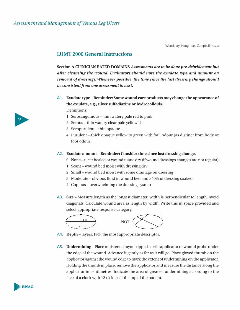
LUMT 2000 General Instructions
Section A CLINICIAN RATED DOMAINS Assessments are to be done pre-debridement but
after cleansing the wound. Evaluators should note the exudate type and amount on
removal of dressings. Whenever possible, the time since the last dressing change should
be consistent from one assessment to next.
A1. Exudate type – Reminder: Some wound care products may change the appearance of
the exudate, e.g., silver sulfadiazine or hydrocolloids.
Definitions:
1 Serosanguinous – thin watery pale red to pink
2 Serous – thin watery clear pale yellowish
3 Seropurulent – thin opaque
4 Purulent – thick opaque yellow to green with foul odour (as distinct from body or
foot odour)
A2. Exudate amount – Reminder: Consider time since last dressing change.
0 None – ulcer healed or wound tissue dry (if wound dressings changes are not regular)
1 Scant – wound bed moist with dressing dry
2 Small – wound bed moist with some drainage on dressing
3 Moderate – obvious fluid in wound bed and >50% of dressing soaked
4 Copious – overwhelming the dressing system
A3. Size – Measure length as the longest diameter; width is perpendicular to length. Avoid
diagonals. Calculate wound area as length by width. Write this in space provided and
select appropriate response category.
NOT
A4. Depth – layers. Pick the most appropriate descriptor.
A5. Undermining – Place moistened rayon-tipped sterile applicator or wound probe under
the edge of the wound. Advance it gently as far as it will go. Place gloved thumb on the
applicator against the wound edge to mark the extent of undermining on the applicator.
Holding the thumb in place, remove the applicator and measure the distance along the
applicator in centimetres. Indicate the area of greatest undermining according to the
face of a clock with 12 o’clock at the top of the patient.
l 90
w
Woodbury, Houghton, Campbell, Keast
98
Assessment and Management of Venous Leg Ulcers

A6. Necrotic tissue type – Reminder: The wound should be thoroughly cleansed before
evaluating. Pick the predominant type of necrotic tissue, e.g., if most of the wound bed
is attached fibrin with small amount of black eschar, choose attached fibrin as tissue type.
A7. Necrotic tissue amount of predominant type selected in A6. The sum of the percentages
in A7 and A9 may be less than but should not exceed 100%.
A8. Granulation tissue type – Choose predominant type of granulation tissue.
A9. Granulation tissue amount – (The sum of the percentages in A7 and A9 may be less
than but should not exceed 100%.) The percentage of granulation tissue refers only to
the non-epithelialized (open) portion of the wound. The advancing border of epithelium
is not considered part of the wound surface.
A10. Edges – Definition: Indistinct borders - where you would not be able to trace the wound edge.
1 More than half of advancing borders may be indistinct because most of wound is
epithelializing.
Advancing wound edge is
2 Less than half of the wound edge is advancing (the process of epidermal resurfacing
appears smooth and shiny).
3 Attached, no advancing border – unable to probe. Looks like
4 Unattached wound edge is undermined wound edge is
A11. Periulcer skin viability – Select the following items that are present; count the number
selected; then use this total to determine appropriate response category.
Definitions: Callus – thick dry epidermis
Scaling dermatitis – scaling red skin which may be weeping
Maceration – wet white opaque skin
Induration – feels firmer than surrounding skin when pressed
Erythema – skin redness (bright red)
Woodbury, Houghton, Campbell, Keast
99
N u r s i n g B e s t P r a c t i c e G u i d e l i n e

A12. Leg edema type – Indicate the worst edema type located anywhere on leg.
Definition: lipodermatosclerosis – waxy white firm tissue.
A13. Leg edema location – Indicate the most proximal location of any type of edema. Clinical
example: pitting edema ankles with non-pitting edema to mid calf: For A10, leg edema
type = 2 ‘pitting’ , A11, leg edema location = 3 to ‘mid calf’.
A14 Assessment of bioburden
1 Lightly colonized: small amount of serous- type exudate.
2 Heavily colonized: large amount of seropurulent drainage with foul odour and no
other cardinal signs of inflammation.
3 Localized infection: large amount of seropurulent drainage with foul odour and
either induration, erythema, warmth, or pain.
4 Systemic infection: advancing cellulitis or osteomyelitis.
Section B PATIENT (PROXY) RATED DOMAINS Read the questions “as they are” to the
patient. It is important to qualify that the questions refer to the last 24 hours. If the patient
is unable to understand the questions due to cognition or language deficits, section B
should not be completed or it may be completed by a proxy only if the proxy knows the
patient well and has been with the patient for most of the last 24 hours. The same person
should provide proxy information for each assessment; otherwise do not complete section B.
B1. Pain amount as it relates to the leg ulcer in the last 24 hours. Determine the rating based on
a numerical rating scale ranging from 0 - 10, then place response in appropriate category.
B2. Pain frequency as it relates to the leg ulcer in the last 24 hours. How often patient
experienced pain in the last 24 hours.
B3. Quality of life as it relates to the leg ulcer in the last 24 hours.
© Woodbury, Houghton, Campbell, Keast, 2000
Reprinted with permission from Dr. M. Gail Woodbury, Investigator, Lawson Health Research
Institute, London, Ontario, Canada.
100
Assessment and Management of Venous Leg Ulcers

Appendix F: Quality of Life Assessment ToolPrognostic indication:The prognostic indication considers the benefits and risks, requirements, and outcomes of
the selected therapy. It takes into account the patient compliance, the physical, cognitive and
emotional state, the social integration in the family, the circle of acquaintances and the living
conditions together with the everyday and occupational capabilities of the patient.
A treatment plan is produced with therapeutic aims broken down into separate aims. The
necessary cooperation of the patient is allocated to the various parts of the break-down.
Alternative aims of the treatment plan are:
� Maintenance/re-establishment of the ability to work,
� Avoidance of the necessity for lifelong professional nursing care,
� Improvement of the quality of life,
� Prolongation of the years of survival.
The patient’s estimate of the quality of life should be included both initially in the discussion
of the treatment plan with the patient, and once the ulcer has healed.
The outcome of the treatment as a result of the physician-patient and the nurse-patient
interaction is influenced by:
� Perception of the severity of the disorder by the patient
� Assessment of the efficacy of the treatment by the patient
� Duration of treatment and disorder
� Complexity of the therapy.
Reprinted with permission.
Compliance Network Physicians/Health Force Initiative, Inc. (1999). Guideline for the
outpatient treatment – venous and venous-arterial mixed leg ulcer. Compliance Network
Physicians/Health Force Initiative, Inc., Berlin, Germany [On-line]. Available:
http://www.cnhfi.de/index-engl.html
101
N u r s i n g B e s t P r a c t i c e G u i d e l i n e

Assess for Quality of LifeCollect information on quality of life and impact of illness on a regular basis and assess for
change over time. Use existing measure if your agency uses a quality of life assessment instrument
(e.g., Medical Outcome Study – SF-36 or the SF-12 quality of life scale) or develop generic, simple
questions to be incorporated into the nursing assessment.
Example:
How would you describe your current health status?
❏ Very good ❏ Good ❏ Fair ❏ Bad ❏ Very Bad
How does the leg ulcer impact your day-to-day living?
❏ Very little ❏ Moderately ❏ A lot
Periodic reassessment is recommended.
Set treatment goals with the client consistent with the values of the individual, family, and
caregiver.
Arrange interventions to meet identified psychosocial needs and goals. Follow-up should be
planned in cooperation with the individual, caregiver, and consultations with appropriate
interdisciplinary team members.
102
Assessment and Management of Venous Leg Ulcers

Appendix G: Pain Assessment ToolsSAMPLE 1 – Visual Analogue Scale (VAS)
No Pain Pain as bad as it could possibly be
The patient indicates intensity of pain on a 10cm. line marked from no pain at one end
to pain as bad as it could possibly be at the other end.
SAMPLE 2 – Numeric Rating Scale (NRS)
0 1 2 3 4 5 6 7 8 9 10The patient rates pain on a scale from 0 to 10.
SAMPLE 3 – Verbal Rating Scale (VRS)
No Pain Mild Pain Moderate Severe Very Worst Pain Pain Severe Pain Possible
PainThe patient rates the pain on a Likert scale verbally, e.g. “none”, “mild pain”, “moderate pain”,
“severe pain”, “very severe pain” or “worst possible pain”.
103
N u r s i n g B e s t P r a c t i c e G u i d e l i n e

Facial Grimace & Behaviour Checklist Flow Charts
Name: Active � Resting � Time:
Regular pain Medication: Rescue/PRN medication
Month:Date or Time FACIAL SCORE 10 8 6 4 2 0 PRN medication
Facial Grimace Score: The facial grimace scale scores the level of pain (from 0-10 on the left) as assessed by thecaregiver observing the facial expressions of the resident. Assessment is done once daily or more (14 days areindicated above). This assessment of the degree of discomfort should be done at the same time every day andduring the same level of activity. Note if rescue/PRN medication is given; yes (y), no (n) or dose.
Behaviour Checklist10 – always 8 – mostly 6 – often 4 - occasionally 2 – rarely 0 - neverDate or Time BEHAVIOUR eats poorly tense quiet indicates pain calls out paces noisy breathing sleeps poorly picks
PRN medication
Behaviour Checklist: Behaviour changes can be used to assess pain or distress, and thereby evaluate the efficacyof interventions. At the top of the scoring graph, when the specific behaviour has been observed, it can berated from 10 (always) to 0 (never). The behaviours being rated and scored over 24 hours are listed down theleft column. This chart scores 9 different behaviours over 14 days. The caregiver can expand on the checklist,i.e., rocking, screams, etc. Note if rescue/PRN medication given. Both tools may be adapted for individual use.
Reprinted with permission. Brignell, A. (Ed) (2000). Guidelines for developing a pain management program. Aresource guide for long-term care facilities, 3rd edition.
0 2 4 6 8 10no pain mild discomforting distressing horrible excruciating
104
Assessment and Management of Venous Leg Ulcers

Pain Assessment Tool
Assessment Date: Name:
Location of Pain: Use letters to identify different pains.
Intensity: Use appropriate pain tool to rate pain subjectively/objectively on a scale of 0-10.
Location Pain A Pain B Pain C Other
What is your/their present level of pain?
What makes the pain better?
What is the rate when the pain is at it’s least?
What makes the pain worse?
What is the rate when the pain is at it’s worst?
Is the pain continuous or intermittent (comes & goes)?
When did this pain start?
What do you think is the cause of this pain?
What level of pain are you satisfied with?
Quality: Indicate the words that describe the pain using the letter of the pain (A,B,C) being described.
aching � throbbing � shooting � stabbing � gnawing � sharp �
burning � tender � exhausting � tiring � penetrating � numb �
nagging � hammering � miserable � unbearable � tingling � stretching �
pulling � other:
Originally adapted with permission from Grey Bruce Palliative Care/Hospice Association Manual.Reprinted with Permission. Brignell, A. (Ed) (2000). Guideline for developing a pain management program. A resource guide for long-term care facilities, 3rd edition.
0 2 4 6 8 10no pain mild discomforting distressing horrible excruciating
Right
Left
Left Left LeftRight Right
Right
105
N u r s i n g B e s t P r a c t i c e G u i d e l i n e

Appendix H: Cleansing Agents and Their Associated ToxicitiesRelative toxicity indices of nonantimicrobal and antimicrobial wound cleansers
Reprinted with permission.
Rodeheaver, G. T. (2001). Wound cleansing, wound irrigation, wound disinfection. In
D. L. Krasner, G. T. Rodeheaver & R. G. Sibbald (Eds.), Chronic wound care: A clinical source
book for healthcare professionals,Third Edition. (pp. 369-383). Wayne, PA: HMP Communications.
Product (nonantimicrobial)
Dermagran®
Shur-Clens®
Biolex™Cara-Klenz™ Wound & Skin CleanserSaf-Clens® Chronic Wound CleanserClinswound™Constant-Clens™ Dermal Wound CleanserCuraklense™ Wound CleanserCurasol™Gentell Wound Cleanser™Sea-Clens® Wound CleanserUltra-Klenz™ Wound Cleanser
Product (antimicrobial)
Clinical Care® Dermal Wound CleanserDermal Wound CleanserMicroKlenz™ Antimicrobial Wound CleanserPuri-Clens™ Wound Deodorizer & CleanserRestore™Royl-Derm™SeptiCare™ Antimicrobial Wound Cleanser
Manufacturer
Derma Sciences, Inc.Conva Tec®
Bard Medical Division, C.R. Bard Inc.Carrington Laboratories Inc.Conva Tec®
Sage Laboratories, Inc.Sherwood Medical-Davis & GeckKendall Healthcare Products Co.Healthpoint MedicalGentellColoplast Sween Corp.Carrington Laboratories, Inc.
Manufacturer
Care-Tech® Laboratories, Inc.Smith & Nephew United, Inc.Carrington Laboratories, Inc.Coloplast Sween Corp.Hollister Inc.Acme United Corp.Sage Laboratories, Inc.
Toxicity Index
1010
100100100
1,0001,0001,0001,0001,0001,0001,000
Toxicity Index
1,00010,00010,00010,00010,00010,00010,000
106
Assessment and Management of Venous Leg Ulcers

Appendix I: Potential Allergens
Commonly Reported Allergens In Patients With Chronic Ulcers
Reprinted with permission from Dr. Gary Sibbald.
Allergen
Topical antibioticse.g., framycetin, neomycin, gentamicin
Lanolin (wood alcohols, amerchol L 101)
Cetyl stearyl alcohol
Colophony (Rosin, Esters of Rosin)
Rubber Chemicalse.g., Thiuram mix, including latex
Preservativese.g., parabens and chloroxylenol
Antibacterials and antisepticse.g., quinoline mix, chlorhexidine
a. Balsam of Peru/fragrance mixb. Benzocaine
Tixocortol pivalate
Source
Medicamentse.g., some tulles, powders, creams and ointments
Many creams, ointments and emollients
Present in many creams preparations e.g., in aqueous cream and some corticosteroid cream.Also in some ointments e.g., emulsifying ointment, andin some paste bandages.
Sticking plaster, adhesive in some bandages and somehydrocolloid dressings.
Bandages, tubular elastic bandages, elastic stockingscontaining natural rubber and latex gloves worn by carer
In many medicaments and some paste bandages
Solutions, creams, tulles
Home care preparations:a. with perfumeb. with local anaesthetic action
Marker of corticosteroid hypersensitivity particular tohydrocortisone
107
N u r s i n g B e s t P r a c t i c e G u i d e l i n e

Appendix J: Topical Antimicrobial Agents
TOPICAL ANTIMICROBIAL AGENTS
Safe &Effective
SelectiveUse
CadexomerIodine
Ionized Silver
SilverSulphadiazine
Polymyxin BSulphate –Bacitracin Zinc
Metronidazolegel/cream
Benzyl/Peroxide
Acetic Acid
Comments
Broad spectrum.Effective for fungi& virus. Widely available.Sheet requireswound contact.Caution if on thyroidmedication.
Broad spectrum.Effective for fungi& virus. Sheet requireswound contact.
Limited potentialfor resistance.Available in pasteor ointment.Do not use ifsulfa sensitive.
Sheet requireswound contact.
Reserve foranaerobes &odour control.
Reserve forMRSA & otherresistant grampositive.
Used in 0.25%(e.g.,1/4 of 1.0%maximum concentration).
SpectrumSA
�
�
�
�
�
MRSA
�
�
�
�
Strep
�
�
�
�
�
PS
�
�
�
�
�
�
F
�
�
An-aerobic�
�
�
�
�
�
VRE
�
Agent
108
Assessment and Management of Venous Leg Ulcers
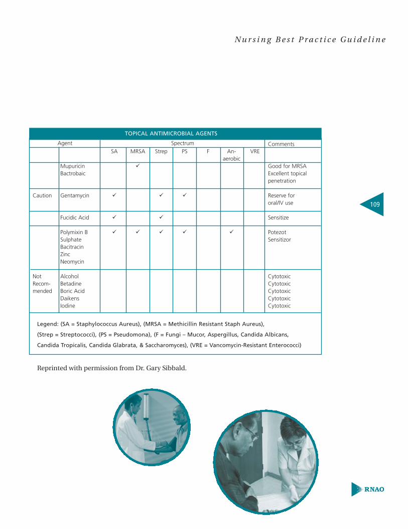
Caution
NotRecom-mended
MupuricinBactrobaic
Gentamycin
Fucidic Acid
Polymixin BSulphateBacitracin Zinc Neomycin
AlcoholBetadineBoric AcidDaikensIodine
Comments
Good for MRSAExcellent topicalpenetration
Reserve fororal/IV use
Sensitize
PotezotSensitizor
CytotoxicCytotoxicCytotoxicCytotoxicCytotoxic
Spectrum
TOPICAL ANTIMICROBIAL AGENTS
TOPICAL ANTIMICROBIAL AGENTS
Legend: (SA = Staphylococcus Aureus), (MRSA = Methicillin Resistant Staph Aureus),
(Strep = Streptococci), (PS = Pseudomona), (F = Fungi – Mucor, Aspergillus, Candida Albicans,
Candida Tropicalis, Candida Glabrata, & Saccharomyces), (VRE = Vancomycin-Resistant Enterococci)
Reprinted with permission from Dr. Gary Sibbald.
SA
�
�
�
MRSA
�
�
Strep
�
�
�
PS
�
�
F An-aerobic
�
VREAgent
109
N u r s i n g B e s t P r a c t i c e G u i d e l i n e

110
Assessment and Management of Venous Leg UlcersA
pp
end
ix K:C
lasses of Com
pression
Ban
dages
Below
are examp
les of co
mp
ression
ban
dages co
mm
on
ly used
in th
e man
agemen
t of ven
ou
s leg ulcers:
Perform
ance C
haracteristics
Sustained compression; can be w
orn continuously for up to 1 week; can be w
ashed and re-used, but may slip.
Can use flexible cohesive for slippage.
Designed to apply 40 m
mH
g pressure at the ankle, graduating to 17 mm
Hg at the knee; sustainable for 1 w
eek.
Reusable with slight stretch giving low
resting pressure but high pressure during activity.
Non com
pliant, plaster-type dressing. Need ankle m
obility if slippage problem.
Bandages can be made by com
bining Kling and a Tensor (spiral or figure 8) and a flexible cohesive bandage on
top. Com
ponents can be re-used.
Self-adherent to prevent slippage; useful over non-adhesive bandages such as elastocrepe and paste bandages;com
pression well sustained.Provides approxim
ately 23 mm
Hg or higher at the ankle graduating to approxim
atelyone-half
this pressure at the knee.
Low pressure obtained; used alone it gives only light support; a single w
ash reduces pressure by about 20 percent.
For holding dressings in place, as a layer within
the multilayer bandage, for light support of m
inor strains and sprains; pressures alone are too low
to be effective in managem
entof venous ulcers; 40 to 60 percent of pressure lostin first 20 m
inutes after application.
Type o
f Co
mp
ression
High elastic com
pression(Long stretch)
Multilayer high com
pression
Inelastic Com
pression
Unna’s Boot
Multilayer bandages
Cohesive bandages
Light compression single
layer elastic
Light support only(inelastic)
Reprinted with perm
ission
from D
r. Gary Sibbald.
Examp
les
Surepress* (Convatec)
Surepress and flexible cohesivebandage.
Profore* (Smith &
Nephew
) 4 layerbandage com
prising of orthopedicpadding; crepe; Elset; C
oban.
Short-stretch bandage, e.g.,C
omprilon (Beiersdorf)
Profore light
Coban (3M
), Roflex
Kling/orthopedic w
ool
Tensor/Elastocrepe
*indicates trademark
HIG
H (40 m
mH
g p
ressure an
d h
igh
er)
MED
IUM
(20-40 mm
Hg
pressu
re)
LOW
(15-20 mm
Hg
pressu
re)
No
tes:1.Ap
plyin
g in a figu
re 8 increases com
pression
by 10 to 15 mm
Hg over sp
iral, for any
ban
dage.
2.Latex-free pro
du
cts – Un
na’s p
aste bo
ot an
d o
ther sp
ecially marked
latex-free
pro
du
cts. 3.Su
pp
ort- in
elastic ban
dages u
sually req
uire m
ore freq
uen
t chan
ging w
ith
redu
ced ed
ema. 4.In
form
clients to
no
tify health
care pro
fession
al if there is p
ain in
foo
t,
or if it tu
rns b
lue o
r black.

Appendix L: Description of the ToolkitToolkit: Implementation of Clinical Practice Guidelines
Best practice guidelines can only be successfully implemented if there are: adequate planning,
resources, organizational and administrative support as well as appropriate facilitation.
RNAO, through a panel of nurses, researchers and administrators has developed a Toolkit:
Implementation of Clinical Practice Guidelines, based on available evidence, theoretical
perspectives and consensus. The Toolkit is recommended for guiding the implementation of
any clinical practice guideline in a healthcare organization.
The Toolkit provides step-by-step directions to individuals and groups involved in planning,
coordinating, and facilitating the guideline implementation. Specifically, the Toolkit
addresses the following key steps:
1. Identifying a well-developed, evidence-based clinical practice guideline.
2. Identification, assessment and engagement of stakeholders.
3. Assessment of environmental readiness for guideline implementation.
4. Identifying and planning evidence-based implementation strategies.
5. Planning and implementing evaluation.
6. Identifying and securing required resources for implementation.
Implementing guidelines in practice that result in successful practice changes and positive
clinical impact is a complex undertaking. The Toolkit is one key resource for managing
this process.
111
N u r s i n g B e s t P r a c t i c e G u i d e l i n e
The Toolkit is available through the Registered Nurses Association of
Ontario. The document is available in a bound format for a nominal
fee, and is also available free of charge from the RNAO website. For
more information, an order form or to download the Toolkit, please
visit the RNAO website at www.rnao.org/bestpractices.

Notes:
112
Assessment and Management of Venous Leg Ulcers

Nursing Best Practice GuidelineShaping the future of Nursing
Supplement IntegrationThis supplement to the nursing best
practice guideline Assessment and
Management of Venous Leg Ulcers is the
result of a three year scheduled revision
of the guideline. Additional material has
been provided in an attempt to provide
the reader with current evidence to sup-
port practice. Similar to the original
guideline publication, this document
needs to be reviewed and applied, based
on the specific needs of the organization
or practice setting/environment, as well
as the needs and wishes of the client.
This supplement should be used in con-
junction with the guideline as a tool to
assist in decision making for individualized
client care, as well as ensuring that
appropriate structures and supports are
in place to provide the best possible care.
Since the original publication of this
guideline, venous leg ulcers have continued
to be a significant and chronic problem
for large numbers of Canadians, with
major implications to quality of life and
overall healthcare costs (Harrison et al., 2005).
A review of the most recent literature and
relevant guidelines published since 2004
does not suggest dramatic changes to
our assessment and management
approach to venous leg ulcers, but rather
suggest some refinements and stronger
evidence for our approach.
Assessment and Management of Venous Leg Ulcers
Guideline supplement
Revision Panel Members
Patricia Coutts, RN, IIWCCTeam LeaderWound Care and Clinical Trials CoordinatorDebary DermatologicalMississauga, Ontario
Patti Barton, RN, PHN, ETET Wound ConsultantSpecialty ET ServicesToronto, Ontario
Cathy Burrows, RN, BScNStaff NurseVascular Surgery Leg Ulcer ClinicQE II Health Sciences CentreHalifax, Nova Scotia
Karen E. Campbell, RN, MScN, PhD(c), ACNPNurse Practitioner/Clinical Nurse SpecialistLondon Health Sciences Center, University HospitalLondon, Ontario
Moira Coates, RN, BScN, ETEnterostomal Therapy NurseSt. Joseph’s Care GroupThunder Bay, Ontario
Theresa Hurd, RN, MScN, MSN, ACNPAdvanced Practice NurseHamilton Niagara Haldimand Brant CCACSt. Catharines, Ontario
Karen Lorimer, RN, MScN, CCHNAdvanced Practice NurseCarefor Health & Community ServicesOttawa, Ontario
Nancy Parslow, RN, ETWound Care Nurse SpecialistSouthlake Regional Health CentreNewmarket, Ontario
Christine Pearson, RN, IIWCCCommunity Wound ClinicianNorth Vancouver, British Columbia
Laura Teague, RN, MN, ACNPNurse PractitionerSt. Michael’s HospitalToronto, Ontario
Samantha Mayo, RN, BScN, MNProgram CoordinatorNursing Best Practice Guidelines ProgramRegistered Nurses’ Association of OntarioToronto, Ontario
Meagan Cleary, BAProject CoordinatorNursing Best Practice Guidelines ProgramRegistered Nurses’ Association of OntarioToronto, Ontario
March 2007

2
Revision ProcessThe Registered Nurses’ Association of
Ontario (RNAO) has made a commitment
to ensure that this practice guideline is
based on the best available evidence. In
order to meet this commitment, a
monitoring and revision process has
been established for each guideline
every 3 years. The revision panel
members (experts from a variety of
practice settings) are given a mandate
to review the guideline focusing on the
recommendations and the original
scope of the guideline.
Members of the panel critically
appraised three international guide-
lines, published since 2004, using the
Appraisal of Guidelines For Research
and Evaluation (AGREE Collaboration, 2001)
instrument. This review resulted in the
following guideline and accompanying
document being used as a supportive
resource during the revision process of
this RNAO guideline:
Royal College of Nursing (RCN). (2006).
Clinical practice guideline: The nursing
management of patients with venous
leg ulcers. [Online].
Available: http://www.rcn.org.uk/pub-
lications/pdf/guidelines/venous_leg_u
lcers.pdf.
Accompanying document:
Prodigy. (2004). Leg ulcer – venous.
[Online]. Available:
http://www.cks.library.nhs.uk/leg_ulcer
_venous
Review/Revision ProcessFlow Chart
Literature Search
Yield 203 abstracts
54 studies met the inclusion criteria
Quality appraisal of studies
Develop evidence summary table
Revisions based on new evidence
Supplement published
Dissemination
New Evidence
Look for the following resources to support the implementation of this guideline (available for freedownload at www.rnao.org/bestpractices):
• Learning Package: Assessment and Management of Venous Leg Ulcers• Health Education Fact Sheet: Taking Care of Your Legs

3
Summary of EvidenceThe following content reflects the evidence reviewed that either supports the original guideline recom-mendations or provides evidence for revision. Through the review process, many recommendationswere re-worded, removed or combined to reflect current knowledge and to enhance the clarity of thedocument. Two new recommendations have also been added. As a result of these changes, the num-bering of the recommendations has been revised. The following are the recommendations supportedby the revision process. Details regarding the omitted recommendations can be found on page 14.
Practice Recommendations: Comprehensive AssessmentThe section heading was modified to emphasize that a comprehensive assessment, where a complete clientassessment precedes evaluation of limb and ulcer characteristics, is essential to determine the underlying ulceretiology and appropriate treatment approaches
Recommendation 1
Assessment and clinical investigations should be undertaken by healthcare professional(s) trained and experienced in leg
ulcer management.
(Level C)
Additional Literature Supports
RCN, 2006
Recommendation 2
A comprehensive clinical history and physical examination includes:
■ blood pressure measurement;
■ weight;
■ blood glucose level;
■ doppler measurement of Ankle Brachial Pressure Index (ABPI);
■ any other tests relevant to presenting patient’s condition;
■ ulcer history;
■ ulcer treatment history;
■ medical history;
■ medication;
■ bilateral limb assessment;
■ pain;
■ nutrition;
■ allergies;
■ psychosocial status (including quality of life); and
■ functional, cognitive, emotional status and ability for self-care.
The above should be documented in a structured format for a client presenting with either their first or recurrent leg ulcer
and should be ongoing thereafter.
(Level C)
This recommendation has been modified to incorporate original Recommendations 2, 3, 4, 6, 7, 13 and 26, inorder to emphasize the essential components of a comprehensive clinical assessment. Any other tests relevantto presenting patient’s condition, ulcer treatment history, medical history and medications have also beenadded to reflect components of an assessment that may provide valuable information towards the developmentof an appropriate treatment plan.
A comprehensive clinical history and physical examination will provide important information regarding theunderlying etiology of the ulcer. There are several types of leg ulcers whose treatment is beyond the scope of theguideline. The recommendations presented here were developed specifically for the management of leg ulcersrelated to venous disease. Appendix M (page 16 of this supplement) provides a description of physical findingsthat would indicate venous disease versus arterial disease.
� unchanged
� changed
+ additional information
NEW new recommendation
�
+
�
�
+

4
�
+
�
�
+
�
Recommendation 3
A comprehensive assessment of an ulcer should include:
■ measurement of the wound and undermining;
■ amount and quality of exudate;
■ wound bed appearance;
■ condition of the wound edge;
■ infection;
■ presence or absence of patient suffering; and
■ re-evaluation.
(Level C)
Measure the surface areas of ulcers, at regular intervals, to monitor progress. Maximum length and width, or tracings onto
a transparency are useful methods.
(Level B)
Key elements of ulcer assessment have been added to this recommendation to reflect that ulcer condition is bestevaluated using a comprehensive approach. The panel cautions, however, that the presence of undermining israre with venous ulceration and therefore warrants further assessment to rule out other etiologies (i.e. arterial disease).
Additional Literature Supports
Sibbald et al., 2006
Recommendation 4
Regular ulcer assessment is essential to monitor treatment effectiveness and healing goals.
(Level C)
Practice Recommendations: Diagnostic Evaluation
Recommendation 5
An Ankle Brachial Pressure Index (ABPI) measurement should be performed by a trained practitioner to rule out the
presence of peripheral arterial disease, particularly prior to the application of compression therapy.
(Level B)
This recommendation has been modified to emphasize the role of Ankle Brachial Pressure Index (ABPI)measurement within the scope of nursing practice and represents a combination of original Recommendations9 and 10. In the management of venous leg ulcers, ABPI measurement offers valuable information as a screeningtool for lower extremity peripheral arterial disease (Hirsch et al., 2006). Where peripheral arterial disease is suspected,compression therapy treatments designed for venous leg ulcers may be contraindicated. ABPI may also offerprognostic data that are useful to predict limb survival, wound healing and patient survival. The use of ABPImeasurement for diagnosis is generally outside of the scope of nursing practice. Furthermore, only thosepractitioners with the appropriate knowledge, skill and judgement to perform this measurement should do so.
Additional Literature Supports
Hirsch et al., 2006; RCN, 2006; Smith et al., 2003
Recommendation 6
An Ankle Brachial Pressure Index (ABPI) >1.2 and <0.8 warrants referral for further medical assessment.
(Level C )

5
+
�
+
�
+
�
NEW
+
NEW
This recommendation has been modified to emphasize the importance of specialized assessment in cases ofabnormal ABPI. People with abnormally low or abnormally high ABPI should be further investigated forperipheral arterial disease (Hirsch et al., 2006). For example, an ABPI >1.3 is considered indicative of non-compressible vessels that are found in individuals with diabetes, chronic renal failure and who are older than 70years of age. In these cases, compression therapy may not be recommended.
Additional Literature Supports
Hirsch et al., 2006; Partsch & Partsch, 2005; Schroder & Diehm, 2006
Recommendation 7
Prior to debridement, vascular assessment, such as Ankle Brachial Pressure Index (ABPI), is recommended for ulcers in
lower extremities to rule out vascular compromise and ensure healability.
(Level C)
The wording of this recommendation has been modified to indicate that vascular assessment (including ABPI)may also provide information regarding the healability of the wound. Adequate perfusion to these woundsindicates that healing is supported by the microvascular supply and that debridement, when appropriate, maybe safely performed.
Practice Recommendations: Pain
Recommendation 8
Pain may be a feature of both venous and arterial disease, and should be addressed.
(Level B)
A comprehensive approach to pain is beyond the scope of this guideline. Refer to the RNAO Best PracticeGuideline Assessment and Management of Pain (Revised) (RNAO, 2007) for strategies by which to address pain.
Additional Literature Supports
Charles, 2002; Nemeth et al., 2004
Recommendation 9
Prevent or manage pain associated with debridement. Consult with a physician and pharmacist as needed.
(Level C)
Practice Recommendations: Venous Ulcer Care
Recommendation 10
Develop treatment goals mutually agreed upon by the patient and healthcare professionals, based on clinical findings,
current evidence, expert opinion and patient preference.
(Level C)
Prior to commencing wound care, a treatment plan must be developed based on the goals of care for theparticular client. The healing potential of the wound is one factor that may impact on the goals of care. Potentialfor healing is affected by local, host, and environmental factors. For any issues identified in the assessment thatmay impair healing, a plan needs to be developed that addresses these as well. Appendix N (page 16 of thissupplement) provides a list of factors that may impact healing potential.
Recommendation 11
Local wound bed preparation includes debridement when appropriate, moisture balance and bacterial balance.
(Level C)

6
+
�
�
+
�
The panel strongly recommends a systematic approach to wound care. The aim of local wound bed preparationis the removal of barriers to healing. Appendix O (page 17 of this supplement) outlines important considerationswhen preparing the wound bed.
Not all wounds require debridement. The need for debridement is based on the goals of the client. Other issueswhen considering the appropriateness of debridement include the practice setting, skill of the clinician and theability of the patient to tolerate the procedure. When choosing between particular techniques, considerationsshould include type, quantity and location of nonviable tissue, the depth of the wound, the amount of wound fluidand the general condition and goals of the client. Appendix P (page 17 of this supplement) compares the methodsof debridement based on several key considerations. Refer to the original guideline (RNAO, 2004) for a more detaileddiscussion of debridement.
Moisture balance refers to the management of exudate and maintenance of fluids in the wound bed. Whenhealing is the goal, a balanced moist wound environment ensures that there is sufficient moisture to facilitatecellular growth but not in excess to cause further skin breakdown. The primary approach to achieving moisturebalance is through the appropriate use of moisture-balancing dressings (Okan et al., 2007).
Bacterial balance describes the bacterial level present in a wound and their ability to cause damage or infection. Allchronic wounds contain bacteria. However, the impact of these bacteria on healing is dependent on several factors,including the number of organisms, the virulence of these organisms and host resistance (Sibbald, Woo, & Ayello, 2006).Promoting host resistance, through optimizing overall health of the client, is an important aspect of achievingbacterial balance. The management of existing infection is addressed in the following section of this supplement.
Additional Literature Supports
Sibbald et al., 2006; Sibbald, Woo, & Ayello, 2006; Okan et al., 2007
Recommendation 12
Cleansing of the ulcer should be kept simple; warm tap water or saline is usually sufficient.
(Level B)
Recommendation 13
First-line and uncomplicated dressings must be simple, low adherent, acceptable to the client and should be cost-effective.
(Level A)
A recent systematic review by Palfreyman et al. (2007) found insufficient evidence to support the use of aparticular first-line and uncomplicated dressing type (beneath compression therapy) for the treatment of venousleg ulcers. As such, the authors suggested that decisions regarding dressings should be based on local costs andpractitioner or patient preferences (Palfreyman et al., 2007). Similarly, the RCN guideline (2006) states that for patientsnot requiring frequent compression bandaging, the cost-effectiveness of the dressing will be related to its abilityto remain in place and manage the exudate. The wording of this recommendation was modified to reflect aconsideration of cost-effectiveness when choosing a dressing.
In a systematic review by O’Donnell and Lau (2006), despite the findings that certain complex wound dressingsresulted in better outcomes in both the proportion of ulcers healed and time to healing, the authors maintainedthat the selection of the specific dressing will depend on the dressing characteristics for ease of application,patient comfort, wound drainage absorption and expense.
Additional Literature Supports
O’Donnell & Lau, 2006; Palfreyman et al., 2007; RCN, 2006; Schulze et al., 2001; Smith et al., 2004; Vanscheidt, Sibbald, & Eager, 2004; Vin, Teot, &
Meaume, 2002
Recommendation 14
Avoid products that are known to cause skin sensitivity, such as those containing lanolin, phenol alcohol, or some topical
antibiotic and antibacterial preparations.
(Level C)

7
+
�
+
�
+
�
�
+
�
+
The wording of this recommendation has been modified to indicate that not all topical antibiotic andantibacterial preparations are known to cause skin sensitivity. The change in the wording is for clarification only,and there has been no change in the intent of the recommendation.
Recommendation 15
Choose a dressing that optimizes the wound environment and patient tolerance.
(Level C)
This recommendation was revised to indicate that there are many considerations when choosing an appropriatedressing. Dressings are usually applied beneath the compression to aid healing, comfort and to control exudate.Wounds heal quicker in a moist environment and dressings are used to absorb excess fluid or retain fluid in anotherwise dry wound (Palfreyman et al., 2007).
Additional Literature Supports
Palfreyman et al., 2007; Schulze et al., 2001
Recommendation 16
No specific dressing has been demonstrated to encourage ulcer healing.
(Level A)
Additional Literature Supports
Nelson, Cullum, & Jones, 2006; O’Donnell & Lau, 2006; Palfreyman et al., 2007; Vin, Teot, & Meaume, 2002; Wollina et al., 2005
Recommendation 17
In contrast to drying out, moist wound conditions allow optimal cell migration, proliferation, differentiation and
neovascularization.
(Level A)
Recommendation 18
Refer clients with suspected sensitivity reactions to a dermatologist for patch testing. Following patch testing, identified
allergens must be avoided, and medical advice on treatment should be sought.
(Level B)
The panel notes that for most nurses, referral is outside their scope of practice. However, nurses are encouragedto advocate for appropriate referrals as necessary.
Recommendation 19
Venous surgery followed by graduated compression hosiery is an option for consideration in clients with superficial
venous insufficiency.
(Level A)
The level of evidence has been updated in the context of recent research that indicates a significant reduction inulcer recurrence after venous surgery for clients with superficial venous insufficiency.
Additional Literature Supports
Barnwell et al., 2004; Obermayer et al., 2006; Roka, Binder, & Bohler-Sommeregger, 2006; Ting et al., 2006; van Gent et al., 2006

8
�
+
�
+
�
+
�
+
Practice Recommendations: Infection
Recommendation 20
Assess for signs and symptoms of infection
(Level A)
The wording of this recommendation was modified to appropriately reflect nurses’ scope of practice. It isimperative that the assessment and treatment of infection be addressed as it can influence the plan of care, suchas the use of local dressings, compression modalities and adjunctive therapies. Refer to Appendix Q (page 18 ofthis supplement) for a list of signs and symptoms of infection.
Recommendation 21
Manage wound infection with cleansing and debridement, as appropriate. Where there is evidence of cellulitis, treatment
of infection involves systemic antibiotics.
(Level B)
This recommendation, which reflects a combination of original Recommendations 29 and 30, was modifiedbased on infectious disease guidelines and current evidence. Cellulitis is characterised by the presence of someof the following signs or symptoms: pyrexia, increasing pain, increasing erythema of surrounding skin; purulentexudate; and rapid increase in ulcer size.
Additional literature supports
Cutting and White, 2005; Gardner, Franz, & Doebbeling, 2001; Grayson et al., 2002; Rosser et al., 2005;
Recommendation 22
The use of topical antiseptics to reduce bacteria in wound tissue should be reserved for situations in which concern for
bacterial load is higher than that of healability.
(Level C)
This recommendation has been modified in the context of current evidence that suggests that the use of topicalantiseptics may be beneficial for short term use, particularly when bacterial levels are sufficiently high to causetissue destruction and the goal of care is the maintenance of the wound. Some antiseptics have been shown tobe safe and efficacious for this purpose in wound care. Appendix R (page 18 of this supplement) provides asummary of evidence for various antiseptics.
Additional literature supports
Drosou, Falabella & Kirsner, 2003
Practice Recommendations: Compression
Recommendation 23
The treatment of choice for venous ulceration uncomplicated by other factors is graduated compression bandaging,
properly applied and combined with exercise. (Level A)■ In venous ulceration, high compression achieves better healing than low compression. (Level A)■ Compression bandages should only be applied by a suitably trained and experienced practitioner. (Level B)■ The concepts, practice, and hazards of graduated compression should be fully understood by those prescribing and
fitting compression stockings. (Level B)■ Ankle circumference should be measured at a distance of 2.5 cm (one inch) above the medial malleolus. (Level C)
Original Recommendations 33, 34, 35, 43 & 44 have been combined and modified to emphasize importantconsiderations when providing compression therapy in the care of venous ulceration. This recommendation nolonger addresses care of venous eczema as treatment approaches for conditions that could lead to ulceration,dermatological or otherwise, was beyond the scope of the current review. The level of evidence for original

9
+
�
+
�
+
�
+
�
�
Recommendation 44 has also been updated to reflect current knowledge. The panel notes that despite theevidence to support the use of high compression bandages for the treatment of uncomplicated venous ulcers,there is insufficient evidence to support the choice between using elastic versus non-elastic bandaging systems.
Additional Literature Supports
Brooks et al., 2004; Edwards et al., 2005; Franks et al., 2004; Iglesias et al., 2004; Nelson, Cullum, & Jones, 2006; Nelson et al., 2004; O’Brien et al., 2003;
Padberg, Johnston, & Sisto, 2004; Partsch & Partsch, 2005; Partsch et al., 2001; Polignano et al., 2004; Polignano, Guarnera, & Bonadeo, 2004
Recommendation 24
External compression applied using various forms of pneumatic compression pumps can be indicated for individuals
with chronic venous insufficiency.
(Level A)
The wording of this recommendation has been modified to emphasize the need for clinical judgement indetermining whether this treatment is appropriate for a particular client. The change in the wording is forclarification only, and there has been no change in the intent of the recommendation.
Recommendation 25
The client should be prescribed regular vascular exercise by means of intensive controlled walking and exercises to
improve the function of the ankle joint and calf muscle pump.
(Level A)
The wording of this recommendation has been modified for anatomical accuracy. The change in the wording isfor clarification only, and there has been no change in the intent of the recommendation.
Additional Literature Supports
Edwards et al., 2005; Padberg, Johnston, & Sisto, 2004.
Practice Recommendations: Complementary Therapies
Recommendation 26
Consider electrical stimulation in the treatment of venous leg ulcers.
(Level B)
Additional Literature Supports
Cherry & Ryan, 2005
Recommendation 27
Therapeutic ultrasound may be used to reduce the size of chronic venous ulcers.
(Level A)
Practice Recommendations: Reassessment
Recommendation 28
If signs of healing are not evident, a comprehensive assessment and re-evaluation of the treatment plan should be carried
out at three month intervals, or sooner if clinical condition deteriorates.
(Level C)

10
+
�
+
�
+
�
+
The wording of this recommendation has been modified to indicate the importance of wound assessment indetermining healing. Recommendation 3 (page 4 of this supplement) addresses aspects of ulcer assessment. Animprovement in these attributes indicates healing. If an improvement in these attributes is not evident, or ifulcer condition worsens, a comprehensive patient assessment is warranted. The change in the wording is forclarification only, and there has been no change in the intent of the recommendation.
Additional Literature Supports
Flanagan, 2003; Prodigy, 2004
Recommendation 29For resolving and healing venous leg ulcers, routine assessment at six-month intervals should include:
■ physical assessment;
■ Ankle Brachial Pressure Index (ABPI);
■ replacement of compression stockings; and
■ reinforcement of teaching.
(Level C)
Additional Literature Supports
Prodigy, 2004
Practice Recommendations:Client Education for Secondary Prevention
The section heading was modified to emphasize the importance of client education as a valuable means to
promoting effective preventative measures.
Recommendation 30
Inform the client of measures to prevent recurrence after healing:
■ daily wear of compression stockings, cared for as per manufacturer’s instructions and replaced at a minimum every
six months;
■ discouragement of self-treatment with over-the-counter preparations;
■ avoidance of accidents or trauma to legs;
■ rest periods throughout the day with elevation of affected limb above level of heart;
■ early referral at first sign of skin breakdown or trauma to limb;
■ need for exercise and ankle-joint mobility;
■ appropriate skin care avoiding sensitizing products; and
■ compression therapy for life with reassessment based on symptoms.
(Level C)
This recommendation has been modified to incorporate Recommendations 46, 47, 56 and 57 from the originalpublication, thereby providing a concise list of essential attributes of patient education with regards tosecondary prevention of leg ulcers. Furthermore, although it is recognized that compression therapy for life is acommon preventative strategy, the recommendation has also been modified to emphasize the importance ofreassessment given the potential for changing client needs. Appendix S (page 19 of this supplement) provides aClient Education Checklist to support the implementation of this recommendation.
Additional Literature Supports
Edwards et al., 2005; Prodigy, 2004

11
�
+
�
+
�
+
�
�
Education Recommendations
Recommendation 31
Guidelines are more likely to be effective if they take into account local circumstances and are disseminated by an ongoing
education and training program.
(Level C)
Additional Literature Supports
Brooks et al., 2004; McGuckin et al., 2001
Recommendation 32
Using principles of adult learning, present information at an appropriate level for the target audience, including
healthcare providers, clients, family members and caregivers.
(Level C)
The wording of this recommendation has been modified to emphasize the importance of considering adultlearning principles when designing educational programs. The change in the wording is for clarification only,and there has been no change in the intent of the recommendation.
Additional Literature Supports
Brooks et al., 2004; McGuckin et al., 2001
Recommendation 33
All healthcare professionals who manage lower limb ulcers should be trained in leg ulcer assessment and management.
(Level C)
The wording of this recommendation has been modified to recognize that particular health care professionals,and not necessarily all health professionals, require specialized training in leg ulcer care. The change in thewording is for clarification only, and there has been no change in the intent of the recommendation.
Additional Literature Supports
Brooks et al., 2004
Recommendation 34
Design, develop, and implement educational programs that reflect a continuum of care. The program should begin with
a structured, comprehensive, and organized approach to prevention and should culminate in effective treatment
protocols that promote healing as well as prevent recurrence.
(Level C)
Recommendation 35
Education programs for healthcare professionals who manage lower limb ulcers should include:
■ pathophysiology of leg ulceration;
■ leg ulcer assessment;
■ need for Doppler ultrasound to measure Ankle Brachial Pressure Index (ABPI);
■ normal and abnormal wound healing;
■ compression therapy theory, management, and application;
■ dressing selection;
■ principles of debridement;
■ principles of cleansing and infection control;
■ skin care of the lower leg;

12
+
�
+
�
+
�
■ peri-wound skin care and management;
■ psychological impact of venous stasis disease;
■ quality of life;
■ pain management;
■ teaching and support for care provider;
■ health education;
■ preventing recurrence;
■ principles of nutritional support with regard to tissue integrity;
■ mechanisms for accurate documentation and monitoring of pertinent data, including treatment interventions and
healing progress; and
■ criteria for referral for specialized assessment.
(Level C)
The wording of this recommendation has been modified to recognize that particular healthcare professionals,and not necessarily all health professionals, require specialized training in leg ulcer care. The change in thewording is for clarification only, and there has been no change in the intent of the recommendation.
Recommendation 36
Healthcare professionals with recognized training in leg ulcer care should mentor and transfer their knowledge and skills
to local healthcare teams.
(Level C)
This wording of this recommendation was modified to suggest the means by which healthcare professionals candisseminate their specialized knowledge and skill regarding leg ulcer care. The change in the wording is forclarification only, and there has been no change in the intent of the recommendation.
Recommendation 37
The knowledge and understanding of the healthcare professional is a major factor in adherence to treatment regimens.
(Level C)
Additional Literature Supports
Brooks et al., 2004; McGuckin et al., 2001
Organization and Policy Recommendatons
Recommendation 38
Successful implementation of a venous ulcer treatment policy/strategy requires:
■ dedicated funding
■ integration of healthcare services
■ support from all levels of government
■ management support
■ human resources
■ financial resources
■ functional space
■ commitment
■ collection of baseline information about vulnerable populations
■ resources and existing knowledge
■ interpretation of above data and identification of organizational problems.
(Level C)

13
�
Recommendation 39
Nursing best practice guidelines can be successfully implemented only where there are adequate planning, resources,
organizational and administrative support, as well as appropriate facilitation. Organizations may wish to develop a plan
for implementation that includes:
■ An assessment of organizational readiness and barriers to education.
■ Involvement of all members (whether in a direct or indirect supportive function) who will contribute to the
implementation process.
■ Dedication of a qualified individual to provide the support needed for the education and implementation process.
■ Ongoing opportunities for discussion and education to reinforce the importance of best practices.
■ Opportunities for reflection on personal and organizational experience in implementing guidelines.
In this regard, RNAO (through a panel of nurses, researchers and administrators) has developed the Toolkit:
Implementation of Clinical Practice Guidelines, based on available evidence, theoretical perspectives and consensus. The
RNAO strongly recommends the use of this Toolkit for guiding the implementation of the best practice guideline on
Assessment and Management of Venous Leg Ulcers.
(Level C)
Implementation Strategies
The Registered Nurses’ Association of Ontario and the guideline panel have compiled a list of implementation strategies toassist health care organizations or health care disciplines who are interested in implementing this guideline. A summary ofthese strategies follows:
■ Have at least one dedicated person such as an advanced practice nurse or a clinical resource nurse who will providesupport, clinical expertise and leadership. The individual should have good interpersonal, facilitation and projectmanagement skills.
■ Conduct an organizational needs assessment related to the care of adults with leg ulcers to identify current knowledgebase and further educational requirements.
■ Create a vision to help direct the change effort and develop strategies for achieving and sustaining the vision.■ Establish a steering committee comprised of key stakeholders and interdisciplinary members committed to leading the
change initiative. Identify short term and long-term goals. ■ Identify and support designated best practice champions on each unit to promote and support implementation.
Celebrate milestones and achievements, acknowledging work well done (Davies & Edwards, 2004).■ Provide organizational support such as having the structures in place to facilitate best practices in leg ulcer care. For
example, having an organizational philosophy that reflects the value of best practices through policies and procedures.Develop new assessment and documentation tools (Davies & Edwards, 2004).
Research Gaps and Implications
In reviewing the evidence for the revision of this guideline, it is clear that future research opportunities involve: ■ Effective training strategies to improve clinician skill in providing compression therapy■ Evaluation of the effect of practice guidelines on delivery of care, adherence to treatment, and recurrence■ The effect of preventative measures and healthy lifestyles on rates of recurrence■ The effect of various follow-up practices and policies on rates of recurrence■ Further treatment interventions to improve rates of healing■ Comparing overall healthcare costs of prevention versus treatment
Some of the recommendations in this guideline are based on consensus or expert opinion. Further substantive research isrequired to validate the expert opinion. Increasing the research can impact knowledge that will lead to improved practice andoutcomes for patients with venous leg ulcers.

Deleted RecommendationsIn order to maintain the clarity and rigour of this practice guideline, the following recommendations from the original publicationwere withdrawn as part of the revision process based on panel consensus and current research evidence, where indicated.
Recommendation 25
Biological wound coverings and growth factor treatments should not be applied in cases of wound infection. (Level C)
This recommendation was removed as it was considered too limited in scope. It has been more appropriately integrated intothe discussion of revised Recommendation 20 (page 8 of this supplement), where it is described that wound infection mayimpact on the appropriateness of several different treatment options.
Recommendation 28
An infection is indicated when >105 bacteria/gram tissue is present. (Level B)
This recommendation was removed as it is a definition of infection and not a recommendation for practice. Clinicalassessment of infection is addressed in revised Recommendation 20 (page 8 of this supplement).
Recommendation 32
Topical antibiotics and antibacterial agents are frequent sensitizers and should be avoided. (Level B)
This recommendation was removed because it no longer reflects current knowledge and clinical practice. Topical antibioticsand antibacterial agents have a wide range of characteristics and may not all be frequent sensitizers and may not all have to beavoided. The appropriateness and safety of these agents in the management of venous leg ulcers should be based onconsideration of the particular client, clinician and product. The recommendation to avoid topical antibiotics and antibacterialagents that are known to cause skin sensitivity is reflected in the revised Recommendation 14 (page 6 of this supplement).
Recommendation 36
Venous ulceration should be treated with high compression bandaging to achieve a pressure between 35-40 mmHg, at the ankle,
graduating to half at calf in the normally shaped limb, as per La Place’s Law. (Level C)
This recommendation was removed based on the difficulty of evaluating these measurements in clinical practice.
Recommendation 37
Use protective padding over bony prominences when applying high compression. (Level C)
This recommendation was removed based on its high specificity towards certain products. The practice of using protectivepadding over bony prominences may be contraindicated or inappropriate based on the particular compression therapyproducts being used.
Recommendation 38
Arterial insufficiency is a contraindication to the use of high compression. A modified form of compression may be used under specialist
supervision. (Level C)
This recommendation was removed based on the ambiguity related to what constitutes appropriate compression therapy inthe cases of arterial insufficiency. As such, this recommendation was considered difficult to apply to clinical practice. Thecaution required related to the application of compression therapy in these cases has been incorporated into the revisedRecommendation 5.
Recommendation 39
Use compression with caution in clients with diabetes, those with connective tissue disease and the elderly. (Level C)
This recommendation was removed based on the ambiguity related to what constitutes appropriate compression therapy inthe cases of diabetes, those with connective tissue disease and the elderly.
14
15
Recommendation 40
Compression therapy should be modified until clinical infection is treated. (Level C)
This recommendation was removed based on a lack of evidence to support the modification of compression therapy based onthe presence of clinical infection alone. Such intervention is considered part of clinical decision making based on acomprehensive physical assessment.
Recommendation 41
Bandages should be applied according to manufacturer’s recommendations. (Level C)
This recommendation was removed as it is more appropriately a standard of practice regarding safe product usage and not arecommendation for practice.
Recommendation 42
When using elastic systems such as “high compression” bandages, the ankle circumference must be more than or padded to equal 18cms.
(Level C)
This recommendation was removed based on its high specificity towards certain products. Circumference specifications maydiffer based on the particular compression bandaging systems used.
Recommendation 45
Graduated compression hosiery should be measured and fitted by a certified fitter. (Level C)
This recommendation was removed by panel consensus as its meaning is reflected in the revised Recommendation 23.
Recommendation 47
Graduated compression hosiery should be prescribed for life. (Level C)
This recommendation was combined into the revised Recommendation 36 to emphasize the role of ongoing compressiontherapy as part of secondary prevention for future venous leg ulcers as well as the need for ongoing reassessment based onchanges in the client’s condition.
Recommendation 51
Hyperbaric oxygen may reduce ulcer size in non-diabetic, non-atherosclerotic leg ulcers. (Level A)
This recommendation was removed as it is no longer supported by current evidence. A recent systematic review indicates thathyperbaric oxygen may reduce amputation in patients with diabetic foot ulcers, but the effect of this therapy on venous ulcersis not clear (Kranke et al., 2006).
Recommendation 55
Measures to prevent recurrence of a venous leg ulcer include: wearing compression stockings, regular follow-up to monitor Ankle Brachial
Pressure Index (ABPI), discouragement of self-treatment with over-the-counter preparations, and avoidance of accidents or trauma to
legs. (Level C)
This recommendation, except for the component regarding follow-up ABPI measurement, was combined into the revisedRecommendation 36 to reflect that secondary prevention from the clinician’s perspective most commonly involves patienteducation of preventative measures. Although it is recognized that measurement of Ankle Brachial Pressure Index (ABPI) maybe included as part of a comprehensive reassessment, this follow-up measure is not considered part of routine preventativecare unless indicated by a change in client condition, such as the emergence of symptoms or skin breakdown.

16
Appendices
The review/revision process identified a need for updates to the Glossary and the inclusion of additional appendices:
Appendix B: Glossary of Terms
The glossary has been updated to add:
Bacterial Balance: The bacterial level present in a wound and their ability to cause damage or infection. The impact of these bacteria
on healing is dependent on several factors, including the number of organisms, the virulence of these organisms and host resistance
(Sibbald, Woo, & Ayello, 2006).
Moisture Balance: The management of exudate and maintenance of fluids in the wound bed. When healing is the goal, a balanced moist
wound environment ensures that there is sufficient moisture to facilitate cellular growth but not in excess to cause further skin breakdown.
Appendix M: Physical Findings of Venous Disease versus Arterial Disease
Physical Findings of Venous Disease versus Arterial Disease
Source: RNAO, 2004
Appendix N: Factors Affecting Healing Potential
Factors Affecting Healing Potential
Adapted from RNAO, 2005.
Venous Disease
Usually shallow, moist ulcers
Situated on gaiter area of leg
Edema
Eczema
Ankle flare
Lipodermatosclerosis
Varicose veins
Hyperpigmentation
Atrophie blanche
Arterial Disease
Ulcers with a “punched out” appearance
Base of wound poorly perfused, pale, dry
Cold legs/feet (in a warm environment)
Shiny, taut skin
Dependent rubor
Pale or blue feet
Gangrenous toes
Local
■ Necrosis
■ Infection
■ Microvascular supply
■ Foreign body
■ Iatrogenic
• Cytotoxic agents
Host
■ Co-morbidities
• Inflammatory condition
• Nutrition
• Peripheral vascular disease
• Coronary artery disease
■ Adherence to plan of care
■ Cultural/personal beliefs
Environment
■ Access to care
■ Family support
■ Healthcare sector
■ Geographic
■ Socioeconomic status
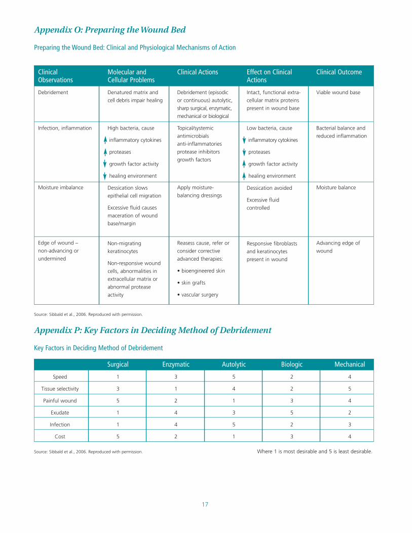
17
Appendix O: Preparing the Wound Bed
Preparing the Wound Bed: Clinical and Physiological Mechanisms of Action
Source: Sibbald et al., 2006. Reproduced with permission.
Appendix P: Key Factors in Deciding Method of Debridement
Key Factors in Deciding Method of Debridement
Source: Sibbald et al., 2006. Reproduced with permission. Where 1 is most desirable and 5 is least desirable.
ClinicalObservations
Debridement
Infection, inflammation
Moisture imbalance
Edge of wound –
non-advancing or
undermined
Molecular andCellular Problems
Denatured matrix and
cell debris impair healing
High bacteria, cause
inflammatory cytokines
proteases
growth factor activity
healing environment
Dessication slows
epithelial cell migration
Excessive fluid causes
maceration of wound
base/margin
Non-migrating
keratinocytes
Non-responsive wound
cells, abnormalities in
extracellular matrix or
abnormal protease
activity
Clinical Actions
Debridement (episodic
or continuous) autolytic,
sharp surgical, enzymatic,
mechanical or biological
Topical/systemic
antimicrobials
anti-inflammatories
protease inhibitors
growth factors
Apply moisture-
balancing dressings
Reasess cause, refer or
consider corrective
advanced therapies:
• bioengineered skin
• skin grafts
• vascular surgery
Effect on ClinicalActions
Intact, functional extra-
cellular matrix proteins
present in wound base
Low bacteria, cause
inflammatory cytokines
proteases
growth factor activity
healing environment
Dessication avoided
Excessive fluid
controlled
Responsive fibroblasts
and keratinocytes
present in wound
Clinical Outcome
Viable wound base
Bacterial balance and
reduced inflammation
Moisture balance
Advancing edge of
wound
Speed
Tissue selectivity
Painful wound
Exudate
Infection
Cost
Surgical
1
3
5
1
1
5
Enzymatic
3
1
2
4
4
2
Autolytic
5
4
1
3
5
1
Biologic
2
2
3
5
2
3
Mechanical
4
5
4
2
3
4

18
Appendix Q: Signs and Symptoms of Venous Leg Ulcer Infection
Signs and Symptoms of Venous Leg Ulcer InfectionCompiled by P. Coutts & L. Teague (2007). Published with permission.
References: Cutting & Harding, 1994; Gardner et al., 2001; Sibbald et al., 2006
Appendix R: Antiseptic Use
Antiseptic UseCompiled by P. Coutts & L. Teague (2007). Published with permission.
Adapted from Drosou, Falabella & Krisner, 2003.
Local
Increased pain
New areas of wound breakdown
Friable granulation tissue
Foul odour
Increased exudate
Bright red granulation tissue
Bridging of soft tissue and epithelium
Erythema >2cm beyond the border of the wound
Increase in ulcer size
Systemic
Fever
Rigors
Chills
Hypotension
Antiseptic
Acetic Acid
0.25% or 0.5%
Chlorhexadine
0.05% or 0.2%
Silver Compounds
Povidone Iodine
Cadexomer Iodine
Hydrogen Peroxide
3% solution
In-vitro studiessupport use
�
�
�
�
�
In-vivo studiessupport use
�
�
�
�
�
Requires morehuman trials to
assess efficacy andsafety
�
�
�
Level of Evidence
A
A
A

19
Appendix S: Client Education Checklist
Inform the client of measures to prevent recurrence after healing:
�
Daily wear of compression stockings, cared for as per manufacturer’s instructions and replaced ata minimum every six months
Discouragement of self-treatment with over-the-counter preparations
Avoidance of accidents or trauma to legs
Rest periods throughout the day with elevation of affected limb above level of heart
Early referral at first sign of skin breakdown or trauma to limb
Need for exercise and ankle-joint mobility
Appropriate skin care avoiding sensitizing products
Compression therapy for life with reassessment based on symptoms
(RNAO Consensus Panel, 2007)

20
ReferencesAGREE Collaboration. (2001). Appraisal of Guidelines for Research and Evaluation (AGREE) Instrument. [Online]. Available: www.agreetrust.org.
Barnwell, J. R., Davies, C. E. ,Deacon, J., Harvey, K., Minor, J., Sassano, A., et al. (2004). Comparison of surgery and compression with compressionalone in chronic venous ulceration (ESCHAR STUDY): randomized controlled trial. The Lancet, 363,1854-1859.
Brooks, J., Ersser, S. J., Lloyd, A., & Ryan, T. J. (2004). Nurse-led education sets out to improve patient concordance and prevent recurrence of legulcers. Journal of Wound Care, 13(3), 111-116.
Charles, H. (2002). Venous leg ulcer pain and its characteristics. Journal of Tissue Viability, 12(4), 154-158.
Cherry, G. W. & Ryan, T. J. (2005). Using cycloidal vibration to heal venous leg ulcers: a cost-analysis based on retrospective data. Journal ofWound Care, 14(4), 177-178.
Cutting, K. F. & Harding, K. G. (1994). Criteria for identifying wound infection. Journal of Wound Care, 3(4), 198-201.
Cutting, K. F. & White, R. J. (2005). Criteria for identifying wound infection – revisited. Ostomy/Wound Management, 51(1), 28-34.
Davies, B. & Edwards, N. (2004). RNs measure effectiveness of best practice guidelines. Registered Nurse Journal, 16(1), 21-23.
Drosou, A., Falabella, A., & Kirsner, R. S. (2003). Antiseptics on wounds: an area of controversy. Wounds, 15(5), 149-166.
Edwards, H., Courtney, M., Finlayson, K., Lewis, C., Lindsay, E., & Dumble, J. (2005). Improved healing rates for chronic venous leg ulcers: pilotstudy results from a randomized controlled trial of a community nursing intervention. International Journal of Nursing Practice, 11, 169-176.
Flanagan, M. (2003). Improving accuracy of wound measurement in clinical practice. Ostomy/Wound Management, 49(10), 28-40.
Franks, P. J., Moody, M., Moffatt, C. J., Martin, R., Blewett, R., Seymour, E. et al. (2004). Randomized trial of cohesive short-stretch versus four-layerbandaging in the management of venous ulceration. Wound Repair and Regeneration, 12, 157-162.
Gardner, S. E., Frantz, R. A., & Doebbeling, B. N. (2001). The validity of the clinical signs and symptoms used to identify localized chronic woundinfection. Wound Repair and Regeneration, 9, 178-186.
Grayson, M. L., McDonald, M., Gibson, K., Athan, E., Munckhof, W. J., Paull, P. et al. (2002). Once-daily intravenous cefazolin plus oral probenecidis equivalent to once-daily intravenous ceftriaxone plus oral placebo for the treatment of moderate-to-severe cellulitis in adults. Clinical InfectiousDiseases, 34, 1440-1448.
Harrison, M. B., Graham, I. D., Lorimer, K., Friedberg, E., Pierscianowski, T., & Brandys, T. (2005). Leg-ulcer care in the community, before andafter implementation of an evidence-based service. Canadian Medical Association Journal, 172(11), 1447-1452.
Hirsch, A. T., Haskal, Z. J., Hertzer, N. R., Bakal, C. W., Creager, M. A., Halperin, J. L., et al. (2006). Peripheral Arterial Disease: ACC/AHA 2005Guidelines for the Management of Patients With Peripheral Arterial Disease (Lower Extremity, Renal, Mesenteric, and Abdominal Aortic): ACollaborative Report from the American Association for Vascular Surgery/Society for Vascular Surgery, Society for Cardiovascular Angiography andInterventions, Society for Vascular Medicine and Biology, Society of Interventional Radiology, and the ACC/AHA Task Force on Practice Guidelines(Writing Committee to Develop Guidelines for the Management of Patients With Peripheral Arterial Disease). Journal of the American College ofCardiology, 47, 1239-1312.
Iglesias, C., Nelson, E. A., Cullum, N. A., Torgerson, D. J. on behalf of the VenUS Team. (2004). VenUS I: a randomised controlled trial of two typesof bandage for treating venous leg ulcers. Health Technology Assessment, 8(29), 1-105.
Kranke, P., Bennett, M., Roeckl-Wiedmann, I., & Debus, S. (2006). Hyperbaric oxygen therapy for chronic wounds. The Cochrane Database ofSystematic Reviews, Issue 2. John Wiley & Sons, Ltd.
McGuckin, M., Williams, L., Brooks, J., & Cherry, G. (2001). Guidelines in practice: the effect on healing of venous ulcers. Advances in Skin &Wound Care, 14, 33-36.
Nelson, E. A., Iglesias, C. P., Cullum, N., & Torgerson, D. J. (2004). Randomized clinical trial of four-layer and short-stretch compression bandagesfor venous leg ulcers (VenUS I). British Journal of Surgery, 91, 1292-1299.
Nelson, E. A., Cullum, N., & Jones, J. (2006). Venous leg ulcers. Clinical Evidence, 15, 1-3.
Nemeth, K. A., Harrison, M. B., Graham, I. D., & Burke, S. (2004). Understanding venous leg ulcer pain: results of a longitudinal study.Ostomy/Wound Management, 50(1), 34-46.
Obermayer, A., Gostl, K., Walli, G., & Benesch, T. (2006). Chronic venous leg ulcers benefit from surgery: long-term results from 173 legs. Journalof Vascular Surgery, 44, 572-579.
O’Brien, J. F., Grace, P. A., Perry, I. J., Hannigan, A., Clarke Moloney, M., & Burke, P. E. (2003). Randomized clinical trial and economic analysis offour-layer compression bandaging for venous ulcers. British Journal of Surgery, 90, 794-798.
O’Donnell, T. F. & Lau, J. (2006). A systematic review of randomized controlled trials of wound dressings for chronic venous ulcer. Journal ofVascular Surgery, 44, 1118-1125.
Okan, D., Woo, K., Ayello, E. A., & Sibbald, R. G. (2007). The role of moisture balance in wound healing. Advances in Skin & Wound Care, 20, 39-53.
Padberg, F. T., Johnston, M. V., & Sisto, S. A. (2004). Structured exercise improves calf muscle pump function in chronic venous insufficiency: arandomized trial. Journal of Vascular Surgery, 39, 79-87.
Palfreyman, S. J., Nelson, E. A., Lochiel, R., & Michaels, J. A. (2007). Dressings for healing venous leg ulcers. The Cochrane Database of SystematicReviews, Issue 1. John Wiley & Sons, Ltd.

21
Partsch, B. & Partsch, H. (2005). Calf compression required to achieve venous closure from supine to standing positions. Journal of VascularSurgery, 42, 734-738.
Partsch, H., Damstra, R. J., Tazelaar, D. J., Schuller-Petrovic, S., Velders, A. J., de Rooij, M. J. M. et al. (2001). Multicentre, randomised controlledtrial of four-layer bandaging versus short-stretch bandaging in the treatment of venous leg ulcers. VASA, 30, 108-113.
Polignano, R., Bonadeo, P., Gasbarro, S., & Allegra, C. (2004). A randomised controlled study of four-layer compression versus Unna’s Boot forvenous ulcers. Journal of Wound Care, 13(1), 21-24.
Polignano, R., Guarnera, G., & Bonadeo, P. (2004). Evaluation of SurePress Comfort: a new compression system for the management of venousleg ulcers. Journal of Wound Care, 13(9), 387-391.
Prodigy. (2004). Leg ulcer – venous. [Online]. Available: http://www.cks.library.nhs.uk/leg_ulcer_venous
Registered Nurses’ Association of Ontario (2007). Assessment and Management of Pain (Revised). Toronto, Ontario: Registered Nurses’ Associationof Ontario.
Registered Nurses’ Association of Ontario (2005). Assessment and Management of Foot Ulcers for People with Diabetes. Toronto, Ontario:Registered Nurses’ Association of Ontario.
Registered Nurses’ Association of Ontario (2004). Assessment and Management of Venous Leg Ulcers. Toronto, Ontario: Registered Nurses’Association of Ontario.
Registered Nurses’ Association of Ontario (2002). Toolkit: Implementation of Clinical Practice Guidelines. Toronto, Ontario: Registered Nurses’Association of Ontario.
Roka, F., Binder, M., & Bohler-Sommeregger, K. (2006). Mid-term recurrence rate of incompetent perforating veins after combined superficial veinsurgery and subfascial endoscopic perforating vein surgery. Journal of Vascular Surgery, 44, 359-363.
Rosser, W. W., Pennie, R. A., Pillia, N. J. and the Anti-infective Review Panel. (2005). Anti-infective Guidelines for Community-Acquired Infections.Toronto, Ontario: MUMS Guideline Clearinghouse.
Royal College of Nursing (RCN). (2006). Clinical practice guideline: The nursing management of patients with venous leg ulcers. [Online].Available: http://www.rcn.org.uk/publications/pdf/guidelines/venous_leg_ulcers.pdf.
Schroder, F. & Diehm, N. (2006). A modified calculation of ankle-brachial pressure index is far more sensitive in the detection of peripheral arterialdisease. Journal of Vascular Surgery, 44, 531-536.
Schulze, H. J., Lane, C., Charles, H., Ballard, K., Hampton, S., & Moll, I. (2001). Evaluating a superabsorbent hydropolymer dressing for exudingvenous leg ulcers. Journal of Wound Care, 10(1), 511-518.
Sibbald, R. G., Orsted, H. L., Coutts, P. M., & Keast, D. H. (2006). Best practice recommendations for preparing the wound bed: Update 2006.Wound Care Canada, 4, 15-29.
Sibbald, R. G., Woo, K., & Ayello, E. A.. (2006). Increased bacterial burden and infection: the story of NERDS and STONES. Advances in Skin &Wound Care, 19, 447-461.
Smith, J., Hill, J., Barrett, S., Hayes, W., Kirby, P., Walsh, S. et al. (2004). Evaluation of Urgotul plus K-Four compression for venous leg ulcers.British Journal of Nursing, 13(6), S20-S28.
Smith, F. B., Lee, A, J., Price, J. F., van Wijk, M. C., & Fowkes, F. G. (2003). Changes in ankle brachial index in symptomatic and asymptomatic subjectsin the general population. Journal of Vascular Surgery, 38, 1323-1330.
Ting, A. C. W., Cheng, S. W. K., Ho, P., Poon, J. T. C., Wu, L. L. H., & Cheung, G. C. Y. (2006). Reduction in deep vein reflux after concomitantsubfascial endoscopic perforating vein surgery and superficial vein ablation in advanced primary chronic venous insufficiency. Journal of VascularSurgery, 43, 546-550.
van Gent, W. B., Hop, W. C., van Pragg, M. C., Mackaay, A. J., De Boer, E. M., & Wittens, C. H. (2006). Conservative versus surgical treatment ofvenous leg ulcers: a prospective, randomized, multicenter trial. Journal of Vascular Surgery, 44, 563-571.
Vanscheidt, W., Sibbald, R. G., & Eager, C. A. (2004). Comparing a foam composite to a hydrocellular foam dressing in the management ofvenous leg ulcers: a controlled clinical study. Ostomy/Wound Management, 50(11), 42-55.
Vin, F., Teot, L., & Meaume, S. (2002). The healing properties of Promogran in venous leg ulcers. Journal of Wound Care, 11(9), 335-341.
Wollina, U., Schmidt, W-D., Kronert, C., Nelskamp, C., Scheibe, A., & Fassler, D. (2005). Some effects of a topical collagen-based matrix on themicrocirculation and wound healing in patients with chronic venous leg ulcers: preliminary observations. International Journal of Lower ExtremityWounds, 4(4), 214-224.

Nursing Best Practice Guidelineassessment & management of venous leg ulcers
This project is funded by the Ontario Ministry of Health and Long-Term Care
ISBN 0-920166-42-3
March 2004