Embed Size (px)
Citation preview
3799Research Article
IntroductionPoliovirus (PV) is a member of the Enterovirus genus in thePicornaviridae family and is the causative agent of poliomyelitis.This virus contains a single positive-strand RNA as genome, whichencodes a large polyprotein that is cleaved to generate the matureand functional proteins. This proteolytic processing is exerted bytwo virus-encoded proteases known as 2Apro and 3Cpro (Seipelt etal., 1999), which have been also implicated in the inhibition of hostgene expression in infected cells (Lloyd, 2006; Weidman et al.,2003). Both proteases induce the blockade of protein synthesis bycleaving eukaryotic translation initiation factors (eIFs). 2Apro
hydrolyzes eIF4GI and eIF4GII, whereas 3Cpro cleaves poly(A)-binding protein (PABP) (Lloyd, 2006). PV RNA contains aninternal ribosome entry site (IRES) that ensures translation despiteeIF4G cleavage (Martinez-Salas and Fernandez-Miragall, 2004).2Apro and 3Cpro also target some nuclear factors, including severaltranscription factors (Weidman et al., 2003) and, in the case of 2Apro,the structural component of small nuclear ribonucleoproteins(snRNPs) gemin-3, which is implicated in eukaryotic intron removalmediated by the spliceosome machinery (Almstead and Sarnow,2007).
The traffic of biological molecules between the nucleus and thecytoplasm occurs through a macromolecular structure known as thenuclear pore complex (NPC). The metazoan NPC is composed ofmultiple copies of ~30 different proteins, known as nucleoporins(Nup). NPC forms large structures (~125 MDa) embedded in thenuclear membrane and generates a channel through the nuclear
envelope. Eight filaments of ~50 nm project into the cytoplasm anda basket-like structure extends 100 nm into the nucleoplasm. TheseNPC extensions might function as initial cargo-docking sites duringnuclear-cytoplasmic transport (Cullen, 2003; Kohler and Hurt,2007). Some viruses severely damage NPC architecture, which leadsto the inhibition of protein trafficking between the nucleus and thecytoplasm (Belov et al., 2004; Gustin and Sarnow, 2001; Gustinand Sarnow, 2002; Park et al., 2008; Porter and Palmenberg, 2009),inhibition of RNA export from the nucleus (Her et al., 1997; Satterlyet al., 2007), and, in some instance, an increase of the nuclearmembrane permeability at later times post-infection (Belov et al.,2004; Lidsky et al., 2006). Here, we report that PV 2Apro inducesalterations in the NPC, which inhibits nuclear export of U snRNA,rRNA and mRNA but not that of tRNAs. The inhibition oftrafficking of de-novo-synthesized mRNAs occurs early after 2Apro
expression, suggesting that this protease could prevent hostresponses to viral infection.
ResultsPV 2Apro alters nuclear export of mRNAs, U snRNAs andrRNAs but not tRNAsTranslation of newly synthesized mRNAs is blocked by PV 2Apro
earlier than that of mRNAs involved in ongoing protein synthesis(Castello et al., 2006a; Novoa and Carrasco, 1999), suggesting thatthis protease might interfere with an early step of gene expression.Previous reports showed that the NPC was altered in PV-infectedcells (Belov et al., 2004; Gustin and Sarnow, 2001; Gustin and
Cytopathic viruses have developed successful strategies to blockor, at least, to attenuate host interference with their replication.Here, we have analyzed the effects of poliovirus 2A protease onRNA nuclear export. 2A protease interferes with trafficking ofmRNAs, rRNAs and U snRNAs from the nucleus to thecytoplasm, without any apparent effect on tRNA transport.Traffic of newly produced mRNAs is more strongly affected thantraffic of other mRNAs over-represented in the cytoplasm, suchas mRNA encoding -actin. Inhibition of RNA nuclear exportin HeLa cells expressing 2A protease is concomitant with thecleavage of Nup98, Nup153, Nup62 and their subsequentsubcellular redistribution. The expression of an inactive 2Aprotease failed to interfere with RNA nuclear export. Inaddition, other related proteases, such as poliovirus 3C or foot
and mouth disease virus Lpro did not affect mRNA distributionor Nup98 integrity. Treatment of HeLa cells with interferon(IFN)- increased the relative amount of Nup98. Under suchconditions, the cleavage of Nup98 induced by 2A protease ispartial, and thus IFN- prevents the inhibition of RNA nuclearexport. Taken together, these results are consistent with aspecific proteolysis of Nup98 by 2A protease to prevent de novomRNA traffic in poliovirus-infected cells.
Supplementary material available online athttp://jcs.biologists.org/cgi/content/full/122/20/3799/DC1
Key words: 2A protease, RNA nuclear export, Nucleoporin,Interferon, Gene expression control
Summary
RNA nuclear export is blocked by poliovirus 2Aprotease and is concomitant with nucleoporincleavageAlfredo Castelló*, José M. Izquierdo, Ewelina Welnowska and Luis Carrasco‡
Centro de Biología Molecular ‘Severo Ochoa’ (CSIC-UAM), C/Nicolás Cabrera, 1. Universidad Autónoma de Madrid. Cantoblanco, 28049 Madrid,Spain*Present address: European Molecular Biology Laboratory (EMBL), Meyerhofstrasse 1, 69117 Heidelberg, Germany‡Author for correspondence ([email protected])
Accepted 12 August 2009Journal of Cell Science 122, 3799-3809 Published by The Company of Biologists 2009doi:10.1242/jcs.055988
Jour
nal o
f Cel
l Sci
ence
3800
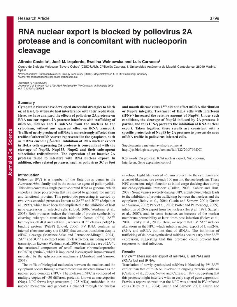
Sarnow, 2002; Park et al., 2008). Thus, we investigated the impactof PV 2Apro on the export of cellular RNAs from nucleus tocytoplasm. First, we analyzed the cellular distribution of de-novo-synthesized luciferase mRNAs in Tet-off HeLa X1/5 cells thatexpress PV 2Apro (Castello et al., 2006a). Luciferase mRNAsynthesis was induced by removal of tetracycline (Tet) from theculture medium (Novoa and Carrasco, 1999), and the expressionof PV 2Apro was achieved by the electroporation of 1 or 9 g of anin-vitro-synthesized mRNA that contained the encephalomyocarditisvirus (EMCV) IRES followed by the PV 2Apro coding sequence(IRES-2A mRNA) (Castello et al., 2006a). At 8 hours post-electroporation (hpe), luciferase activity and the integrity of eIF4GIand eIF4GII were estimated.
Luciferase activity decreased by about 90% in 2Apro-expressingcells compared with control cells, irrespective of the IRES-2AmRNA dose employed (supplementary material Fig. S1A).However, 9 g IRES-2A mRNA was required to strongly inhibitongoing -actin protein synthesis (supplementary material Fig.S1B). As expected, eIF4GI was proteolyzed in cells electroporatedwith 1 g IRES-2A mRNA, whereas eIF4GII remained intact. Bycontrast, electroporation of 9 g IRES-2A mRNA induced totalcleavage of both eIF4GI and eIF4GII (supplementary material Fig.
S1C). To determine whether the decrease in luciferase synthesis in2Apro-expressing cells was due to an inhibition of RNA nuclearexport, nuclear and cytoplasmic fractions were prepared from thedifferent electroporated cells. Total RNA was then isolated fromeach subcellular fraction and the steady-state levels of luciferaseor -actin mRNAs were quantified by real-time reverse transcriptase(RT)-PCR. The effectiveness of fractionation was checked bywestern blot analysis against a cytoplasmic protein such as a-tubulinor a nuclear protein such as poly-ADP-ribose polymerase (PARP).
As expected, a-tubulin was present only in the cytoplasmicfraction, whereas PARP was only detected in the nuclear fraction(supplementary material Fig. S1D). Interestingly, the ~80 KDacleavage product related to caspase-3-mediated cleavage of PARPwas not detected in either case (supplementary material Fig. S1D),suggesting that apoptosis was not induced by the viral protease atthese particular times and doses. The total amount of luciferase and-actin mRNAs was similar in control and in 2Apro-expressing cells,suggesting that transcription was not hampered by the PV protease(Fig. 1A). The distribution of -actin mRNA in nuclear andcytoplasmic fractions was not significantly altered in 2Apro-expressing cells as compared with that in control cells (Fig. 1B).The cytoplasmic to nuclear (C/N) ratio of this mRNA was
Journal of Cell Science 122 (20)
Fig. 1. Nuclear-cytoplasmic distribution of cellular mRNAs, 18S rRNA, U2 snRNA and tRNA-val in PV 2A-expressing cells. HeLa X1/5 cells, incubated with 20ng/ml Tet, were electroporated with either 1 or 9g IRES-2A mRNA, or with transcription buffer as a control. During electroporation, Tet was removed from theculture medium. Total, nuclear and cytoplasmic fractions were obtained in each case at 8 hpe. Luciferase, COX-1, COX-2, IL-6, IL-2, c-myc, p53 and -actinmRNAs and 18S rRNA, U2 snRNA and tRNA-val were quantified in each fraction by real-time RT-PCR using specific oligonucleotides and Taqman probes.(A)Relative amount of each mRNA in the total fraction from control cells (black bars) or cells electroporated with 1 (grey bars) or 9g (white bars) IRES-2AmRNA. (B)Relative quantitation of each mRNA in the nuclear and cytoplasmic fractions. (C)C/N ratio calculated for each mRNA in the presence or absence of2Apro. (D)Relative level of 18S rRNA, U2 snRNA and tRNA-Val in the total fraction of control (black bars) or 2A-expressing cells (grey and white bars).(E)Relative quantitation of 18S rRNA, U2 snRNA and tRNA-Val in nuclear and cytoplasmic fractions. (F)C/N ratio calculated for 18S rRNA, U2 snRNA andtRNA-Val. Values are means ± s.d. *P<0.05, **P<0.01 compared with control, calculated using the Student’s t-test.
Jour
nal o
f Cel
l Sci
ence
3801Poliovirus 2A protein blocks RNA traffic
approximately 2-3 in each case (Fig. 1C). By contrast, thedistribution of luciferase mRNA was strongly altered in 2Apro-expressing cells. Whereas the amount of luciferase mRNA in thecytoplasmic fraction decreased, it increased in the nuclear fraction(Fig. 1B,C). It is worth noting that the C/N ratio of luciferase mRNAwas 9.2% that of -actin in control cells (Fig. 1C). This could bebecause luciferase mRNA is capped and polyadenylated by hostmachinery, but it is not spliced. Hence, this mRNA might beexported by an inefficient pathway that involves the CRM1transporter (Cullen, 2003; Kohler and Hurt, 2007). The C/N ratioof luciferase mRNA decreased from 0.26 to ~0.1, and to ~0.07 incells electroporated with 1 g and 9 g IRES-2A mRNA,respectively (Fig. 1C). These results suggest that nuclear export ofde-novo-synthesized mRNAs is more sensitive to 2Apro than theexport of mRNAs that are over-represented in the cytoplasm.
To rule out the possibility that the CRM1-mediated exportpathway could be more susceptible to 2Apro activity, the subcellulardistribution of different endogenous spliced mRNAs involved inimmunological responses against pathogens [cyclooxygenase(COX)-1, COX-2, interleukin (IL)-2, IL-6], in cell survival anddeath (p53) and in cell proliferation (c-myc) was analyzed in thesecells. As expected, the most abundant mRNA in the total fractionof control cells was -actin, whereas IL-2 mRNA was not detected(supplementary material Fig. S1E). Steady-state levels of IL-6, c-myc and p53 mRNAs increased in cells electroporated with IRES-2A mRNA in a dose-dependent manner (Fig. 1A). Probably, thepresence of PV 2Apro induced a host gene response. Similarly tothe luciferase mRNA, relative levels of COX-1, COX-2, IL-6, c-myc and p53 mRNAs increased in the nuclear fraction and decreasedor, at least, were maintained in the cytoplasmic fraction of cellselectroporated with IRES-2A mRNA (Fig. 1B). Consequently, theC/N ratio of each mRNA diminished in 2Apro-expressing cells inall cases (Fig. 1C). Therefore, 2Apro not only interferes with thenuclear export of luciferase mRNA but also inhibits the traffic ofspliced mRNAs. -actin appears to be insensitive to 2Apro, althoughthe contribution of the half-life of each mRNA might account, atleast in part, for this effect.
rRNAs, snRNAs and tRNAs are exported to the cytoplasm byother pathways than those used to transport mRNAs. To analyze thefunctionality of these RNA export pathways in 2Apro-expressing cells,the distribution of 18S rRNA, U2 snRNA and tRNA-Val werestudied. Expression of 2Apro did not affect the total relative amountof 18S rRNA or tRNA-Val but moderately increased the level of U2snRNA (~1.5-fold) (Fig. 1D). Looking at the subcellular distribution,18S rRNA decreased in the cytoplasm of 2Apro-expressing cells ina dose-dependent manner but accumulated in the nuclear fraction(Fig. 1E). The C/N ratio of rRNA 18S was reduced from 6 to 3.2by the electroporation of 1 or 9 g of IRES-2A mRNA, respectively(Fig. 1F). The relative level of U2 snRNA was maintained in thecytoplasm but increased in the nucleus (Fig. 1E). Consequently, theC/N ratio of U2 snRNA was also reduced by the PV protease in adose-dependent manner (Fig. 1F). By contrast, distribution of tRNA-Val in cytoplasmic and nuclear fractions was not affected by 2Apro
(Fig. 1E,F). Therefore, 2Apro blocks rRNA and U snRNA transportfrom the nucleus to the cytoplasm, whereas the pathway used bytRNA export remains functional.
To analyze the contribution of the half-life of -actin mRNA tothe insensitivity to 2Apro, we added actinomycin D, a potent inhibitorof cellular transcription, to HeLa X1/5 culture cells and analyzedprotein synthesis at 4 and 8 hpe. Transcription was halted by thiscompound because luciferase activity was dramatically reduced in
treated cells (supplementary material Fig. S2A). By contrast,treatment with actinomycin D weakly inhibited -actin synthesis,suggesting that -actin mRNA is very stable in HeLa cells(supplementary material Fig. S2B).
PV 2Apro hampers nuclear export of cellular polyadenylatedmRNAsNext, we decided to assay the impact of PV 2Apro on the export ofbulk cellular mRNAs using in-situ-hybridization assays with afluorescein-labeled oligo d(T) probe against poly(A) tail. Oligo d(A)probe or a mixture of both probes [either 1:1 or 5:1 ratio of oligod(A) to oligo d(T)] was used as a control (supplementary materialFig. S3A). Nuclei and cytoplasm were stained by indirectimmunofluorescence using antibodies raised against Ref-1/Aly anda-tubulin. Fluorescent oligo d(T) localized in the nucleus andcytoplasm of HeLa cells, whereas no fluorescent signal was detectedfrom the oligo d(A) probe (supplementary material Fig. S3A). Asexpected, abrogation of oligo d(T) staining occurred when oligod(A) was added to the hybridization mixture in a dose-dependentmanner, suggesting that exogenous oligo d(A) competes withpoly(A) tail by binding to oligo d(T) (supplementary material Fig.S3A). Therefore, we can conclude that our hybridization assays witholigo d(T) specifically detect polyadenylated mRNAs.
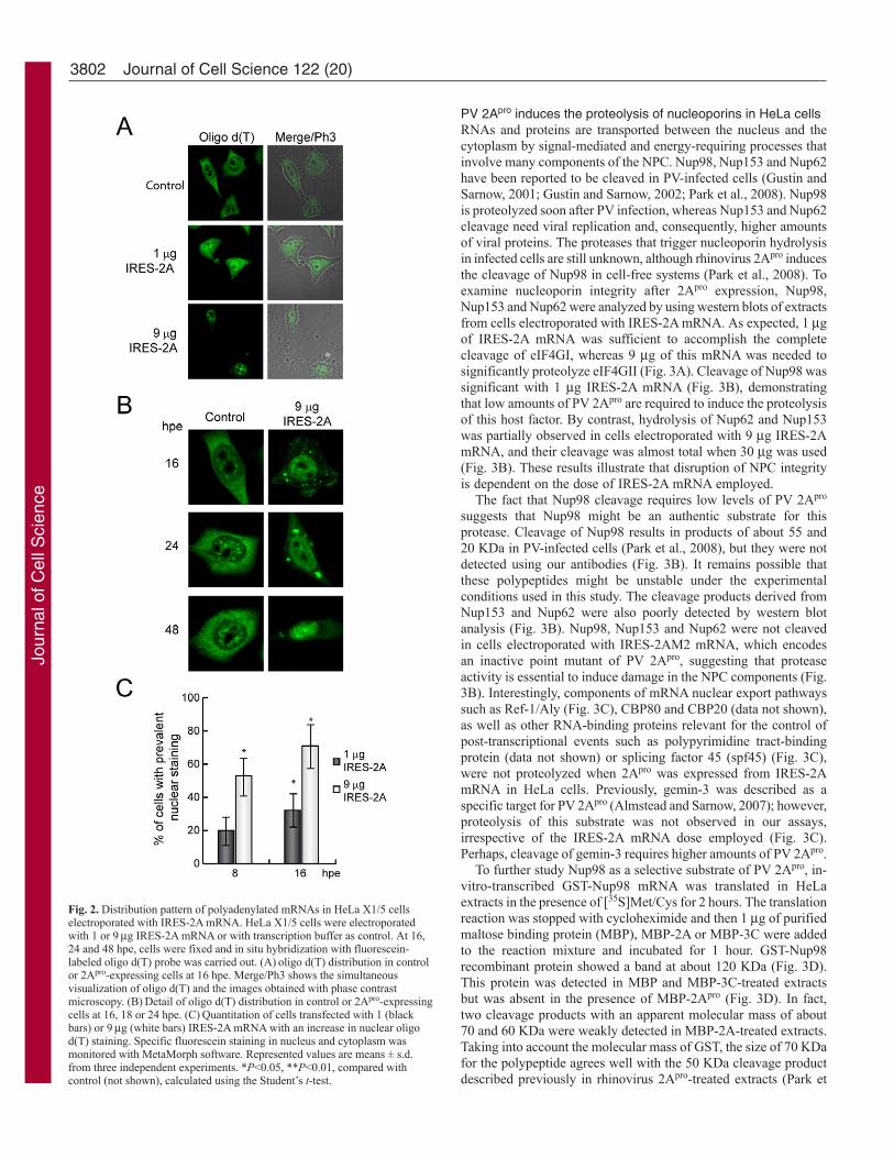
Next, in situ hybridization with oligo d(T) probe was carriedout at 16, 24 and 48 hpe in control cells or cells expressing IRES-2A. In control cells, poly(A)-containing mRNAs localized tobrightly staining foci in the nucleus, which were excluded fromnuclear structures consistent with nucleoli (Fig. 2A,B). Conversely,a diffuse distribution of mRNAs was observed in the cytoplasm(Fig. 2A,B). Nuclear fluorescence increased slightly in cellselectroporated with 1 g IRES-2A mRNA but no significantdifferences were observed in the intensity and the distribution ofhybridized oligo d(T) in the cytoplasm (Fig. 2A). These dataindicate that 2Apro synthesized in low quantities has a moderateeffect on mRNA export. However, cells electroporated with 9 gIRES-2A mRNA exhibited notable differences in the fluorescencepattern.
The evolution of these alterations could progress as follows: (i)dissolution of nuclear foci, which is accompanied by increasedfluorescence throughout the nucleus excluded from nucleoli (Fig.2A,B); (ii) generation of mRNA-containing granules in thecytoplasm, possibly corresponding to stress granules (Fig. 2A,B);(iii) reduction of relative levels of cytoplasmic mRNAs (Fig. 2B);and (iv) chromatin condensation, which is apparent at 24-48 hpewhen To-Pro-3 is used, suggesting the induction of an apoptosisresponse (data not shown). Interestingly, expression of an inactivepoint mutant of PV 2A protease from IRES-2AM2 mRNA (Ventosoet al., 1998) did not affect the distribution of the polyadenylatedpool of cellular mRNAs as compared with that in control HeLacells (supplementary material Fig. S3B). Together, these dataindicate that PV 2Apro activity abrogates mRNA export from thenucleus. Interestingly, a moderate accumulation of mRNAs in thenuclei of PV 2Apro-expressing cells was detected at earlier timepoints (8 hpe), but no effect was observed on mRNA levels in thecytoplasm (supplementary material Fig. S4), which is in agreementwith the findings reported for PV infection at early time-points (Parket al., 2008). These data indicate that PV 2Apro might block thetransport of some induced cellular mRNAs soon after theirexpression, but that there is a weak effect on the distribution ofstable and constitutively synthesized mRNAs over-represented inthe cytoplasm, such as -actin.
Jour
nal o
f Cel
l Sci
ence
3802
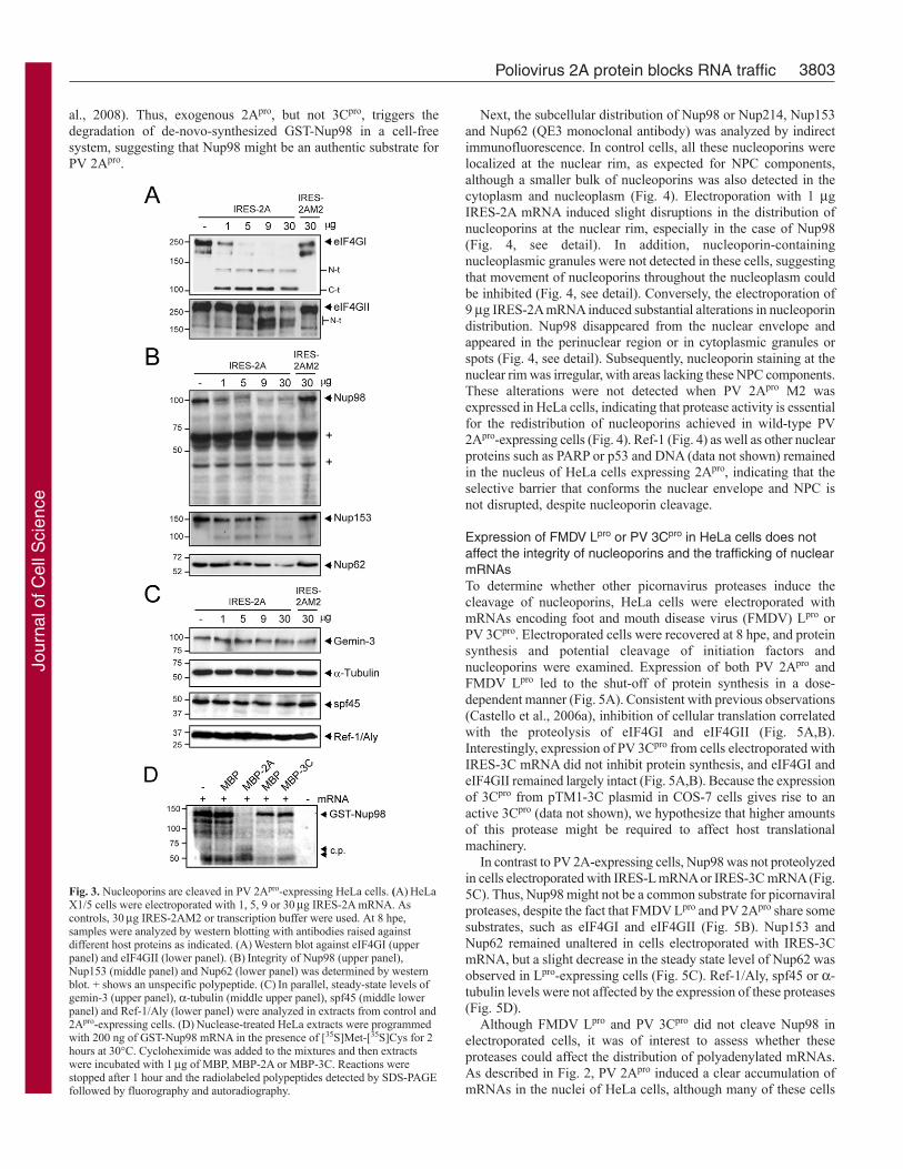
PV 2Apro induces the proteolysis of nucleoporins in HeLa cellsRNAs and proteins are transported between the nucleus and thecytoplasm by signal-mediated and energy-requiring processes thatinvolve many components of the NPC. Nup98, Nup153 and Nup62have been reported to be cleaved in PV-infected cells (Gustin andSarnow, 2001; Gustin and Sarnow, 2002; Park et al., 2008). Nup98is proteolyzed soon after PV infection, whereas Nup153 and Nup62cleavage need viral replication and, consequently, higher amountsof viral proteins. The proteases that trigger nucleoporin hydrolysisin infected cells are still unknown, although rhinovirus 2Apro inducesthe cleavage of Nup98 in cell-free systems (Park et al., 2008). Toexamine nucleoporin integrity after 2Apro expression, Nup98,Nup153 and Nup62 were analyzed by using western blots of extractsfrom cells electroporated with IRES-2A mRNA. As expected, 1 gof IRES-2A mRNA was sufficient to accomplish the completecleavage of eIF4GI, whereas 9 g of this mRNA was needed tosignificantly proteolyze eIF4GII (Fig. 3A). Cleavage of Nup98 wassignificant with 1 g IRES-2A mRNA (Fig. 3B), demonstratingthat low amounts of PV 2Apro are required to induce the proteolysisof this host factor. By contrast, hydrolysis of Nup62 and Nup153was partially observed in cells electroporated with 9 g IRES-2AmRNA, and their cleavage was almost total when 30 g was used(Fig. 3B). These results illustrate that disruption of NPC integrityis dependent on the dose of IRES-2A mRNA employed.
The fact that Nup98 cleavage requires low levels of PV 2Apro
suggests that Nup98 might be an authentic substrate for thisprotease. Cleavage of Nup98 results in products of about 55 and20 KDa in PV-infected cells (Park et al., 2008), but they were notdetected using our antibodies (Fig. 3B). It remains possible thatthese polypeptides might be unstable under the experimentalconditions used in this study. The cleavage products derived fromNup153 and Nup62 were also poorly detected by western blotanalysis (Fig. 3B). Nup98, Nup153 and Nup62 were not cleavedin cells electroporated with IRES-2AM2 mRNA, which encodesan inactive point mutant of PV 2Apro, suggesting that proteaseactivity is essential to induce damage in the NPC components (Fig.3B). Interestingly, components of mRNA nuclear export pathwayssuch as Ref-1/Aly (Fig. 3C), CBP80 and CBP20 (data not shown),as well as other RNA-binding proteins relevant for the control ofpost-transcriptional events such as polypyrimidine tract-bindingprotein (data not shown) or splicing factor 45 (spf45) (Fig. 3C),were not proteolyzed when 2Apro was expressed from IRES-2AmRNA in HeLa cells. Previously, gemin-3 was described as aspecific target for PV 2Apro (Almstead and Sarnow, 2007); however,proteolysis of this substrate was not observed in our assays,irrespective of the IRES-2A mRNA dose employed (Fig. 3C).Perhaps, cleavage of gemin-3 requires higher amounts of PV 2Apro.
To further study Nup98 as a selective substrate of PV 2Apro, in-vitro-transcribed GST-Nup98 mRNA was translated in HeLaextracts in the presence of [35S]Met/Cys for 2 hours. The translationreaction was stopped with cycloheximide and then 1 g of purifiedmaltose binding protein (MBP), MBP-2A or MBP-3C were addedto the reaction mixture and incubated for 1 hour. GST-Nup98recombinant protein showed a band at about 120 KDa (Fig. 3D).This protein was detected in MBP and MBP-3C-treated extractsbut was absent in the presence of MBP-2Apro (Fig. 3D). In fact,two cleavage products with an apparent molecular mass of about70 and 60 KDa were weakly detected in MBP-2A-treated extracts.Taking into account the molecular mass of GST, the size of 70 KDafor the polypeptide agrees well with the 50 KDa cleavage productdescribed previously in rhinovirus 2Apro-treated extracts (Park et
Journal of Cell Science 122 (20)
Fig. 2. Distribution pattern of polyadenylated mRNAs in HeLa X1/5 cellselectroporated with IRES-2A mRNA. HeLa X1/5 cells were electroporatedwith 1 or 9g IRES-2A mRNA or with transcription buffer as control. At 16,24 and 48 hpe, cells were fixed and in situ hybridization with fluorescein-labeled oligo d(T) probe was carried out. (A)oligo d(T) distribution in controlor 2Apro-expressing cells at 16 hpe. Merge/Ph3 shows the simultaneousvisualization of oligo d(T) and the images obtained with phase contrastmicroscopy. (B)Detail of oligo d(T) distribution in control or 2Apro-expressingcells at 16, 18 or 24 hpe. (C)Quantitation of cells transfected with 1 (blackbars) or 9g (white bars) IRES-2A mRNA with an increase in nuclear oligod(T) staining. Specific fluorescein staining in nucleus and cytoplasm wasmonitored with MetaMorph software. Represented values are means ± s.d.from three independent experiments. *P<0.05, **P<0.01, compared withcontrol (not shown), calculated using the Student’s t-test.
Jour
nal o
f Cel
l Sci
ence
3803Poliovirus 2A protein blocks RNA traffic
al., 2008). Thus, exogenous 2Apro, but not 3Cpro, triggers thedegradation of de-novo-synthesized GST-Nup98 in a cell-freesystem, suggesting that Nup98 might be an authentic substrate forPV 2Apro.
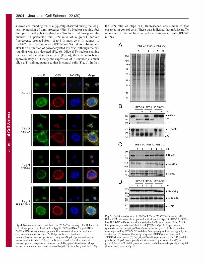
Next, the subcellular distribution of Nup98 or Nup214, Nup153and Nup62 (QE3 monoclonal antibody) was analyzed by indirectimmunofluorescence. In control cells, all these nucleoporins werelocalized at the nuclear rim, as expected for NPC components,although a smaller bulk of nucleoporins was also detected in thecytoplasm and nucleoplasm (Fig. 4). Electroporation with 1 gIRES-2A mRNA induced slight disruptions in the distribution ofnucleoporins at the nuclear rim, especially in the case of Nup98(Fig. 4, see detail). In addition, nucleoporin-containingnucleoplasmic granules were not detected in these cells, suggestingthat movement of nucleoporins throughout the nucleoplasm couldbe inhibited (Fig. 4, see detail). Conversely, the electroporation of9 g IRES-2A mRNA induced substantial alterations in nucleoporindistribution. Nup98 disappeared from the nuclear envelope andappeared in the perinuclear region or in cytoplasmic granules orspots (Fig. 4, see detail). Subsequently, nucleoporin staining at thenuclear rim was irregular, with areas lacking these NPC components.These alterations were not detected when PV 2Apro M2 wasexpressed in HeLa cells, indicating that protease activity is essentialfor the redistribution of nucleoporins achieved in wild-type PV2Apro-expressing cells (Fig. 4). Ref-1 (Fig. 4) as well as other nuclearproteins such as PARP or p53 and DNA (data not shown) remainedin the nucleus of HeLa cells expressing 2Apro, indicating that theselective barrier that conforms the nuclear envelope and NPC isnot disrupted, despite nucleoporin cleavage.
Expression of FMDV Lpro or PV 3Cpro in HeLa cells does notaffect the integrity of nucleoporins and the trafficking of nuclearmRNAsTo determine whether other picornavirus proteases induce thecleavage of nucleoporins, HeLa cells were electroporated withmRNAs encoding foot and mouth disease virus (FMDV) Lpro orPV 3Cpro. Electroporated cells were recovered at 8 hpe, and proteinsynthesis and potential cleavage of initiation factors andnucleoporins were examined. Expression of both PV 2Apro andFMDV Lpro led to the shut-off of protein synthesis in a dose-dependent manner (Fig. 5A). Consistent with previous observations(Castello et al., 2006a), inhibition of cellular translation correlatedwith the proteolysis of eIF4GI and eIF4GII (Fig. 5A,B).Interestingly, expression of PV 3Cpro from cells electroporated withIRES-3C mRNA did not inhibit protein synthesis, and eIF4GI andeIF4GII remained largely intact (Fig. 5A,B). Because the expressionof 3Cpro from pTM1-3C plasmid in COS-7 cells gives rise to anactive 3Cpro (data not shown), we hypothesize that higher amountsof this protease might be required to affect host translationalmachinery.
In contrast to PV 2A-expressing cells, Nup98 was not proteolyzedin cells electroporated with IRES-L mRNA or IRES-3C mRNA (Fig.5C). Thus, Nup98 might not be a common substrate for picornaviralproteases, despite the fact that FMDV Lpro and PV 2Apro share somesubstrates, such as eIF4GI and eIF4GII (Fig. 5B). Nup153 andNup62 remained unaltered in cells electroporated with IRES-3CmRNA, but a slight decrease in the steady state level of Nup62 wasobserved in Lpro-expressing cells (Fig. 5C). Ref-1/Aly, spf45 or a-tubulin levels were not affected by the expression of these proteases(Fig. 5D).
Although FMDV Lpro and PV 3Cpro did not cleave Nup98 inelectroporated cells, it was of interest to assess whether theseproteases could affect the distribution of polyadenylated mRNAs.As described in Fig. 2, PV 2Apro induced a clear accumulation ofmRNAs in the nuclei of HeLa cells, although many of these cells
Fig. 3. Nucleoporins are cleaved in PV 2Apro-expressing HeLa cells. (A)HeLaX1/5 cells were electroporated with 1, 5, 9 or 30g IRES-2A mRNA. Ascontrols, 30g IRES-2AM2 or transcription buffer were used. At 8 hpe,samples were analyzed by western blotting with antibodies raised againstdifferent host proteins as indicated. (A)Western blot against eIF4GI (upperpanel) and eIF4GII (lower panel). (B)Integrity of Nup98 (upper panel),Nup153 (middle panel) and Nup62 (lower panel) was determined by westernblot. + shows an unspecific polypeptide. (C)In parallel, steady-state levels ofgemin-3 (upper panel), a-tubulin (middle upper panel), spf45 (middle lowerpanel) and Ref-1/Aly (lower panel) were analyzed in extracts from control and2Apro-expressing cells. (D)Nuclease-treated HeLa extracts were programmedwith 200 ng of GST-Nup98 mRNA in the presence of [35S]Met-[35S]Cys for 2hours at 30°C. Cycloheximide was added to the mixtures and then extractswere incubated with 1g of MBP, MBP-2A or MBP-3C. Reactions werestopped after 1 hour and the radiolabeled polypeptides detected by SDS-PAGEfollowed by fluorography and autoradiography.
Jour
nal o
f Cel
l Sci
ence
3804
showed cell rounding that is a typically observed during the long-term expression of viral proteases (Fig. 6). Nuclear staining focidisappeared and polyadenylated mRNAs localized throughout thenucleus. In particular, the C/N ratio of oligo-d(T)-derivedfluorescence dropped from ~2 to 1 in most cells. In contrast toPV2Apro, electroporation with IRES-L mRNA did not substantiallyalter the distribution of polyadenylated mRNAs, although the cellrounding was also detected (Fig. 6). Oligo d(T) nuclear stainingfoci were observed in these cells (Fig. 6), the C/N ratio beingapproximately 1.7. Finally, the expression of 3C induced a similaroligo d(T) staining pattern to that in control cells (Fig. 6). In fact,
the C/N ratio of oligo d(T) fluorescence was similar to thatobserved in control cells. These data indicated that mRNA trafficseems not to be inhibited in cells electroporated with IRES-LmRNA.
Journal of Cell Science 122 (20)
Fig. 4. Nucleoporins are redistributed in PV 2Apro-expressig cells. HeLa X1/5cells electroporated with either 1 or 9g IRES-2A mRNA, 9g of IRES-2AM2 mRNA or with transcription buffer as a control, were seeded afterelectroporation on coverslips. At 16 hpe, cells were fixed andimmunofluorescence was performed using anti-Nup98 (green) and mousemonoclonal antibody QE5 (red). Cells were visualized with a confocalmicroscope and images were processed with Huygens 3.0 software. Mergeshows the simultaneous visualization of Nup98, QE5 antibody and Ref-1/Aly.
Fig. 5. Nup98 remains intact in FMDV Lpro- or PV 3Cpro-expressing cells.HeLa X1/5 cells were electroporated with either 1 or 9g of IRES-2A, IRES-L or IRES-3C mRNAs or with transcription buffer as a control. From 7 to 8hpe, protein synthesis was labeled with [35S]Met/Cys. At 8 hpe, proteinsynthesis and the integrity of host factors were analyzed. (A)Total proteinswere separated by SDS-PAGE and then fluorography and autoradiography wascarried out. (B)Western blot analysis against eIF4GI (upper panel) andeIF4GII (lower panel). (C)Integrity of Nup98 (upper panel), Nup153 (middlepanel) and Nup62 (lower panel) was determined by western blot. (D)Inparallel, levels of Ref-1/Aly (upper panel), a-tubulin (middle panel) and spf45(lower panel) were analyzed.
Jour
nal o
f Cel
l Sci
ence
3805Poliovirus 2A protein blocks RNA traffic
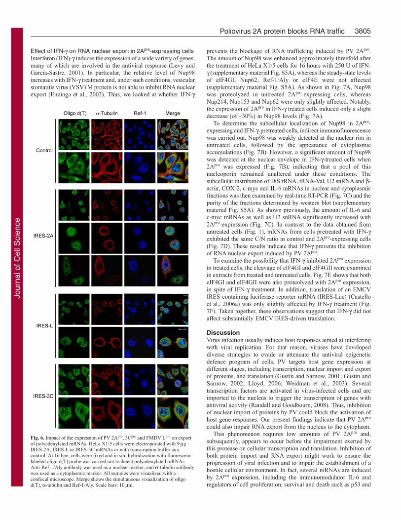
Effect of IFN- on RNA nuclear export in 2Apro-expressing cellsInterferon (IFN)- induces the expression of a wide variety of genes,many of which are involved in the antiviral response (Levy andGarcia-Sastre, 2001). In particular, the relative level of Nup98increases with IFN- treatment and, under such conditions, vesicularstomatitis virus (VSV) M protein is not able to inhibit RNA nuclearexport (Enninga et al., 2002). Thus, we looked at whether IFN-
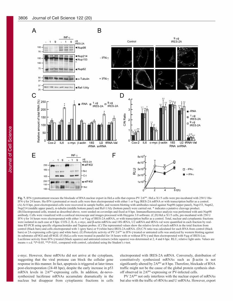
prevents the blockage of RNA trafficking induced by PV 2Apro.The amount of Nup98 was enhanced approximately threefold afterthe treatment of HeLa X1/5 cells for 16 hours with 250 U of IFN- (supplementary material Fig. S5A), whereas the steady-state levelsof eIF4GI, Nup62, Ref-1/Aly or eIF4E were not affected(supplementary material Fig. S5A). As shown in Fig. 7A, Nup98was proteolyzed in untreated 2Apro-expressing cells, whereasNup214, Nup153 and Nup62 were only slightly affected. Notably,the expression of 2Apro in IFN- treated cells induced only a slightdecrease (of ~30%) in Nup98 levels (Fig. 7A).
To determine the subcellular localization of Nup98 in 2Apro-expressing and IFN--pretreated cells, indirect immunofluorescencewas carried out. Nup98 was weakly detected at the nuclear rim inuntreated cells, followed by the appearance of cytoplasmicaccumulations (Fig. 7B). However, a significant amount of Nup98was detected at the nuclear envelope in IFN--treated cells when2Apro was expressed (Fig. 7B), indicating that a pool of thisnucleoporin remained unaltered under these conditions. Thesubcellular distribution of 18S rRNA, tRNA-Val, U2 snRNA and -actin, COX-2, c-myc and IL-6 mRNAs in nuclear and cytoplasmicfractions was then examined by real-time RT-PCR (Fig. 7C) and thepurity of the fractions determined by western blot (supplementarymaterial Fig. S5A). As shown previously, the amount of IL-6 andc-myc mRNAs as well as U2 snRNA significantly increased with2Apro-expression (Fig. 7C). In contrast to the data obtained fromuntreated cells (Fig. 1), mRNAs from cells pretreated with IFN-exhibited the same C/N ratio in control and 2Apro-expressing cells(Fig. 7D). These results indicate that IFN- prevents the inhibitionof RNA nuclear export induced by PV 2Apro.
To examine the possibility that IFN- inhibited 2Apro expressionin treated cells, the cleavage of eIF4GI and eIF4GII were examinedin extracts from treated and untreated cells. Fig. 7E shows that botheIF4GI and eIF4GII were also proteolyzed with 2Apro expression,in spite of IFN- treatment. In addition, translation of an EMCVIRES containing luciferase reporter mRNA (IRES-Luc) (Castelloet al., 2006a) was only slightly affected by IFN- treatment (Fig.7F). Taken together, these observations suggest that IFN- did notaffect substantially EMCV IRES-driven translation.
DiscussionVirus infection usually induces host responses aimed at interferingwith viral replication. For that reason, viruses have developeddiverse strategies to evade or attenuate the antiviral epigeneticdefence program of cells. PV targets host gene expression atdifferent stages, including transcription, nuclear import and exportof proteins, and translation (Gustin and Sarnow, 2001; Gustin andSarnow, 2002; Lloyd, 2006; Weidman et al., 2003). Severaltranscription factors are activated in virus-infected cells and areimported to the nucleus to trigger the transcription of genes withantiviral activity (Randall and Goodbourn, 2008). Thus, inhibitionof nuclear import of proteins by PV could block the activation ofhost gene responses. Our present findings indicate that PV 2Apro
could also impair RNA export from the nucleus to the cytoplasm.This phenomenon requires low amounts of PV 2Apro and,
subsequently, appears to occur before the impairment exerted bythis protease on cellular transcription and translation. Inhibition ofboth protein import and RNA export might work to ensure theprogression of viral infection and to impair the establishment of ahostile cellular environment. In fact, several mRNAs are inducedby 2Apro expression, including the immunomodulator IL-6 andregulators of cell proliferation, survival and death such as p53 and
Fig. 6. Impact of the expression of PV 2Apro, 3Cpro and FMDV Lpro on exportof polyadenylated mRNAs. HeLa X1/5 cells were electroporated with 9gIRES-2A, IRES-L or IRES-3C mRNAs or with transcription buffer as acontrol. At 16 hpe, cells were fixed and in situ hybridization with fluorescein-labeled oligo d(T) probe was carried out to detect polyadenylated mRNAs.Anti-Ref-1/Aly antibody was used as a nuclear marker, and a-tubulin antibodywas used as a cytoplasmic marker. All samples were visualized with aconfocal microscope. Merge shows the simultaneous visualization of oligod(T), a-tubulin and Ref-1/Aly. Scale bars: 10m.
Jour
nal o
f Cel
l Sci
ence
3806
c-myc. However, these mRNAs did not arrive at the cytoplasm,suggesting that the viral protease can block the cellular generesponse in this manner. In fact, apoptosis is triggered at later timespost-electroporation (24-48 hpe), despite the early increase in p53mRNA levels in 2Apro-expressing cells. In addition, de-novo-synthesized luciferase mRNAs accumulate dramatically in thenucleus but disappear from cytoplasmic fractions in cells
electroporated with IRES-2A mRNA. Conversely, distribution ofconstitutively synthesized mRNAs such as -actin is notsignificantly altered by 2Apro at 8 hpe. Therefore, blockade of RNAtraffic might not be the cause of the global protein synthesis shut-off observed in 2Apro-expressing or PV-infected cells.
PV 2Apro not only interferes with the nuclear export of mRNAsbut also with the traffic of rRNAs and U snRNAs. However, export
Journal of Cell Science 122 (20)
Fig. 7. IFN- pretreatment rescues the blockade of RNA nuclear export in HeLa cells that express PV 2Apro. HeLa X1/5 cells were pre-incubated with 250 U Hu-IFN- for 24 hours. Hu-IFN- pretreated or mock cells were then electroporated with either 1 or 9g IRES-2A mRNA or with transcription buffer as a control.(A)At 8 hpe, post-electroporated cells were recovered in sample buffer, and western blotting with antibodies raised against Nup98 (upper panel), Nup153, Nup62,Nup214 (middle upper panel), a-tubulin (middle bottom panel) and Ref-1/Aly (bottom panel) were carried out. * indicates a putative cleavage product.(B)Electroporated cells, treated as described above, were seeded on coverslips and fixed at 8 hpe. Immunofluorescence analysis was performed with anti-Nup98antibody. Cells were visualized with a confocal microscope and images processed with Huygens 3.0 software. (C,D)HeLa X1/5 cells, pre-incubated with 250 UIFN- for 16 hours were electroporated with either 1 or 9g of IRES-2A mRNA, or with transcription buffer as a control. Total, nuclear and cytoplasmic fractionswere isolated in each case at 8 hpe. COX-2, IL-6, c-myc, and -actin mRNAs and 18S rRNA, U2 snRNA and tRNA-val were quantified in each fraction by real-time RT-PCR using specific oligonucleotides and Taqman probes. (C)The represented values show the relative levels of each mRNA in the total fraction fromcontrol (black bars) and cells electroporated with 1 (grey bars) or 9 (white bars) IRES-2A mRNA. (D)C/N ratio was calculated for each RNA from control (blackbars) or 2A-expressing cells (grey and white bars). (E)Proteolytic activity of PV 2Apro in IFN- treated or untreated cells was analyzed by western blotting againstits substrates eIF4GI and eIF4GII. (F)HeLa cells were treated in parallel for 16 hours with or without IFN- and then electroporated with 9g of IRES-Luc.Luciferase activity from IFN- treated (black squares) and untreated extracts (white squares) was determined at 2, 4 and 6 hpe. RLU, relative light units. Values aremeans ± s.d. *P<0.05, **P<0.01, compared with control, calculated using the Student’s t-test.
Jour
nal o
f Cel
l Sci
ence
3807Poliovirus 2A protein blocks RNA traffic
of tRNAs from the nucleus is not affected by the PV protease. Asimilar result was observed for vesicular stomatitis virus (VSV) Mprotein, suggesting a common target for both viral proteins (Her etal., 1997; Petersen et al., 2000). Nevertheless, the possible role ofthe inhibition of rRNA and U snRNA trafficking in PV infectionneeds further investigation. We can speculate that both the proteinsynthesizing machinery and/or the splicing activity might also beaffected.
Exchange of macromolecules between the nucleus and cytoplasmtakes place exclusively through the NPC. In fact, each NPC is ableto orchestrate hundreds of transport events per minute (Ball andUllman, 2005). This abundant selective traffic makes the NPC akey target during viral infections because the virus aims to blockthe transport of RNAs and proteins between these two subcellularcompartments. 2Apro targets the NPC directly or indirectly bycleaving different nucleoporins such as Nup153, Nup62 andspecially Nup98. Immunofluorescence assays revealed that theamount of the three nucleoporins recognized by QE5 antibody(Nup153, Nup62 and Nup214) diminished in 2Apro-expressing cells,whereas Nup98 disappeared from the nuclear rim and wasrelocalized in the cytoplasm. These nucleoporins are essentialcomponents of the NPC and mediate import and export of proteinsand RNAs through the NPC by means of interactions with crucialcomponents of the different traffic pathways (Galy et al., 2003;Harborth et al., 2001). For example, Nup153 binds importin-5/Kap3/RanBP5, importin-7/RanBP7, exportin-5, exportin-t,exportin-1, NXF1/Tap (the export receptor of mRNAs), Impa2 andNTF2 (the import receptor of Ran) (Ball and Ullman, 2005).Furthermore, Nup98 interacts with Rae-1, an adaptor for NXF1/Tap-dependent export (Pritchard et al., 1999).
Interaction domains in nucleoporins are related to an unfoldedsubset of glycosylated FG (phenylalanine-glycine) repeats locatedat the C-terminal portion of the protein. The relevance of this domainin RNA export was demonstrated using specific antibodies(Featherstone et al., 1988; Neuman de Vegvar and Dahlberg, 1990;Terns and Dahlberg, 1994). Therefore, alteration of the NPC by PV2Apro might uncouple export and import machinery from the NPC.In this regard, VSV M protein also targets the NPC. This viralprotein interacts with Rae-1/mrnp41 and with Nup98 at the FGdomain, inhibiting cellular RNA export (Faria et al., 2005; vonKobbe et al., 2000). In addition, influenza virus NS1 protein formsan inhibitory complex with NXF1/TAP, p15/NXT, Rae-1/mrnp41and E1B-AP5 and downregulates Nup98 (Satterly et al., 2007).Analogies between VSV M, influenza virus, NS1 and PV 2Apro
could point to the NPC as an important target for several nuclearand cytoplasmic viruses.
Nup98 and Nup153 are mobile components of the NPC and theirdynamics are dependent on active transcription, suggesting a linkbetween RNA cargo formation and the status of ongoing porereconfiguration. In particular, mobility of Nup98 and Nup153 arerelated to RNA polymerase (Pol) I and II activity (Griffis et al.,2002; Griffis et al., 2004). Given that Pol I transcribes long rRNAs,Pol II synthesizes U snRNAs and mRNAs, and Pol III generatestRNAs, Nup98 and Nup153 might be involved in export of rRNAs,U snRNAs and mRNAs but not of tRNAs (Kohler and Hurt, 2007).For that reason, tRNAs might be insensitive to the cleavage ofnucleoporins (Fig. 1D-F). Alternatively, exportin-t, the transporterinvolved in tRNA export (Cullen, 2003; Kohler and Hurt, 2007),could work despite damage to the NPC induced by PV 2Apro orVSV M. Cleavage of nucleoporins Nup153, Nup62 and Nup98 waspreviously observed in PV and rhinovirus-infected cells (Gustin and
Sarnow, 2001; Gustin and Sarnow, 2002; Park et al., 2008).However, the protease(s) involved in NPC damage is still unknown.
Interestingly, alterations in the NPC induced in vitro by PV areblocked with PV 2Apro inhibitors such as elastatinal, elastase andMPCMK (Belov et al., 2004). These data strongly suggest that PV2Apro is directly or indirectly responsible for nucleoporin cleavage.Caspase-3 and/or caspase-9 can also hydrolyze Nup153, renderinga cleavage product similar to that observed in PV 2Apro-expressingcells. However, Nup62 is not proteolyzed by caspases in HeLa cellsafter induction of apoptosis (Buendia et al., 1999). Moreover, PVinfection also alters the NPC in cells lacking active caspase-3 andcaspase-9 (Belov et al., 2004). These results indicate that theseapoptosis executors are not involved in the proteolysis ofnucleoporins in PV-infected cells. Our data indicate that expressionof PV 2Apro alone is sufficient to induce nucleoporin cleavage intransfected HeLa cells or in cell-free systems. By contrast,expression of an inactive mutant of PV 2Apro did not affect theintegrity of nucleoporins. In addition, electroporation of IRES-3CmRNA (which encodes 3Cpro, the other protease of PV) did notinduce the proteolysis of nucleoporins nor the inhibition of mRNAtraffic. Taking these results together, we hypothesize that PV 2Apro
is the protease involved in directly or indirectly targeting the NPCduring PV infection. Interestingly, other related proteases, such asFMDV Lpro, share with PV 2Apro some substrates such as eIF4GIand eIF4GII but both proteases differ in their ability to cleaveNup98. Thus, low doses of PV 2Apro (1 g IRES-2A mRNA) giverise to the cleavage of Nup98, whereas this protein remainsunaltered after FMDV Lpro expression. Consequently, FMDV Lpro
did not affect substantially the distribution of the pool ofpolyadenylated mRNAs. Curiously, L protein from EMCV inducesthe phosphorylation of nucleoporins, leading to the inactivation ofnucleus-cytoplasm traffic of macromolecules (Porter et al., 2006;Porter and Palmenberg, 2009). However, we could not observe theseeffects after expression of FMDV Lpro.
IFN- triggers a host response against viral infection (Randalland Goodbourn, 2008). Strikingly, two components of the RNAnuclear export machinery, Nup98 and Rae-1, are overexpressed incells treated with IFN- (Enninga et al., 2002; Faria et al., 2005).These data suggest that RNA and protein trafficking through theNPC is a key target for successful infection of cytopathic viruses.In fact, inhibition of nuclear RNA export by VSV M is avoidedafter treatment with IFN-, perhaps due to the overproduction ofNup98 and Rae-1 (Enninga et al., 2002; Faria et al., 2005). Inaddition, we have also observed a specific increase in the Nup98steady-state level in HeLa cells pre-incubated with 250 U IFN-.Under these experimental conditions, Nup98 was partiallyproteolyzed in 2Apro-expressing cells and a substantial poolremained at the nuclear membrane. Nevertheless, the amount ofNup153 is not increased when HeLa cells are treated with IFN-.These data point to the idea that Nup98 might be an essentialcomponent of the nucleus-cytoplasm traffic machinery. Theineffective cleavage of Nup98 in IFN--treated cells is consistentwith the restoration of nuclear-cytoplasmic RNA trafficking.Collectively, these findings suggest that IFN- is an important hostfactor in preventing alterations in cellular gene expression inducedby viral infections. Therefore, secretion of IFN- from immune cellsmight induce an effective gene response against virus infection incompetent neighbouring cells. Future efforts will focus ondetermining whether cellular components involved in the RNAexport machinery are also targeted by other proteins from othercytopathic viruses.
Jour
nal o
f Cel
l Sci
ence
3808
Materials and MethodsCell culturesHela X1/5 cells were previously described (Novoa and Carrasco, 1999). Hu-IFN-was generously provided by Ali Alejo and Antonio Alcamí (Centro de BiologíaMolecular ‘Severo Ochoa’, Madrid, Spain).
Plasmids, in vitro transcription and RNA transfectionThe plasmids pTM1-2A, pTM1-2AM2, pTM1-3C and pTM1-L were used astemplates to synthesize in vitro IRES-2A, IRES-2AM2, IRES-3C and IRES-LmRNAs, respectively. The in vitro transcription reaction and transfection of resultingmRNAs were carried out as described previously (Castello et al., 2006a). pGEX-4T-Nup98 CBP20 (a generous gift from Elisa Izaurralde, Max Planck Institute forDevelopmental Biology, Tübingen, Germany) was previously described (von Kobbeet al., 2000).
Protein synthesis, subcellular fractionation, western blot andluciferase activity analysesDetermination of protein synthesis was performed by metabolic labeling with 50 Ciof [35S]Met-[35S]Cys/ml (Promix; Amersham Biosciences) for 1 hour, followed bySDS-PAGE, fluorography and autoradiography. Subcellular fractions were obtainedusing a buffer containing NP-40, RNaseOUT (Invitrogen) and a cocktail of proteaseinhibitors (Sigma-Aldrich) as described previously (Perales et al., 2003). eIF4GI wasdetected with antisera raised against peptides derived from the N-terminal and C-terminal, at 1:1000 dilution (Aldabe et al., 1995). Rabbit antisera against N-terminaland C-terminal region of eIF4GII (a generous gift from Nahum Sonenberg, McGillUniversity, Montreal, Canada) were employed at 1:500 dilution. Mouse antibodyagainst PARP (BD Pharmigen) was used at 1:250 dilution. Mouse antibodies againstPABP, gemin-3 (Abcam), PTB, a-tubulin (Sigma) were used at 1:200, 1:1000, 1:1000,and 1:5000 dilutions, respectively. Rabbit antisera against Ref-1/Aly, CBP80, CBP20(a generous gift from Elisa Izaurralde, Max Planck Institute for DevelopmentalBiology, Tübingen, Germany) and spf45 (a generous gift from Juan Valcárcel, Centrefor Genomic Regulation, Barcelona, Spain) were used at 1:1000 dilution. Mousemonoclonal antibody [QE5] (Abcam) was used at 1:1000 dilution for western blotanalysis and 1:50 for immunofluorescence. Nup98 was detected inimmunofluorescence with rat monoclonal antibody at 1:100 dilution (a generous giftfrom Maureen A. Powers, Emory University School of Medicine, Atlanta, GA), andwith rabbit antisera (Abcam) for western blot analysis at 1:500 dilution. Anti-rabbit(Amersham) and anti-mouse (Promega) immunoglobulin G antibodies coupled toperoxidase were used at 1:5000 dilution. The percentage of protein synthesis and thepercentage of intact proteins were determined by densitometric scanning. Measurementof luciferase activity was determined with the Luciferase Assay System kit (Promega)with a Monolight 2010 apparatus (Analytical Luminescence Laboratory, San Diego,CA).
In vitro translationThe HeLa S3 extracts and the translation reaction mix were obtained as previouslydescribed (Franco et al., 2005) and was kindly provided by David Franco (AaronDiamond AIDS Research Center, The Rockefeller University, New York, NY). GST-Nup98 cleavage was analyzed by metabolic labeling with 50 Ci of [35S]Met-[35S]Cys/ml (Promix; Amersham Biosciences), followed by SDS-PAGE, fluorographyand autoradiography.
Purification of recombinant proteinsThe chimeric MBP (maltose-binding protein), MBP-2Apro and MBP-3Cpro werepurified by affinity chromatography, as described previously (Novoa and Carrasco,1999).
Real-time RT-PCR analysesTotal RNA was isolated from the different fractions using the kit RNAspin mini (GEHealthcare). Analysis of -actin, luciferase, U2 snRNA and 18S rRNA levels wasperformed as described previously (Castello et al., 2006a). IL-6, COX-1, COX-2, c-myc and p53 mRNAs were quantified as described previously using primers andprobes designed by Applied Biosystems (a generous gift from Manuel Fresno, Centrode Biología Molecular ‘Severo Ochoa’, Madrid, Spain). Real-time RT-PCR witholigonucleotides designed against tRNA-Val (Roche) was carried out using MasterSYBR Green I Kit (Roche) (Castello et al., 2006b). Data analysis was carried outusing the SDS-7000 software (Version 1.1).
Immunofluorescence microscopy and FISH assayFixation, permeabilization and confocal microscopy were performed as describedpreviously (Madan et al., 2008), employing a confocal LSM510 lens coupled to anAxiovert 200 M microscope (Zeiss). Image processing was performed with Huygens3.0 software. Detection of polyadenylated mRNAs by fluorescence in situhybridization (FISH) was carried out using fluorescein-labeled oligo d(T) or oligod(A) probes (Gene link). Cells were fixed and permeabilized and then washed threetimes: first with 1� phosphate-buffered saline (PBS), then with 1� PBS and 1�saline-sodium citrate buffer (SSC), and finally with 2� SSC. Next, cells wereincubated at 37°C with pre-hybridation buffer (2� SSC, 20% deionized formamide,
0.2% BSA and 1 mg/ml yeast tRNA). Afterwards, cells were incubated at 37°C for4 hours with hybridization buffer (2� SSC, 20% deionized formamide, 0.2% BSA,1 mg/ml yeast tRNA, 10% dextran sulphate and 1 pmol/l oligo d(T) probe).Preparations were washed four times at 42°C for 5 minutes: the first wash wasperformed with 2� SSC mixed with 20% formamide; the second with 2� SSC; thethird with 1� SSC and 1� PBS; and the last with 1� PBS. FISH was carried outusing the immunofluorescence protocol described previously (Madan et al., 2008).
This study was supported by a DGICYT Grant (BFU 2006-02182)and an institutional grant awarded to the Centro de Biología Molecular‘Severo Ochoa’ by the Fundación Ramón Areces. We thank AntonioAlcamí, Ali Alejo, David Franco, Elisa Izaurralde, Manuel Fresno,Maureen A. Powers, Nahum Sonenberg and Juan Valcárcel for someof the antibodies and reagents used in this study. We are very gratefulfor the help of Aurelie Rakotondrafara in the preparation of themanuscript.
ReferencesAldabe, R., Feduchi, E., Novoa, I. and Carrasco, L. (1995). Efficient cleavage of p220
by poliovirus 2Apro expression in mammalian cells: effects on vaccinia virus. Biochem.Biophys. Res. Commun. 215, 928-936.
Almstead, L. L. and Sarnow, P. (2007). Inhibition of U snRNP assembly by a virus-encoded proteinase. Genes Dev. 21, 1086-1097.
Ball, J. R. and Ullman, K. S. (2005). Versatility at the nuclear pore complex: lessonslearned from the nucleoporin Nup153. Chromosoma 114, 319-330.
Belov, G. A., Lidsky, P. V., Mikitas, O. V., Egger, D., Lukyanov, K. A., Bienz, K. andAgol, V. I. (2004). Bidirectional increase in permeability of nuclear envelope uponpoliovirus infection and accompanying alterations of nuclear pores. J. Virol. 78, 10166-10177.
Buendia, B., Santa-Maria, A. and Courvalin, J. C. (1999). Caspase-dependent proteolysisof integral and peripheral proteins of nuclear membranes and nuclear pore complexproteins during apoptosis. J. Cell Sci. 112, 1743-1753.
Castello, A., Alvarez, E. and Carrasco, L. (2006a). Differential cleavage of eIF4GI andeIF4GII in mammalian cells. Effects on translation. J. Biol. Chem. 281, 33206-33216.
Castello, A., Sanz, M. A., Molina, S. and Carrasco, L. (2006b). Translation of Sindbisvirus 26S mRNA does not require intact eukariotic initiation factor 4G. J. Mol. Biol.355, 942-956.
Cullen, B. R. (2003). Nuclear RNA export. J. Cell Sci. 116, 587-597.Enninga, J., Levy, D. E., Blobel, G. and Fontoura, B. M. (2002). Role of nucleoporin
induction in releasing an mRNA nuclear export block. Science 295, 1523-1525.Faria, P. A., Chakraborty, P., Levay, A., Barber, G. N., Ezelle, H. J., Enninga, J.,
Arana, C., van Deursen, J. and Fontoura, B. M. (2005). VSV disrupts the Rae1/mrnp41mRNA nuclear export pathway. Mol. Cell 17, 93-102.
Featherstone, C., Darby, M. K. and Gerace, L. (1988). A monoclonal antibody againstthe nuclear pore complex inhibits nucleocytoplasmic transport of protein and RNA invivo. J. Cell Biol. 107, 1289-1297.
Franco, D., Pathak, H. B., Cameron, C. E., Rombaut, B., Wimmer, E. and Paul, A.V. (2005). Stimulation of poliovirus RNA synthesis and virus maturation in a HeLa cell-free in vitro translation-RNA replication system by viral protein 3CDpro. Virol. J. 2, 86.
Galy, V., Mattaj, I. W. and Askjaer, P. (2003). Caenorhabditis elegans nucleoporins Nup93and Nup205 determine the limit of nuclear pore complex size exclusion in vivo. Mol.Biol. Cell 14, 5104-5415.
Griffis, E. R., Altan, N., Lippincott-Schwartz, J. and Powers, M. A. (2002). Nup98 isa mobile nucleoporin with transcription-dependent dynamics. Mol. Biol. Cell 13, 1282-1297.
Griffis, E. R., Craige, B., Dimaano, C., Ullman, K. S. and Powers, M. A. (2004). Distinctfunctional domains within nucleoporins Nup153 and Nup98 mediate transcription-dependent mobility. Mol. Biol. Cell 15, 1991-2002.
Gustin, K. E. and Sarnow, P. (2001). Effects of poliovirus infection on nucleo-cytoplasmictrafficking and nuclear pore complex composition. EMBO J. 20, 240-249.
Gustin, K. E. and Sarnow, P. (2002). Inhibition of nuclear import and alteration of nuclearpore complex composition by rhinovirus. J. Virol. 76, 8787-8796.
Harborth, J., Elbashir, S. M., Bechert, K., Tuschl, T. and Weber, K. (2001). Identificationof essential genes in cultured mammalian cells using small interfering RNAs. J. CellSci. 114, 4557-4565.
Her, L. S., Lund, E. and Dahlberg, J. E. (1997). Inhibition of Ran guanosinetriphosphatase-dependent nuclear transport by the matrix protein of vesicular stomatitisvirus. Science 276, 1845-1848.
Kohler, A. and Hurt, E. (2007). Exporting RNA from the nucleus to the cytoplasm. Nat.Rev. Mol. Cell Biol. 8, 761-773.
Levy, D. E. and Garcia-Sastre, A. (2001). The virus battles: IFN induction of the antiviralstate and mechanisms of viral evasion. Cytokine Growth Factor Rev. 12, 143-156.
Lidsky, P. V., Hato, S., Bardina, M. V., Aminev, A. G., Palmenberg, A. C., Sheval, E.V., Polyakov, V. Y., van Kuppeveld, F. J. and Agol, V. I. (2006). Nucleocytoplasmictraffic disorder induced by cardioviruses. J. Virol. 80, 2705-2717.
Lloyd, R. E. (2006). Translational control by viral proteinases. Virus Res. 119, 76-88.Madan, V., Castello, A. and Carrasco, L. (2008). Viroporins from RNA viruses induce
caspase-dependent apoptosis. Cell Microbiol. 10, 437-451.Martinez-Salas, E. and Fernandez-Miragall, O. (2004). Picornavirus IRES: structure
function relationship. Curr. Pharm. Des. 10, 3757-3767.
Journal of Cell Science 122 (20)
Jour
nal o
f Cel
l Sci
ence
3809Poliovirus 2A protein blocks RNA traffic
Neuman de Vegvar, H. E. and Dahlberg, J. E. (1990). Nucleocytoplasmic transport andprocessing of small nuclear RNA precursors. Mol. Cell. Biol. 10, 3365-3375.
Novoa, I. and Carrasco, L. (1999). Cleavage of eukaryotic translation initiation factor4G by exogenously added hybrid proteins containing poliovirus 2Apro in HeLa cells:effects on gene expression. Mol. Cell. Biol. 19, 2445-2454.
Park, N., Katikaneni, P., Skern, T. and Gustin, K. E. (2008). Differential targeting ofnuclear pore complex proteins in poliovirus-infected cells. J. Virol. 82, 1647-1655.
Perales, C., Carrasco, L. and Ventoso, I. (2003). Cleavage of eIF4G by HIV-1 protease:effects on translation. FEBS Lett. 533, 89-94.
Petersen, J. M., Her, L. S., Varvel, V., Lund, E. and Dahlberg, J. E. (2000). The matrixprotein of vesicular stomatitis virus inhibits nucleocytoplasmic transport when it is inthe nucleus and associated with nuclear pore complexes. Mol. Cell. Biol. 20, 8590-8601.
Porter, F. W. and Palmenberg, A. C. (2009). Leader-induced phosphorylation ofnucleoporins correlates with nuclear trafficking inhibition by cardioviruses. J. Virol. 83,1941-1951.
Porter, F. W., Bochkov, Y. A., Albee, A. J., Wiese, C. and Palmenberg, A. C. (2006).A picornavirus protein interacts with Ran-GTPase and disrupts nucleocytoplasmictransport. Proc. Natl. Acad. Sci. USA 103, 12417-12422.
Pritchard, C. E., Fornerod, M., Kasper, L. H. and van Deursen, J. M. (1999). RAE1is a shuttling mRNA export factor that binds to a GLEBS-like NUP98 motif at the nuclearpore complex through multiple domains. J. Cell Biol. 145, 237-254.
Randall, R. E. and Goodbourn, S. (2008). Interferons and viruses: an interplay betweeninduction, signalling, antiviral responses and virus countermeasures. J. Gen. Virol. 89,1-47.
Satterly, N., Tsai, P. L., van Deursen, J., Nussenzveig, D. R., Wang, Y., Faria, P. A.,Levay, A., Levy, D. E. and Fontoura, B. M. (2007). Influenza virus targets the mRNAexport machinery and the nuclear pore complex. Proc. Natl. Acad. Sci. USA 104, 1853-1858.
Seipelt, J., Guarne, A., Bergmann, E., James, M., Sommergruber, W., Fita, I. andSkern, T. (1999). The structures of picornaviral proteinases. Virus Res. 62, 159-168.
Terns, M. P. and Dahlberg, J. E. (1994). Retention and 5� cap trimethylation of U3 snRNAin the nucleus. Science 264, 959-961.
Ventoso, I., Barco, A. and Carrasco, L. (1998). Mutational analysis of poliovirus 2Apro.Distinct inhibitory functions of 2apro on translation and transcription. J. Biol. Chem.273, 27960-27967.
von Kobbe, C., van Deursen, J. M., Rodrigues, J. P., Sitterlin, D., Bachi, A., Wu, X.,Wilm, M., Carmo-Fonseca, M. and Izaurralde, E. (2000). Vesicular stomatitis virusmatrix protein inhibits host cell gene expression by targeting the nucleoporin Nup98.Mol. Cell 6, 1243-1252.
Weidman, M. K., Sharma, R., Raychaudhuri, S., Kundu, P., Tsai, W. and Dasgupta,A. (2003). The interaction of cytoplasmic RNA viruses with the nucleus. Virus Res. 95,75-85.
Jour
nal o
f Cel
l Sci
ence