Embed Size (px)
Citation preview
RNA Isolation and Technology Applications
Nadine NassifSenior Research Scientist
Promega Corporation
Overview
• Brief overview of basic RNA/DNA chemistry.• Overview of total and poly(A+) RNA isolation.• Discuss the problem of RNase contamination.• Discuss some major RNA analysis techniques.

Nucleic Acid Chemistry• DNA (deoxyribonucleic acid)
and RNA (ribonucleic acid) store and transfer genetic information in living organisms.
• DNA: – major constituent of the nucleus – stable representation of an
organism’s complete genetic makeup
• RNA: – found in the nucleus and the
cytoplasm– key to information flow within a cell

Nitrogenous Bases: Purines and Pyrimidines
• Purines: fused 5- and 6-member rings
• Pyrimidines: 6-member ring

CH2
O
OH
B
Ribose
Nitrogen-containing Base
(pyrimidine or purine)
1’
3’
5’PO
OO
O
Structure of RNA nucleotides
OHphosphate
Sugar •in RNA, the sugar is ribose•Hydroxyl group at position 2 of sugar ring
2’
4’
Nitrogen-containing Base
(pyrimidine or purine)
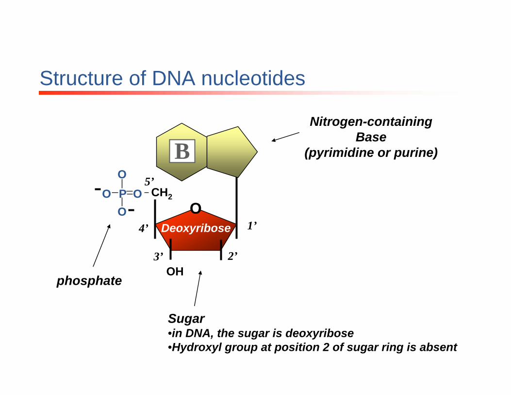
Structure of DNA nucleotides
Sugar •in DNA, the sugar is deoxyribose•Hydroxyl group at position 2 of sugar ring is absent
phosphate
CH2
ODeoxyribose 1’
3’
5’PO
OO
O
B
2’OH
4’

C
CH2
O
P
O
O
O
O CH2
O
P
O
O
O
O CH2
O
P
O
O
O
O CH2
O
P
O
O
O
O CH2
O
OH
OH
OH
OH
OH
C
A
G
U
O
OO P O
RNA is a Polymer of rNTPs
• Sugar-phosphate backbone– 5’ position of one sugar is
connected to the 3’ position of the next sugar via the phosphate group
• Single-stranded• Purines: Adenine, Guanine• Pyrimidines: Cytosine, Uracil
5’
3’
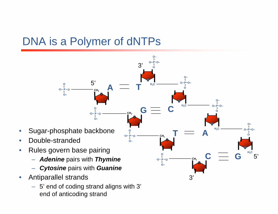
DNA is a Polymer of dNTPs
3’
5’ H2C
O
CH2
O
CH2
O
CH2
O
CH2
O
A
G
T
C
T
O
P
O
O–
O–
O
H2CC
O
O
H2C
H2C
A
G
3’
5’
P
O
O–
O–
O
P
O
O–
O–
O
• Sugar-phosphate backbone• Double-stranded• Rules govern base pairing
– Adenine pairs with Thymine– Cytosine pairs with Guanine
• Antiparallel strands– 5’ end of coding strand aligns with 3’
end of anticoding strand
–O P O
O–
O
–O P O
O–
O
–O P O
O–
O
–O P O
O–
O
P
O
O–
O–
O
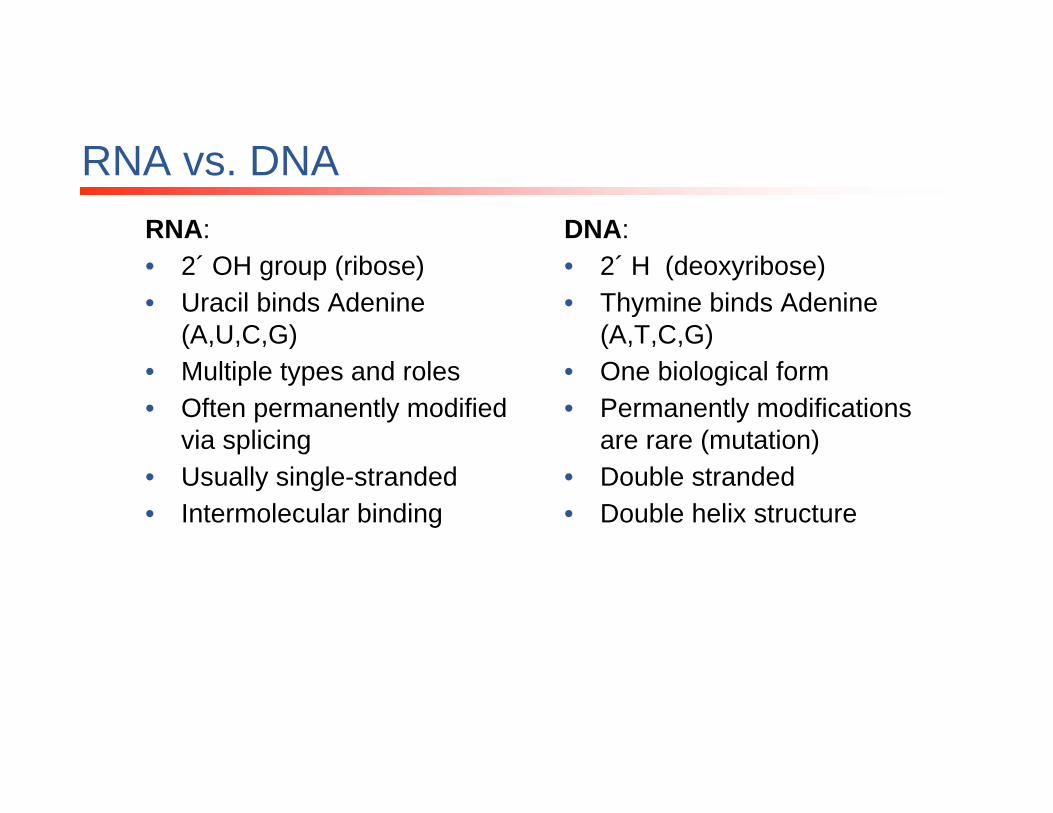
RNA vs. DNARNA:• 2´ OH group (ribose)• Uracil binds Adenine
(A,U,C,G)• Multiple types and roles• Often permanently modified
via splicing• Usually single-stranded• Intermolecular binding
DNA:• 2´ H (deoxyribose)• Thymine binds Adenine
(A,T,C,G)• One biological form• Permanently modifications
are rare (mutation)• Double stranded• Double helix structure
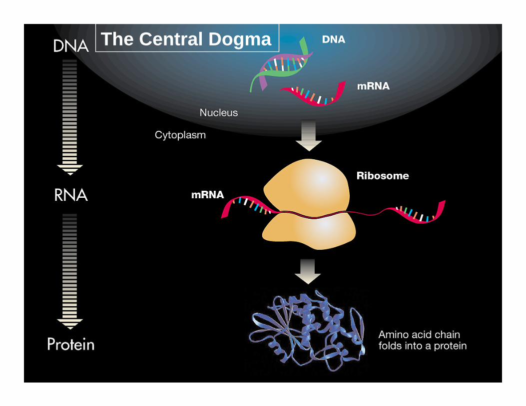
The Central Dogma

Cellular “total” RNA
• Messenger RNA (mRNA): 1-5% Serves as a template for protein synthesis
• Ribosomal RNA (rRNA): >80%Structural component of ribosomes
• Transfer RNA (tRNA): 10-15%Translates mRNA information into the appropriate
amino acid

RNA Isolation Techniques
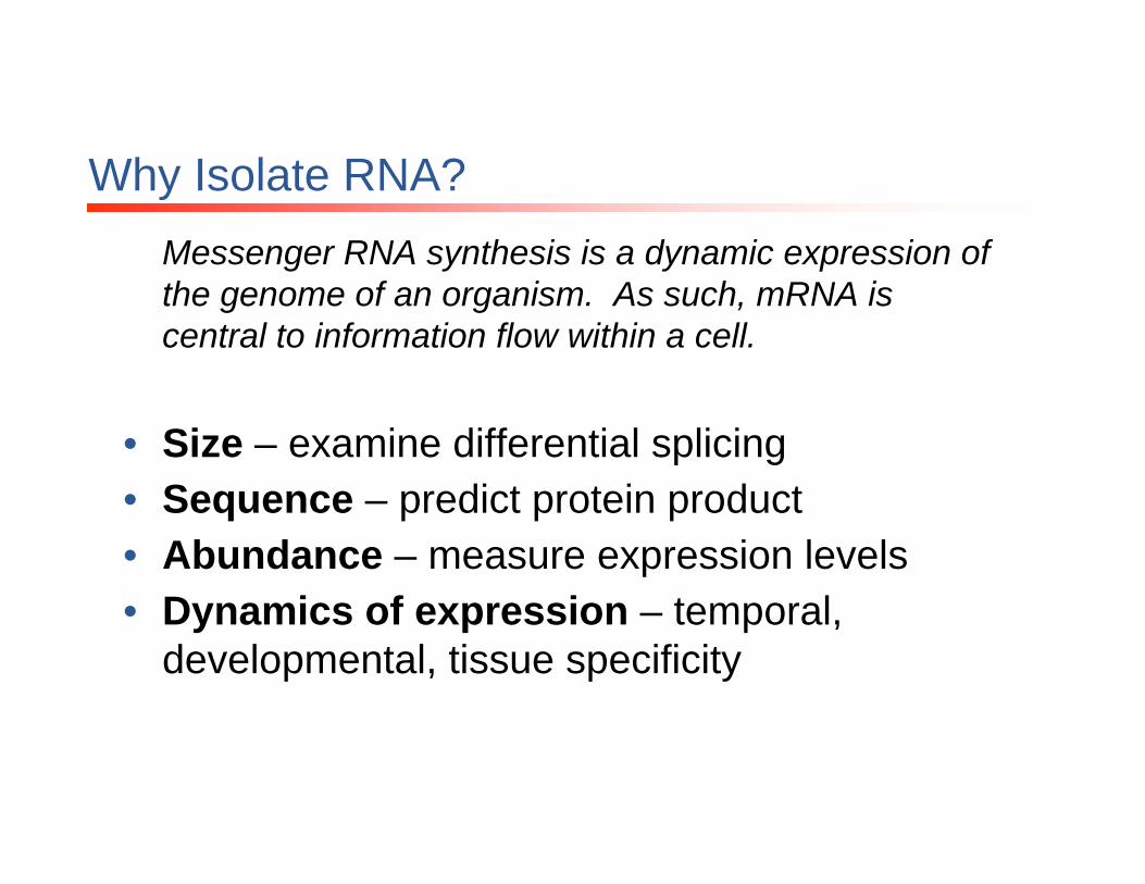
Why Isolate RNA?Messenger RNA synthesis is a dynamic expression of the genome of an organism. As such, mRNA is central to information flow within a cell.
• Size – examine differential splicing• Sequence – predict protein product• Abundance – measure expression levels • Dynamics of expression – temporal,
developmental, tissue specificity

Tissue Cells
Purity Integrity
Break open the cell
RNA Purification
Quality Parameters
RNA isolation
RNA Purification
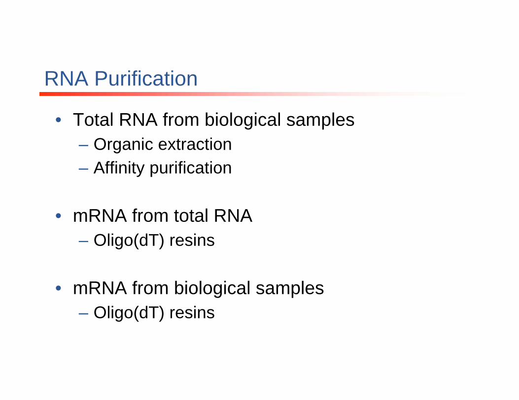
• Total RNA from biological samples– Organic extraction– Affinity purification
• mRNA from total RNA– Oligo(dT) resins
• mRNA from biological samples– Oligo(dT) resins

Total RNA Purification
• Goal: Isolate RNA from other cellular components– Cells or tissue must be rapidly and efficiently
disrupted– Inactivate RNases– Denature nucleic acid-protein complexes– RNA selectively partitioned from DNA and
protein
• Isolation from different tissues/sources raises different issues
Ribonucleases (RNases)
• RNases are naturally occurring enzymes that degrade RNA
• Common laboratory contaminant (from bacterial and human sources)
• Also released from cellular compartments during isolation of RNA from biological samples
• Can be difficult to inactivate

Protecting against RNase
• Wear gloves at all times• Use RNase-free tubes and pipet tips• Use dedicated, RNase-free, chemicals• Pre-treat materials with extended heat (180oC for
several hours), wash with DEPC-treated water, NaOHor H2O2
• Supplement reactions with RNase inhibitors• Include a chaotropic agent (guanidine) in the procedure
– Chaotropic agents such as guanidine inactivate and precipitate RNases and other proteins
Organic Extraction of total RNA
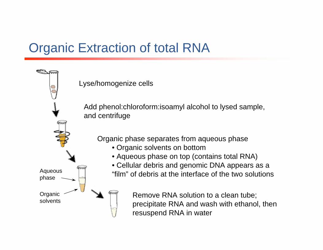
Lyse/homogenize cells
Add phenol:chloroform:isoamyl alcohol to lysed sample, and centrifuge
Organic phase separates from aqueous phase• Organic solvents on bottom• Aqueous phase on top (contains total RNA)• Cellular debris and genomic DNA appears as a “film” of debris at the interface of the two solutions
Remove RNA solution to a clean tube; precipitate RNA and wash with ethanol, then resuspend RNA in water
Aqueous phase
Organic solvents
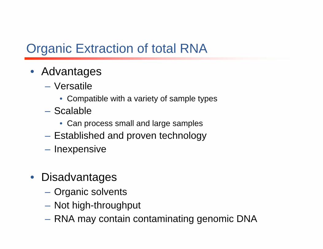
Organic Extraction of total RNA
• Advantages– Versatile
• Compatible with a variety of sample types– Scalable
• Can process small and large samples– Established and proven technology– Inexpensive
• Disadvantages– Organic solvents– Not high-throughput– RNA may contain contaminating genomic DNA
Affinity purification of total RNA
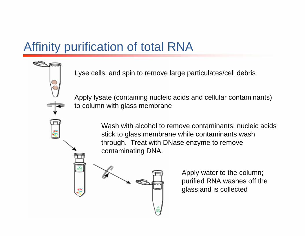
Lyse cells, and spin to remove large particulates/cell debris
Apply lysate (containing nucleic acids and cellular contaminants) to column with glass membrane
Wash with alcohol to remove contaminants; nucleic acids stick to glass membrane while contaminants wash through. Treat with DNase enzyme to remove contaminating DNA.
Apply water to the column; purified RNA washes off the glass and is collected

• Advantages– Eliminates need for organic solvents– Compatible with a variety of sample types (tissue,
tissue culture cells, white blood cells, plant cells, bacteria, yeast, etc.)
– DNase treatment eliminates contaminating genomic DNA
– Excellent RNA purity and integrity
Affinity purification of total RNA
Messenger RNA Isolation
• mRNA molecules have a tail of A’s at the 3’ end (polyA tail)
• Oligo(dT) probes can be used to purify mRNA from other RNAs
• mRNA can be eluted from oligo(dT) matrix using water or low-salt buffer
Messenger RNA Isolation
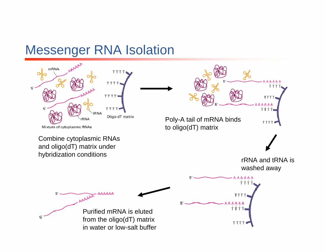
Combine cytoplasmic RNAsand oligo(dT) matrix under hybridization conditions
Poly-A tail of mRNA binds to oligo(dT) matrix
rRNA and tRNA is washed away
Purified mRNA is eluted from the oligo(dT) matrix in water or low-salt buffer
Messenger RNA Isolation
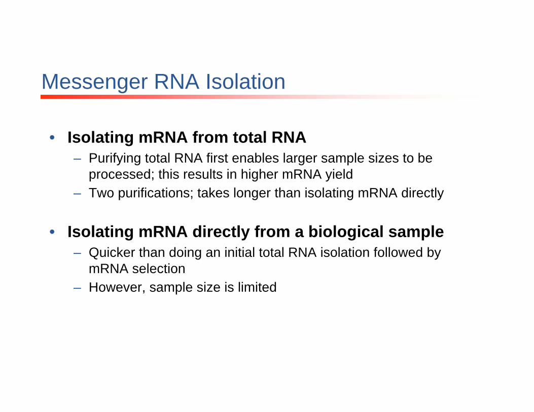
• Isolating mRNA from total RNA– Purifying total RNA first enables larger sample sizes to be
processed; this results in higher mRNA yield– Two purifications; takes longer than isolating mRNA directly
• Isolating mRNA directly from a biological sample– Quicker than doing an initial total RNA isolation followed by
mRNA selection– However, sample size is limited

• RNA sample absorbances are determined on the spectrophotometer at 260nm, 280nm, and 230nm
– 260nm: nucleic acid (DNA, RNA, nucleotides)– 280nm: protein– 230nm: guanidine
Estimating RNA quality by spectrophotometry

Quantitation of RNA
• Nucleic acids absorb UV light maximally at 260nm
• For RNA:1 OD260 Unit = 40 µg/ml of ssRNA
• The concentration in the sample is calculated by using the formula:
A260 x dilution x 40 = [RNA] µg/ml
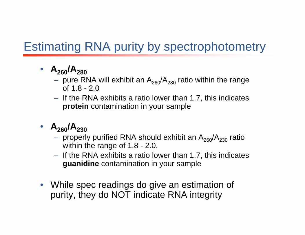
• A260/A280– pure RNA will exhibit an A260/A280 ratio within the range
of 1.8 - 2.0– If the RNA exhibits a ratio lower than 1.7, this indicates
protein contamination in your sample
• A260/A230– properly purified RNA should exhibit an A260/A230 ratio
within the range of 1.8 - 2.0. – If the RNA exhibits a ratio lower than 1.7, this indicates
guanidine contamination in your sample
• While spec readings do give an estimation of purity, they do NOT indicate RNA integrity
Estimating RNA purity by spectrophotometry
• Two major ribosomal subunits: 28S and 18S
• Approximately 80% of total RNA is associated with these two subunits
• Integrity of the total RNA sample can be determined by evaluating the 28S and 18S rRNA– 28S and 18S rRNA should appear as distinct bands– Ratio of 28S:18S should be approximately 2:1
Evaluating RNA integrity

Evaluating RNA integrity
• High quality total RNA imaged on an Agilent 2100 Bioanalyzer• 28S rRNA and 18S rRNA appear as distinct, intact bands• Ratio of 28S : 18S approximately 2 : 1
Poor quality RNA
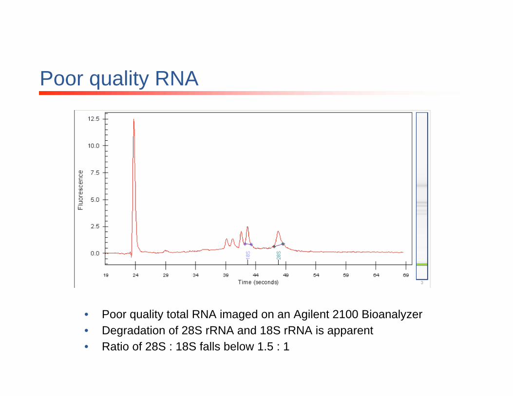
• Poor quality total RNA imaged on an Agilent 2100 Bioanalyzer• Degradation of 28S rRNA and 18S rRNA is apparent• Ratio of 28S : 18S falls below 1.5 : 1

mRNA isolated from Total RNA
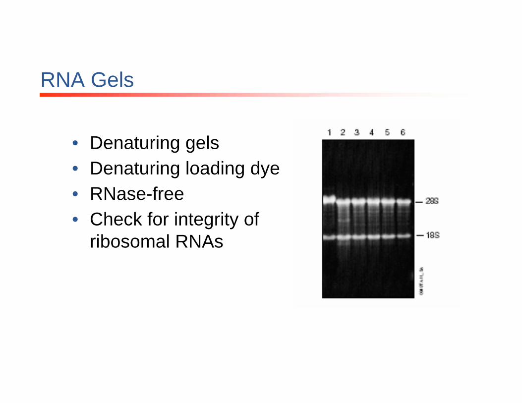
RNA Gels
• Denaturing gels• Denaturing loading dye• RNase-free• Check for integrity of
ribosomal RNAs

Applications for RNA Analysis
Techniques for RNA analysis: Northern Blotting
• Once a gene has been identified, it is useful to determine the size and quantity of the mRNA message produced and whether alternative splice variants of different sizes exist
• Northern Blotting: method to determine the abundance and size of a RNA molecule in a sample, by hybridization with a gene-specific probe.
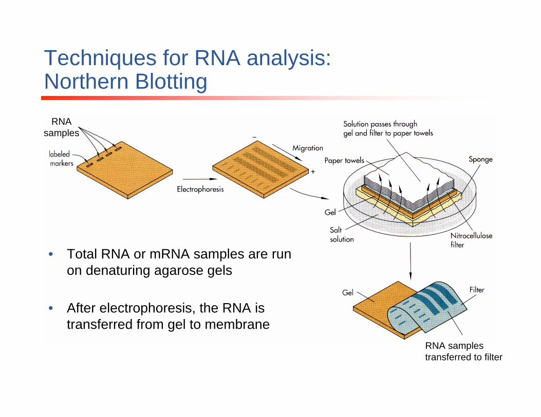
Techniques for RNA analysis: Northern Blotting
RNA samples
RNA samples transferred to filter
• Total RNA or mRNA samples are run on denaturing agarose gels
• After electrophoresis, the RNA is transferred from gel to membrane

Techniques for RNA analysis: Northern Blotting
Labeled probe
• A labeled probe that is specific to the gene of interest is applied to the RNA samples on the filter
• Labeling methods:– Radioactive– Chemiluminescent– Fluorescent
• Analysis reveals the size and quantity of the mRNA message in each sample

Techniques for RNA analysis: Northern Blotting
• Size and quantity of message can be determined
Probe used is specific to themRNA sequence of interest
Techniques for RNA analysis:RNase Protection Assay
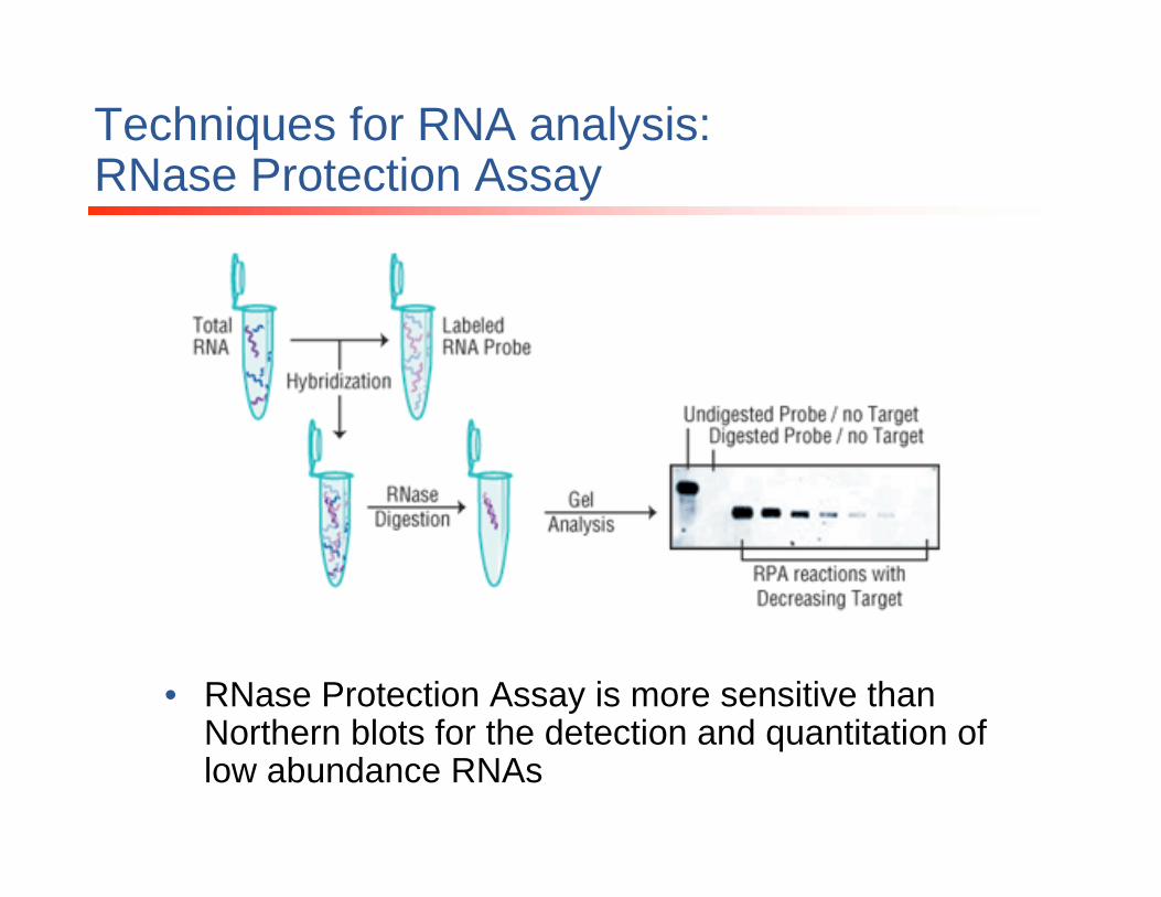
• RNase Protection Assay: sensitive technique for the quantitation of specific RNA transcripts in solution
• Can be performed using total RNA or mRNA as the target• A probe that is complementary to the sequence of interest is used
– defined length– radioactively labeled
• The RNA target and RNA probe are hybridized in solution• Following hybridization, the RNA is digested with RNases specific
for single-stranded nucleic acids– Any remaining unhybridized single-stranded RNA target and
probe is degraded– Target:probe hybridized sequences are left intact
• Following RNase inactivation, RNA samples are run on denaturing polyacrylamide gels
• The amount of intact probe observed on the gel is proportional to the amount of target RNA in the original sample
Techniques for RNA analysis:RNase Protection Assay
• RNase Protection Assay is more sensitive than Northern blots for the detection and quantitation of low abundance RNAs

Techniques for RNA analysis:RT-PCR
• Reverse Transcription-Polymerase Chain Reaction
• Amplification of a specific RNA molecule into many copies of a cDNA molecule
• Potential applications:– Determine the presence or absence of a
transcript– Estimate mRNA expression levels – Convert RNA message to cDNA for cloning
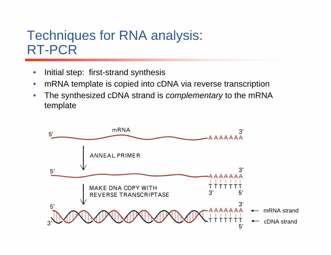
• Initial step: first-strand synthesis• mRNA template is copied into cDNA via reverse transcription• The synthesized cDNA strand is complementary to the mRNA
template
mRNA strand
cDNA strand
Techniques for RNA analysis:RT-PCR
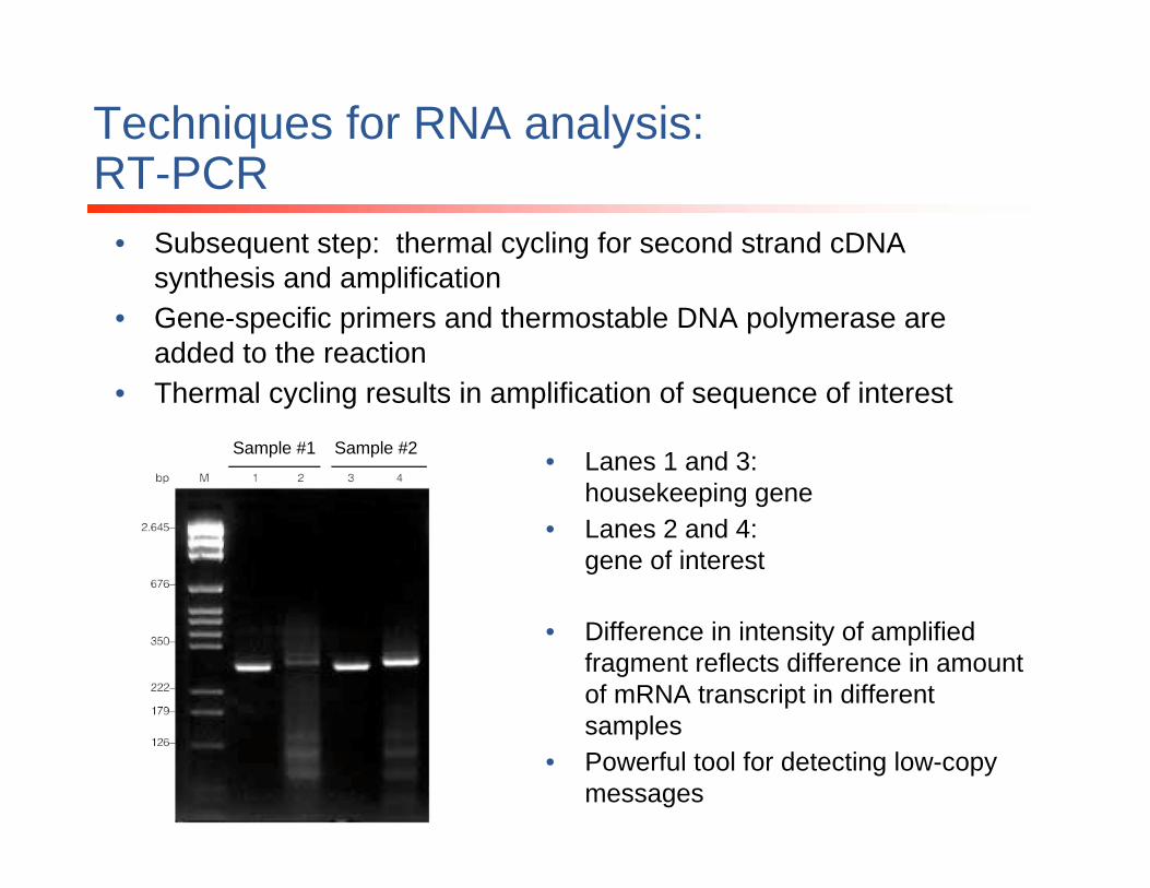
• Subsequent step: thermal cycling for second strand cDNAsynthesis and amplification
• Gene-specific primers and thermostable DNA polymerase are added to the reaction
• Thermal cycling results in amplification of sequence of interest
Sample #1 Sample #2 • Lanes 1 and 3: housekeeping gene
• Lanes 2 and 4: gene of interest
• Difference in intensity of amplified fragment reflects difference in amount of mRNA transcript in different samples
• Powerful tool for detecting low-copy messages
Techniques for RNA analysis:RT-PCR
Techniques for RNA analysis:qRT-PCR
• In traditional PCR assays, too much amplification makes it impossible to quantitate the amount of starting nucleic acid material.
• Real-Time PCR (or, quantitative PCR) addresses this problem of end-point analysis
• Real-Time PCR monitors the product accumulation as the PCR amplification proceeds, allowing for highersensitivity.

Techniques for RNA analysis:qRT-PCR
Reference housekeeping gene:Gene of interest:
• Cells from the eye were evaluated for expression of IL1-beta• Experimental group (“vit”): treated with vitreous humor• Control group (“con”): no treatment• IL1-beta shows dramatic difference in mRNA levels in treated vs control
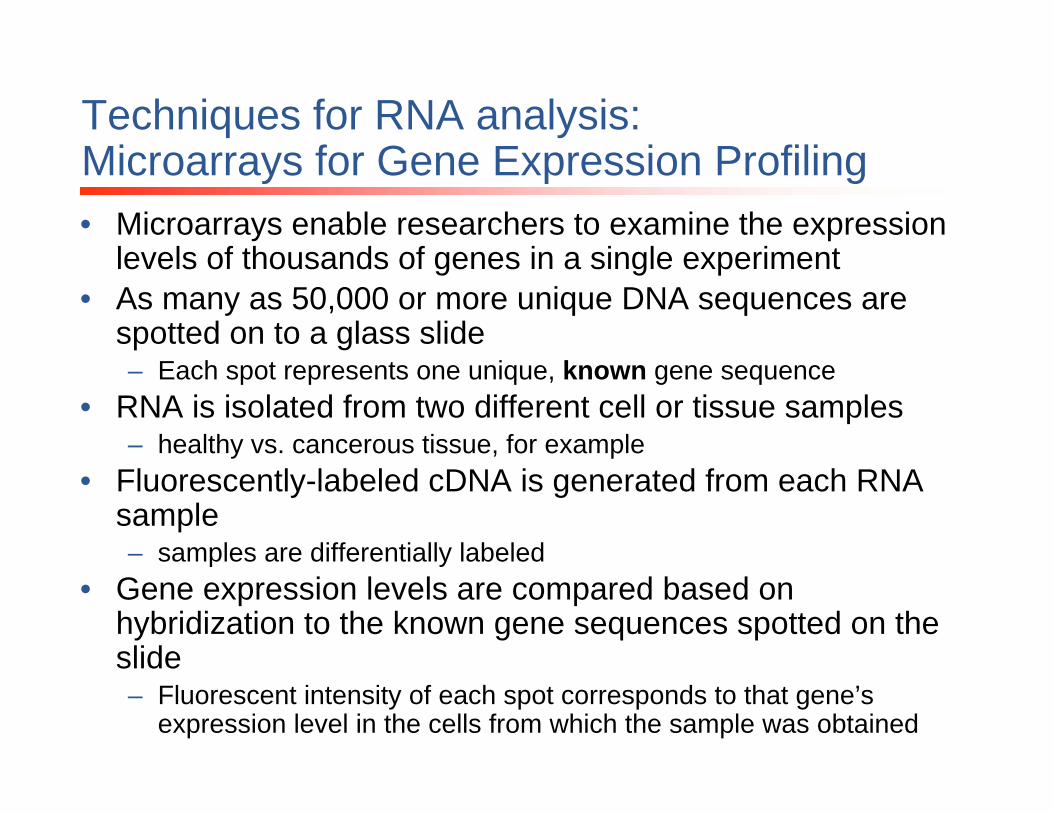
Techniques for RNA analysis:Microarrays for Gene Expression Profiling• Microarrays enable researchers to examine the expression
levels of thousands of genes in a single experiment • As many as 50,000 or more unique DNA sequences are
spotted on to a glass slide – Each spot represents one unique, known gene sequence
• RNA is isolated from two different cell or tissue samples– healthy vs. cancerous tissue, for example
• Fluorescently-labeled cDNA is generated from each RNA sample– samples are differentially labeled
• Gene expression levels are compared based on hybridization to the known gene sequences spotted on the slide– Fluorescent intensity of each spot corresponds to that gene’s
expression level in the cells from which the sample was obtained
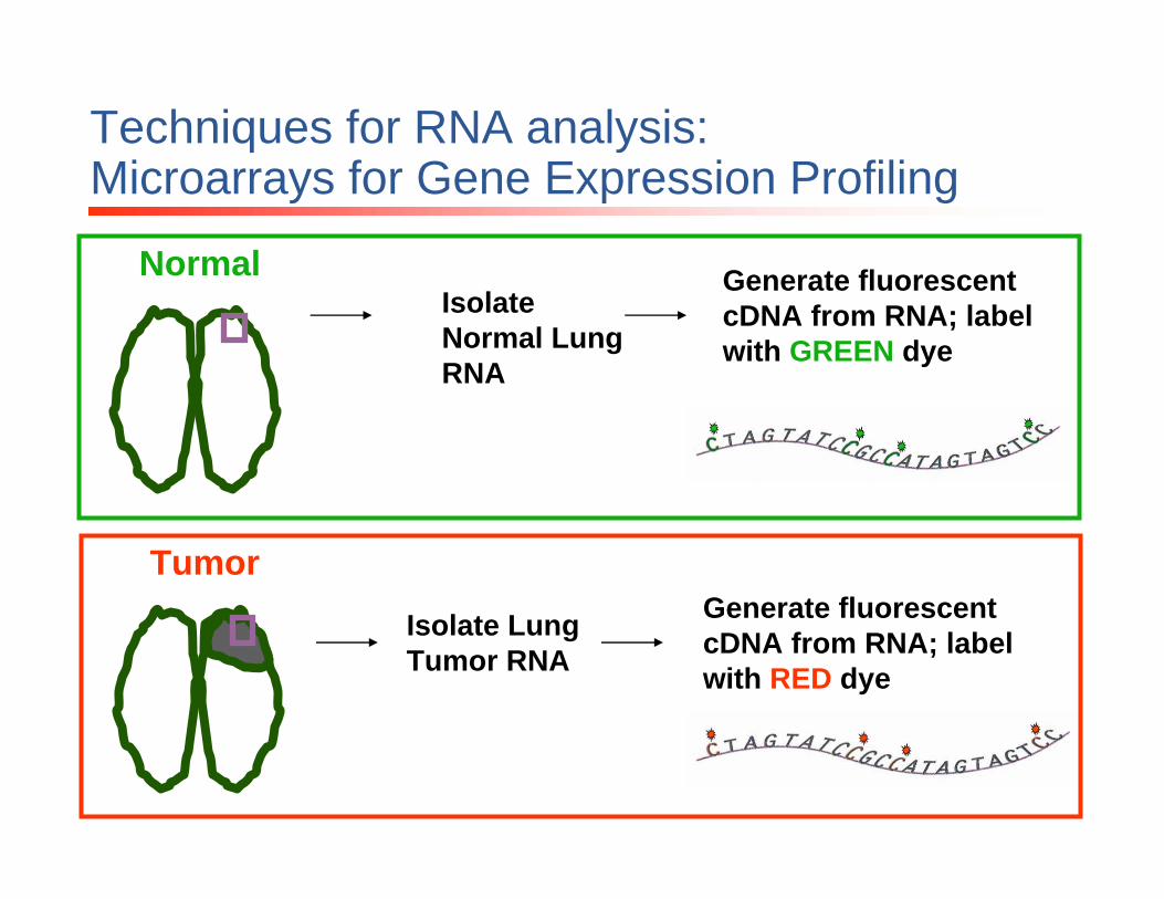
Normal
Tumor
Isolate Normal Lung RNA
Isolate Lung Tumor RNA
Techniques for RNA analysis:Microarrays for Gene Expression Profiling
Generate fluorescent cDNA from RNA; label with GREEN dye
Generate fluorescent cDNA from RNA; label with RED dye

cDNA generated from normal lung
cDNA generated from lung tumor
GREEN RED
Techniques for RNA analysis:Microarrays for Gene Expression Profiling
• Mix normal lung cDNA (labeled with green fluorescent dye) with lung tumor cDNA(labeled with red fluorescent dye)
• Apply both fluorescent cDNAs to spotted slide

Techniques for RNA analysis:Microarrays for Gene Expression Profiling• Normal lung sample
was labeled with green dye
• The green fluorescent intensity of each spot indicates the level of expression of that gene in the normal sample
– bright spots correspond to strongly expressed genes
– dim spots indicate weak expression
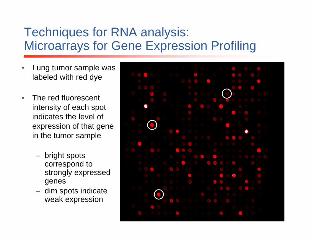
Techniques for RNA analysis:Microarrays for Gene Expression Profiling
• Lung tumor sample was labeled with red dye
• The red fluorescent intensity of each spot indicates the level of expression of that gene in the tumor sample
– bright spots correspond to strongly expressed genes
– dim spots indicate weak expression

Techniques for RNA analysis:Microarrays for Gene Expression Profiling
By overlaying red and green data from the same slide, you can view the differential expression of various genesGreen = gene expressed in normal sample
Red = gene expressed in tumor sample
Yellow = gene expressed in BOTH normal and tumor samples
Techniques for RNA analysis:Microarrays for Gene Expression Profiling
• Gene expression data generated in microarray experiments is often confirmed using qRT-PCR technology
• Microarrays: most useful when screening many genes of interest
• Real-Time PCR: most useful when many treatment conditions on a handful of genes
• Example:– Screen a tumor sample vs a normal sample for expression
differences in 10,000 genes: MICROARRAY– Screen the effect of 10 different drug treatments on the
expression of one or two gene: REAL-TIME PCR

Techniques for RNA analysis:Microarrays for Gene Expression Profiling
• Some Examples of Potential Applications– Gene expression studies in disease– Drug discovery and development– Detection of bacteria, viruses in environmental samples
• water quality management– Use as a diagnostic tool in the field of healthcare
• Disease detection• Newborn screening
– Detection of bioagents• Anthrax• biological weapons

Techniques for RNA analysis:In vitro Translation
• In vitro Translation: Expression of a protein product encoded by an RNA molecule in a cell-free system– Commonly used cell-free translation systems
consist of extracts from rabbit reticulocytes, wheat germ and Escherichia coli
– All are prepared as crude extracts containing all the macromolecular components required for translation of exogenous RNA
– Standard translation systems, such as reticulocytelysates and wheat germ extracts, use RNA as a template

Techniques for RNA analysis:In vitro Translation
• Making proteins in vitro is an important tool for:– studying protein mutations – protein-DNA binding studies– protein activity assays– protein-protein interaction studies.
Techniques for RNA analysis:RNA Interference (RNAi)
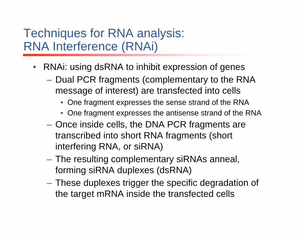
• RNAi: using dsRNA to inhibit expression of genes– Dual PCR fragments (complementary to the RNA
message of interest) are transfected into cells• One fragment expresses the sense strand of the RNA• One fragment expresses the antisense strand of the RNA
– Once inside cells, the DNA PCR fragments are transcribed into short RNA fragments (short interfering RNA, or siRNA)
– The resulting complementary siRNAs anneal, forming siRNA duplexes (dsRNA)
– These duplexes trigger the specific degradation of the target mRNA inside the transfected cells
Techniques for RNA analysis:RNA Interference (RNAi)
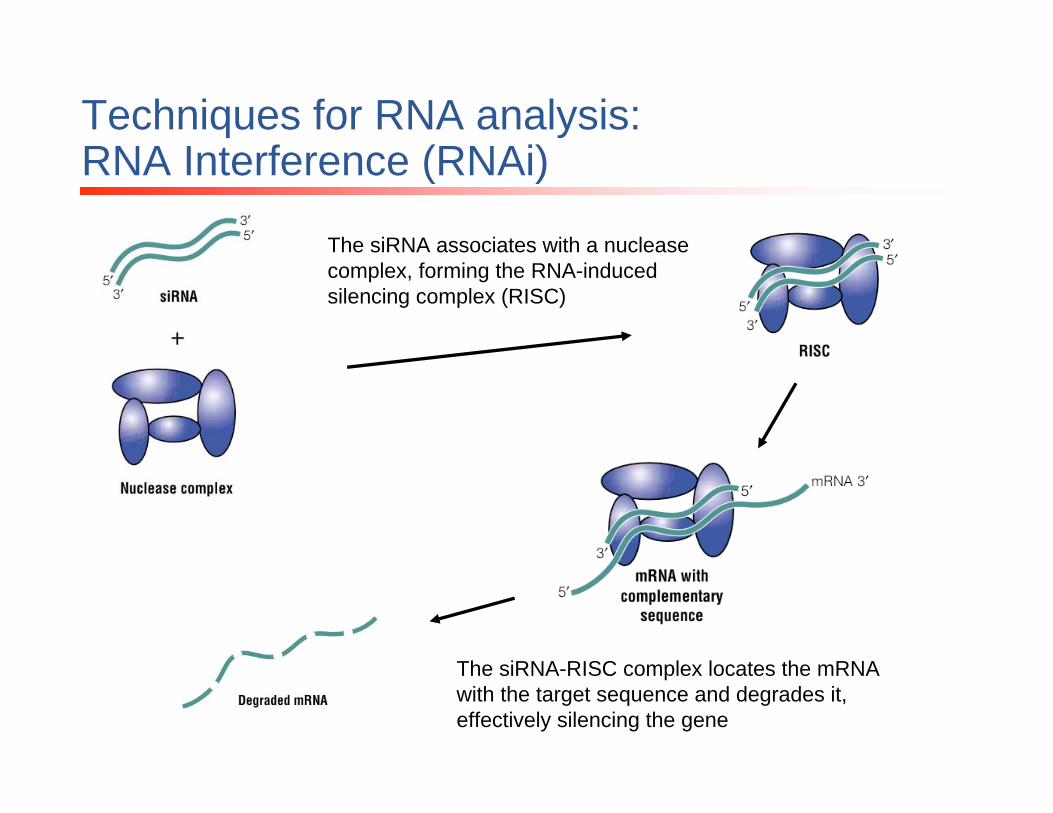
The siRNA associates with a nuclease complex, forming the RNA-induced silencing complex (RISC)
The siRNA-RISC complex locates the mRNA with the target sequence and degrades it, effectively silencing the gene

Techniques for RNA analysis:RNA Interference (RNAi)
• Gene knockdown is a powerful tool for analyzing the function of a single gene
• Selectively downregulating the expression of a particular gene allows researchers to determine its function in many cellular processes
• Particularly useful in studies such as:– Mapping cellular pathways– Developing gene therapies

Questions?


![RNA Recognition and Stress Granule Formation by TIA Proteins · 2017-07-24 · Int. J. Mol. Sci. 2014, 15 23379 targeted mRNA transcripts [23,24]. This review, however, focuses on](https://img.pdfslide.us/doc/110x75/5ecf668ad7b7bd0f216d76a5/rna-recognition-and-stress-granule-formation-by-tia-proteins-2017-07-24-int-j.jpg)