Embed Size (px)
Citation preview

ORIGINAL SCIENTIFIC REPORT
Rives Technique for the Primary Larger Inguinal Hernia Repair:A Prospective Study of 1000 Repairs
Enrique J. Grau-Talens1,8• Carlos D. Ibanez2
• Jacob Motos-Mico3•
Francisco Garcıa-Olives4• Martina Arribas-Jurado5
• Carlos Jordan-Chaves6•
Jose M. Aparicio-Gallego7• Jose F. Salgado2
Published online: 8 May 2017
� The Author(s) 2017. This article is an open access publication
Abstract
Objective We report a prospective study of repairs using the Rives technique of the more difficult primary inguinal
hernias, focusing on the immediate post-operative period, clinical recurrence, testicular atrophy, and chronic pain. A
mesh placed in the preperitoneal space can reduce recurrences and chronic pain.
Methods For the larger primary inguinal hernias (Types 3, 4, 6, and some 7), we favour preperitoneal placement of a
mesh, covering the myopectineal orifice by means of a transinguinal (Rives technique) approach. The Rives tech-
nique was performed on 943 patients (1000 repairs), preferably under local anaesthesia plus sedation in ambulatory
surgery.
Results The mean operative time was 31.8 min. Pain assessment after 24 h with an Andersen scale and a categorical
scale gave two patients with intense pain on the Andersen scale, and four patients who thought their state was bad.
Surgical wound complications were below 1%, and urinary retention was 1.2% mostly associated with spinal
anaesthesia and, in one case, bladder perforation. There was spermatic cord and testicular oedema with some degree
of orchitis in 17 patients. The clinical follow-up of 849 repairs (86.4%), mean (range) 30.0 (12–192) months, gave
five recurrences (0.6%), three cases (0.4%) of testicular atrophy, and 37 (4.3%) of post-operative chronic pain (8
patients with visual analogue scale of 3–10).
Conclusions The Rives technique requires a sound knowledge of inguinal preperitoneal space anatomy, but it is an
excellent technique for the larger and difficult primary inguinal hernias, giving a low rate of recurrences and chronic
pain.
& Enrique J. Grau-Talens
Carlos D. Ibanez
Jacob Motos-Mico
Martina Arribas-Jurado
Carlos Jordan-Chaves
Jose M. Aparicio-Gallego
Jose F. Salgado
1 Hospital Siberia-Serena, Carretera Talarrubias-Agudo, SN,
06640 Talarrubias, Badajoz, Spain
2 Hospital Siberia-Serena, Calle Zafra, 3, 3D,
06400 Don Benito, Badajoz, Spain
3 Hospital Siberia-Serena, Calle Canaleja, 4, 28 A,06400 Don Benito, Badajoz, Spain
4 Verge Del Toro Hospital, Menorca. Fort de L’eau 96,
07701 Mahon, Balearic Islands, Spain
123
World J Surg (2017) 41:2480–2487
DOI 10.1007/s00268-017-4038-z

Introduction
The value of a surgical intervention lies in the short and
long term results (quality) provided at a reasonable cost
[1]. The primary repair of an inguinal hernia aims to cure
the symptoms and prevent immediate post-operative com-
plications, chronic pain, and recurrences. Mesh repair has
reduced the recurrence of inguinal hernias, but there is still
a rate of 4% of recurrence and 12% of post-operative
chronic pain, moderate-to-severe, with the Lichtenstein
procedure [2].
The aim of our work is the prospective study of the
Rives technique for repair of the larger primary inguinal
hernias (transinguinal insertion of a mesh in the
preperitoneal space), focusing on the immediate post-
operative period, clinical recurrence, and chronic pain.
The mesh placed in the preperitoneal space, covering
the myopectineal orifice of Fruchaud and separated
from the neurological plane, is the ideal location
hydrostatically and anatomically to prevent the recur-
rence of the three potential hernia orifices and chronic
pain.
Patients and methods
From January 1992 to April 2014, we performed 1000
primary inguinal hernia repairs in 943 patients using the
Rives technique: 198 patients at the Verge del Toro
Hospital, Mahon, Menorca (Balearic Islands), Spain
(1992–2001); and 745 patients from October 2007 until
April 2014 in ambulatory/short-stay surgery at the Siberia-
Serena Hospital, Talarrubias (Badajoz), Spain. In 1994, all
the data of the operated patients, which had been collected
in paper form, were input into an Access-form database.
All data collected thereafter were input into the said
database on a daily basis and analysed with Excel.
Between 2001 and 2006, the first author did not perform
abdominal wall surgery because he was assigned to the
coloproctology section of the Infanta Cristina University
Hospital, Badajoz, Spain.
Inclusion criteria and perioperative measures
and follow-up
We use Gilbert’s classification with additions by Rutkow
and Robbins [3]. The Rives technique was used in Types 2
(when the deep inguinal ring was about 4 cm in diameter),
3, 4, 6, and some of the 7.
In ambulatory surgery, all patients are contacted via
telephone within 24 h of surgery and pain is evaluated at
rest and mobilization using the Andersen scale [4]. This is
concluded with a subjective assessment by the patient on a
categorical scale of bad, fair, good, or excellent overall
condition. After a week they are called again.
The first 467 patients were clinically reviewed in the
office within 7–10 days of discharge (for the rest of the
patients, we opted for telephone inquiry to spare them from
coming to the hospital, where the wound was observed, and
pain was evaluated at rest and with movement (standing
up, sitting down, and walking), using a VAS (visual ana-
logue scale) in 154 consecutive patients, as a representative
sample size for post-operative pain.
They are reviewed clinically by the service’s surgeons
again after a year or earlier if the patient experiences pain,
and called again 3 or more years later. If pain is found, this
is evaluated as being with movement, spontaneous, episo-
dic, or constant in nature, and measured with a VAS. The
inguinal region is examined. A mass, reducible or other-
wise, is regarded as a recurrence. Testicular examination is
made for atrophy or other pathology, and inquiry for late
infection.
Preoperative preparation includes the prophylaxis of
nausea and vomiting with dexamethasone 8 mg IV. Pro-
phylaxis of the surgical site infection (cefazoline 2 g IV) is
not performed routinely. It was done in obese, diabetics,
and bilateral hernia repair. Glove change and wound pro-
tection with gauze before inserting the mesh were the rule.
Lastly, rinsing wound preceded the skin sutures.
The technique now
We perform the technique preferably under local anaes-
thesia and sedation, with mepivacaine 1%, 400–500 mg
(6 mg/kg) according to the technique of Ponka and
Flanagan [5]. An anaesthesiologist provides sedation. The
sedation begins with midazolam (0.03 mg/kg IV) followed
by propofol (25–50 mc/kg/min). Upon finishing the inter-
vention, we infiltrate the surgical field with 20–30 ml of
0.25% bupivacaine. In very large inguinoscrotal or non-
reducible hernias, spinal or general anaesthesia is used.
The surgical technique comprises the insertion of a mesh
in the preperitoneal space through the groin as described by
Rives [6–10]. We use a 15 9 15 cm polypropylene mesh
(standard weight 80 g/m2, and macro-porous
5 Calle La Cola, 10, 14300 Villaviciosa de Cordoba, Cordoba,
Spain
6 Hospital Siberia-Serena, Calle Santiago, 32, 06900 Llerena,
Badajoz, Spain
7 Calle 18 de mayo, 71, 48, 06400 Don Benito, Badajoz, Spain
8 Calle Castillo 15, 06006 Zalamea de la Serena, Badajoz,
Spain
World J Surg (2017) 41:2480–2487 2481
123
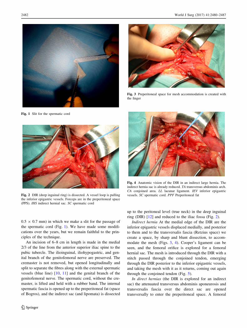
0.5 9 0.7 mm) in which we make a slit for the passage of
the spermatic cord (Fig. 1). We have made some modifi-
cations over the years, but we remain faithful to the prin-
ciples of the technique.
An incision of 6–8 cm in length is made in the medial
2/3 of the line from the anterior superior iliac spine to the
pubic tubercle. The ilioinguinal, iliohypogastric, and gen-
ital branch of the genitofemoral nerve are preserved. The
cremaster is not removed, but opened longitudinally and
split to separate the fibres along with the external spermatic
vessels (blue line) [10, 11] and the genital branch of the
genitofemoral nerve. The spermatic cord, without the cre-
master, is lifted and held with a rubber band. The internal
spermatic fascia is opened up to the preperitoneal fat (space
of Bogros), and the indirect sac (and lipomata) is dissected
up to the peritoneal level (true neck) in the deep inguinal
ring (DIR) [12] and reduced to the iliac fossa (Fig. 2).
Indirect hernia At the medial edge of the DIR are the
inferior epigastric vessels displaced medially, and posterior
to them and to the transversalis fascia (Retzius space) we
create a space, by sharp and blunt dissection, to accom-
modate the mesh (Figs. 3, 4). Cooper’s ligament can be
seen, and the femoral orifice is explored for a femoral
hernial sac. The mesh is introduced through the DIR with a
stitch passed through the conjoined tendon, emerging
through the DIR posterior to the inferior epigastric vessels,
and taking the mesh with it as it returns, coming out again
through the conjoined tendon (Fig. 5).
In direct hernias (the DIR is explored for an indirect
sac) the attenuated transversus abdominis aponeurosis and
transversalis fascia over the direct sac are opened
transversally to enter the preperitoneal space. A femoral
Fig. 1 Slit for the spermatic cord
Fig. 2 DIR (deep inguinal ring) is dissected. A vessel loop is pulling
the inferior epigastric vessels. Forceps are in the preperitoneal space
(PPS). IHS indirect hernial sac. SC spermatic cord
Fig. 3 Preperitoneal space for mesh accommodation is created with
the finger
Fig. 4 Anatomic vision of the DIR in an indirect large hernia. The
indirect hernia sac is already reduced. TA transversus abdominis arch.
CA conjoined area. LL lacunar ligament. IEV inferior epigastric
vessels. SC spermatic cord. PPF Preperitoneal fat
2482 World J Surg (2017) 41:2480–2487
123

sac, if any, is reduced. The inferior epigastric vessels are
released and preserved (Fig. 6), and the mesh, inserted
through Hesselbach’s triangle, slides (preferably) under
them or pushes them down and overlies them (less desir-
able case) [13].
Mesh fixation The mesh is fixed with 5–7 stitches of
polypropylene monofilament with an appropriate needle
(premilene� No. 0, HR80, B Braun). The first stitch in the
conjoined tendon (as described above) acts as an anchor.
The second stitch takes the Cooper’s ligament (close to the
femoral ring) and the mesh, at approximately 4 cm from
the edge (Fig. 7), slid over it and covering the femoral and
obturator rings. Particular attention has to be paid to the
anastomotic pubic branch, and sometimes to the anterior
pubic branch artery, obturator accessory, or aberrant artery
and corresponding veins. In indirect hernias, due to the
limited space of the DIR, it is easier to put the stitch into
the Coopers ligament before mesh introduction and fixation
to the posterior face of the conjoined tendon. If access to
the Cooper ligament is difficult, the mesh is attached to the
iliopubic tract (less desirable), with care taken of the
inferior epigastric vessels.
The spermatic cord is positioned in the slit of the mesh,
and wrapped with it in the manner of a tunnel (Fig. 8). The
mesh is slipped with Crile forceps behind the transversus
abdominis muscle in the preperitoneal space. The third
stitch fixes the mesh to the vascular fascia/iliopubic tract,
taking care with the genital branch of the genitofemoral
nerve, the fourth stitch is placed into the transversus
abdominis arch, and the fifth into the most lateral zone of
the transverse arch. Additional sutures are inserted if nee-
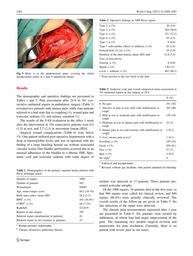
ded. The mesh covers and exceeds entirely, by at least
4 cm, the myopectineal orifice (Fig. 9).
Fig. 5 Mesh ready to be passed under epigastric vessels to lie under
conjoined area. A stitch running through the conjoined area near the
pubic tubercle, coming out through the DIR under the inferior
epigastric vessels, takes the mesh and returns the same way it came to
the conjoined area
Fig. 6 Transversalis fascia over a direct sac has been opened and the
hernia reduced. A wound swab is introduced. Forceps are pointing at
the Cooper ligament (CL). Inferior epigastric vessels (IEV) are pulled
by a vessel loop. The spermatic cord (SC) is held with a rubber band
Fig. 7 A stitch (white arrow) is passed through Cooper ligament
Fig. 8 Spermatic cord is positioned in the slit and a tunnel-like flap is
made around it, as seen in the lower image A
World J Surg (2017) 41:2480–2487 2483
123
Results
The demographic and operative findings are presented in
Tables 1 and 2. Pain assessment after 24 h in 761 con-
secutive unilateral repairs in ambulatory surgery (Table 3)
revealed two patients with intense pain, while four patients
referred to a bad state due to coughing (1), wound pain and
testicular oedema (2), and urinary retention (1).
The results of the VAS evaluation in the office 1 week
after the intervention in 154 consecutive patients were 0.5
(1.5) at rest, and 2.7 (2.1) in movement [mean (SD)].
Surgical wound complications (Table 4) were below
1%. One patient suffered post-operative hypotension with a
drop in haemoglobin levels and was re-operated with the
finding of a large bleeding hernial sac without associated
vascular lesion. One bladder perforation occurred due to an
unusual adherence of the bladder to a fibrotic DIR. Sper-
matic cord and testicular oedema with some degree of
orchitis was detected in 17 patients. Three patients pre-
sented testicular atrophy.
Of the 1000 repairs, 16 patients died in the first year, so
that 984 repairs were called for clinical review, and 849
repairs (86.4%) were actually clinically reviewed. The
overall results of the follow-up are given in Table 5. No
late infections of the repair were detected.
The chronic pain measurements registered after 1 year
are presented in Table 6. Six patients were treated by
infiltration, of whom four had major improvement of the
pain. The remaining two needed surgical revision and
neurectomy for pain resolution. Currently, there is no
patient with severe pain in our series.
Fig. 9 Mesh is in the preperitoneal space covering the whole
myopectineal orifice in a type 6 (pantaloon) hernia
Table 1 Demographics of the primary inguinal hernia patients with
Rives technique repair
Number of repairs 1000
Number of patients 943
Women/men 56/887
Age, mean (range) years 60.2 (18–93)
Body mass index, mean (SD) 28.2 (3.7)
BPHa, n (%) 164 (18.4%)
COPDb, n (%) 68 (7.2%)
Right/left 521/479
Repairs in day-surgery 785
Bilateral repair–simultaneous (n patients) 24
Bilateral repair–in two sessions (n patients) 19
a Benign prostatic hypertrophyb Chronic obstructive pulmonary disease
Table 2 Operative findings in 1000 Rives repairs
Type 2, n (%) 36 (3.6)
Type 3, n (%) 564 (56.4)
Type 4, n (%) 321 (32.1)
Type 6, n (%) 45 (4.5)
Type 7, n (%) 8 (0.8)
Type 7 with another (direct or indirect), n (%) 26 (2.6)
Femoral hole >2 cm, n (%) 29 (2.9)
Duration of the intervention, mean (SD) mina 31.8 (7.5)
Type of anaesthesia
General, n (%) 8 (0.8)
Spinal, n (%) 130 (13)
Local ? sedation, n (%) 862 (86.2)
a From incision to the last stitch in the skin
Table 3 Andersen scale and overall categorical status assessment in
761 unilateral repairs in day-surgery at 24 h
N (%)
0. No pain 291 (38)
1. Absence of pain at rest, mild with mobilization or
cough
302 (40)
2. Mild at rest or moderate pain with mobilization or
cough
139 (18)
3. Moderate at rest or intense pain with mobilization or
cough
23 (3)
4. Intense pain at rest and extreme with mobilization or
cougha1 (0.1)
5. Very intense pain at resta 1 (0.1)
Excellent, n (%) 239 (32)
Good, n (%) 456 (61)
Fair, n (%) 57 (7)
Bad, n (%) 4 (0.5)
No replyb 4
a Followed and asymptomaticb Revised, without any incidents. One patient admitted for bleeding
2484 World J Surg (2017) 41:2480–2487
123

Discussion
All surgeons who operate inguinal hernias must have deep
knowledge of the inguinal region’s anatomy, and this is
especially true for the Rives repair technique [12–17].
The preperitoneal space is the most suitable site for the
insertion of a mesh [2] because the hydrostatic pressure of
the abdominal cavity itself will fix the mesh against the
abdominal wall, provided there is sufficient mesh extension
(4 cm) around the hernial ring. There is less intra-abdom-
inal pressure on the mesh if it is located in the preperitoneal
space due to the smaller radius (Laplace’s law).
The internal spermatic fascia is a continuation of the
fascia transversalis [14], and in indirect hernia the dissec-
tion of the hernial sac must be prolonged until the
preperitoneal fat appears. Medially to the true deep ingu-
inal ring, the inferior epigastric vessels can be seen more or
less clearly enveloped in a condensation of transversalis
fascia. Sharp dissection, opening the scissor’s tip posterior
to the epigastric vessels, allows for the exploration of the
wall posterior to the transversalis fascia. This sharp dis-
section is likely to be necessary because the Retzius space
is laterally closed along the length of the inferior epigastric
vessels [16, 18]. The preperitoneal fat must emerge clearly
behind the epigastric vessels to allow the exploration,
without risk or difficulty, of the Bogros and Retzius spaces.
The inferior epigastric vein and artery are spared. In a
few cases of difficult hernias in elderly patients, they were
ligated to improve repair. The presence of communicating
arteries, aberrant or accessory obturator, which may be as
frequent as 30–69% [19], must be demonstrated by blunt
dissection, and, generally speaking, it is easy to avoid
puncturing the vessels. Occasionally it is risky to insert a
stitch into the Cooper’s ligament, and then we settle for the
iliopubic tract and the femoral sheath.
The lymphoadipose tissue can simulate the presence of a
hernial sac in the femoral canal, and when in doubt we
explore the subinguinal region to rule out the presence of a
sac in the oval fossa.
Independently of the existence of a clear femoral hernia,
the presence of a wide femoral ring arouses suspicions of a
future femoral hernia [20]. Nevertheless, given that an epi-
gastric hernia can develop through a ring only a few mil-
limetres wide, the femoral ring’s diameter loses importance,
while the need to protect it from future hernias with a
prosthesis anchored in Cooper’s ligament gains in strength.
In Lichtenstein’s technique, the femoral ring is strength-
ened when judged large, while in the Rives technique this is
done systematically. Between 7.9 and 40% [21–23] of the
recurrences in an inguinal hernia occur through the femoral
ring. Whether they are really recurrences or rather undis-
covered hernias is unknown. In any case, Mikkelsen’s
study’s [21] main finding was a 15-fold greater incidence of
femoral hernia repair in patients who had previously had an
inguinal hernia repair compared with the spontaneous
operation rate for a femoral hernia. The rate of recurrences
after Lichtenstein’s operation is taken by the European
Hernia Society [2] to be 4%, although the reality is probably
different [24] with a rate twice or thrice that. Even if hernia
recurrence has indeed dropped, we still cannot be sure that it
is not the main concern in the repair of a primary inguinal
hernia. Our recurrence rate of 0.6% is within the range
Table 4 Post-operative complications in 1000 Rives technique
repairs
N (%)
Haematomaa 7 (0.7)
Seroma 9 (0.9)
Infectionb 9 (0.9)
Sinus 1 (0.1)
Bladder injury 1 (0.1)
Urinary retention 12 (1.2)
Testicular pain and swelling 17 (1.7)
Re-operationc 1 (0.1)
Death 0
a Four admitted for observation, and three evacuated and suturedb One patient with Fournier gangrenec For a larger bleeding hernial sac
Table 5 Overall clinical follow-up of 984 Rives technique repairs
Followed up, n (%) 849 (86.4)
Mean (range), month 30.0 (12–192)
Median, month 23
Recurrence, n (%) 5 (0.6)
Testicular atrophy, n (%) 3 (0.4)
Hydrocele, n (%) 6 (0.7)
Pseudocyst of spermatic cord, n (%) 5 (0.6)
Table 6 Chronic pain at 1 year
Total, n (%) 37 (4.3)
Pain in movements, n (%) 17 (2.0)
VASa\ 3, n 11
VAS = 3–6, n 3
VAS = 6–10, n 3
Pain episodic spontaneous, n (%) 22 (2.5)
VAS\ 3, n 21
VAS = 3–6, n 2
Pain constant, n (%) 2 (0.3)
VAS\ 3, n 2
a VAS visual analogue scale
World J Surg (2017) 41:2480–2487 2485
123

(0–1.2%) of those of Read [9], Pelissier et al. [25], and
Koning et al. [26, 27]. However, we believe that recurrence
should be zero or anecdotal for a technique that covers all
potential hernial orifices and turns the slit into a flap for the
passage of the spermatic cord avoiding indirect recurrences.
The Nyhus posterior preperitoneal prosthetic placement has
a similar conception to the transinguinal way and has pro-
vided with excellent results when the prosthesis was
extended to buttress the abdominal wound [28, 29].
Post-operative chronic pain is defined by the Interna-
tional Guidelines as a pain that was absent prior to the
intervention or is different from that prior to the interven-
tion, and lasts for more than 3 months [2]. Franneby et al.
[30] found 30% of inguinal herniorrhaphy patients who
reported pain or discomfort and nearly 6% high-intensity
pain with inability to perform daily activities. There is a
generalized consensus that identification and protection of
the three inguinal nerves lowers the risk of developing
severe chronic post-operative pain [11, 30, 31]. From the
results of a double-blind, randomized controlled trial, Mui
et al. [32] advocated that prophylactic ilioinguinal
neurectomy should be incorporated into the essential steps
of Lichtenstein hernia repair.
The incidence of chronic pain of any kind in our patients
after 1 year was 4.3% and moderate/severe pain 1.0%. With
these results, we can conclude that the polypropylene mesh in
preperitoneal position and preservation of the three nerves
gives a low chronic post-operative pain rate. However, we
needed two neurectomies and four infiltrations for there to be
no patient with any pain greater than some mild discomfort.
There are simpler options for inguinal hernia repair than
the Rives technique. However, our patients presented her-
nias through large orifices, with weak walls, and frequently
with associated co-morbidity.
Acknowledgements Our sincerest thanks to the extraordinary labour
of the Admission Service of the Siberia-Serena Hospital, especially
Dr. Felix Miranda, Ms. Guadalupe Dıaz, and Ms. Ines Parralejo in
citing patients to achieve a high follow-up rate. To the operating room
assistant nurses Ms.: Carmen Becerra, Rocıo Romero, Lourdes Perez,
Antonia Isabel Cuevas, Marıa Jose Castro, and Alejandra Alcaide.
Finally, to Mr. Carlos J. Grau-Polo for his aid in the preparation of
this manuscript. The work is supported by the public service ‘‘Ser-
vicio Extremeno de Salud’’ (Extremadure Health Service).
Compliance with ethical standards
Conflict of interest The authors declare that they have no competing
interests.
Open Access This article is distributed under the terms of the
Creative Commons Attribution 4.0 International License (http://crea
tivecommons.org/licenses/by/4.0/), which permits unrestricted use,
distribution, and reproduction in any medium, provided you give
appropriate credit to the original author(s) and the source, provide a
link to the Creative Commons license, and indicate if changes were
made.
References
1. Traverso LW (1996) Technology and surgery. Dilemma of the
gimmick, true advances, and cost effectiveness. Surg Clin North
Am 76:129–138
2. Simons MP, Aufenacker T, Bay-Nielsen M et al (2009) European
Hernia society guidelines on the treatment of inguinal hernia in
adult patients. Hernia 13:343–403
3. Rutkow IM, Robbins AW (1998) Classification systems and groin
hernias. Surg Clin North Am 78:117–1127
4. ZaragozaGarcıa F, LandaGarcia I, LarrainzarGarijoR et alNormas
basicas para un tratamiento adecuado del dolor postoperatorio
(2005) In: Dolor Postoperatorio en Espana. Primer Document
Consenso, Imago concept& ImageDevelopment,Madrid, pp 16–24
5. Flanagan L, Bascom JV (1984) Repair of the groin hernia: out-
patient approach with local anestesia. Surg Clin North Am
64:256–267
6. Rives J (1978) Cure chirurgicale des hernies de laine. Definitions.
Objectifs de lintervention. Encyl Med Chir, Paris, Tehniques
chirurgicales, appareil digestif, 3.14.02, 40090
7. Rives J, Lardennois B, Hibon J (1978) Traitement moderne des
hernies de laine et de leurs recidives. Encycl Med Chir Tech Chir
1(40110):1–12
8. Flament JB, Avisse C, Palot JP, Rives J, The Rives technique:
treatment of groin hernias with a mersilene mesh by an inguinal
approach (2001) In: Bendavid R et al (eds) Adbominal wall
hernias: principles and management. Springer, New York,
pp 401–406
9. Read RC, Barone GW, Hauer-Hensen M et al (1993) Properi-
toneal prosthetic placement through the groin. Sur Clin North Am
73:545–555
10. Grau Talens EJ, Tecnica de Rives en la reparacion de la hernia
inguinal. Reparacion protesica preperitoneal total por vıa anterior
(1997) In: Porrero JL (ed) Cirugıa de la pared abdominal. Mas-
son, Barcelona, pp 91–96
11. Alfieri S, Amid PK, Campanelli G et al (2011) Internacional
guidelines for prevention and management of post-operative
chronic pain following inguinal hernia surgery. Hernia
15:239–249
12. Read RC (2003) Recent advances in the repair of groin hernia-
tion. Curr Prob Surg 40:1–80
13. Flament JB, Avisse C, Delattre JF, Anatomy of the abdominal
wall (2001) In: Bendavid R et al (eds) Adbominal wall hernias:
principles and management. Springer, New York, pp 39–63
14. McVay CB Abdominal wall (1984) In: McVay (ed) Anson and
McVay surgical anatomy, 6th edn.: WB Saunders Co, Philadel-
phia, pp 484–580
15. Skandalakis JE, Gray SW, Skandalakis LJ et al (1989) Surgical
anatomy of the inguinal area. World J Surg 13:490–498
16. Kingsnorth AN, Skandalakis PN, Colborn GL et al (2000)
Embriology, anatomy, and surgical applications of the preperi-
toneal space. Surg Clin North Am 80:1–24
17. Bendavid R, Howarth D (2000) Transversalis fascia rediscovered.
Surg Clin North Am 80:25–33
18. Korobkin M, Silverman PM, Quint LE et al (1992) CT of the
preperitoneal space: normal anatomy and fluid collections. AJR
159:933–945
19. Skandalakis LJ, Androulakis J, Colborn GL et al (2000) Obtu-
rator hernia. Embriology, anatomy, and surgical applications.
Surg Clin North Am 80:71–84
20. Anson BJ, Morgan EH, McVay CB (1960) Surgical anatomy of
the inguinal region based upon study of 500 body-halves. Surg
Gyn Obst 111:707–725
21. Mikkelsen T, Bay-Nielsen M, Kehlet H (2000) Risk of femoral
hernia after inguinal herniorraphy. Br J Surg 89:486–488
2486 World J Surg (2017) 41:2480–2487
123

22. Balen EM, Ferrer JV, Blazquez L et al (2000) Recurrences after
prosthetic repair of inguinal hernias by Lichtenstein technique.
Hernia 4:13–16
23. Chan G, Chan CK (2011) The characteristics of inguinal hernia
recurrence in the modern era and the long-term outcomes after
reoperation. Hernia 15:193–199
24. Eker HH, Langeveld HR, Klitsie PJ et al (2012) Randomized
clinical trial of total extraperitoneal inguinal hernioplasty vs
Lichtenstein repair. Arch Surg 147:256–260
25. Pelissier EP, Blum D, Ngo P, Monek O (2008) Transinguinal
preperitoneal repair with the Polysoft patch: prospective evalua-
tion of recurrence and chronic pain. Hernia 12:51–56
26. Koning GG, Koole D, de Jongh MA et al (2011) The transin-
guinal preperitoneal hernia correction vs Lichtenstein’s tech-
nique; is TIPP top? Hernia 15:19–22
27. Koning GG, Keus F, Koeslag L et al (2012) Randomized clinical
trial of chronic pain after the transinguinal preperitoneal
technique compared with Lichtenstein’s method for inguinal
hernia repair. Br J Surg 99:1365–1373
28. Nyhus LM, Pollak R, Bombeck CT et al (1988) The preperitoneal
approach and prosthetic buttress repair for recurrent hernia. The
evolution of a technique. Ann Surg 208:733–737
29. Patino JF, Garcıa-Herreros LG, Zundel N (1998) Inguinal hernia
repair. The Nyhus posterior preperitoneal operation. Surg Clin
North Am 78:1063–1074
30. Franneby U, Sandbloom G, Nordin P et al (2006) Risk factors for
long-term pain after hernia surgery. Ann Surg 244:212–219
31. Wantz GE (1993) Testicular atrophy and chronic residual neu-
ralgia as risks of inguinal hernioplasty. Surg Clin North Am
73:571–581
32. Mui WL, Ng CS, Fung TM (2006) Prophylactic ilioinguinal
neurectomy in open inguinal hernia repair: a double-blind ran-
domized controlled trial. Ann Surg 244:27–33
World J Surg (2017) 41:2480–2487 2487
123