Embed Size (px)
Citation preview

radimage.com
Automated Analysis For These Phantoms:
• ISOCube-kV/MVMatching
• DISCPlusforKVImaging(30x30cm)
• IBAPrimusLforkVImaging(30x30cm)
• PTWNORMI4FLUforkVImaging(30x30cm)
• PTWEPIDforMVImaging(25x25cm)
• RITLRADRadiation/LightField
• LasVegasforMVcontrast
• LeedsTOR18FGforkVImaging
• QC-3forkVImaging
• CatphanforkV&ConeBeam
• SiemensforMV&ConeBeam
RIT software is the single vendor solution
that performs or trends
Every Test recommended in TG142
2
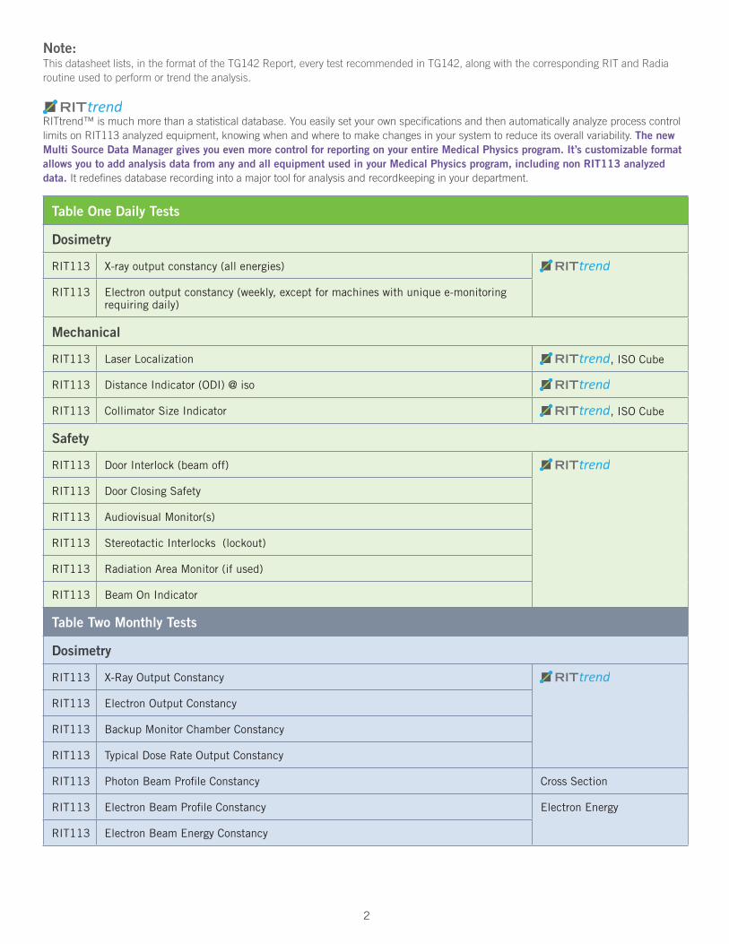
Table One Daily Tests
Dosimetry
RIT113 X-ray output constancy (all energies)
RIT113 Electron output constancy (weekly, except for machines with unique e-monitoring requiring daily)
Mechanical
RIT113 Laser Localization , ISO Cube
RIT113 Distance Indicator (ODI) @ iso
RIT113 Collimator Size Indicator , ISO Cube
Safety
RIT113 Door Interlock (beam off)
RIT113 Door Closing Safety
RIT113 Audiovisual Monitor(s)
RIT113 Stereotactic Interlocks (lockout)
RIT113 Radiation Area Monitor (if used)
RIT113 Beam On Indicator
Table Two Monthly Tests
Dosimetry
RIT113 X-Ray Output Constancy
RIT113 Electron Output Constancy
RIT113 Backup Monitor Chamber Constancy
RIT113 Typical Dose Rate Output Constancy
RIT113 Photon Beam Profile Constancy Cross Section
RIT113 Electron Beam Profile Constancy Electron Energy
RIT113 Electron Beam Energy Constancy
Note: Thisdatasheetlists,intheformatoftheTG142Report,everytestrecommendedinTG142,alongwiththecorrespondingRITandRadiaroutineusedtoperformortrendtheanalysis.
RITtrend™ismuchmorethanastatisticaldatabase.YoueasilysetyourownspecificationsandthenautomaticallyanalyzeprocesscontrollimitsonRIT113analyzedequipment,knowingwhenandwheretomakechangesinyoursystemtoreduceitsoverallvariability.The new Multi Source Data Manager gives you even more control for reporting on your entire Medical Physics program. It’s customizable format allows you to add analysis data from any and all equipment used in your Medical Physics program, including non RIT113 analyzed data. Itredefinesdatabaserecordingintoamajortoolforanalysisandrecordkeepinginyourdepartment.
2 3
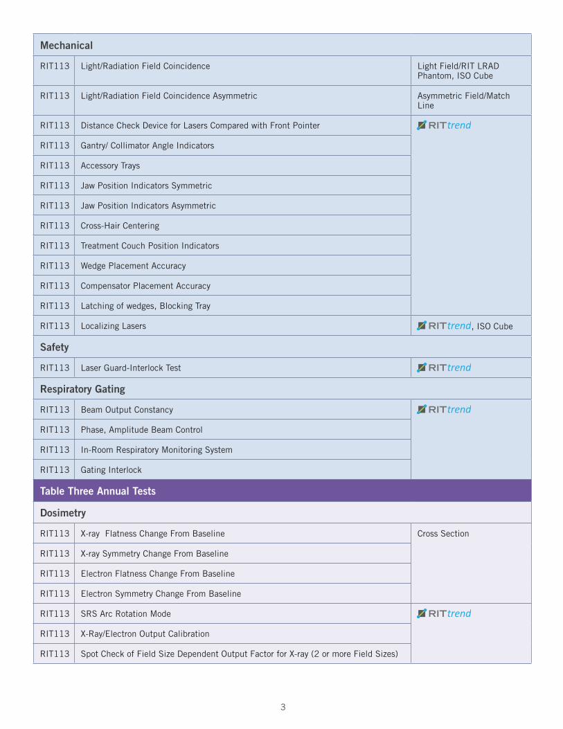
Mechanical
RIT113 Light/Radiation Field Coincidence Light Field/RIT LRAD Phantom, ISO Cube
RIT113 Light/Radiation Field Coincidence Asymmetric Asymmetric Field/Match Line
RIT113 Distance Check Device for Lasers Compared with Front Pointer
RIT113 Gantry/ Collimator Angle Indicators
RIT113 Accessory Trays
RIT113 Jaw Position Indicators Symmetric
RIT113 Jaw Position Indicators Asymmetric
RIT113 Cross-Hair Centering
RIT113 Treatment Couch Position Indicators
RIT113 Wedge Placement Accuracy
RIT113 Compensator Placement Accuracy
RIT113 Latching of wedges, Blocking Tray
RIT113 Localizing Lasers , ISO Cube
Safety
RIT113 Laser Guard-Interlock Test
Respiratory Gating
RIT113 Beam Output Constancy
RIT113 Phase, Amplitude Beam Control
RIT113 In-Room Respiratory Monitoring System
RIT113 Gating Interlock
Table Three Annual Tests
Dosimetry
RIT113 X-ray Flatness Change From Baseline Cross Section
RIT113 X-ray Symmetry Change From Baseline
RIT113 Electron Flatness Change From Baseline
RIT113 Electron Symmetry Change From Baseline
RIT113 SRS Arc Rotation Mode
RIT113 X-Ray/Electron Output Calibration
RIT113 Spot Check of Field Size Dependent Output Factor for X-ray (2 or more Field Sizes)

4
RIT113 Output Factors For Electron Applicators (Spot Check Of One Applicator/Energy)
RIT113 X-ray Beam Quality (PDD10 or TMR20
10)
RIT113 Electron Beam Quality (R50) Electron Energy
RIT113 Physical Wedge Transmission (Factor Constancy)
RIT113 X-Ray Monitor Unit Linearity (Output Constancy)
RIT113 Electron Monitor Unit Linearity (Output Constancy) Electron Energy
RIT113 X-ray Output Constancy vs Dose Rate RapidArc Analysis
RIT113 X-ray Output Constancy vs Gantry Angle
RIT113 Electron Output Constancy vs Gantry Angle
RIT113 Electron and X-ray Off-Axis Factor
RIT113 Arc Mode (Expected MU, Degrees)
RIT113 TBI/TSET Mode
RIT113 PDD or TMR and OAF Constancy
RIT113 TBI/TSET Output Calibration
RIT113 TBI/TSET Accessories
Mechanical
RIT113 Collimator Rotation Isocenter Star Shot, ISO Cube
RIT113 Gantry Rotation Isocenter
RIT113 Couch Rotation Isocenter
RIT113 Electron Applicator Interlocks
Radia/RIT113
Coincidence of Radiation and Mechanical Isocenter Stereotactic Routines / ISO Cube Phantom
RIT113 Table Top Sag , ISO Cube
RIT113 Table Angle
RIT113 Table Travel Maximum Range
RIT113 Stereotactic accessories, lockouts, etc.
Safety
RIT113 Follow manufacturer’s test procedures.
Respiratory Gating
Radia Beam Energy Constancy PTW EPID / PTW EPID Phantom
RIT113 Temporal Accuracy of Phase/Amplitude Gate On
RIT113 Calibration of Surrogate for Respiratory Phase/Amplitude
RIT113 Interlock Testing
4 5
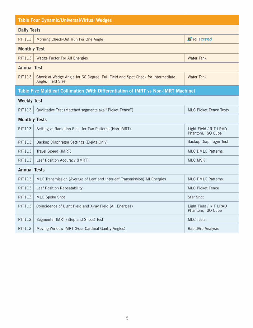
Table Four Dynamic/Universal/Virtual Wedges
Daily Tests
RIT113 Morning Check-Out Run For One Angle
Monthly Test
RIT113 Wedge Factor For All Energies Water Tank
Annual Test
RIT113 Check of Wedge Angle for 60 Degree, Full Field and Spot Check for Intermediate Angle, Field Size
Water Tank
Table Five Multileaf Collimation (With Differentiation of IMRT vs Non-IMRT Machine)
Weekly Test
RIT113 Qualitative Test (Matched segments aka “Picket Fence”) MLC Picket Fence Tests
Monthly Tests
RIT113 Setting vs Radiation Field for Two Patterns (Non-IMRT) Light Field / RIT LRAD Phantom, ISO Cube
RIT113 Backup Diaphragm Settings (Elekta Only) Backup Diaphragm Test
RIT113 Travel Speed (IMRT) MLC DMLC Patterns
RIT113 Leaf Position Accuracy (IMRT) MLC MSK
Annual Tests
RIT113 MLC Transmission (Average of Leaf and Interleaf Transmission) All Energies MLC DMLC Patterns
RIT113 Leaf Position Repeatability MLC Picket Fence
RIT113 MLC Spoke Shot Star Shot
RIT113 Coincidence of Light Field and X-ray Field (All Energies) Light Field / RIT LRAD Phantom, ISO Cube
RIT113 Segmental IMRT (Step and Shoot) Test MLC Tests
RIT113 Moving Window IMRT (Four Cardinal Gantry Angles) RapidArc Analysis

6
Table Six Imaging Tests
Daily Tests
Planar kV and MV (EPID) Imaging
RIT113 Collision Interlocks
RIT113 Position/Reposition , ISO Cube
Radia Imaging and Treatment Coordinate Coincidence (Single Gantry Angle) IGRT / ISO Cube Phantom
Cone-beam CT (kV and MV)
RIT113 Collision Interlocks
Radia Imaging and Treatment Coordinate Coincidence IGRT / ISO Cube Phantom
RIT113 Position/Repositioning , ISO Cube
Monthly Tests
Planar MV Imaging (EPID)
Radia Imaging and Treatment Coordinate Coincidence (Four Cardinal Angles) IGRT / ISO Cube Phantom
Radia Scaling PTW EPID, QC-3, Las Vegas Phantoms
Radia Spatial Resolution
Radia Contrast
Radia Uniformity and Noise
Planar kV Imaging
Radia Imaging and Treatment Coordinate Coincidence (Four Cardinal Angles) IGRT / ISO Cube Phantom
Radia Scaling DISC Plus, Primus L, NORMI 4, TOR 18FG PhantomsRadia Spatial Resolution
Radia Contrast
Radia Uniformity and Noise
Cone-beam CT (kV and MV)
Radia Geometric Distortion Catphan or Siemens OBI/CT, Elekta XVI Phantoms
Radia Spatial Resolution
Radia Contrast
Radia HU Constancy
Radia Uniformity and Noise

6 7
Annual Tests
Planar MV Imaging (EPID)
RIT113 Full Range of Travel SDD
RIT113 Imaging dose
Planar kV Imaging
RIT113 Beam Quaility/Energy
RIT113 Imaging dose
Cone Beam CT (kV and MV)
RIT113 Imaging dose
Visit www.radimage.comCall 719-590-1077Email [email protected]
Catphan is a Registered trademark of The Phantom LaboratoriesPrimus is a Registered trademark of IBACyberknife is a Registered trademark of AccurayNORMI 4 is a Registered trademark of PTW
© Radiological Imaging Technology, Inc., July, 2012
ClassicRIT113
RIT113 Family of Products