Embed Size (px)
Citation preview

1
EU Reference Laboratory for E. coli Department of Veterinary Public Health and Food Safety
Unit of Foodborne Zoonoses Istituto Superiore di Sanità
Risultati del 9° test inter-laboratorio nazionale per l’identificazione della
presenza di ceppi di E. coli produttori di verocitotossina in campioni di
semi di bietola - 2012
Il nono studio inter-laboratorio (Proficiency test, PT) sulla ricerca e l’identificazione dei
ceppi di E. coli produttori di verocitotossina (VTEC) è stato organizzato nel 2012 dal
Laboratorio Nazionale di Riferimento (LNR) per E. coli presso l’Istituto Superiore di Sanità
ai fini della valutazione esterna di qualità dei laboratori coinvolti nel controllo ufficiale degli
alimenti ed è stato dedicato alla ricerca di VTEC appartenenti ai cinque principali
sierogruppi patogeni (O157, O26, O103, O111, O145) in campioni di semi di bietola.
Poiché l’LNR per E. coli è anche Laboratorio Europeo di Riferimento (EU-RL) per questo
patogeno, lo studio nazionale è stato condotto contestualmente a quello dedicato agli LNR
per E. coli degli Stati Membri della UE, che ha visto la partecipazione di 37 LNR attivi nel
settore della sanità pubblica veterinaria e della sicurezza alimentare, rappresentanti 26
Stati Membri dell’Unione Europea, la Norvegia, la Svizzera, la Turchia, la Russia e l’Egitto.
Il report dello studio europeo è disponibile al sito web dell’EU-RL
(http://www.iss.it/vtec/neww/cont.php?id=147&lang=2&tipo=15).
1. PARTECIPANTI
Allo studio nazionale hanno partecipato i 18 laboratori di seguito elencati:
− Centro Servizi Sanitari, Laboratorio di Sanità Pubblica, Trento
− IZS Abruzzo e Molise "G. Caporale", Reparto Igiene degli Alimenti, Teramo
− IZS Puglia e Basilicata, Ricerca e Sviluppo Scientifico, Foggia
− IZS Lombardia ed Emilia Romagna, Reparto di Microbiologia, Laboratorio
Microbiologia, Brescia
− IZS Lombardia ed Emilia Romagna, Sezione di Bologna

2
− IZS Lombardia ed Emilia Romagna, Sezione di Piacenza, Gariga di Podenzano (PC)
− IZS Lazio e Toscana, Roma
− IZS Lazio e Toscana, Dir. Op. Controllo degli Alimenti, Roma
− IZS del Mezzogiorno, Sezione di Salerno, U.O. Microbiologia Alimentare, Fuorni (SA)
− IZS del Mezzogiorno, U.O.S."Biotecnologie applicate agli alimenti-OGM", Portici
(Napoli)
− IZS della Sicilia, Area Microbiologia degli Alimenti, Palermo
− IZS della Sardegna, Laboratorio di Microbiologia e Terreni Colturali, Sassari
− IZS Piemonte, Liguria e Valle d'Aosta, Laboratorio Controllo Alimenti, Torino
− IZS Piemonte, Liguria e Valle d'Aosta, S.C. Biotecnologie, Torino
− IZS Umbria e Marche, Laboratorio Contaminanti Biologici PGCB, Perugia
− IZS Umbria e Marche, Laboratorio Controllo Alimenti, Sezione di Fermo
− IZS delle Venezie, Sezione Pordenone e Udine, Cordenons (PN)
− IZS delle Venezie, OIE/National Reference Laboratory for Salmonellosis, Legnaro (PD)
2. OBIETTIVI E STRUTTURA DEL TEST INTERLABORATORIO
La scelta dei semi destinati alla produzione di germogli come matrice per questo PT è
dovuta al fatto che, negli ultimi anni, numerosi episodi epidemici di infezione da VTEC
sono stati associati al consumo di germogli contaminati. In questi episodi, la
contaminazione del prodotto finito e pronto per il consumo era frequentemente dovuta alla
presenza del contaminante nei semi utilizzati per la produzione.
Gli obiettivi dello studio erano quindi: i) accrescere l’esperienza dei laboratori nell’uso del
metodo molecolare standard per la ricerca dei VTEC; ii) valutare l’efficacia del metodo
stesso su una matrice ad oggi non compresa nel suo campo di applicazione. Infatti, in
assenza di un metodo standard specifico, il metodo sviluppato per la ricerca dei VTEC negli
alimenti è stato adattato per l’analisi dei semi.
Ai laboratori partecipanti è stato richiesto di ricercare i VTEC appartenenti ai 5 sierogruppi
maggiormente coinvolti nelle infezioni umane (O157, O111, O26, O103, and O145),
utilizzando lo standard ISO/TS 13136: Horizontal method for the detection of Shiga toxin-
producing Escherichia coli (STEC) and the determination of O157, O111, O26, O103 and
O145 serogroups. La procedura di laboratorio fornita ai laboratori, riportata in Allegato,
includeva anche indicazioni per il trattamento dei campioni da effettuare prima
dell’applicazione dello lo standard ISO/TS 13136.
3
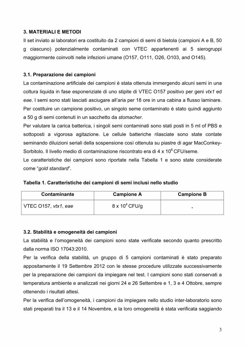
3. MATERIALI E METODI
Il set inviato ai laboratori era costituito da 2 campioni di semi di bietola (campioni A e B, 50
g ciascuno) potenzialmente contaminati con VTEC appartenenti ai 5 sierogruppi
maggiormente coinvolti nelle infezioni umane (O157, O111, O26, O103, and O145).
3.1. Preparazione dei campioni
La contaminazione artificiale dei campioni è stata ottenuta immergendo alcuni semi in una
coltura liquida in fase esponenziale di uno stipite di VTEC O157 positivo per geni vtx1 ed
eae. I semi sono stati lasciati asciugare all’aria per 18 ore in una cabina a flusso laminare.
Per costituire un campione positivo, un singolo seme contaminato è stato quindi aggiunto
a 50 g di semi contenuti in un sacchetto da stomacher.
Per valutare la carica batterica, i singoli semi contaminati sono stati posti in 5 ml of PBS e
sottoposti a vigorosa agitazione. Le cellule batteriche rilasciate sono state contate
seminando diluizioni seriali della sospensione così ottenuta su piastre di agar MacConkey-
Sorbitolo. Il livello medio di contaminazione riscontrato era di 4 x 10⁶ CFU/seme.
Le caratteristiche dei campioni sono riportate nella Tabella 1 e sono state considerate
come “gold standard”.
Tabella 1. Caratteristiche dei campioni di semi inclusi nello studio
Contaminante Campione A Campione B
VTEC O157, vtx1, eae 8 x 10⁴ CFU/g -
3.2. Stabilità e omogeneità dei campioni
La stabilità e l’omogeneità dei campioni sono state verificate secondo quanto prescritto
dalla norma ISO 17043:2010.
Per la verifica della stabilità, un gruppo di 5 campioni contaminati è stato preparato
appositamente il 19 Settembre 2012 con le stesse procedure utilizzate successivamente
per la preparazione dei campioni da impiegare nel test. I campioni sono stati conservati a
temperatura ambiente e analizzati nei giorni 24 e 26 Settembre e 1, 3 e 4 Ottobre, sempre
ottenendo i risultati attesi.
Per la verifica dell’omogeneità, i campioni da impiegare nello studio inter-laboratorio sono
stati preparati tra il 13 e il 14 Novembre, e la loro omogeneità è stata verificata saggiando

4
12 campioni positivi e 12 campioni negativi, selezionati casualmente subito dopo la
preparazione. I test sono stati effettuati il 15 Novembre e hanno prodotto i risultati attesi.
3.3. Invio dei campioni
I campioni, identificati con codici numerici a tre o quattro cifre assegnati casualmente e
diversi per ogni laboratorio, sono stati mantenuti a temperatura ambiente fino al
trasferimento in contenitori per la spedizione. Questa è stata effettuata il 19 Novembre
mediante corriere. Ai laboratori è stato richiesto di iniziare le analisi entro 18 ore dall’arrivo
stesso.
3.4. Raccolta ed elaborazione dei risultati
I laboratori hanno inviato i loro risultati direttamente via WEB, usando pagine dedicate
accessibili attraverso la Restricted Area del sito web dell’EU-RL VTEC (www.iss.it/vtec),
previa introduzione di User ID e Password, inviate a ogni laboratorio insieme al codice
identificativo e alle istruzioni necessarie per il log in. Al termine del test, i partecipanti
hanno avuto la possibilità di stampare direttamente il proprio test-report con i risultati
inviati e quelli attesi.
3.5. Valutazione della performance dei laboratori
La performance è stata valutata in termini di:
- Concordanza (Kappa di Cohen)
- Sensibilità
- Specificità
Per il calcolo di tali parametri sono stati confrontati i risultati delle determinazioni analitiche
ottenute dai laboratori con i valori reali (gold standard) dei campioni oggetto di analisi.
La valutazione del livello di concordanza è stata ottenuta attraverso il calcolo del Kappa di
Cohen che permette di stimare l’accordo tra il risultato analitico e il valore gold standard,
indipendentemente dalla componente imputabile al caso. Per la valutazione
dell’accettabilità del valore Kappa è stata utilizzata la griglia di giudizio proposta da Fleiss
J.L. (Statistical methods for rates and proportions, 1981) secondo la quale valori di
K ≥ 0,75 indicano livelli di concordanza eccellenti, valori 0,40 ≤ K < 0,75 buona
concordanza e valori K < 0,40 livelli di concordanza scarsi. La sensibilità diagnostica è
stata definita come la proporzione di campioni positivi correttamente identificati (presenza
dei geni vtx1, vtx2, eae e sierogruppo specifici). La specificità diagnostica è stata definita
5
come la proporzione di campioni negativi identificati correttamente (assenza dei geni vtx1,
vtx2, eae e sierogruppo specifici). Per tutti i parametri è stato calcolato il relativo intervallo
di confidenza (95 % I.C.).
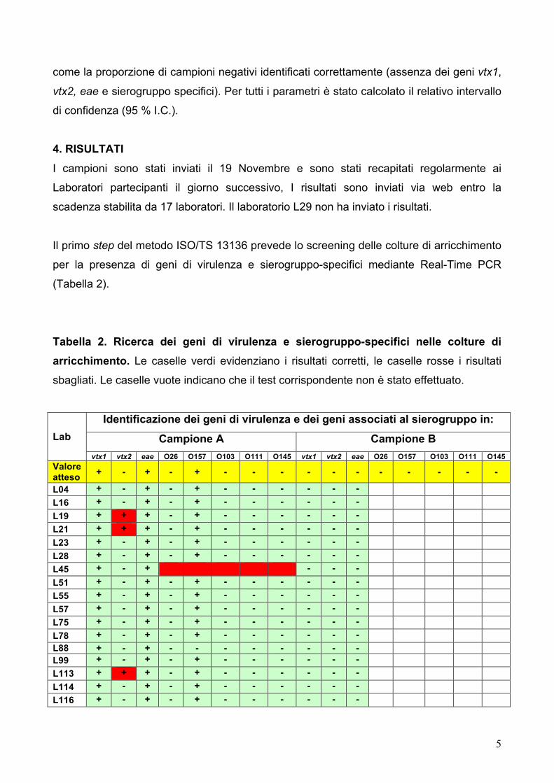
4. RISULTATI
I campioni sono stati inviati il 19 Novembre e sono stati recapitati regolarmente ai
Laboratori partecipanti il giorno successivo, I risultati sono inviati via web entro la
scadenza stabilita da 17 laboratori. Il laboratorio L29 non ha inviato i risultati.
Il primo step del metodo ISO/TS 13136 prevede lo screening delle colture di arricchimento
per la presenza di geni di virulenza e sierogruppo-specifici mediante Real-Time PCR
(Tabella 2).
Tabella 2. Ricerca dei geni di virulenza e sierogruppo-specifici nelle colture di
arricchimento. Le caselle verdi evidenziano i risultati corretti, le caselle rosse i risultati
sbagliati. Le caselle vuote indicano che il test corrispondente non è stato effettuato.
Lab Identificazione dei geni di virulenza e dei geni associati al sierogruppo in:
Campione A Campione B vtx1 vtx2 eae O26 O157 O103 O111 O145 vtx1 vtx2 eae O26 O157 O103 O111 O145
Valore atteso + - + - + - - - - - - - - - - -
L04 + - + - + - - - - - - L16 + - + - + - - - - - - L19 + + + - + - - - - - - L21 + + + - + - - - - - - L23 + - + - + - - - - - - L28 + - + - + - - - - - - L45 + - + - - - L51 + - + - + - - - - - - L55 + - + - + - - - - - - L57 + - + - + - - - - - - L75 + - + - + - - - - - - L78 + - + - + - - - - - - L88 + - + - - - - - - - - L99 + - + - + - - - - - - L113 + + + - + - - - - - - L114 + - + - + - - - - - - L116 + - + - + - - - - - -

6
Questa parte dello studio è stata effettuata correttamente da 13 laboratori che, hanno
quindi ottenuto valori individuali di sensibilità e specificità del 100% e valori di
concordanza, valutati tramite il Kappa di Cohen, pari a 1, con limiti di confidenza pari a
0,41 – 1.
Tre laboratori (L19, L21, L113) hanno utilizzato kit commerciali che individuano la
presenza dei geni vtx ma non sono in grado di distinguere i geni vtx1 da quelli vtx2. Questi
laboratori hanno quindi riportato correttamente la presenza di geni vtx nel campione A,
segnalando però la presenza di entrambe le varianti geniche. Per questi laboratori la
sensibilità era pari al 100%, la specificità all’ 87,5% e il valore di K era 0,79, con limiti di
confidenza pari a 0,21 - 1.
Un laboratorio (L45) non ha effettuato la ricerca dei geni sierogruppo-specifici, ottenendo
sensibilità e specificità del 100% e un valore di K pari a 1, con limiti di confidenza pari a
0,21 - 1, visto il minor numero di test effettuati.
La successiva fase dell’isolamento dei ceppi VTEC dai campioni PCR-positivi è stata
condotta da 15 laboratori, mentre I laboratori L45 e L113 non hanno inviato i risultati
relativi all’isolamento.Questi 15 laboratori, dopo aver identificato correttamente la
presenza del gene associato al sierogruppo O157 nelle colture di arricchimento, hanno
anche isolato e geno-tipizzato correttamente il ceppo VTEC O157 dal campione A, con
l’eccezione del laboratorio L21, che ha segnalato la presenza di entrambe le varianti dei
geni vtx. L’errore era ancora dovuto all’utilizzo di kit commerciali non in grado di
distinguere i geni vtx1 da quelli vtx2.

7
Tabella 3. Isolamento e geno-tipizzazione dei ceppi VTEC dalle colture di arricchimento Real Time PCR-positive. Le caselle verdi evidenziano i risultati corretti, le
caselle rosse i risultati sbagliati. Le caselle vuote indicano che il test corrispondente non è
stato effettuato.
5. Considerazioni - Lo studio è stato condotto a termine da 17 laboratori coinvolti nel controllo ufficiale degli
alimenti, che includevano 9 Istituti Zooprofilattici Sperimentali.
- Tutti i laboratori hanno effettuato la fase di screening molecolare delle colture di
arricchimento mediante Real-time PCR, identificando correttamente il campione
contaminato da VTEC.
- Quindici laboratori hanno effettuato la fase dell’isolamento del ceppo di VTEC O157
dalla coltura PCR-positive, riportando una performance analitica eccellente.
- Questo studio ha confermato che il metodo ISO/TS 13136 basato sullo screening
mediante Real-time PCR delle colture di arricchimento rappresenta uno strumento
robusto per la ricerca dei VTEC anche quando applicato a matrici non strettamente
comprese nel suo campo di applicazione.
Laboratorio Isolamento e geno-tipizzazione dei ceppi VTEC dalle colture di
arricchimento Campione A Campione B
VTEC Genotipo VTEC Genotipo Valore atteso O157 vtx1 vtx2 eae Nessuno vtx1 vtx2 eae
+ - + - - - L04 O157 + - + L16 O157 + - + L19 O157 + - + L21 O157 + + + L23 O157 + - + L28 O157 + - + L51 O157 + - + L55 O157 + - + L57 O157 + - + L75 O157 + - + L78 O157 + - + L88 O157 + - + L99 O157 + - + L114 O157 + - + L116 O157 + - +

8
ALLEGATO
EU Reference Laboratory for E.coli Department of Veterinary Public Health and Food Safety
Unit of Foodborne Zoonoses Istituto Superiore di Sanità
9th inter-laboratory study on the detection of Verocytotoxin-producing E. coli (VTEC) belonging to the serogroups most involved in human infections (O157, O111, O26,
O103, and O145) in samples of seeds intended for sprout production
Laboratory Guideline Introduction In the absence of specific international standards for the detection of VTEC in seeds, the
method developed for the detection of VTEC in foodstuffs, CEN/ISO TS 13136
“Microbiology of food and animal feed -- Real-time polymerase chain reaction (PCR)-
based method for the detection of food-borne pathogens -- Horizontal method for the
detection of Shiga toxin-producing Escherichia coli (STEC) and the determination of O157,
O111, O26, O103 and O145 serogroups”, has been adapted to this matrix. In particular,
the following aspects have been considered:
1. Seeds are generally contaminated at very low levels. Nonetheless, the sprouting
process is characterized by conditions (humidity, heat) favoring the pathogen’s
enrichment. Therefore, 50 gr of seeds are analyzed instead of the usual 25 gr of food
items, in order to increase the sensitivity of the assay.
− Seeds are generally dried. Therefore, the contaminating pathogens are supposed to be
stressed.
− The contamination may occur on the surface of the seeds as well as inside their body.
− The enrichment cultures of seeds may contain inhibitors of the DNA polymerase used
for the PCR screening of the samples.
The procedure comprises the following sequential steps:
- Smashing of the seeds to allow the release of possibly internalized bacteria;
- Transfer of the sample to the enrichment medium;
- Microbial enrichment;

9
- Nucleic acid extraction;
- Detection of virulence genes;
- Detection of serogroup-associated genes;
- Isolation from positive samples.
1. Treatment of the seed samples The samples are constituted by 50 gr of seeds placed in a stomacher bag.
1. The seeds are smashed directly in the stomacher bag, using a mortar with pestel or
other similar tools, before adding the enrichment broth. It is advisable to put the bag
with the sample into another sterile stomacher bag, to limit the possibility of spill over
due to cuts in the bag.
2. The smashed seeds are added with 450 ml BPW and incubated for 24 hrs at 37°C
(either static or in agitation). Check carefully for the integrity of the stomacher bag after
smashing the seeds. In case of damages evidence transfer the smashed seeds
aseptically to a sterile container (flask or a new stomacher bag) before adding the
culture medium. This operation must be done under a Biohazard laminar flow hood.
3. A 5 ml aliquot of the enrichment culture is taken, mixed by vortex (in order to detach as
much as possible the bacteria possibly adhering to seeds), centrifuged at 500 X g 1
min to sediment the seeds’ debris.
4. One ml aliquot of the supernatant is taken at this stage and used for DNA preparation.
2. Nucleic acid extraction, detection of virulence and serogroup-associated genes, and isolation of VTEC This procedure is described in the Annex 1. Briefly, it is based on a Real-time PCR
screening of enrichment cultures to detect the presence of virulence genes (vtx1 and vtx2,
and eae) and serogroup-specific genes for O26, O103, O111 and O145, followed by the
isolation of VTEC from PCR-positive samples, accomplished by an immuno-concentration
enrichment step specific for the serogroups identified in the PCR step.
One ml aliquot of the enrichment culture performed and treated as described in the
previous paragraph is used for DNA extraction and purification, accomplished according
to the ISO 20837 “Microbiology of food and animal feeding stuffs - Polymerase chain
reaction (PCR) for the detection of foodborne pathogens - Requirements for sample
preparation for qualitative detection”. The remaining culture shall be stored at 4°C for the
isolation steps that will follow a positive PCR result.

10
To perform the Real-time PCR, the DNA sample is diluted 1:10 before use. In the case of
absence of amplification of the internal amplification control (IAC), the DNA template is
used at the dilution of 1:30.
The method is sequential:
Step 1: Detection of the genes vtx1, vtx2 and eae.
Step 2: Samples positive for both vtx and eae are tested for the serogroup-associated
genes (molecular serogrouping).
Step 3: Isolation of the VTEC strain; samples positive at the same time for vtx, eae, and at
least one of the serogroup-associated genes are submitted to a further step aimed
at isolation of the VTEC strain. This requires serogroup-specific enrichment based
on IMS or other immuno-capture suitable approaches. A guideline for the isolation
of the different VTEC serogroups is also included in the Annex 1.
Step 4: Characterization of the isolate i.e. identification, detection of vtx genes, the eae
gene and the serogroup gene.

11
Annex 1
EU Reference Laboratory for E.coli Department of Veterinary Public Health and Food Safety
Unit of Foodborne Zoonoses Istituto Superiore di Sanità
Laboratory procedure for the detection of Shiga toxin (Verocytotoxin)-producing Escherichia coli (STEC/VTEC) belonging to O157, O111, O26,
O103 and O145 serogroups in seed samples - Qualitative Method
Introduction Shiga toxin-producing Escherichia coli (STEC) cause severe disease in humans such as
haemorrhagic colitis and haemolitic uraemic syndrome (HUS). Although STEC may belong
to a large number of serogroups, those that have been firmly associated with severe
human disease, in particular HUS, belong to O157, O26, O111, O103, O145 (1), and
represent the targets of this Laboratory Procedure.
In this Laboratory Procedure, the wording Shiga toxin (Stx) is synonymous of
Verocytotoxin (Vtx).
The following nomenclature has been adopted throughout the text:
stx: Shiga toxin genes (synonymous of vtx)
Stx: Shiga toxin (synonymous of Vtx)
STEC: Shiga toxin-producing Escherichia coli (synonymous of VTEC: Verocytotoxin-
producing Escherichia coli).
1. Scope
This Laboratory Procedure describes a method for the detection of (i) the major virulence
genes of STEC (2,3), and (ii) the genes associated with the serogoups O157, O111, O26,
O103 and O145 (3,4).
In the case of detection of these genes, the isolation of the strain is attempted, to confirm
the simultaneous presence of the genes in the same live bacterial cell.

12
The analytical approach is based on the use of the Real-time PCR..
This Laboratory Procedure is applicable to enrichment cultures from seed samples.
2. Normative references
ISO/DIS 7218
General requirements and guidance for microbiological examinations
ISO/DIS 20837
Requirements for sample preparation for qualitative detection
ISO/DIS 20838
Requirements for amplification and detection of qualitative methods
ISO/DIS 22174
General method specific requirements
3. Terms and definitions
3.1 Shiga toxin-producing Escherichia coli (STEC): Microrganism possessing the Stx-
coding genes
3.2 Shiga toxin-producing Escherichia coli (STEC) potentially pathogenic to humans:
Microrganism possessing the Stx-coding genes and the intimin-coding gene eae.
3.3 Shiga toxin-producing Escherichia coli (STEC) highly pathogenic to humans:
Microrganism possessing the Stx-coding genes, the intimin-coding gene eae and
belonging to one of the serogroups in the scope of the present Laboratory Procedure.
4. Principle
4.1 General The detection of STEC and of the 5 serogroups comprises the following sequential steps:
1. Microbial enrichment
2. Nucleic acid extraction
3. Detection of virulence genes
4. Detection of serogroup-associated genes in samples positive to point 3
5. Isolation from samples positive to points 3 and 4.

13
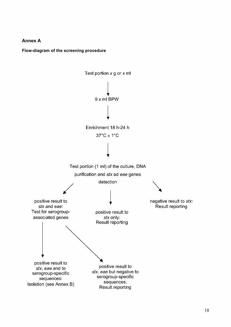
A flow-diagram of the whole procedure is given in Annex A.
4.1.1 Microbial enrichment The number of STEC cells to be detected is increased by incubating the test portion in the
non-selective liquid nutrient medium Buffered peptone water (BPW).
The BPW is to be used to analyse the seeds samples since they are supposed to contain
stressed target bacteria (dryed matrix).
4.1.2 Nucleic acid extraction
Bacterial cells are separated from the enrichment medium and lysed. The nucleic acid is
then purified according to any protocol allowing the production of template DNA suitable
for Real Time PCR.
4.1.3 Target genes The purified nucleic acid is used for the detection of the following target genes:
- the main virulence genes for STEC: stx genes, encoding the Shiga toxins and the
eae gene, encoding a 90KDa protein, the intimin, involved in the attaching and
effacing mechanism of adhestion, a typical feature of the pathogenic STEC strains.
- the rfbE (O157), wbdl(O111), wzx(O26), ihp1(O145) and wzx(O103) genes, to
identify the corresponding serogroups.
4.1.4 Detection The Real-time PCR products are detected by light emission in a 5’ nuclease PCR assay.
4.1.5 Isolation Once a STEC is detected and the serogroup identified, in order to isolate the STEC strain,
a serogroup-specific enrichment is performed followed by plating onto the agar Trpytone-
bile-glucuronic medium (TBX) or onto a specific selective medium where available (see
note in Annex A).
14
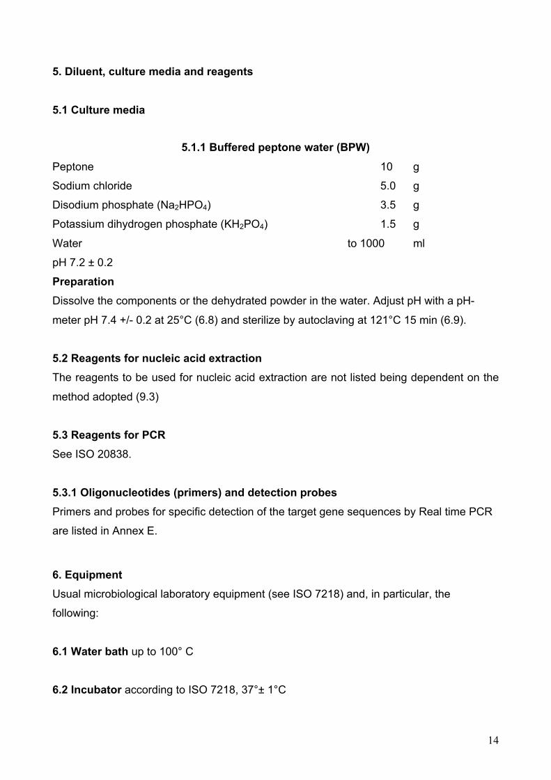
5. Diluent, culture media and reagents 5.1 Culture media
5.1.1 Buffered peptone water (BPW) Peptone 10 g
Sodium chloride 5.0 g
Disodium phosphate (Na2HPO4) 3.5 g
Potassium dihydrogen phosphate (KH2PO4) 1.5 g
Water to 1000 ml
pH 7.2 ± 0.2
Preparation Dissolve the components or the dehydrated powder in the water. Adjust pH with a pH-
meter pH 7.4 +/- 0.2 at 25°C (6.8) and sterilize by autoclaving at 121°C 15 min (6.9).
5.2 Reagents for nucleic acid extraction The reagents to be used for nucleic acid extraction are not listed being dependent on the
method adopted (9.3)
5.3 Reagents for PCR See ISO 20838.
5.3.1 Oligonucleotides (primers) and detection probes Primers and probes for specific detection of the target gene sequences by Real time PCR
are listed in Annex E.
6. Equipment Usual microbiological laboratory equipment (see ISO 7218) and, in particular, the
following:
6.1 Water bath up to 100° C
6.2 Incubator according to ISO 7218, 37°± 1°C

15
6.3 Appatarus for nucleic acid extraction Appropriate equipment according to the method adopted. If needed.
6.4 Pipettes for volumes between 1µl and 1000 µl
6.5 Thin walled Real-Time PCR microtubes (0,2 ml /0,5 ml reaction tubes), multi-well
PCR microplates or other suitable light transparent disposable plasticware. 6.6 Thermal cycler Several brands of apparatus are available and can be chosen
according to the laboratory policies.
6.7 Apparatus for detection of the PCR product Light emission following 5’ nuclease PCR assay is detected by the Real-time PCR
apparatus
6.8 pH-meter capable of measuring to an accuracy of +/- 0.05 pH units and its resolution
shall be 0.01 pH units
6.9 Autoclave according to ISO 7218
6.10 Stomacher peristaltic blender with sterile bags possibly with a device for adjustin
speed and time
7. Sampling In the absence of specific International Standards dealing with sampling of the product
concerned, it is recommended that the parties concerned come to an agreement on this
subject.
8. Preparation of test sample Prepare the test sample in accordance with the specific International Standard dealing with
the product concerned where available, or other suitable guidelines.

16
9. Procedure 9.1 Test portion and initial suspension 9.1.1 General Use the necessary quantity of enrichment medium to yield a final dilution of 1/10 of the
original test portion (50 gr seeds + 450 ml BPW).
9.2 Enrichment 9.2.1 Incubation
Incubate the stomacher bag or the tube/bottle (9.1.2) at 37°C ± 1°C for 16 h to18 h.
9.2.2 Process control (for Real-time PCR)
Perform process control according to ISO 22174.
Guidance on internal amplification control (IAC) and process control are given in Annex D.
9.3 Nucleic acid preparation
Use an appropriate nucleic acid extraction procedure for Gram-negative bacteria. A
comprehensive collection of methods can be found in (7). Alternatively, commercial kits
may be used according to the manufacturers’ instructions.
9.4 Real-time PCR amplification 9.4.1 General Follow all requirements for the PCR amplification as described in ISO 20838 “Microbiology
of food and animal feeding stuffs – Polymerase chain reaction for the detection of food
pathogens – amplification and detection”.
Primers and detection probes for conducting the real-time PCR are described in Annex E.
9.4.2 PCR controls In accordance with ISO 22174, examples of PCR controls are given in Annex D.
9.4.3 Detection of PCR products Light emission is captured by the apparatus once generated during the amplification.

17
9.4.4 Interpretation of PCR results The PCR results obtained, including the controls specified in ISO 22174 and in Annex D,
are interpreted by the software linked to the apparatus. During amplification, the Software
monitors 5’-nuclease PCR amplification by analysing fluorescence emissions (Rn) of the
reporter dye for each sample. ΔRn was Rn minus the baseline reporter dye intensity
established in the first few cycles. At the end of the PCR, a reaction was considered
positive if its ΔRn curve exceeded the threshold, defined as 10 times the standard
deviation of the mean baseline emission calculated between the first few cycles. The cycle
threshold (Ct) was defined as the cycle number at which a sample’s ΔRn fluorescence
crossed the determined threshold value.
If the results are ambiguous, check the emission curves. Positive samples give a curve
with a clear increase in fluorescence, starting from a number of cycles corresponding to
the Ct.
If the controls yield unexpected results, repeat the procedure.
9.5 Strain isolation The pattern of virulence genes, exhibited by STEC which are considered to be pathogenic
to humans, is complex and it is possible that different strains presenting only part of the
virulence gene pattern may be present in the food sample at the same time. Therefore, the
isolation of the STEC strains is required to confirm that the positive PCR signals are
generated from genes that are simultaneously present in the same live bacterial cell.
A serogroup-specific enrichment followed by direct plating onto suitable solid media and
screening of the colonies for the presence of the virulence genesis are required.
The real-time PCR protocol described in this Laboratory Procedure may be used in order
to confirm the presence of the virulence genes in the isolated colonies. In alternative other
equivalent PCR protocols can be downloaded from the EU RL VTEC website
(http://www.iss.it/vtec/work/cont.php?id=152&lang=2&tipo=3).
STEC isolation is described in the flow chart of Annex B
18
Annex A
Flow-diagram of the screening procedure

19
Annex B Flow-diagram of the isolation procedure
20
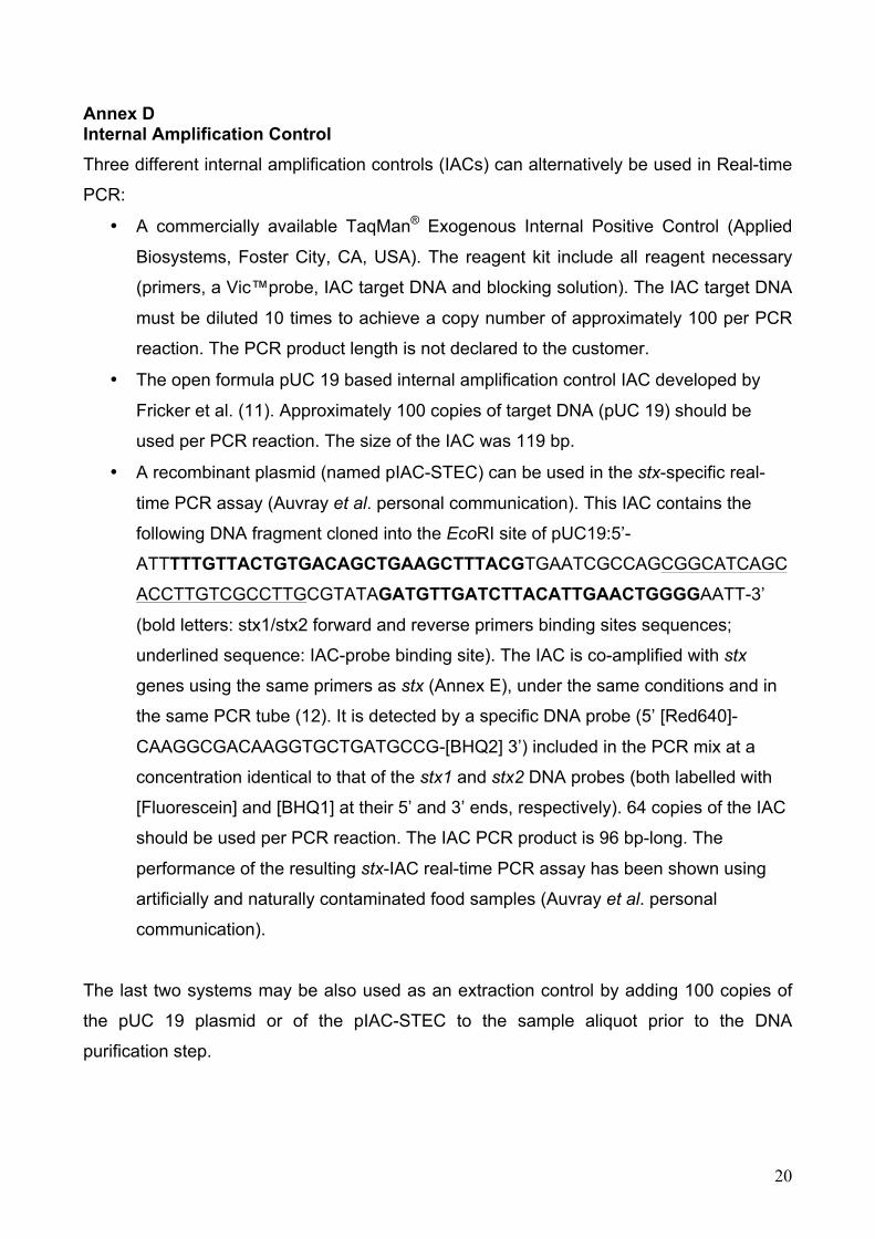
Annex D Internal Amplification Control Three different internal amplification controls (IACs) can alternatively be used in Real-time
PCR:
• A commercially available TaqMan® Exogenous Internal Positive Control (Applied
Biosystems, Foster City, CA, USA). The reagent kit include all reagent necessary
(primers, a Vic™probe, IAC target DNA and blocking solution). The IAC target DNA
must be diluted 10 times to achieve a copy number of approximately 100 per PCR
reaction. The PCR product length is not declared to the customer.
• The open formula pUC 19 based internal amplification control IAC developed by
Fricker et al. (11). Approximately 100 copies of target DNA (pUC 19) should be
used per PCR reaction. The size of the IAC was 119 bp.
• A recombinant plasmid (named pIAC-STEC) can be used in the stx-specific real-
time PCR assay (Auvray et al. personal communication). This IAC contains the
following DNA fragment cloned into the EcoRI site of pUC19:5’-
ATTTTTGTTACTGTGACAGCTGAAGCTTTACGTGAATCGCCAGCGGCATCAGC
ACCTTGTCGCCTTGCGTATAGATGTTGATCTTACATTGAACTGGGGAATT-3’
(bold letters: stx1/stx2 forward and reverse primers binding sites sequences;
underlined sequence: IAC-probe binding site). The IAC is co-amplified with stx
genes using the same primers as stx (Annex E), under the same conditions and in
the same PCR tube (12). It is detected by a specific DNA probe (5’ [Red640]-
CAAGGCGACAAGGTGCTGATGCCG-[BHQ2] 3’) included in the PCR mix at a
concentration identical to that of the stx1 and stx2 DNA probes (both labelled with
[Fluorescein] and [BHQ1] at their 5’ and 3’ ends, respectively). 64 copies of the IAC
should be used per PCR reaction. The IAC PCR product is 96 bp-long. The
performance of the resulting stx-IAC real-time PCR assay has been shown using
artificially and naturally contaminated food samples (Auvray et al. personal
communication).
The last two systems may be also used as an extraction control by adding 100 copies of
the pUC 19 plasmid or of the pIAC-STEC to the sample aliquot prior to the DNA
purification step.

21
ANNEX E The Real-time PCR protocol described is based on the use of the following primers and
probes which shall be considered as refererence reagents. However, other primers and
probes may be used provided that they have been recognised equivalent to those
indicated in the tables E.1 and E.2 according to the ISO 16140 rules.
Primers and probes for the PCR assays
Tables E.1 and E.2 provides respectively the primers and probes sequences for:
- the detection of stx and eae genes by real-time-PCR (PCR A);
- the detection of serogroup-related genes genes using real-time-PCR (PCR B).
In these tables , the chemistry of the reporter and quencher phluorophores is not indicated,
being largely dependent on the real time PCR instruments available in each laboratory.
Table E.1: Degenerate primers and TaqMan probes used for 5’ nuclease PCR assays. (§3 and *2)
Target gene
Forward primer, reverse primer and probe sequences (5’-3’)a
Amplicon size (bp)
Location within sequence
GenBank accession number
stx1§ TTTGTYACTGTSACAGCWGAAGCYTTACG
CCCCAGTTCARWGTRAGRTCMACRTC Probe-CTGGATGATCTCAGTGGGCGTTCTTATGTAA
131 878–906 983–1008
941–971
M16625
stx2§ b
TTTGTYACTGTSACAGCWGAAGCYTTACG
CCCCAGTTCARWGTRAGRTCMACRTC
Probe-TCGTCAGGCACTGTCTGAAACTGCTCC
128 785–813 785–813 838–864
X07865
eae* CAT TGA TCA GGA TTT TTC TGG TGA TA
CTC ATG CGG AAA TAG CCG TTA
Probe-ATAGTCTCGCCAGTATTCGCCACCAATACC
102 899-924
1000-979
966-936
Z11541
a In the sequence Y is (C, T), S is (C, G), W is (A, T), R is (A, G), M is (A, C). b This combination of primer/probe recognises all the stx2 variants but the stx2f

22
Table E.2. Primers and probes used for amplification of O antigen specific genes in 5’ nuclease PCR assays. (§3 and *4)
Target gene
(serogroup) Forward primer, reverse primer and probe sequences
(5’-3’) Amplicon
size (bp) Location within
sequence GenBank accession
number §rfbE (O157) TTTCACACTTATTGGATGGTCTCAA
CGATGAGTTTATCTGCAAGGTGAT
Probe-AGGACCGCAGAGGAAAGAGAGGAATTAAGG
88 348–372
412–435 381–410
AF163329
§wbdI (O111) CGAGGCAACACATTATATAGTGCTTT
TTTTTGAATAGTTATGAACATCTTGTTTAGC
Probe-TTGAATCTCCCAGATGATCAACATCGTGAA
146 3464–3489
3579–3609
3519–3548
AF078736
§wzx (O26) CGCGACGGCAGAGAAAATT
AGCAGGCTTTTATATTCTCCAACTTT
Probe-CCCCGTTAAATCAATACTATTTCACGAGGTTGA
135 5648–5666
5757–5782
5692–5724
AF529080
§ihp1 (O145) CGATAATATTTACCCCACCAGTACAG
GCCGCCGCAATGCTT
Probe-CCGCCATTCAGAATGCACACAATATCG
132 1383–1408
1500–1514
1472–1498
AF531429
*wzx (O103) CAAGGTGATTACGAAAATGCATGT
GAAAAAAGCACCCCCGTACTTAT
Probe-CATAGCCTGTTGTTTTAT
99 4299–4323
4397–4375
4356–4373
AY532664

23
Annex F Isolation of STEC strains
Follow the procedure described below to isolate STEC strains from real time PCR positive
samples:
1) Perform a serogroup-specific enrichment (SSE) on the remaining enrichment culture
(see Note 1)
2) Streak SSE onto TBX or other suitable medium (see note 2). Incubate for 18 to 24 hours
at 37°C
3) Pick up 10 to 50 colonies with E. coli morphology or with characteristic aspect (see Note
5) and point-inoculate on nutrient agar (NA) (see Note 3) and H2O (the colonies may be
pooled in water up to a number of ten per pool).
4) Perform the detection of the stx-coding gene and the eae gene on the isolated colonies
or the H2O pools (see Note 4).
5) If a pool is positive, go back to NA and assay the individual colonies forming the positive
pool in order to select one single positive colony.
6) Identify the colonies as E. coli and confirm the serogroup the sample was positive to in
the screening PCR assay (e.g. by PCR B in the Annex E), see Note 5.
7) Isolates may be sent to the a Reference Laboratory for further characterization.
NOTE 1: Serogroup-specific enrichment may be achieved by using immunocapture
systems such as immuno-magnetic separation (IMS) or equivalent. Generally, refer to the
instruction supplied by the manufacturer.
For O157 positive samples, use ISO 16654 or alternative methods validated according to
ISO 16140.
NOTE 2: For O157 positive samples, use ISO 16654 or alternative methods validated
according to ISO 16140. Sorbitol-fermenting E. coli O157 are susceptible to tellurite
contained in the CT SMAC medium indicated in ISO 16654. Therefore the use of a second
SMAC isolation plate without antibiotics is recommended. In the absence of Sorbitol-
negative colonies on the plates, the screening of Sorbitol-positive colonies is suggested.
For STEC O26 isolation, a differential solid media (MacConkey) containing Rhamnose
instead of lactose is commercially available (RMAC). It is very effective in distinguishing
STEC O26 strains, which do not ferment Rhamnose, from other E. coli.
NOTE 3: There are several types of nutrient agar media commercially available either
ready to use plates or prepared in house from dehydrated powders. Every type of non-
24
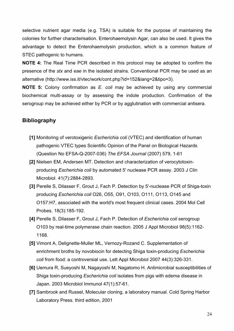
selective nutrient agar media (e.g. TSA) is suitable for the purpose of maintaining the
colonies for further characterisation. Enterohaemolysin Agar, can also be used. It gives the
advantage to detect the Enterohaemolysin production, which is a common feature of
STEC pathogenic to humans.
NOTE 4: The Real Time PCR described in this protocol may be adopted to confirm the
presence of the stx and eae in the isolated strains. Conventional PCR may be used as an
alternative (http://www.iss.it/vtec/work/cont.php?id=152&lang=2&tipo=3).
NOTE 5: Colony confirmation as E. coli may be achieved by using any commercial
biochemical multi-assay or by assessing the indole production. Confirmation of the
serogroup may be achieved either by PCR or by agglutination with commercial antisera.
Bibliography
[1] Monitoring of verotoxigenic Escherichia coli (VTEC) and identification of human
pathogenic VTEC types Scientific Opinion of the Panel on Biological Hazards
(Question No EFSA-Q-2007-036) The EFSA Journal (2007) 579, 1-61
[2] Nielsen EM, Andersen MT. Detection and characterization of verocytotoxin-
producing Escherichia coli by automated 5' nuclease PCR assay. 2003 J Clin
Microbiol. 41(7):2884-2893.
[3] Perelle S, Dilasser F, Grout J, Fach P. Detection by 5'-nuclease PCR of Shiga-toxin
producing Escherichia coli O26, O55, O91, O103, O111, O113, O145 and
O157:H7, associated with the world's most frequent clinical cases. 2004 Mol Cell
Probes. 18(3):185-192.
[4] Perelle S, Dilasser F, Grout J, Fach P. Detection of Escherichia coli serogroup
O103 by real-time polymerase chain reaction. 2005 J Appl Microbiol 98(5):1162-
1168.
[5] Vimont A, Delignette-Muller ML, Vernozy-Rozand C. Supplementation of
enrichment broths by novobiocin for detecting Shiga toxin-producing Escherichia
coli from food: a controversial use. Lett Appl Microbiol 2007 44(3):326-331.
[6] Uemura R, Sueyoshi M, Nagayoshi M, Nagatomo H. Antimicrobial susceptibilities of
Shiga toxin-producing Escherichia coli isolates from pigs with edema disease in
Japan. 2003 Microbiol Immunol 47(1):57-61.
[7] Sambrook and Russel, Molecular cloning, a laboratory manual. Cold Spring Harbor
Laboratory Press. third edition, 2001

25
[8] Paton AW, Paton JC. Detection and characterization of Shiga toxigenic Escherichia
coli by using multiplex PCR assays for stx1, stx2, eaeA, enterohemorrhagic E. coli
hlyA, rfbO111, and rfbO157. 1998J Clin Microbiol 36: 598-602.
[9] O'Brien AD, Newland JW, Miller SF, Holmes RK, Smith HW, Formal SB. Shiga-like
toxin-converting phages from Escherichia coli strains that cause hemorrhagic colitis
or infantile diarrhea. 1984 Science 226: 694-696.
[10] Perna N.T. et al. Genome sequence of enterohaemorrhagic Escherichia coli
O157:H7. 2001 Nature 409: 529-533.
[11] Fricker M, Messelhäusser U, Busch U, Scherer S, Ehling-Schulz M. Diagnostic real-
time PCR assays for the detection of emetic Bacillus cereus strains in foods and
recent food-borne outbreaks. 2007 Appl Environ Microbiol 73(6):1892-1898.
[12] Hoorfar J, Malorny B, Abdulmawjood A, Cook N, Wagner M, Fach P. Practical
considerations in design of internal amplification controls for diagnostic PCR
assays. 2004 J Clin Microbiol 42(5):1863-1868.
[13] Beutin L, Jahn S, Fach P. Evaluation of the 'GeneDisc' real-time PCR system for
detection of enterohaemorrhagic Escherichia coli (EHEC) O26, O103, O111, O145
and O157 strains according to their virulence markers and their O- and H-antigen-
associated genes. 2009. J Appl Microbiol. 106:1122-32.