Embed Size (px)
Citation preview
Risks
High-energy ESW can destroy non myelinated nerves but afterwards there is an even faster regeneration (Lee 2007, Hausdorf 1 2008, Wu 2008, Ohtori 2013)
Low energy ESW do not damage motoric or somatosensible nerves. (Manganonotti 2012) They have no influence on the nociceptive system. (Haake 2002).
Avoid Pain! Move the therapy source constantly! Stimulation: Yes! Irritation: No! ESW on painful or sensitive structures are painful Too many ESW on one spot cause pain.
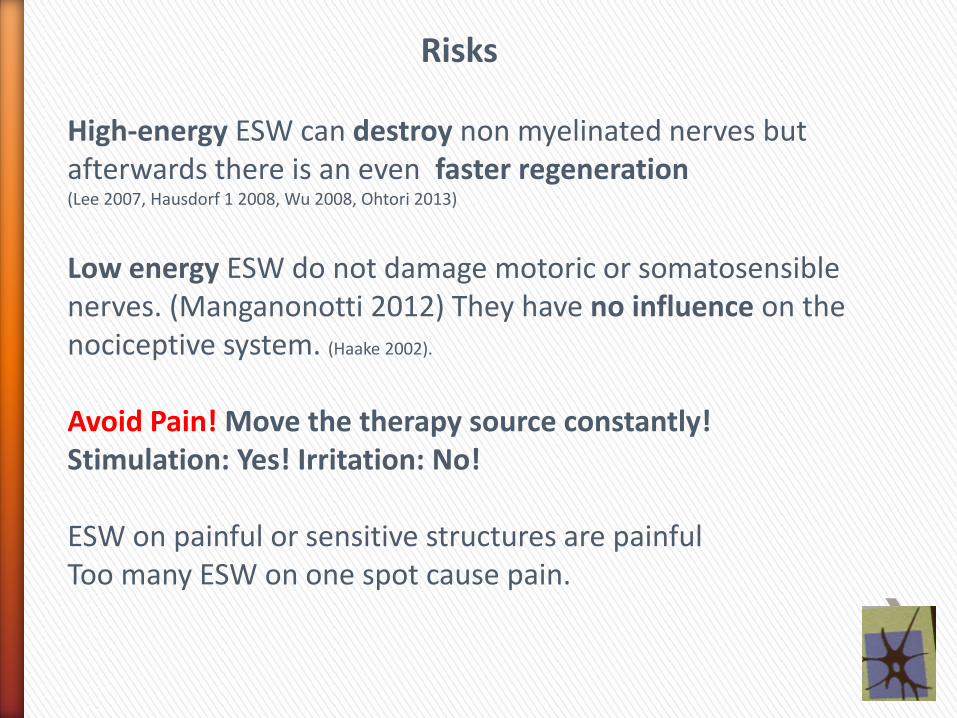
I’m throwing you a ball. Please, Play with it.
Photo of a shock wave by O. Wess Storz Medical
Focused extracorporeal shock waves Improve pareses
in 8 cases of spinal cord injury and 3 cases of myelomeningocele.
H. Lohse-Busch, U. Reime, R. Failand
Rheintalklinik D-79189 Bad Krozingen
The first author received research grant f From Storz Medical AG Tagerwilen
Methods
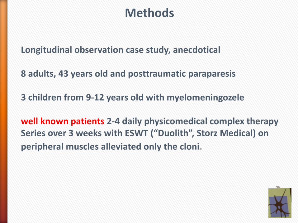
Longitudinal observation case study, anecdotical 8 adults, 43 years old and posttraumatic paraparesis 3 children from 9-12 years old with myelomeningozele well known patients 2-4 daily physicomedical complex therapy Series over 3 weeks with ESWT (“Duolith”, Storz Medical) on
peripheral muscles alleviated only the cloni.
Methods
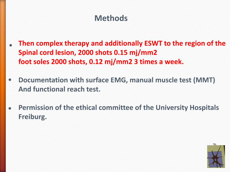
Then complex therapy and additionally ESWT to the region of the Spinal cord lesion, 2000 shots 0.15 mj/mm2 foot soles 2000 shots, 0.12 mj/mm2 3 times a week. Documentation with surface EMG, manual muscle test (MMT) And functional reach test. Permission of the ethical committee of the University Hospitals Freiburg.
●
●
●
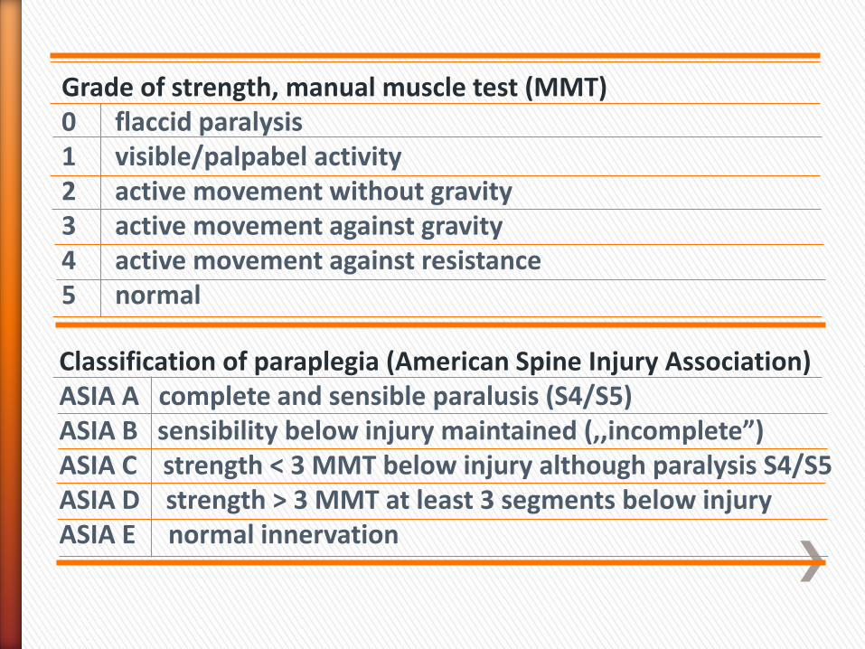
Grade of strength, manual muscle test (MMT) 0 flaccid paralysis 1 visible/palpabel activity 2 active movement without gravity 3 active movement against gravity 4 active movement against resistance 5 normal
Classification of paraplegia (American Spine Injury Association) ASIA A complete and sensible paralusis (S4/S5) ASIA B sensibility below injury maintained (,,incomplete”) ASIA C strength < 3 MMT below injury although paralysis S4/S5 ASIA D strength > 3 MMT at least 3 segments below injury ASIA E normal innervation
Results
Previously non-innervated muscles Showed increasing EMG activity and an average improvement in
strength of about 2.5 points in the MMT.
The functional reach tests Improved with seated patients
Of about 8.5 cm on average.
Deep and superficial sensibility was better.
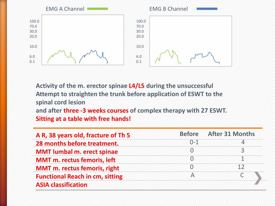
Activity of the m. erector spinae L4/L5 during the unsuccessful Attempt to straighten the trunk before application of ESWT to the spinal cord lesion and after three -3 weeks courses of complex therapy with 27 ESWT. Sitting at a table with free hands! A R, 38 years old, fracture of Th 5 28 months before treatment. MMT lumbal m. erect spinae MMT m. rectus femoris, left MMT m. rectus femoris, right Functional Reach in cm, sitting ASIA classification
EMG A Channel EMG B Channel
100.0 70.0 30.0 20.0 10.0 6.0 0.1
100.0 70.0 30.0 20.0 10.0 6.0 0.1
Before After 31 Months 0-1 4 0 3 0 1 0 12 A C
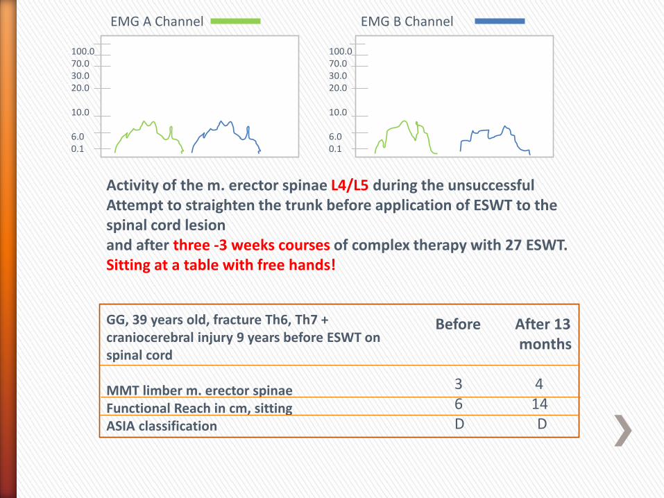
Activity of the m. erector spinae L4/L5 during the unsuccessful Attempt to straighten the trunk before application of ESWT to the spinal cord lesion and after three -3 weeks courses of complex therapy with 27 ESWT. Sitting at a table with free hands!
GG, 39 years old, fracture Th6, Th7 + craniocerebral injury 9 years before ESWT on spinal cord MMT limber m. erector spinae Functional Reach in cm, sitting ASIA classification
Before After 13 months 3 4 6 14 D D
EMG A Channel EMG B Channel
100.0 70.0 30.0 20.0 10.0 6.0 0.1
100.0 70.0 30.0 20.0 10.0 6.0 0.1
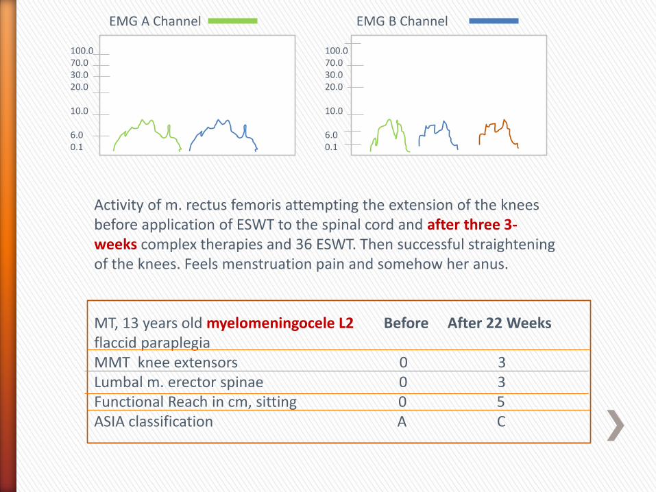
Activity of m. rectus femoris attempting the extension of the knees before application of ESWT to the spinal cord and after three 3- weeks complex therapies and 36 ESWT. Then successful straightening of the knees. Feels menstruation pain and somehow her anus. MT, 13 years old myelomeningocele L2 Before After 22 Weeks flaccid paraplegia MMT knee extensors 0 3 Lumbal m. erector spinae 0 3 Functional Reach in cm, sitting 0 5 ASIA classification A C
EMG A Channel EMG B Channel
100.0 70.0 30.0 20.0 10.0 6.0 0.1
100.0 70.0 30.0 20.0 10.0 6.0 0.1
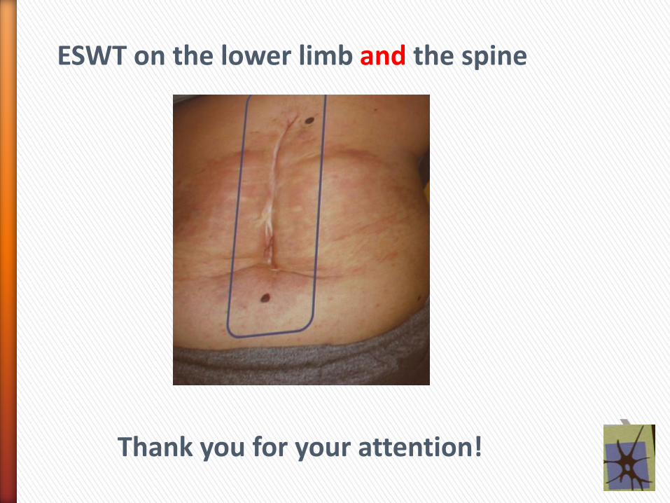
ESWT on the lower limb and the spine
Thank you for your attention!
Methods
Longitudinal observation case study, pilot study 5 patients, 8 to 18 years after the brain lesion, unresponsive Wakefulness syndrome of different severity, 4 PEG feeding tubes. 3 had stabilized epilepsy. All patients had palliative surgical Interventions on muscles. 2-5 previous physicomedical complex therapy regimes over 3 Weeks each with ESWT on periphereal muscles improved the cloni but not the vigilance.
●
●
●
Methods • Then the patients received 3 sessions weekly with transcranial extrcorporeal shock waves (TESWT, device Duolith, Storz Medical) during 4-week physicomedical complex therapies moving the therapy source 5,ooo ESW with 0.15 – 0.2 mj/mm2. • Documentation with the German Coma Remission Scale (KRS) and the Glasgow Coma Scale. Permission of the ethical committee of the University Hospitals Freiburg
Results After 2-4 years
and an average of 5.2 treatment series (27 TESTWT sessions on average)
the total improvement on the Coma remission Scale for the 5 patients is 135.9% (motor area 64.3%)/
43.6% improvement on the Glasgow Coma Scale.
Three PEG feeding tubes could be removed
A nonverbal communication initiated 4 times.
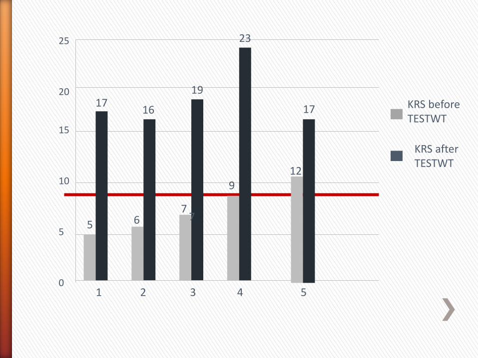
25 20 15 10 5 0
1 2 3 4 5
5 6 7
7
9 12
17
23
19
16 17 KRS before
TESTWT
KRS after TESTWT
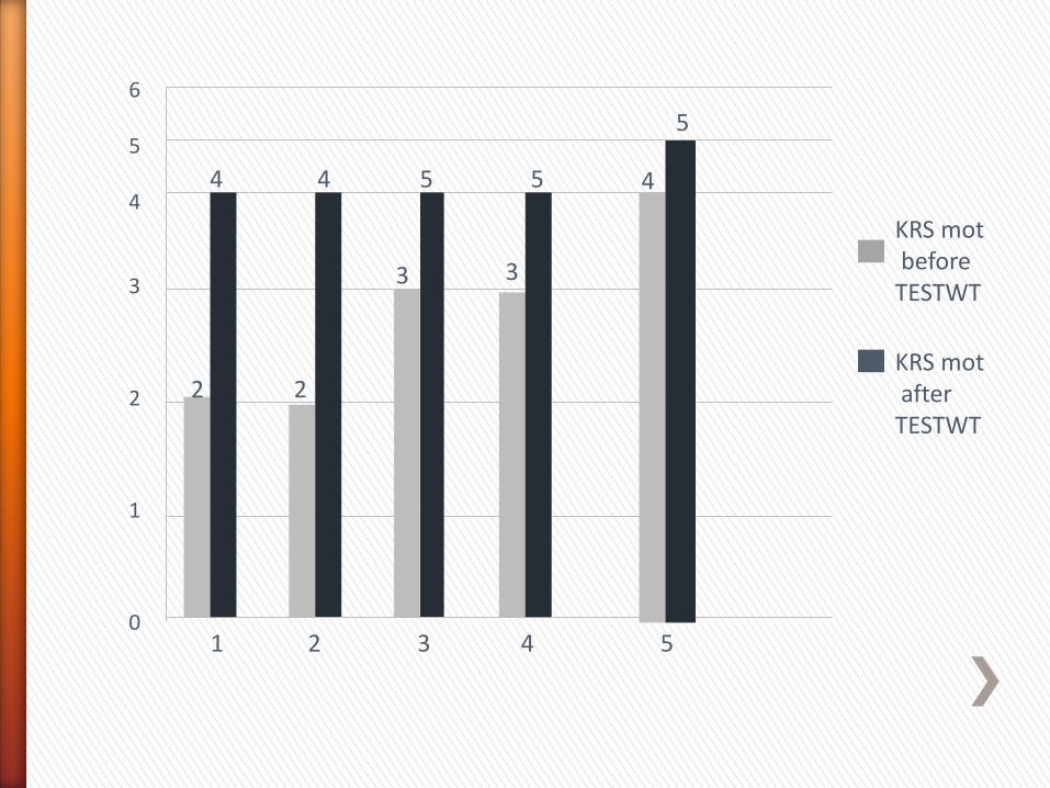
6 5 4 3 2 1 0
1 2 3 4 5
2 2
3 3
4
5
5 5 4 4
KRS mot before TESTWT
KRS mot after TESTWT
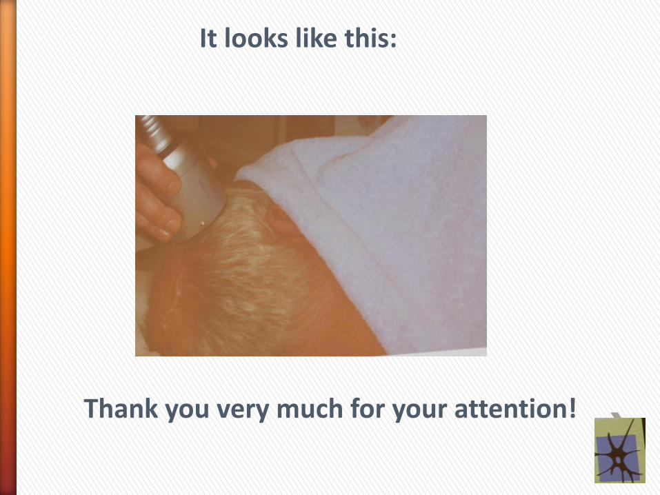
It looks like this:
Thank you very much for your attention!
Focused low-energy Extracorporeal shock waves
With distally Symmetric polyneuropathy (DSPNP)
A pilot study.
Lohse -Busch, H (1); Marlinghaus, E. (2); Reime, U. (1) Mowis, U. (1)
(1) Rheintalklinik, D-79189 Bad Krozingen, Germany. (2) Storz Medical AG, CH-8274 Tagerwilen, Swtzerland.
The first author received a research grant From Storz Medical AG Tagerwilen
The second author is an employee of Storz Medical
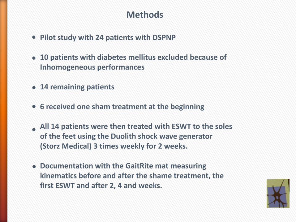
Methods
Pilot study with 24 patients with DSPNP 10 patients with diabetes mellitus excluded because of Inhomogeneous performances 14 remaining patients 6 received one sham treatment at the beginning All 14 patients were then treated with ESWT to the soles of the feet using the Duolith shock wave generator (Storz Medical) 3 times weekly for 2 weeks. Documentation with the GaitRite mat measuring kinematics before and after the shame treatment, the first ESWT and after 2, 4 and weeks.
●
●
●
●
●
●
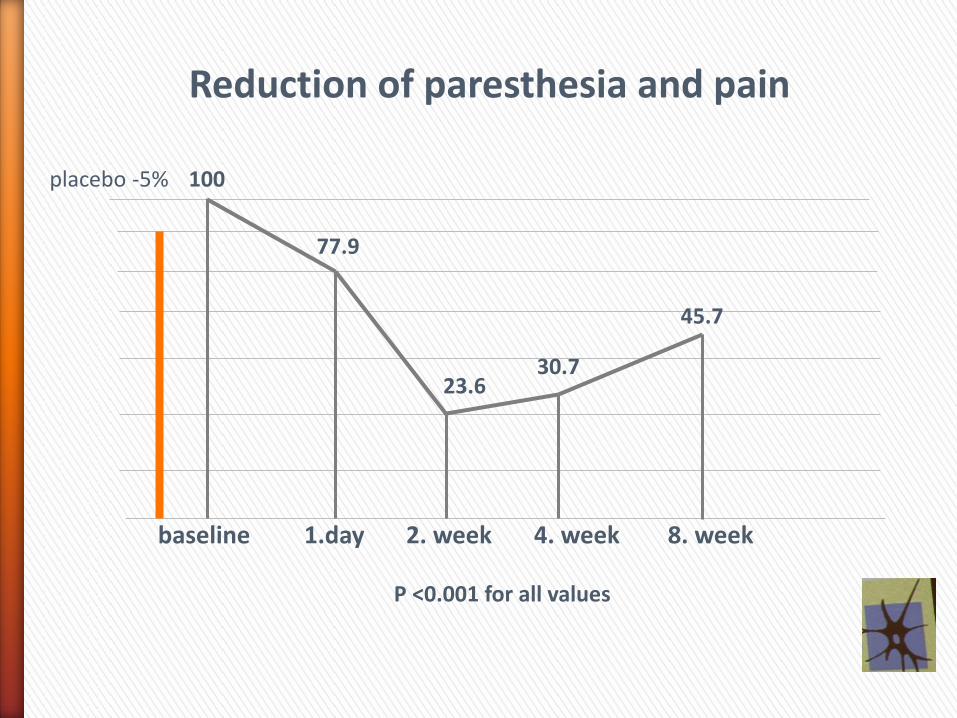
Reduction of paresthesia and pain
placebo -5% 100
77.9
23.6 30.7
45.7
baseline 1.day 2. week 4. week 8. week P <0.001 for all values
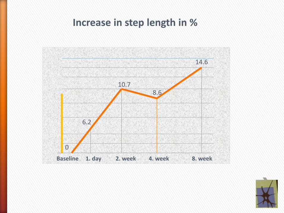
Increase in step length in %
14.6
0
6.2
10.7 8.6
Baseline 1. day 2. week 4. week 8. week
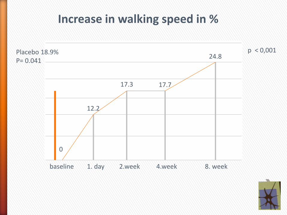
Increase in walking speed in %
24.8
17.7 17.3
12.2
0
baseline 1. day 2.week 4.week 8. week
Placebo 18.9% P= 0.041
p < 0,001
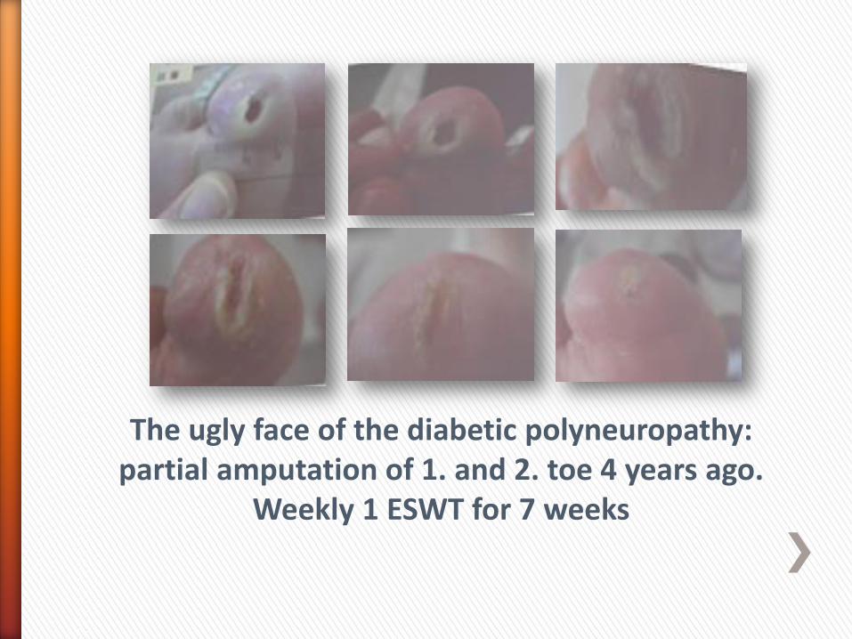
The ugly face of the diabetic polyneuropathy: partial amputation of 1. and 2. toe 4 years ago.
Weekly 1 ESWT for 7 weeks
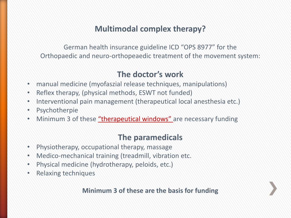
Multimodal complex therapy?
German health insurance guideline ICD “OPS 8977” for the Orthopaedic and neuro-orthopeaedic treatment of the movement system:
The doctor’s work • manual medicine (myofaszial release techniques, manipulations) • Reflex therapy, (physical methods, ESWT not funded) • Interventional pain management (therapeutical local anesthesia etc.) • Psychotherpie • Minimum 3 of these “therapeutical windows” are necessary funding
The paramedicals • Physiotherapy, occupational therapy, massage • Medico-mechanical training (treadmill, vibration etc. • Physical medicine (hydrotherapy, peloids, etc.) • Relaxing techniques
Minimum 3 of these are the basis for funding
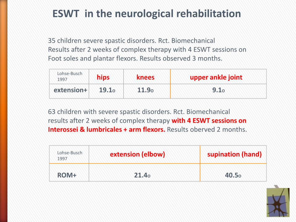
ESWT in the neurological rehabilitation
35 children severe spastic disorders. Rct. Biomechanical Results after 2 weeks of complex therapy with 4 ESWT sessions on Foot soles and plantar flexors. Results observed 3 months. 63 children with severe spastic disorders. Rct. Biomechanical results after 2 weeks of complex therapy with 4 ESWT sessions on Interossei & lumbricales + arm flexors. Results oberved 2 months.
hips knees upper ankle joint Lohse-Busch 1997
extension+ 19.1O 11.9O 9.1O
extension (elbow) supination (hand)
ROM+ 21.4O 40.5O
Lohse-Busch 1997
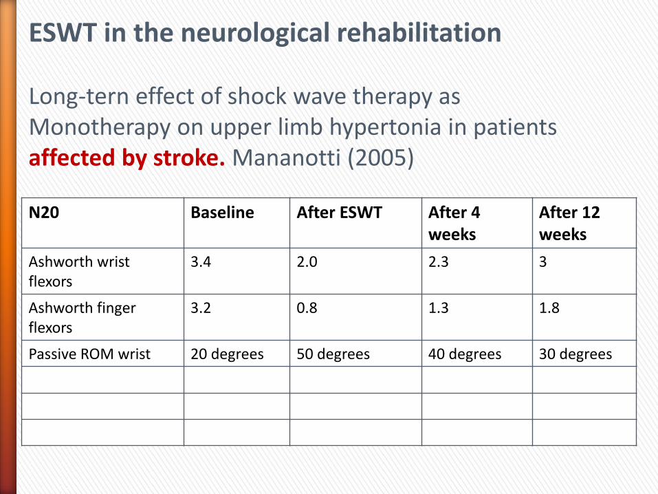
N20 Baseline After ESWT After 4 weeks
After 12 weeks
Ashworth wrist flexors
3.4 2.0 2.3 3
Ashworth finger flexors
3.2 0.8 1.3 1.8
Passive ROM wrist 20 degrees 50 degrees 40 degrees 30 degrees
ESWT in the neurological rehabilitation Long-tern effect of shock wave therapy as Monotherapy on upper limb hypertonia in patients affected by stroke. Mananotti (2005)
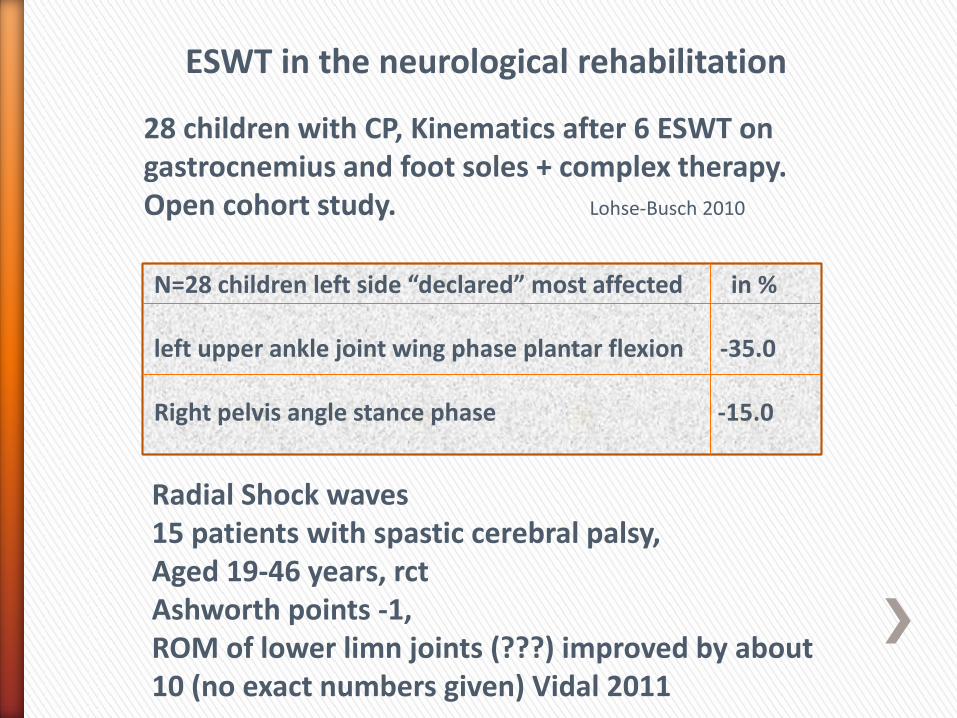
ESWT in the neurological rehabilitation
28 children with CP, Kinematics after 6 ESWT on gastrocnemius and foot soles + complex therapy. Open cohort study. Lohse-Busch 2010
N=28 children left side “declared” most affected in % left upper ankle joint wing phase plantar flexion -35.0 Right pelvis angle stance phase -15.0
Radial Shock waves 15 patients with spastic cerebral palsy, Aged 19-46 years, rct Ashworth points -1, ROM of lower limn joints (???) improved by about 10 (no exact numbers given) Vidal 2011
The newest one: 15 children with CP. Kinematics after 15 ESWT on spastic muscles of the lower limb. Gait analysis. Rct. The stride length, cadence, speed, cycle time, and stance phase improved by about 25%.
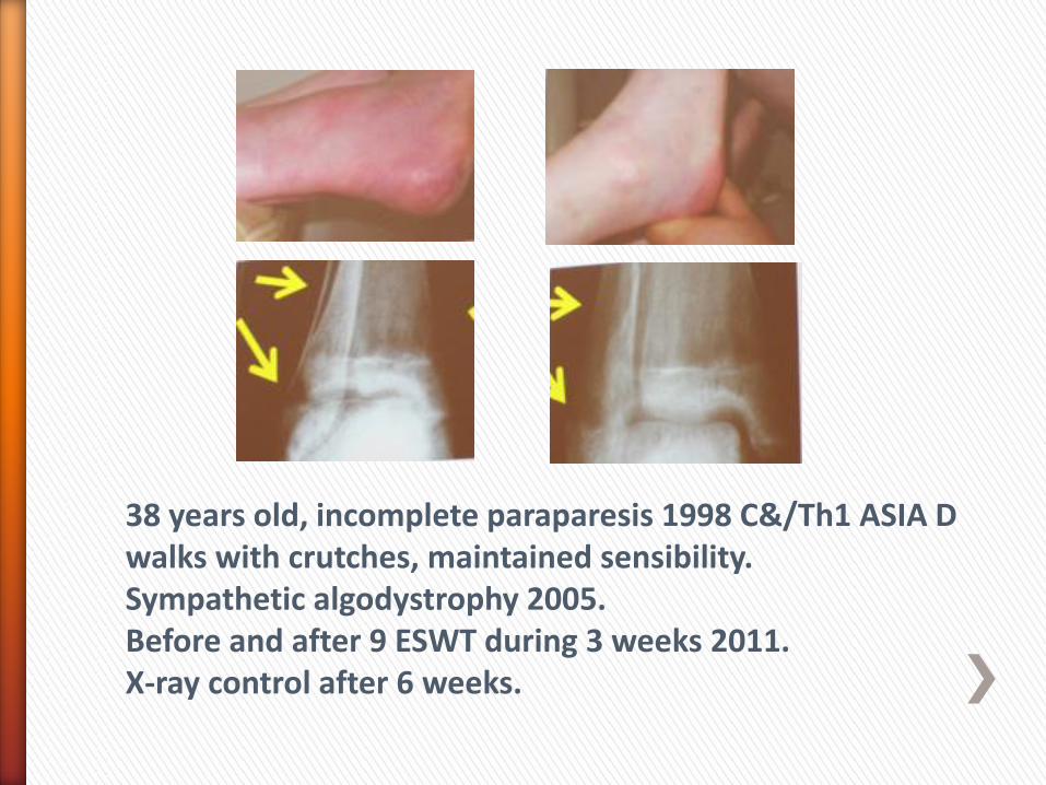
38 years old, incomplete paraparesis 1998 C&/Th1 ASIA D walks with crutches, maintained sensibility. Sympathetic algodystrophy 2005. Before and after 9 ESWT during 3 weeks 2011. X-ray control after 6 weeks.
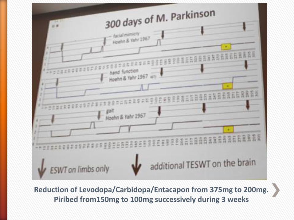
Reduction of Levodopa/Carbidopa/Entacapon from 375mg to 200mg. Piribed from150mg to 100mg successively during 3 weeks
Parkinsons Patient Video I
Quad Kicking Ball