Embed Size (px)
Citation preview
“Risk Stratification after Myocardial Infarction Using
Dobutamine Stress Echocardiography”
Dissertation Submitted to
THE TAMIL NADU DR. M.G.R. MEDICAL UNIVERSITY
In partial fulfillment of the regulations
For the award of the degree of
D.M. BRANCH –II
CARDIOLOGY
STANLEY MEDICAL COLLEGE,
CHENNAI
ACKNOWELEDGEMENT
I express my deepest respect and most sincere gratitude to my beloved teacher and
mentor Prof. Dr. R. Subramanian M.D., D.M., (Cardiology) Professor & Head of the
Department of Cardiology, for his precious guidance and encouragement through out the
study.
I am extremely thankful to our Additional Professor Dr. M. Somasundaram M.D.,
D.M., (Cardiology) for his unfailing support and valuable guidance during the study.
I am grateful and obliged to all our Assistant Professors of Cardiology for their
amazing encouragement, timely advices and enormous support during the study.
I thank the DEAN Dr. Ravindran M.D (Chest) DTCD Government Stanley Hospital
and Medical College, Chennai, for permitting me to utilize the hospital materials during the
course of this study.
I express my thanks to Dr. Nancy, M. Sc, PhD., Biostatistician, for her help in
statistical analysis.
Last but not the least; I thank all the patients who ungrudgingly lent themselves to
undergo the study without which this study would not have seen the light of the day.
CERTIFICATE
This is to certify that the dissertation entitled “Risk Stratification after
Myocardial Infarction Using Dobutamine Stress Echocardiography” is the bonafide original work of
Dr. Anand Gnanaraj, M.D., in partial fulfillment of the requirements for D.M branch-II
(CARDIOLOGY) Examination of the Tamilnadu Dr.M.G.R. Medical University to be held in
February 2006.
Prof. Dr. R. SUBRAMANIAN, M.D., D.M.,
PROFESSOR AND HEAD, DEPT. OF
CARDIOLOGY GOVT. STANLEY MEDICAL
COLLEGE & HOSPITAL, CHENNAI.
THE DEAN
GOVT. STANLEY MEDICAL
COLLEGE & HOSPITAL
CHENNAI.
THE TAMIL NADU DR. M.G.R. MEDICAL UNIVERSITY
CHENNAI.
FEBRUARY 2006
CONTENTS
1. Introduction 1
2. Aim of the Study 2
3. Review of Literature
a. Dobutamine Stress Echocardiography 3
i. Stress Echocardiography 3
ii. Physiological Basis 4
iii. Historical Development of DSE 6
iv. Pharmacology of Dobutamine 7
v. Pathophysiology of DSE 8
vi. Hypotension and LVOT obstructions 10
b. Dobutamine Stress Echocardiography Protocols 11
i. Early Protocols 11
ii. Infusion Endpoints 12
iii. Laboratory Setup and Personnel 13
iv. Sample Protocol for DSE 13
v. ECHO and ECG Vital Signs Monitoring 14
vi. Safety and Complications of DSE 15
vii. Contraindications to DSE 16
viii. Clinical Uses of DSE 17
ix. Evaluation of a Patient with Chest Pain 17
x. Clinical Accuracy Compared to Other Non Invasive Testing 19

xi. Evaluation of a Patient with Known Coronary Artery Disease 20
xii. Post Myocardial Infarction 20
xiii. Myocardial Viability 21
xiv. Post Percutaneous Coronary Interventions and Other Patients 21
c. Risk Stratification 22
i. The Value of a Negative Test 22
ii. Patients With Known Coronary Artery Disease 22
iii. Perioperative Risk Stratification for Non Cardiac Surgery 23
iv. DSE Compared with Nuclear Perfusion Imaging 23
v. Application of DSE in the Post Infarction Risk Stratification 25
vi. Dobutamine Stress Echocardiography 26
vii. Assessment of Myocardial Viability 27
viii. Myocardial Stunning 28
ix. Prediction of Reversible Dysfunction with DSE after MI 29
x. Clinical Relevance of Myocardial Viability after MI 29
xi. Myocardial Hibernation 31
xii. Detection of Myocardial Hibernation with DSE 33
xiii. Contractile Function Versus Perfusion 35
xiv. Conclusion 37
4. The Study
a. Materials and Methods 40
b. Statistical Analysis 44
c. Results 45
d. Analysis of Pretest Variables 46
e. Complications 48

f. Protocol Completion 49
g. Analysis of Follow Up 49
h. Discussion 51
i. Prognostic Implication of Myocardial Viability 52
j. Conclusion 53
5. Bibliography 54
6. Annexure
a. Proforma 64
b. Master Chart 67
c. Coding Sheet 74
d. Glossary 78
Risk Stratification afterRisk Stratification after
Myocardial Infarction UsingMyocardial Infarction Using
Dobutamine StressDobutamine Stress
EchocardiographyEchocardiography
Introduction
Pharmacological stress testing has emerged as an important diagnostic tool in the evaluation
and management of patients with known coronary artery disease, especially those who cannot perform
physical exercise. It has also become an important modality to assess myocardial viability, hibernating
myocardium and valvular heart disease. Unlike exercise stress testing in which cardiac imaging is not
always needed, pharmacological stress test needs some form of imaging to detect myocardial
ischaemia.
Pharmacological stress tests with vasodilatation, using dipyridamole or adenosine, have been
used extensively in conjunction with nuclear perfusion imaging1. Recently pharmacological stress
echocardiography has become an accepted alternative to exercise stress testing and to stress testing
with nuclear imaging. Many pharmacological agents have been used for stress testing. Dipyridamole
and adenosine are used commonly in some places in combination with echocardiographic imaging.
Dobutamine is the preferred agent in some parts of the world, like the United States. More and more
cardiac laboratories consider dobutamine as the drug of choice for stress testing. A newer drug
Arbutamine is also approved for this purpose.
Aims of the Study
• To assess the myocardial viability after myocardial infarction using dobutamine stress
echocardiography
• To study the various high risk variables that affect the viability of the myocardium after
myocardial infarction
• To detect the significance of regional wall motion abnormality during the dobutamine stress
echocardiography in the prediction of coronary events after myocardial infarction
• To evaluate the factors that predict the coronary events and the presence of myocardial viability
after myocardial infarction
• To stratify the risk of the patients for coronary events following myocardial infarction using the
dobutamine stress echocardiographic responses at low doses and high doses
• To assess the negative predictive value of dobutamine stress echocardiography in the post
myocardial infarction setting
DOBUTAMINE STRESS ECHOCARDIOGRAPHY
Stress Echocardiography
Stress echocardiography is based on the fundamental causal relationship between induced
myocardial ischaemia and left ventricular regional wall motion abnormalities. The potential for using
echocardiography for this purpose was first reported in 1979 when two groups of investigators
demonstrated the proof of concept. Mason and colleagues2 used M-mode echocardiography to study 13
patients with coronary artery disease and 11 age matched controls during supine bicycle exercise.
Stress induced wall motion changes were observed in 19 of the 22 segments on the stenotic coronary
artery territory. Although this was the first demonstration of transient ischaemia being detected with
ultrasound, the inherent limitations of M-mode echocardiography was apparent. Wann and coworkers3
applied an early 2D, 30 degree sector imaging system to demonstrate inducible wall motion
abnormalities during supine bicycle exercise and subsequent improvement of the wall motion response
after revascularization. These early studies were limited by image quality and a reliance on video tape
analysis, factors that would slow the growth of the field in its early years.
In the 1980s, improvement in image quality and the development of digital acquisition
technology or frame grabbers, contributed to greater accuracy and increased the practicality of using
stress echocardiography in clinical situations. Most important, the digitization of echocardiographic
images reduces the problem of respiratory interference by permitting selection of cardiac cycles that
were devoid of lung interference and the creation of cine loops that permitted side by side analysis of
rest and stress images. This allowed more accurate interpretation of wall motion, largely by permitting
relatively subtle changes in stress induced wall motion, to be detected. Digital technology also

shortened the acquisition time for post exercise imaging and facilitated display, storage and
transmission of echocardiographic data. More than any other single factor, the application of digital
imaging led to the rapid development of stress echocardiography as a clinical tool.
Physiological Basis
In the early days of echocardiography Tennant and Wiggers observed the relationship between
systolic contraction and myocardial blood supplied to the left ventricle. With the induction of
ischaemia, these investigators demonstrated the rapid and predictable development of systolic bulging
or dyskinesis. This observation established the link between induced ischaemia and transient regional
myocardial dyssynergy, recorded echocardiographically as the development of wall motion abnormality
after the application of a stressor.
In the absence of flow limiting coronary stenosis, physiologic stress results in an increase in
heart rate and contractility that is maintained via an increase in myocardial blood flow. Systolic wall
thickening, endocardial excursion, and global contractility all increase, leading to a decrease in end
systolic volume and an increase in the ejection fraction compared with the baseline. Although this
response may be blunted in the setting of advanced age and hypertension or in the presence of β-
blocker therapy, absence of the hypercontractile state in response to stress should generally be
considered an abnormal response.
In the presence of coronary stenosis, the increase in myocardial oxygen demand that occurs in
response to stress is not matched by an appropriate increase in supply. If the supply demand mismatch
persists, a complex sequence of events known as the ischaemic cascade will develop. Soon after the
development of a regional perfusion defect, a wall motion abnormality occurs, characterized
echocardiographically as a reduction in systolic thickening and endocardial excursion. The severity of

the wall motion abnormality (hypokinesis vs dyskinesis) will depend on several factors, including the
magnitude of the blood flow change, the spatial extent of the defect, the presence of collateral blood
flow, left ventricular pressure and wall stress, and the duration of ischaemia. Deterioration in regional
wall motion however, is a specific and predictable marker of regional ischaemia that generally precedes
such traditional manifestations as angina or electrocardiographic abnormalities.
Once the stressor is eliminated, myocardial oxygen demand decreases and ischaemia resolves.
Normalization of wall motion abnormality may occur rapidly, although typically the complete recovery
of normal function takes 1 to 2 minutes, largely depending on severity and duration of ischaemia.
Stunned myocardium is the term applied when functional abnormalities persist after transient ischaemia
for a longer period. Although a reversible process, stunning may last days or even weeks if the
ischaemia is severe and prolonged.
The utility of echocardiography in conjunction with stress testing is contingent on the ability to
record wall motion and left ventricular function at baseline and then to detect changes after the
induction of stress, either exercise or pharmacologic. At baseline, the presence of regional wall motion
abnormality generally implies the presence of previous myocardial infarction. Less often
cardiomyopathy and stunning or hibernating myocardium cause resting wall motion abnormalities.
Historical Development of Dobutamine Stress Echocardiography
Dobutamine was used in the year 1984 for cardiac stress imaging in conjunction with thallium
scintigraphy and was reported in The American Heart Journal2. Following this report various modalities
of detecting ischaemia induced by dobutamine, including ECG and echocardiographic imaging were
reported4. Coma-Canella4 reported 95% sensitivity and 78% specificity of the dobutamine stress
echocardiography using ST changes or angina as markers of positive test in nearly 100 patients. The
sensitivity and specificity reported with dobutamine stress ECG were much lower.5
At present dobutamine stress testing is almost always used with either echocardiography or
nuclear imaging. Between these two, echocardiography has emerged as the most powerful tool to detect
ischaemia. The role of DSE (Dobutamine Stress Echocardiography) has expanded from evaluation of a
patient with suspected coronary artery disease to include patients with known coronary artery disease,
post myocardial infarction patients, cardiac risk stratification prior to noncardiac surgery and
assessment of myocardial viability.
Technical advancements and improvements in echocardiographic image quality have
contributed to increasing use of echocardiography as a means of imaging the heart both during exercise
and pharmacological stress. The development of digital imaging has enhanced the use of
echocardiography6 and especially DSE.7 The ability to capture a single loop of cardiac cycle and
display it as a continuous loop with the pre and post exercise images running side by side has greatly
improved the detection of subtle hypokinesia, increased the ease of use and shortened the reporting
times.
Pharmacology of Dobutamine
Dobutamine is a synthetic catecholamine developed by Tuttle and Mills8 as an inotropic agent
with less chronotropic and peripheral vascular effects than other catecholamines like isoprenaline,
dopamine and norepinephrine. The drug was developed in the mid 1970s and was first available
clinically in 1978. It was initially used as a positive inotropic agent to augment cardiac output in
patients with congestive cardiac failure.9

Dobutamine has a predominant β1 agonist activity.9 It also has a relatively weak β2 and α1 agonist
activity.9 Through its β1 action it increases the heart rate and contractility with resultant increase in
cardiac output. Peripheral resistance may fall due to the β2 effects. The haemodynamic effects of
dobutamine are similar to exercise (increase in heart rate, blood pressure and contractility). There is an
augmentation of myocardial oxygen demand MVO2 and increase in coronary blood flow in normal
vessels.10 In the setting of an obstructed coronary artery, the regional myocardial perfusion may be
impaired when there is an increased metabolic demand.11
There is a good correlation between dobutamine dose, plasma levels and haemodynamic
effects.9 With a continuous infusion of the drug, the onset of action is within 2 minutes and maximum
effects are seen at 10 minutes. Steady state is not achieved till the 10 th minute. The half life of
dobutamine is about 2 minutes and the drug is eliminated and metabolized in 10 to 12 minutes after the
termination of a continuous infusion.9 Dobutamine is metabolized by catechol-o-methyltransferase to
pharmacologically inactive metabolites that are excreted in the urine.9
In addition to the expected cardiac effects, dobutamine may produce a lot of noncardiac
symptoms like headache, anxiety and tremors.9 Patients may also develop chills and shivering.
Pathophysiology of Dobutamine Stress Echocardiography
The clinical applications of DSE rest on the following principles
1. The infusion of dobutamine can cause regional myocardial ischaemia in areas supplied by
obstructed coronary arteries.
2. The regional ischaemia results is systolic contractile dysfunction.
3. The two-dimensional echocardiography is an accurate and reliable imaging modality to detect

systolic regional dyssynergy and dysfunction.
Dobutamine increases MVO2 by increasing the heart rate (chronotropic effect), myocardial
contractility (inotropic effect) with a variable effect on blood pressure.11 Dobutamine also influences
the regional myocardial blood flow.10 The normal response of the myocardium to dobutamine is to
increase myocardial thickening with an augmentation of ejection fraction. In the setting of obstructive
coronary artery disease, increases in MVO2 induced by dobutamine result in ischaemia due to supply-
demand mismatch. The ischaemic cascade results initially in diastolic dysfunction, followed by systolic
dysfunction, ECG changes and finally symptoms of ischaemia. Therefore when areas of myocardium
become ischaemic, echocardiography may detect areas of hypokinesis, akinesis or dyskinesis.12
Reduced regional systolic wall thickening is another finding with myocardial ischaemia. Depending on
the response of the nonischaemic regions, global ejection fraction may increase, fall or remain
unchanged. Regions of the myocardium that are infarcted and have no viable tissue remain hypokinetic
or akinetic. In the regions with viable and infarcted myocardium, dobutamine may cause an increased
wall thickening. If this area is supplied by a critically stenosed coronary artery, they may show
subsequent worsening of contractile function (the so called biphasic response) at higher doses of
dobutamine, reflecting viable but ischaemic myocardium.
Several other potential markers of myocardial ischaemia induced by dobutamine may serve as
adjuncts to an analysis of wall motion. None are currently used alone to define a positive DSE. These
markers include left ventricular cavity dilatation, alterations in transmitral flow by pulsed wave
doppler, a decrease in transaortic flow, the development of new worsening mitral regurgitation, the
development of sinus node acceleration, development of ischaemic ECG changes and the development
of hypotension.13 Despite some initial enthusiasm, development of ECG changes has shown to be an
insensitive marker of ischaemia.5 The hypotension following dobutamine also does not mean serious
ischaemia.13
By producing cardiac effects similar to exercise, inducing an increased heart rate, cardiac output
and systolic blood pressure, with a resultant increase in MVO2, a graded dobutamine infusion provides
a rational alternative to exercise as a way to stress the heart and provoke myocardial ischaemia.
Although the ECG response is a relatively insensitive marker of ischaemia, two-dimentional
echocardiography provides a reliable method of detecting ischaemia because regional wall motion
abnormalities are early and predictable response to myocardial ischaemia. Therefore the combination of
dobutamine and echocardiography provide a reasonable alternative to exercise testing.
Hypotension and Left Ventricular Tract Outflow Obstruction
Significant decreases in systolic blood pressure are not uncommon during dobutamine stress
echocardiography. The definition of hypotension is generally accepted as a fall of more than 20
mmHg.14 Unlike the prognostic significance of hypotension during exercise stress testing, a fall in
blood pressure with dobutamine is not associated with adverse prognosis and is not a marker of severe
CAD.15
Several mechanisms have been proposed as the cause for the hypotension in the absence of
worsening left ventricular function. A secondary vasodepressor response, development of a dynamic
intraventricular gradient13, 79, failure to increase the cardiac output, decrease in systemic vascular
resistance and left ventricular cavity obliteration may work alone or in concert to cause hypotension.
Dobutamine Stress Echocardiography Protocols
There is no universal standardized infusion protocol, including initial dose (2.5 µg/kg/min to 10
µg/kg/min), stage duration (2 to 10 minutes) and maximum dose (30 to 50 µg/kg/min). 16 Many
protocols used an age predicted maximum heart rate and 85% of the target heart rate. The infusion
protocol is often a function of the clinical setting, perceived pretest probability of disease, resting left
ventricular function and the clinical question.
Initial experience with DSE emphasized safety concerns and thus early protocols began with
relatively low doses. As the experience with dobutamine increased the initial infusion rates of 5 to 10
µg/kg/min and increased by 10 µg/kg/min every 2 to 3 minutes to a maximum of 40 to 50 µg/kg/min
are being used.7
Prolonged duration of stages may potentially improve the sensitivity of the test, decrease the
need for atropine and require a lower peak dose to achieve target heart rate. It also allows better
evaluation of myocardial viability with fewer side effects. At present most laboratories use 3 minute
stages. For patients who do not achieve 85% of the target heart rate after the maximum dobutamine
dose, 0.2 to 0.5 mg of atropine every 1 to 3 minutes to a maximum of 1 to 2 mg is given along with the
dobutamine infusion. Patients on β blockers are more likely to require the addition of atropine. Unless
the goal of the test is to assess medical therapy, β blockers should be discontinued 24 to 48 hours prior
to the testing.
It is also reported that a heart rate of less than 70 beats per minute at the 20 µg/kg/min dose
stage or an increase in heart rate from the base line at 20 µg/kg/min of less than 5 beats per minute are
accurate predictors of the need for atropine. If the above mentioned finding is noticed at the 20
µg/kg/min stage atropine can be used before achieving the 40 µg/kg/min stage.17 This significantly
shortens the time of study but the impact on the sensitivity and specificity are not clearly known.
Isometric hand grip can also be used to achieve the target heart rate in the later stages.18

Infusion Endpoints
The dobutamine infusion is continued until one or more predefined end points are reached. In
the absence of other endpoints or intolerable side effects, the infusion is continued until the patient’s
heart rate reaches 85% of the age predicted maximum heart rate.
The other end points are
1. Significant new or worsening regional wall motion abnormalities
2. Severe symptoms, either cardiac or noncardiac
3. Severe increase in blood pressure (SBP > 240 mmHg or DBP > 120 mmHg) or symptomatic
hypotension
4. Significant ECG changes, especially if accompanied by echocardiographic findings
5. Significant ventricular or supraventricular tachyarrhythmias
6. Completion of protocol usually at 40 – 50 µg/kg/min
Laboratory Setup and Personnel
The testing area should satisfy the following requirements19
1. Adequate space, with a minimum of 300 sq ft. is recommended.
2. Appropriate resuscitatory equipment – Crash cart, Oxygen and suction equipment
3. Readily available medication to reverse the effects of dobutamine or to treat arrhythmias –
Intravenous β blocker and IV calcium channel blockers
4. Infusion pump to regulate the delivery of dobutamine
5. Automate or manual blood pressure measuring apparatus
6. A 12 lead ECG machine
7. A echocardiography equipment that can record and replay digital images
Sample Protocol for Dobutamine Stress Echocardiography
1. Baseline echocardiographic views, 12 lead ECG and blood pressure. Continuous ECG
recording with printout every minute.
2. Initial starting dose of 2.5 µg/kg/min for viability studies, 5 µg/kg/min for standard study or
10 µg/kg/min for low risk group.
3. Echocardiography images obtained and evaluated approximately 1 minute after the start of
dobutamine and before each increase.
4. Heart rate, blood pressure and 12 lead ECG at each stage. Each stage lasts 3 minutes.
5. After 20 µg/kg/min the dose is increased by 10 µg/kg/min intervals up to a maximum
dosage of 40 – 50 µg/kg/min.
6. If infusion end point is not reached, atropine is given at 0.25 to 0.5 mg intravenously,
continuing the current dobutamine dose. Additional doses of atropine can be given till 2 mg
is reached.
7. The dobutamine infusion is discontinued if one or more of the end points mentioned above
are reached.
Echocardiographic, Electrocardiographic and Vital Sign Monitoring
Baseline heart rate, blood pressure and 12 lead ECG are recorded and baseline
echocardiographic images are obtained. A secure peripheral line is established for the delivery of
dobutamine. Following the review of baseline data after a brief history from the patient and
determining the indication for the study, the dobutamine infusion is begun. ECG is monitored
continuously during the study. A 12 lead ECG is taken every minute and at the end of each stage, or if
symptoms develop. Blood pressure is measured after 1.5 to 2 minutes into each stage or if symptoms of
hypertension or hypotension develop.
Patients are monitored for a minimum of 5 minutes post infusion or until vital signs have
returned to baseline (heart rate below 100 beats per minute or 20 bpm of baseline, blood pressure
returning to baseline or dysrhythmias resolved) and symptoms have resolved. Patient is monitored till
all the ECG and echocardiographic changes have returned to baseline.
Safety and Complications of Dobutamine Stress Echocardiography
Dobutamine stress echocardiography has an excellent safety profile. Several large studies have
specifically examined the safety issues in a variety of populations.2 They have reported low rates of
significant complications. The few serious complications that have been reported are
haemodynamically significant dysrhythmias. There are rare reports of acute myocardial infarction
during DSE. This may be due to α1 mediated vasospasm, and the occurrence appears to be uncommon.
The largest series reported by Seckuns and Marwick21 evaluated a large number of patients and
were followed up for 5 years. In spite of the aggressive protocols, there was no increase in the major
side effects. Arrhythmias and hypotension were the most commonly reported complications. Serious
complications like myocardial infarction and sustained ventricular tachycardia occurred in 0.3% of
patients. No deaths and incidences of ventricular fibrillation occurred.
The safety of DSE in post myocardial infarction patients was evaluated by Mertes20 and he
reported no deaths, myocardial infarctions or sustained ventricular tachycardias. The commonest cause
for stopping the test was for reaching the target heart rate (52%). Angina during the test was managed
with sublingual nitrates or intravenous β blockers. Ventricular and atrial premature beats were observed
in 15% and 8% respectively. Atrial flutter and fibrillation was noted in 1% of patients, which reverted
spontaneously after stopping the infusion.
In a multicentric study of a large number of patients, Picano22 reported a relatively high
incidence (0.5%) of significant and potentially life-threatening events with DSE. Patients suffered from
myocardial infarction, ventricular tachycardia, ventricular fibrillation, hypotension and prolonged
angina. They accounted for 12% of terminations of the test. Various others have evaluated the safety of
DSE in other high risk groups like aortic aneurysm, early post MI and patients with organized apical
thrombus23. One patient has suffered myocardial rupture at the 10 µg/kg/min stage and survived.24
Contraindications to Dobutamine Stress Echocardiography
The contraindications to DSE are similar to those of exercise stress testing.
1. Acute coronary syndromes including unstable angina and acute myocardial infarction.
2. Uncontrolled heart failure
3. Uncontrolled ventricular or supraventricular tachycardia
4. Marked hypertension with a systolic blood pressure of more than 200 mmHg or a diastolic
blood pressure of more than 110 mmHg.
5. Hypertrophic obstructive cardiomyopathy
6. Severe aortic stenosis
7. The use of atropine is contraindicated in patients with glaucoma and prostatic obstruction.
Clinical Uses of Dobutamine Stress Echocardiography
Dobutamine stress echocardiography is used in a variety of clinical settings including patients
with chest pain syndromes for evaluation of coronary artery disease. It is also used in patients with
established coronary artery disease to detect the functional status, to detect viability of myocardium in
infarcted areas, in identification of the culprit vessel when the angiographic lesion shows borderline
stenosis, to evaluate the response to medical, percutaneous or surgical therapy and risk stratification
following myocardial infarction. Additionally, DSE can also be used in patients after cardiac
transplantation, risk stratification in patients before noncardiac surgery and in evaluation of patients
with valvular heart disease.
Evaluation of Patients with Chest Pain
Many tests are available to the clinician to evaluate patients with chest pain that is suspected of
being caused by myocardial ischaemia. These include the standard exercise ECG, the stress perfusion
imaging and pharmacological stress imaging. Of these modalities, DSE has emerged as an important
tool in the assessment of chest pain. It becomes more important in patients who cannot exercise due to
orthopaedic complications.
The initial study by Sawada for evaluating a patient for CAD with angiographic correlation
showed a sensitivity of 89% and a specificity of 85% using the 30 µg/kg/min maximum dose protocol.
All false negatives occurred in patients with single vessel disease and in those with a diameter stenosis
of 40 – 50 %. In patients with resting regional wall motion abnormality, the sensitivity and specificity
to detect ischaemia at a distance was 81% and 87% respectively. In another group Segar et al, reported
a sensitivity of 95% and a specificity of 82% in a group of patients who underwent DSE and
quantitative angiography. Many other groups have reported similar figures in patients with single and
multivessel disease.7
In a review of 28 studies published from 1991 to 1996, Geleijnse et al13 reported that based on a
total of 2246 patients undergoing DSE, the sensitivity, specificity and accuracy of the test to detect
CAD were 80%, 84% and 92% respectively. The mean sensitivity for detecting single, double and
triple vessel disease were 74%, 86% and 92% respectively. The sensitivity of DSE for the detection of
CAD is purely a function of severity of the underlying disease, the protocol used and the achievement
of adequate heart rate. Diagnostic sensitivity is compromised when adequate heart rate is not achieved.
Addition of atropine to increase the heart rate has been shown to improve the diagnostic
accuracy. False positive studies may occur due to true ischaemia in the absence of angiographically
significant stenosis, over interpretation of regional wall motion abnormality and regional differences in
the myocardial thickening. The tendency to get false positive test is more in the basal posterior
circulation.
The diagnostic accuracy of noninvasive testing in patients with LBBB and left ventricular
hypertrophy is often challenging. Perfusion images often result in false positive studies in patients with
LBBB. Small studies indicate that DSE may be more accurate in evaluating these patients with LBBB
or LVH.13 This observation needs to be confirmed by larger trials.
Clinical Accuracy Compared to Other noninvasive Testing
In general, DSE compares well in terms of sensitivity, specificity and overall accuracy, with
stress nuclear perfusion imaging, pharmacological stress imaging using vasodilators and exercise stress
echocardiography and is superior to the exercise ECG and dobutamine stress ECG. When compared
with treadmill ECG in patients who were evaluated for CAD, DSE proved better than treadmill ECG
testing and dobutamine ECG testing. The direct comparison of dobutamine stress echocardiography,
treadmill stress echocardiography and dipyridamole stress echocardiography showed that they were
comparable with angiography as the gold standard. Another study by Martin et al showed that DSE was
more sensitive and better tolerated than either adenosine or dipyridamole.
Marwick25 compared dobutamine and adenosine in combination with echocardiography and
99mTc-MIBI single photon emission computed tomography imaging and noted virtually identical
sensitivity between DSE and adenosine perfusion imaging. Thus dobutamine stress testing appears to
be a reliable and accurate method of noninvasively assessing patients with chest pain.

Evaluation of Patients with Known Coronary Artery DiseaseFor patients with known coronary artery disease, there is often a need for a functional test to aid
in the clinical decision making. Dobutamine stress echocardiography has become an important and
reliable method to evaluate such patients who are unable to exercise.
Owing to the ability of echocardiography to detect regional changes in left ventricular systolic
function, DSE can help to localize myocardial ischaemia to a specific vascular territory. There is often
a need to evaluate patients in a variety of clinical circumstances. These include post MI risk
stratification, viability in the infarcted zone, to detect ischaemia at a distance, to detect functional
significance of angiographically borderline lesions, to detect the culprit vessel in multivessel disease
before revascularization, to provide long term prognostic information, to assess response to medical
and surgical revascularization and to evaluate patients with left ventricular dysfunction for the presence
of viable and residual ischaemic myocardium.
Post Myocardial Infarction
The goals of post MI assessment including the evaluation of residual ischaemia, or viability
with or without ischaemia, in the infarct zone and evaluation of inducible ischaemia remote from the
infarct13, which is an evidence of multivessel disease. Stress echocardiography including DSE, has been
shown to be a safe and reliable tool in evaluation of this patient group.5, 13
Myocardial Viability
The differentiation of dysfunctional but viable myocardium, which can be a hibernating or
stunned myocardium, from irreversible damaged myocardial scar is important in the assessment of
patients with depressed left ventricular function, in whom revascularization is contemplated. Overall
left ventricular systolic function is an important determinant in the outcome of patients with chronic
CAD. Thus identifying viable myocardium prior to attempts at revascularization is important because

the contractility of the myocardium that is successfully revascularized may improve, leading to
improved global left ventricular function and long term outcome. Dobutamine stress echocardiography
is a reliable method of assessing viability of the myocardium.26
Post PCI and Other Patient Populations
Dobutamine stress echocardiography has been used in patients following percutaneous
revascularization procedures and after transmyocardial laser revascularization to determine the success
of such revascularization procedures and to evaluate patients for the presence of residual ischaemia.27
There is also a growing population of post cardiac transplantation patients who undergo DSE as a
screening test to detect coronary allograft vasculopathy.28
RISK STRATIFICATION
The utility of DSE as a tool to stratify individuals according to risk factors has been evaluated in
a variety of clinical settings.
The Value of a Negative Test
Dobutamine stress echocardiography not only plays a role in the diagnostic evaluation of
patients presenting with chest pain but also in risk stratification of these patients. Patients with a
negative DSE in general have an excellent long term prognosis.16
A group of 200 patients were studied by Geleijnse16 who presented with chest pain, a normal
resting ECG and a negative DSE. They included patients with known CAD but excluded those who had
undergone revascularization procedures. The patients were classified into low risk (<10%),
intermediate risk (10–80%) and high risk (>80%) pretest probability of CAD depending on the clinical
variables. On follow up, the patients who had events were the ones with high pretest probability and the
ones who has a positive DSE.
Patients with Known Coronary Artery Disease
Steinberg et al followed a significant number of patients who underwent both DSE and coronary
angiography for a period of 3-6 years. They found that cardiac mortality was significantly more in the
positive DSE group compared with the negative DSE group (9% versus 0%). Dobutamine stress
echocardiography had an overall sensitivity and specificity for all cardiac events of 78.8% and 51.9%
respectively and 100% and 37.2% for cardiac death respectively. The negative predictive value for
cardiac death was 100% and was 95.2% for myocardial infarction.
Preoperative Risk Stratification for Non-cardiac Surgery
Beginning in the early 1990s, pharmacological stress imaging has been used extensively in the
assessment of patients prior to non cardiac surgery, especially in patients with peripheral vascular
diseases.
The first large multicenric experience with DSE in the post infarct period was reported by Echo
Dobutamine International Cooperative (EDIC) Study Group. Sicari et al published data on 778 patients
on whom DSE was done 12 days after myocardial infarction. They found that echocardiographic
images were adequate for analysis in 97% of patients and the complication rate was extremely low
(0.5%). Mortality was not significantly different between the groups with or without ischaemia (2.2%
vs 1.1%) and the only predictors were the presence of ischaemia and left ventricular function.
Dobutamine Stress Echocardiography Compared with Nuclear Perfusion Imaging
The overall accuracy of pharmacological stress echocardiography and nuclear perfusion
imaging is similar. Therefore diagnostic test performance is usually not an issue in deciding between an

echocardiographic versus a nuclear study. The experience and expertise of the local laboratories should
play a role in deciding which test is to be performed.
One potential clinical advantage of DSE compared with pharmacologic perfusion imaging with
vasodilators is the ability of DSE to detect an ischaemic threshold. Owing to the ability to continuously
monitor left ventricular function as the heart rate and blood pressure change during the infusion, one is
able to determine the heart rate, and the rate pressure product, at which ischaemia becomes evident.
This may correlate with the severity of the underlying CAD and may also have implications for risk
stratification prior to noncardiac surgery.
Dobutamine stress echocardiography does have many other advantages over nuclear techniques.
It is clearly less costly not only in terms of initial startup cost but also in ongoing costs to maintain the
laboratory and in the cost of the test to the patient. Because most hospitals and many outpatient offices
already have cardiac ultrasound systems in place the need to acquire additional specialized imaging
equipment other than the ability to have digital storage and display of the echocardiograms is avoided.
Because no radiation is involved, it is potentially safer for both patients and the staff and the additional
cost in terms of materials and personnel such as nuclear medicine technicians and complexity of
working with radioactive materials are avoided. Patients with significant bronchospasm can safely
undergo a DSE whereas dipyrimadole is relatively contraindicated in these patients. Echocardiography
also allows for a more thorough evaluation of the patient's cardiac status including an assessment of left
ventricular systolic and diastolic function, valvular anatomy, the proximal aorta and the pericardium in
addition to the evaluation for evidence of myocardial ischaemia.
Echocardiography also has the advantage of immediate online result. It is more convenient for
the patient and allows for the referring physician to be contacted with results immediately following the
test. There is no need for the patient to return for delayed images or for the images to be processed for
subsequent interpretation.
Application of Stress Echocardiography to Post infarct Risk Stratification
The validation of stress echocardiographic techniques in the diagnostic and prognostic
assessment of chronic CAD has been discussed extensively. As this technique may define left
ventricular dysfunction and exercise capacity and detect ischaemia they may be very effective in
defining irreversible risk after myocardial infarction. The evaluation of these different approaches has
been based on the ability to predict multivessel disease as a surrogate marker of reversible risk or direct
evaluation of outcomes after various test results.
A number of different stresses have been employed in the post infarction patient. The following
modalities are most often employed as a means of stress for echocardiographic imaging
1. Exercise Echocardiography
2. Dipyridamole stress echocardiography
3. Dobutamine stress echocardiography
Dobutamine stress echocardiography
The methodology of dobutamine stress echocardiography is detailed earlier. Low doses of
dobutamine engender augmentation of function in viable myocardium. High doses of dobutamine,
especially with co-administration of atropine, to achieve maximum heart rate response increased
myocardial oxygen demand and thereby induced new or worsening regional wall motion abnormalities
in regions supplied by stenotic coronary arteries. Thus, patients may be further stratified according to
the presence of ischaemia, viability, or both.
The contribution of ECG responses to the data supplied by DSE is probably limited. ST

segment depression is not an important contributor to the diagnosis of ischaemia. The meaning of ST
segment elevation is ambiguous. In the normal left ventricle, it is almost pathognomonic for ischaemia.
Some authors have suggested that the more common finding of ST segment elevation occurring in
leads with Q waves is indicative of myocardial viability. The preponderance of data however, suggests
that this finding is not a predictor of viability. It may perhaps be a passive phenomenon related to
dyskinesis induced in akinetic segments.29 In subjects treated with thrombolysis,29 ST segment
elevation during DSE may predict infarct related artery occlusion but it does not predict viability or
ischaemic responses during DSE.
Although the use of DSE for the detection of multivessel disease after MI was originally
reported a decade ago, the initial prognostic study of DSE following MI was reported only in 1996. Of
41 subjects, 36 had a positive DSE with five negative tests. After an average 9.5 months of follow up,
cardiac events including revascularization, occurred in 42% of DSE positive subjects versus 20% of
DSE negative subjects. Carlos et al examined 214 subjects after a mean of 4.5 days post infarction,
among whom 80 subjects had cardiac events during follow up, with similar frequency in the 83
revascularized and 131 medically treated subjects. The sensitivity and specificity for multivessel
disease were 66% and 98% respectively, and in the 6 patients with events who had multivessel disease
on angiography missed by DSE, there were no hard events. Multivariate predictors of events were
infarct size (>3segments) on low dose DSE nonviability of the infarcted region and presence of
multivessel disease on peak dose DSE, which was a better predictor in medically treated subjects. Left
anterior descending artery and multivessel disease predicted events in both revascularized and
medically treated groups.

Assessment of Myocardial Viability
Over the past two decades, there has been an increased realization that systolic myocardial
dysfunction, outside of the setting of acute ischaemia, does not necessarily imply irreversible
myocardial injury. Viable myocardium is traditionally defined as dysfunctional myocardium with
reduced contractility which improves after restoration of adequate coronary blood flow.
Myocardial viability has been described in two closely linked syndromes, myocardial stunning
and myocardial hibernation. Stunned myocardium is a state of persistent contractile dysfunction with
delayed recovery of function after transient ischaemia despite adequate reperfusion. A phenomenon
originally described in animal models, myocardial stunning is clinically observed in the setting of acute
coronary syndromes. On the other hand, hibernating myocardium refers to chronic ventricular
dysfunction associated with severe coronary artery disease, which exhibits complete or partial recovery
of function after revascularization. Despite being considered separate entities, myocardial hibernation
and stunning frequently co-exist clinically, particularly in patients with significant coronary artery
stenosis.
Several, modalities have been used for the identification of viable myocardium. These include
dobutamine stress echocardiography, magnetic resonance imaging, membrane integrity with
radionuclide techniques and metabolic activity with the use of positron emission tomography. More
recently myocardial contrast echocardiography has shown promise as an additional method for
detecting viable myocardium by assessing microvascular integrity.
Myocardial Stunning
Myocardial stunning was initially described in experimental animals after a period of coronary
occlusion followed by reperfusion. After a period of coronary occlusion for 2 hours followed by
reperfusion, regional dysfunction improved over a period of 2 – 3 weeks. The postischaemic contractile
dysfunction may be multifactorial, the result of oxygen free radicals, abnormalities in calcium flux, and
local accumulation of neutrophils. In the clinical setting, myocardial stunning can be observed after
reperfusion therapy for acute myocardial infarction and in patients with unstable angina coronary
syndromes or exercise induced ischaemia.
Prediction of Reversible Dysfunction with DSE after Myocardial Infarction
The demonstration of inotropic reserve forms the basis of detection of viable myocardium with
DSE. In the early phase of applying these concepts in a clinical setting, Pierard et al, evaluated low
dose dobutamine (10 µg/kg/min) in patients after acute myocardial infarction and compared results
with positron emission tomography. A 79% concordance in the evaluation of viability was found
between the two techniques. Since then, it is now well established that augmentation of regional
function in response to low dose dobutamine, soon after myocardial infarction (less than 2 weeks),
accurately predicts recovery of residual viable myocardium.
Smart et al studied patients within 7 days of myocardial infarction. Forty three percent showed
improvement in resting regional wall motion abnormality with low dose DSE (4 µg/kg/min), with a
sensitivity of 86%. Congruent data has been obtained by Watada et al in patients with reperfused
anterior myocardial infarction, with a sensitivity and specificity of 83% and 86% respectively, for
detecting improvement in function. Previtali et al examined patients who were thrombolysed within 6
hours of presentation. DSE was performed at a mean of 8 + 4 days after infarction and showed a
sensitivity of 79% and a specificity of 68% in predicting contractile recovery.
Clinical Relevance of Myocardial Viability after Myocardial Infarction
The presence of myocardial viability shortly after acute myocardial infarction has been well
documented and may be a predictor of subsequent ischaemic events in the presence of residual
coronary artery stenosis. Ventricular impairment, which may exist because of a compendium of scar
and viable myocardium in the post infarction setting, may significantly affect the outcome of these
patients. An analysis of patients from GISSI-2 study has shown that early left ventricular failure and
recovery phase left ventricular dysfunction are two of the most powerful predictors of 6 month
mortality following acute myocardial infarction.
Recently, Bolognese et al have demonstrated progressive LV dilatation in patients who showed
no evidence of residual viability after reperfusion for acute myocardial infarction. Thus, revascularized
viable tissue may favorably affect ventricular remodeling, which is a major determinant of mortality
following myocardial infarction. The potential role of viable myocardium at risk in promoting
ischaemia, electrophysiological instability, and ventricular remodeling post infarction have also been
proposed as plausible mechanisms by which prognosis may be altered.
The prognostic impact of DSE in patients following an acute myocardial infarction has been
evaluated recently. In the two trials that evaluated this group, Sicara et al30 performed DSE, up to 40
µg/kg/min, in a large group of patients at 12 + 5 days after a first uncomplicated myocardial infarction.
The presence of viability at low dose DSE was the strongest predictor of spontaneous events,
predominantly unstable angina. Wall motion score index at peak stress and remote ischaemia, however,
were much stronger predictors of hard events like cardiac events and nonfatal myocardial infarction.

Carlos et al31 studied patients 2 to 7 days after acute myocardial infarction and found that infarction
zone nonviability and ischaemia or infarction at a distance was among the strongest predictors for
adverse outcome. The uncontrolled medical and surgical management of these patients, coupled with
differences in timing of DSE in relation to the onset of infarction and recovery of function, limit
inferences regarding whether viability by itself is a good or bad prognostic variable in the post infarct
period. The impact of residual viability may depend on the period of study after the infarction, the
initial management of infarction and the presence of residual ischaemia in the infarct territory. The
presence of provocable ischaemia remains an important indicator for prognostication.30 It also appears
to be superior information than coronary anatomy at cardiac catheterization.31
Myocardial Hibernation
It is postulated that in some patients with coronary artery disease, the myocardium may respond
to chronic hypoperfusion by downregulating contractile function, thereby reducing cardiac energy
demands.32 Myocardial hibernation, a name originally coined by Rahimtoola, describes such a state in
patients with chronic ischaemic heart disease, in whom restoration of coronary flow allows recovery of
ventricular contractile function.33,34
Although early studies suggested that resting blood flow in this syndrome is reduced, more
recent data with quantitative flow measurements indicate that resting blood flow may be normal or is
moderately reduced, with disproportionate decline in contractile function.35,36 This had led to the
proposal that contractile dysfunction in myocardial hibernation may involve, at least in part, an element
of repetitive myocardial stunning, which manifests as protracted contractile dysfunction. Patients with
myocardial hibernation have an increased risk for future cardiac events, hence the importance of
identification of such patients that may benefit from revascularization.37,38
The cellular and molecular mechanisms underlying hibernating myocardium36 remain
incompletely understood. This is largely due to the paucity of human data and inadequacy of
experimental data in chronic hibernation. Myocardial hibernation is an adaptive mechanism that allows
myocyte survival in areas of marginal perfusion. Data from transmural biopsies from patients
undergoing coronary artery bypass surgery have shown a variety of histological changes, including
depletion of contractile apparatus, glycogen accumulation and loss of organized endoplasmic
reticulum.41 Borgers et al41 have suggested that the distribution of fibronectin, α-smooth muscle actin,
cardiotin and titin in hibernating myocytes reflects dedifferentiation in viable myocardium with
regression of fetal phenotypes. This had been challenged by others, however, and recent studies have
been inconsistent in this regard.45
Newer data have started appearing correlating myocardial histopathology with functional
recovery in hibernating myocardium, showing that the extent of myocardial fibrosis is an important
determinant of postoperative recovery of function.42, 43, 44 Recent data also suggests that the severity of
histological change may also depend on the duration of myocardial hibernation, with progression
toward irreversibility with time.45, 46 These findings coupled with data on the prognostic impact of
myocardial hibernation, further stress the importance of increased awareness and early detection of
myocardial viability in patients with depressed ventricular function and significant coronary artery
disease.
Detection of Myocardial Hibernation with Dobutamine Stress Echocardiography
It is well established that hibernating myocardium exhibits contractile reserve in response to
inotropic stimulation. This has been documented as early as the 1970s during cardiac catheterization.47,
48 This has also been shown in experimental models of short term hibernation. Further clinical evidence
has been more recently accumulated with the use of dobutamine stress echocardiography.49 Several

studies in chronic left ventricular dysfunction have now corroborated the view that contractile reserve
demonstrated by dobutamine stress echocardiography is highly predictive of functional improvement
after revascularization.49 Cigarroa et al51 evaluated a few patients with multivessel coronary disease and
left ventricular dysfunction and found an 82% predictive value for functional recovery. Since then other
studies have been published using low dose dobutamine stress echocardiography of 10 to 20
µg/kg/min. Augmentation of regional function with dobutamine in dysfunctional segments was
predictive of recovery of function after revascularization. Sensitivity for recovery of function has
ranged from 74% abd 88%, with specificity between 73% and 87%.49 Sensitivity in akinetic segments
appears to be lower than in hypokinetic segments.52
Clinical and experimental observations demonstrate that contractile reserve is present but
limited in myocardial hibernation.49 An increasing inotropic stimulation leads to depletion of energy
stores, resulting in ischaemia. The use of high dose in addition to low dose dobutamine in patients with
suspected myocardial hibernation has unmasked differences in contractile reserve49, that have
significant implications for the prediction of recovery after revascularization. Some data49 have
established the importance of using high dose dobutamine (40 µg/kg/min) in conjunction with low dose
dobutamine to detect hibernating myocardium.
Myocardium showing regional dysfunction showed one of the four characteristic responses to
dobutamine.
1. Biphasic response, with augmentation at low dose followed by deterioration at higher doses.
2. Sustained response; improvement at low dose that persisted or further improved at higher doses.
3. No change.
4. Worsening of function, without contractile reserve.
5. New onset regional wall motion abnormality with dobutamine
A biphasic contractile response had the highest predictive value of 72% for recovery of function,
followed by worsening only (35%). Sustained improvement and no change in contractile function
showed poor predictive values (15% and 13% respectively). The deterioration of function of the
segments with biphasic response was usually seen at 20 µg/kg/min of dobutamine. History of
exertional angina alone was a poor predictor of functional recovery (36%). Thus the presence of
viability with ischaemia appears to be the most predictive of functional recovery. An increased in
sensitivity for recovery of function occurs when considering other responses in addition to biphasic
response as a criteria for viability.49
The low predicative value of sustained improvement response to dobutamine for functional
recovery emphasizes the difference between ‘myocardial viability’ and functional recovery after
revascularization in patients with chronic ventricular dysfunction. Although augmentation of function
with dobutamine is inherently an indicator of viability, the continued improvement in myocardial
function with increasing doses of dobutamine supports the notion that these segments are not
ischaemic, even during stress. Chronic resting ischaemia or repetitive stress induced ischaemia are
therefore not the underlying mechanism for resting dysfunction. Revascularization in this situation
would be expected not to improve the resting wall motion abnormality. In patients with coronary artery
disease, these segments probably represent areas of subendocardial infarction with residual stenosis
that is not flow limiting or tethered myocardium. The sustained improvement response to dobutamine
would also be expected in patients with nonischaemic cardiomyopathy.53 Arnese et al54 The sensitivity
of DSE of in comparison to myocardial perfusion techniques may be lower (74% vs 89%), but the
specificity is higher (95% vs 48%). Similar findings were observed by Perrone-Filardi et al.55
Contractile function Versus Perfusion
The factors that determine the contractile response of viable myocardium to dobutamine are
severity of coronary stenosis, coronary reserve, extent of collaterals, cellular degeneration, cellular
metabolism and myocardial tethering. Radionuclide myocardial uptake and myocardial thickening at
low dose have been shown to relate to the extent of fibrosis evaluated from biopsies at surgery.42 As
mentioned previously, contractile reserve is generally less sensitive but more specific than perfusion
imaging for predicting recovery of function after revascularization.
It is now well established that the subendocardium contributes more significantly to myocardial
thickening than the subepicardium especially from animal models. Implications of the extent of
infarction on recovery of resting function and on detection of viability by techniques relying on
contractile reserve, using dobutamine stress echocardiography or perfusion using nuclear techniques
have been published. Discordance between these techniques is usually seen in patients with
intermediate extent of subepicardial viability and not in the patients with high or low likelihood of
viability. Depending on the extent of necrosis, dobutamine may or may not elicit thickening and resting
function may or may not recover, whereas perfusion imaging is more likely to be positive. Data from
explanted hearts and from myocardial biopsies at surgeries have supported this postulate and
demonstrated that when discordance in the determination of viability exists between contractile reserve
and perfusion parameters, the presence myocardial fibrosis is intermediate. Whether revascularization
of myocardium with evidence of viability but without subsequent recovery of resting function has
beneficial implications for heart failure and ventricular remodeling, electrical instability and survival
remain to be determined.

Some studies56 have shown recovery of contractile function in areas that showed no recovery of rest
function showing that recovery of resting function may underestimate the presence of viable
myocardium and the therapeutic benefit of revascularization. Reduced left ventricular systolic
dysfunction is one of the strongest predictors of poor outcome in patients with coronary artery disease.
Hibernating myocardium may strongly contribute to ventricular impairment in these patients. Recovery
of left ventricular dysfunction after revascularization of viable myocardium has been shown in many
studies. This also may translate directly into improved prognosis.

Conclusion
The presence of viable myocardium, detected by either dobutamine stress echocardiography or
nuclear techniques, has consistently appeared as an important prognostic marker in patients with left
ventricular systolic dysfunction. Several features of viable myocardium may explain the benefits
derived from its revascularization: ventricular dysfunction, which contributes directly to reduce
exercise tolerance and increased mortality, may be reversed; progression to frank ischaemia and
necrosis may further worsen prognosis. Long-term ventricular dysfunction secondary to structural
remodeling may be affected by the extent and location of viable myocardium. Viable tissue may be a
potential source of electrophysiological instability. Several factors in addition to viability, however, are
involved in the decision making process to revascularize patients with significant coronary artery
disease and left ventricular dysfunction. These include the presence and severity of angina or
ischaemia, severity and extent of coronary artery disease, adequacy of target vessels for
revascularization, ventricular function and overall risk of bypass surgery or percutaneous
revascularization. Patients who have significant angina, multivessel coronary artery disease and left
ventricular dysfunction benefit from revascularization.80 Viability, its presence and extent are important
determinants in the prognosis in patients with coronary artery disease and left ventricular dysfunction.
The remaining issue is the best therapeutic approach to patients without significant demonstrable
viability.78 With the current advances in medical treatment for heart failure and interventional
techniques for coronary artery disease, whether the absence of myocardial viability is best treated
medically or with coronary interventions remains to be determined in larger trials.
Materials and Methods
The study was conducted in the Department of Cardiology at the Stanley
Government Hospital, Chennai. Patients who were admitted with acute
myocardial infarction to our department, who fulfilled the eligibility
criteria and did not have the exclusion criteria, were enrolled for the study.
Patients were studied either before discharge or called for the study on a predefined date.
Patients were followed up at monthly intervals when they came for their biweekly drug review.
They were examined clinically and all the vital parameters were recorded. A rest ECG was taken and a
detailed history regarding their symptoms was obtained. Episodes of unstable angina and symptoms of
heart failure were recorded and analyzed. If the patients had undergone any procedures the same was
recorded and the patients were advised accordingly. Patients were followed up for six months.
All the patients who participated in the study were patients admitted to our hospital. An
informed consent was obtained and the purpose of the study was explained in detail. Patients were
advised to undergo coronary angiography according to their dobutamine stress echocardiography
results. Coronary angiography was not advised for all the patients because angiographic parameters
were not included in the risk stratification protocol.
Methods
The patients for the study were selected from the population that was admitted to our hospital.
The study was conducted from January 2005 to August 2005 and the patients who were admitted in the
time period were studied. Though DSE is done regularly at our institution, the prognostic implications
were not studied earlier. The inclusion criteria included
1. Patients who had myocardial infarction as evidenced by ST elevation, of more than 2 mm in
chest leads and more than 1 mm in limb leads, in more than two concordant leads associated
with chest pain.
2. Patients who were willing for the study and had the motivation for a regular 6 month follow up.
3. Patients with adequate echocardiographic window during early assessment of left ventricular
function.
The exclusion criteria included the following parameters
1. Patients with ongoing myocardial ischaemia, congestive cardiac failure and recurrent
arrhythmias
2. Patients who had poor acoustic window during initial examination
3. Patients with glaucoma or other contraindications for atropine
4. Patients who were above 75 years
5. Patients who were unwilling and did not want follow up.
After the initial screening and exclusion, 46 patients were selected for
the study and were enrolled in the Dobutamine Stress Echocardiography Register. All the patients
underwent dobutamine stress echocardiography at our echocardiography lab after 11.89 + 4.44 days
using the Aloka ProSound 4000 machine, which is installed with the dobutamine stress
echocardiography package. Since heart failure, persistent angina and shock have been shown to be
strongly associated with mortality and morbidity those parameters were excluded to avoid the masking
of subtle parameters.
The following protocol was followed to evaluate the patients. The protocol was adopted from the
standard text books and articles.
1. Baseline echocardiographic views, 12 lead ECG and blood pressure were recorded.
2. A Continuous ECG recording with a printout every minute was obtained.
3. An initial starting dose of 2.5 µg/kg/min, for viability studies, 5 µg/kg/min for standard
study or 10 µg/kg/min for low risk group was used.
4. Echocardiography images were obtained and were evaluated approximately 1 minute
after the start of dobutamine and before each dose increase.
5. Heart rate, blood pressure and 12 lead ECGs were recorded at each stage. Each stage
lasted for 3 minutes.
6. After 20 µg/kg/min the dose is increased by 10 µg/kg/min intervals up to a maximum
dosage of 40 – 50 µg/kg/min.
7. If infusion end point was not reached, atropine was given at 0.25 to 0.5 mg
intravenously, continuing the current dobutamine dose. Additional doses of atropine
were given till 2 mg is reached, if needed.
8. The dobutamine infusion was discontinued if one or more of the end points mentioned
above were reached.
The following parameters were assessed during the study.
1. Myocardial function – Left ventricular systolic function using Teichloz method and using
modified Simpsons method in patients with regional contractile dysfunction
2. Regional wall motion assessment, according to the American Society of Echocardiography,
with 16 segmental wall motion grading was done.
a. Normal = 1
b. Hypokinesia = 2
c. Akinesia = 3
d. Dyskinesia = 4
3. The wall motion score index (WMSI) was derived by dividing the sum of individual segment
scores by the number of segments studied.
Test positivity was defined as the occurrence of one of the following features
1. New dyssynergy in a region with normal function
2. Worsening of resting dyssynergy
3. Biphasic response
4. Remote ischaemia
The presence of viability was defined as an improvement in regional function of grade 1 or more at
low dose dobutamine (up to 10 µg/kg/min). Echocardiographic monitoring was performed throughout
the study and for 5 minutes after the discontinuation of the infusion. The test was terminated for the
routine indications for termination.
Statistical Analysis
The collected data was analyzed using the Statistical Package for Social Sciences software
(SPSS Version-12). The individual effects of certain variables were analyzed with the use of the Cox
model. The variables selected for examination were age, gender, presence of diabetes, presence of
hypertension, smoking, refractory angina, Killips class, thrombolysis, AWMI, effort tolerance after MI,
left ventricular function after MI and WMSI at peak dobutamine dosage.
Various variables were compared by the unpaired two tailed t test. Proportions were compared
by the Pearson chi-square value. Fisher exact test was used when appropriate. A p value of < 0.05 was
considered statistically significant. Patients were grouped to comparable groups to obtain relevance if
there were smaller number of patients. The final cardiac end points were also grouped into events and
no events.
Results
Descriptive Statistics
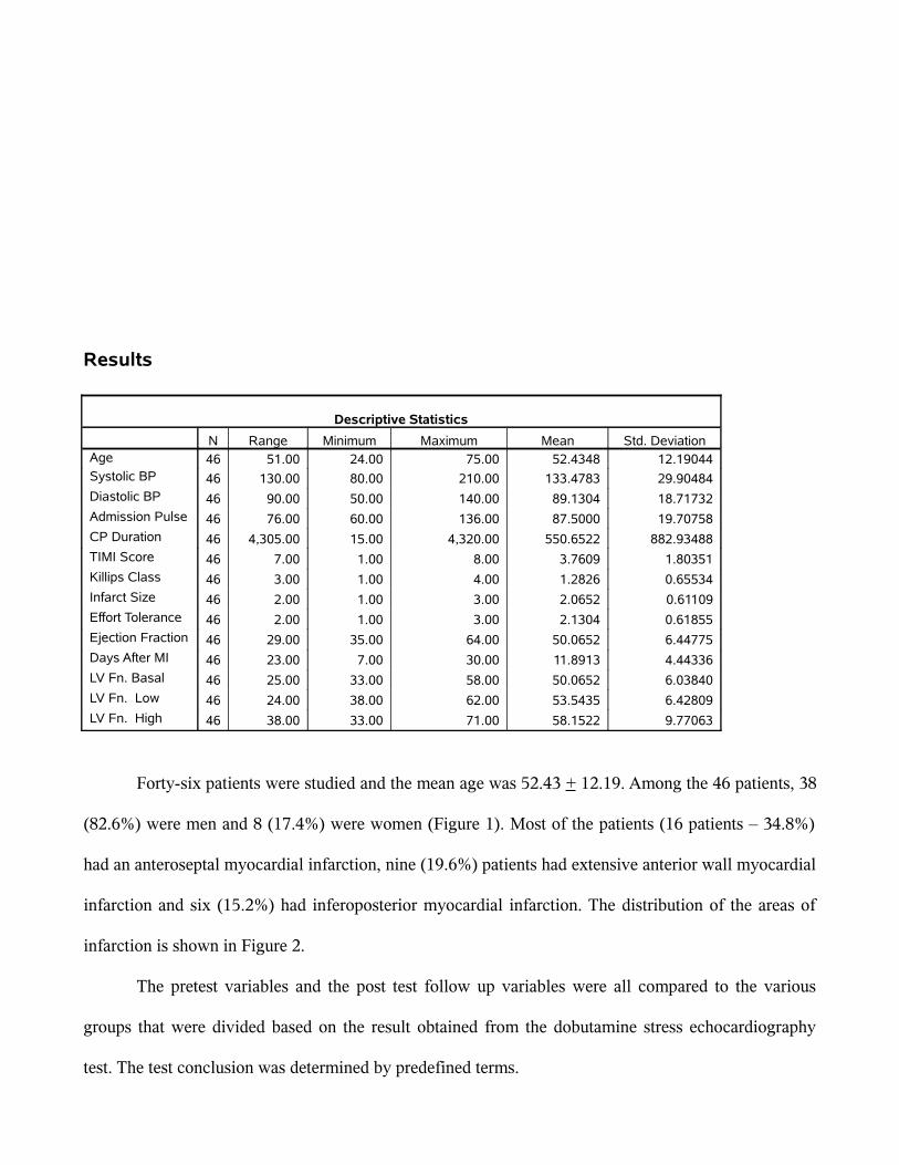
N Range Minimum Maximum Mean Std. DeviationAge 46 51.00 24.00 75.00 52.4348 12.19044Systolic BP 46 130.00 80.00 210.00 133.4783 29.90484Diastolic BP 46 90.00 50.00 140.00 89.1304 18.71732Admission Pulse 46 76.00 60.00 136.00 87.5000 19.70758CP Duration 46 4,305.00 15.00 4,320.00 550.6522 882.93488TIMI Score 46 7.00 1.00 8.00 3.7609 1.80351Killips Class 46 3.00 1.00 4.00 1.2826 0.65534Infarct Size 46 2.00 1.00 3.00 2.0652 0.61109Effort Tolerance 46 2.00 1.00 3.00 2.1304 0.61855Ejection Fraction 46 29.00 35.00 64.00 50.0652 6.44775Days After MI 46 23.00 7.00 30.00 11.8913 4.44336LV Fn. Basal 46 25.00 33.00 58.00 50.0652 6.03840LV Fn. Low 46 24.00 38.00 62.00 53.5435 6.42809LV Fn. High 46 38.00 33.00 71.00 58.1522 9.77063
Forty-six patients were studied and the mean age was 52.43 + 12.19. Among the 46 patients, 38
(82.6%) were men and 8 (17.4%) were women (Figure 1). Most of the patients (16 patients – 34.8%)
had an anteroseptal myocardial infarction, nine (19.6%) patients had extensive anterior wall myocardial
infarction and six (15.2%) had inferoposterior myocardial infarction. The distribution of the areas of
infarction is shown in Figure 2.
The pretest variables and the post test follow up variables were all compared to the various
groups that were divided based on the result obtained from the dobutamine stress echocardiography
test. The test conclusion was determined by predefined terms.

• Group I – No regional wall motion abnormality at rest and no worsening of wall motion,
with normal increase in contractility to dobutamine, which indicates absence of
significant stenosis.
• Group II - Regional wall motion abnormality is present at rest and it improves with low
dose dobutamine and worsens with high dose (the classical biphasic response), which
indicates viability with significant stenosis causing ischaemia.
• Group III - Regional wall motion abnormality is present at rest, which improves with
dobutamine and continues to improve with increasing doses of dobutamine, which
signifies stunned myocardium capable of improving with revascularization.
• Group IV - Regional wall motion abnormality is present at rest and does not improve or
worsen with dobutamine, indicating absence of viability.
• Group V - Regional wall motion abnormality may or may not be present but the regional
wall motion abnormality worsens with dobutamine, which signifies critical narrowing of
the related coronary artery.
The results of the test are shown in the graph Figure 6. Group I had all the patients who showed
a normal response to the test. This comprised of two (4.3%) patients who had no regional wall motion
abnormality at rest and showed no worsening on stress. Group II had 18 (39.1%) patients who had
viability without ischaemia with Group III having seven (15.2%) patients showing viability without
ischaemia. Group IV had 14 (30.4%) patients who showed no reaction to dobutamine indicating
myocardial scar. Group V had five (10.9%) patients who showed worsening ischaemia who probably
had critical stenosis of the respective coronary artery.
Analysis of the Pretest Variables
The results of the Dobutamine Stress Echocardiography (Normal, viable without ischaemia,
viable with ischaemia, not viable and ischaemia on stress) were compared with the pretest parameters.
The various risk factors and their distribution are shown in Figure 4. Cross tabulations were made with
all the individual variables and compared. Both men and women had similar findings during DSE, in
other words, sex did not make a significant difference in the result after MI. Surprisingly the location of
the infarct and the use of streptokinase also did not show significant differences in the conclusion of the
test. The presence of a prior MI was a significant factor in influencing the presence of viability and a
significant number of patients with a prior MI had absent viability as shown in Figure 13 (p=0.043).
The presence of diabetes also significantly influenced the presence of ischaemia in viable tissue as
depicted in Figure 11 (p=0.026). The normoglycaemics had more viable myocardium without
ischaemia than the ones with diabetes. Patients with hypertension also had a noticeable decrease in
viability than normotensives but was not significant (p=0.09).
Factors such as hypotension on admission, presence of an anterior wall myocardial infarction,
ST elevation myocardial infarction and arrhythmias did not have a significant influence on viability
during the post-infarction dobutamine stress echocardiography. Effort tolerance after MI was a
significant factor (p=0.029) in predicting viability after MI.
The TIMI score and the Killips score for myocardial infarction were used; there was no
significant difference when the individual results of the study were compared. If the absence of
ischaemia was taken as a combined entity and if group 1 and 2 were combined, then the scores were
significant in predicting the presence of ischaemia and viability.
One-way analysis of the variables and the results of DSE (Normal, viable without ischaemia,
viable with ischaemia, not viable and ischaemia on stress) showed that the older patients had less
viability than the younger patients did and also more incidence of prior MI. The Killips score was
highest (mean = 1.5 + 0.94) for the group that showed no viability. The pulse rate was also significantly
different between the groups (p=0.016) and so was the basal myocardial function (p=0.001).
When the ischaemia and nonischaemic groups were compared with presence and absence of
events the difference between the groups was significant (p = 0.026). The use of Student’s T- test
revealed a significant influence of TIMI score and ejection fraction. During the test the left ventricular
ejection fraction during low dose and high dose were also significantly related to the presence of
ischaemia (p<0.001). The presence of prior MI also showed significant influence in the presence of
ischaemia in the viable zone.
The analysis of the pretest variables with the conclusion of the test revealed that the following
variables predicted the presence of ischaemia in the presence of viability
1. The presence of prior myocardial infarction
2. The presence of Diabetes mellitus
3. The patient being a hypertensive
4. Patients with more than Killips I
5. Effort tolerance after MI of Class II or more
6. Tachycardia on admission
7. Basal left ventricular systolic function

Complications
Analysis of the test per se showed that it is a safe test and had very low complication rate.
Among the patients studied 37(80.4%) patients had no complications, 2 (4.3%) patients had
hypotension, 1 (2.2%) patient had headache, 5 (10.9%) patients had ventricular ectopic activity and
none had life threatening complications, as shown in Figure 5.
Protocol Completion
It was observed that most of the patients (87%) attained their target heart rate, which was
defined as 85% of the age predicted maximum heart rate (Predicted maximum heart rate = 220 – Age),
with the routine protocol. Only two (4.3%) had inadequate response to dobutamine and out of that one
patient was on a beta-blocker. These patients were included because their heart rate was 82% and 84%
of the target heart rate respectively.
In two patients who had a poor chronotropic response to dobutamine at 20 µg/kg/min, atropine
was added earlier at 30 µg/kg/min and they were able to achieve their target heart rate.
Analysis of the Follow Up
The patients were followed up for 6 months at monthly intervals or whenever symptoms were
present. The analysis was based on the conclusion of the test and the events related to the conclusion.
Initial analysis of individual conclusions of the DSE (Normal, viable without ischaemia, viable
with ischaemia, not viable and ischaemia on stress) were analyzed with the individual events like
unstable angina, myocardial infarction, heart failure or death. Since the number of patients studied were
small and were distributed in the various DSE results, a statistical significance could not be observed.
Once again, the conclusion was clubbed; as one with ischaemia or the one without ischaemia, since
many earlier studies have shown that it is ischaemia that correlates with mortality and not the viability
per se. The events were also clubbed to show if the patients had an event or not instead of showing the
individual events.
There were a significant number of more events in the patients who had ischaemia with viability
than in the patients who showed only viability – Figure 17. The events were seen more often in the
third month of follow up (8.7%), probably because of the return to manual work and because of
irregularity in taking their prescribed medications once they went for work. The patients who had poor
viability and poor ejection fractions presented with more heart failures especially in the second month
of follow up (6.5%). There was a steep increase in the procedure rate in the third month of follow up
(10.9%). This was attributed to the waiting list at the Government General Hospital for the diagnostic
procedure and the usual delay of two months to get the procedure done after a referral.
In the final analysis, when the DSE result was compared with the events during follow up, 17
(85%) patients who had no ischaemia had no ischaemic events – Figure 12 & Figure 14. Among the
patients who showed ischaemia, 12 (46.2%) patients had ischaemia events which was statistically
significant (p = 0.026). The presence of ischaemia during stress after myocardial infarction, detected by
dobutamine stress echocardiography, influenced the prediction of events more than the presence of
viability alone.

Discussion
The strong negative predictive value of dobutamine stress echocardiography was recognized in
our study, which showed no events in the patients with a normal study. The patients who underwent the
study were all post myocardial infarction patients, who were expected to have some amount of regional
wall motion abnormality. Two patients had no regional wall motion abnormality at baseline and did not
progress to show any deterioration of wall motion.
The presence of left ventricular dysfunction and regional wall motion abnormality are dominant
factors in the prognosis of coronary artery disease. Similar patterns were seen in our study with a high
level of significance (p < 0.001), when left ventricular function was compared between all the groups.
The presence of viability has a major impact on the events after myocardial infarction, not just on
mortality. The analysis of pretest factors showed that certain factors contributed to viability and certain
factors predicted ischaemia. Presence of diabetes predicted the presence of ischaemia, than in
euglycaemics who showed more viability without ischaemia. Surprisingly patients who were
thrombolysed showed significantly more number of patients with viability and ischaemia. The exact
relevance of this observation is not clearly understood in this study and probably a study with more
patients would clarify this issue.
Similarly, smokers had more of non-viable myocardium than their non-smoking counterparts
did. It is also interesting to note that none of the smokers had a normal dobutamine stress
echocardiography and none of the non-smokers had an ischaemic response from normal baseline. This
goes to show that smoking not only has an important role in the pathogenesis of myocardial infarction,
it also has a significant role in the prognostic features by affecting the amount of viable myocardium,
probably by influencing the response to treatment.

The detection of the presence of ischaemia and viability using dobutamine stress
echocardiography has been proven beyond doubt. The study focused on the variables that provided
information on the prognostic details. Peak dobutamine wall motion score index and an integrated
assessment of the extent and severity of ventricular dysfunction at peak stress correlated with the
severity of the underlying coronary artery disease and is the strongest predictor of subsequent cardiac
events and death. Stress echocardiographic testing assesses the physiological significance of the
coronary stenosis but cannot predict events largely unrelated to plaque size, thrombus, ulceration of
plaque, fissuring of plaque that leads to coronary occlusion.
Prognostic Meaning of Myocardial Viability
After the pioneering study by Pierard et al, several groups have confirmed the observations that
contractility with dobutamine predicts functional recovery of the dysfunctioning myocardium. In the
light of these well-defined facts, it would be expected that myocardial viability would carry a potential
positive prognostic impact. If the segment has inotropic reserve after dobutamine, it is likely to recover,
and left ventricular function, a major prognostic determinant, is likely to improve. It is worth
remembering that the presence of ischaemia affects the events and long term survival that the presence
of viability alone. Active revascularization should be considered in all patients who show viability with
ischaemia, since they have the best chance of improving left ventricular function with significant long
term benefits.
Conclusion
In our post myocardial infarction dobutamine stress echocardiography study, which also
assessed the risk stratification and follow up for 6 months, the following conclusions were made
1. It is safe to do Dobutamine Stress Echocardiography in post myocardial infarction patients,
even in the early post infarction period, with very low complication rate and risk to life.
2. Thrombolysis had a significant effect on viability, especially in the group, which showed a
biphasic response.
3. Smoking affected the presence of myocardial viability significantly.
4. Patients with diabetes showed more biphasic response, signifying an increased prevalence of
obstructive coronary artery disease.
5. Patients with normal dobutamine stress echocardiography had no events on follow up,
establishing its negative predictive value.
6. Patients who had a biphasic response had the maximum number of events compared to all the
other groups. This group should be considered for early coronary revascularization.
7. The presence of a prior myocardial infarction predicted absence of viability and the absence of
prior myocardial infarction predicted presence of viability.
8. Ejection fraction is a major predictor of events especially heart failure. It also predicted the
coronary events significantly.
BIBLIOGRAPHY
1. Shaw LJ, Eagle KA, Gersh BJ, et al: Meta-analysis of intravenous dipyridamole-thallium-201
imaging and dobutamine echocardiography for risk stratification before vascular surgery. J Am
Coll Cardiol 27:787, 1996
2. Mason JR, palac RT, Freeman ML, et al: Thallium scintigraphy during dobutamine infusion:
non-exercise dependant screening for coronary disease. Am Heart J 107:481, 1984.
3. Wann LS, Faris JV, Childress RH, et al: Exercise cross-sectional echocardiography in ischaemic
heart disease. Circulation 1979; 60:1300-1308
4. Coma-Canella I: Dobutamine stress test to diagnose the presence and severity of coronary
artery disease. Am J Cardiol 12, 1198, 1991
5. Berthe C, Pierard LA, Hiernaux M, et al: Predicting the extent and location of coronary artery
disease in acute myocardial infarction by echocardiography during dobutamine infusion. Am J
Cardiol 58:1167, 1986
6. Armstrong WF, O’Donnel J, Dillon JC, et al: Complementary value of two-dimentional exercise
echocardiography to routine treadmill exercise testing. Ann Intern Med 105:829, 1996
7. Cohen JL, Green TO, Ottenweller J,et al: Dobutamine digital echocardiography for detecting
coronary artery disease. Am J Cardiol 67:1311, 1991
8. Tuttle RR, Mills J: Dobutamine: Development of a new catecholamine to selectively increase
cardiac contractility. Circulation Res 36:185, 1975
9. Leier CV, Unverferth DV: Drugs five years later: Dobutamine. Ann Intern Med 99:490, 1983
10. Chatterjee K, De Marco T: Central and peripheral adrenergic agonists in heart failure. Eur Heart
J 10 (Suppl B):55, 1989
11. Fung AY, Gallagher KP, Buda AJ: Physiological basis of dobutamine as compared with

dipyridamole stress interventions in the assessment of critical coronary stenosis. Circulation
76:943, 1987
12. Corday E, Hajduczki I, O’Byrne GT, et al: Echocardiographic criteria to distinguish reversible
from irreversible myocardial ischaemia. Eur Heart J 9:29, 1988
13. Geleijnse ML, Fioretti PM, Roelandt JR: Methodology, feasibility, safety and diagnostic
accuracy of dobutamine stress echocardiography. J Am Coll Cardiol 30:595, 1997
14. Elhendy A, van Domburg RT, Nierop PR, et al: Impaired systolic blood pressure response to
dobutamine stress testing. A marker of more severe functional abnormalities in patients with
myocardial infarction. J Am Soc Echocardiogr 11:436, 1998
15. Marcovitz PA, Bach DS, Mathias W, et al: Paradoxical hypotension during Dobutamine stress
echocardiography: Clinical and diagnostic implications. J Am Coll Cardiol 21:1080, 1993
16. Geleijnse ML, Elhendy A, van Domburg RT, et al: Prognostic implications of a normal
dobutamine-atropine stress echocardiogram in patients with chest pain. J Am Soc Echocardiogr
11:606, 1998
17. Hepner AM, Bach DS, Armstrong WF: Early chronotropic incompetence predicts the need for
atropine during dobutamine stress echocardiography. Am J Cardiol 79:336, 1997
18. Chauvel C, BoginoE, Reynaud P, et al: Usefulness of isometric exercise during dobutamine
administration for stress echocardiography. Am J Cardiol 81:225, 1998
19. Kisslo J, Byrd BF, Geiser EA, et al: Recommendations for continous quality improvement in
echocardiography. J Am Soc Echocardiogr 8(S):S1, 1995
20. Mertes H, Sawada SG, Ryan T, et al: Symptoms, adverse effects and complications associated
with dobutamine stress echocardiography: Experience in 1118 patients. Circulation 88:15, 1993
21. Seknus M, Marwick TH: Evolution of dobutamine echocardiography protocols and indications:
Safety and sideeffects in 3011 studies over 5 years. J Am Coll Cardiol 29:1234, 1997
22. Picano E, Mathias W, Pingitore A, et al: Safety and tolerability of dobutamine-atropine stress
echocardiography: A prospective, multicentric trial. Lancet 334:1190, 1994
23. Cusik DA, Bonow RO, Chaudhry FA: Safety of dobutamine stress echocardiography in patients
with left ventricular apical thrombus. Am J Cardiol 80:1252, 1997
24. Daniels CJ, Orsinelli DA: Cardiac rupture with dobutamine stress echocardiography. J Am Soc
Echocardiogr 10:979, 1997
25. Marwick T, Willermart B, D’Hondt AM, et al: Selection of optimal nonexercise stress for the
evaluation of ischaemic regional myocardial dysfunction and malperfusion: Comparision of
dobutamine and adenosine using echocardiography and 99mTc-MIBI single photon emission
tomography. Circulation 87:345, 1993
26. Afridi I, Grayburn PA, Panza JA: Myocardial viability during dobutamine echocardiography
predicts survival in patients with coronary artery disease and severe systolic dysfunction. J Am
Coll Cardiol 32:921, 1998
27. Donovan CL, Landolfo KP, Lowe JE, et al: Improvement in inducible ischaemia during
dobutamine stress echocardiography after transmyocardial laser revascularization in patients
with refractory angina pectoris. J Am Coll Cardiol 30:607, 1997
28. Akosah KO, McDaniel S, Hanrahan JS, et al: Dobutamine stress echocardiography early after
heart transplantation predicts development of allogtraft coronary artery disease and outcome. J
Am Coll Cardiol 31: 1607, 1998
29. Lanzarini L, Fetiveau R, Poli A, et al: Significance of ST segment elevation during dobutamine
stress echocardiogroaphy in patients with acute myocardial infarction treated with thrombolysis.
Eur Heart J 17:1008, 1996
30. Sicara R, Picana E, Landi P, et al: On behalf of the Echo Dobutamine International Cooperative
(EDIC) study: Prognostic value of dobutamine-atropine stress echocardiography. J Am Coll
Cardiol 29:254,1997
31. Carlos ME, Smart SC, Wynsen JC, et al: Dobutamine stress echocardiography for risk
stratification after myocardial infarction. Circulation 95:1402, 1997
32. Ross J Jr: Myocardial perfusion-contraction matching: Implications for coronary heart disease
and hibernation. Circulation 83:1076, 1991
33. Rhimtoola SH: The hibernating myocardium. Am Heart J 117:211, 1994
34. Rahimtoola SH: A perspective on the three large multicentric randomized clinical trials of
coronary bypass surgery for chronic stable angina. Circulation 72:V123, 1985
35. Sun KT, Czernin J, Krivokapich J, et al: Effects of dobutamine stimulation on myocardial blood
flow, glucose metabolism amd wall motion in normal and dysfunctional myocardium.
Circulation 94:3146, 1996
36. Vanoverschelde JJ, Wijns W, Depre C,et al: Chronic myocardial hibernation in humans. From
bedside to bench. Circulation 95:1961, 1997
37. Afridi I, Grayburn PA, Panza JA, et al: Myocardial viability during dobutamine
echocardiography predicts survival in patients with coronary artery disease and severe left
ventricular systolic dysfunction. J Am Coll Cardiol 32:921, 1998
38. Eitzman D, al-Aour Z, Kanter HL, et al: Clinical outcome of patients with advanced coronary
artery disease after viability studies with positron emission tomography. J Am Coll Cardiol
20:559, 1992
39. Pagley PR, Beller GA, Watson DD, et al: Improved outcome after coronary bypass surgery in
patients with ischaemic cardiomyopathy and residual myocardial viability. Circulation 21:445,
1997
40. William MJ, Odabashin J, Lauer MS, et al: Prognostic value of dobutamine echocardiography
in patients with left ventricular dysfunction. J Am Coll Cardiol 27:132, 1996
41. Borgers M, Ausma J,: Structural aspects of the chronic hibernating myocardium in man. Basic
res Cardiol 90:44, 1995
42. Dakik HA, Howell JF, Lawrie GM, et al: Assessment of myocardial viability with 99mTc

sestamibi tomography before coronary bypass graft surgery: Correlation with histopathology
and postoperative improvement in cardiac function. Circulation 88: 430, 1993
43. Depre C, Vanoverschelde JL, Melin JA, et al: Structural and metabolic correlates of the
reversibility of chronic left ventricular ischaemic dysfunction in humans. Am J physiol
268:111265, 1995
44. Flameng W, Wouters L, Sergeant P, et al: Multivariate analysis of angiographic, histologic and
electrocardiographic data in patients with coronary heart disease. Circulation 70:7, 1984
45. Elsasser A, Schlepper M, Klovekorn WP, et al: Hibernating myocardium. An incomplete
adaptation to ischaemia. Circulation 96:2920, 1997
46. Schwarz ER, Schaper J, Vom Dahl J, et al: Myocyte degeneration and cell death in hibernating
human myocardium. J Am Coll Cardiol 27:1577, 1996
47. Horn HR, Teichholz LE, Cohn PF, et al: Augmentation of left ventricular contraction pattern in
coronary artery disease by an inotropic catecholamine. The epinephrine ventriculogram.
Circulation 49:1063, 1974
48. Popio KA, Gorlin R, Bechtel D, et al: Postextrasystolic potentiation as a predictor of potential
myocardial viability: Preoperative analysis compared with studies after coronary bypass
surgery. Am J Cardiol 39:944, 1977
49. Afridi I, Kleiman NS, Raizner AE, et al: Dobutamine echocardiography in myocardial
hibernation. Optimal dose and accuracy in predicting recovery of ventricular function after
coronary angioplasty. Circulation 91:663, 1995
50. Arnse M, Cornel JH, Salustri A, et al: Prediction of improvement of regional left ventricular
function after surgical revascularization. A comparison of low dose dobutamine
echocardiography with 201Tl single photon emission computed tomography. Circulation
91:2748, 1995
51. Cigarroa CG, deFilippi CR, Brickner ME, et al: Dobutamine stress echocardiography identifies
hibernating myocardium and predicts recovery of left ventricular function after coronary
revascularization. Circulation 88:430, 1993
52. Perrone-Filardi P, Pace L, Prastro M, et al: Dobutamine echocardiography predicts
improvement of hypoperfusion dysfunctional myocardium after revascularization in patients
with coronary artery disease. Circulation 91:2556, 1995
53. Sharp SM, Sawada SG, Segar DS, et al: Dobutamine stress echocardiography: Detection of
coronary artery disease in patients with dilated cardiomyopathy. J Am Coll Cardiol 24:934,
1994
54. Arnese M, Cornel JH, Salustri A, et al: Prediction of improvement of regional left ventricular
function after surgical revscularization. A comparison of low-dose dobutamine
echocardiography with 201Ti single-photon emission computed tomography. Circulation
91:2748, 1995
55. Perrone-Filardi P, Pace L, Prastro M, et al: Assessment of myocardial viability in patients with
chronic coronary artery disease. Rest-4-hour-24-hour 201Tl tomography versus dobutamine
echocardiography. Circulation 94:2712, 1996
56. Afridi I, Qureshi U, Kopelen HA, et al: Serial changes in response of hibernating myocardium
to inotropic stimulation after revascularization: A dobutamine echocardiographic study. J Am
Coll Cardiol 30:1233, 1997
57. Tillish J, Brunken R, Marshall R et al: Reversibility of cardiac wall motion abnormalities
predicted by positron tomography. N Eng J Med 314:884, 1986
58. Di Carli MF, Davidson M, Little R, et al: Value of metabolic imaging with positron emission
tomography for evaluating prognosis in patients with coronary artery disease and left
ventricular dysfunction. Am J Cardiol 73:5
59. Bach DS, Eagle KA: Dobutamine stress echocardiography: Stressing the indications for
preoperative testing. Circulation 95:8, 1997

60. Bach DS, Cohen JL, Fioretti PM, et al: Safety and efficacy of closed loop arbutamine stress
echocardiography for the detection of coronary artery disease. Am J Cardiol 81:32, 1998
61. Bach DS, Muller DWM, Gros BJ, et al: False positive stress echocardiograms: Characterization
of clinical, echocardiographic and angiographic findings. J Am Coll Cardiol 24:928, 1994
62. Beleslin BD, Ostojic M, Stepanovic J, et al: Stress echocardiography in the detection of
myocardial ischaemia: Head-to-head comparison of exercise, dobutamine and dipyridamole
tests. Circulation 90:1168, 1994
63. Davila-Roman VG, Waggoner AD, Sicard GA, et al: Dobutamine stress echocardiography
predicts surgical outcome in patients with aortic aneurysm and periopheral vascular disease. J
Am Coll Cardiol 21:957, 1993
64. deFilippi CR, Willett DL, Brickner E, et al: Usefullness of dobutamine stress echocardiography
in distinguishing severe from nonsevere valvular
65. Fioretti PM, Poldermans D, Salustri A, et al: Atropine increases the accuracy of dobutamine
stress echocardiography in patients taking β-blockers. Eur Heart J, 15:355, 1994
66. Lalka SG, Sawada SG, Dalsing MC, et al: Dobutamine stress echocardiography as a predictor
of cardiac events associated with aortic surgery. J Vasc Surg 15:831, 1992
67. Lane RT, Sawada SG, Segar DS, et al: Dobutamine stress echocardiography for assessment of
cardiac risk before noncardiac surgery. Am J Cardiol 68: 976, 1991
68. Langan EM III, Youkey JR, Franklin DP, et al: Dobutamine stress echocardiography for cardiac
risk assessment before aortic surgery:J Vasc Surg 18:905, 1993
69. Lewandowski TJ, Armstrong WF, Bach DS: Reduced test time by early identification of
patients requiring atropine during Dobutamine stress echocardiography. J Am Soc Echocardiogr
11:236, 1998
70. Ling LH, Pellikka PA, Mahoney DW, et al: Atropine augmentation in dobutamine stress
echocardiography in patients taking β blockers. J Am Coll Cardiol 28:551, 1996
71. Mannering D, Cripps T, Leech G, et al: The Dobutamine stress test as an alternative to exercise
testing after acute myocardial infarction. Br Heart J 59:521, 1988
72. Martin TW, Seaworth JF, Johns JP, et al: Comparison of adenosine, dipyridamole and
dobutamine in stress echocardiography. Ann Intern Med 116:190, 1992
73. Mazeika PK, Nadazdin PK, Oakley DM: Dobutamine stress echocardiography for detecting and
assessment of coronary artery disease. J Am Coll Cardiol 19:1203, 1992
74. McNeill AJ, Fioretti PM, El-Said EM, et al: Dobutamine stress echocardiography before and
after coronary angioplasty. Am J Cardiol 69:740, 1992
75. Meyer SL, Curry GC, Donsky MS, et al: Influence of dobutamine on haemodynamics and
coronary blood flow with and without coronary artery disease. Am J Cardiol 38:103, 1993
76. Mukherjee S, Davidoff R: Stress echocardiography: An evolving technology for the 90s.
Cardiol Rev 1:350, 1993
77. Murakami H, Nishimura M, Urabe K: Relation between dynamic midventricular obstruction
and unexplained chest pain in patients with normal echocardiograms at rest. Am J Cardiol
78:1063, 1996
78. Nagueh SF, Vaduganathan P, Ali N, et al: Identification of hibernating myocardium:
Comparative accuracy of myocardial contrast echocardiography, rest redistribution thallium-
201 tomography and dobutamine echocardiography J Am Coll Cardiol 28:985, 1997
79. Pellikka PA, Oh JK, Bailey KR, et al: Dynamic intraventricular obstruction during dobutamine
stress echocardiography: A new observation Circulation 86:1429, 1992
80. Pellika PA, Roger VL, Oh JK, et al: Stress echocardiography. Part II. Dobutamine stress
echocardiography: Techniques, implementation, clinical applications and correlations. Mayo
Clin Proc 70:16, 1995
81. Picano E: From pathophysiological toy to diagnostic tool. Circulation 85:1604, 1992
82. Picano E, Mathias W, Pingitore A, et al: Safety and tolerability of dobutamine-atropine stress

echocardiography: A prospective multicentric study. Lancet 334:1190, 1994
DOBUTAMINE STRESS ECHO STUDY
Department of Cardiology, Government Stanley Hospital, Chennai.
Name Age Sex CD Number
Address: Phone:
Study Case Number Date Category I / II / III
Clinical Details
Basic Details
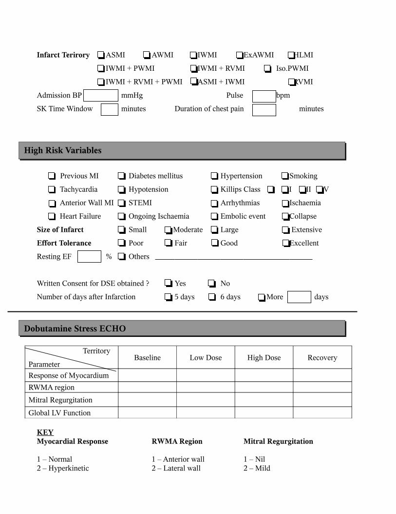
Infarct Terirory ASMI AWMI IWMI ExAWMI HLMI
IWMI + PWMI IWMI + RVMI Iso.PWMI
IWMI + RVMI + PWMI ASMI + IWMI RVMI
Admission BP mmHg Pulse bpm
SK Time Window minutes Duration of chest pain minutes
Previous MI Diabetes mellitus Hypertension Smoking
Tachycardia Hypotension Killips Class I II III IV
Anterior Wall MI STEMI Arrhythmias Ischaemia
Heart Failure Ongoing Ischaemia Embolic event Collapse
Size of Infarct Small Moderate Large Extensive
Effort Tolerance Poor Fair Good Excellent
Resting EF % Others
Written Consent for DSE obtained ? Yes No
Number of days after Infarction 5 days 6 days More days
Territory
ParameterBaseline Low Dose High Dose Recovery
Response of Myocardium
RWMA region
Mitral Regurgitation
Global LV Function
KEYMyocardial Response RWMA Region Mitral Regurgitation
1 – Normal 1 – Anterior wall 1 – Nil2 – Hyperkinetic 2 – Lateral wall 2 – Mild
High Risk Variables
Dobutamine Stress ECHO

3 – Hypokinetic 3 – Inferior wall 3 – Moderate 4 – Akinetic 4 – Posterior wall 4 – Moderately severe5 – Dyskinetic 5 – Septum 5 – Severe6 – Biphasic 6 – Apex
DSE Conclusion:
1 st Follow Up: Date
Any Events ? Unstable Angina Heart Failure Arrhythmias Infarction Death
If Other Events – Details
Any Procedure ? Coronary Angiography PTCA CABG
If Yes Details
2 nd Follow Up: Date
Any Events ? Unstable Angina Heart Failure Arrhythmias Infarction Death
If Other Events – Details
Any Procedure ? Coronary Angiography PTCA CABG
If Yes Details 3 rd Follow Up: Date
Any Events ? Unstable Angina Heart Failure Arrhythmias Infarction Death
If Other Events – Details
Any Procedure ? Coronary Angiography PTCA CABG
If Yes Details
4 th Follow Up: Date
Any Events ? Unstable Angina Heart Failure Arrhythmias Infarction Death
If Other Events – Details
Any Procedure ? Coronary Angiography PTCA CABG
Follow Up
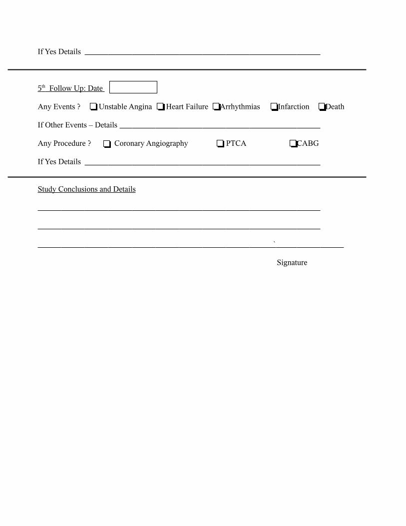
If Yes Details
5 th Follow Up: Date
Any Events ? Unstable Angina Heart Failure Arrhythmias Infarction Death
If Other Events – Details
Any Procedure ? Coronary Angiography PTCA CABG
If Yes Details
Study Conclusions and Details
Signature

Coding Sheet
Specific Codes General Coding
Sex: 0 – No
1 – Male 1 – Yes
2 – Female
Infarct Area:
1 – Anteroseptal Myocardial Infarction
2 – Anterior Wall Myocardial Infarction
3 – Inferior Wall Myocardial Infarction
4 – Extensive Anterior Wall Myocardial Infarction
5 – High Lateral Myocardial Infarction
6 – Inferior Wall Myocardial Infarction + Posterior Wall Myocardial Infarction

7 - Inferior Wall Myocardial Infarction + Right Ventricular Myocardial Infarction
8 – Isolated Posterior Wall Myocardial Infarction
9 - Inferior Wall Myocardial Infarction
+ Posterior Wall Myocardial Infarction + Right Ventricular Myocardial Infarction
10 - Anteroseptal Myocardial Infarction + Inferior Wall Myocardial Infarction
11 - Right Ventricular Myocardial Infarction
12 – Inferolateral Myocardial Infarction
13 – Isolated Right Ventricular Myocardial Infarction
Regional Wall Motion Abnormality:
1 – Normal
2 – Hyperkinetic
3 – Hypokinetic
4 – Akinetic
5 – Dyskinetic
6 – Biphasic
Regional Wall Motion Abnormality – Location:
1 – Anterior Wall
2 – Lateral Wall
3 - Inferior Wall
4 – Posterior Wall
5 – Septum

6- Apex
Mitral Regurgitation:
1 – None
2 – Mild
3 – Moderate
4 – Moderately Severe
5 – Severe
Protocol Completion:
1 – Completed
2 – Achieved Target Heart Rate at low dose
3 – Inadequate heart rate
4 – Stopped due to complications
Complications:
1 – None
2 – Hypotension
3 – Headache
4 – Atrial fibrillation
5 – Ventricular premature beats
6 – Supraventricular tachycardia
7 - Collapse

8 – Death
9 - Myocardial Infarction
10 – Persistent Angina
Conclusion:
1 – Normal
2 – Viability without ischaemia
3 – Viability with ischaemia
4 – No viability
5 – Worsening Ischaemia
Follow Up Events:
1 – None
2 – Unstable Angina
3 – Heart Failure
4 – Arrhythmias
5 – Infarction
6 – Death
Follow Up Procedures:
1 – None
2 – Coronary Angiography

3 – Percutaneous Coronary Intervention
4 – Coronary Artery Bypass Surgery
5 – Death during Procedure
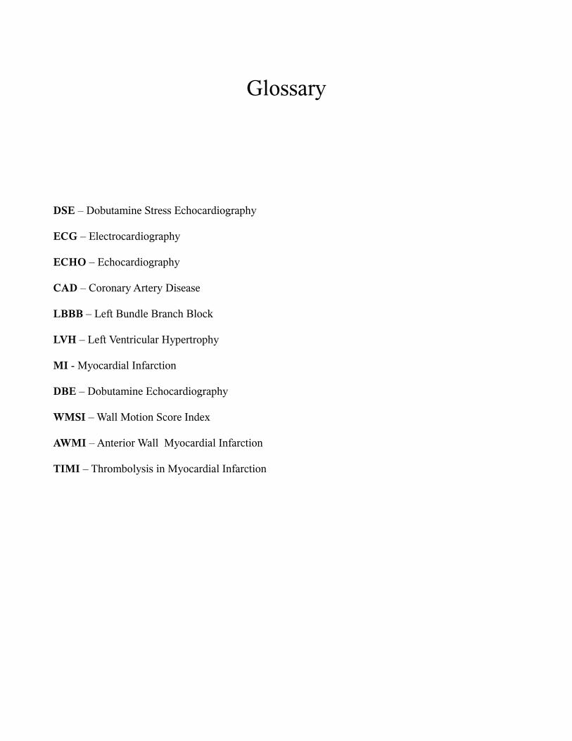
Glossary
DSE – Dobutamine Stress Echocardiography
ECG – Electrocardiography
ECHO – Echocardiography
CAD – Coronary Artery Disease
LBBB – Left Bundle Branch Block
LVH – Left Ventricular Hypertrophy
MI - Myocardial Infarction
DBE – Dobutamine Echocardiography
WMSI – Wall Motion Score Index
AWMI – Anterior Wall Myocardial Infarction
TIMI – Thrombolysis in Myocardial Infarction
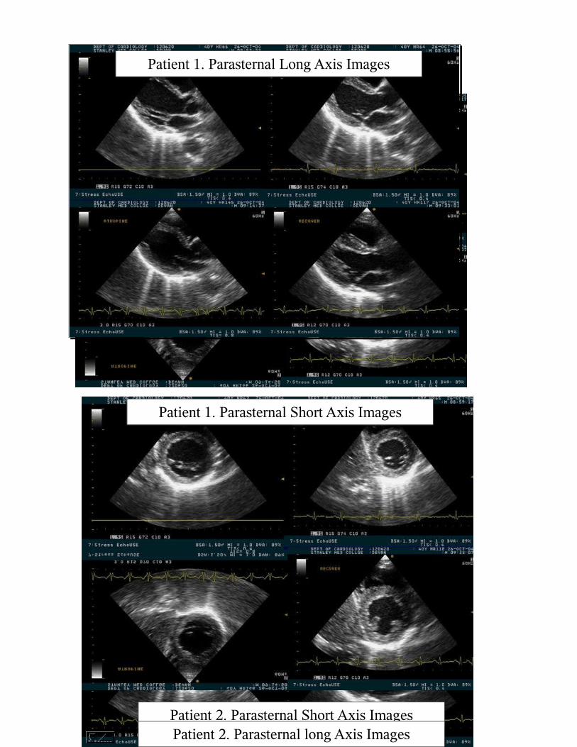
Patient 1. Parasternal Long Axis Images
Patient 1. Parasternal Short Axis Images
Patient 2. Parasternal Short Axis ImagesPatient 2. Parasternal long Axis Images

Figure 1. Sex Distribution

Figure 2. Infarction Territory

Figure 4. High Risk Variables
Figure 5. Complications

Figure 6. Viability Status
Figure 7. Sex & Viability after MI
Figure 8. Thrombolysis & Viability after MI
Figure 9. Smoking & Viability after MI

Figure 10. Peak WMSI & Viability after MI
P < 0.001 P < 0.001
Figure 11. Diabetes & Viability after MI
P = 0.026

Figure 12. Events & Viability after MI
P = 0.047
Figure 13. Prior MI & Viability after MI

Figure 14. Presence of Ischaemia & Coronary Events
Figure 15. Factors Predicting Ischaemia
p = 0.01
p = 0.023
p < 0.001.
p < 0.001.


![Longdom - Early ventricular dysfunction in type II diabetes role ......control population but also more than patients with coronary artery disease [8]. Dobutamine stress echocardiography](https://img.pdfslide.us/doc/110x75/613c808d4c23507cb6356ca8/longdom-early-ventricular-dysfunction-in-type-ii-diabetes-role-control.jpg)