Embed Size (px)
Citation preview
Risk of Acute Kidney Injury After IntravenousContrast Media Administration
Jeremiah S. Hinson, MD, PhD*; Michael R. Ehmann, MD, MPH, MS; Derek M. Fine, MD;Elliot K. Fishman, MD, FACR; Matthew F. Toerper, BS; Richard E. Rothman, MD, PhD; Eili Y. Klein, MS, PhD
*Corresponding Author. E-mail: [email protected].
Study objective: The study objective was to determine whether intravenous contrast administration for computedtomography (CT) is independently associated with increased risk for acute kidney injury and adverse clinical outcomes.
Methods: This single-center retrospective cohort analysis was performed in a large, urban, academic emergencydepartment with an average census of 62,179 visits per year; 17,934 ED visits for patients who underwent contrast-enhanced, unenhanced, or no CT during a 5-year period (2009 to 2014) were included. The intervention was CT scanwith or without intravenous contrast administration. The primary outcome was incidence of acute kidney injury.Secondary outcomes included new chronic kidney disease, dialysis, and renal transplantation at 6 months. Logisticregression modeling and between-groups odds ratios with and without propensity-score matching were used to test foran independent association between contrast administration and primary and secondary outcomes. Treatmentdecisions, including administration of contrast and intravenous fluids, were examined.
Results: Rates of acute kidney injury were similar among all groups. Contrast administration was not associated withincreased incidence of acute kidney injury (contrast-induced nephropathy criteria odds ratio¼0.96, 95% confidenceinterval 0.85 to 1.08; and Acute Kidney Injury Network/Kidney Disease Improving Global Outcomes criteria oddsratio¼1.00, 95% confidence interval 0.87 to 1.16). This was true in all subgroup analyses regardless of baseline renalfunction and whether comparisons were made directly or after propensity matching. Contrast administration was notassociated with increased incidence of chronic kidney disease, dialysis, or renal transplant at 6 months. Clinicians wereless likely to prescribe contrast to patients with decreased renal function and more likely to prescribe intravenous fluidsif contrast was administered.
Conclusion: In the largest well-controlled study of acute kidney injury following contrast administration in the ED todate, intravenous contrast was not associated with an increased frequency of acute kidney injury. [Ann Emerg Med.2016;-:1-10.]
Please see page XX for the Editor’s Capsule Summary of this article.
0196-0644/$-see front matterCopyright © 2016 by the American College of Emergency Physicians.http://dx.doi.org/10.1016/j.annemergmed.2016.11.021
INTRODUCTIONBackground
Intravenous iodinated contrast media are routinelyused to improve the diagnostic accuracy of computedtomography (CT). Although more than 80 million doses ofintravenous contrast media are administered annually,1
clinical decisionmaking in regard to their use is complicatedby concerns related to their potential for precipitating renaldysfunction.2-6 Indeed, contrast media administration iscited as the third most common cause of iatrogenic acutekidney injury6,7 and has been linked to increased risk ofmajor adverse events, including initiation of dialysis, renalfailure, stroke, myocardial infarction, and death.3,8,9 Recentstudies performed in the emergency department (ED),where intravenous administration of contrast media for
enhancement of CT imaging is often necessary to diagnoseacute critical conditions, have reported an incidence ofcontrast-induced nephropathy as high as 14% and linkedcontrast-induced nephropathy to a 2-fold increased risk ofmajor adverse events within 1 year.3,10-12 Although thesereports are concerning, the causal relationship betweenadministration of intravenous contrast media and thedevelopment of acute kidney injury has recently beenchallenged.13-19
ImportanceCurrent understanding of contrast-induced nephropathy
is complicated by studies that predate widespread use oflow- and iso-osmolar contrast media and extrapolation offindings from arterial angiographic studies to the use of
Volume -, no. - : - 2016 Annals of Emergency Medicine 1
IMAGING/ORIGINAL RESEARCH
Editor’s Capsule Summary
What is already known on this topicMany providers defer intravenous contrastenhancement with computed tomography (CT)because of concerns about acute kidney injury.
What question this study addressedHow often does acute kidney injury occur afterenhanced and nonenhanced emergency department(ED) CT?
What this study adds to our knowledgeUsing a propensity-matched case-control design atone site, the frequency of later acute kidney injury in7,201 patients undergoing contrast-enhanced CT,5,499 undergoing unenhanced CT, and 5,234 withno imaging did not differ (10.2% to 10.9%).
How this is relevant to clinical practiceThis study suggests fear of triggering acute kidneyinjury after intravenous contrast during ED CT isdisproportionate to objective data. A randomized trialis needed to confirm this finding.
intravenous contrast media. Additionally, the majority ofstudies examining acute kidney injury after contrast mediaadministration, including those performed in EDpatients,3,10-12 were performed without control populationsthat did not receive contrast media. Indeed, serumcreatinine level fluctuations meeting criteria for contrast-induced nephropathy occur in patients undergoingunenhanced CT at rates similar to those published aftercontrast-enhanced CT,13 and systematic reviews and meta-analyses of the few existing studies analyzing intravenouscontrast media administration with adequate controlsfound no increased risk of acute kidney injury associatedwith contrast media.14,16
Historically, randomized controlled trials designed toelucidate the true incidence of contrast-inducednephropathy have been perceived as unethical because ofthe presumption that contrast media administration is adirect cause of acute kidney injury. To date, all controlledstudies of contrast-induced nephropathy have beenobservational, and conclusions from these studies areseverely limited by selection bias associated with the clinicaldecision to administer contrast media. Two research groupshave recently used propensity-score analysis to control forthis bias, approximating randomization by matchingnonrandomized populations from large single-center
databases according to their probability of treatmentassignment.18,20 These investigators, however, reachedopposite conclusions. McDonald et al15,17,18 found noincreased risk of acute kidney injury, emergency dialysis, ormortality after contrast media administration in any patientgroup regardless of baseline renal function, whereasDavenport et al21 reported an increased risk of acute kidneyinjury after contrast media administration in patients withpreexisting renal dysfunction. Potential explanations forthese discrepant results include different strategies forpropensity matching, variances in institutional contrast-enhanced CT protocols, and widely discordant subgroupsample sizes, especially at the lowest baseline renal function.
Goals of This InvestigationIn this study, we sought to clarify the incidence of acute
kidney injury attributable to intravenous contrast mediaadministration by testing the hypothesis that such injuryoccurs at higher rates in patients undergoing contrast-enhanced CT than in those not receiving contrast media.To minimize biases associated with comparison ofnonrandomized populations, we used 2 distinct controlpopulations that did not receive contrast media, usedpropensity-score analysis to minimize bias associated withtreatment assignment, and analyzed large numbers ofpatients in all subgroups of baseline renal function. We alsoexamined selected clinician practice patterns that may affectthe incidence of acute kidney injury after contrast mediaadministration.
MATERIALS AND METHODSStudy Design and Setting
This was a single-center retrospective cohort studyconducted in a large urban academic ED. During the studyperiod, our mean annual ED census was 62,179 total visits(range 55,955 to 69,249), with a mean annual admissionrate of 23.4% (range 22.2% to 25.1%). This study wasapproved by our university institutional review board.
An experienced data user (E.Y.K.) extracted all clinicalinformation from a relational database that underlies theelectronic medical record of our ED. Queries usingstructured query language were performed separately forpatients who did and did not undergo CT. For patientswho underwent CT, data were extracted for all encountersthat had both an order and result interpretation for at leastone CT scan during the study period. Two authors (J.S.H.and M.R.E.) not involved in data extraction classified CTstudies as enhanced or unenhanced based on specific andstandardized order and result interpretation identifiers.When order and result interpretation identifiers differed
2 Annals of Emergency Medicine Volume -, no. - : - 2016
Risk of Acute Kidney Injury After Intravenous Contrast Administration Hinson et al
(which occurred in cases in which the radiology team, inconsultation with the ordering ED clinician, performed astudy that differed from the original order), resultinterpretation identifiers were used. Any discrepancies wereresolved through consensus. For patients who did notundergo CT, data were extracted for all encounters thatlacked an order or result for a CT study. All data relating tovital signs, medication administration, and preexistingdiagnoses entered in the ED, as well as laboratory resultsand new procedure or diagnostic codes entered from anypoint in our hospital system, were extracted with the samestructured query language for every patient. Medicaldiagnoses and procedures were identified with InternationalClassification of Diseases, Ninth Edition, ClinicalModification (ICD-9-CM) diagnostic and procedure codes.
Selection of ParticipantsPatients aged 18 years and older who received a CT
with or without contrast enhancement in the ED betweenJanuary 1, 2009, and June 30, 2014, and had both aninitial serum creatinine level measured in the 8 hoursbefore CT and a second level measured 48 to 72 hoursafter CT were included. To minimize bias associated withthe decision to obtain imaging, we included a secondcontrol group of contrast media-unexposed ED patientsaged 18 years and older and treated during the studyperiod who did not undergo CT imaging, and had both aninitial serum creatinine level measured in the ED and asecond level measured 48 to 72 hours later. Exclusioncriteria included initial serum creatinine level less than 0.4mg/dL (to minimize inclusion of random laboratory erroras cases of acute kidney injury) or equal to or greater than4.0 mg/dL (already meeting partial criteria for severe acutekidney injury), insufficient serum creatinine level data, ahistory of renal transplant or ongoing or previous dialysis,an ED visit in the 6 months before the study start date, aCT scan performed in the 6 months preceding the indexED visit, and contrast-enhanced CT performed within 72hours of ED departure. We chose the antecedent 6-monthwindow to minimize potential confounding residualeffects of previously administered contrast media and thesubsequent 72-hour window to minimize potential groupcrossover of patients undergoing CT scan after EDdeparture within the period defined by contrast-inducednephropathy criteria. CT scans were classified as contrastenhanced or unenhanced. Consecutive CT acquisitions atdifferent anatomic locations were treated as a single-scanevent, and those performed with and without contrastmedia were treated as a single contrast-enhanced CT. Alleligible patients during a 5-year period were included,
resulting in a sample size powered to detect a difference inincidence of acute kidney injury between populations aslow as 1.5%.
The primary variable of interest was administration ofintravenous contrast media. Control variables included age,sex, race, initial serum creatinine level, initial estimatedglomerular filtration rate,20 and chronic comorbidities andacute illness severity indicators previously shown topredispose to the development of contrast-associated acutekidney injury.22-24 Chronic comorbidities includeddiabetes mellitus, hypertension, HIV/AIDS, congestiveheart failure, chronic kidney disease, and history of renaltransplantation (all identified by ICD-9-CM codes25).Acute illness severity indicators included hypotension(systolic blood pressure <80 mm Hg), designation by anED attending physician as a patient requiring critical care,anemia (hematocrit level <39% or <36% for men andwomen, respectively), and hypoalbuminemia (<3.5 g/dL)during the index ED visit. Additional control variablesincluded ED administration of nephrotoxic ornephroprotective medications (see Figure E1, availableonline at http://www.annemergmed.com, for full list)26
and ED administration of intravenous crystalloid fluids inany amount.
All contrast media administration was performedaccording to institutional protocols, available online athttp://www.ctisus.com/protocols. Patients who underwentcontrast-enhanced CT were administered either iohexol oriodixanol intravenously, as dictated by radiologic study-specific protocol, and volumes of administration rangedfrom 80 to 120 mL. According to institutional policy,patients provided consent before administration ofintravenous contrast, and for patients with serum creatininelevel greater than 1.7 mg/dL, treating clinicians cosignedconsent for contrast-enhanced CT.
Outcome MeasuresThe primary outcome variable was incidence of acute
kidney injury. The clinical definition of acute kidney injuryhas undergone multiple revisions, with published studies ofcontrast-induced nephropathy using varied equations tocalculate its incidence. We estimated the incidence of acutekidney injury for all study participants by using both themost frequently published criteria for contrast-inducednephropathy (absolute increase in serum creatinine level"0.5 mg/dL or "25% increase over baseline serumcreatinine level at 48 to 72 hours after imaging or, fornon-CT patients, after initial serum creatinine levelmeasurement)27 and for acute kidney injury as definedby the Acute Kidney Injury Network/Kidney Disease
Hinson et al Risk of Acute Kidney Injury After Intravenous Contrast Administration
Volume -, no. - : - 2016 Annals of Emergency Medicine 3
Improving Global Outcomes guidelines.28,29 By AcuteKidney Injury Network/Kidney Disease Improving GlobalOutcomes creatinine-based criteria, acute kidney injury isstaged (stage 1: absolute increase in serum creatinine level"0.3 mg/dL or a 1.5- to 1.9-fold increase over baselineserum creatinine level; stage 2: 2.0- to 2.9-fold increaseover baseline serum creatinine level; stage 3: 3-fold increaseover baseline serum creatinine level, increase to serumcreatinine level "4.0 mg/dL, or initiation of dialysis). Weclassified any patient meeting Acute Kidney InjuryNetwork/Kidney Disease Improving Global Outcomesstage 1 criteria as having acute kidney injury and performedsubanalyses to compare patients meeting criteria for stages2 and 3. Patient-centered outcomes were assessed,including newly diagnosed chronic kidney disease,initiation of dialysis, and renal transplantation (eachassessed by ICD-9-CM or procedure code documentationin our institutional electronic medical record) within 6months of the index ED visit.
Primary Data AnalysisDichotomous variables are displayed as percentages,
categorical data as relative frequencies (in percentages), andcontinuous data as medians with interquartile ranges. Amultivariable logistic regression model was used to ascertainhow contrast media administration was associated with therisk of acute kidney injury in the entire study populationafter controlling for demographic variables and medicalconditions previously reported to increase risk fordeveloping such injury.23,24 Incidence of acute kidneyinjury was calculated as the percentage of visits withoccurrence of acute kidney injury.
The association between contrast media and acutekidney injury was first assessed with the test of proportionsto compare incidence of acute kidney injury in patientswho underwent contrast-enhanced CT with those whounderwent unenhanced CT and with all patients who didnot receive contrast media. To reduce potential selectionbias inherent to administration of contrast media, we alsoused propensity-score matching to estimate the effect ofcontrast media on acute kidney injury. The clinical decisionto administer contrast media is guided by patient pathologyand conditional patient-related factors that mightcontraindicate contrast media administration. Theconditional patient factors included in the estimation of thepropensity scores were sex, age, race, initial serumcreatinine level or estimated glomerular filtration rate,crystalloid fluid administration, nephrotoxic medicationadministration, chronic comorbidities (as noted above),and whether the patient was designated as requiring critical
care. Propensity-score matching was performed withdefault parameters (nearest neighbor of one, no caliperrestriction, and sampling with replacement), and theaverage treatment effect was calculated. All comparisonswere made for the entire study population and forsubgroups stratified by initial serum creatinine level andestimated glomerular filtration rate. Matching forpropensity score was performed by group for subgroupanalyses. Results are presented as odds ratios (ORs). Allanalysis, including propensity-score matching, wasconducted in Stata (version 14.1; StataCorp, CollegeStation, TX).
RESULTSDuring the study period, there were 82,729 patient
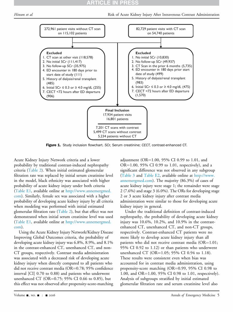
visits in which a CT was performed among 54,740 uniquepatients. Of these, 12,700 patient visits by 11,567 patientsmet all inclusion and no exclusion criteria. Of all CT scans,56.7% were contrast enhanced. There were 272,961patient visits during the study period wherein patients didnot undergo CT. Of these, 5,234 met all inclusion and noexclusion criteria. Thus, a total of 17,934 patient visitsfrom 16,801 unique patients were included in the finalanalysis (Figure 1).
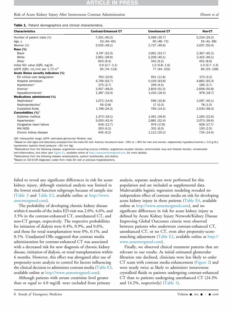
All 3 patient groups analyzed (contrast-enhanced CT,unenhanced CT, and non-CT) were demographicallysimilar, although the unenhanced CT group was slightlyolder (Table 1). Patients in the contrast-enhanced CTgroup were less likely to have diabetes, congestive heartfailure, or chronic kidney disease. Initial serum creatininevalues were similar across groups, although the contrast-enhanced CT group had a higher estimated glomerularfiltration rate.
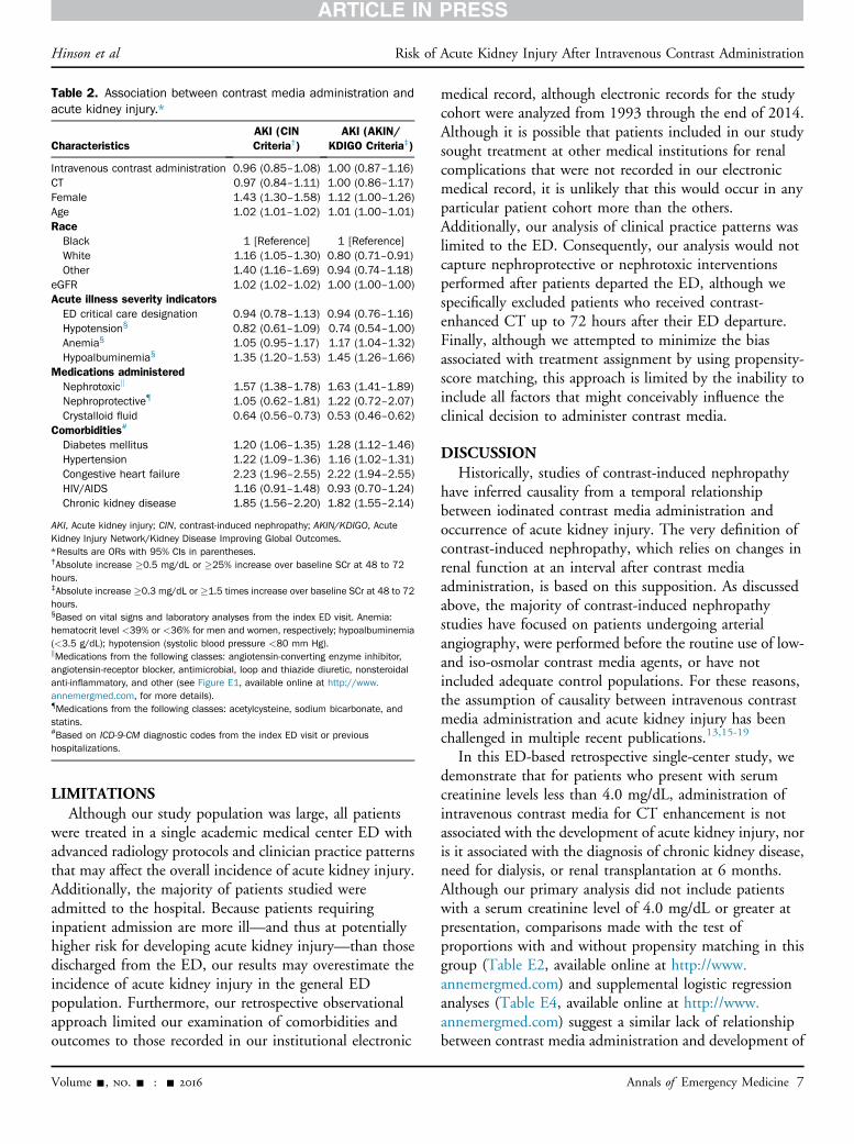
Multivariable logistic regression modeling of the entirestudy population, with inclusion of predictor variablespreviously reported to affect the incidence of acute kidneyinjury, revealed no independent effect of contrast mediaon the probability of developing acute kidney injury(Table 2 and Table E1, available online at http://www.annemergmed.com). Factors associated most strongly withan increased probability of acute kidney injury wereincreased age, administration of nephrotoxic medication(s),preexisting diagnosis of congestive heart failure or chronickidney disease, and hypoalbuminemia. Administration ofintravenous crystalloids was associated with a lowerprobability of developing acute kidney injury. Whenmodeling was performed with initial estimated glomerularfiltration rate as a predictor variable, the effect of race onacute kidney injury differed by criteria: black ethnicity wasassociated with higher probability of acute kidney injury by
Risk of Acute Kidney Injury After Intravenous Contrast Administration Hinson et al
4 Annals of Emergency Medicine Volume -, no. - : - 2016
Acute Kidney Injury Network criteria and a lowerprobability by traditional contrast-induced nephropathycriteria (Table 2). When initial estimated glomerularfiltration rate was replaced by initial serum creatinine levelin the model, black ethnicity was associated with higherprobability of acute kidney injury under both criteria(Table E1, available online at http://www.annemergmed.com). Similarly, female sex was associated with a higherprobability of developing acute kidney injury by all criteriawhen modeling was performed with initial estimatedglomerular filtration rate (Table 2), but that effect was notdemonstrated when initial serum creatinine level was used(Table E1, available online at http://www.annemergmed.com).
Using the Acute Kidney Injury Network/Kidney DiseaseImproving Global Outcomes criteria, the probability ofdeveloping acute kidney injury was 6.8%, 8.9%, and 8.1%in the contrast-enhanced CT, unenhanced CT, and non-CT groups, respectively. Contrast media administrationwas associated with a decreased risk of developing acutekidney injury when directly compared to all patients whodid not receive contrast media (OR¼0.78; 95% confidenceinterval [CI] 0.70 to 0.88) and patients who underwentunenhanced CT (OR¼0.75; 95% CI 0.66 to 0.85), butthis effect was not observed after propensity-score-matching
adjustment (OR¼1.00, 95% CI 0.99 to 1.01, andOR¼1.00, 95% CI 0.99 to 1.01, respectively), and asignificant difference was not observed in any subgroup(Table 3 and Table E2, available online at http://www.annemergmed.com). The majority (86.3%) of cases ofacute kidney injury were stage 1; the remainder were stage2 (7.6%) and stage 3 (6.0%). The ORs for developing stage2 or 3 acute kidney injury after contrast mediaadministration were similar to those for developing acutekidney injury in general.
Under the traditional definition of contrast-inducednephropathy, the probability of developing acute kidneyinjury was 10.6%, 10.2%, and 10.9% in the contrast-enhanced CT, unenhanced CT, and non-CT groups,respectively. Contrast-enhanced CT patients were nomore likely to develop acute kidney injury than allpatients who did not receive contrast media (OR¼1.01;95% CI 0.92 to 1.12) or than patients who underwentunenhanced CT (OR¼1.05; 95% CI 0.94 to 1.18).These results were consistent even when bias wasaccounted for in contrast media administration, usingpropensity-score matching (OR¼0.99, 95% CI 0.98 to1.00, and OR¼1.00, 95% CI 0.98 to 1.01, respectively).Analysis of subgroups stratified by initial estimatedglomerular filtration rate and serum creatinine level also
Figure 1. Study inclusion flowchart. SCr, Serum creatinine; CECT, contrast-enhanced CT.
Hinson et al Risk of Acute Kidney Injury After Intravenous Contrast Administration
Volume -, no. - : - 2016 Annals of Emergency Medicine 5
failed to reveal any significant differences in risk for acutekidney injury, although statistical analysis was limited inthe lowest renal function subgroups because of sample size(Table 3 and Table E2, available online at http://www.annemergmed.com).
The probability of developing chronic kidney diseasewithin 6 months of the index ED visit was 2.0%, 4.6%, and3.5% in the contrast-enhanced CT, unenhanced CT, andnon-CT groups, respectively. The respective probabilitiesfor initiation of dialysis were 0.4%, 0.9%, and 0.6%,and those for renal transplantation were 0%, 0.1%, and0.1%. Unadjusted ORs suggested that contrast mediaadministration for contrast-enhanced CT was associatedwith a decreased risk for new diagnosis of chronic kidneydisease, initiation of dialysis, or renal transplantation within6 months. However, this effect was abrogated after use ofpropensity-score analysis to control for factors influencingthe clinical decision to administer contrast media (Table E3,available online at http://www.annemergmed.com).
Although patients with serum creatinine level greaterthan or equal to 4.0 mg/dL were excluded from primary
analysis, separate analyses were performed for thispopulation and are included as supplemental data.Multivariable logistic regression modeling revealed noindependent effect of contrast media on risk for developingacute kidney injury in these patients (Table E4, availableonline at http://www.annemergmed.com), and nosignificant differences in risk for acute kidney injury asdefined by Acute Kidney Injury Network/Kidney DiseaseImproving Global Outcomes criteria were observedbetween patients who underwent contrast-enhanced CT,unenhanced CT, or no CT, even after propensity-score-matching adjustment (Table E2, available online at http://www.annemergmed.com).
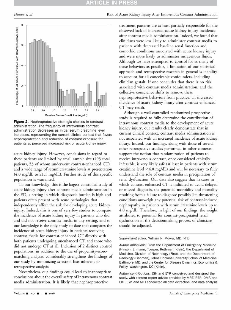
Finally, we observed clinical treatment patterns that arerelevant to our results. As initial estimated glomerularfiltration rate declined, clinicians were less likely to orderCT scans with contrast media enhancement (Figure 2) andwere nearly twice as likely to administer intravenouscrystalloid fluids to patients undergoing contrast-enhancedCT than to patients undergoing unenhanced CT (24.3%and 14.2%, respectively) (Table 1).
Table 1. Patient demographics and clinical characteristics.
Characteristics Contrast-Enhanced CT Unenhanced CT Non-CT
Number of patient visits (%) 7,201 (40.2) 5,499 (30.7) 5,234 (29.2)Age, y 53 (40–65) 60 (48–73) 55 (41–68)Women (%) 3,535 (49.1) 2,727 (49.6) 2,637 (50.4)Race (%)Black 3,747 (52.0) 2,951 (53.7) 2,367 (45.2)White 2,851 (39.6) 2,206 (40.1) 2,415 (46.1)Other 603 (8.4) 342 (6.2) 452 (8.6)
Initial SCr value (IQR), mg/dL 0.9 (0.7–1.1) 1.0 (0.8–1.6) 1.0 (0.7–1.3)eGFR (IQR), mL/min per 1.73 m2 95 (74–114) 77 (44–102) 84 (55–108)Acute illness severity indicators (%)ED critical care designation 763 (10.6) 651 (11.8) 170 (3.2)Hospital admission 6,749 (93.7) 5,159 (93.8) 4,882 (93.3)Hypotension* 270 (3.7) 249 (4.5) 196 (3.7)Anemia* 3,457 (48.0) 2,819 (51.3) 2,658 (50.8)Hypoalbuminemia* 1,387 (19.3) 1,010 (18.4) 978 (18.7)
Medications administered (%)Nephrotoxic† 1,072 (14.9) 596 (10.8) 2,097 (40.1)Nephroprotective‡ 56 (0.8) 17 (0.3) 78 (1.5)Crystalloid fluids 1,748 (24.3) 783 (14.2) 2,530 (48.3)
Comorbidities (%)§
Diabetes mellitus 1,373 (19.1) 1,461 (26.6) 1,183 (22.6)Hypertension 3,050 (42.4) 2,881 (52.4) 2,073 (39.6)Congestive heart failure 918 (12.7) 979 (17.8) 928 (17.7)HIV/AIDS 303 (4.2) 331 (6.0) 130 (2.5)Chronic kidney disease 445 (6.2) 1,112 (20.2) 735 (14.0)
IQR, Interquartile range; eGFR, estimated glomerular filtration rate.*Based on vital signs and laboratory analyses from the index ED visit. Anemia: hematocrit level <39% or <36% for men and women, respectively; hypoalbuminemia (<3.5 g/dL);hypotension (systolic blood pressure <80 mm Hg).†Medications from the following classes: angiotensin-converting enzyme inhibitor, angiotensin-receptor blocker, antimicrobial, loop and thiazide diuretic, nonsteroidalanti-inflammatory, and other (see Figure E1, available online at http://www.annemergmed.com, for more details).‡Medications from the following classes: acetylcysteine, sodium bicarbonate, and statins.§Based on ICD-9-CM diagnostic codes from index ED visit or previous hospitalizations.
Risk of Acute Kidney Injury After Intravenous Contrast Administration Hinson et al
6 Annals of Emergency Medicine Volume -, no. - : - 2016
LIMITATIONSAlthough our study population was large, all patients
were treated in a single academic medical center ED withadvanced radiology protocols and clinician practice patternsthat may affect the overall incidence of acute kidney injury.Additionally, the majority of patients studied wereadmitted to the hospital. Because patients requiringinpatient admission are more ill—and thus at potentiallyhigher risk for developing acute kidney injury—than thosedischarged from the ED, our results may overestimate theincidence of acute kidney injury in the general EDpopulation. Furthermore, our retrospective observationalapproach limited our examination of comorbidities andoutcomes to those recorded in our institutional electronic
medical record, although electronic records for the studycohort were analyzed from 1993 through the end of 2014.Although it is possible that patients included in our studysought treatment at other medical institutions for renalcomplications that were not recorded in our electronicmedical record, it is unlikely that this would occur in anyparticular patient cohort more than the others.Additionally, our analysis of clinical practice patterns waslimited to the ED. Consequently, our analysis would notcapture nephroprotective or nephrotoxic interventionsperformed after patients departed the ED, although wespecifically excluded patients who received contrast-enhanced CT up to 72 hours after their ED departure.Finally, although we attempted to minimize the biasassociated with treatment assignment by using propensity-score matching, this approach is limited by the inability toinclude all factors that might conceivably influence theclinical decision to administer contrast media.
DISCUSSIONHistorically, studies of contrast-induced nephropathy
have inferred causality from a temporal relationshipbetween iodinated contrast media administration andoccurrence of acute kidney injury. The very definition ofcontrast-induced nephropathy, which relies on changes inrenal function at an interval after contrast mediaadministration, is based on this supposition. As discussedabove, the majority of contrast-induced nephropathystudies have focused on patients undergoing arterialangiography, were performed before the routine use of low-and iso-osmolar contrast media agents, or have notincluded adequate control populations. For these reasons,the assumption of causality between intravenous contrastmedia administration and acute kidney injury has beenchallenged in multiple recent publications.13,15-19
In this ED-based retrospective single-center study, wedemonstrate that for patients who present with serumcreatinine levels less than 4.0 mg/dL, administration ofintravenous contrast media for CT enhancement is notassociated with the development of acute kidney injury, noris it associated with the diagnosis of chronic kidney disease,need for dialysis, or renal transplantation at 6 months.Although our primary analysis did not include patientswith a serum creatinine level of 4.0 mg/dL or greater atpresentation, comparisons made with the test ofproportions with and without propensity matching in thisgroup (Table E2, available online at http://www.annemergmed.com) and supplemental logistic regressionanalyses (Table E4, available online at http://www.annemergmed.com) suggest a similar lack of relationshipbetween contrast media administration and development of
Table 2. Association between contrast media administration andacute kidney injury.*
CharacteristicsAKI (CINCriteria†)
AKI (AKIN/KDIGO Criteria‡)
Intravenous contrast administration 0.96 (0.85–1.08) 1.00 (0.87–1.16)CT 0.97 (0.84–1.11) 1.00 (0.86–1.17)Female 1.43 (1.30–1.58) 1.12 (1.00–1.26)Age 1.02 (1.01–1.02) 1.01 (1.00–1.01)RaceBlack 1 [Reference] 1 [Reference]White 1.16 (1.05–1.30) 0.80 (0.71–0.91)Other 1.40 (1.16–1.69) 0.94 (0.74–1.18)
eGFR 1.02 (1.02–1.02) 1.00 (1.00–1.00)Acute illness severity indicatorsED critical care designation 0.94 (0.78–1.13) 0.94 (0.76–1.16)Hypotension§ 0.82 (0.61–1.09) 0.74 (0.54–1.00)Anemia§ 1.05 (0.95–1.17) 1.17 (1.04–1.32)Hypoalbuminemia§ 1.35 (1.20–1.53) 1.45 (1.26–1.66)
Medications administeredNephrotoxick 1.57 (1.38–1.78) 1.63 (1.41–1.89)Nephroprotective{ 1.05 (0.62–1.81) 1.22 (0.72–2.07)Crystalloid fluid 0.64 (0.56–0.73) 0.53 (0.46–0.62)
Comorbidities#
Diabetes mellitus 1.20 (1.06–1.35) 1.28 (1.12–1.46)Hypertension 1.22 (1.09–1.36) 1.16 (1.02–1.31)Congestive heart failure 2.23 (1.96–2.55) 2.22 (1.94–2.55)HIV/AIDS 1.16 (0.91–1.48) 0.93 (0.70–1.24)Chronic kidney disease 1.85 (1.56–2.20) 1.82 (1.55–2.14)
AKI, Acute kidney injury; CIN, contrast-induced nephropathy; AKIN/KDIGO, AcuteKidney Injury Network/Kidney Disease Improving Global Outcomes.*Results are ORs with 95% CIs in parentheses.†Absolute increase "0.5 mg/dL or "25% increase over baseline SCr at 48 to 72hours.‡Absolute increase "0.3 mg/dL or "1.5 times increase over baseline SCr at 48 to 72hours.§Based on vital signs and laboratory analyses from the index ED visit. Anemia:hematocrit level <39% or <36% for men and women, respectively; hypoalbuminemia(<3.5 g/dL); hypotension (systolic blood pressure <80 mm Hg).kMedications from the following classes: angiotensin-converting enzyme inhibitor,angiotensin-receptor blocker, antimicrobial, loop and thiazide diuretic, nonsteroidalanti-inflammatory, and other (see Figure E1, available online at http://www.annemergmed.com, for more details).{Medications from the following classes: acetylcysteine, sodium bicarbonate, andstatins.#Based on ICD-9-CM diagnostic codes from the index ED visit or previoushospitalizations.
Hinson et al Risk of Acute Kidney Injury After Intravenous Contrast Administration
Volume -, no. - : - 2016 Annals of Emergency Medicine 7
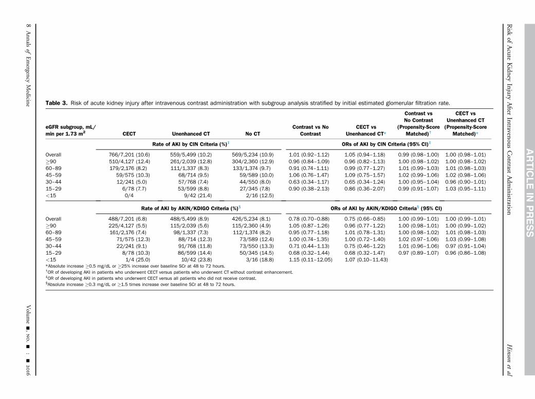
Table 3. Risk of acute kidney injury after intravenous contrast administration with subgroup analysis stratified by initial estimated glomerular filtration rate.
eGFR subgroup, mL/min per 1.73 m2 CECT Unenhanced CT No CT
Contrast vs NoContrast
CECT vsUnenhanced CT*
Contrast vsNo Contrast
(Propensity-ScoreMatched)†
CECT vsUnenhanced CT(Propensity-Score
Matched)*
Rate of AKI by CIN Criteria (%)‡ ORs of AKI by CIN Criteria (95% CI)‡
Overall 766/7,201 (10.6) 559/5,499 (10.2) 569/5,234 (10.9) 1.01 (0.92–1.12) 1.05 (0.94–1.18) 0.99 (0.98–1.00) 1.00 (0.98–1.01)"90 510/4,127 (12.4) 261/2,039 (12.8) 304/2,360 (12.9) 0.96 (0.84–1.09) 0.96 (0.82–1.13) 1.00 (0.98–1.02) 1.00 (0.98–1.02)60–89 179/2,176 (8.2) 111/1,337 (8.3) 133/1,374 (9.7) 0.91 (0.74–1.11) 0.99 (0.77–1.27) 1.01 (0.99–1.03) 1.01 (0.98–1.03)45–59 59/575 (10.3) 68/714 (9.5) 59/589 (10.0) 1.06 (0.76–1.47) 1.09 (0.75–1.57) 1.02 (0.99–1.06) 1.02 (0.98–1.06)30–44 12/241 (5.0) 57/768 (7.4) 44/550 (8.0) 0.63 (0.34–1.17) 0.65 (0.34–1.24) 1.00 (0.95–1.04) 0.96 (0.90–1.01)15–29 6/78 (7.7) 53/599 (8.8) 27/345 (7.8) 0.90 (0.38–2.13) 0.86 (0.36–2.07) 0.99 (0.91–1.07) 1.03 (0.95–1.11)<15 0/4 9/42 (21.4) 2/16 (12.5)
Rate of AKI by AKIN/KDIGO Criteria (%)§ ORs of AKI by AKIN/KDIGO Criteria§ (95% CI)
Overall 488/7,201 (6.8) 488/5,499 (8.9) 426/5,234 (8.1) 0.78 (0.70–0.88) 0.75 (0.66–0.85) 1.00 (0.99–1.01) 1.00 (0.99–1.01)"90 225/4,127 (5.5) 115/2,039 (5.6) 115/2,360 (4.9) 1.05 (0.87–1.26) 0.96 (0.77–1.22) 1.00 (0.98–1.01) 1.00 (0.99–1.02)60–89 161/2,176 (7.4) 98/1,337 (7.3) 112/1,374 (8.2) 0.95 (0.77–1.18) 1.01 (0.78–1.31) 1.00 (0.98–1.02) 1.01 (0.98–1.03)45–59 71/575 (12.3) 88/714 (12.3) 73/589 (12.4) 1.00 (0.74–1.35) 1.00 (0.72–1.40) 1.02 (0.97–1.06) 1.03 (0.99–1.08)30–44 22/241 (9.1) 91/768 (11.8) 73/550 (13.3) 0.71 (0.44–1.13) 0.75 (0.46–1.22) 1.01 (0.96–1.06) 0.97 (0.91–1.04)15–29 8/78 (10.3) 86/599 (14.4) 50/345 (14.5) 0.68 (0.32–1.44) 0.68 (0.32–1.47) 0.97 (0.89–1.07) 0.96 (0.86–1.08)<15 1/4 (25.0) 10/42 (23.8) 3/16 (18.8) 1.15 (0.11–12.05) 1.07 (0.10–11.43)*Absolute increase "0.5 mg/dL or "25% increase over baseline SCr at 48 to 72 hours.†OR of developing AKI in patients who underwent CECT versus patients who underwent CT without contrast enhancement.‡OR of developing AKI in patients who underwent CECT versus all patients who did not receive contrast.§Absolute increase "0.3 mg/dL or "1.5 times increase over baseline SCr at 48 to 72 hours.
Risk
ofAcute
Kidney
InjuryAfter
IntravenousContrast
Adm
inistrationHinson
etal
8Annals
ofEmergency
Medicine
Volum
e-,no.-
:-
2016
acute kidney injury. However, conclusions in regard tothese patients are limited by small sample size (493 totalpatients, 53 of whom underwent contrast-enhanced CT)and a wide range of serum creatinine levels at presentation(4.0 mg/dL to 21.1 mg/dL). Further study of this specificpopulation is warranted.
To our knowledge, this is the largest controlled study ofacute kidney injury after contrast media administration inthe ED, a setting in which diagnostic burden is high andpatients often present with acute pathologies thatindependently affect the risk for developing acute kidneyinjury. Indeed, this is one of very few studies to comparethe incidence of acute kidney injury in patients who didand did not receive contrast media in any setting, and toour knowledge is the only study to date that compares theincidence of acute kidney injury in patients receivingcontrast media for contrast-enhanced CT directly withboth patients undergoing unenhanced CT and those whodid not undergo CT at all. Inclusion of 2 distinct controlpopulations, in addition to the use of propensity-score-matching analysis, considerably strengthens the findings ofour study by minimizing selection bias inherent toretrospective analysis.
Nevertheless, our findings could lead to inappropriateconclusions about the overall safety of intravenous contrastmedia administration. It is likely that nephroprotective
treatment patterns are at least partially responsible for theobserved lack of increased acute kidney injury incidenceafter contrast media administration. Indeed, we found thatclinicians were less likely to administer contrast media topatients with decreased baseline renal function andcomorbid conditions associated with acute kidney injuryand were more likely to administer intravenous fluids.Although we have attempted to control for as many ofthese behaviors as possible, a limitation of our statisticalapproach and retrospective research in general is inabilityto account for all conceivable confounders, includingclinician gestalt. If one concludes that there is no riskassociated with contrast media administration, and thecollective conscience shifts to remove thesenephroprotective behaviors from practice, an increasedincidence of acute kidney injury after contrast-enhancedCT may result.
Although a well-controlled randomized prospectivestudy is required to fully determine the contribution ofintravenous contrast media to the development of acutekidney injury, our results clearly demonstrate that incurrent clinical context, contrast media administration isnot associated with an increased incidence of acute kidneyinjury. Indeed, our findings, along with those of severalother retrospective studies performed in other contexts,support the notion that randomization of patients toreceive intravenous contrast, once considered ethicallyinfeasible, is very likely safe (at least in patients with serumcreatinine level <4.0 mg/dL) and will be necessary to fullyunderstand the role of contrast media in precipitation ofrenal dysfunction. Our data also suggest that in cases inwhich contrast-enhanced CT is indicated to avoid delayedor missed diagnosis, the potential morbidity and mortalityresulting from a failure to diagnose possibly life-threateningconditions outweigh any potential risk of contrast-inducednephropathy in patients with serum creatinine levels up to4.0 mg/dL. Therefore, in light of our findings, the weightattributed to potential for contrast-precipitated renaldysfunction in the decisionmaking process of cliniciansshould be adjusted.
Supervising editor: William R. Mower, MD, PhD
Author affiliations: From the Department of Emergency Medicine(Hinson, Ehmann, Toerper, Rothman, Klein), the Department ofMedicine, Division of Nephrology (Fine), and the Department ofRadiology (Fishman), Johns Hopkins University School of Medicine,Baltimore, MD; and the Center for Disease Dynamics, Economics &Policy, Washington, DC (Klein).
Author contributions: JSH and EYK conceived and designed thestudy, with content expert advice provided by MRE, RER, DMF, andEKF. EYK and MFT conducted all data extraction, and data analysis
Figure 2. Nephroprotective strategic choices in contrastadministration. The frequency of intravenous contrastadministration decreases as initial serum creatinine levelincreases, representing the current clinical context that favorsnephroprotection and reduction of contrast exposure forpatients at perceived increased risk of acute kidney injury.
Hinson et al Risk of Acute Kidney Injury After Intravenous Contrast Administration
Volume -, no. - : - 2016 Annals of Emergency Medicine 9
was performed primarily by EYK. JSH and MRE drafted themanuscript, and all authors contributed substantially to itsrevision. JSH takes responsibility for the paper as a whole.
All authors attest to meeting the four ICMJE.org authorship criteria:(1) Substantial contributions to the conception or design of thework; or the acquisition, analysis, or interpretation of data for thework; AND (2) Drafting the work or revising it critically for importantintellectual content; AND (3) Final approval of the version to bepublished; AND (4) Agreement to be accountable for all aspects ofthe work in ensuring that questions related to the accuracy orintegrity of any part of the work are appropriately investigated andresolved.
Funding and support: By Annals policy, all authors are required todisclose any and all commercial, financial, and other relationshipsin any way related to the subject of this article as per ICMJE conflictof interest guidelines (see www.icmje.org). The authors have statedthat no such relationships exist.
Publication dates: Received for publication May 11, 2016.Revision received September 5, 2016. Accepted for publicationNovember 14, 2016.
REFERENCES1. Katzberg RW, Haller C. Contrast-induced nephrotoxicity: clinical
landscape. Kidney Int. 2006;69:S3-S7.2. Subramanian S, Tumlin J, Bapat B, et al. Economic burden of contrast-
induced nephropathy: implications for prevention strategies. J MedEcon. 2007;10:119-134.
3. Mitchell AM, Kline JA, Jones AE, et al. Major adverse events one yearafter acute kidney injury after contrast-enhanced computedtomography. Ann Emerg Med. 2015;66:267-274.e4.
4. Schräder R. Contrast material–induced renal failure: an overview.J Interv Cardiol. 2005;18:417-423.
5. McCullough PA, Adam A, Becker CR, et al. Epidemiology and prognosticimplicationsof contrast-inducednephropathy.AmJCardiol. 2006;98:5-13.
6. Hou SH, Bushinsky DA, Wish JB, et al. Hospital-acquired renalinsufficiency: a prospective study. Am J Med. 1983;74:243-248.
7. Nash K, Hafeez A, Hou S. Hospital-acquired renal insufficiency. Am JKidney Dis. 2002;39:930-936.
8. Coca SG, Peixoto AJ, Garg AX, et al. The prognostic importance of a smallacute decrement in kidney function in hospitalized patients: a systematicreview and meta-analysis. Am J Kidney Dis. 2007;50:712-720.
9. Solomon RJ, Mehran R, Natarajan MK, et al. Contrast-inducednephropathy and long-term adverse events: cause and effect? Clin JAm Soc Nephrol. 2009;4:1162-1169.
10. Mitchell AM, Kline JA. Contrast nephropathy following computedtomography angiography of the chest for pulmonary embolism in theemergency department. J Thromb Haemost. 2007;5:50-54.
11. Mitchell AM, Jones AE, Tumlin J, et al. Incidence of contrast-inducednephropathy after contrast-enhanced computed tomography in theoutpatient setting. Clin J Am Soc Nephrol. 2010;5:4-9.
12. Mitchell AM, Jones AE, Tumlin J, et al. Prospective study of theincidence of contrast-induced nephropathy among patients evaluatedfor pulmonary embolism by contrast-enhanced computed tomography.Acad Emerg Med. 2012;19:618-625.
13. Newhouse JH, Kho D, Rao QA, et al. Frequency of serum creatininechanges in the absence of iodinated contrast material: implications forstudies of contrast nephrotoxicity. AJR Am J Roentgenol. 2008;191:376-382.
14. Rao QA, Newhouse JH. Risk of nephropathy after intravenousadministration of contrast material: a critical literature analysis.Radiology. 2006;239:392-397.
15. McDonald JS, McDonald RJ, Carter RE, et al. Risk of intravenouscontrast material–mediated acute kidney injury: a propensityscore–matched study stratified by baseline-estimated glomerularfiltration rate. Radiology. 2014;271:65-73.
16. Mcdonald JS, McDonald RJ, Comin J, et al. Frequency of acute kidneyinjury following intravenous contrast medium administration: asystematic review and meta-analysis. Radiology. 2013;267:119-128.
17. McDonald RJ, McDonald JS, Carter RE, et al. Intravenous contrastmaterial exposure is not an independent risk factor for dialysis ormortality. Radiology. 2014;273:714-725.
18. Mcdonald RJ, McDonald JS, Bida JP, et al. Contrast material-inducednephropathy: causal or coincident phenomenon? Radiology.2013;267:103-118.
19. Sinert R, Brandler E, Subramanian RA, et al. Does the currentdefinition of contrast-induced acute kidney injury reflect a true clinicalentity? Acad Emerg Med. 2012;19:1261-1267.
20. Levey AS, Stevens LA, Schmid CH, et al. A new equation to estimateglomerular filtration rate. Ann Intern Med. 2009;150:604-612.
21. Davenport MS, Khalabari S, Cohan RH, et al. Contrastmaterial–induced nephrotoxicity and intravenous low-osmolalityiodinated contrast material: risk stratification by using estimatedglomerular filtration rate. Radiology. 2013;268:719-728.
22. Kooiman J, Klok FA, Mos IC, et al. Incidence and predictors ofcontrast-induced nephropathy following CT-angiography for clinicallysuspected acute pulmonary embolism. J Thromb Haemost.2010;8:409-411.
23. Mehran R, Aymong ED, Nikolsky E, et al. A simple risk score forprediction of contrast-induced nephropathy after percutaneouscoronary intervention. J Am Coll Cardiol. 2004;44:1393-1399.
24. Mehran R, Nikolsky E. Contrast-induced nephropathy: definition,epidemiology, and patients at risk. Kidney Int Suppl. 2006;100:S11-S15.
25. Elixhauser A, Steiner C, Harris DR, et al. Comorbidity measures foruse with administrative data. Med Care. 1998;36:8-27.
26. Naughton CA. Drug-induced nephrotoxicity. Am Fam Physician.2008;78:743-750.
27. Solomon R. Contrast-medium-induced acute renal failure. Kidney Int.1998;53:230-242.
28. Mehta RL, Kellum JA, Shah SV, et al. Acute Kidney Injury Network:report of an initiative to improve outcomes in acute kidney injury. CritCare. 2007;11:R31.
29. Kidney Disease: Improving Global Outcomes (KDIGO) Acute KidneyInjury Work Group. KDIGO clinical practice guideline for acute kidneyinjury. Kidney Int Suppl. 2012;2:1-138.
Risk of Acute Kidney Injury After Intravenous Contrast Administration Hinson et al
10 Annals of Emergency Medicine Volume -, no. - : - 2016
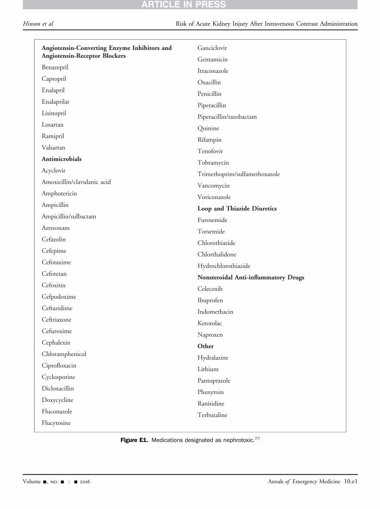
Figure E1. Medications designated as nephrotoxic.26
Hinson et al Risk of Acute Kidney Injury After Intravenous Contrast Administration
Volume -, no. - : - 2016 Annals of Emergency Medicine 10.e1
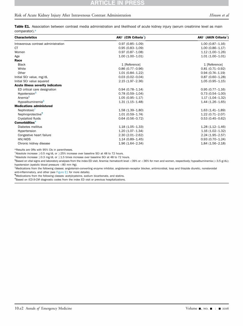
Table E1. Association between contrast media administration and likelihood of acute kidney injury (serum creatinine level as maincomparator).*
Characteristics AKI* (CIN Criteria†) AKI* (AKIN Criteria‡)
Intravenous contrast administration 0.97 (0.85–1.09) 1.00 (0.87–1.16)CT 0.95 (0.83–1.09) 1.00 (0.86–1.17)Women 0.97 (0.87–1.08) 1.12 (1.00–1.26)Age 1.00 (1.00–1.01) 1.01 (1.00–1.01)RaceBlack 1 [Reference] 1 [Reference]White 0.86 (0.77–0.96) 0.81 (0.71–0.92)Other 1.01 (0.84–1.22) 0.94 (0.74–1.19)
Initial SCr value, mg/dL 0.03 (0.02–0.04) 0.87 (0.60–1.28)Initial SCr value squared 2.15 (1.97–2.36) 1.05 (0.95–1.15)Acute illness severity indicatorsED critical care designation 0.94 (0.78–1.14) 0.95 (0.77–1.16)Hypotension§ 0.78 (0.59–1.04) 0.73 (0.54–1.00)Anemia§ 1.05 (0.95–1.17) 1.17 (1.04–1.32)Hypoalbuminemia§ 1.31 (1.15–1.48) 1.44 (1.26–1.65)
Medications administeredNephrotoxick 1.58 (1.39–1.80) 1.63 (1.41–1.89)Nephroprotective{ 1.01 (0.59–1.74) 1.22 (0.71–2.07)Crystalloid fluids 0.64 (0.56–0.72) 0.53 (0.45–0.62)
Comorbidities#
Diabetes mellitus 1.18 (1.05–1.33) 1.28 (1.12–1.46)Hypertension 1.20 (1.07–1.34) 1.16 (1.02–1.32)Congestive heart failure 2.30 (2.01–2.62) 2.24 (1.95–2.57)HIV/AIDS 1.14 (0.89–1.45) 0.93 (0.70–1.24)Chronic kidney disease 1.96 (1.64–2.34) 1.84 (1.56–2.18)
*Results are ORs with 95% CIs in parentheses.†Absolute increase "0.5 mg/dL or "25% increase over baseline SCr at 48 to 72 hours.‡Absolute increase "0.3 mg/dL or "1.5 times increase over baseline SCr at 48 to 72 hours.§Based on vital signs and laboratory analyses from the index ED visit. Anemia: hematocrit level <39% or <36% for men and women, respectively; hypoalbuminemia (<3.5 g/dL);hypotension (systolic blood pressure <80 mm Hg).kMedications from the following classes: angiotensin-converting enzyme inhibitor, angiotensin-receptor blocker, antimicrobial, loop and thiazide diuretic, nonsteroidalanti-inflammatory, and other (see Figure E1 for more details).{Medications from the following classes: acetylcysteine, sodium bicarbonate, and statins.#Based on ICD-9-CM diagnostic codes from the index ED visit or previous hospitalizations.
Risk of Acute Kidney Injury After Intravenous Contrast Administration Hinson et al
10.e2 Annals of Emergency Medicine Volume -, no. - : - 2016
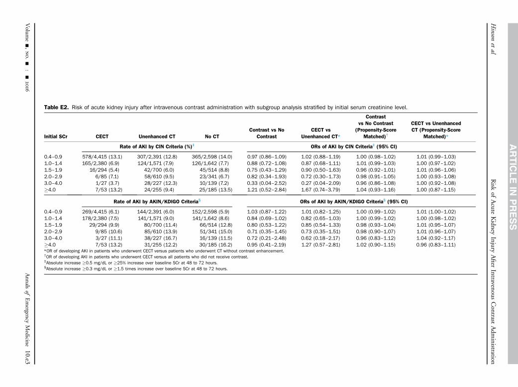
Table E2. Risk of acute kidney injury after intravenous contrast administration with subgroup analysis stratified by initial serum creatinine level.
Initial SCr CECT Unenhanced CT No CTContrast vs No
ContrastCECT vs
Unenhanced CT*
Contrastvs No Contrast
(Propensity-ScoreMatched)†
CECT vs UnenhancedCT (Propensity-Score
Matched)*
Rate of AKI by CIN Criteria (%)‡ ORs of AKI by CIN Criteria‡ (95% CI)
0.4–0.9 578/4,415 (13.1) 307/2,391 (12.8) 365/2,598 (14.0) 0.97 (0.86–1.09) 1.02 (0.88–1.19) 1.00 (0.98–1.02) 1.01 (0.99–1.03)1.0–1.4 165/2,380 (6.9) 124/1,571 (7.9) 126/1,642 (7.7) 0.88 (0.72–1.08) 0.87 (0.68–1.11) 1.01 (0.99–1.03) 1.00 (0.97–1.02)1.5–1.9 16/294 (5.4) 42/700 (6.0) 45/514 (8.8) 0.75 (0.43–1.29) 0.90 (0.50–1.63) 0.96 (0.92–1.01) 1.01 (0.96–1.06)2.0–2.9 6/85 (7.1) 58/610 (9.5) 23/341 (6.7) 0.82 (0.34–1.93) 0.72 (0.30–1.73) 0.98 (0.91–1.05) 1.00 (0.93–1.08)3.0–4.0 1/27 (3.7) 28/227 (12.3) 10/139 (7.2) 0.33 (0.04–2.52) 0.27 (0.04–2.09) 0.96 (0.86–1.08) 1.00 (0.92–1.08)"4.0 7/53 (13.2) 24/255 (9.4) 25/185 (13.5) 1.21 (0.52–2.84) 1.67 (0.74–3.79) 1.04 (0.93–1.16) 1.00 (0.87–1.15)
Rate of AKI by AKIN/KDIGO Criteria§ ORs of AKI by AKIN/KDIGO Criteria§ (95% CI)
0.4–0.9 269/4,415 (6.1) 144/2,391 (6.0) 152/2,598 (5.9) 1.03 (0.87–1.22) 1.01 (0.82–1.25) 1.00 (0.99–1.02) 1.01 (1.00–1.02)1.0–1.4 178/2,380 (7.5) 141/1,571 (9.0) 141/1,642 (8.6) 0.84 (0.69–1.02) 0.82 (0.65–1.03) 1.00 (0.99–1.02) 1.00 (0.98–1.02)1.5–1.9 29/294 (9.9) 80/700 (11.4) 66/514 (12.8) 0.80 (0.53–1.22) 0.85 (0.54–1.33) 0.98 (0.93–1.04) 1.01 (0.95–1.07)2.0–2.9 9/85 (10.6) 85/610 (13.9) 51/341 (15.0) 0.71 (0.35–1.45) 0.73 (0.35–1.51) 0.98 (0.90–1.07) 1.01 (0.96–1.07)3.0–4.0 3/27 (11.1) 38/227 (16.7) 16/139 (11.5) 0.72 (0.21–2.48) 0.62 (0.18–2.17) 0.96 (0.83–1.12) 1.04 (0.92–1.17)"4.0 7/53 (13.2) 31/255 (12.2) 30/185 (16.2) 0.95 (0.41–2.19) 1.27 (0.57–2.81) 1.02 (0.90–1.15) 0.96 (0.83–1.11)*OR of developing AKI in patients who underwent CECT versus patients who underwent CT without contrast enhancement.†OR of developing AKI in patients who underwent CECT versus all patients who did not receive contrast.‡Absolute increase "0.5 mg/dL or "25% increase over baseline SCr at 48 to 72 hours.§Absolute increase "0.3 mg/dL or "1.5 times increase over baseline SCr at 48 to 72 hours.
Hinson
etal
Risk
ofAcute
Kidney
InjuryAfter
IntravenousContrast
Adm
inistration
Volum
e-,no.-
:-
2016
Annals
ofEmergency
Medicine
10.e3
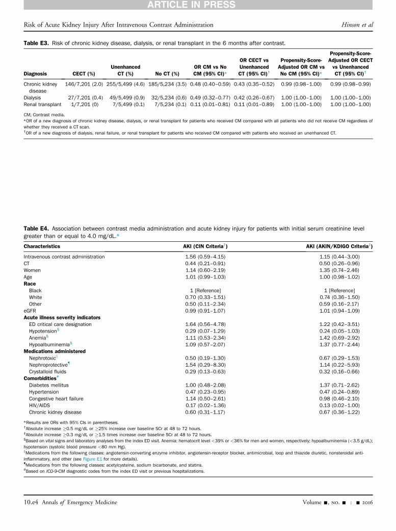
Table E3. Risk of chronic kidney disease, dialysis, or renal transplant in the 6 months after contrast.
Diagnosis CECT (%)Unenhanced
CT (%) No CT (%)OR CM vs NoCM (95% CI)*
OR CECT vsUnenhancedCT (95% CI)†
Propensity-Score-Adjusted OR CM vsNo CM (95% CI)*
Propensity-Score-Adjusted OR CECTvs UnenhancedCT (95% CI)†
Chronic kidneydisease
146/7,201 (2.0) 255/5,499 (4.6) 185/5,234 (3.5) 0.48 (0.40–0.59) 0.43 (0.35–0.52) 0.99 (0.98–1.00) 0.99 (0.98–0.99)
Dialysis 27/7,201 (0.4) 49/5,499 (0.9) 32/5,234 (0.6) 0.49 (0.32–0.77) 0.42 (0.26–0.67) 1.00 (1.00–1.00) 1.00 (1.00–1.00)Renal transplant 1/7,201 (0) 7/5,499 (0.1) 7/5,234 (0.1) 0.11 (0.01–0.81) 0.11 (0.01–0.89) 1.00 (1.00–1.00) 1.00 (1.00–1.00)
CM, Contrast media.*OR of a new diagnosis of chronic kidney disease, dialysis, or renal transplant for patients who received CM compared with all patients who did not receive CM regardless ofwhether they received a CT scan.†OR of a new diagnosis of dialysis, renal failure, or renal transplant for patients who received CM compared with patients who received an unenhanced CT.
Table E4. Association between contrast media administration and acute kidney injury for patients with initial serum creatinine levelgreater than or equal to 4.0 mg/dL.*
Characteristics AKI (CIN Criteria†) AKI (AKIN/KDIGO Criteria‡)
Intravenous contrast administration 1.56 (0.59–4.15) 1.15 (0.44–3.00)CT 0.44 (0.21–0.91) 0.50 (0.26–0.96)Women 1.14 (0.60–2.19) 1.35 (0.74–2.46)Age 1.01 (0.99–1.03) 1.00 (0.98–1.02)RaceBlack 1 [Reference] 1 [Reference]White 0.70 (0.33–1.51) 0.74 (0.36–1.50)Other 0.50 (0.11–2.34) 0.59 (0.16–2.17)
eGFR 0.99 (0.91–1.07) 1.01 (0.94–1.09)Acute illness severity indicatorsED critical care designation 1.64 (0.56–4.78) 1.22 (0.42–3.51)Hypotension§ 0.29 (0.07–1.29) 0.24 (0.05–1.03)Anemia§ 1.11 (0.53–2.34) 1.42 (0.69–2.92)Hypoalbuminemia§ 1.09 (0.57–2.07) 1.37 (0.77–2.44)
Medications administeredNephrotoxick 0.50 (0.19–1.30) 0.67 (0.29–1.53)Nephroprotective{ 1.54 (0.29–8.30) 1.14 (0.22–5.93)Crystalloid fluids 0.29 (0.13–0.63) 0.32 (0.16–0.66)
Comorbidities#
Diabetes mellitus 1.00 (0.48–2.08) 1.37 (0.71–2.62)Hypertension 0.47 (0.23–0.95) 0.47 (0.24–0.89)Congestive heart failure 1.14 (0.50–2.61) 0.98 (0.46–2.10)HIV/AIDS 0.17 (0.02–1.36) 0.13 (0.02–1.00)Chronic kidney disease 0.60 (0.31–1.17) 0.67 (0.36–1.22)
*Results are ORs with 95% CIs in parentheses.†Absolute increase "0.5 mg/dL or "25% increase over baseline SCr at 48 to 72 hours.‡Absolute increase "0.3 mg/dL or "1.5 times increase over baseline SCr at 48 to 72 hours.§Based on vital signs and laboratory analyses from the index ED visit. Anemia: hematocrit level <39% or <36% for men and women, respectively; hypoalbuminemia (<3.5 g/dL);hypotension (systolic blood pressure <80 mm Hg).kMedications from the following classes: angiotensin-converting enzyme inhibitor, angiotensin-receptor blocker, antimicrobial, loop and thiazide diuretic, nonsteroidal anti-inflammatory, and other (see Figure E1 for more details).{Medications from the following classes: acetylcysteine, sodium bicarbonate, and statins.#Based on ICD-9-CM diagnostic codes from the index ED visit or previous hospitalizations.
Risk of Acute Kidney Injury After Intravenous Contrast Administration Hinson et al
10.e4 Annals of Emergency Medicine Volume -, no. - : - 2016