Embed Size (px)
Citation preview
Risk Analysis, Vol. 28, No. 5, 2008 DOI: 10.1111/j.1539-6924.2008.01080.x
Risk Communication Planning for the Aftermathof a Plague Bioattack
Elizabeth A. Casman1∗ and Baruch Fischhoff1,2
We create an influence diagram of how a plague bioattack could unfold and then use it toidentify factors shaping infection risks in many possible scenarios. The influence diagramand associated explanations provide a compact reference that allows risk communicators toidentify key messages for pre-event preparation and testing. It can also be used to answerspecific questions in whatever unique situations arise, considering both the conditions of theattack and the properties of the attacked populations. The influence diagram allows a quick,visual check of the factors that must be covered when evaluating audience information needs.The documentation provides content for explaining the resultant advice. We show how thesetools can help in preparing for crises and responding to them.
KEY WORDS: Bioterrorism; influence diagram; plague ecology; risk communication; rodents; Yersiniapestis
1. INTRODUCTION
In a public health emergency, there is little timeto develop health communications. Unless that workhas been done in advance, public health officials mustimprovise—at the risk of saying wrong things (be-cause the situation has not been analyzed properly)or of saying right things wrongly (because messageshave not been tested for effectiveness). If officials failthe public, then they can cede the stage to less quali-fied voices, offering confident, incompetent, and con-tradictory messages.
There are guidelines for systematically develop-ing and evaluating communications for well-specifiedrisks.(1,2) But what happens when a threat’s de-tails cannot be predicted in advance? We propose
1 Department of Engineering and Public Policy, Carnegie MellonUniversity.
2 Department of Social and Decision Sciences, Carnegie MellonUniversity.
∗Address correspondence to Elizabeth A. Casman, Carnegie Mel-lon University, Department of Engineering and Public Policy,Pittsburgh, PA 15213 USA; [email protected].
a method for developing communications for suchsituations. It uses an influence diagram to organizethe facts relevant to the decisions that individualsmight face. In advance of an emergency, prototypemessages wouldbe developed and evaluated for sce-narios spanning the range of possible emergencies.When an actual emergency arose, the prototype mes-sages would be adapted to the specific circumstances,drawing on the information organized with the influ-ence diagram. We demonstrate the approach with aplague bioattack, one threat with multiple possiblescenarios.
1.1. Current Risk Communication Planningfor Plague Attack
In focus groups convened by the Centers for Dis-ease Control and Prevention (CDC), participants re-ported wanting information that would help themto prevent and detect exposures, identify symptoms,and treat infections, along with background informa-tion providing them with basic understanding of thehazard.(3) Such information is available on the CDCbioterrorism website.(4)
1327 0272-4332/08/0100-1327$22.00/1 C© 2008 Society for Risk Analysis

1328 Casman and Fischhoff
As seen below, plague risk reflects the in-teraction of multiple, complex processes. Withoutdecision-focused analysis, communications can misscritical facts or bury them in irrelevant details. Mem-bers of the public cannot be expected to set informa-tion priorities about topics where they lack expertise,even with more systematic data-collection methodsthan focus groups, whose only proper use is in themost preliminary, formative stages of research.(5)
1.2. Influence-Diagram-Based RapidCommunication Method
There are large peer-reviewed and gray liter-atures about plague, its natural ecology, and con-trol with contributions from many disciplines. Influ-ence diagrams can organize such disparate facts,(6,7)
representing critical factors as nodes and their depen-dencies as connecting arrows.
Section 2 gives an overview of naturally occur-ring and postattack plague risk. Section 3 presents abasic influence diagram model of a plague bioattack,focused on factors relevant to decision making. Sec-tion 4 focuses on measures that disrupt model links.Section 5 describes how the model can be used toidentify and organize facts needed for effective com-munication. We focus on the United States, althoughmany conclusions apply elsewhere.
2. PLAGUE ATTACK SCENARIO ANDBACKGROUND INFORMATION
Plague is a rapidly progressing, often fatal dis-ease caused by the bacterium Yersinia pestis. It is aCDC Category A select agent, i.e., an organism suit-able for bioterrorism. Plague can infect many warm-blooded animals, often lethally.(8,9)
In order to infect humans, plague bacteria mustbe inhaled, swallowed, or enter broken skin. Natu-rally occurring cases are mostly bubonic, transmit-ted by flea bite and characterized by painful swollenlymph nodes (buboes). Some flea-borne infectionsbecome septicemic, infecting the bloodstream. Pneu-monic plague is an infection of the airways, andis usually contracted by inhaling infectious fluiddroplets.
If begun within 18 hours of the first symp-toms, antibiotics can treat most naturally circulatingplague strains,(4) though some drug-resistant strainshave been observed in Africa.(10) The Soviet Unionis thought to have developed multidrug-resistantstrains. Because creating antibiotic-resistant bacte-ria is straightforward, preventing transmission is crit-
ical in bioterror attacks. That requires behavioralmeasures—and communications supporting them.
2.1. Naturally Occurring Plague in the United States
Plague arrived by ship from China more than acentury ago, causing rat-borne human epidemics inport cities on the Pacific and Gulf coasts. Aggres-sive rat control and plague surveillance stopped itsspread.(11) Similar measures prevented further urbanoutbreaks, but not before plague had moved into na-tive rural rodent populations in grassland, forest, andshrubland habitats, where it is now endemic in thewestern United States.(9,12)
CDC receives about a dozen reports of humanplague cases annually, with 78% traced to flea bites,20% to direct contact with infected animals, and 2%to inhalation (the latter almost always involving do-mestic cats).(13–15) Although epizootics (epidemics inanimals) can be geographically widespread, few hu-man cases have resulted.
2.2. Zoonotic Potential of a Plague Bioattack
Most analyses of plague bioattacks have ignoredthe zoonotic dimension. For example, a major WorldHealth Organization assessment assumed no ani-mal uptake in a scenario involving 50 kg of plaguebacteria dropped from a plane.(16) One of the De-partment of Homeland Security’s (DHS) 15 disas-ter planning scenarios has aerosol releases causingthousands of human cases of pneumonic plague,but no zoonotic involvement;(17) DHS’s first threeTOPOFF planning exercises also had no zoonoticdimension.
In nature, plague is a zoonotic disease of ro-dents (rats, mice, chipmunks, squirrels moles, voles)and lagomorphs (hares, rabbits, pikas), presumablysusceptible to aerosol infection.(18) Flea bites andcontact with dead animals can infect humans andcompanion animals like cats and dogs. The risk to hu-mans lasts until an epizootic burns through suscepti-ble animal populations.(13) That could be prolonged,if illness (or fear) undermined the municipal servicesthat control plague risk: pest extermination, garbagecollection, lawn mowing, sewer maintenance, animalshelters, etc.(19–21)
3. INFLUENCE DIAGRAMDOCUMENTATION FOR APLAGUE BIOATTACK
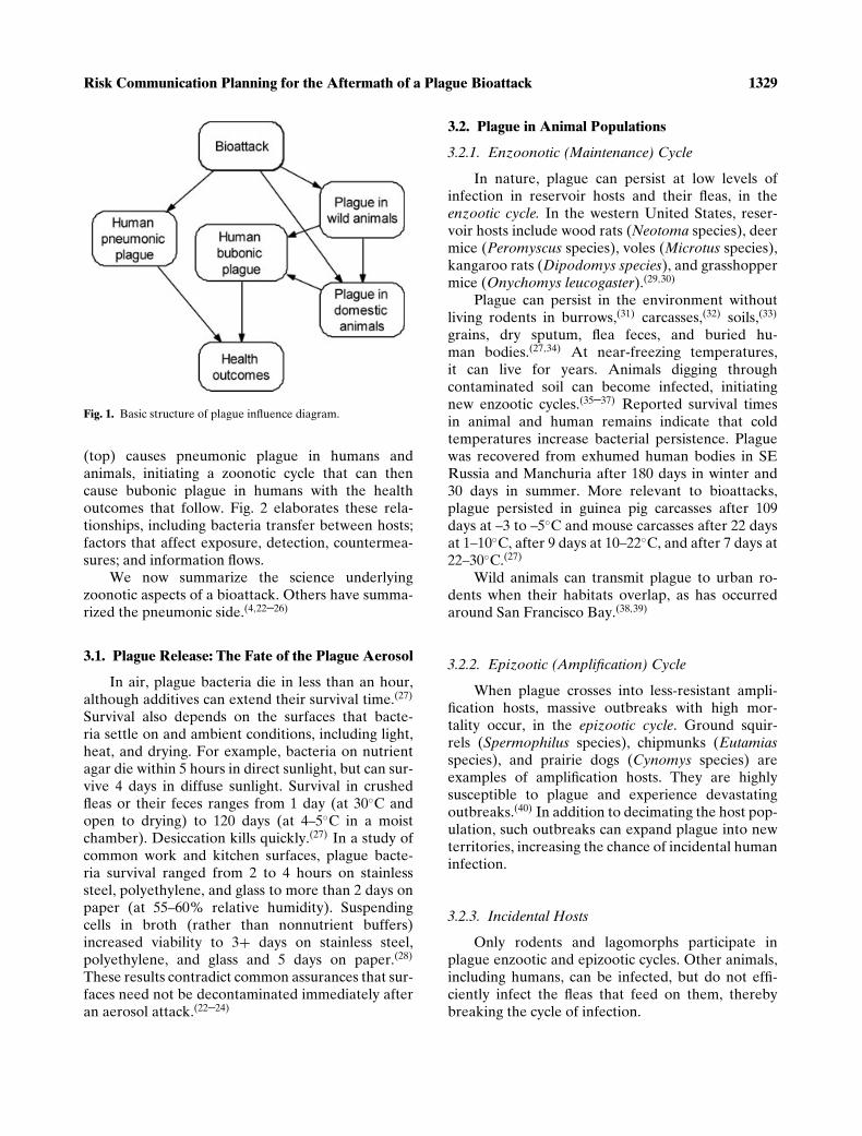
Fig. 1 shows the basic relationships between hu-man and animal plague infections. An aerosol release
Risk Communication Planning for the Aftermath of a Plague Bioattack 1329
Fig. 1. Basic structure of plague influence diagram.
(top) causes pneumonic plague in humans andanimals, initiating a zoonotic cycle that can thencause bubonic plague in humans with the healthoutcomes that follow. Fig. 2 elaborates these rela-tionships, including bacteria transfer between hosts;factors that affect exposure, detection, countermea-sures; and information flows.
We now summarize the science underlyingzoonotic aspects of a bioattack. Others have summa-rized the pneumonic side.(4,22–26)
3.1. Plague Release: The Fate of the Plague Aerosol
In air, plague bacteria die in less than an hour,although additives can extend their survival time.(27)
Survival also depends on the surfaces that bacte-ria settle on and ambient conditions, including light,heat, and drying. For example, bacteria on nutrientagar die within 5 hours in direct sunlight, but can sur-vive 4 days in diffuse sunlight. Survival in crushedfleas or their feces ranges from 1 day (at 30◦C andopen to drying) to 120 days (at 4–5◦C in a moistchamber). Desiccation kills quickly.(27) In a study ofcommon work and kitchen surfaces, plague bacte-ria survival ranged from 2 to 4 hours on stainlesssteel, polyethylene, and glass to more than 2 days onpaper (at 55–60% relative humidity). Suspendingcells in broth (rather than nonnutrient buffers)increased viability to 3+ days on stainless steel,polyethylene, and glass and 5 days on paper.(28)
These results contradict common assurances that sur-faces need not be decontaminated immediately afteran aerosol attack.(22–24)
3.2. Plague in Animal Populations
3.2.1. Enzoonotic (Maintenance) Cycle
In nature, plague can persist at low levels ofinfection in reservoir hosts and their fleas, in theenzootic cycle. In the western United States, reser-voir hosts include wood rats (Neotoma species), deermice (Peromyscus species), voles (Microtus species),kangaroo rats (Dipodomys species), and grasshoppermice (Onychomys leucogaster).(29,30)
Plague can persist in the environment withoutliving rodents in burrows,(31) carcasses,(32) soils,(33)
grains, dry sputum, flea feces, and buried hu-man bodies.(27,34) At near-freezing temperatures,it can live for years. Animals digging throughcontaminated soil can become infected, initiatingnew enzootic cycles.(35–37) Reported survival timesin animal and human remains indicate that coldtemperatures increase bacterial persistence. Plaguewas recovered from exhumed human bodies in SERussia and Manchuria after 180 days in winter and30 days in summer. More relevant to bioattacks,plague persisted in guinea pig carcasses after 109days at –3 to –5◦C and mouse carcasses after 22 daysat 1–10◦C, after 9 days at 10–22◦C, and after 7 days at22–30◦C.(27)
Wild animals can transmit plague to urban ro-dents when their habitats overlap, as has occurredaround San Francisco Bay.(38,39)
3.2.2. Epizootic (Amplification) Cycle
When plague crosses into less-resistant ampli-fication hosts, massive outbreaks with high mor-tality occur, in the epizootic cycle. Ground squir-rels (Spermophilus species), chipmunks (Eutamiasspecies), and prairie dogs (Cynomys species) areexamples of amplification hosts. They are highlysusceptible to plague and experience devastatingoutbreaks.(40) In addition to decimating the host pop-ulation, such outbreaks can expand plague into newterritories, increasing the chance of incidental humaninfection.
3.2.3. Incidental Hosts
Only rodents and lagomorphs participate inplague enzootic and epizootic cycles. Other animals,including humans, can be infected, but do not effi-ciently infect the fleas that feed on them, therebybreaking the cycle of infection.

1330 Casman and Fischhoff
Fig. 2. Influence diagram of a plague bioattack, including consequences and public health responses. Numbers refer to text sections.
Carnivores, such as domestic dogs, domesticferrets (but not the indigenous black-footed ferret),black bears, badgers, coyotes, and skunks, can beinfected by flea bite, inhalation, wound, or inges-tion. They have fairly strong resistance to plague in-fection, typically exhibiting mild or no symptoms,after ingesting plague-infected rodents. Felines andblack-footed ferrets are exceptions; they becomemortally ill when infected. Even if uninfected, car-nivores can physically transfer plague bacteria andplague-infected fleas from other animals.
In a study performed in states where plague isendemic, 16% of tested carnivores had antibodies toY. pestis, evidence of plague infection. The sampleincluded coyotes, badgers, raccoons, foxes, weasels,martens, skunks, bobcats, lynxes, mountain lions, andwild boars. Rates for some individual species were:badger 55%, weasels 43%, coyotes 13–14%, raccoons6–14%, and bears 3%.(41)
Hoofed animals are rarely infected, hence poselittle threat to humans. Birds are resistant to plague,
but may transport infected fleas between susceptiblehosts. Reptiles and fish are resistant.(9)
If infected, these incidental hosts can pose somerisk to humans by transporting infected fleas, infect-ing wounds and scratches, or emitting respirable in-fectious droplets.(42)
3.2.4. Zoonotic Flea-Borne Transmission
Flea bite is the main transmission mode for bothenzootics and epizootics with their course depend-ing on the flea species. Although fleas have distinctpreferences among animal hosts, most infested ani-mals carry several species, including species that pre-fer other hosts.
About 80 flea species are susceptible to Y. pestisinfection.(43) In a few species, after an infected bloodmeal, Y. pestis clogs the entrance to the midgutwith a biofilm-and-bacteria plug.(44) This blockagenot only increases the number of bacteria transmit-ted per bite, but starves the flea, encouraging it to

Risk Communication Planning for the Aftermath of a Plague Bioattack 1331
feed more aggressively. When the flea bites, bacteriaflow from the plug into the bite wound. Oriental ratfleas (Xenopsylla cheopis), associated with historicalplague pandemics, are particularly prone to block-age. Although blocking greatly enhances transmis-sion, most U.S. human cases come from unblockedground squirrel fleas (Oropsylla montanus), whichbecome infectious immediately after feeding, andthen transmit plague efficiently.(45)
3.2.5. Fleas and Climate
Seasonal variation is common in both the pres-ence of different flea species and their infestationlevels on different hosts.(46–49) High temperatureand low humidity reduce adult flea survival, espe-cially when they leave their hosts.(50) Flea infesta-tions are generally higher around nests and burrowsthan on roaming adult rodents. In hot weather, bur-rows and nests have moderate temperature and hu-midity, thereby improving juvenile flea survival.(51)
As a result, environmental conditions cause season-ality in flea-borne human plague coincident with lo-cal “flea seasons.” Interannual variation in humanplague cases has been linked to precipitation ef-fects on vegetation, rodent abundance, and flea sur-vival.(52)
3.3. Flea-to-Human Transmission
Human plague cases are often associated withepizootic die-offs of rodent hosts after fleas leavedead animals to seek new hosts, including humanswithin jumping range.(53) Cat fleas can jump 50cm horizontally and 25 cm vertically in a singlejump.(54)
Any flea that can bite through skin can transmitplague, with fewer than 100 bacteria needed to in-fect a human. Scratching bites can introduce bacte-ria left by fleas into abrasions. The threat posed byflea vectors depends on their access to humans andtheir plague transmission efficiency. That said, theinefficient cat flea (C. felis) and human flea (P. irri-tans) have both been involved in outbreaks, includingJapan’s use of weaponized plague-infected P. irritansduring World War II.(55)
Fleas’ biting ability and host preferences also af-fect their risk to humans.(47,51,56–58) For example, al-though prairie dogs have been involved in the mostwidespread U.S. plague epizootics, they account foronly 3–6% of human cases, as their fleas prefer other
hosts.(9) In contrast, 40% involve the ground squirrelflea, which readily bites humans. Although cat anddog fleas feed on humans, they cause less than 5%because they are poor vectors, with limited access toplague-infected rodents.(51)
Most human-flea interactions in the UnitedStates involve companion animals. In the humid east,cat fleas infest cats, dogs, rabbits, and other species,typically feeding on humans only when infestationsare high.
In the arid west, the cat flea cannot survive.There, the most problematic fleas infest wild animals,biting humans when those animals die or abandonnests near homes. These species include the humanflea, which lives on skunks, opossums, and foxes, andOrchopeas howardi, a fox squirrel flea, which dogscan carry into homes.(59)
Several factors inhibit flea-to-human transmis-sion. One is that only a fraction of the fleas frominfected hosts and burrows are typically infectedwith plague.(60,61) Second, although most mammalscan contract plague, only certain species can infectfleas.(62) Third, most flea species transmit plaguepoorly, either because their mouthparts cannot pene-trate human skin or because they prefer other hosts.Fourth, plague bacteria kill many infected fleas be-fore they can transmit disease.(55)
City dwellers typically have little contact withfleas, except for the homeless, shelter workers,and people living in substandard housing. Hikers,campers, and rural residents are sometimes bitten.Small animal veterinarians and assistants, animalshelter workers, and exterminators also face expo-sure. The largest group of people with regular fleaexposure is owners of outdoor dogs and cats.(14,63,64)
For humans in plague-endemic areas, the great-est risks come from contact with infected ani-mal tissues or fluids, rodent harborage, and foodsources around the home (e.g., pet food), and fleason roaming pets.(14,64) Minor risk factors are skin-ning or cooking rabbits, coyotes, and foxes, andcamping.(15,65–67)
3.4. Urban Rodents
3.4.1. Risks to Humans from Rats
Domestic rats are not currently involved inplague transmission in the United States. The twocommon urban rat species are, however, compe-tent vectors. The black rat, Rattus rattus, inhabitssoutheastern coastal states from southeast Virginia

1332 Casman and Fischhoff
to Texas and all western coastal states and Hawaii.The larger Norway rat, Rattus norvegicus, is foundin all states.(68) Norway rats nest mostly in bur-rows, usually close to human habitation. They arewell adapted to living with humans, frequentingtrash heaps, alleys, and sewer systems, especiallyolder combined storm and sanitary sewers. Blackrats are climbers, nesting mainly in roofs, attics, andtrees.
Rat population size is limited principally by food.A female Norway rat can, theoretically, produce 180offspring a year, with a gestation period of 23 daysand estrus 18 hours after giving birth. The actual an-nual number of successful weanlings is 10–20.(69,70)
Female pups reach sexual maturity in four months,males in three.
The United States has not had a case of humanplague from urban rats since 1924. Rats are oc-casionally involved in epizootic-plague transmis-sion.(11,13) For half a century, densities of rats andtransmitting flea species have remained steady, withepisodic “hot” spots or seasons.(49) A city’surbanrat population is said to be roughly equal to itshuman population, with great local variation indensity.(71)
A 1990 survey in Baltimore found that ratsand mice were often seen outdoors, but seldom in-side residences; only 1.2% of respondents reportedever being bitten by a rodent.(72) Estimating rat-human interaction from rat-bite frequencies is dif-ficult, as few cases are reported to health authori-ties.(70) In New York City between 1974 and 1978,the annual average incidence of reported rat biteswas 2/100,000, ranging from 8.5/100,000 (Lower EastSide) to 0.3/100,000 (Forest Hills, Queens). Abouthalf of the incidences occurred while people wereasleep.(73–75)
No U.S. city appears conducive to sustained rat-mediated flea-borne transmission to humans, givencurrent rat densities, flea species densities, and con-tact frequency with other potential hosts,(9) althoughsome neighborhoods may be exceptions.(49) As a re-sult, if the plague infected the urban rats, it would,in most instances, eventually disappear or retreat torural hosts.
3.4.2. Risk to Humans from House Mice
Although house mice, Mus musculus, can be in-fected with Y. pestis, they have not been implicatedin human plague.(76,77) Feral house mice sometimesmigrate seasonally to human dwellings, where they
can exchange fleas with domestic mice. However, thistransmission pathway is probably very minor.(78) Thelargest risk to humans comes from cats infected fromeating plague-killed mice.(79)
3.4.3. Risks to Humans from Other Urbanand Suburban Fauna
Other urban and suburban animals involved inplague transmission include squirrels, chipmunks,voles, and rabbits. The fox squirrel (Sciurus niger)has participated in urban plague circulation in Col-orado for at least the last 40 years, with few jumps tohumans.(9,13,80,81)
3.5. Companion Animals
More than half of American households havedogs or cats, with more than 30% having at least onecat.(82) Pets living entirely indoors (e.g., rabbits, ger-bils, hamsters) have little chance of exposure unlessthey interact with infectious outdoor pets.
3.5.1. Plague in Cats
Unlike other carnivores, cats are highly sus-ceptible to plague. Like humans, they can developbubonic plague (sometimes progressing to secondarypneumonic plague), septicemic plague, and primarypneumonic plague. Recognizing plague in cats canbe difficult, as typical symptoms resemble other fe-line diseases, fever (103–106◦F; normal temperatureis 101◦F), anorexia, lethargy, and enlarged, some-times abscessed lymph nodes (buboes), especially un-der the jaw (Table I). These abscesses rupture eas-ily, producing exudates loaded with Y. pestis. The
Table I. Most Common Symptoms of 119 Plague-InfectedDomestic Cats in New Mexico 1977–1988(83)
Lethargy 82%Anorexia 77%Fever (greater than 39.2◦C, 102.6◦F) 71%Abscesses (open sores) 34%Difficulty breathing 14%Discharge from mouth or nose 14%Coughing or sneezing 9%Lethargy, anorexia, fever, buboes 34%Lethargy, anorexia, fever, abscesses∗ 24%
∗Sixty-one percent of abscesses were of buboes, that is, on lymphnodes. Other locations: on or beneath the tongue, mouth, face,lips, jaw, buttocks, hind limb, forelimb, chest, and abdomen.

Risk Communication Planning for the Aftermath of a Plague Bioattack 1333
Table II. Exposures Leading to Cat-AssociatedHuman Plague(42)
Activity Frequency
Cared for sick cat, handled cat, buried dead cat 35%Face-to-face contact, slept with cat, inhalation 30%Bite 22%Scratch 13%
mortality rate is about 10% in cats treated withantibiotics, 14% for untreated bubonic, 70% for un-treated septicemic, and 83% for untreated pneumon-icplague.(42,83)
Infected cats were the source of 8% of the 297U.S. cases of human plague from 1977 to 1998,and accounted for all but one case of pneumonicplague.(42,67,84–87) Nearly all these cases involvedclose physical contact with sick cats; about a quar-ter occurred in veterinarians or assistants (Table II).Unlike most human plague, cat-related cases do notpeak in the summer along with flea abundance.(42)
Feces and urine from infected cats seldom contain Y.pestis.(79)
3.5.2. Plague in Dogs
Dogs can become infected through flea bites, in-gestion, or contacting infected animals. Most dogs re-cover without antibiotics.(67) Although rare, plaguefatalities have occurred in both dogs and their own-ers, with owners being infected by fleas carried bydogs.(88) Plague-infected dogs have no symptomsor nonspecific ones, like moderate fever (105◦F vs.normal =100.5–102.5◦F), lethargy, unresponsiveness,oral cavity lesions, anorexia, coughing, and drooling.A study found that dogs deliberately infected subcu-taneously (to simulate flea-bite) developed swellingand inflammation within three days. All still had le-sions with plague bacteria on Day 10. Plague bac-teria were found after 10 days in throat swabs ofhalf the dogs who were fed plague-containing rat vis-cera.(89) In another study, all dogs infected via inhala-tiondied.(90)
3.6. Human-to-Human Transmission
Human-to-human plague transmission (pneu-monic plague) is typically through infectious aerosoldroplets.
4. RISK-REDUCTION MEASURES
Risks of human exposure are reduced by inter-rupting pathways of plague transmission in natureand in the urban environment including home.
4.1. Measures Focused on Wildlife
4.1.1. Environmental Monitoring
Plague epizootics typically go unnoticed, as thehosts are small or reclusive and die unobserved.In a bioattack, concerted environmental monitoringwould be needed in order to formulate control strate-gies and determine when the danger has past.
4.1.2. Plague Suppression
Rodent eradication is not practical, or even pos-sible, for wild species. However, traps and rodenti-cides can reduce rodent densities, while pesticidescan reduce flea burdens (e.g., dusting burrow en-trances and runways). Experimental live-virus, bait-delivered vaccines exist for prairie dogs.(91)
4.2. Measures Focused on Urban Rodents
After a plague bioattack, rats and mice, bothlive and dead, should be considered potentiallyinfectious.
4.2.1. Rat Control
Effective methods include deploying poison baitand traps, destroying rat harborage, and repairingsewer pipes. These activities are usually municipalfunctions, and cash-strapped cities have experiencedwidespread problems after eliminating their rodentcontrol programs.3 Poisoning rats without first con-trolling fleas can raise human risk by increasing thenumber of questing fleas.
4.2.2. Property Maintenance
Residents can discourage rodents by clearingdebris, using metal garbage cans, removing food(e.g., pet food, bird seed, animal feces), and block-ing entrances (e.g., holes in foundations, doors, win-dows).(70) Commercially available traps and poisons
3 For example, the city of Pittsburgh eliminated its Rodent Con-trol Division in 2003. Complaints increased from 37 (2002) to 81(2003) to 136 (2004).(92)

1334 Casman and Fischhoff
can reduce infestations. Burning rat harborage anddebris, however, poses fire risks. In 1900, a large sec-tion of Honolulu burned down as a result of clearingdebris while combating plague.(93)
4.2.3. Treating the Outside Environment
If plague is detected in wild rodents, yardswith flea infestations will require pesticide treat-ment before reoccupation. Protective clothing (longpants with cuffs tucked into socks, long sleeves,gloves) and insect repellent with DEET should beworn.
Dead animals should not be buried, as roaminganimals might exhume the infectious remains. Fleascan jump nearly 2 feet, so carcasses should be movedusing a long handled shovel. Disposal of remains insturdy plastic bags in outside garbage cans is recom-mended.(94)
4.3. Measures Focused on Companion Animals
4.3.1 Plague Prevention
If a bioattack infects urban rodents, roamingcompanion animals could be affected. Keeping dogsand cats from hunting and eating rodents and rab-bits is critical to preventing Y. pestis infection. Free-roaming pets should be treated with quick-actinginsecticide, preferably flea powder, and then kept in-side. If symptoms appear, prompt treatment is ad-vised. Until neighborhoods are declared plague-free,dogs should be walked with short leashes and keptfrom contact with rodents and dead animals. Normalwaste disposal practices should suffice.
4.3.2. Preferred Methods for Flea Control
Control measures must work quickly. The quick-est “knock down” of adult fleas is with insecticidalpowders, shampoos, dips, and sprays. “Spot-on” sys-temic treatments, fast-drying liquids appliedbetweenpets’ shoulder blades or along the backbone, takea day to kill all adult fleas (killing 98% within12 hours), during which time some fleas may biteor move elsewhere. Thus, spot-on treatments leavesome small risk, so treated pets should be isolatedfrom humans for a day while the fleas die. Whendefleaing an animal, people should wear protectiveclothing and insect repellent (Section 4.3.5). To pre-vent reinfestation, the environment may need to betreated(Section 4.3.6).
4.3.3. Flea Treatments That Don’t Work
As mentioned, some topical flea treatments worktoo slowly to provide instantaneous protection. Allsystemic pesticides are imperfect, as fleas ingest themwhen they bite, which is too late to prevent plaguetransmission. Gas-emitting flea collars work onlyaround the neck, leaving fleas elsewhere. Herbalremedies (e.g., garlic, onions, thiamine, fleabane,brewer’s yeast, eucalyptus) provide no protection.Flea pills do not kill adult fleas, but prevent flea eggsfrom emerging into the larval stage.
Insecticides can kill or sicken kittens youngerthan five months old, so nonpesticidal methods maybe necessary. These include flea combs and pesticide-free shampoo.(95) These methods put the groomer atincreased risk of flea bite and release host-less, livefleas. Nonpesticidal methods should be employedonly if the animal has not been exposed to plague.
4.3.4. Illegal Pesticides
EPA’s website warns about counterfeit fleatreatments resembling registered products, whichmight become more common after a plague bioat-tack.(96) Illegally imported unsafe pesticides are an-other threat, especially to immigrants from the im-porting countries.(97)
4.3.5. Handling a Sick Pet
Only veterinarians can confirm plague. If theydo, family members need prophylactic antibiotics.If professional help is unavailable, as might happenduring a plague attack, symptomatic pets should betreated as though infected. After flea treatment, (po-tentially) sick animals should be isolated and allowedto recover or die by themselves. Antibiotics pre-scribed to humans should not be shared with animals,as neither humans nor animals will get proper doses.
People who handle sick pets should wear protec-tive clothing, work gloves, insect repellent contain-ing DEET, and eye and breathing protection. Whendone, they should wash immediately with soap andwater, launder clothing in hot water and detergent,and disinfect any surfaces that animals have touchedwith a 10% solution of household chlorine-basedbleach. Facemasks should be discarded in a plasticbag, along with other contaminated items that can-not be cleaned.
As a precaution, even asymptomatic pets shouldget flea treatment and be kept off beds. Owners

Risk Communication Planning for the Aftermath of a Plague Bioattack 1335
should avoid nuzzling, scratches, bites, and contactwith sores and saliva.
4.3.6. Treating the House for Fleas
If a house is infested with fleas, daily vacu-uming is recommended, especially of carpets, un-der furniture, flooring cracks, baseboards, windows,doorframes, and places where animals rest. A fleacollar or mothballs inside the vacuum bag will killfleas caught there. Pet bedding should be washed orsteam cleaned, and areal insecticidal sprays or fleabombs used for persistent infestations. Treatmentsmust be repeated until all flea pupae have hatched,which could take months.
4.4. Measures Focused on Infectious Humans
Because human-to-human plague transmission(pneumonic plague) typically involves infectiousaerosol droplets, barrier methods, like masks, couldprotect patients’ caregivers.(98) If the strain is sensi-tive to antibiotics, prophylactic antibiotics could re-duce disease incidence. A formalin-inactivated vac-cine exists for bubonic plague. However, it requiresseveral doses spread over months and does not pro-tect against pneumonic plague.(14) New vaccines areunder development, based on F1 and V antigens ofY. pestis. Once shown safe and effective, their use-fulness in a bioattack will depend on how availablethey are and how quickly they stimulate an immuneresponse.(99)
5. COMMUNICATIONS INBIOATTACK RESPONSE
Focusing communications on the risk factors thatdetermine human exposures and the measures thatmight control them makes best use of citizens’ limitedtime, energy, and resources—while protecting themagainst the false security of intuitively appealing, butineffective measures.
Once identified, plague-related advice is rela-tively easy to explain. The communication challengeis identifying the few critical facts, in this complexdomain. Fig. 2 structured that process by summa-rizing the possible human exposures, the factorsleading to them, and the opportunities for theirreduction.
Once the content of communications has beenselected, it must be made comprehensible. Often,that requires affording recipients a mental model of
why the actions are recommended and how theycan be adapted to specific circumstances. Fortu-nately, many plague facts are special cases of familiarprocesses. For example, plague is caused by a bac-terium that antibiotics can treat (unless it has beengenetically altered); that dies quickly in sunlight, butpersists in cool, humid environments; and that istransmitted by close contact (fleas, droplets, woundexudates). For individuals who understand these coreconcepts, specific messages (e.g., not nuzzling pets,keeping a safe distance from dead animals) shouldtake little additional explanation.
5.1. Communications Based on Core Concepts
Our analysis points to the following core con-cepts:
• Plague is a deadly infectious disease caused bya bacterium.
• Early symptoms in humans resemble flu or di-gestive upset.
• Starting antibiotics as soon as symptoms ap-pear increases the chance of survival.
• Plague can infect many mammals and fleas thatcan, in turn, infect humans.
• Plague can be spread by flea and animal bites,inhalation, and cuts.
• Plague bacteria can survive for days in humid,cool, dark places.
Before an emergency, research should establishempirically how well people understand these coreconcepts, and then find ways to make needed im-provements. Research can then build on these coreconcepts to explain the measures(14) described in Sec-tion 4, with the key ones being:
1. reduce rodent harborage and food sourcesnear the home;
2. use insect repellents when outdoors;3. keep cats and dogs indoors;4. use fast-acting insecticides to kill fleas on cats
and dogs;5. avoid sick or dead animals; and6. avoid sick cats’ fleas, open sores, or respira-
tory droplets.
When treatments have potential side effects,those should be acknowledged in quantitative termsshowing the size of the risks and allowing compar-isons to benefits.

1336 Casman and Fischhoff
Fig. 3. Relevant nodes and text sections for pet care information.
5.2. Using the Diagram to ConstructCommunications
5.2.1. Exposure-Specific Messages
Fig. 2 highlights the exposure routes that thesemeasures seek to control. Assembling the infor-mation regarding a particular exposure route in-volves tracing the arrows from the communicationnode to the relevant exposures (double oval nodes)and, then, to the factors contributing to that expo-sure. These factors are labeled with the numbersof the sections (in this article) providing explana-tory material. Messages should convey this content,following the diagram’s causal structure and invok-ing the core concepts. As ever, messages shouldbe evaluated empirically. Communications currentlyused in plague-endemic areas might be adapted tobioattacks.(85,100)
As an example, Fig. 3 highlights the portion ofthe model for pet care advice. It shows three classes
of exposures: “home,” “outdoor & recreational,” and“pet-related.” The arrows connect these exposuresto the contributing risk factors (round-cornered ob-long nodes). This information determines the rele-vance and effectiveness of possible protective mea-sures (rectangles). Invoking the core concepts shouldgive the recommendations credibility and help peo-ple to apply them appropriately.
5.2.2. Answering Context-Specific Questions
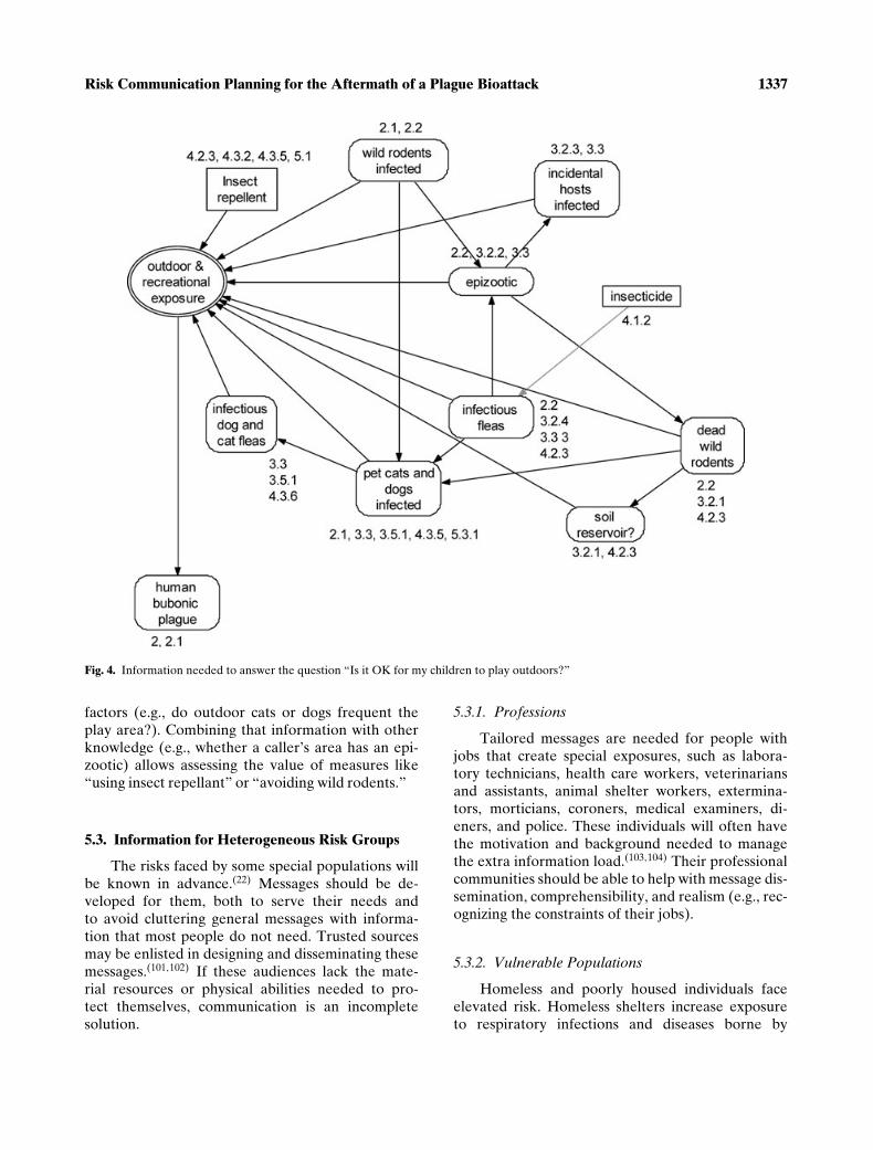
Any generic message will omit situations impor-tant to some people. The diagram can help to answersuch questions. Consider, for example, hotline callersasking: “Is it OK for my children to play outdoors?”The hotline operator would locate the exposure node“outdoor & recreational exposure” in Fig. 2 and re-view the factors pointing to it (Fig. 4). The operatorcould then walk the caller through the potential risk
Risk Communication Planning for the Aftermath of a Plague Bioattack 1337
Fig. 4. Information needed to answer the question “Is it OK for my children to play outdoors?”
factors (e.g., do outdoor cats or dogs frequent theplay area?). Combining that information with otherknowledge (e.g., whether a caller’s area has an epi-zootic) allows assessing the value of measures like“using insect repellant” or “avoiding wild rodents.”
5.3. Information for Heterogeneous Risk Groups
The risks faced by some special populations willbe known in advance.(22) Messages should be de-veloped for them, both to serve their needs andto avoid cluttering general messages with informa-tion that most people do not need. Trusted sourcesmay be enlisted in designing and disseminating thesemessages.(101,102) If these audiences lack the mate-rial resources or physical abilities needed to pro-tect themselves, communication is an incompletesolution.
5.3.1. Professions
Tailored messages are needed for people withjobs that create special exposures, such as labora-tory technicians, health care workers, veterinariansand assistants, animal shelter workers, extermina-tors, morticians, coroners, medical examiners, di-eners, and police. These individuals will often havethe motivation and background needed to managethe extra information load.(103,104) Their professionalcommunities should be able to help with message dis-semination, comprehensibility, and realism (e.g., rec-ognizing the constraints of their jobs).
5.3.2. Vulnerable Populations
Homeless and poorly housed individuals faceelevated risk. Homeless shelters increase exposureto respiratory infections and diseases borne by

1338 Casman and Fischhoff
rats, fleas, lice, and ticks.(105–107) Children mighthave limited understanding and unusual exposures(e.g., befriending animals that act strangely). Lan-guage barriers, social isolation, and distrust may in-crease the vulnerability of individuals in immigrantcommunities.
5.3.3. Transient Populations
An attack will catch some individuals awayfrom their usual surroundings, without needed re-sources or knowledge (e.g., campers, hikers, hunters,travelers). These individuals will need special mes-sages (e.g., about local medical resources) and help,perhaps conveyed through local professionals (e.g.,rangers, police, hotel staff).
6. CONCLUSION
Urban plague risk has been eliminated in theUnited States for a century, thanks to citizens andprofessionals who have maintained clean surround-ings and suppressed outbreaks. A bioattack could un-dermine these strategies (e.g., through absenteeismamong sanitation workers), while introducing newvectors (e.g., pets). Although the full picture is com-plex (Fig. 2), each exposure route is much simpler(Figs. 3 and 4). Moreover, all routes share familiarprocesses, summarized in the core concepts, with spe-cialized information available for dealing with spe-cific exposures (e.g., how far fleas jump; how to han-dle a sick cat). As a result, this complex topic can bereduced to a small set of measures that should be rel-atively easy to understand andexecute.
Whether that potential is realized depends onthe quality of the research implementing it. Messagetesting is needed to reveal how robust existing be-liefs are and where they require correction, as wellas where messages make unrealistic demands (e.g.,requiring material resources or physical abilities thatpeople lack). Messages will undermine trust if theymake no sense or ask people to do the impossible.(108)
They will strengthen public morale if they afford awarranted feeling of self-efficacy, as well as confi-dence in authorities who have demonstrated theirability to meet the public’s needs.
The method proposed here can facilitate antici-pating information needs, composing messages, pre-dicting noncompliance, and responding to emergingevents. Combined with empirical message testing, it
can help health communications officials get aheadand stay abreast of an attack.
Once derived, this advice may seem somewhatobvious. However, some of the advice in this arti-cle contradicts the assumptions in prominent plaguebioterrorism scenarios. These scenarios assume that(1) aerosolized plague will die in an hour, ignoringconditions that increase survival times; (2) house-hold surfaces need not be disinfected, ignoring sub-strate effects; (3) mice play no role, ignoring theirability to infect cats; (4) antibiotics will work, ig-noring the possibility of engineered strains; (5)adequate personnel and supplies will be available,ignoring epidemics’ disruptive effects; and (6) peo-ple will follow recommendations, ignoring barriers tounderstanding and execution. Most importantly, offi-cial scenarios ignore the roles of zoonotic processes.These questionable assumptions were revealed byanalyzing these scenarios in terms of the advice thatordinary citizens need.
ACKNOWLEDGMENTS
This work was supported by EPA/DHSCAMRA Center Grant RD832362, NSF GrantSES-0433152, and the MacArthur Foundation. Wethank David Wagner, Julie Downs, and MichaelDeKay for their suggestions and insights.
REFERENCES
1. Fischhoff, B. (2005). Decision research strategies. Health Psy-chology, 21(4), S9–16.
2. Fischhoff, B. (2008). Risk perception and risk communica-tion. In R. Detels, et al. (Eds.), Oxford Textbook of PublicHealth. Oxford: Oxford University Press.
3. Wray, R., & Jupka, K. (2004). What does the public want toknow in the event of a terrorist attack using plague? Biosecu-rity and Bioterrorism: Biodefense Strategy, Practice, and Sci-ence, 2(3), 208–215.
4. Centers for Disease Control and Prevention. (2006). PlagueInformation. 2006 May 2 [accessed June 14, 2006]. Availableat http://www.bt.cdc.gov/agent/plague/.
5. Merton, R. K. (1987). The focused interview and focusgroups: Continuities and discontinuities. Public OpinionQuarterly, 51(4), 550–566. Available at http://links.jstor.org/sici?sici=0033-362X%28198724%2951%3A4%3C550%3ATFIAFG%3E2.0.CO%3B2-0 [accessed May 20, 2008].
6. Casman, E., Fischhoff, B., Small, M., Palmgren, C., & Wu, F.(2000). An integrated risk model of a drinking-water-bornecryptosporidiosis outbreak. Risk Analysis, 20(4), 493–509.
7. Bruine de Bruin, W., et al. (2006). Expert judgments of pan-demic influenza risks. Global Public Health, 1(2), 178–193.
8. Poland, J., & Barnes, A. (1979). Plague. In J. H. Steele (Ed.),CRC Handbook Series in Zoonoses (pp. 515–597). BocaRaton, FL: CRC Press.
9. Barnes, A. M. (1982). Surveillance and control of bubonicplague in the United States. Symposia of the Zoological Soci-ety of London, 50, 237–270.

Risk Communication Planning for the Aftermath of a Plague Bioattack 1339
10. Chanteau, S., Ratsitorahina, M., Rahalison, L., Rasoa-manana, B., Chan, F., Boisier, P., Rabeson, D., & Roux, J.(2000). Current epidemiology of human plague in Madagas-car. Microbes and Infection, 2(1), 25–31.
11. Link, V. B. (1955). Public Health Monograph No. 26:A History of Plague in the United States. Washington,DC: U.S. Department of Health, Education, and Welfare;Public Health Service.
12. Cully, J. F. Jr., & Williams, E. S. (2001). Interspecific compar-isons of sylvatic plague in prairie dogs. Journal of Mammol-ogy, 82(4), 894–905.
13. Poland, J. D., & Barnes, A. M. (1970). Current Status ofPlague and Plague Control in the United States. Available athttp://digitalcommons.unl.edu/cgi/viewcontent.cgi?article=1009&context=vpcfour [accessed 2006 May 16 ].
14. Gage, K. L., Dennis, D. T., & Tsai, T. F. (1996). Preven-tion of plague: Recommendations of the Advisory Commit-tee on Immunization Practices (ACIP), Centers for DiseaseControl and Prevention. MMWR Morb Mortal Wkly Rep, 45(RR-14), 1–16.
15. Bertram-Sosa, L., Jaso, C., Valdez, A., Nix, B., Jones, R.,Sidwa, T., Walker, J., Anglirn, A., Reporter, R., Mascola, L.,Van Gordon, G., Ramirez, J. C., Fritz, C. L., Davis, R., Ross,J., Chongsiriwatana, K., DiMenna, M., Sheyka, J., Ettestad,P., Smelser, C. B., Powers, N., Reynolds, P., Fowler, J., Pape,J., Tanda, D., Griffith, K., Gage, K. L., Dietrich, G., Kubota,K., Young, J. A., & Gould, L.H. (2006). Human plague—Four states. Journal of the American Medical Association,296(14), 1722–1724.
16. World Health Organization. (1970). Health Aspects of Chem-ical and Biological Weapons (pp. 97–99). Geneva.
17. Howe, D. (2004). Planning Scenarios, Executive Summaries.Washington, DC: Homeland Security Council.
18. Lathem, W. W., Crosby, S.D., Miller, V.L., & Goldman, W.E.(2005). Progression of primary pneumonic plague: A mousemodel of infection, pathology, and bacterial transcriptionalactivity. Proceedings of National Academy of Sciences USA,102(49), 17786–17791.
19. Qureshi, K., Gershon, R.R., Sherman, M.F., Straub, T.,Gebbie, E., McCollum, M., Erwin, M.J., & Morse, S.S. (2005).Health care workers’ ability and willingness to report to dutyduring catastrophic disasters. Journal of Urban Health, 82(3),378–388.
20. Dimaggio, C., Markenson, D., T Loo, G., & Redlener, I.(2005). The willingness of U.S. Emergency medical techni-cians to respond to terrorist incidents. Biosecurity and Bioter-rorism: Biodefense Strategy, Practice, and Science, 3(4), 331–337.
21. Balicer, R. D., Omer, S. B., Barnett, D. J., & Everly Jr., G.S.(2006). Survey of local public health workers’ perceptions to-ward responding to an influenza pandemic. Journal of Health-care Protection Management, 22(2), 1–14.
22. Inglesby, T.V., Dennis, D.T., Henderson, D.A., Bartlett, J.G.,Ascher, M.S., Eitzen, E., Fine, A.D., Friedlander, A.M.,Hauer, J., Koerner, J.F., Layton, M., McDade, J., Oster-holm, M.T., O’Toole, T., Parker, G., Perl, T.M., Russell, P.K.,Schoch-Spana, M., & Tonat, K. (2000). Plague as a biologi-cal weapon: Medical and public health management. Work-ing Group on Civilian Biodefense. Journal of the Ameri-can Medical Association, 283(17), 2281–2290, Available athttp://jama.ama.assn.org/cgi/content/short/283/17/2281.
23. O’Toole, T., & Inglesby, T. V. (2001). Epidemic response sce-nario: Decision making in a time of plague. Public Health Re-ports, 116(Suppl 2), 92–103.
24. Inglesby, T. V., Grossman, R., & O’Toole, T. (2001). Aplague on your city: Observations from TOPOFF. ClinicalInfectious Diseases, 32(3), 436–445. Available at http://www.journals.uchicago.edu/doi/abs/10.1086/318513 [accessed May20, 2008].
25. Center for Infectious Disease Research and Policy. (2006).Plague: Current, Comprehensive Information on Patho-genesis, Microbiology, Epidemiology, Diagnosis, andTreatment. Available at: http://www.cidrap.umn.edu/cidrap/content/bt/plague/biofacts/plaguefactsheet.html [accessedSeptember 12, 2006].
26. Centers for Disease Control and Prevention. (2004). Emer-gency Preparedness & Response: Plague. 2004 September4, last reviewed 2007 February 12 [accessed 2008 Jan-uary 10] Available at http://www.bt.cdc.gov/agent/plague/trainingmodule/index.asp.
27. Mitscherlich, E., & Marth, E. H. (1984). Microbial Survival inthe Environment: Bacteria and Rickettsiae Important in Hu-man and Animal Health. Heidelberg: Springer-Verlag.
28. Rose, L. J., Donlan, R., Banerjee, S. N., & Arduino, M. J.(2003). Survival of Yersinia pestis on environmental surfaces.Applied and Environmental Microbiology, 69(4), 2166–2171.
29. Girard, J. M., Wagner, D.M., Vogler, A.J., Keys, C.,Allender, C.J., Drickamer, L.C., & Keim, P. (2004).Differential plague-transmission dynamics determineYersinia pestis population genetic structure on local, re-gional, and global scales. Proceedings of the NationalAcademy of Sciences of the United States of Amer-ica, 101(22), 8408–8413. Available at http://www.pnas.org/cgi/reprint/101/22/8408 [accessed May 20, 2008].
30. Gage, K. L., & Kosoy, M. Y. (2005). Natural history ofplague: Perspectives from more than a century of research.Annual Review of Entomology, 50, 505–528.
31. Lechleitner, R. R., Kartman, L., Goldenberg, M.I., Hudson,B. W. (1968). An epizootic of plague in Gunnison’s prairiedogs (Cynomys gunnisoni) in south-central Colorado. Ecol-ogy, 49, 734–743.
32. Breneva, N. V., Maramovich, A. S., & Klimov, V. T.(2005). Ecological regularities of the existence of pathogenicYersinia in soil ecosystems. Abstract. Article in Russian.Zhurnal Mikrobiologii Epidemiologii I Immunobiologii, (6),82–88.
33. Breneva, N. V., Maramovich, A. S., & Klimov, V. T. (2006).The population variability of Yersinia pestis in soil samplesfrom the natural focus of plague. Abstract. Article in Russian.Zhurnal Mikrobiologii Epidemiologii I Immunobiologii, (2),7–11.
34. U.S. Army Medical Research Institute of Infectious Diseases.(2004). USAMRIID’s Medical Management of BiologicalCasualties Handbook. 5th ed. Fort Detrick, Frederick,MD. Available at http://www.usamriid.army.mil/education/bluebookpdf/USAMRIID%20Blue%20Book%205th%20Edition.pdf [accessed May 20, 2008].
35. Drancourt, M., Houhamdi, L., & Raoult, D. (2006). Yersiniapestis as a telluric, human ectoparasite-borne organism.Lancet Infectious Diseases, 6, 234–241.
36. Mollaret, H. H., Karimi, Y., Eftekhari, M., & Blatazard,M. (1963). Burrowing plague. Bull Soc PatholExot Filiales, 56, 1183–1186, as cited in Drancourt,et al. (2006) Yersinia pestis as a telluric, human ectoparasite-borne organism. Lancet Infectious Diseases, 6, 234–241.
37. Butler, T., Fu, Y.S., Furman, L., Almeida, C., & Almeida, A.(1982). Experimental Yersinia pestis infection in rodents af-ter intragastric inoculation and ingestion of bacteria. Infec-tion and Immunity, 36(3), 1160–1167.
38. Miles, V. I., Kinney, A. R., & Stark, H. E. (1957). Flea-hostrelationships of associated Rattus and native wild rodents inthe San Francisco Bay area of California, with special refer-ence to plague. American Journal of Tropical Medicine andHygiene, 6(4), 752–760.
39. Hudson, B. W., & Quan, T. J. (1975). Serologic observationsduring an outbreak of rat borne plague in the San FranciscoBay area of California. Journal of Wildlife Diseases, 11(3),431–436.

1340 Casman and Fischhoff
40. Pauli, J. N., Buskirk, S.W., Williams, E.S., & Edwards, W.H.(2006). A plague epizootic in the black-tailed prairie dog(Cynomys ludovicianus). Journal of Wildlife Diseases, 42(1),74–80.
41. Salkeld, D. J., & Stapp, P. (2006). Seroprevalence ratesand transmission of plague (Yersinia pestis) in mammaliancarnivores. Vector-Borne and Zoonotic Diseases, 6(3), 231–239.
42. Gage, K. L., Dennis, D.T., Orloski, K.A., Ettestad, P., Brown,T.L., Reynolds, P.J., Pape, W.J., Fritz, C.L., Carter, L.G.,& Stein, J.D. (2000). Cases of cat-associated human plaguein the Western US, 1977–1998. Clinical Infectious Diseases,30(6), 893–900.
43. Pollitzer, R. (1954). Plague. Geneva: World Health Organi-zation.
44. Erickson, D. L., Jarrett, C.O., Wren, B.W., & Hin-nebusch, B.J. (2006). Serotype differences and lack ofbiofilm formation characterize Yersinia pseudotuberculo-sis infection of the Xenopsylla cheopis flea vector ofYersinia pestis. Journal of Bacteriology, 188(3), 1113–1119.
45. Eisen, R. J., Bearden, S. W., Wilder, A.P., Montenieri, J.A.,Antolin, M.F., & Gage, K. L. (2006). Early-phase transmis-sion of Yersinia pestis by unblocked fleas as a mechanismexplaining rapidly spreading plague epizootics. Proceedingsof National Academy of Sciences of USA, 103(42), 15380–15385.
46. Lang, J. D. (1996). Factors affecting the seasonal abundanceof ground squirrel and wood rat fleas (Siphonaptera) in SanDiego County, California. Journal of Medical Entomology,33(5), 790–804.
47. Schwan, T. G., Thompson, D., & Nelson, B. C. (1985). Fleason roof rats in six areas of Los Angeles County, California:Their potential role in the transmission of plague and murinetyphus to humans. American Journal of Tropical Medicineand Hygiene, 34(2), 372–379.
48. Ryckman, R. E. (1971). Plague vector studies Part 1. The rateof transfer of fleas among Citellus, Rattus, and Sylvilagus un-der field conditions in southern California. Journal of MedicalEntomology, 8(5), 535–540.
49. Nelson, B. C., Madon, M. B., & Tilzer, A. (1986). The com-plexities at the interface among domestic/wild rodents, fleas,pets, and man in urban plague ecology in Los Angeles,County, California. In Proceedings Twelfth Vertebrate PestConference. UC-Davis.
50. Anderson, S. H., & Williams, E. S. (1997). Plague in a com-plex of white-tailed prairie dogs and associated small mam-mals in Wyoming. Journal of Wildlife Diseases, 33(4), 720–732.
51. Perry, R. D., & Fetherston, J. D. (1997). Yersinia pestis–etiologic agent of plague. Clinical Microbiology Reviews,10(1), 35–66.
52. Parmenter, R. R., Yadav, E., Parmenter, C.A., Ettestad, P.,& Gage, K.L. (1999). Incidence of plague associated with in-creased winter-spring precipitation in New Mexico. AmericanJournal of Tropical Medicine and Hygiene, 61(5), 814–821.
53. Keeling, M. J., & Gilligan, C. A. (2000). Bubonic plague: Ametapopulation model of a zoonosis. Proceedings. BiologicalSciences/The Royal Society, 267(1458), 2219–2230.
54. Cadiergues, M. C., Joubert, C., & Franc, M. (2000). Acomparison of jump performances of the dog flea, Cteno-cephalides canis (Curtis, 1826) and the cat flea, Cteno-cephalides felis felis (Bouche, 1835). Veterinary Parasitology,92(3), 239–241.
55. McGovern, T. W., & Friedlander, A. M. (1997).Plague. In R. Zajtchuk & R. J. Bellamy (Eds.), Text-book of Military Medicine: Medical Aspects of Chem-ical and Biological Warfare (ch. 23). Washington,DC: Office of the Surgeon General, Borden Insti-
tute, Walter Reed Army Medical Center. Available athttp://www.bordeninstitute.army.mil/published volumes/chemBio/chembio.html [accessed May 20, 2008].
56. Burroughs, A. L. (1947). The vector efficiency of nine speciesof fleas compared with Xenopsylla cheopis. Journal of Hy-giene (Lond), 45, 371–396.
57. Gratz, N. (1999). Rodent reservoirs & flea vectors ofnatural foci of plague. In Plague Manual: Epidemiology,Distribution, Surveillance and Control, WHO/CDS/CSR/EDC/99.2 (ch. 4). Geneva: World Health Organization.
58. Hinnebusch, B. J. (2004). Interactions of Yersinia pestis withits flea vector that lead to the transmission of plague. InS. H. Gillespie, G. L. Smith, & A. Osbourn (Eds.), SGM Sym-posium 63: Microbe-Vector Interactions in Vector-Borne Dis-eases (pp. 331–343). Cambridge, UK: Cambridge UniversityPress.
59. Cranshaw, W., & Wilson, R. K. (2004). Fleas and Plague.[Accessed 2008 April 14.] Colorado State University Exten-sion Service. Household Insect Factsheet, number 5.600, Re-viewed/Revised Fabruary 2007. Available at http://www.ext.colostate.edu/Pubs/insect/05600.html.
60. Humphreys, F. A., Campbell, A. G., & Smith, E. S. (1951).Plague infection in western Canada, Information gleanedfrom rodent surveys 1938–1950 (incl.). Canadian Journal ofPublic Health, 42, 437–438.
61. Engelthaler, D. M., & Gage, K. L. (2000). Quantities ofYersinia pestis in fleas (Siphonaptera: Pulicidae, Ceratophyl-lidae, and Hystrichopsyllidae) collected from areas of knownor suspected plague activity. Journal of Medical Entomology,37(3), 422–426.
62. Lorange, E. A., Race, B.L., Sebbane, F., & Hinnebusch, B.J.(2005). Poor vector competence of fleas and the evolution ofhypervirulence in Yersinia pestis. Journal of Infectious Dis-eases, 191(11), 1907–1912.
63. Centers for Disease Control and Prevention. (1984). Plaguepneumonia—California. Morbidity and Mortality WeeklyReport, 33(34), 481–483. Available at http://www.cdc.gov/mmwr/preview/mmwrhtml/00000394.htm [accessed May 20,2008].
64. Mann, J. M., Martone, W.J., Boyce, J.M., Kaufmann, A.F.,Barnes, A.M., & Weber, N.S. (1979). Endemic human plaguein New Mexico: Risk factors associated with infection. Jour-nal of Infectious Diseases, 140(3), 397–401.
65. Butler, T. (1989). The black death past and present. 1. Plaguein the 1980s. Transactions of the Royal Society of TropicalMedicine and Hygiene, 83(4), 458–60.
66. von Reyn, C. F., Barnes, A.M., Weber, N.S., & Hodgin, U.G.(1976). Bubonic plague from exposure to a rabbit: A docu-mented case, and a review of rabbit-associated plague casesin the United States. American Journal of Epidemiology,104(1), 81–87.
67. Orloski, K. A., & Eidson, M. (1995). Yersinia pestis infec-tion in three dogs. Journal of American Medical Association,207(3), 316–318.
68. Howard, W. E., & Marsh, R. E. (1980). The rat: Its bi-ology and control. In Div. Agric. Sci. Publ. 2896. 1980,UC–Davis. p. 30. Available at http://icwdm.org/handbook/rodents/ro b125.pdf [accessed May 20, 2008].
69. Timm, R. M. (1994). Prevention and Control of WildlifeDamage: Norway Rats. Available at http://wildlifedamage.unl.edu/handbook/handbook/allPDF/ro b105.pdf [accessed2006 May 22].
70. Clinton, J. M. (1969). Rats in urban America. Public HealthReports, 84(1), 1–7.
71. Easterbrook, J. D., Shields, T., Klein, S.L., & Glass, G.E.(2005). Norway rat population in Baltimore, Maryland, 2004.Vector Borne and Zoonotic Diseases, 5(3), 296–299.
72. Childs, J. E., Glass, G. E., & LeDuc, J. W. (1991). Ro-dent sightings and contacts in an inner-city population of

Risk Communication Planning for the Aftermath of a Plague Bioattack 1341
Baltimore, Maryland, U.S.A. Bulletin of the Society of Vec-tor Ecology, 16, 245–255.
73. Coombe, N., & Marr, J. S. (1980). Rat bites support needfor in-home control: An epidemiologic study of rat bites inNew York City, 1974–1978. Journal of Environmental Health,42(6), 321–326.
74. Hirschhorn, R. B., & Hodge, R. R. (1999). Identifica-tion of risk factors in rat bite incidents involving humans.Pediatrics, 104(3), e35–40. Available at http://pediatrics.aappublications.org/cgi/content/full/104/3/e35 [accessed May20, 2008].
75. Childs, J. E., McLafferty, S.L., Sadek, R., Miller, G.L., Khan,A.S., DuPree, E.R., Advani, R., Mills, J.N., & Glass, G.E.(1998). Epidemiology of rodent bites and prediction of ratinfestation in New York City. American Journal Epidemiol-ogy, 148(1), 78–87.
76. Mittal, V., Rana, U.V., Jain, S.K., Kumar, K., Pal, I.S.,Arya, R.C., Ichhpujani, R.L., Lal, S., & Agarwal, S.P.(2004). Quick control of bubonic plague outbreak in UttarKashi, India. Journal of Communicative Diseases, 36(4), 233–239.
77. Zhao, Y. (2001). Geographical divisions of rat transmissiblediseases (Abstract. Article in Chinese). Wei Sheng Yan Jiu,30(2), 93–97.
78. Krasnov, B. R., & Khokhlova, I. S. (2001). The effect of be-havioural interactions on the transfer of fleas (Siphonaptera)between two rodent species. Journal of Vector Ecology,26(2), 181–190.
79. Gasper, P. W., Barnes, A.M., Quan, T.J., Benziger, J.P.,Carter, L.G., Beard, M.L., & Maupin, G.O. (1993). Plague(Yersinia pestis) in cats: Description of experimentally in-duced disease. Journal of Medical Entomology, 30(1), 20–26.
80. Boulder County Public Health. (2005). Squirrel Found in Cityof Boulder Tests Positive for Plague. Available at http://www.co.boulder.co.us/health/pr/2005/06142005squirrelPositivePlague.htm [accessed Jul 14, 2006].
81. Boulder County Public Health. (2005). Public Health Of-ficials Investigate Plague Die-Off in Lyons. Available athttp://www.co.boulder.co.us/health/pr/2005/05262005plagueLyons.htm [accessed July 14, 2006].
82. American Veterinary Medicine Association. (2002). U.S. PetOwnership and Demographics Sourcebook. AVMA.
83. Eidson, M., Thilsted, J. P., & Rollag, O. J. (1991). Clini-cal, clinicopathologic, and pathologic features of plague incats: 119 cases (1977–1988). Journal of American VeterinaryMedicine Association, 199(9), 1191–1197.
84. Brown, C. (2003). Virchow revisited: Emerging zoonoses.ASM News, 69(10), 493–497.
85. Centers for Disease Control and Prevention. (1994). Humanplague–United States, 1993–1994. Morbidity and MortalityWeekly Report, 43(13), 242–246.
86. White, M. E., Rosenbaum, R.J., Canfield, T.M., & Poland,J.D. (1981). Plague in a neonate. American Journal of Dis-eases of Children, 135(5), 418–419.
87. Lowell, J. L., Wagner, D.M., Atshabar, B., Antolin, M.F.,Vogler, A.J., Keim, P., Chu, M.C., & Gage, K.L. (2005). Iden-tifying sources of human exposure to plague. Journal of Clin-ical Microbiology, 43(2), 650–656.
88. Eidson, M., Tierney, L.A., Rollag, O.J., Becker, T., Brown,T., & Hull, H.F. (1988). Feline plague in New Mexico: Riskfactors and transmission to humans. American Journal ofPublic Health, 78(10), 1333–1335.
89. Rust, J. H. J., Cavanaugh, D.C., O’Shita, R., & Marshall Jr.,J.D. (1971). The role of domestic animals in the epidemiologyof plague. I. Experimental infection of dogs and cats. Journalof Infectious Diseases, 124, 522–526.
90. Strong, R. P., & Teague, O. (1912). Studies on pneumonicplague and plague immunization VIII. Susceptibility of ani-
mals to pneumonic plague. Philippine Journal of Science, 7B,223-228, as cited in Salkeld, D. J., & Stapp, P. (2006). Sero-prevalence rates and transmission of plague (Yersinia pestis)in mammalian carnivores. Vector-Borne and Zoonotic Dis,6(3), 231–9.
91. Mencher, J. S., Smith, S.R., Powell, T.D., Stinchcomb, D.T.,Osorio, J.E., & Rocke, T.E. (2004). Protection of black-tailedprairie dogs (Cynomys ludovicianus) against plague aftervoluntary consumption of baits containing recombinant rac-coon poxvirus vaccine. Infection and Immunity, 72(9), 5502–5505.
92. Boren, J. (2005). Exam-fee feud halts rat-control ef-forts. Tribune-Review. Pittsburgh: Pittsburgh Tribune-Review Publishing Co. Available at http://pittsburghlive.com/x/tribune-review/trib/pittsburgh/s 364393.html[accessed May 20, 2008].
93. Mohr, J. C. (2005). Plague and Fire: Battling Black Death andthe 1900 Burning of Honolulu’s Chinatown. New York: Ox-ford University Press.
94. Boulder County Public Health. (2005). How to Disposeof Dead Animals. Available at http://www.co.boulder.co.us/health/environ/vector/deadAnimal.htm [accessed July 13,2006].
95. U.S. Environmental Protection Agency. (2006). TakingCare of Fleas and Ticks on Your Pet. Available at http://www.epa.gov/pesticides/factsheets/flea-tick.htm [accessedJune 12, 2006].
96. U.S. Environmental Protection Agency. (2006). Coun-terfeit Pesticide Products for Dogs and Cats. Availableat http://www.epa.gov/pesticides/factsheets/petproduct.htm[accessed June 12, 2006].
97. U.S. Environmental Protection Agency. (2006). IllegalPesticide Products. Available at: http://www.epa.gov/pesticides/health/illegalproducts/index.htm [accessed June12, 2006].
98. Kool, J. L. (2005). Risk of person-to-person transmission ofpneumonic plague. Clinical Infectious Diseases, 40(8), 1166–1172.
99. Santi, L., Giritch, A., Roy, C.J., Marillonnet, S., Klimyuk, V.,Gleba, Y., Webb, R., Arntzen, C.J., & Mason, H.S. (2006).Protection conferred by recombinant Yersinia pestis antigensproduced by a rapid and highly scalable plant expression sys-tem. Proceedings of National Academy of Sciences of USA,103(4), 861–866.
100. Boulder County Public Health. (2005). PreventingPlague. Available at http://www.co.boulder.co.us/health/hpe/cdc/diseases/plague/prevention.htm [accessed July 13,2006].
101. Nations, M. K., & Monte, C. M. (1996). “I’m not dog, no!”:Cries of resistance against cholera control campaigns. SocialScience and Medicine, 43(6), 1007–1024.
102. McGough, M., Frank, L.L., Tipton, S., Tinker, T.L., &Vaughan, E. (2005). Communicating the risks of bioterrorismand other emergencies in a diverse society: A case study ofspecial populations in North Dakota. Biosecurity and Bioter-rorism: Biodefense Strategy, Practice, and Science, 3(3), 235–245.
103. Nolte, K. B. (2000). Safety precautions to limit exposure fromplague-infected patients. Journal of American Medical Asso-ciation, 284(13), 1648.
104. Nolte, K. B., & Yoon, S. S. (2003). Theoretical risk for oc-cupational blood-borne infections in forensic pathologists.Infection Control and Hospital Epidemiology, 24(10), 772–773.
105. O’Connell, J. J. (1991). Nontuberculosus respiratory infec-tions among the homeless. Seminars in Respiratory Infections,6(4), 247–253.
106. Smith, H. M., Reporter, R., Rood, M.P., Linscott, A.J.,Mascola, L.M., Hogrefe, W., & Purcell, R.H. (2002).

1342 Casman and Fischhoff
Prevalence study of antibody to ratborne pathogens andother agents among patients using a free clinic in down-town Los Angeles. Journal of Infectious Diseases, 186(11),1673–1676.
107. Brouqui, P., Stein, A., Dupont, H.T., Gallian, P., Badiaga,S., Rolain, J.M., Mege, J.L., La Scola, B., Berbis, P., &
Raoult, D. (2005). Ectoparasitism and vector-borne diseasesin 930 homeless people from Marseilles. Medicine (Balti-more), 84(1), 61–68.
108. Florig, H. K., & Fischhoff, B. (2007). Individuals’ decisionsaffecting radiation exposures after a nuclear event. HealthPhysics, 92, 475–83.