Embed Size (px)
Citation preview
THE OXYGENATIONOF CONCENTRATEDVERSUSNORMALBLOODS
By GEO. B. RAY, C. I. THOMASAND J. E. STRONG1
(From the Department of Physiology, Western Reserve University, School of Medicine,Clevland)
(Received for publication June 28, 1933)
While the physiological activity of normal or dilute bloods representsone of the most widely studied fields of investigation, the problemspresented by high concentrations of hemoglobin have not received theattention they merit. As far as the writers have been able to determinecomparatively little attention has been paid to the ability of polycythemicbloods to take up oxygen under normal conditions. Barcroft and Murray(1) compared the effect of concentrating blood by centrifugating andremoving part of the plasma. For a given 02 tension they found ahigher degree of saturation in the concentrated blood than in normalblood which in turn took up more 02 than did a sample diluted with theplasma which had been taken from the concentrated blood. Thisvariation was considered secondary to a disturbance in the carbon-dioxide equilibrium. Richards and Strauss (2) found no differencebetween the dissociation curve of the blood of a polycythemic patientand that of blood from a normal individual.
The experiments reported in this paper fall into two groups: thefirst designed to repeat the tests of the oxygen-combining power ofconcentrated blood in vitro, and the second to observe and compare theoxygenation of concentrated blood with that of normal blood in theperfused lung where the experimenter has a maximum control over thefactors associated with oxygenation. Such a series of experimentsshould offer a concrete and definite answer to the question, does a plethoraof red cells, per se, influence the respiratory function of the blood.
Experiments on the 02-combining power of concentrated bloodThe technique employed in the handling of the blood and gases in
these experiments was essentially that described by Austin et al. (3).The concentrated blood was obtained by centrifugating blood of normaldogs and pipetting away most of the supernatant plasma. A measuredamount of this blood, of known oxygen capacity, was placed in a smalltonometer bearing at one end a two-way stopcock and at the other a
1 Crile Research Scholar.1051
OXYGENATIONOF BLOOD
tube of fairly large bore. The tubular end was connected to a similarend of a large tonometer of the same type having a capacity of 250 to300 cc. Care was taken to see that the blood filled the smaller tonometerwell into the rubber connection. This connection was then clamped andthe pair of tonometers connected with a manifold whereby one couldeither evacuate the vessel or supply C02-free air, nitrogen, or carbondioxide. The tonometer was repeatedly evacuated and washed withnitrogen after which sufficient C02-free air was admitted to give thedesired oxygen tension, following which the CO2 tension was adjustedto 40 millimeters. Pressure within the tonometer was then brought toatmospheric by the admission of nitrogen. With the cocks closed thetonometer was removed from the manifold and the clamp between theblood and gas chamber taken off, allowing the blood to flow into thelarger chamber. In this manner portions of each sample of blood werebrought into equilibrium with gas mixtures of five different oxygentensions. The tonometers were rotated simultaneously in an air bathat 370 C. for thirty minutes. At the end of the period of equilibrationthe blood was allowed to return to the small chamber and the clampreplaced. The chambers were then disconnected, and samples taken foranalysis by forcing the blood into a pipette with mercury pressure.At no time was the blood allowed to come in contact with the room air.The analyses for oxygen content were made with the Van Slyke andNeill constant volume manometric apparatus; oxygen capacity wasestimated by means of the spectrophotometer (Ray, Blair and Thomas(4)). The final 02 tensions existing in the tonometers were correctedfor oxygen lost from the blood and for the effect of temperature change.
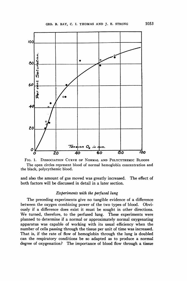
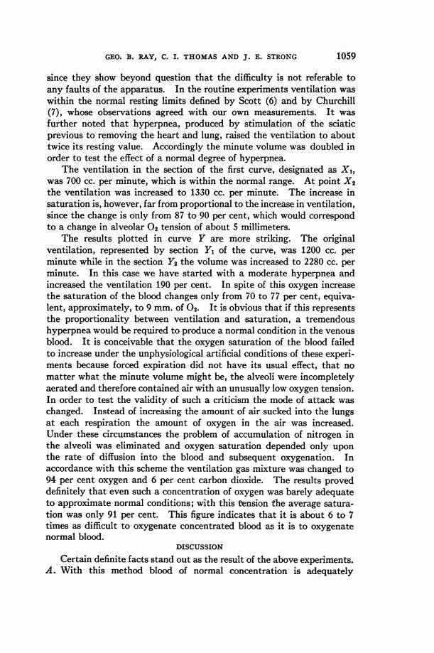
The results of a characteristic series of experiments are given inFigure 1. The open circles represent the results found in the normalblood and the black those for the polycythemic blood. Both sampleswere from the same original blood in order to avoid any chemical factorswhich might influence the results. These data show beyond any doubtthat the oxygen-combining power of the two bloods is identical, underthe conditions of the experiments. In fact the distribution of the twosets of points coincides quite as well as would points secured from twosamples of the same concentration. The variation of any point fromthe mean curve is within the experimental error of the methods employed.
There is a point of distinction which can not be demonstrated inthe graph, but was definitely apparent during the course of the experi-ment. The time required for the concentrated blood to reach an equi-librium was markedly longer than that needed for normal blood. Whilethe normal sample showed its maximal color change in five to ten minutesafter the start of equilibration the concentrated needed almost double thetime for the same change. This was a natural sequence of the concen-tration of the blood since diffusion is inversely proportional to viscosity
1052
GEO. B. RAY, C. I. THOMASAND J. E. STRONG
.0 ~~~~~~~~~~0
.0040
60 a -4.
4. 0~0
00
60 ~,
0 20 40 60 .0 100
FIG. 1. DISSOCIATION CURVEOF NORMALAND POLYCYTHEMICBLOODSThe open circles represent blood of normal hemoglobin concentration and
the black, polycythemic blood.
and also the amount of gas moved was greatly increased. The effect ofboth factors will be discussed in detail in a later section.
Experiments uith the perfused lung
The preceding experiments give no tangible evidence of a differencebetween the oxygen combining power of the two types of blood. Obvi-ously if a difference does exist it must be sought in other directions.We turned, therefore, to the perfused lung. These experiments were
planned to determine if a normal or approximately normal oxygenatingapparatus was capable of working with its usual efficiency when thenumber of cells passing through the tissue per unit of time was increased.That is, if the rate of flow of hemoglobin through the lung is doubledcan the respiratory conditions be so adapted as to produce a normaldegree of oxygenation? The importance of blood flow through a tissue
rinT 1 1 1
1053
,_ _
OXYGENATIONOF BLOOD
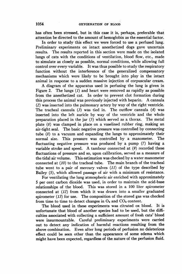
has often been stressed, but in this case it is, perhaps, preferable thatattention be directed to the amount of hemoglobin as the essential factor.
In order to study this effect we were forced to use a perfused lung.Preliminary experiments on intact anesthetized dogs gave uncertainresults. The results reported in this section were made on the isolatedlungs of cats with the conditions of ventilation, blood flow, etc., madeto simulate as closely as possible, normal conditions, while allowing fullcontrol over every variable. It was thus possible to study the respiratoryfunction without the interference of the generalized compensatorymechanisms which were likely to be brought into play in the intactanimal in response to a sudden massive injection of corpuscular cream.
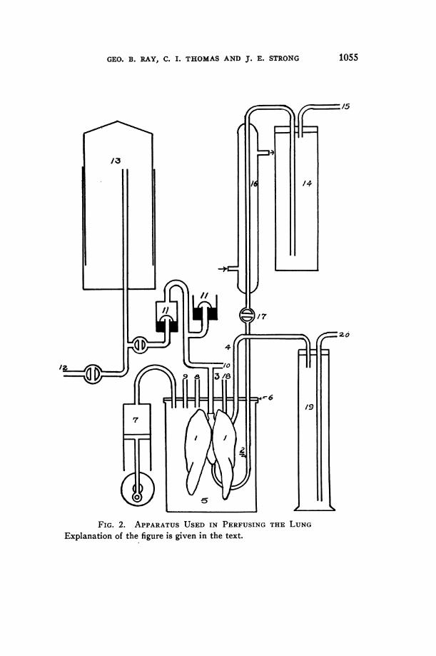
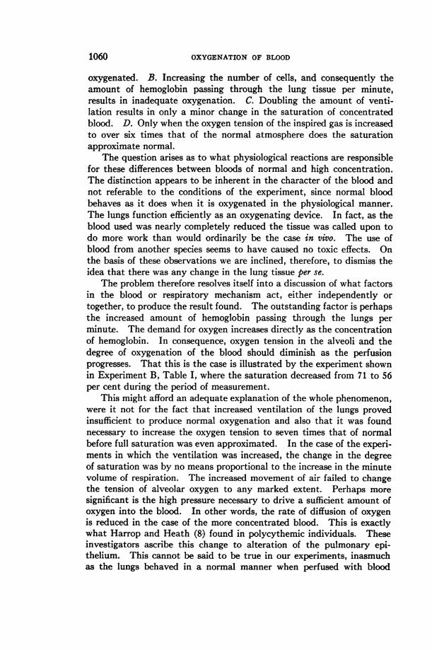
A diagram of the apparatus used in perfusing the lung is given inFigure 2. The lungs (1) and heart were removed as rapidly as possiblefrom the anesthetized cat. In order to prevent clot formation duringthis process the animal was previously injected with heparin. A cannula(2) was inserted into the pulmonary artery by way of the right ventricle.The tracheal cannula (3) was tied in. The outflow cannula (4) wasinserted into the left auricle by way of the ventricle and the wholepreparation placed in the jar (5) which served as a thorax. The metalplate (6) was clamped in place on a vaselined rubber ring, making anair-tight seal. The basic negative pressure was controlled by connectingtube (8) to a vacuum and expanding the lungs to approximately theirnormal size. This pressure was controlled by a manometer. Thefluctuating negative pressure was produced by a pump (7) having avariable stroke and speed. A tambour connected at (9) recorded thesefluctuations of pressure and so, upon calibration, served as a measure ofthe tidal air volume. This estimation was checked by a water manometerconnected at (10) to the tracheal tube. The main branch of the trachealtube went to a pair of mercury valves (11) of the type described byBailey (5), which allowed passage of air with a minimum of resistance.
For ventilating the lung atmospheric air enriched with approximately5 per cent carbon dioxide was used, in order to maintain the acid-baserelationships of the blood. This was stored in a 100 liter spirometerconnected at (12) from which it was drawn into a smaller graduatedspirometer (13) for use. The composition of the stored gas was checkedfrom time to time to detect changes in 02 and CO2 content.
The blood used in these experiments was citrated ox blood. It isunfortunate that blood of another species had to be used, but the diffi-culties associated with collecting a sufficient amount of fresh cats' bloodwere insurmountable. Careful preliminary experiments were carriedout to detect any indication of harmful reactions resulting from theabove combination. Even after long periods of perfusion no deleteriouseffect could be seen other than the appearance of some edema whichmight have been expected, regardless of the nature of the perfusion fluid.
1054
GEO. B. RAY, C. I. THOMASAND J. E. STRONG
:20
FIG. 2. APPARATUSUSED IN PERFUSING THE LUNGExplanation of the figure is given in the text.
1055
OXYGENATIONOF BLOOD
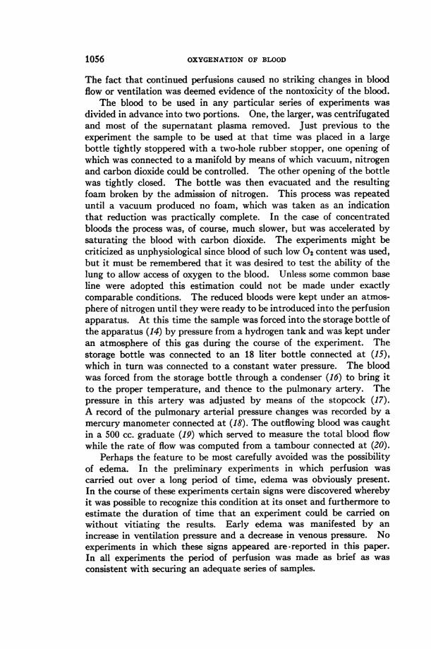
The fact that continued perfusions caused no striking changes in bloodflow or ventilation was deemed evidence of the nontoxicity of the blood.
The blood to be used in any particular series of experiments wasdivided in advance into two portions. One, the larger, was centrifugatedand most of the supernatant plasma removed. Just previous to theexperiment the sample to be used at that time was placed in a largebottle tightly stoppered with a two-hole rubber stopper, one opening ofwhich was connected to a manifold by means of which vacuum, nitrogenand carbon dioxide could be controlled. The other opening of the bottlewas tightly closed. The bottle was then evacuated and the resultingfoam broken by the admission of nitrogen. This process was repeateduntil a vacuum produced no foam, which was taken as an indicationthat reduction was practically complete. In the case of concentratedbloods the process was, of course, much slower, but was accelerated bysaturating the blood with carbon dioxide. The experiments might becriticized as unphysiological since blood of such low 02 content was used,but it must be remembered that it was desired to test the ability of thelung to allow access of oxygen to the blood. Unless some common baseline were adopted this estimation could not be made under exactlycomparable conditions. The reduced bloods were kept under an atmos-phere of nitrogen until they were ready to be introduced into the perfusionapparatus. At this time the sample was forced into the storage bottle ofthe apparatus (14) by pressure from a hydrogen tank and was kept underan atmosphere of this gas during the course of the experiment. Thestorage bottle was connected to an 18 liter bottle connected at (15),which in turn was connected to a constant water pressure. The bloodwas forced from the storage bottle through a condenser (16) to bring itto the proper temperature, and thence to the pulmonary artery. Thepressure in this artery was adjusted by means of the stopcock (17).A record of the pulmonary arterial pressure changes was recorded by amercury manometer connected at (18). The outflowing blood was caughtin a 500 cc. graduate (19) which served to measure the total blood flowwhile the rate of flow was computed from a tambour connected at (20).
Perhaps the feature to be most carefully avoided was the possibilityof edema. In the preliminary experiments in which perfusion wascarried out over a long period of time, edema was obviously present.In the course of these experiments certain signs were discovered wherebyit was possible to recognize this condition at its onset and furthermore toestimate the duration of time that an experiment could be carried onwithout vitiating the results. Early edema was manifested by anincrease in ventilation pressure and a decrease in venous pressure. Noexperiments in which these signs appeared are-reported in this paper.In all experiments the period of perfusion was made as brief as wasconsistent with securing an adequate series of samples.
1056
GEO. B. RAY, C. i. THOMASAND J. E. STRONG
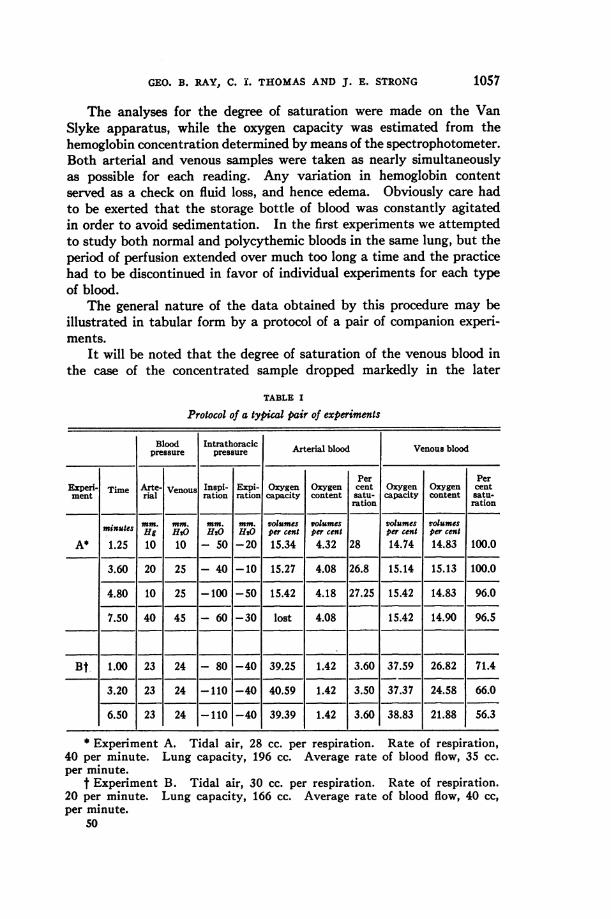
The analyses for the degree of saturation were made on the VanSlyke apparatus, while the oxygen capacity was estimated from thehemoglobin concentration determined by means of the spectrophotometer.Both arterial and venous samples were taken as nearly simultaneouslyas possible for each reading. Any variation in hemoglobin contentserved as a check on fluid loss, and hence edema. Obviously care hadto be exerted that the storage bottle of blood was constantly agitatedin order to avoid sedimentation. In the first experiments we attemptedto study both normal and polycythemic bloods in the same lung, but theperiod of perfusion extended over much too long a time and the practicehad to be discontinued in favor of individual experiments for each typeof blood.
The general nature of the data obtained by this procedure may beillustrated in tabular form by a protocol of a pair of companion experi-ments.
It will be noted that the degree of saturation of the venous blood inthe case of the concentrated sample dropped markedly in the later
TABLE I
Protocol of a typical pair of experiments
Blood Intrathoracic Arterial blood Venous bloodpressure pressure
Per PerExperi- Time Arte- Venous Inspi- Expi- Oxygen Oxygen cent Oxygen Oxygen centment rial ration ration capacity content satu- capacity content satu-
mation ration
mm. mm. mm. mm. volumes volumes volumes volumesminutes Hg HgQ H20 H20 per cent per cent per cent per centA* 1.25 10 10 - 50 -20 15.34 4.32 28 14.74 14.83 100.0
3.60 20 25 - 40 -10 15.27 4.08 26.8 15.14 15.13 100.0
4.80 10 25 -100 -50 15.42 4.18 27.25 15.42 14.83 96.0
7.50 40 45 - 60 -30 lost 4.08 15.42 14.90 96.5
Bt 1.00 23 24 - 80 -40 39.25 1.42 3.60 37.59 26.82 71.4
3.20 23 24 -110 -40 40.59 1.42 3.50 37.37 24.58 66.0
6.50 23 24 -110 -40 39.39 1.42 3.60 38.83 21.88 56.3
* Experiment A. Tidal air, 28 cc. per respiration. Rate of respiration,40 per minute. Lung capacity, 196 cc. Average rate of blood flow, 35 cc.per minute.
t Experiment B. Tidal air, 30 cc. per respiration. Rate of respiration.20 per minute. Lung capacity, 166 cc. Average rate of blood flow, 40 cc,per minute.
50
1057
1058 OXYGENATIONOF BLOOD
readings. As far as we could determine this was not due to any changein the lung tissue but rather to a continually diminishing tension ofoxygen in the alveolar air resulting from the demands of the concentratedblood. Spectrophotometric evidence proved, in addition, that thehemoglobin had retained its ability to combine with oxygen. The slightvariations noted in oxygen capacity are without doubt due to sedimenta-tion, which could not be completely controlled.
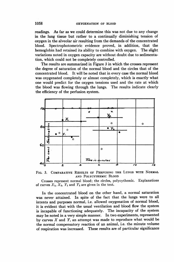
The results are summarized in Figure 3 in which the crosses represent
the degree of saturation of the normal blood and the circles that of theconcentrated blood. It will be noted that in every case the normal bloodwas oxygenated completely or almost completely, which is exactly whatone would predict for the oxygen tensions used and the rate at whichthe blood was flowing through the lungs. The results indicate clearlythe efficiency of the perfusion system.
0 o
0~~~~~
0 / /2
FIG. 3. COMPARATIVERESULTS OF PERFUSING THE LUNGS WITH NORMALAND POLYCYTHEMICBLOOD
Crosses represent normal blood; the circles, polycythemic. Explanationsof curves X1, X2, Y, and Y2 are given in the text.
In the concentrated blood on the other hand, a normal saturationwas never attained. In spite of the fact that the lungs were to allintents and purposes normal, i.e. allowed oxygenation of normal blood,it is evident that with the usual ventilation and blood flow the systemis incapable of functioning adequately. The incapacity of the systemmay be noted in a very simple manner. In two experiments, representedby curves X and Y, an attempt was made to reproduce what would bethe normal compensatory reaction of an animal, i.e. the minute volumeof respiration was increased. These results are of particular significance
GEO. B. RAY, C. I. THOMASAND J. E. STRONG
since they show beyond question that the difficulty is not referable toany faults of the apparatus. In the routine experiments ventilation waswithin the normal resting limits defined by Scott (6) and by Churchill(7), whose observations agreed with our own measurements. It wasfurther noted that hyperpnea, produced by stimulation of the sciaticprevious to removing the heart and lung, raised the ventilation to abouttwice its resting value. Accordingly the minute volume was doubled inorder to test the effect of a normal degree of hyperpnea.
The ventilation in the section of the first curve, designated as Xi,was 700 cc. per minute, which is within the normal range. At point X2the ventilation was increased to 1330 cc. per minute. The increase insaturation is, however, far from proportional to the increase in ventilation,since the change is only from 87 to 90 per cent, which would correspondto a change in alveolar 02 tension of about 5 millimeters.
The results plotted in curve Y are more striking. The originalventilation, represented by section Y1 of the curve, was 1200 cc. perminute while in the section Y2 the volume was increased to 2280 cc. perminute. In this case we have started with a moderate hyperpnea andincreased the ventilation 190 per cent. In spite of this oxygen increasethe saturation of the blood changes only from 70 to 77 per cent, equiva-lent, approximately, to 9 mm. of 02. It is obvious that if this representsthe proportionality between ventilation and saturation, a tremendoushyperpnea would be required to produce a normal condition in the venousblood. It is conceivable that the oxygen saturation of the blood failedto increase under the unphysiological artificial conditions of these experi-ments because forced expiration did not have its usual effect, that nomatter what the minute volume might be, the alveoli were incompletelyaerated and therefore contained air with an unusually low oxygen tension.In order to test the validity of such a criticism the mode of attack waschanged. Instead of increasing the amount of air sucked into the lungsat each respiration the amount of oxygen in the air was increased.Under these circumstances the problem of accumulation of nitrogen inthe alveoli was eliminated and oxygen saturation depended only uponthe rate of diffusion into the blood and subsequent oxygenation. Inaccordance with this scheme the ventilation gas mixture was changed to94 per cent oxygen and 6 per cent carbon dioxide. The results proveddefinitely that even such a concentration of oxygen was barely adequateto approximate normal conditions; with this tension the average satura-tion was only 91 per cent. This figure indicates that it is about 6 to 7times as difficult to oxygenate concentrated blood as it is to oxygenatenormal blood.
DISCUSSION
Certain definite facts stand out as the result of the above experiments.A. With this method blood of normal concentration is adequately
1059
OXYGENATIONOF BLOOD
oxygenated. B. Increasing the number of cells, and consequently theamount of hemoglobin passing through the lung tissue per minute,results in inadequate oxygenation. C. Doubling the amount of venti-lation results in only a minor change in the saturation of concentratedblood. D. Only when the oxygen tension of the inspired gas is increasedto over six times that of the normal atmosphere does the saturationapproximate normal.
The question arises as to what physiological reactions are responsiblefor these differences between bloods of normal and high concentration.The distinction appears to be inherent in the character of the blood andnot referable to the conditions of the experiment, since normal bloodbehaves as it does when it is oxygenated in the physiological manner.The lungs function efficiently as an oxygenating device. In fact, as theblood used was nearly completely reduced the tissue was called upon todo more work than would ordinarily be the case in vivo. The use ofblood from another species seems to have caused no toxic effects. Onthe basis of these observations we are inclined, therefore, to dismiss theidea that there was any change in the lung tissue per se.
The problem therefore resolves itself into a discussion of what factorsin the blood or respiratory mechanism act, either independently ortogether, to produce the result found. The outstanding factor is perhapsthe increased amount of hemoglobin passing through the lungs perminute. The demand for oxygen increases directly as the concentrationof hemoglobin. In consequence, oxygen tension in the alveoli and thedegree of oxygenation of the blood should diminish as the perfusionprogresses. That this is the case is illustrated by the experiment shownin Experiment B, Table I, where the saturation decreased from 71 to 56per cent during the period of measurement.
This might afford an adequate explanation of the whole phenomenon,were it not for the fact that increased ventilation of the lungs provedinsufficient to produce normal oxygenation and also that it was foundnecessary to increase the oxygen tension to seven times that of normalbefore full saturation was even approximated. In the case of the experi-ments in which the ventilation was increased, the change in the degreeof saturation was by no means proportional to the increase in the minutevolume of respiration. The increased movement of air failed to changethe tension of alveolar oxygen to any marked extent. Perhaps moresignificant is the high pressure necessary to drive a sufficient amount ofoxygen into the blood. In other words, the rate of diffusion of oxygenis reduced in the case of the more concentrated blood. This is exactlywhat Harrop and Heath (8) found in polycythemic individuals. Theseinvestigators ascribe this change to alteration of the pulmonary epi-thelium. This cannot be said to be true in our experiments, inasmuchas the lungs behaved in a normal manner when perfused with blood
1060
GEO. B. RAY, C. I. THOMASAND J. E. STRONG
containing a low concentration of cells. It is obvious, therefore, thatthe reason for the faulty oxygenation of the blood must be sought else-where. It will be recalled that in the experiments upon the dissociationcurves a longer time was needed for complete equilibration of the concen-trated blood. It would seem, therefore, that a certain fraction of theunsaturation can be attributed to the character of the concentratedblood itself.
Still another factor to be considered is the reaction which occurs inthe capillaries. In the table cited it will be noted that the amount ofblood flowing through the system is approximately the same in bothexperiments. The viscosity of the concentrated blood is much greaterthan normal, yet if the second readings of both types of experiments arecompared it will be noted that the same pressure produces the same flow.The simplest explanation that can be suggested for this adjustment isdilatation of the smaller vessels. The appearance of the lungs in thetwo cases certainly substantiated this view. If such a compensationinvolved the alveolar capillaries a second reason for the impairment ofoxygen diffusion is found in the increased distance the gas must pass inorder to complete the process of oxygenation. In addition to this effect,the engorgement of the vessels would decrease the alveolar space, i.e.,the vital capacity, thereby adding to the factors which promote anoxemia.
SUMMARY
Studies of the dissociation curves of normal and artificial poly-cythemic bloods, show no difference in the tension of oxygen required toproduce a given saturation. When these types of blood are oxygenatedby the perfused lung the normal blood becomes completely oxygenatedwhile the concentrated blood is never fully saturated. Increasing theoxygen tension increases the saturation of the concentrated blood.
The difference between the two bloods is ascribed to the greater rateat which hemoglobin in the polycythemic blood passes through the lungs,coupled with a delayed diffusion resulting from capillary dilatation.
BIBLIOGRAPHY1. Barcroft, J., and Murray, C. D., Phil. Trans. Roy. Soc., (B), 1923, ccxi, 469.
Some Secondary Effects of Increasing the Proportion of Red Corpusclesin Blood.
2. Richards, D. W., Jr., and Strauss, M. L., J. Clin. Invest., 1927, iv, 105.Oxyhemoglobin Dissociation Curves of Whole Blood in Anemia.
3. Austin, J., Cullen, G. E., Hastings, A. B., McLean, F. C., Peters, J. P., andVan Slyke, D. D., J. Biol. Chem., 1922, liv, 121. Studies of Gas andElectrolyte Equilibria in Blood. I. Technique for Collection andAnalysis of Blood and for its Saturation with Gas Mixtures of KnownComposition.
4. Ray, G. B., Blair, H. A., and Thomas, C. I., J. Biol. Chem., 1932, xcviii, 63.The Spectrophotometric Determination of Certain Blood Pigments.
1061
1062 OXYGENATIONOF BLOOD
S. Bailey, C. V., Proc. Soc. Exper. Biol. and Med., 1926, xxiv, 184. A LowResistance Air Valve.
6. Scott, R. W., Am. J. Physiol., 1917, xliv, 196. The Effect of the Accumula-tion of Carbon Dioxide on the Tidal Air and on the H-ion Concentrationof the Arterial Blood in the Decerebrate Cat.
7. Churchill, E. D., Am. J. Physiol., 1928, lxxxvi, 274. The Effect of IncreasedBlood Flow on the Ratio between Oxygen Consumption and PulmonaryVentilation.
8. Harrop, G. A., Jr., and Heath, E. H., J. Clin. Invest., 1927, iv, 53. Pul-monary Gas Diffusion in Polycythemia Vera.